Case Report Heart of Lymphoma: Primary Mediastinal Large B...
5
Hindawi Publishing Corporation Case Reports in Oncological Medicine Volume 2013, Article ID 814291, 4 pages http://dx.doi.org/10.1155/2013/814291 Case Report Heart of Lymphoma: Primary Mediastinal Large B-Cell Lymphoma with Endomyocardial Involvement Elisa Rogowitz, 1 Hani M. Babiker, 2 Ravitharan Krishnadasan, 2 Clint Jokerst, 3 Thomas P. Miller, 2 and Michael Bookman 2 1 University of Arizona College of Medicine, 1501 N. Campbell Avenue, Tucson, AZ 85724, USA 2 Division of Hematology-Oncology, University of Arizona Cancer Center, 1515 N. Campbell Avenue, Tucson, AZ 85724, USA 3 Department of Radiology, University of Arizona College of Medicine, 1501 N. Campbell Avenue, Tucson, AZ 85724, USA Correspondence should be addressed to Hani M. Babiker; [email protected] Received 25 August 2013; Accepted 17 September 2013 Academic Editors: L. Beex, D. V. Jones, L. Lu, and F. A. Mauri Copyright © 2013 Elisa Rogowitz et al. is is an open access article distributed under the Creative Commons Attribution License, which permits unrestricted use, distribution, and reproduction in any medium, provided the original work is properly cited. Primary mediastinal B-cell lymphoma (PMBCL) is an uncommon aggressive subset of diffuse large B-cell lymphomas. Although PMBCL frequently spreads locally from the thymus into the pleura or pericardium, it rarely invades directly through the heart. Herein, we report a case of a young Mexican female diagnosed with PMBCL with clear infiltration of lymphoma through the cardiac wall and into the right atrium and tricuspid valve leading to tricuspid regurgitation. is was demonstrated by cardiac MRI and transthoracic echocardiogram. In addition, cardiac MRI and CT scan of the chest revealed the large mediastinal mass completely surrounding and eroding into the superior vena cava (SVC) wall causing a collar of stokes. e cardiac and SVC infiltration created a significant therapeutic challenge as lymphomas are very responsive to chemotherapy, and treatment could potentially lead to vascular wall rupture and hemorrhage. Despite the lack of conclusive data on chemotherapy-induced hemodynamic compromise in such scenarios, her progressive severe SVC syndrome and respiratory distress necessitated urgent intervention. In addition to the unique presentation of this rare lymphoma, our case report highlights the safety of R-CHOP treatment. 1. Case Report A 23-year-old Mexican female presented to the emergency room with a relentless cough for three days. e cough and dyspnea started six weeks prior to presentation and were gradually worsening, causing her to sleep upright. She also experienced fatigue, prominent facial swelling, engorged neck vasculature, headaches, and a 25-pound weight loss. Climbing even four individual stairs caused this former soc- cer athlete significant fatigue and lightheadedness. She denied having fevers, chills, or night sweats. She is a full-time college student living with her parents and sibling in Mexico. She was evaluated there and diagnosed with Cushing syndrome and hypothyroidism and was prescribed levothyroxine. She presented to our emergency department aſter her condition deteriorated during her visit to the USA. e patient had a temperature of 37.2 ∘ C, a pulse of 120 beats per minute, a blood pressure of 96/57 mm Hg, a respiratory rate of 24 breaths per minute, and an oxygen saturation of 98% on room air. She had significant facial, neck, and upper trunk swelling with visible engorged vessels. A collar of stokes was present, and her right upper extremity was more edematous than the leſt. A faint diastolic murmur was heard best over the right sternal border. No lymphadenopathy was noted. Labs revealed a WBC of 11.4 (3.4–10.4 1000/uL) with 81% neutrophils, 11% lymphocytes, 6% monocytes, 1% eosinophils, 1% basophils, and an absolute neutrophil count of 9234/microL. Serum LDH was 1308 (125–243 IU/L). In addition, her potassium was 3.2 (3.5–5.1 mMol/L), calcium 9.8 (8.6–10.6 mg/dL), phosphorus 4.2 (2.3–4.7 mg/dL), and magnesium 2 (1.6–2.6 mg/dL). Beta-2-microglobulin was 1.82 (0.97–2.64 mg/L), and uric acid was 4.4 (2.6–6 mg/dL). A chest X-ray demonstrated a large anterior mediastinal mass. Follow-up contrast enhanced chest CT revealed a large lobulated anterior mediastinal mass near the right atrium with complete encasement and effacement of the superior vena cava (SVC) and invasion into the right atrium (Figures 1 and 2). Tumor almost completely filled the right atrium
Transcript of Case Report Heart of Lymphoma: Primary Mediastinal Large B...

Hindawi Publishing CorporationCase Reports in Oncological MedicineVolume 2013, Article ID 814291, 4 pageshttp://dx.doi.org/10.1155/2013/814291
Case ReportHeart of Lymphoma: Primary Mediastinal Large B-CellLymphoma with Endomyocardial Involvement
Elisa Rogowitz,1 Hani M. Babiker,2 Ravitharan Krishnadasan,2 Clint Jokerst,3
Thomas P. Miller,2 and Michael Bookman2
1 University of Arizona College of Medicine, 1501 N. Campbell Avenue, Tucson, AZ 85724, USA2Division of Hematology-Oncology, University of Arizona Cancer Center, 1515 N. Campbell Avenue, Tucson, AZ 85724, USA3Department of Radiology, University of Arizona College of Medicine, 1501 N. Campbell Avenue, Tucson, AZ 85724, USA
Correspondence should be addressed to Hani M. Babiker; [email protected]
Received 25 August 2013; Accepted 17 September 2013
Academic Editors: L. Beex, D. V. Jones, L. Lu, and F. A. Mauri
Copyright © 2013 Elisa Rogowitz et al. This is an open access article distributed under the Creative Commons Attribution License,which permits unrestricted use, distribution, and reproduction in any medium, provided the original work is properly cited.
Primary mediastinal B-cell lymphoma (PMBCL) is an uncommon aggressive subset of diffuse large B-cell lymphomas. AlthoughPMBCL frequently spreads locally from the thymus into the pleura or pericardium, it rarely invades directly through the heart.Herein, we report a case of a youngMexican female diagnosedwith PMBCLwith clear infiltration of lymphoma through the cardiacwall and into the right atrium and tricuspid valve leading to tricuspid regurgitation. This was demonstrated by cardiac MRI andtransthoracic echocardiogram. In addition, cardiac MRI and CT scan of the chest revealed the large mediastinal mass completelysurrounding and eroding into the superior vena cava (SVC) wall causing a collar of stokes.The cardiac and SVC infiltration createda significant therapeutic challenge as lymphomas are very responsive to chemotherapy, and treatment could potentially lead tovascular wall rupture and hemorrhage. Despite the lack of conclusive data on chemotherapy-induced hemodynamic compromisein such scenarios, her progressive severe SVC syndrome and respiratory distress necessitated urgent intervention. In addition tothe unique presentation of this rare lymphoma, our case report highlights the safety of R-CHOP treatment.
1. Case Report
A 23-year-old Mexican female presented to the emergencyroom with a relentless cough for three days. The coughand dyspnea started six weeks prior to presentation andwere gradually worsening, causing her to sleep upright. Shealso experienced fatigue, prominent facial swelling, engorgedneck vasculature, headaches, and a 25-pound weight loss.Climbing even four individual stairs caused this former soc-cer athlete significant fatigue and lightheadedness. She deniedhaving fevers, chills, or night sweats. She is a full-time collegestudent living with her parents and sibling in Mexico. Shewas evaluated there and diagnosed with Cushing syndromeand hypothyroidism and was prescribed levothyroxine. Shepresented to our emergency department after her conditiondeteriorated during her visit to the USA.
The patient had a temperature of 37.2∘C, a pulse of120 beats per minute, a blood pressure of 96/57mmHg, arespiratory rate of 24 breaths per minute, and an oxygen
saturation of 98%on roomair. She had significant facial, neck,and upper trunk swelling with visible engorged vessels. Acollar of stokeswas present, andher right upper extremitywasmore edematous than the left. A faint diastolic murmur washeard best over the right sternal border.No lymphadenopathywas noted. Labs revealed a WBC of 11.4 (3.4–10.4 1000/uL)with 81% neutrophils, 11% lymphocytes, 6% monocytes, 1%eosinophils, 1% basophils, and an absolute neutrophil countof 9234/microL. Serum LDH was 1308 (125–243 IU/L). Inaddition, her potassium was 3.2 (3.5–5.1mMol/L), calcium9.8 (8.6–10.6mg/dL), phosphorus 4.2 (2.3–4.7mg/dL), andmagnesium 2 (1.6–2.6mg/dL). Beta-2-microglobulinwas 1.82(0.97–2.64mg/L), and uric acid was 4.4 (2.6–6mg/dL).
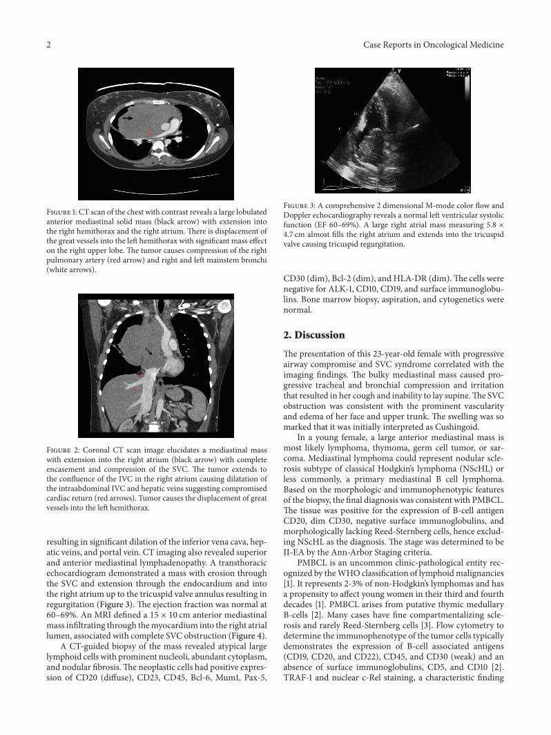
A chest X-ray demonstrated a large anterior mediastinalmass. Follow-up contrast enhanced chest CT revealed a largelobulated anterior mediastinal mass near the right atriumwith complete encasement and effacement of the superiorvena cava (SVC) and invasion into the right atrium (Figures 1and 2). Tumor almost completely filled the right atrium
2 Case Reports in Oncological Medicine
Figure 1: CT scan of the chest with contrast reveals a large lobulatedanterior mediastinal solid mass (black arrow) with extension intothe right hemithorax and the right atrium. There is displacement ofthe great vessels into the left hemithorax with significant mass effecton the right upper lobe. The tumor causes compression of the rightpulmonary artery (red arrow) and right and left mainstem bronchi(white arrows).
Figure 2: Coronal CT scan image elucidates a mediastinal masswith extension into the right atrium (black arrow) with completeencasement and compression of the SVC. The tumor extends tothe confluence of the IVC in the right atrium causing dilatation ofthe intraabdominal IVC and hepatic veins suggesting compromisedcardiac return (red arrows). Tumor causes the displacement of greatvessels into the left hemithorax.
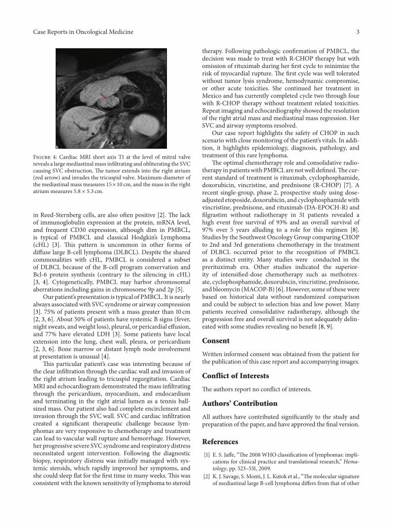
resulting in significant dilation of the inferior vena cava, hep-atic veins, and portal vein. CT imaging also revealed superiorand anterior mediastinal lymphadenopathy. A transthoracicechocardiogram demonstrated a mass with erosion throughthe SVC and extension through the endocardium and intothe right atrium up to the tricuspid valve annulus resulting inregurgitation (Figure 3). The ejection fraction was normal at60–69%. An MRI defined a 15 × 10 cm anterior mediastinalmass infiltrating through themyocardium into the right atriallumen, associated with complete SVC obstruction (Figure 4).
A CT-guided biopsy of the mass revealed atypical largelymphoid cells with prominent nucleoli, abundant cytoplasm,and nodular fibrosis.The neoplastic cells had positive expres-sion of CD20 (diffuse), CD23, CD45, Bcl-6, Mum1, Pax-5,
Figure 3: A comprehensive 2 dimensional M-mode color flow andDoppler echocardiography reveals a normal left ventricular systolicfunction (EF 60–69%). A large right atrial mass measuring 5.8 ×4.7 cm almost fills the right atrium and extends into the tricuspidvalve causing tricuspid regurgitation.
CD30 (dim), Bcl-2 (dim), and HLA-DR (dim).The cells werenegative for ALK-1, CD10, CD19, and surface immunoglobu-lins. Bone marrow biopsy, aspiration, and cytogenetics werenormal.
2. Discussion
The presentation of this 23-year-old female with progressiveairway compromise and SVC syndrome correlated with theimaging findings. The bulky mediastinal mass caused pro-gressive tracheal and bronchial compression and irritationthat resulted in her cough and inability to lay supine.The SVCobstruction was consistent with the prominent vascularityand edema of her face and upper trunk. The swelling was somarked that it was initially interpreted as Cushingoid.
In a young female, a large anterior mediastinal mass ismost likely lymphoma, thymoma, germ cell tumor, or sar-coma. Mediastinal lymphoma could represent nodular scle-rosis subtype of classical Hodgkin’s lymphoma (NScHL) orless commonly, a primary mediastinal B cell lymphoma.Based on the morphologic and immunophenotypic featuresof the biopsy, the final diagnosis was consistent with PMBCL.The tissue was positive for the expression of B-cell antigenCD20, dim CD30, negative surface immunoglobulins, andmorphologically lacking Reed-Sternberg cells, hence exclud-ing NScHL as the diagnosis. The stage was determined to beII-EA by the Ann-Arbor Staging criteria.
PMBCL is an uncommon clinic-pathological entity rec-ognized by theWHOclassification of lymphoidmalignancies[1]. It represents 2-3% of non-Hodgkin’s lymphomas and hasa propensity to affect young women in their third and fourthdecades [1]. PMBCL arises from putative thymic medullaryB-cells [2]. Many cases have fine compartmentalizing scle-rosis and rarely Reed-Sternberg cells [3]. Flow cytometry todetermine the immunophenotype of the tumor cells typicallydemonstrates the expression of B-cell associated antigens(CD19, CD20, and CD22), CD45, and CD30 (weak) and anabsence of surface immunoglobulins, CD5, and CD10 [2].TRAF-1 and nuclear c-Rel staining, a characteristic finding
Case Reports in Oncological Medicine 3
Figure 4: Cardiac MRI short axis T1 at the level of mitral valvereveals a largemediastinalmass infiltrating and obliterating the SVCcausing SVC obstruction. The tumor extends into the right atrium(red arrow) and invades the tricuspid valve. Maximum diameter ofthe mediastinal mass measures 15×10 cm, and the mass in the rightatrium measures 5.8 × 5.3 cm.
in Reed-Sternberg cells, are also often positive [2]. The lackof immunoglobulin expression at the protein, mRNA level,and frequent CD30 expression, although dim in PMBCL,is typical of PMBCL and classical Hodgkin’s Lymphoma(cHL) [3]. This pattern is uncommon in other forms ofdiffuse large B-cell lymphoma (DLBCL). Despite the sharedcommonalities with cHL, PMBCL is considered a subsetof DLBCL because of the B-cell program conservation andBcl-6 protein synthesis (contrary to the silencing in cHL)[3, 4]. Cytogenetically, PMBCL may harbor chromosomalaberrations including gains in chromosome 9p and 2p [5].
Our patient’s presentation is typical of PMBCL. It is nearlyalways associated with SVC syndrome or airway compression[3]. 75% of patients present with a mass greater than 10 cm[2, 3, 6]. About 50% of patients have systemic B signs (fever,night sweats, andweight loss), pleural, or pericardial effusion,and 77% have elevated LDH [3]. Some patients have localextension into the lung, chest wall, pleura, or pericardium[2, 3, 6]. Bone marrow or distant lymph node involvementat presentation is unusual [4].
This particular patient’s case was interesting because ofthe clear infiltration through the cardiac wall and invasion ofthe right atrium leading to tricuspid regurgitation. CardiacMRI and echocardiogram demonstrated the mass infiltratingthrough the pericardium, myocardium, and endocardiumand terminating in the right atrial lumen as a tennis ball-sized mass. Our patient also had complete encirclement andinvasion through the SVC wall. SVC and cardiac infiltrationcreated a significant therapeutic challenge because lym-phomas are very responsive to chemotherapy and treatmentcan lead to vascular wall rupture and hemorrhage. However,her progressive severe SVC syndrome and respiratory distressnecessitated urgent intervention. Following the diagnosticbiopsy, respiratory distress was initially managed with sys-temic steroids, which rapidly improved her symptoms, andshe could sleep flat for the first time in many weeks. This wasconsistent with the known sensitivity of lymphoma to steroid
therapy. Following pathologic confirmation of PMBCL, thedecision was made to treat with R-CHOP therapy but withomission of rituximab during her first cycle to minimize therisk of myocardial rupture. The first cycle was well toleratedwithout tumor lysis syndrome, hemodynamic compromise,or other acute toxicities. She continued her treatment inMexico and has currently completed cycle two through fourwith R-CHOP therapy without treatment related toxicities.Repeat imaging and echocardiography showed the resolutionof the right atrial mass and mediastinal mass regression. HerSVC and airway symptoms resolved.
Our case report highlights the safety of CHOP in suchscenario with close monitoring of the patient’s vitals. In addi-tion, it highlights epidemiology, diagnosis, pathology, andtreatment of this rare lymphoma.
The optimal chemotherapy role and consolidative radio-therapy in patientswith PMBCLare notwell defined.The cur-rent standard of treatment is rituximab, cyclophosphamide,doxorubicin, vincristine, and prednisone (R-CHOP) [7]. Arecent single-group, phase 2, prospective study using dose-adjusted etoposide, doxorubicin, and cyclophosphamidewithvincristine, prednisone, and rituximab (DA-EPOCH-R) andfilgrastim without radiotherapy in 51 patients revealed ahigh event free survival of 93% and an overall survival of97% over 5 years alluding to a role for this regimen [8].Studies by the Southwest Oncology Group comparing CHOPto 2nd and 3rd generations chemotherapy in the treatmentof DLBCL occurred prior to the recognition of PMBCLas a distinct entity. Many studies were conducted in theprerituximab era. Other studies indicated the superior-ity of intensified-dose chemotherapy such as methotrex-ate, cyclophosphamide, doxorubicin, vincristine, prednisone,and bleomycin (MACOP-B) [6].However, some of thesewerebased on historical data without randomized comparisonand could be subject to selection bias and low power. Manypatients received consolidative radiotherapy, although theprogression free and overall survival is not adequately delin-eated with some studies revealing no benefit [8, 9].
Consent
Written informed consent was obtained from the patient forthe publication of this case report and accompanying images.
Conflict of Interests
The authors report no conflict of interests.
Authors’ Contribution
All authors have contributed significantly to the study andpreparation of the paper, and have approved the final version.
References
[1] E. S. Jaffe, “The 2008 WHO classification of lymphomas: impli-cations for clinical practice and translational research,” Hema-tology, pp. 523–531, 2009.
[2] K. J. Savage, S.Monti, J. L. Kutok et al., “Themolecular signatureof mediastinal large B-cell lymphoma differs from that of other

4 Case Reports in Oncological Medicine
diffuse large B-cell lymphomas and shares features with classicalHodgkin lymphoma,” Blood, vol. 102, no. 12, pp. 3871–3879,2003.
[3] M. Martelli, A. J. M. Ferreri, and P. Johnson, “Primary medi-astinal large B-cell lymphoma,” Critical Reviews in Oncology/Hematology, vol. 68, no. 3, pp. 256–263, 2008.
[4] A. Rosenwald, G. Wright, K. Leroy et al., “Molecular diagnosisof primary mediastinal B cell lymphoma identifies a clinicallyfavorable subgroup of diffuse large B cell lymphoma relatedto Hodgkin lymphoma,” Journal of Experimental Medicine, vol.198, no. 6, pp. 851–862, 2003.
[5] M. Bentz, T. FE. Barth, S. Bruderlein et al., “Gain of chromo-some arm9p is characteristic of primarymediastinal B-cell lym-phoma (MBL): comprehensive molecular cytogenetic analysisand presentation of a novel MBL cell line,”Genes, Chromosomesand Cancer, vol. 30, no. 4, pp. 393–401, 2001.
[6] P. L. Zinzani, M. Martelli, M. Magagnoli et al., “Treatment andclinical management of primary mediastinal large B-cell lym-phoma with sclerosis: MACOP-B regimen and mediastinalradiotherapy monitored by 67Gallium scan in 50 patients,”Blood, vol. 94, no. 10, pp. 3289–3293, 1999.
[7] K. J. Savage, “Primary mediastinal large B-cell lymphoma,”TheOncologist, vol. 11, no. 5, pp. 488–495, 2006.
[8] K. Dunleavy, S. Pittalunga, L. S. Maeda et al., “Dose-adjustedEPOCH-rituximab therapy in primary mediastinal B-cell lym-phoma,” The New England Journal of Medicine, vol. 386, pp.1408–1416, 2013.
[9] K. J. Savage, N. Al-Rajhi, N. Voss et al., “Favorable outcome ofprimary mediastinal large B-cell lymphoma in a single institu-tion: the British Columbia experience,” Annals of Oncology, vol.17, no. 1, pp. 123–130, 2006.

Submit your manuscripts athttp://www.hindawi.com
Stem CellsInternational
Hindawi Publishing Corporationhttp://www.hindawi.com Volume 2014
Hindawi Publishing Corporationhttp://www.hindawi.com Volume 2014
MEDIATORSINFLAMMATION
of
Hindawi Publishing Corporationhttp://www.hindawi.com Volume 2014
Behavioural Neurology
EndocrinologyInternational Journal of
Hindawi Publishing Corporationhttp://www.hindawi.com Volume 2014
Hindawi Publishing Corporationhttp://www.hindawi.com Volume 2014
Disease Markers
Hindawi Publishing Corporationhttp://www.hindawi.com Volume 2014
BioMed Research International
OncologyJournal of
Hindawi Publishing Corporationhttp://www.hindawi.com Volume 2014
Hindawi Publishing Corporationhttp://www.hindawi.com Volume 2014
Oxidative Medicine and Cellular Longevity
Hindawi Publishing Corporationhttp://www.hindawi.com Volume 2014
PPAR Research
The Scientific World JournalHindawi Publishing Corporation http://www.hindawi.com Volume 2014
Immunology ResearchHindawi Publishing Corporationhttp://www.hindawi.com Volume 2014
Journal of
ObesityJournal of
Hindawi Publishing Corporationhttp://www.hindawi.com Volume 2014
Hindawi Publishing Corporationhttp://www.hindawi.com Volume 2014
Computational and Mathematical Methods in Medicine
OphthalmologyJournal of
Hindawi Publishing Corporationhttp://www.hindawi.com Volume 2014
Diabetes ResearchJournal of
Hindawi Publishing Corporationhttp://www.hindawi.com Volume 2014
Hindawi Publishing Corporationhttp://www.hindawi.com Volume 2014
Research and TreatmentAIDS
Hindawi Publishing Corporationhttp://www.hindawi.com Volume 2014
Gastroenterology Research and Practice
Hindawi Publishing Corporationhttp://www.hindawi.com Volume 2014
Parkinson’s Disease
Evidence-Based Complementary and Alternative Medicine
Volume 2014Hindawi Publishing Corporationhttp://www.hindawi.com