Primary mediastinal B-cell lymphoma and mediastinal gray zone ...
27
Primary mediastinal B-cell lymphoma and mediastinal gray zone lymphoma: Do they require a unique therapeutic approach? Kieron Dunleavy and Wyndham H. Wilson Lymphoid Malignancies Branch, Center for Cancer Research, National Cancer Institute, Bethesda, MD, USA Correspondence: Wyndham H. Wilson, M.D. PhD, Metabolism Branch, National Cancer Institute, Building 10, Room 4N-115, 9000 Rockville Pike, Bethesda, MD 20892. Email: [email protected] Key words: mediastinal lymphoma; primary mediastinal B-cell lymphoma; grey-zone; thymic B-cell; obviating radiation; janus kinase 2. Blood First Edition Paper, prepublished online December 11, 2014; DOI 10.1182/blood-2014-05-575092 Copyright © 2014 American Society of Hematology For personal use only. on February 17, 2018. by guest www.bloodjournal.org From
-
Upload
nguyenkien -
Category
Documents
-
view
235 -
download
1
Transcript of Primary mediastinal B-cell lymphoma and mediastinal gray zone ...
1
Primary mediastinal B-cell lymphoma and mediastinal gray zone lymphoma: Do they require a unique therapeutic
approach? Kieron Dunleavy and Wyndham H. Wilson Lymphoid Malignancies Branch, Center for Cancer Research, National Cancer Institute, Bethesda, MD, USA Correspondence: Wyndham H. Wilson, M.D. PhD, Metabolism Branch, National Cancer Institute, Building 10, Room 4N-115, 9000 Rockville Pike, Bethesda, MD 20892. Email: [email protected] Key words: mediastinal lymphoma; primary mediastinal B-cell lymphoma; grey-zone; thymic B-cell; obviating radiation; janus kinase 2.
Blood First Edition Paper, prepublished online December 11, 2014; DOI 10.1182/blood-2014-05-575092
Copyright © 2014 American Society of Hematology
For personal use only.on February 17, 2018. by guest www.bloodjournal.orgFrom
2
ABSTRACT
Primary mediastinal B-cell lymphoma (PMBL) is a subtype of diffuse large B-cell
lymphoma (DLBCL) that is putatively derived from a thymic B-cell. Accounting for up
to 10% of cases of DLBCL, this subtype predominantly affects females in the third and
fourth decades of life. Its clinical and molecular characteristics are distinct from other
subtypes of DLBCL and in fact, closely resemble those of nodular sclerosing Hodgkin
lymphoma (NSHL). Recently, mediastinal lymphomas with features intermediate
between PMBL and NSHL – called mediastinal grey-zone lymphomas – have been
described. The optimal management of PMBL is controversial and most standard
approaches include a combination of immunochemotherapy and mediastinal radiation.
Recently, the recognition that mediastinal radiation is associated with significant long-
term toxicities has led to the development of novel approaches for PMBL that have
shown excellent efficacy and challenge the need for routine mediastinal radiation.
INTRODUCTION
Primary mediastinal B-cell lymphoma (PMBL), originally described in the
1980’s, accounts for up to 10% of diffuse large B-cell lymphomas (DLBCL). It is
epidemiologically, clinically and biologically distinct from the other subtypes of DLBCL
(germinal center B-cell like (GCB) and activated B-cell like (ABC)). Similar to nodular
sclerosing Hodgkin lymphoma (NSHL) arising in the mediastinum, it is likely derived
from a thymic B-cell and typically presents in adolescents and young adults with an
anterior mediastinal mass, which may invade local structures. Studies of gene expression
profiling demonstrate a significant overlap between PMBL and NSHL and interestingly,
For personal use only.on February 17, 2018. by guest www.bloodjournal.orgFrom

3
mediastinal lymphomas, with pathologic features intermediate and transitional between
PMBL and NSHL (mediastinal grey zone lymphomas) have been described.
The optimal therapeutic approach to PMBL is controversial with a paucity of
prospective studies. Though there are many retrospective studies, one of the challenges in
interpreting them is that older studies likely included cases that would not meet the
clinicopathologic definition of PMBL today. For the most part, it has been treated in the
same way as the other subtypes of DLBCL with R-CHOP. However, the high efficacy of
increased dose intensity regimens in this disease suggests that it requires a unique
therapeutic approach. The major controversies in PMBL therapeutics are the need for
consolidation radiation, the role of FDG-PET scanning and whether or not there are
superior chemotherapy platforms to CHOP.
CLINICAL FEATURES
PMBL usually affects adolescents and young adults, with a female propensity,
and typically presents in the third and fourth decades of life, which is much earlier than
the other subtypes of DLBCL(1). Symptoms at diagnosis are typically caused by an
anterior mediastinal mass and complications such as the superior vena cava (SVC)
syndrome are common at presentation. PMBL tends to stay confined to the mediastinum
and sometimes, may invade local structures such as the anterior chest wall and lungs.
Disseminated disease may occur at diagnosis when extranodal sites such as the kidney,
liver and adrenal gland may be involved. Nodular sclerosing Hodgkin lymphoma -
arising in the mediastinum - shares many clinical characteristics with PMBL and also
typically presents in young females. Recently, mediastinal grey zone lymphomas
For personal use only.on February 17, 2018. by guest www.bloodjournal.orgFrom
4
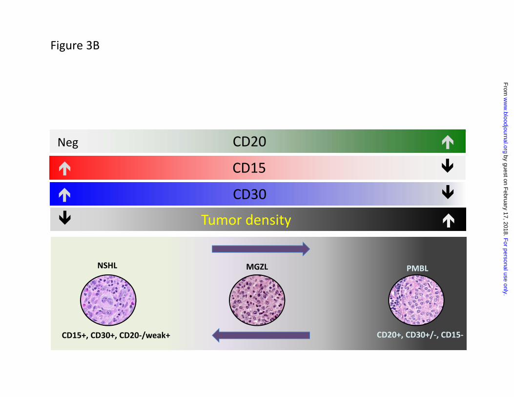
(MGZL) with clinical and pathologic features intermediate between PMBL and classical
Hodgkin lymphoma have been recognized. MGZL predominantly affect males and
appear to have an inferior outcome compared to PMBL(1, 2).
Pathology
PMBL is putatively derived from a medullary thymic B-cell. Morphologically,
these are medium to large cells having round or lobulated nuclei and abundant cytoplasm.
In most cases, compartmentalizing sclerosis is observed and sometimes tumor cells can
resemble Hodgkin/Reed Sternberg cells. The nodal architecture is typically diffuse with
occasional cases showing focal nodularity and necrosis is sometimes seen(3). PMBL has
a B-cell phenotype and expresses CD20 and pan B-cell markers such as CD79a, but
tumor cells do not express surface immunoglobulin– therefore, monoclonality can not be
established by kappa and lambda staining in contrast to most B-cell neoplasms (4, 5). B-
cell transcription factors including PAX5, OCT2 and BOB1 are typically strongly
expressed. CD30 is typically expressed but is dim in comparison to classical HL whereas
CD15 is usually negative (3-5). The germinal center markers CD10, BCL6 and CD23 are
expressed in most cases of PMBL, in keeping with its thymic B-cell origin(6, 7).
Distinguishing PMBL from nodular sclerosis Hodgkin lymphoma can sometimes be
challenging for the pathologist – NSHL has a nodular pattern of growth and the presence
of lacunar variants of HRS cells with a characteristic immunophenotype. In contrast to
PMBL, cells are typically CD15 positive and strongly positive for CD30. The expression
of B-cell markers like CD20, CD79a and PAX5 is often weak or negative(8, 9).
For personal use only.on February 17, 2018. by guest www.bloodjournal.orgFrom
5
The morphological and immunohistochemical features of MGZL are intermediate
and transitional between PMBL and NSHL(10-12). As in the case of both PMBL and
NSHL, surface immunoglobulin is not expressed. B-cell markers such as CD20 and
CD79a are typically expressed, CD30 is usually positive and there is variable expression
of CD15. PAX5, OCT2 and BOB1 are also typically expressed. In MGZL cases, an
asynchrony between morphology and immunophenotype can be seen – cases can have a
PMBL-like morphology but with immunophenotypical features of nodular sclerosis
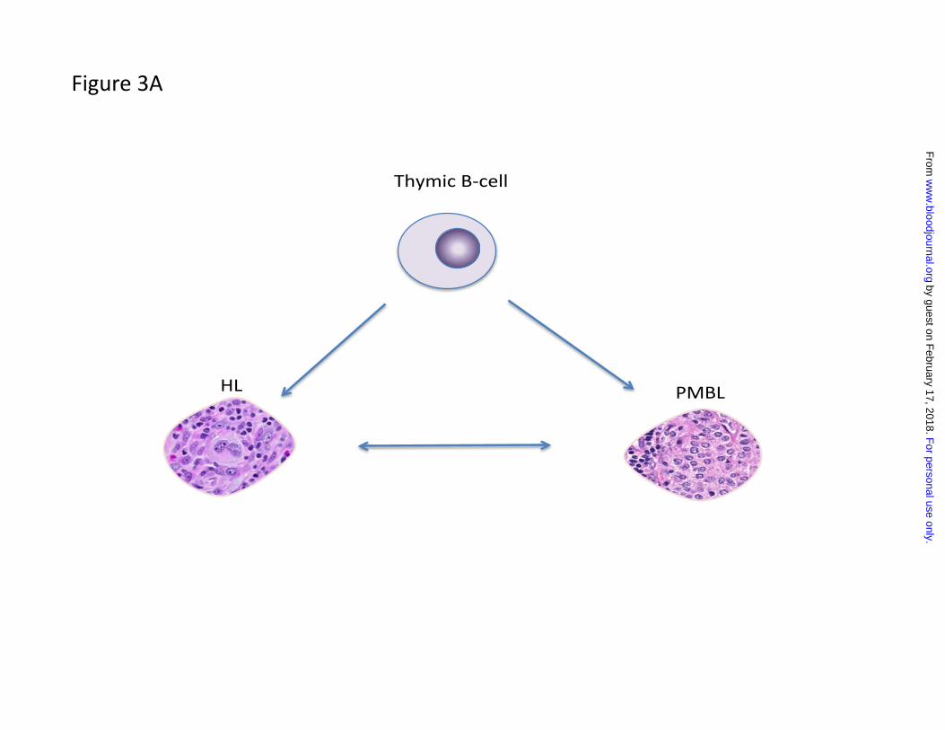
Hodgkin lymphoma or vice versa. The factors that transform a thymic B-cell into one or
other of these diseases are not well understood but it is likely that there is plasticity in
these events given that it is not uncommon to see them recur as one of the other entities
(e.g. PMBL recurs as NSHL or NSHL as MGZL etc.)(figure 3A)(11).
Genetic and Molecular Characteristics of PMBL
Gene expression profiling studies have demonstrated that the genotype of PMBL
has much more in common with that of NSHL than the other subtypes of DLBCL (i.e.
germinal center B-cell like (GCB) and activated B-cell like (ABC))(13, 14). In fact,
PMBL shares a third of its genes with NSHL(13). Among the most common genetic
alterations in PMBL are abnormalities on chromosome 9p (up to 75%) and 2p
(approximately 50%) – while these have been described in NSHL, they are typically not
found in the other DLBCL subtypes (15). The 9p region encodes janus kinase 2 (JAK2)
which then activates the transcription factor STAT 6 through phosphorylation (13, 16).
Recent work has demonstrated that phosphorylated STAT 6 can transcriptionally repress
BCL6 in PMBL(17). Suppressor of cytokine signaling 1 (SOCS1) suppresses JAK
For personal use only.on February 17, 2018. by guest www.bloodjournal.orgFrom
6
signaling and is mutated in a high proportion of PMBL and CHL cases(18). Also in the
9p region, programmed death ligands (PDL) 1 and 2 are rearranged at a frequency of
20% while gains or amplifications of c-REL may be seen at 2p(13, 19). Approximately
one third of PMBL cases may have gains in chromosome X. Recently, whole-genome
and whole-transcriptome sequencing has identified recurrent somatic coding-sequence
mutations in the PTPN1 gene – these are also commonly found in Hodgkin lymphoma
cases(20). In PMBL, in contrast to other subtypes of DLBCL, rearrangements of BCL2,
BCL6 and MYC are typically absent (15). PMBL and CHL both have constitutively
activated nuclear factor kappa-B (NFκB) and PMBL cell line survival is dependent on
NFκB target genes.
Due to its rarity, the molecular characteristics of GZL have not been well studied.
However, a study that looked at chromosomal aberrations in GZL showed gains
including amplifications in 2p16.1(REL/BCL11A locus) in 33% of all cases, alterations of
the JAK2/PDL2 locus in 9p24.1 in 55%, rearrangement of the CIITA locus at 16p13.13 in
27% and gains of 8q24 (MYC) in 27%(10). A recent large-scale methylation analysis that
included PMBL, CHL and MGZL cases, showed that these entities shared many
epigenetic characteristics and that MGZL had a distinct epigenetic signature(21).
Diagnosis of PMBL and Prognostic Factors
The diagnostic work up for PMBL should include the same routine tests that are
performed for any other DLBCL patient. Most importantly, the tissue biopsy should be
evaluated by a pathologist expert in the diagnosis of lymphoma. For the aforementioned
reasons, it can sometimes be challenging to distinguish PMBL from NSHL. A thorough
For personal use only.on February 17, 2018. by guest www.bloodjournal.orgFrom
7
history and physical examination, complete evaluation of hematological and biochemical
parameters, computerized tomography of the chest, abdomen and pelvis and a bone
marrow aspirate and biopsy should be performed. Though CNS involvement is very rare
at initial diagnosis, the CSF should be checked by cytology and flow cytometry in the
presence of clinical characteristics that are associated with a higher risk of CNS
spread(22). It is common for pleural and pericardial effusions to occur at presentation so
it may be useful to perform an echocardiogram. While the international prognostic index
(IPI) is useful in DLBCL, its utility in PMBL specifically is limited by the young age
distribution of the disease and its typical confinement to the mediastinum(23). Various
studies have looked at the role of IPI but it is not clear that it is helpful in predicting
outcome (24). Some retrospective studies have suggested that factors like LDH level,
male sex, performance status and advanced stage disease may be useful predictors of
survival but this is controversial and has not been validated in prospective studies(25,
26).
Primary Treatment and Outcome
As PMBL is relatively rare and only recently described, there is a paucity of
prospective treatment data and a lack of randomized studies. Therefore controversies
abound about what the optimal therapeutic approach should be and which regimen is
best. The cure rate for progressive or recurrent disease following primary therapy is low
so it is critical to optimize up-front outcomes. Most effective approaches to date have
incorporated consolidation radiation – though high cure rates are achieved with combined
modality therapy, it is increasingly apparent, especially from follow-up data on long-term
For personal use only.on February 17, 2018. by guest www.bloodjournal.orgFrom
8
survivors of HL, that mediastinal radiation is associated with significant late sequelae –
particularly a high risk of breast tumors in females (27, 28). Though some studies suggest
that lower doses of radiation and more focused treatment fields may reduce these
complications, this has not been clearly demonstrated and some studies contest this(29).
It is therefore important in PMBL to develop platforms that obviate the need for routine
radiation and thus eliminate these complications.
Radiation and Dose Intensity
Early studies in PMBL suggested that consolidation radiation was a critical
component of curative therapy. One of these that led to this widely held acceptance was a
study of MACOP-B (methotrexate, leucovorin, doxorubicin, cyclophosphamide,
vincristine, prednisone and bleomycin) followed by mediastinal radiation therapy in 50
untreated PMBL patients(30). While 66% of patients were gallium scan positive (this
study was done in the pre FDG-PET era) at the end of chemotherapy, only 19% were
gallium positive after radiation and this supported a combined modality approach, which
is taken by most today. Early (albeit retrospective) studies also suggested a benefit to
increasing dose intensity, which has been shown to be important in Hodgkin lymphoma,
a closely related disease clinically and biologically. One such study retrospectively
compared MACOP-B and VACOP-B (etoposide, doxorubicin, cyclophosphamide,
vincristine, prednisone, bleomycin) to CHOP in 138 patients – those who received CHOP
had a worse outcome, suggesting a role for dose-intensity(31). The largest study to look
at the dose intensity question was conducted by the International Extranodal Lymphoma
Study Group (IELSG) and evaluated 426 newly diagnosed PMBL patients who received
For personal use only.on February 17, 2018. by guest www.bloodjournal.orgFrom
9
MACOP-B, VACOP-B, ProMACECytaBom or CHOP (26). Although response rates
were similar among the groups, projected long-term PFS and OS survival rates were
higher in patients who received third-generation regimens. Additional retrospective
series from the British Columbia Cancer Agency and MSKCC groups also suggested that
increased dose-intensity regimens might be superior to CHOP-like approaches in this
disease(24, 25). Nonetheless, prospective comparisons of increased dose-intensity versus
CHOP-like regimens have not been done in PMBL – though the Southwest Oncology
Group (SWOG) prospectively compared second and third generation regimens to CHOP
in DLBCL, the outcome of PMBL was not assessed as it was not a recognized disease
entity at this time(32).
The Role of Rituximab
While the addition of rituximab to CHOP chemotherapy in DLBCL has been
shown to improve survival in several different studies, this has not been well studied or
established in PMBL due to the rarity of the disease(33). A subgroup analysis of the
prospective, randomized, phase III MabThera International Trial (MInT) evaluated the
role of rituximab in combination with CHOP-like regimens in PMBL patients with an age
adjusted IPI of ≤ 1 (34). The rituximab arm was clearly superior in terms of 3-year event-
free survival (78% versus 52% in the chemotherapy arm alone), but no statistically
significant difference in OS was detected due to small numbers of patients. When the
outcome of patients who received DA-EPOCH alone in the pre-rituximab era was
compared to those who received DA-EPOCH-R, albeit a non-randomized comparison,
there was a significantly better event-free and overall survival in the group who received
For personal use only.on February 17, 2018. by guest www.bloodjournal.orgFrom
10
rituximab (35). Though an earlier retrospective study from the British Columbia Cancer
Agency in the pre and post rituximab period demonstrated no survival advantage in
patients when rituximab was added to CHOP, a more recent report from the group
showed an improved time to progression and longer overall survival in those receiving
rituximab(25, 36).
Can mediastinal radiation be omitted?
Despite a lack of prospective studies, there does appear to be a benefit to adding
rituximab to chemotherapy in PMBL. Therefore, can rituximab abrogate any advantage
of dose-intensive platforms over CHOP and obviate the need for routine radiation? While
the subset analysis in the MInT study demonstrated improved responses and EFS in
patients receiving rituximab, preplanned radiotherapy was still administered to 73% of
patients in the immunochemotherapy arm and adding radiation improved remission rates.
In addition and importantly, the study was confined to patients with a low IPI score (≤1)
who truly represent very favorable subset of patients without the presence of unfavorable
characteristics at diagnosis. A recent retrospective analysis of R-CHOP (followed by
mediastinal radiation in 77% of responders) in 58 PMBL patients that included all IPI
groups showed a high rate of primary induction failures (21%) and an overall PFS of
68% at 5 years(37). Another retrospective analysis from the MSKCC group evaluated R-
CHOP followed by ifosfamide, cyclophosphamide and etoposide (ICE) without radiation
and reported a PFS of 78% at 3 years(38). A British Columbia study that looked at the
outcome of PMBL in the rituximab era reported on a subset of patients in whom an FDG-
PET guided RT approach (i.e. FDG-PET negative cases were not radiated) was used.
For personal use only.on February 17, 2018. by guest www.bloodjournal.orgFrom
11
Despite this, a sizeable proportion of PET negative cases subsequently relapsed(36). Due
to the observation that dose intensity has been important in PMBL, an NCI study
evaluated dose adjusted EPOCH-R without radiation in PMBL and included all clinical
risk groups(39-41). In 51 patients, at a median follow-up of 5 years, EFS and OS were
93% and 97% respectively and just 2 patients required consolidation radiation(35). In an
additional 16 PMBL patients who received the regimen at Stanford, both EFS and OS
were 100% without radiation This approach is now being tested in multicenter studies
and an early report from a pediatric/adolescent study suggested high efficacy without the
need for radiation in this population(42).
FDG-PET evaluation following therapy
At the completion of treatment for PMBL, a residual mediastinal mass is
commonly present, particularly in cases where there had been a large mass at initial
diagnosis or a large fibrotic component to the mass. It is not uncommon for these masses
to persist for several months after the completion of therapy and this is an important
consideration in interpretation of follow-up imaging. Hence, computed tomography (CT)
alone is limited in its scope to assess for the presence of residual disease. Gallium
scanning was used as an adjunct imaging test in the past for this purpose but it is now
regarded as a cumbersome test and is infrequently used today. While studies looking at
the role of end of treatment FDG-PET and its ability to guide the use/need for
consolidation radiation are limited, the technique has been found to have a very high
negative predictive value in this disease(35). However, the positive predictive value of
FDG-PET in PMBL is poor in contrast to the high clinical accuracy of FDG-PET in other
For personal use only.on February 17, 2018. by guest www.bloodjournal.orgFrom
12
aggressive lymphomas(35, 42). Following DA-EPOCH-R, 18 of 36 patients who
underwent an FDG-PET scan, had a maximum standardized value (SUV) above the
mediastinal blood pool but only 3 of these were found to have residual lymphoma. One
retrospective study that looked at interim PET in PMBL patients receiving R-VACOP-B
also reported a low positive predictive value(43). This is likely due to ongoing
inflammatory activity in residual mediastinal masses that may be FDG-PET avid.
Therefore, it is not a very accurate technique for determining the presence of residual
disease at the end of treatment and alternative, more specific imaging modalities should
be investigated.
Therapeutic Decision Making
In making decisions about the initial treatment of PMBL, one must consider the
long-term complications of mediastinal radiation in this population who are
predominantly young females(27). While R-CHOP followed by radiation has been
effective in low-risk patients, it appears to be insufficient therapy for patients with high-
risk disease and is associated with a high rate of primary refractory disease(44). Based on
its promising efficacy in an NCI study, we recommend DA-EPOCH-R without radiation
while confirmatory studies are in progress. Following this regimen without radiation, end
of therapy FDG-PET has an excellent negative predictive value but low positive
predictive value so end of therapy positive FDG-PETs need to be interpreted cautiously
with regard to decisions about consolidation radiation. A prospective study of FDG-PET
directed therapy by the IELSG is currently underway.
For personal use only.on February 17, 2018. by guest www.bloodjournal.orgFrom
13
Treatment of Mediastinal grey-zone lymphomas
These very rare tumors have histological and immunophenotypic features that are
transitional between PMBL and nodular sclerosing Hodgkin lymphoma. Due to their
rarity and recent identification, they have been poorly studied. In the past, these tumors
were likely called “anaplastic large-cell lymphoma Hodgkin-like” and that entity was
reported to have a poor prognosis with short median survivals(45). Their indeterminate
pathobiology has led to uncertainty about what the optimal therapeutic strategy should
be. One retrospective study reported that the 5-year event-free survival for this entity was
worse than that for classical Hodgkin lymphoma (International Database on Hodgkin’s
Disease), suggesting adverse biology and a high rate of treatment resistance (45).
Recently, a prospective study looked at the clinical characteristics and outcome of MGZL
following treatment with DA-EPOCH-R and reported a worse outcome compared to that
of PMBL, despite a patient population with similar clinical characteristics (EFS and OS
of 62% and 74% versus 93% and 97% for PMBL) (46, 47). Studies investigating the
molecular characteristics and biological basis for this inferior outcome are ongoing.
Treatment of relapsed or refractory disease
In PMBL, relapses tend to occur relatively early following the completion of
treatment and most are observed in the first year or 18 months after therapy. Relapsed
disease may stay confined to the mediastinum or spread to extra-nodal sites such as the
liver, kidneys or central nervous system. . Optimal therapy for relapsed disease has not
been well defined and should be decided based on the pattern of relapse and prior
treatments received. For relapses that are localized to the mediastinum, chemotherapy
For personal use only.on February 17, 2018. by guest www.bloodjournal.orgFrom

14
with radiation treatment (with or without autologous stem cell transplantation) may be a
curative option, especially in patients who did not receive mediastinal radiation initially.
For non-localized relapses, salvage chemotherapy followed by high dose therapy may be
considered(48). Allogeneic transplantation is another experimental option that can be
considered. The outcome for patients with MGZL is inferior to that of PMBL and
relapses should be approached similarly to PMBL.
Novel Agents
The antibody drug conjugate directed against CD30 – brentuximab vedotin - has shown
activity in patients with relapsed Hodgkin lymphoma and is currently being studied in
PMBL, where CD30 is variably expressed(49). Novel strategies in PMBL and other
mediastinal lymphomas should focus on combining targeted agents with effective
immunochemotherapy platforms. As the NF-kappa B signaling pathway is one of the
most important deregulated pathways in PMBL, inhibitors of this pathway are rational
strategies in PMBL. A region on chromosome 9p24 is amplified in 70% of cases of
PMBL that constitutes critical targets including janus kinase 2 (JAK2) and the PD1
ligands programmed cell death ligand 1 (PD-L1) and PD-L2 (50-52). Selective JAK2
inhibition has been shown to specifically decrease mediastinal large B-cell lymphoma
growth in vitro and in vivo (53). Agents such as JAK-STAT pathway inhibitors or
neutralizing antibodies to PD-1, like pidilizumab, are worthwhile investigating in
PMBL.(54)
Conclusions
For personal use only.on February 17, 2018. by guest www.bloodjournal.orgFrom
15
Controversies abound as to what the optimal regimen is for PMBL but evidence
from several studies suggests a benefit to regimens with increased dose-intensity.
Recent data demonstrates that DA-EPOCH-R can obviate the need for routine
mediastinal radiation. These results are now being validated in multicenter settings in
distinct patient groups. Though FDG-PET is routinely used for end of treatment
assessment in PMBL, it has a low positive predictive value that limits its usefulness in the
assessment of residual masses.
Authorship
KD and WHW contributed to the writing of the manuscript and approved the final
version. KD and WHW have no relevant conflicts of interest.
The authors receive funding from the Intramural Program of the National Cancer
Institute.
References
1. Swerdlow S, Campo E, Harris N, JAffe E, Pileri S, Stein H, et al. WHO Classification
of Tumors of Haematopoeitic and Lymphoid Tissues. IARC: Lyon 2008.
2. Dunleavy K, Grant C, Eberle FC, Pittaluga S, Jaffe ES, Wilson WH. Gray zone
lymphoma: better treated like hodgkin lymphoma or mediastinal large B-cell
lymphoma? Curr Hematol Malig Rep. 2012;7(3):241-7. Epub 2012/07/27.
3. Pileri SA, Zinzani PL, Gaidano G, Falini B, Gaulard P, Zucca E, et al. Pathobiology
of primary mediastinal B-cell lymphoma. Leuk Lymphoma. 2003;44 Suppl 3:S21-6. Epub
2004/06/19.
4. Moller P, Moldenhauer G, Momburg F, Lammler B, Eberlein-Gonska M, Kiesel S,
et al. Mediastinal lymphoma of clear cell type is a tumor corresponding to terminal
steps of B cell differentiation. Blood. 1987;69(4):1087-95. Epub 1987/04/01.
For personal use only.on February 17, 2018. by guest www.bloodjournal.orgFrom
16
5. Pileri SA, Gaidano G, Zinzani PL, Falini B, Gaulard P, Zucca E, et al. Primary
mediastinal B-cell lymphoma: high frequency of BCL-6 mutations and consistent
expression of the transcription factors OCT-2, BOB.1, and PU.1 in the absence of
immunoglobulins. Am J Pathol. 2003;162(1):243-53. Epub 2003/01/01.
6. Calaminici M, Piper K, Lee AM, Norton AJ. CD23 expression in mediastinal large
B-cell lymphomas. Histopathology. 2004;45(6):619-24. Epub 2004/12/01.
7. Salama ME, Rajan Mariappan M, Inamdar K, Tripp SR, Perkins SL. The value of
CD23 expression as an additional marker in distinguishing mediastinal (thymic) large B-
cell lymphoma from Hodgkin lymphoma. Int J Surg Pathol. 2010;18(2):121-8. Epub
2009/02/19.
8. Schmid C, Pan L, Diss T, Isaacson PG. Expression of B-cell antigens by Hodgkin's
and Reed-Sternberg cells. Am J Pathol. 1991;139(4):701-7. Epub 1991/10/01.
9. Zukerberg LR, Collins AB, Ferry JA, Harris NL. Coexpression of CD15 and CD20 by
Reed-Sternberg cells in Hodgkin's disease. Am J Pathol. 1991;139(3):475-83. Epub
1991/09/01.
10. Eberle FC, Salaverria I, Steidl C, Summers TA, Jr., Pittaluga S, Neriah SB, et al.
Gray zone lymphoma: chromosomal aberrations with immunophenotypic and clinical
correlations. Mod Pathol. 2011;24(12):1586-97. Epub 2011/08/09.
11. Traverse-Glehen A, Pittaluga S, Gaulard P, Sorbara L, Alonso MA, Raffeld M, et al.
Mediastinal gray zone lymphoma: the missing link between classic Hodgkin's lymphoma
and mediastinal large B-cell lymphoma. Am J Surg Pathol. 2005;29(11):1411-21. Epub
2005/10/15.
12. Garcia JF, Mollejo M, Fraga M, Forteza J, Muniesa JA, Perez-Guillermo M, et al.
Large B-cell lymphoma with Hodgkin's features. Histopathology. 2005;47(1):101-10.
Epub 2005/06/29.
13. Rosenwald A, Wright G, Leroy K, Yu X, Gaulard P, Gascoyne RD, et al. Molecular
diagnosis of primary mediastinal B cell lymphoma identifies a clinically favorable
subgroup of diffuse large B cell lymphoma related to Hodgkin lymphoma. J Exp Med.
2003;198(6):851-62. Epub 2003/09/17.
14. Savage KJ, Monti S, Kutok JL, Cattoretti G, Neuberg D, de Leval L, et al. The
molecular signature of mediastinal large B-cell lymphoma differs from that of other
diffuse large B-cell lymphomas and shares features with classical Hodgkin's lymphoma.
Blood. 2003:2003-06-1841.
15. Swerdlow SH, Campo E, Harris NL, Jaffe ES, Pileri SA, Stein H, et al. WHO
Classification of Tumours of Haematopoietic and Lymphoid Tissues. IARC: Lyon 2008.
16. Guiter C, Dusanter-Fourt I, Copie-Bergman C, Boulland ML, Le Gouvello S,
Gaulard P, et al. Constitutive STAT6 activation in primary mediastinal large B-cell
lymphoma. Blood. 2004;104(2):543-9. Epub 2004/03/27.
17. Ritz O, Rommel K, Dorsch K, Kelsch E, Melzner J, Buck M, et al. STAT6-mediated
BCL6 repression in primary mediastinal B-cell lymphoma (PMBL). Oncotarget.
2013;4(7):1093-102. Epub 2013/07/16.
18. Melzner I, Bucur AJ, Bruderlein S, Dorsch K, Hasel C, Barth TF, et al. Biallelic
mutation of SOCS-1 impairs JAK2 degradation and sustains phospho-JAK2 action in the
MedB-1 mediastinal lymphoma line. Blood. 2005;105(6):2535-42. Epub 2004/12/02.
For personal use only.on February 17, 2018. by guest www.bloodjournal.orgFrom
17
19. Twa DD, Chan FC, Ben-Neriah S, Woolcock BW, Mottok A, Tan KL, et al. Genomic
rearrangements involving programmed death ligands are recurrent in primary
mediastinal large B-cell lymphoma. Blood. 2014;123(13):2062-5. Epub 2014/02/06.
20. Gunawardana J, Chan FC, Telenius A, Woolcock B, Kridel R, Tan KL, et al.
Recurrent somatic mutations of PTPN1 in primary mediastinal B cell lymphoma and
Hodgkin lymphoma. Nat Genet. 2014;46(4):329-35. Epub 2014/02/18.
21. Eberle FC, Rodriguez-Canales J, Wei L, Hanson JC, Killian JK, Sun HW, et al.
Methylation profiling of mediastinal gray zone lymphoma reveals a distinctive signature
with elements shared by classical Hodgkin's lymphoma and primary mediastinal large B-
cell lymphoma. Haematologica. 2011;96(4):558-66. Epub 2011/04/02.
22. van Besien K, Ha CS, Murphy S, McLaughlin P, Rodriguez A, Amin K, et al. Risk
factors, treatment, and outcome of central nervous system recurrence in adults with
intermediate-grade and immunoblastic lymphoma. Blood. 1998;91(4):1178-84. Epub
1998/03/07.
23. A predictive model for aggressive non-Hodgkin's lymphoma. The International
Non-Hodgkin's Lymphoma Prognostic Factors Project. N Engl J Med. 1993;329(14):987-
94.
24. Hamlin PA, Portlock CS, Straus DJ, Noy A, Singer A, Horwitz SM, et al. Primary
mediastinal large B-cell lymphoma: optimal therapy and prognostic factor analysis in
141 consecutive patients treated at Memorial Sloan Kettering from 1980 to 1999. Br J
Haematol. 2005;130(5):691-9. Epub 2005/08/24.
25. Savage KJ, Al-Rajhi N, Voss N, Paltiel C, Klasa R, Gascoyne RD, et al. Favorable
outcome of primary mediastinal large B-cell lymphoma in a single institution: the British
Columbia experience. Ann Oncol. 2006;17(1):123-30. Epub 2005/10/21.
26. Zinzani PL, Martelli M, Bertini M, Gianni AM, Devizzi L, Federico M, et al.
Induction chemotherapy strategies for primary mediastinal large B-cell lymphoma with
sclerosis: a retrospective multinational study on 426 previously untreated patients.
Haematologica. 2002;87(12):1258-64. Epub 2002/12/24.
27. Castellino SM, Geiger AM, Mertens AC, Leisenring WM, Tooze JA, Goodman P, et
al. Morbidity and mortality in long-term survivors of Hodgkin lymphoma: a report from
the Childhood Cancer Survivor Study. Blood. 2011;117(6):1806-16. Epub 2010/11/03.
28. Dunleavy K, Bollard CM. Sobering realities of surviving Hodgkin lymphoma.
Blood. 2011;117(6):1772-3. Epub 2011/02/12.
29. O'Brien MM, Donaldson SS, Balise RR, Whittemore AS, Link MP. Second
malignant neoplasms in survivors of pediatric Hodgkin's lymphoma treated with low-
dose radiation and chemotherapy. J Clin Oncol. 2010;28(7):1232-9. Epub 2010/02/04.
30. Zinzani PL, Martelli M, Magagnoli M, Pescarmona E, Scaramucci L, Palombi F, et
al. Treatment and clinical management of primary mediastinal large B-cell lymphoma
with sclerosis: MACOP-B regimen and mediastinal radiotherapy monitored by
(67)Gallium scan in 50 patients. Blood. 1999;94(10):3289-93.
31. Todeschini G, Secchi S, Morra E, Vitolo U, Orlandi E, Pasini F, et al. Primary
mediastinal large B-cell lymphoma (PMLBCL): long-term results from a retrospective
multicentre Italian experience in 138 patients treated with CHOP or MACOP-B/VACOP-B.
Br J Cancer. 2004;90(2):372-6. Epub 2004/01/22.
For personal use only.on February 17, 2018. by guest www.bloodjournal.orgFrom
18
32. Fisher RI, Gaynor ER, Dahlberg S, Oken MM, Grogan TM, Mize EM, et al.
Comparison of a standard regimen (CHOP) with three intensive chemotherapy regimens
for advanced non-Hodgkin's lymphoma. N Engl J Med. 1993;328(14):1002-6.
33. Coiffier B, Lepage E, Briere J, Herbrecht R, Tilly H, Bouabdallah R, et al. CHOP
chemotherapy plus rituximab compared with CHOP alone in elderly patients with diffuse
large-B-cell lymphoma. N Engl J Med. 2002;346(4):235-42. Epub 2002/01/25.
34. Rieger M, Osterborg A, Pettengell R, White D, Gill D, Walewski J, et al. Primary
mediastinal B-cell lymphoma treated with CHOP-like chemotherapy with or without
rituximab: results of the Mabthera International Trial Group study. Ann Oncol.
2011;22(3):664-70. Epub 2010/08/21.
35. Dunleavy K, Pittaluga S, Maeda LS, Advani R, Chen CC, Hessler J, et al. Dose-
adjusted EPOCH-rituximab therapy in primary mediastinal B-cell lymphoma. N Engl J
Med. 2013;368(15):1408-16. Epub 2013/04/12.
36. Savage KJ, Yenson PR, Shenkier T, Klasa R, Villa D, Goktepe O, et al. The Outcome
of Primary Mediastinal Large B-Cell Lymphoma (PMBCL) in the R-CHOP Treatment Era.
Blood (ASH Annual Meeting Abstracts) 2012 120: Abstract 303.
37. Soumerai JD, Hellmann MD, Feng Y, Sohani AR, Toomey CE, Barnes JA, et al.
Treatment of primary mediastinal B-cell lymphoma with rituximab, cyclophosphamide,
doxorubicin, vincristine and prednisone is associated with a high rate of primary
refractory disease. Leuk Lymphoma. 2014;55(3):538-43. Epub 2013/06/06.
38. Moskowitz C, Hamlin PA, Maraguilia J, Meikle J, Zelenetz AD. Sequential Dose-
Dende R-CHOP followed by ICE consolidation (MSKCC Protocol 01-142) without
radiotherapy for patients with primary mediastinal large B-cell lymphoma. Blood (ASH
Annual Meeting Abstracts) 2010 116: #420.
39. Wilson WH, Bryant G, Bates S, Fojo A, Wittes RE, Steinberg SM, et al. EPOCH
chemotherapy: toxicity and efficacy in relapsed and refractory non-Hodgkin's
lymphoma. J Clin Oncol. 1993;11(8):1573-82.
40. Wilson WH, Dunleavy K, Pittaluga S, Hegde U, Grant N, Steinberg SM, et al. Phase
II study of dose-adjusted EPOCH and rituximab in untreated diffuse large B-cell
lymphoma with analysis of germinal center and post-germinal center biomarkers. J Clin
Oncol. 2008;26(16):2717-24. Epub 2008/04/02.
41. Wilson WH PP, Hurd D, et al. Phase II study of Dose-Adjusted EPOCH-R in
untreated de novo CD20+ diffuse large B-cell lymphoma (DLBCL)-CALGB 50103. Proc Am
Soc Clin Oncol. 2005;24.
42. Woessmann W, Lisfeld J, Burkhardt B. Therapy in primary mediastinal B-cell
lymphoma. N Engl J Med. 2013;369(3):282. Epub 2013/07/19.
43. Avigdor A, Sirotkin T, Kedmi M, Ribakovsy E, Berkowicz M, Davidovitz Y, et al. The
impact of R-VACOP-B and interim FDG-PET/CT on outcome in primary mediastinal large
B cell lymphoma. Ann Hematol. 2014. Epub 2014/03/07.
44. Soumerai JD, Hellmann MD, Feng Y, Sohani AR, Toomey CE, Barnes JA, et al.
Treatment of Primary Mediastinal B-cell Lymphoma with R-CHOP is Associated with a
High Rate of Primary Refractory Disease. Leuk Lymphoma. 2013. Epub 2013/06/06.
45. Cazals-Hatem D, Andre M, Mounier N, Copin MC, Divine M, Berger F, et al.
Pathologic and clinical features of 77 Hodgkin's lymphoma patients treated in a
For personal use only.on February 17, 2018. by guest www.bloodjournal.orgFrom
19
lymphoma protocol (LNH87): a GELA study. Am J Surg Pathol. 2001;25(3):297-306. Epub
2001/02/27.
46. Dunleavy K, Pittaluga S, Shovlin M, Grant N, Grant C, Chen C, et al. Untreated
primary mediastinal B-cell (PMBL) and mediastinal grey zone (MGZL) lymphomas:
comparison of biological features and clinical outcome following DA-EPOCH-R without
radiation. Annals of Oncology.22 (4) Abstract 149. 2011.
47. Wilson WH, Pittaluga S, Nicolae A, Camphausen K, Shovlin M, Steinberg SM, et
al. A prospective study of mediastinal gray zone lymphoma. Blood. 2014. Epub
2014/07/16.
48. Popat U, Przepiork D, Champlin R, Pugh W, Amin K, Mehra R, et al. High-dose
chemotherapy for relapsed and refractory diffuse large B-cell lymphoma: mediastinal
localization predicts for a favorable outcome. J Clin Oncol. 1998;16(1):63-9. Epub
1998/01/24.
49. de Claro RA, McGinn K, Kwitkowski V, Bullock J, Khandelwal A, Habtemariam B,
et al. U.S. Food and Drug Administration approval summary: brentuximab vedotin for
the treatment of relapsed Hodgkin lymphoma or relapsed systemic anaplastic large-cell
lymphoma. Clin Cancer Res. 2012;18(21):5845-9. Epub 2012/09/11.
50. Joos S, Otano-Joos MI, Ziegler S, Bruderlein S, du Manoir S, Bentz M, et al.
Primary mediastinal (thymic) B-cell lymphoma is characterized by gains of chromosomal
material including 9p and amplification of the REL gene. Blood. 1996;87(4):1571-8. Epub
1996/02/15.
51. Green MR, Monti S, Rodig SJ, Juszczynski P, Currie T, O'Donnell E, et al.
Integrative analysis reveals selective 9p24.1 amplification, increased PD-1 ligand
expression, and further induction via JAK2 in nodular sclerosing Hodgkin lymphoma and
primary mediastinal large B-cell lymphoma. Blood. 2010;116(17):3268-77. Epub
2010/07/16.
52. Rui L, Emre NC, Kruhlak MJ, Chung HJ, Steidl C, Slack G, et al. Cooperative
epigenetic modulation by cancer amplicon genes. Cancer Cell. 2010;18(6):590-605. Epub
2010/12/16.
53. Hao Y, Chapuy B, Monti S, Sun HH, Rodig SJ, Shipp MA. Selective JAK2 inhibition
specifically decreases Hodgkin lymphoma and mediastinal large B-cell lymphoma growth
in vitro and in vivo. Clin Cancer Res. 2014;20(10):2674-83. Epub 2014/03/13.
54. Berger R, Rotem-Yehudar R, Slama G, Landes S, Kneller A, Leiba M, et al. Phase I
safety and pharmacokinetic study of CT-011, a humanized antibody interacting with PD-
1, in patients with advanced hematologic malignancies. Clin Cancer Res.
2008;14(10):3044-51. Epub 2008/05/17.
For personal use only.on February 17, 2018. by guest www.bloodjournal.orgFrom
20
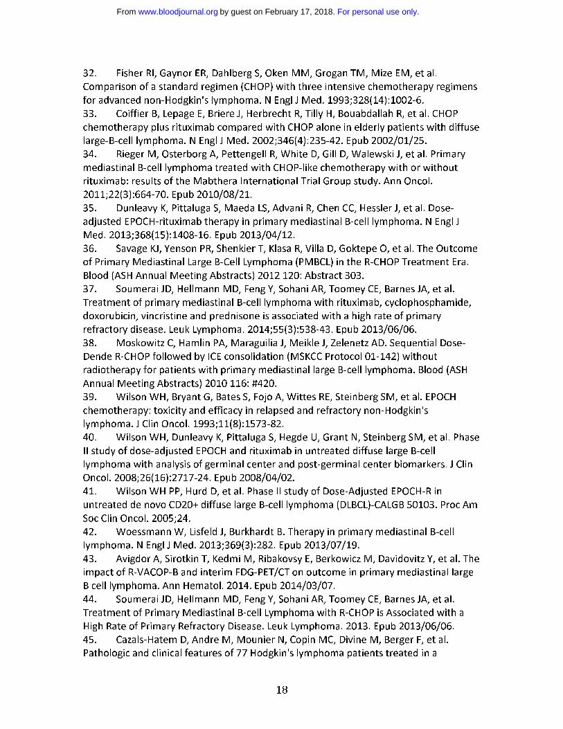
Table 1: Selected published studies of chemotherapy/immunochemotherapy
regimens with and without radiation treatment in PMBL.
For personal use only.on February 17, 2018. by guest www.bloodjournal.orgFrom
21
Figure Legends
Figure 1: Primary mediastinal B-cell lymphoma. H&E is shown and CD20 and
MUM1 staining are positive. MIB-1 scoring is high (Courtesy Stefania Pittlauga).
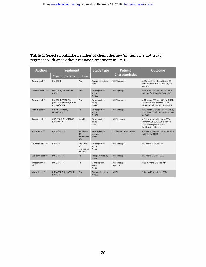
Figure 2: Figure 2. Relationship of PMBL to Hodgkin lymphoma. Relative gene
expression is shown in primary PMBLs (average of all biopsy samples), the PMBL
cell line K1106, three Hodgkin lymphoma (HL) cell lines, and six GCB DLBCL cell
lines. Red represents high gene expression and green low expression. PMBL
signature genes that are also expressed at high levels in Hodgkin lymphoma cell
lines compared with GCB DLBCL cell lines. (Courtesy Louis Staudt).
Figure 3: A. Both PMBL and HL are putatively derived from a thymic B-cell. The
events that transform a thymic B-cell into PMBL or HL are poorly understood but
there appears to be plasticity in these events as HL can recur as PMBL and vice
versa. B. While NSHL is CD15 and CD30 positive and PMBL is CD20 positive, there
are mediastinal lymphomas in between these 2 entitiies with histologic and
immunohistochemical features intermediate and transitional between NSHL and
PMBL. These disease are called mediastinal grey zone lymphomas (MGZLs).
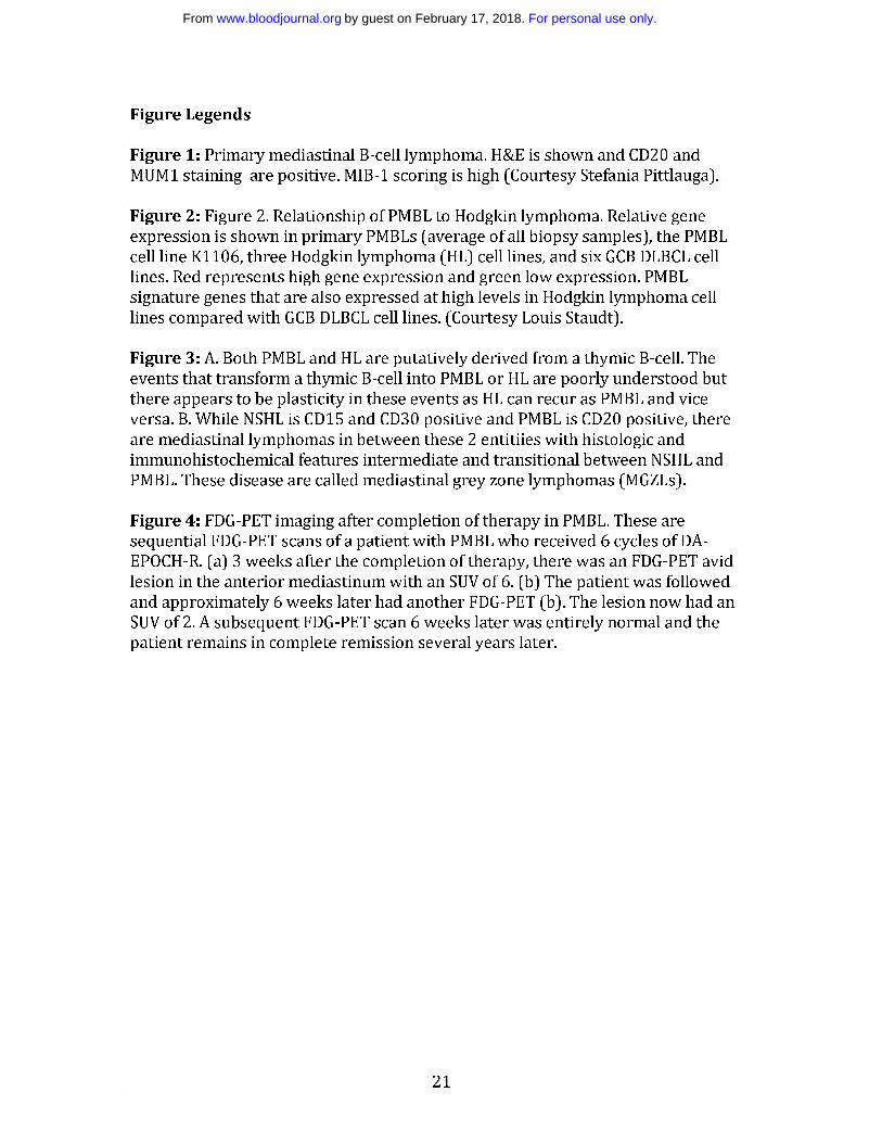
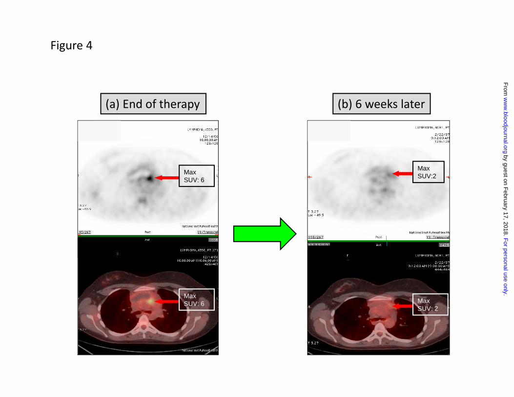
Figure 4: FDG-PET imaging after completion of therapy in PMBL. These are
sequential FDG-PET scans of a patient with PMBL who received 6 cycles of DA-
EPOCH-R. (a) 3 weeks after the completion of therapy, there was an FDG-PET avid
lesion in the anterior mediastinum with an SUV of 6. (b) The patient was followed
and approximately 6 weeks later had another FDG-PET (b). The lesion now had an
SUV of 2. A subsequent FDG-PET scan 6 weeks later was entirely normal and the
patient remains in complete remission several years later.
For personal use only.on February 17, 2018. by guest www.bloodjournal.orgFrom

CD20
MIB-1MUM1
Figure 1
For personal use only.
on February 17, 2018.
by guest
ww
w.bloodjournal.org
From
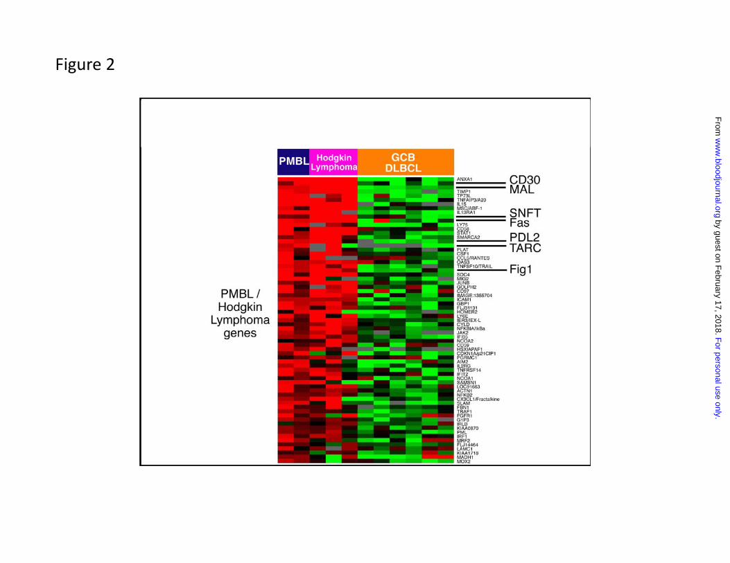
Figure 2
For personal use only.
on February 17, 2018.
by guest
ww
w.bloodjournal.org
From
Thymic B-cell
HL PMBL
Figure 3A
For personal use only.
on February 17, 2018.
by guest
ww
w.bloodjournal.org
From
NSHL PMBL
CD15+, CD30+, CD20-/weak+ CD20+, CD30+/-, CD15-
MGZL
Neg CD20
CD15
CD30
Tumor density
Figure 3B
For personal use only.
on February 17, 2018.
by guest
ww
w.bloodjournal.org
From
Max SUV: 6
Max SUV: 6
Max SUV:2
Max SUV: 2
Observe
(a) End of therapy (b) 6 weeks later
Figure 4
For personal use only.
on February 17, 2018.
by guest
ww
w.bloodjournal.org
From

doi:10.1182/blood-2014-05-575092Prepublished online December 11, 2014;
Kieron Dunleavy and Wyndham H. Wilson lymphoma: do they require a unique therapeutic approach?Primary mediastinal B-cell lymphoma and mediastinal gray zone
http://www.bloodjournal.org/site/misc/rights.xhtml#repub_requestsInformation about reproducing this article in parts or in its entirety may be found online at:
http://www.bloodjournal.org/site/misc/rights.xhtml#reprintsInformation about ordering reprints may be found online at:
http://www.bloodjournal.org/site/subscriptions/index.xhtmlInformation about subscriptions and ASH membership may be found online at:
digital object identifier (DOIs) and date of initial publication. indexed by PubMed from initial publication. Citations to Advance online articles must include final publication). Advance online articles are citable and establish publication priority; they areappeared in the paper journal (edited, typeset versions may be posted when available prior to Advance online articles have been peer reviewed and accepted for publication but have not yet
Copyright 2011 by The American Society of Hematology; all rights reserved.Hematology, 2021 L St, NW, Suite 900, Washington DC 20036.Blood (print ISSN 0006-4971, online ISSN 1528-0020), is published weekly by the American Society of
For personal use only.on February 17, 2018. by guest www.bloodjournal.orgFrom