Primary hypertrophic osteoarthropathy: case report and ... · Hypertrophic osteoarthropathy is a...
5
451 CASE REPORT Bras J Rheumatol 2009;49(4):447-55 INTRODUCTION Hypertrophic osteoarthropathy is a pathology characterized by digital clubbing of the hands and feet, extremity enlargement secondary to bone and periarticular tissue proliferation, joint pain and edema, bilateral eyelid ptosis, leonine face and skin thickness. 1,2 It is classified as either primary (pachydermoperiostosis) or secondary. The primary form, or idiopathic, is considered rare, corresponding to 3-5% of all cases. Here, we describe the clinical and radiological manifestations of a patient with the primary form of hypertrophic osteoarthropathy, and a literature review about the subject, focusing on the therapeutic challenge of refractory joint pain. CASE REPORT Male patient, 31-year-old, admitted in 1997 referring swelling and pain in the knees, ankles and small joints of the hands Primary hypertrophic osteoarthropathy: case report and literature review Aline Biral Zanon 1 , Mellysande Pontes Faccin 2 , Sônia Maria Alvarenga Anti 3 , Felipe José Silva Melo Cruz 4 , Renata Ferreira Rosa 5 Received on 07/14/2008. Approved on 02/02/2009. We declare no conflict of interest. 1. Fifth-year Medical student, ABC Medical School (FMABC) 2. Clinical Semiology Professor (FMABC) 3. Rheumatology Professor (FMABC) 4. Clinical Practice Resident (FMABC) 5. Associate Professor of Rheumatology (FMABC) Correspondence to: Aline Biral Zanon. Rua Piratininga, 790/22, Bairro Barcelona, São Caetano do Sul, SP – Brasil. CEP: 09550-160. E-mail: alinebiralzanon@ gmail.com ABSTRACT Primary hypertrophic osteoarthropathy is a rare syndrome, consisting of clubbed hands, fingers, and feet digits; en- larged extremities secondary to periarticular and bone proliferation; thickened facial skin; painful and swollen joints. The idiopathic form represents 3 to 5 per cent of all cases of hypertrophic osteoarthropaty. Genetic influence, abnormal fibroblasts activity, and changes of the peripherical blood flow appear to be significant on the pathogenesis. Clinical manifestations are variable: the term complete syndrome is used for the patient with pachydermia, coarsening of the face skin and scalp, periostitis, and cutis verticis gyrata); the incomplete form, when there is no sparing of the scalp; and the frusted form for pachydermia with minimal or absent periostitis. The authors describe a 39-year-old white man diagnosed with primary hypertrophic osteoarthropathy. We also report the clinical and radiological carateristics of this syndrome and terapeutical approach of pachydermoperiostosis. Keywords: hypertrophic osteoarthropathy, pachydermoperiostosis. for the last five years, with worsening during exercise and without morning stiffness. Fever or other associated symptoms were denied. The patient had been taken non-steroidal anti- inflammatory drugs (NSAIDs) without improvement. He denied any rheumatologic infection or similar condition in his family. Physical examination, cardiopulmonary auscultation, and abdominal palpation showed no alterations. The osteoarticular examination showed digital clubbing with watch-glass nails, diffuse swollen of the hands and edema of the lower limbs, in addition to arthritis of the ankle (Figures 1 and 2). The thoracic radiological evaluation was normal. X-rays of hands and wrists showed an increase in soft tissue and en- largement of the middle and distal phalanges, associated with cortical thickness. From an X-ray done with mammography technique (Figure 3), hypertrophy of the distal tufts with areas of acroosteolisis was detected. Tibia and fibula X-rays (Figure 4) showed gross irregularities and intense periosteal prolife-
Transcript of Primary hypertrophic osteoarthropathy: case report and ... · Hypertrophic osteoarthropathy is a...

451
CASE REPORT
Bras J Rheumatol 2009;49(4):447-55
INTRODUCTION
Hypertrophic osteoarthropathy is a pathology characterized by digital clubbing of the hands and feet, extremity enlargement secondary to bone and periarticular tissue proliferation, joint pain and edema, bilateral eyelid ptosis, leonine face and skin thickness.1,2
It is classified as either primary (pachydermoperiostosis) or secondary. The primary form, or idiopathic, is considered rare, corresponding to 3-5% of all cases.
Here, we describe the clinical and radiological manifestations of a patient with the primary form of hypertrophic osteoarthropathy, and a literature review about the subject, focusing on the therapeutic challenge of refractory joint pain.
CASE REPORT
Male patient, 31-year-old, admitted in 1997 referring swelling and pain in the knees, ankles and small joints of the hands
Primary hypertrophic osteoarthropathy: case report and literature review
Aline Biral Zanon1, Mellysande Pontes Faccin2, Sônia Maria Alvarenga Anti3,Felipe José Silva Melo Cruz4, Renata Ferreira Rosa5
Received on 07/14/2008. Approved on 02/02/2009. We declare no conflict of interest.1. Fifth-year Medical student, ABC Medical School (FMABC)2. Clinical Semiology Professor (FMABC)3. Rheumatology Professor (FMABC)4. Clinical Practice Resident (FMABC)5. Associate Professor of Rheumatology (FMABC)Correspondence to: Aline Biral Zanon. Rua Piratininga, 790/22, Bairro Barcelona, São Caetano do Sul, SP – Brasil. CEP: 09550-160. E-mail: [email protected]
ABSTRACT
Primary hypertrophic osteoarthropathy is a rare syndrome, consisting of clubbed hands, fingers, and feet digits; en-larged extremities secondary to periarticular and bone proliferation; thickened facial skin; painful and swollen joints. The idiopathic form represents 3 to 5 per cent of all cases of hypertrophic osteoarthropaty. Genetic influence, abnormal fibroblasts activity, and changes of the peripherical blood flow appear to be significant on the pathogenesis. Clinical manifestations are variable: the term complete syndrome is used for the patient with pachydermia, coarsening of the face skin and scalp, periostitis, and cutis verticis gyrata); the incomplete form, when there is no sparing of the scalp; and the frusted form for pachydermia with minimal or absent periostitis. The authors describe a 39-year-old white man diagnosed with primary hypertrophic osteoarthropathy. We also report the clinical and radiological carateristics of this syndrome and terapeutical approach of pachydermoperiostosis.
Keywords: hypertrophic osteoarthropathy, pachydermoperiostosis.
for the last five years, with worsening during exercise and without morning stiffness. Fever or other associated symptoms were denied. The patient had been taken non-steroidal anti-inflammatory drugs (NSAIDs) without improvement. He denied any rheumatologic infection or similar condition in his family.
Physical examination, cardiopulmonary auscultation, and abdominal palpation showed no alterations. The osteoarticular examination showed digital clubbing with watch-glass nails, diffuse swollen of the hands and edema of the lower limbs, in addition to arthritis of the ankle (Figures 1 and 2).
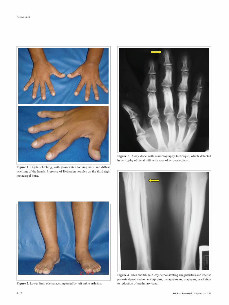
The thoracic radiological evaluation was normal. X-rays of hands and wrists showed an increase in soft tissue and en-largement of the middle and distal phalanges, associated with cortical thickness. From an X-ray done with mammography technique (Figure 3), hypertrophy of the distal tufts with areas of acroosteolisis was detected. Tibia and fibula X-rays (Figure 4) showed gross irregularities and intense periosteal prolife-
Zanon et al.
452 Rev Bras Reumatol 2009;49(4):447-55
Figure 1. Digital clubbing, with glass-watch looking nails and diffuse swelling of the hands. Presence of Heberden nodules on the third right metacarpal bone.
Figure 2. Lower limb edema accompanied by left ankle arthritis.
Figure 3. X-ray done with mammography technique, which detected hypertrophy of distal tuffs with area of acro-osteolisis.
Figure 4. Tibia and fibula X-ray demonstrating irregularities and intense periosteal proliferation in epiphysis, metaphysis and diaphysis, in addition to reduction of medullary canal.

453
Primary hypertrophic osteoarthropathy: case report and review of the literature
Bras J Rheumatol 2009;49(4):447-55
Table ISecondary Hypertrophic Osteoarthropathy Etiology
Pulmonary
Bronchogenic carcinoma
Pulmonary abscesses
Bronchiectasis
Pulmonary emphysema
Hodgkin’s disease
Metastases
Cystic fibrosis
Pleural and diaphragm Mesothelioma
Cardiac Congenital cyanotic heart disease
Abdominal
Portal or biliary cirrhosis
Ulcerative colitis
Crohn’s disease
Gastrointestinal polyps
Neoplasm
Biliary atresia
Others
Nasopharygeal carcinoma
Esophagus carcinoma
Aortic or axillary artery graft infection
ration in the epiphyses, metaphysis and diaphysis, as well as reduction of the spinal canal.
Laboratory analysis showed an erythrocyte sedimentation rate (ESR) of 110 mm in the first hour, C-reactive protein (CRP) negative, serum mucoprotein of 2.9 mg% (normal between 1.9 and 4.9 mg%) and non-reactive rheumatoid factor, antinuclear factor and anti DNA.
No heart, lung or liver diseases were found. Imaging and laboratory tests were performed, which excluded secondary causes of osteoarthropathy.
During his follow up, dyspeptic disease due to the chronic use of NSAIDs was found, in addition to weight loss of 24.25 pounds (11 kg) in four mouths. Upper endoscopy diagnosed a duodenal ulcer with the presence of Helicobacter pylori.
Due to his refractory muscle-skeletal pain, paracetamol associated with tricyclic antidepressant (amitriptyline) was prescribed as an alternative analgesic.
DISCUSSION
Hypertrophic osteoarthropathy was described for the first time by Friedreich, in 1868. It is a rheumatologic condition in which the patient presents with digital clubbing of hands and feet, increase of the extremities secondary to bone and periarticular tissue proliferation, joint pain and edema, bilateral eyelid ptosis, leonine face, and thickened skin.1,2 In the histological evaluation, hyperplasia of the subcutaneous conjunctive tissue was observed.3
The disease is classified as either primary (hereditary or idiopathic) or secondary. Table 1 summarizes the main causes of secondary hypertrophic osteoarthropathy.
The idiopathic form (named pachydermoperiostosis, primary hypertrophic osteoarthropathy or Touraine-Soulente-Golé syndrome) is a rare disease, with unknown etiology, and represents 3 to 5% of all cases of hypertrophic osteoarthropathy.2,4 The presence of digital clubbing, radiographic periostosis and leonine facial features are the main diagnosis criteria.
Although autosomal dominant inheritance transmission with variable expression and incomplete penetrance is recognized, a population study involving 68 families (204 affected patients) found 37 families with dominant autossomal mutation, and recessive autossomal inheritance in the remaining 31. Both forms of transmission differed in the severity of the disease and in the prevalence of some clinical characteristics.5
Pachydermoperiostosis occurs predominantly in men (80% of cases), in which a severe phenotype is often shown. A mutation linked to the X chromosome, in association with hormonal alterations (testosterone-dependent proliferation),
may be involved in the distribution of disease by the gender.5 A higher incidence is also found in African-Americans when compared to Caucasians.
The pathogenesis of the Touraine-Solente-Golé syndrome has not been fully elucidated. A brief study of patients with the disease showed abnormal peripheral blood supply when compared to non-affected individuals; however, additional studies failed to reproduce these findings. Biopsies of skin and bone marrow show an exacerbated proliferation of fibroblasts, associated with diffuse epidermal hyperplasia, partial occlusion of vascular lumen, and lymphohistiocytic infiltration with collagen deposition. An isolated study showed normal fibroblastic proliferation, but with a decrease in total protein rate and collagen synthesis, with significant increase of proteoglycans and sulfated glycosaminoglycans.5
A previous study reported that, in patients with pachydermoperiostosis, there is accumulation of a connective tissue growth modulator, which suggests that the agent responsible for the disease may be a single circulating substance (for example, an hormone) present in abnormal levels in these individuals.6 Subsequent studies in patients with hypertrophic osteoarthropathy showed an increase of plasmatic substances, including osteocalcin,6,7 endothelin-1, ß-tromboglobulin, platelet-derived growth factor,8 von

Zanon et al.
454 Rev Bras Reumatol 2009;49(4):447-55
Willebrand factor, and vascular endothelial growth factor.10 Increased nuclear steroid receptor concentrations in these patients suggest an increase in tissue sensitivity to circulating sex hormones.11
The clinical condition typically begins in adolescece, evolving chronically and insidiously, alternating exacerbation with asymptomatic periods.12 Usually, the disease evolves for about ten years to stabilize spontaneously. Although the evolution of the idiopathic form is less satisfactory (like the patient described in the case), it usually doesn’t interfere with the patient’s life expectancy.
Clinical manifestations are variable, depending on the patients presentation of a complete syndrome (cutaneous face and scalp thickness, pachydermia, periostitis, cutis vertici gyrata), incomplete syndrome (when the scalp is not involved) or even the frustrated form (minimal or absence of pachydermia with periostitis).
Cutaneous alterations include seborrhea, blepharoptosis, acne, palmar and plantar hyperhidrosis, eczema, erythematous lesions in joints, and burning sensation in hands and feet.5 The cosmetic and functional deformities can improve with simple surgical procedures, such as resection of stratum skin and adjacent tissue, followed by primary healing.
The idiopathic form is characterized predominantly by irregular periosteal deposit that appears between metaphysis and epiphysis of tubular bones, as well as in the axial skeleton. Diversely from the patient studied in this case, the pachydermoperiostosis has its first clinical manifestation in youth, with family history and absence of significant pain in the joints.
Gastric manifestations (ulcer and hypertrophic gastritis) are events occurring in 27.6% of the cases with primary hypertrophic osteoarthropathy cases,13 including the description of gastric adenocarcinoma within nine years of disease evolution.12 Due to the small number of cases, this association is not yet proven in the literature. Our patient was submitted to several endoscopies due to his dyspeptic condition; there was no evidence of malignancy in any of these procedures.
The recommended treatment for pachydermoperiostosis is the use of analgesics, NSAIDs, colchicine or courses of oral corticoids, although all of them may be ineffective to control pain. A study using pamidronate in a single dose of 1 mg/kg intravenously, due to conventional treatment failure, achieved significant improvement in 2 out of 3 patients, evidenced by the daily decrease in analgesic drug intake, and with no referred side effects.14 The alternatives for the treatment of the reported case were outworn, especially given the dyspeptic
condition. Low dose tricyclic antidepressants were an option for pain control, although there isn’t sufficient data in the literature about its use in pachydermoperiostosis.
As for genetic counseling, guidance to patients regarding the disease is difficult, due to the heterogeneity transmission of idiopathic hypertrophic osteoarthropathy and the inviability of genetic testing. Nevertheless, a careful clinical and radiological evaluation is mandatory for all first degree relatives.5
CONCLUSION
The identification of secondary osteoarthropathy is relatively common among clinicians. However, it is necessary to know the characteristics of the primary form, a difficult to treat condition, whose prognosis depends on the extent of joint stiffness and the limitations imposed by the periarticular periosteal thickening.
REFERENCES1. Resnick D: Diagnosis of bone and joint disorders, 3 ed., Philadelphia:
W.B. Saunders Company. Capítulo: Enostosis, hyperostosis and periostitis. Resnick, D; Niwayama, G. 1995.
2. Carvalho TN, Araújo CR Jr., Fraguas SRF, Costa MAB, Teixeira KS, Ximenes CA. Osteoartropatia hipertrófica primária (paquidermoperiostose): relato de casos em dois irmãos. Radiol Bras 2004;37(2):147-9.
3. Seyhan T, Ozerdem OR, Aliagaoglu C. Severe complete pachydermoperiostosis (Touraine-Solente-Golé syndrome). Dermatol Surg 2005;31(11 Pt 1):1465-7.
4. Touraine A, Solente G, Golé L. Un syndrome osteodermopathique: la pachydermie plicaturee avec pachyperiostose des extremities. Presse Med 1935; 43:1820-4.
5. Castori M, Sinibaldi L, Mingarelli R, Lachman RS, Rimoin DL, Dallapiccola B. Pachydermoperiostosis: an update. Clinical Genetics 2005;68:477-86.
6. Oikarinen A, Palatsi R, Kylmaniemi M, Keski-Oja J, Risteli J, Kallioinen M. Pachydermoperiostosis: analysis of the connective tissue abnormality in one family. J Am Acad Dermatol 1994;31:947-53.
7. Venencie PY, Boffa GA, Delmas PD, Verola O, Benkaidali I, Frjia J et al. Pachydermoperiostosis with gastric hypertrophy, anemia, and increased serum bone Gla-protein levels. Arch Dermatol 1988;124:1831-4.
8. Silveri F, De Angelis R, Argentati F, Brecciaroli D, Muti S, Cervini C. Hypertrophic osteoarthropathy: endothelium and platelet function. Clin Rheumatol 1996;15:435-9.
9. Matucci-Cerinic M, Martinez-Lavin M, Rojo F, Fonseca C, Kahaleh BM. Von Willebrand factor antigen in hypertrophic osteoarthropathy. J Rheumatol 1992; 19:765-67.
10. Silveira LH, Martinez-Lavin M, Pineda C, Fonseca MC, Navarro C, Nava A. Vascular endothelial growth factor and hypertrophic osteoarthropathy. Clin Exp Rheumatol 2000;18:57-62.

455
Primary hypertrophic osteoarthropathy: case report and review of the literature
Bras J Rheumatol 2009;49(4):447-55
11. Bianchi L, Lubrano C, Carrozzo AM, Iraci S, Tomassoli M, Spera G et al. Pachydermoperiostosis: study of epidermal growth factor and steroid receptors. Br J Dermatol 1995;132:128-33.
12. Jajic Z, Jajic I, Nemcic T. Primary hypertrophic osteoarthropathy: clinical, radiologic, and scintigraphic characteristics. Arch Med Res 2001;32(2):136-42.
13. Ikeda F, Okada H, Mizuno M, Kawamoto H, Okano N, Okazaki H et al. Pachydermoperiostosis associated with juvenile polyps of the stomach and gastric adenocarcinoma. J Gastroenterol 2004;39(4):370-4.
14. Guyot-Drouot MH, Solau-Gervais E, Cortet B, Deprez X, Chastanet P, Cotten A et al. Rheumatologic manifestations of pachydermoperiostosis and preliminary experience with bisphosphonates. Journal of Rheumatology 2000;27(10):2418-23.