Who is Human Capital Management Institute and Sonya Sullins?e
Upload
matt-sullinsCategory
view
76download
0
The Human HeartPresentation by: Matthew
Sullins

The heart is a key part of the Cardiovascular System.
• The role of the heart in this system is to pump oxygenated blood throughout the body by utilizing a network of arteries, capillaries, and veins.

• The heart is a muscular organ about the size of a fist.
• The male heart weighs on average between 10-12 ounces. A female heart typically weighs between 8-10 ounces.
• It is located just behind and slightly left of the breastbone.

The heart is divided into four chambers:
• The two atria (upper chambers) serve as receiving chambers.
• The two ventricles (lower chambers) are the pumping chambers.

These chambers are separated into left and right sides by walls
of tissue known as the interatrial septum and the interventricular septum.

• The right atrium receives blood from the veins and pumps it to the right ventricle.
• The right ventricle receives blood from the right atrium and pumps it to the lungs, where it is loaded with oxygen.

• The left atrium receives oxygenated blood from the lungs and pumps it to the left ventricle.
• The left ventricle pumps oxygen-rich blood to the rest of the body.

• The left ventricle is the strongest chamber.
The contractions of the left ventricle are what create our blood pressure.
Blood is pumped into the aorta, which then carries it to all parts of the body.

The thick wall of the heart consists of three layers:
• The endocardium is the smooth, thin, inner layer of the heart lining. It reduces the
friction of blood flowing between chambers.
• The thick, muscular, middle layer is called the myocardium. This is the muscle
responsible for contraction and blood pressure.
• The outer layer is called the epicardium. This is the inner layer of the pericardium, a fluid sac surrounding the heart that reduces
friction caused by the heart beats.

• The coronary arteries run along the surface of the heart and provide oxygen-rich blood to the myocardium.
• A web of nerve tissue also runs through the heart, conducting the complex signals that govern contraction and relaxation.
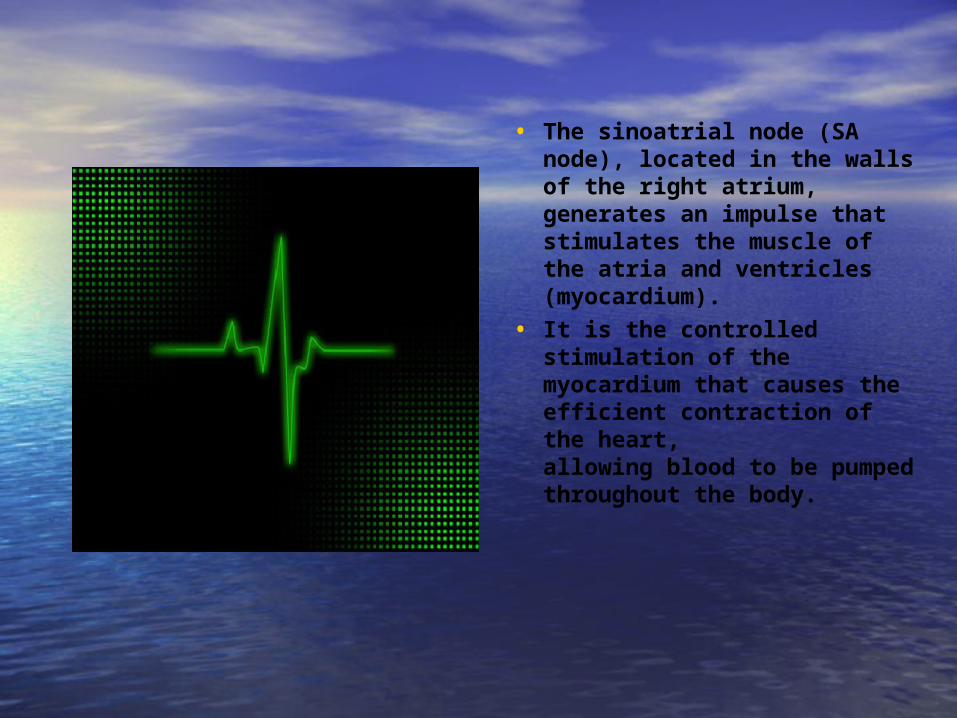
• The sinoatrial node (SA
node), located in the walls of the right atrium, generates an impulse that stimulates the muscle of the atria and ventricles (myocardium).
• It is the controlled stimulation of the myocardium that causes the efficient contraction of the heart, allowing blood to be pumped throughout the body.

The parts of the heart beat in an orderly sequence: Contraction of the atria (atrial
systole) is followed by contraction of ventricles (ventricular systole), and during diastole
(relaxation) all the four chambers are relaxed.

The electrocardiogram (ECG or EKG) is often used to examine the electrical conduction
system of the heart.
• The ECG picks up electrical impulses generated by the polarization and depolarization of cardiac tissue and translates them into a waveform.
• These waveforms are used to measure the rate and regularity of heartbeats, the size and position of the chambers, and the presence of any damage to the heart.

Fun Facts about the Human Heart• An adult heart beats
approximately 100,000 times a day, pumping about 2,000 gallons of blood.
• It has been estimated that the heart will beat about 3 billion times during a 70 year lifetime.

• A kitchen faucet would need to be turned on all the way for at least 45 years to equal the amount of blood pumped by the heart in an average lifetime.
• As a result of the heart having its own electrical impulse, it can continue to beat even when separated from the body, as long as it has an adequate supply of oxygen.
• The heart begins beating at four weeks after conception and does not stop until death.

Thank You.
I hope this presentation has proven helpful and
informational.