PREPARATION AND CHRACTERIZATION OF PLA AND …ethesis.nitrkl.ac.in/3170/1/ethesis.pdf · ·...
30
PREPARATION AND CHRACTERIZATION OF PLA AND PLGA SCAFFOLD AND FILM Thesis submitted to National Institute of Technology, Rourkela For the partial fulfilment of the Master degree in Life science SUPERVISED BY SUBMITTED BY DR.BISMITA NAYAK GOURI SHANKAR HARIPAL ASST.PROFESSOR ROLL NO:-410LS2052 NIT, ROURKELA NIT, ROURKELA DEPARTMENT OF LIFE SCIENCE NATIONAL INSTITUTE OF TECHNOLOGY, ROURKELA-769008 2012
Transcript of PREPARATION AND CHRACTERIZATION OF PLA AND …ethesis.nitrkl.ac.in/3170/1/ethesis.pdf · ·...

PREPARATION AND CHRACTERIZATION
OF
PLA AND PLGA SCAFFOLD AND FILM
Thesis submitted to
National Institute of Technology, Rourkela
For the partial fulfilment of the Master degree in
Life science
SUPERVISED BY SUBMITTED BY
DR.BISMITA NAYAK GOURI SHANKAR HARIPAL
ASST.PROFESSOR ROLL NO:-410LS2052
NIT, ROURKELA NIT, ROURKELA
DEPARTMENT OF LIFE SCIENCE
NATIONAL INSTITUTE OF TECHNOLOGY,
ROURKELA-769008
2012

DEPARTMENT OF LIFE SCIENCE
NATIONAL INSTITUTE OF TECHNOLOGY
ROURKELA-769008
…………………………………………………………………………………………………………………………………………………………….--------------------------------------------------------------------------------------------------------------------------------------
Dr. Bismita Nayak, M.Sc., Ph.D., Ref. No………
Assistant Professor.Date: ..............
Certificate
This is to certify that the thesis entitled “PREPARATION AND
CHARACTERIZATION OF PLA AND PLGA SCAFFOLD AND
FILM” submitted to National Institute of Technology; Rourkela
for the partial fulfilment of the Master degree in Life science is a
faithful record of bonafide and original research work carried out
by Gouri Shankar Haripal under my supervision and guidance.
Dr.Bismita Nayak
Supervisor
................................................................................................................. Phone no.: 0661-2462682 Email:[email protected]
DECLARATION
I hereby declare that the thesis entitled “Preparation and characterization of
PLA and PLGA scaffold and film”, submitted to the Department of Life
Science, National Institute of Technology, Rourkela for the partial fulfilment of
the Master Degree in Life Science is a faithful record of bona fide and original
research work carried out by me under the guidance and supervision of Dr.
Bismita Nayak, Assistant Professor, Department of Life Science, National
Institute of Technology, Rourkela. No part of this thesis has been submitted by
any other research persons or any students.
Date: GOURI SHANKAR HARIPAL
Place:
ACKNOWLEDGEMENT
I express my deep sense of gratitude and reverence to my major advisor, Dr. Bismita Nayak,
assistant professor, Department of Life Science, NIT-Rourkela, for her excellent guidance,
constant and untiring supervision, help and encouragement throughout the investigation and
preparation of this manuscript.
I am extremely grateful and indebted to Dr. S.K. Patra, HOD, Department of Life Science,
NIT-Rourkela, Dr. S.K. Bhutia, Dr. S. Das, Dr. R. Jayabalan, Dr. S. Jha and Dr. B. Mallick
for their inspiring suggestions and valuable advice not only for this investigation but also in
many other fronts without whom it would have been difficult to carry out this work.
I am so much thankful to Dr. S.K. Paria (Chemical Engg.) and faculty of other departments
for their constant help and support.
I am highly obliged to Pradipta Ranjan Rauta, Ph.D Scholar, Department of Life Science,
NIT-Rourkela, for his constant help and encouragement during the period of my project. I am
solely impressed by his great personality.
My heartfelt thanks to my friends N.M. Das, D. Nayak, S. Samal, G.K. Panigrahi, C.K. Das,
C.S. Bhol, D. Bhoi, R.R. Sahoo, S. Swain and all other classmates for their moral support,
help and encouragement throughout the course of this work. I take the pleasure to
acknowledge the constant help and support of my friends has always been cherished.
My sincere obligations are to Mr. B. Das and Murali Mausa, Staff, Department of Life
Science, NIT-Rourkela for their help during this period.
Lastly, I acknowledge with highest sense of regards to my parents, my brother, sister and
other members of my family for their supreme sacrifice, blessings, unwavering support, love
and affection without which the parent investigation would not have been successful in any
sphere of my life.
At the end, I bow down my head to the almighty whose omnipresence has always guided me
and made me energised to carry out such a project.
Date: Gouri Shankar Haripal
Place:

CONTENTS
SL .NO. CHAPTERS PAGE NO.
Abstract
1. Introduction 1-4
2. Review of literature 5-12
2.1 PLA 5-7
2.2 PLA Scaffold 7-9
2.3 PLGA 9-10
2.4 PLGA Scaffold 10-12
3. Objectives 13
4. Materials and Methods 14-17
4.1 Preparation of PLA and
PLGA microparticles
14-15
4.2 Preparation of PLA and
PLGA Scaffolds
15-16
4.3 Preparation of PLA and
PLGA Films
16-17
5. Results 18-20
6. Conclusion 21
7. References 22-23
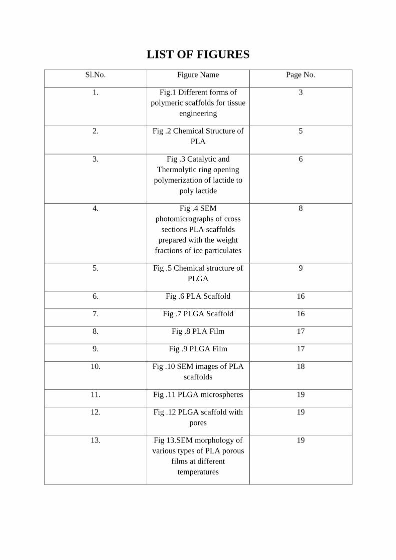
LIST OF FIGURES
Sl.No. Figure Name Page No.
1. Fig.1 Different forms of
polymeric scaffolds for tissue
engineering
3
2. Fig .2 Chemical Structure of
PLA
5
3. Fig .3 Catalytic and
Thermolytic ring opening
polymerization of lactide to
poly lactide
6
4. Fig .4 SEM
photomicrographs of cross
sections PLA scaffolds
prepared with the weight
fractions of ice particulates
8
5. Fig .5 Chemical structure of
PLGA
9
6. Fig .6 PLA Scaffold 16
7. Fig .7 PLGA Scaffold 16
8. Fig .8 PLA Film 17
9. Fig .9 PLGA Film 17
10. Fig .10 SEM images of PLA
scaffolds
18
11. Fig .11 PLGA microspheres 19
12. Fig .12 PLGA scaffold with
pores
19
13. Fig 13.SEM morphology of
various types of PLA porous
films at different
temperatures
19

ABSTRACT
In this research, the PLA/PLGA scaffolds and films were prepared successfully from
PLA/PLGA blank microparticles. The morphology of the PLA/PLGA scaffolds and films are
characterized by means of SEM. Biodegradable and biocompatible scaffolds having a highly
open porous structure and good mechanical strength are needed for cell proliferation,
migration, and differentiation, and guidance for cellular in-growth. Biodegradable porous
scaffolds can be surface engineered to provide an optical microenvironment for better cell
adhesion and tissue in-growth. According to scanning electron microscopy (SEM), a 3-
dimensional porous structure of films could be observed on the surface. As the morphology
of PLA/PLGA scaffolds and films could not be stabilized, we investigated the effect of
temperature and concentration of polymer on the properties of microporous scaffolds and
films. The degradation of polymeric porous films occurs through a homogeneous hydrolytic
chain cleavage mechanism and this process is altered by factors such as molecular weight and
molecular weight distribution. So, the biocompatible film plays an important role in
development of fully biodegradable, tissue compatible active wound dressing material.
Keywords: scaffold; microporous film; PLA; PLGA

1
1. INTRODUCTION
Scaffolds are key components in the tissue engineering paradigm, in which they can
function as templates to allow new tissue growth and provide temporary structural support.
The development of novel scaffolds is very challenging and critical to achieve the appropriate
function for tissue regeneration (Kretlow, 2007). Biodegradable and biocompatible
nanocomposite micro particles are developed as cell scaffolds.
Scaffolds play a critical role in tissue engineering. The regulation of the growth of
cells either cultivated within the porous structure of the scaffold or migrating from
surrounding tissue is one of the major functions of the scaffold. Generally mammalian cells
are anchorage-dependent. So, the scaffold gives a suitable substrate for cell attachment, cell
proliferation, differentiated function and cell migration. The physicochemical properties of
scaffolds are: to support and deliver cells; induce, differentiate, and channel tissue growth;
target cell-adhesion substrates; stimulate cellular response. These are biocompatible and
biodegradable; highly porous with a large surface/volume ratio; possess mechanical strength
and dimensional stability; and have sterilisability. Generally, three-dimensional porous
scaffolds can be fabricated from natural and synthetic polymers.
To achieve the goal of tissue reformation, scaffolds must face some specific
requirements. A high porosity and an appropriate pore size are necessary to facilitate cell
seeding and diffusion throughout the whole structure of both cells and nutrients. The degree
of biodegradability is an essential factor, since scaffolds should preferably be absorbed by the
surrounding tissues without the necessity of a surgical removal. The rate at which degradation
occurs has to coincide as much as possible with the rate of tissue formation: this means that
while cells are fabricating their own natural matrix structure around themselves, the scaffold
must provide the structural integrity within the body and eventually it will break down
leaving the new tissue, newly formed tissue which will take over the mechanical load.
Tissue engineering has emerged as a promising alternative approach to treat the loss
or malfunction of a tissue or organ without the limitations of current therapies (Langer et al.,
1993) Tissue engineering involves the expansion of cells, followed by the culturing of the
cells in temporary three-dimensional scaffolds to form the new organ or tissue.
Therefore, porous three-dimensional temporary scaffolds play an important role in
manipulating cell function and regulation of new organ formation (Peter et al., 1995). Isolated
2
and expanded cells adhere to the temporary scaffold in all three dimensions, generally
proliferate, and secrete their own extracellular matrices, replacing the biodegrading scaffold.
Significant challenges to this approach include the design and manufacture of the scaffolds.
Ideally, scaffolds for tissue engineering should meet several design criteria: (1) the surface
should permit cell adhesion, promote cell growth, and allow the retention of differentiated
cell functions; (2) the scaffolds should be biocompatible; (3) the scaffold should be
biodegradable and eventually eliminated; (4) the porosity should be high enough to provide
sufficient space for cell adhesion, extracellular matrix regeneration and the porous structure
of scaffold must allow even spatial cell distribution throughout the scaffold to facilitate
homogeneous tissue formation; (5) the material should be reproducibly able to process into
three-dimensional structure, and should be mechanically strong.
Many processing techniques have been developed to fabricate natural and synthetic
polymeric scaffolds. Although naturally derived materials are more versatile in providing
various biological functions, synthetic polymeric scaffolds are favoured because they can be
fabricated from a wide range of biodegradable polymers with easy process ability, controlled
degradation, and susceptibility to modification (Peter et al., 1998). While natural polymeric
scaffolds are generally fabricated by freeze drying/cross linking in aqueous solution,
synthetic polymeric scaffolds have been prepared by various methods including solvent
casting, particulate leaching, phase separation, (Mikos et al., 1993). The surfaces of a scaffold
promote cell adhesion by specific cell– matrix interactions (Hubbell et al., 1995).
Furthermore, growth factors can be encapsulated or imbedded within the porous matrices and
delivered in a sustained manner to enhance cell growth and morphogenesis.
Studies on scaffolds releasing DNA encoding the growth factor have also been
suggested as an alternative approach to bypass limitations of protein delivery (Park et al.,
2001). Recently, 3-D matrices based on different structural characteristics or minimally
invasive surgical methods have drawn attention for potential tissue engineering applications
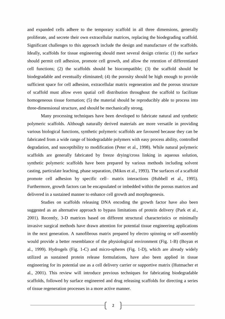
in the next generation. A nanofibrous matrix prepared by electro spinning or self-assembly
would provide a better resemblance of the physiological environment (Fig. 1-B) (Boyan et
al., 1999). Hydrogels (Fig. 1-C) and micro-spheres (Fig. 1-D), which are already widely
utilized as sustained protein release formulations, have also been applied in tissue
engineering for its potential use as a cell delivery carrier or supportive matrix (Hutmacher et
al., 2001). This review will introduce previous techniques for fabricating biodegradable
scaffolds, followed by surface engineered and drug releasing scaffolds for directing a series
of tissue regeneration processes in a more active manner.

3
Fig. 1 Different forms of polymeric scaffolds for tissue engineering: (A) a typical 3-D porous
matrix in the form of a solid foam, (B) a nanofibrousmatrix, (C) a thermo sensitive Sol–gel
transition hydro gel and (D) porous microsphere.
Many biodegradable types of polymers, such as PLA and PLGA, often used in
clinical applications for a very long time. There are several methods for the preparation of
porous, biodegradable film has been developed in the past few years. The method of solvent–
nonsolvent (SNS) phase separation is one of the most convenient route to prepare
multiporous films with micro pores on the surface, and these structures enhance the
hydrophilicity of the films.
Polymers, which are manufactured or processed to be suitable for use in or as a
medical device. Recently, a variety of polymers, biopolymers have been developed
specifically for medical applications.
Polymeric wound dressings that are capable of maintaining a controlled environment
at the wound site have been shown to promote healing (Winter, et al; 1962). Active wound
dressings have been developed to promote healing that enable the controlled, local delivery of
therapeutic agents (Higa et al., 1999).
4
Ideally, the active wound dressing would deliver a nearly instantaneous initial dosage of the
drug at the optimum therapeutic concentration, followed by a sustained constant delivery rate
of the drug that maintains the local concentration at the optimum dosage level for as many
days as necessary to achieve complete and effective wound healing.
In this study, to obtain a novel film structure for wound healing, we investigated the
factors that could affect the micro forming of PLA and PLGA film with micro porous
structure.
5
2. REVIEW OF LITERATURE
Polymer materials have received increasing attention and been widely used for tissue
engineering because of easy control over biodegradability and process ability. There are two
kinds of polymer materials: synthetic polymer, and naturally derived polymers (Thomson, et
al; 1995). The main biodegradable synthetic polymers include polyesters, such as poly
(glycolic acid) (PGA), poly (lactic acid) (PLA), and their copolymer of poly [lactic-co-
(glycolic acid)] (PLGA) are most commonly used for tissue engineering. PLA undergoes
hydrolytic scission to its monomeric form, lactic acid, which is eliminated from the body by
incorporation into TCA cycle. The major elimination path for lactic acid is respiration, and it
is primarily excreted by lungs as CO2. PGA can be broken down by hydrolysis, nonspecific
esterases and carboxypeptidases.
PLA (poly lactic acid) and PLGA (poly lactic acid/glycolic acid) are superior in
biocompatibility and biodegradability, and also act as base material for sustained-release
formulation.
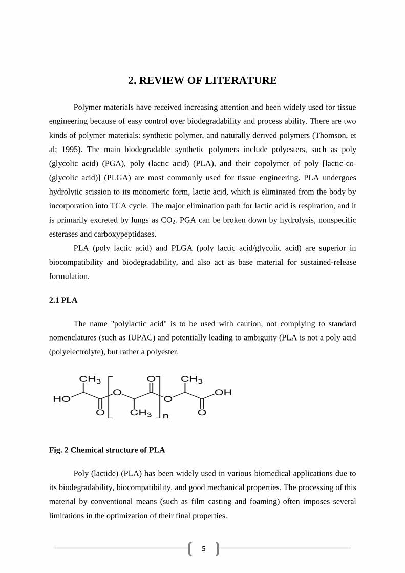
2.1 PLA
The name "polylactic acid" is to be used with caution, not complying to standard
nomenclatures (such as IUPAC) and potentially leading to ambiguity (PLA is not a poly acid
(polyelectrolyte), but rather a polyester.
Fig. 2 Chemical structure of PLA
Poly (lactide) (PLA) has been widely used in various biomedical applications due to
its biodegradability, biocompatibility, and good mechanical properties. The processing of this
material by conventional means (such as film casting and foaming) often imposes several
limitations in the optimization of their final properties.
6
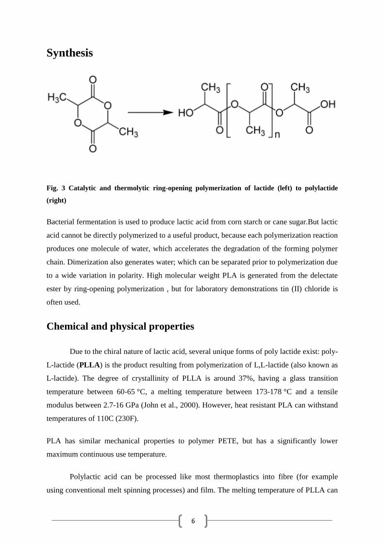
Synthesis
Fig. 3 Catalytic and thermolytic ring-opening polymerization of lactide (left) to polylactide
(right)
Bacterial fermentation is used to produce lactic acid from corn starch or cane sugar.But lactic
acid cannot be directly polymerized to a useful product, because each polymerization reaction
produces one molecule of water, which accelerates the degradation of the forming polymer
chain. Dimerization also generates water; which can be separated prior to polymerization due
to a wide variation in polarity. High molecular weight PLA is generated from the delectate
ester by ring-opening polymerization , but for laboratory demonstrations tin (II) chloride is
often used.
Chemical and physical properties
Due to the chiral nature of lactic acid, several unique forms of poly lactide exist: poly-
L-lactide (PLLA) is the product resulting from polymerization of L,L-lactide (also known as
L-lactide). The degree of crystallinity of PLLA is around 37%, having a glass transition
temperature between 60-65 °C, a melting temperature between 173-178 °C and a tensile
modulus between 2.7-16 GPa (John et al., 2000). However, heat resistant PLA can withstand
temperatures of 110C (230F).
PLA has similar mechanical properties to polymer PETE, but has a significantly lower
maximum continuous use temperature.
Polylactic acid can be processed like most thermoplastics into fibre (for example
using conventional melt spinning processes) and film. The melting temperature of PLLA can

7
be increased 40-50 °C and its heat deflection temperature can be increased from
approximately 60°C to up to 190 °C by physically blending the polymer with PDLA (poly-D-
lactide). A highly regular stereo complex with increased crystallinity is produced by PDLA
and PLLA. The temperature stability is maximised when a 50:50 blend is used, but even at
lower concentrations of PDLA, there is still a substantial improvement. In the latter case,
PDLA acts as a nucleating agent, as a result the crystallization rate is increasing.
Biodegradation of PDLA is slower than for PLA due to the higher crystallinity of PDLA.
PDLA has the useful property of being optically transparent. There is also poly (L-lactide-co-
D,L-lactide) (PLDLLA) – used as PLDLLA/TCP scaffolds for bone engineering.
2.2. PLA SCAFFOLD
Bioresorbable scaffolds of polylactic acid (PLA) offer several benefits. They require
only a single surgery and leave native tissue behind. They gradually transfer load to tissue
during the degradation period, useful for bone remodelling and reducing the risk of re-
fracture. They can be also used to drugs delivery, growth factors or other substances
conducive to the healing of bone locally to the implant site (Middleton, 2000).
The acidic products from the degradation of PLA act to catalyse further degradation
which can cause an accumulation of acidic products at the healing site and elicit an
inflammatory response. Hydroxyapatite (HA) is known to buffer the acidic degradation
products of polylactic acid. Addition of HA to scaffolds results in controlled rate of
degradation and reduced risk of inflammation. Exposure of osteogenic cells to bioactive
ceramics such as HA is known to increase osteoblast differentiation and growth (Jung et al.,
2005).
Biodegradable synthetic polymers including poly (lactic acid) (PLA) are suitable for
biocompatible scaffold constructs but are known to undergo in vitro degradation. This may
limit their potential for use in long-term cultures or loading regimes. This investigation
determines whether it is advantageous to culture cells on scaffolds prior to mechanical
compression

8
Figure.4 SEM photomicrographs of cross sections of PLA scaffolds prepared with the
weight fractions of ice particulates of 70% (a), 80% (b), and 90% (c).
PLA is biodegradable thermoplastic polyester that can be produced through ring-
opening polymerization of lactic acid. Since lactic acid is a chiral molecule, it exists in two
forms, D-PLA and LPLA. PLLA is the result of L-PLA polymerization. While PLLA is
semi-crystalline and it shows a high mechanical strength, poly (D,L-lactic acid) (PLA) is
essentially amorphous, or, at most with a low crystallinity.
9
The methylated version of PGA is PLA ,but is less hydrophilic and, therefore, it degrades
slowly.Poly (lactic acid) PLLA degrades to form lactic acid which is normally present in the
body. This acid then enters TCA cycle and is excreted as water and carbon dioxide.
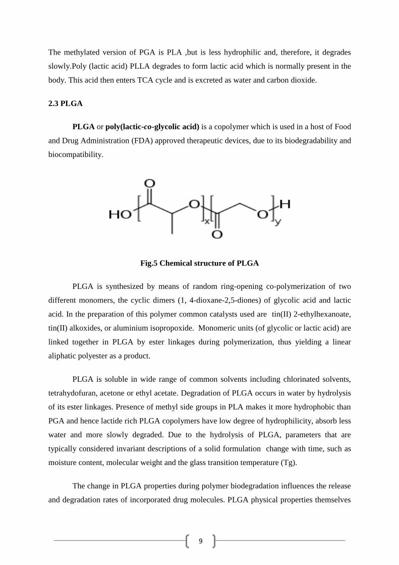
2.3 PLGA
PLGA or poly(lactic-co-glycolic acid) is a copolymer which is used in a host of Food
and Drug Administration (FDA) approved therapeutic devices, due to its biodegradability and
biocompatibility.
Fig.5 Chemical structure of PLGA
PLGA is synthesized by means of random ring-opening co-polymerization of two
different monomers, the cyclic dimers (1, 4-dioxane-2,5-diones) of glycolic acid and lactic
acid. In the preparation of this polymer common catalysts used are tin(II) 2-ethylhexanoate,
tin(II) alkoxides, or aluminium isopropoxide. Monomeric units (of glycolic or lactic acid) are
linked together in PLGA by ester linkages during polymerization, thus yielding a linear
aliphatic polyester as a product.
PLGA is soluble in wide range of common solvents including chlorinated solvents,
tetrahydofuran, acetone or ethyl acetate. Degradation of PLGA occurs in water by hydrolysis
of its ester linkages. Presence of methyl side groups in PLA makes it more hydrophobic than
PGA and hence lactide rich PLGA copolymers have low degree of hydrophilicity, absorb less
water and more slowly degraded. Due to the hydrolysis of PLGA, parameters that are
typically considered invariant descriptions of a solid formulation change with time, such as
moisture content, molecular weight and the glass transition temperature (Tg).
The change in PLGA properties during polymer biodegradation influences the release
and degradation rates of incorporated drug molecules. PLGA physical properties themselves
10
have been shown to depend upon multiple factors, including the initial molecular weight, the
size of the device, exposure to water and storage temperature.
Different forms of PLGA can be obtained depending on the ratio of lactide to
glycolide used for the polymerization. These are usually identified in regard to the monomers'
ratio used (e.g. PLGA 75:25 identifies a copolymer whose composition is 75% lactic acid and
25% glycolic acid). All PLGAs are amorphous rather than crystalline and show a glass
transition temperature in the range of 40-60 °C. PLGA can be dissolved by a wide range of
common solvents, including chlorinated solvents, tetrahydrofuran, acetone or ethyl acetate.
PLGA degrades by hydrolysis of its ester linkages in water. It has been observed that
the time required for degradation of PLGA is related to the monomers' ratio used in
production: the higher the content of glycolide units, less time required for degradation. An
exception to this rule is the copolymer with 50:50 monomers' ratio which exhibits the faster
degradation. The end-capped polymers with esters (as opposed to the free carboxylic acid)
demonstrate longer degradation half-lives.
PLGA has been successful as a biodegradable polymer because it undergoes
hydrolysis in the body as a result of which original monomers are produced, lactic acid and
glycolic acid. Under normal physiological conditions these two monomers are by-products
of various metabolic pathways in the body. As the body effectively deals with the two
monomers, there is minimal toxicity associated with using PLGA for drug delivery or
biomaterial applications. Also, the possibility to tailor the polymer degradation time by
altering the ratio of the monomers used during synthesis has made PLGA a common choice
in the production of a variety of biomedical devices like implants, prosthetic devices, micro
and nanoparticles, grafts and sutures.
2.4. PLGA SCAFFOLD
PLGA and its copolymers are the most extensively used for biodegradable and
biocompatible scaffolds. These polymers which are biocompatible, biodegradable are
approved by the Food and Drug Administration (FDA), and are easily processed into various
3-D matrices like structures.

11
Especially, biodegradable devices made of poly (lactide-co-glycolide) (PLGA)
copolymers are advantageous due to their controlled degradation behaviour and tunable
mechanical properties. The rate of polymer degradation should be carefully controlled to
synchronize with the rate of tissue formation and in-growth to achieve successful
regeneration or repair within a desired time frame.
A variety of techniques have been used for processing biodegradable polymers into
3-D porous scaffolds. The conventional methods include fibre felts, fibre bonding, melt
moulding, solvent casting/particulate leaching, phase separation method, and processing of
high pressure. A number of 3-D porous scaffolds fabricated from various kinds of
biodegradable materials have been developed and used for tissue engineering of liver (Kneser
et al.,1999), bladder (Oberpenning et al.,1999), nerve (Hadlock et al., 2000), skin (
Hansbroughbone et al., 1993), cartilage (Cao et al.,1997) and ligament( Freed et al., 1994)
etc.
The fabrication of three-dimensional (3D) scaffolds that mimic the cellular
microenvironment is of fundamental importance to the success of tissue engineered
constructs. Both scaffold chemistry and architecture can influence the fate and function of
engrafted cells (Bhatia et al., 1999).
Material microstructure, in contrast, is often controlled by process parameters such as
the choice of solvent in phase separation, gas foaming, woven fibres, and controlled ice
crystal formation and subsequent freeze-drying to create pores (Lo H et al., 1995) however,
these scaffolds lack a well-defined organization that is found in most tissues.
Modification of the bulk properties of the porous poly (lactide-co-glycolide) (PLGA)
scaffold was performed by irradiation with a high energy cyclotron proton ion beam. The
PLGA scaffolds were formulated in advance by the gas-foaming method by employing
ammonium bicarbonate particles as porogens.
Irradiation with ion beams was performed with 40 MeV for 3, 6 and 9 min on the
scaffolds at a distance of 30 cm from the beam exit to the scaffold surface. The bulky ion
beam-treated PLGA scaffold apparently demonstrated no color changes. The chemical
structures of the untreated samples seemed to be kept well when analyzed by both Fourier
transformed infrared but a subtle change was observed in its x-ray photoelectron
spectroscopy.
The results of in vitro tissue culture with smooth muscle cells for up to 4 weeks also
demonstrated no significant difference in terms of its handling stability during cell culture

12
and cellular behaviour between the untreated PLGA scaffolds and the ion beam-treated
PLGA scaffolds. However, significant changes were observed in its molecular weight as
measured by gel permeation chromatography, indicating a significant molecular weight
reduction. These results of in vitro tests and GPC measurements indicated that while bulk
modification of the scaffold was processed (Woo et al., 2009).
Scaffolding plays an important role in tissue engineering. In this work, a novel
processing technique has been developed to create three-dimensional biodegradable polymer
scaffolds with well-controlled interconnected pores which are spherical in nature. Paraffin
spheres were fabricated with a dispersion method, and were bonded together strongly through
a heat treatment to form a three-dimensional mold structure. Biodegradable polymers such as
PLLA and PLGA were dissolved in a solvent and cast onto the paraffin sphere assembly.
After dissolving the paraffin, a porous polymer scaffold was formed. The fabrication
parameters have importance in relation to the pore shape, interpore connectivity, pore wall
morphology, and mechanical properties of the polymer scaffolds. The modulus of the
scaffolds decreased with increasing porosity. More time for heat treatment of the paraffin
spheres resulted in larger openings between the pores of the scaffolds. Foams of smaller pore
size (100-200 micron) resulted in significantly lower compressive modulus than that of larger
pore sizes (250-350 or 420-500 micron). The PLLGA foams had a skeletal structure
consisting of small platelets, whereas homogeneous skeletal structures were found in PLGA
foams. The new processing technique can make the polymer scaffolds for a variety of
potential tissue engineering applications because of the well-controlled architecture, interpore
connectivity, and mechanical properties (Ma PX et al., 2001)
Solvent-evaporation method was modified using sucrose as an additive to form large
porous microparticle of poly (d,l-lactic-co-glycolic) (PLGA) polymer. Micro particles
containing hydrophilic polymers (poly vinyl alcohol) incorporated in their internal matrix
structure were also formulated. Different formulations of micro particles were evaluated for
physical properties, cell adhesion, and cell growth in culture. PLGA micro particles
containing poly(vinyl alcohol) (PVA) in the matrix structure (PLGA-PVA) and treated with
serum prior to cell seeding demonstrated better cell adhesion and cell growth than other
formulations of micro particles. (Labhasetwar et al., 2005).

13
3. OBJECTIVES
1. Preparation of blank PLA and PLGA micro particles
2. Preparation of PLA and PLGA Scaffolds
3. Preparation of PLA and PLGA Films
4. Characterization of PLA and PLGA Scaffolds and Films by SEM

14
4. MATERIALS AND METHODS
4.1. Preparation of PLA and PLGA micro particles
4.1.1.: Equipments
Stratos low-temperature high-speed centrifuge(Thermo, Germany)
Cooling centrifuge (REMI)
Magnetic stirrer
Sonicator
Scanning electron microscope (Jeol Jsm-6480 LV)
4.1.2. Materials required:
Sucrose
PLA(Sigma- Aldrich)
DCM
PVA(Sigma- Aldrich)
4.1.3. Procedure:
PLA and PLGA dummy particles were prepared using double solvent evaporation method.
Method is as follows:
1) Internal aqueous phase (200 micro litres):10% sucrose (w/v)
2) Organic phase (4 ml) : PLA 200mg/4 ml DCM
: PLGA 200mg/4 ml DCM
3) External aqueous phase (16 ml) :1% PVA (w/v)
: 10% Sucrose (w/v)
15
4.1.4. Preparation:
Polymer dissolved in organic phase (4 ml) was associated with 200 micro litre
of internal aq.phase was added to make primary emulsion.
For the formation of the secondary emulsion 16ml of external aq.phase was
taken in a beaker and the primary emulsion was added drop wise while
sonicating the primary emulsion. This led to the formation of the secondary
emulsion.
This was kept on a magnetic stirrer for overnight for excess DCM to evaporate.
Particles formed were separated by centrifugation at 15000 RPM which was
performed for a period of 20 minutes.
Separated particles were washed twice with ice cold MQ water. After washing
the particles were lyophilized for 24 hrs to obtain dry particles.
4.2 Preparation of PLA /PLGA Scaffold
For PLA and PLGA
Chemicals required:
Chloroform
Ethanol
Ammonium bicarbonate
Aqueous citric acid
Procedure:
For both PLA and PLGA micro particle
• 100 mg of PLA /PLGA micro particle dissolved in chloroform was precipitated in
ethanol.
• A gel slurry was obtained, 50 mg of ammonium bicarbonate salt particles mixed with
this gel paste were cast in a mold, semi solidified at room temperature and immersed
into 5 ml of aqueous citric acid solution.
• Finally macro porous PLA/PLGA scaffolds with a porosity of over 90% were
obtained.

16
Fig.6: PLA scaffold Fig7: PLGA scaffold
4.3. Preparation of PLA and PLGA films
For PLA and PLGA
Chemicals required:
Dichloro methane(DCM)
Tetrahydrofuran (THF)
Ethylene glycol(EG)
Glycerol
Procedure:
For both PLA and PLGA micro particle
• 100 mg of PLA/PLGA microparticle was first dissolved in 5 ml of DCM at room
temperature.
• After PLA/PLGA microparticle was completely dissolved in 10 ml of THF was then
added into the mixed solution at 550C to ablate DCM by volatilization.
• Since PLA/PLGA was uniformly dissolved in THF the solution was added dropwise
onto the surface of EG/glycerol mixed solution.
• Finally the system was left for volatilization overnight and was microporous
PLA/PLGA film was obtained.

17
Fig 8: PLA Film
Fig 9: PLGA Film

18
5. RESULTS
Characterization of PLA and PLGA scaffolds and films by SEM
The PLA scaffold prepared by this method was highly porous with evenly distributed and
interconnected pore structures. The pore shapes were almost the same as those of the ice
particulates. The degree of interconnection increased as the weight fraction of the ice
particulates increased.
Fig.10 SEM images of PLA scaffolds
The polymer concentration also had some effect on the pore wall structure, i. e., lower
polymer concentration resulted in more porous pore wall structures. The porosity and surface
area/weight ratio increased with the increase of the weight fraction of the ice particulates.
Therefore, the pore structure of the porous temporary scaffolds could be manipulated by
varying the shape, weight fraction, size of the ice particulates, and the polymer concentration.

19
Fig. 11 PLGA microspheres Fig .12 PLGA scaffold with pores
The wettability of a polymer scaffold is considered very important for homogeneous
structure. Under the fabrication conditions used here, the PLGA microspheres have a
spherical morphology with a smooth surface.
Fig .13 SEM morphology of various types of PLA porous films at different temperatures
The morphology observation shown in above indicates that the pore appearances in
different films are conspicuously different and that the films had a smoother surface .The
pores are evenly distributed throughout the film.

20
The volatility of THF at different temperatures was not concordant, and for this
reason, we obtained various PLA films with disparate morphology. We observed the scaffold
prepared at 4°C, 25°C and 50°C, the film had unique micro pores on its surface. This
experiment showed an ambient temperature is beneficial for shaping the surface of the
scaffold with multipores and the obtained films were smoother at a low speed of phase
separation. That is to say, a moderate rate of evaporation is beneficial for formation of
droplets of the solvent, and after these droplets volatilized, spherical holes appeared and
occupied the interface.
21
6. CONCLUSION
In this study, the morphology of PLA/PLGA scaffolds and films prepared from
PLA/PLGA blank microparticle are observed. The results showed that PLA /PLGA scaffolds
possessed higher degree of porosity with evenly distributed and interconnected pore
structures. The pore shapes were almost the same as those of the ice particulates. The
polymer concentration also had some effect on the pore wall structure, i.e., lower polymer
concentration resulted in more porous pore wall structures. These porous scaffolds prepared
by solvent casting/particulate leaching have been intensively studied to provide implantable
devices for tissue regeneration. These polymeric devices are usually surface modified by
immobilizing cell adhesive or growth factor binding moieties to improve its cell adhesive
characteristics or actively induce cell migration, proliferation and differentiation. According
to SEM films had a smoother surface and the pores are evenly distributed throughout the
film. The degradation of PLA and PLGA microfilms occurs through a homogeneous
hydrolytic chain cleavage mechanism where the rates of polymer degradation are similar for
both the surface and the bulk of the microfilms. Factors such as molecular weight and
molecular weight distribution as well as sterilization may also alter the degradation rate of the
biodegradable polyesters in microfilms. In these way biocompatible films represents an
important advance in the development of fully biodegradable, tissue compatible active wound
dressing material capable of delivering a broad range of therapeutic agents.

22
7. REFERENCES
B. D. Boyan, C. H. Lohmann, J. Romero, Z. Schwartz, Clin. Plast. Surg. 1999, 26,629.
Bhatia SN, Chen CS. Tissue engineering at the micro-scale. Biomed Microdevices
1999;2(2):131–44.
D. W. Hutmacher, J. Biomater. Sci., Polym. Ed. 2001, 12,107.
F. Oberpenning, J. Meng, J. J. Yoo, A. Atala, Nat. Biotechnol.1999, 17, 149.
Hadlock T, Sundback C, Hunter D, Cheney M, Vacanti JP. A polymer foam conduit seeded
with Schwann cells promotes guided peripheral nerve regeneration. Tissue Eng
2000;6(2):119–27.
Higa O, Rogero S, Machado L, Mathor M, Lugao A. Biocompatibility study for PVP J. A.
Hubbell, Biotechnology (N. Y.) 1995, 13, 565.
J. F. Hansbrough, J. L. Morgan, G. E. Greenleaf, R. Bartel, J. Burn Care Rehabil. 1993, 14,
485.
Jung Y., Kim S.-S., Young H.K., Kim S.-H., Kim B.-S., Kim S., Cha Y.C., Soo H.K. A
poly(lactic acid)/calcium metaphosphate composite for bone tissue engineering.
Biomaterials. 2005;26(32):6314 .Kretlow, J.D., Advanced Drug Delivery Reviews,
2007. 59(4-5):263-273.
Kretlow, J.D., Advanced Drug Delivery Reviews, 2007. 59(4-5):263-273.
L.E. Freed, G. Vunjak-Novakovic, R.J. Biron, D.B. Eagles, D.C. Lesnoy, S.K. Barlow, R.
Langer, Biodegradable polymer scaffolds for tissue engineering, Nat. Biotechnol. 12
(1994) 689–693.
Lo H, Ponticiello MS, Leong KW. Fabrication of controlled release biodegradable foams by
phase separation. Tissue Eng1995;1:15–28.
Ma PX,Choi JW,2001, Department of Biologic and Material Sciences, Macromolecular
Science and Engineering Center, University of Michigan, Ann Arbor, Michigan
48109-1078.
Middleton J.C., Tipton A.J. Synthetic biodegradable polymers as orthopedic devices.
Biomaterials. 2000;21(23):2335.
Mikos AG, Lyman MD, Freed LE, Langer R. Wetting of poly(l-lactic acid) and poly(dl-
lactic-co-glycolic acid) foams for tissue culture. Biomaterials 1994;15:55–8.
R. C. Thomson, M. J. Yaszemski, J. M. Powers, A. G. Mikos, J. Biomater. Sci., Polymer.
Edn. 1995, 7, 23.
23
S. J. Peter, M. J. Miller, A. W. Yasko, M. J. Yaszemski, A.G. Mikos, J. Biomed. Mater. Res.
1998, 43,422.
S. L. Ishaug, G. M. Crane, M. J. Miller, A. W. Yasko, M. J.Yaszemski, A. G. Mikos, J.
Biomed. Mater. Res. 1997, 36,17.
T. Hadlock, C. Sundback, D. Hunter, M. Cheney, J. P.Vacanti, Tissue Eng. 2000, 6, 119.
U. Kneser, P. M. Kaufmann, H. C. Fiegel, J. M. Pollok, D.Kluth, H. Herbst, X. Rogiers, J.
Biomed. Mater. Res. 1999,47, 494. wound dressing obtained in different conditions.
Radiat Phys Chem 1999; 55:705-707.
Y. Cao, J. P. Vacanti, K. T. Paige, J. Upton, C. A. Vacanti,Plast. Reconstr. Surg. 1997, 100,
297.