Preliminary Evaluation RecombinantOnchocerca volvulus ...jcm.asm.org/content/31/7/1741.full.pdf ·...
5
JOURNAL OF CLINICAL MICROBIOLOGY, JUlY 1993, p. 1741-1745 0095-1137/93/071741-05$02.00/0 Copyright C 1993, American Society for Microbiology Preliminary Evaluation of Recombinant Onchocerca volvulus Antigens for Serodiagnosis of Onchocerciasis A. F. OGUNRINADE,l R. CHANDRASHEKAR,2* M. L. EBERHARD,3 AND G. J. WEIL2 Department of Veterinary Microbiology and Parasitology, University of Ibadan, Ibadan, Nigeria' Departments of Medicine and Molecular Microbiology, Washington University School of Medicine and Jewish Hospital of St. Louis, St. Louis, Missouri 631102; and Division of Parasitic Diseases, Centers for Disease Control and Prevention, Atlanta, Georgia 303333 Received 8 January 1993/Accepted 5 April 1993 Serodiagnostic assays for onchocerciasis based on native antigens are hampered by the scarcity of antigen, and they suffer from poor specificity. The present study was designed to evaluate the diagnostic utility of recently described recombinant Onchocerca volvulus antigens OC 3.6 and OC 9.3 in enzyme immunoassays. The recombinant proteins were expressed as glutathione S-transferase fusions and were tested in several enzyme immunoassay formats to measure immunoglobulin G (IgG) and IgG4 antibodies with sera from patients with onchocerciasis in Nigeria and with various types of control sera. The best results were obtained by measuring IgG4 antibodies to the fusion proteins. Forty of 42 (95%) serum specimens from patients with onchocerciasis were reactive with OC 3.6; the reactivity with OC 9.3 was 81%. Results obtained with sera from experimentally infected chimpanzees suggest that OC 3.6 might be especially useful for detecting prepatent infections in humans, while OC 9.3 mainly detects mature, patent infections. Sera from individuals in Nigeria and the United States residing in areas nonendemic for onchocerciasis were uniformly nonreactive with these antigens in IgG and IgG4 assays, as were sera from patients with bancroftian filariasis, brugian filariasis, loiasis, ascariasis, schistosomiasis, and dracunculiasis. These results suggest that enzyme immunoassays based on the recombinant antigens OC 3.6 and OC 9.3 are useful for the diagnosis of onchocerciasis. Onchocerciasis is an important cause of blindness and severe dermatitis in sub-Saharan Africa and Latin America (32). At present, diagnosis of onchocerciasis in humans depends primarily on the demonstration of microfilariae in skin snips (32). However, skin-snip examination is not sensitive for the detection of early infections or for the diagnosis of onchocerciasis in individuals with low microfi- larial densities in the skin (19, 27). False-negative skin-snip examinations are often seen in children, recently infected adults (especially expatriates), and individuals who develop allergic hypersensitivity to microfilariae ("sowda"). The skin-snip procedure is inconvenient, and special care is needed to ensure that blood-borne viruses are not spread in field studies. A sensitive and specific test for antibodies to the parasite would provide an excellent tool for identifying early infections and also for use in epidemiological surveys (7, 24, 26). Serodiagnostic assays for onchocerciasis based on native antigens are hampered by the scarcity of antigen, and they suffer from poor specificity (1, 30). We have recently iden- tified and characterized two recombinant Onchocerca volvu- lus antigens, OC 3.6 and OC 9.3, which appeared to be promising reagents for use in the diagnosis of onchocerciasis on the basis of preliminary results obtained by Western blotting (immunoblotting) (4). The purpose of the study described here was to systematically evaluate the use of these recombinant antigens in enzyme immunoassays for onchocerciasis. MATERIALS AND METHODS Human sera. Sera were collected from people over 10 years of age who reside in villages in a savannah-type area * Corresponding author. near New Bussa in northern Nigeria with an endemic focus of onchocerciasis. The diagnosis of onchocerciasis required either a positive skin-snip examination for microfilariae or the presence of characteristic subcutaneous nodules or on- chocercal dermatitis on clinical examination, as described previously (29). Subjects were questioned regarding visual symptoms, but formal opthalmological examinations were not performed. Control sera were obtained from residents of Ibadan, Nigeria, which is outside of the onchocerciasis transmission zone (nonendemic area) and from residents of St. Louis, Mo., who have never been exposed to the parasite. Other control sera tested were from patients infected with Wuchereria bancrofti, Brugia malayi, Loa loa, Ascaris lumbricoides, Dracunculus medinensis, and Schistosoma mansoni. These sera were collected in areas that are nonen- demic for onchocerciasis in India, Egypt, and Nigeria. Chimpanzee sera. Five chimpanzees (Pan troglodytes) were used in the present study, and all came from Yerkes Primate Center Colony. All five animals were males, and they ranged in age from 6 to 22 years. They were colony- born chimpanzees, and all were free of preexisting filarial infections, as monitored by two preinoculation blood and skin-snip examinations. Third-stage larvae (L3) were har- vested from blackflies (Simulium yahense) previously in- fected in Liberia and shipped via air to the Centers for Disease Control and Prevention laboratories in Atlanta. Two animals were inoculated with 200 L3 each, two were inocu- lated with 300 L3 each, and one animal received 400 L3. Serum samples were collected at preinoculation and monthly after infection. Skin snips were examined monthly beginning 8 months after inoculation, as described previously (5). Expression of OC 3.6 and OC 9.3 in pGEX-2A. OC 3.6 and OC 9.3 are recombinant Agtll clones with 0. volvulus cDNA inserts which have been described in detail elsewhere (4). 1741 Vol. 31, No. 7 on February 2, 2019 by guest http://jcm.asm.org/ Downloaded from
Transcript of Preliminary Evaluation RecombinantOnchocerca volvulus ...jcm.asm.org/content/31/7/1741.full.pdf ·...

JOURNAL OF CLINICAL MICROBIOLOGY, JUlY 1993, p. 1741-17450095-1137/93/071741-05$02.00/0Copyright C 1993, American Society for Microbiology
Preliminary Evaluation of Recombinant Onchocerca volvulusAntigens for Serodiagnosis of Onchocerciasis
A. F. OGUNRINADE,l R. CHANDRASHEKAR,2* M. L. EBERHARD,3 AND G. J. WEIL2Department of Veterinary Microbiology and Parasitology, University ofIbadan, Ibadan, Nigeria'
Departments ofMedicine and Molecular Microbiology, Washington University School ofMedicine and Jewish Hospital of St. Louis, St. Louis, Missouri 631102; and Division ofParasitic Diseases, Centers for Disease Control and Prevention, Atlanta, Georgia 303333
Received 8 January 1993/Accepted 5 April 1993
Serodiagnostic assays for onchocerciasis based on native antigens are hampered by the scarcity of antigen,and they suffer from poor specificity. The present study was designed to evaluate the diagnostic utility ofrecently described recombinant Onchocerca volvulus antigens OC 3.6 and OC 9.3 in enzyme immunoassays.The recombinant proteins were expressed as glutathione S-transferase fusions and were tested in severalenzyme immunoassay formats to measure immunoglobulin G (IgG) and IgG4 antibodies with sera frompatients with onchocerciasis in Nigeria and with various types of control sera. The best results were obtainedby measuring IgG4 antibodies to the fusion proteins. Forty of 42 (95%) serum specimens from patients withonchocerciasis were reactive with OC 3.6; the reactivity with OC 9.3 was 81%. Results obtained with sera fromexperimentally infected chimpanzees suggest that OC 3.6 might be especially useful for detecting prepatentinfections in humans, while OC 9.3 mainly detects mature, patent infections. Sera from individuals in Nigeriaand the United States residing in areas nonendemic for onchocerciasis were uniformly nonreactive with theseantigens in IgG and IgG4 assays, as were sera from patients with bancroftian filariasis, brugian filariasis,loiasis, ascariasis, schistosomiasis, and dracunculiasis. These results suggest that enzyme immunoassays basedon the recombinant antigens OC 3.6 and OC 9.3 are useful for the diagnosis of onchocerciasis.
Onchocerciasis is an important cause of blindness andsevere dermatitis in sub-Saharan Africa and Latin America(32). At present, diagnosis of onchocerciasis in humansdepends primarily on the demonstration of microfilariae inskin snips (32). However, skin-snip examination is notsensitive for the detection of early infections or for thediagnosis of onchocerciasis in individuals with low microfi-larial densities in the skin (19, 27). False-negative skin-snipexaminations are often seen in children, recently infectedadults (especially expatriates), and individuals who developallergic hypersensitivity to microfilariae ("sowda"). Theskin-snip procedure is inconvenient, and special care isneeded to ensure that blood-borne viruses are not spread infield studies. A sensitive and specific test for antibodies tothe parasite would provide an excellent tool for identifyingearly infections and also for use in epidemiological surveys(7, 24, 26).
Serodiagnostic assays for onchocerciasis based on nativeantigens are hampered by the scarcity of antigen, and theysuffer from poor specificity (1, 30). We have recently iden-tified and characterized two recombinant Onchocerca volvu-lus antigens, OC 3.6 and OC 9.3, which appeared to bepromising reagents for use in the diagnosis of onchocerciasison the basis of preliminary results obtained by Westernblotting (immunoblotting) (4). The purpose of the studydescribed here was to systematically evaluate the use ofthese recombinant antigens in enzyme immunoassays foronchocerciasis.
MATERIALS AND METHODSHuman sera. Sera were collected from people over 10
years of age who reside in villages in a savannah-type area
* Corresponding author.
near New Bussa in northern Nigeria with an endemic focusof onchocerciasis. The diagnosis of onchocerciasis requiredeither a positive skin-snip examination for microfilariae orthe presence of characteristic subcutaneous nodules or on-
chocercal dermatitis on clinical examination, as describedpreviously (29). Subjects were questioned regarding visualsymptoms, but formal opthalmological examinations were
not performed.Control sera were obtained from residents of Ibadan,
Nigeria, which is outside of the onchocerciasis transmissionzone (nonendemic area) and from residents of St. Louis,Mo., who have never been exposed to the parasite. Othercontrol sera tested were from patients infected withWuchereria bancrofti, Brugia malayi, Loa loa, Ascarislumbricoides, Dracunculus medinensis, and Schistosomamansoni. These sera were collected in areas that are nonen-demic for onchocerciasis in India, Egypt, and Nigeria.Chimpanzee sera. Five chimpanzees (Pan troglodytes)
were used in the present study, and all came from YerkesPrimate Center Colony. All five animals were males, andthey ranged in age from 6 to 22 years. They were colony-born chimpanzees, and all were free of preexisting filarialinfections, as monitored by two preinoculation blood andskin-snip examinations. Third-stage larvae (L3) were har-vested from blackflies (Simulium yahense) previously in-fected in Liberia and shipped via air to the Centers forDisease Control and Prevention laboratories in Atlanta. Twoanimals were inoculated with 200 L3 each, two were inocu-lated with 300 L3 each, and one animal received 400 L3.Serum samples were collected at preinoculation and monthlyafter infection. Skin snips were examined monthly beginning8 months after inoculation, as described previously (5).
Expression of OC 3.6 and OC 9.3 in pGEX-2A. OC 3.6 andOC 9.3 are recombinant Agtll clones with 0. volvulus cDNAinserts which have been described in detail elsewhere (4).
1741
Vol. 31, No. 7
on February 2, 2019 by guest
http://jcm.asm
.org/D
ownloaded from
1742 OGUNRINADE ET AL.
These cDNA inserts were subcloned into the plasmid ex-pression vector pGEX-2A, which was kindly supplied byRichard Lucius. This plasmid has been constructed to pro-duce fusion proteins with glutathione S-transferase (GST) ofSchistosoma japonicum at the carboxy terminus to facilitatepurification (23). Competent Eschenichia coli JM101 cellswere transformed with the ligation mixtures. Primaryscreening of transformants for expression of the GST-fusionproteins was performed with 2-ml cultures induced withisopropyl-,-D-thiogalactopyranoside (IPTG; BoehringerMannheim, Indianapolis, Ind.). Bacteria were concentratedby centrifugation and were resuspended in sample buffer forseparation by sodium dodecyl sulfate (SDS)-polyacrylamidegel electrophoresis (PAGE) (10); this was followed by stain-ing with 0.05% Coomassie blue. Transformants expressingthe OC 3.6 and OC 9.3 fusion proteins were confirmed byimmunoblotting with a pool of serum from patients withonchocerciasis (28).Recombinant antigens were purified by affinity chromatog-
raphy with glutathione-agarose (Sigma Chemical Co., St.Louis, Mo.), as described by Smith and Johnson (22).Briefly, a 10-ml overnight culture was inoculated into 1 literof NZCYM medium (GIBCO Bethesda Research Laborato-ries, Gaithersburg, Md.) containing 50 ,ug of ampicillin perml (Sigma). Cultures were grown at 37°C with shaking to anoptical density at 600 nm (OD600) of 1.0. IPTG (final concen-tration, 0.3 mM) was then added, and the culture was grownfor an additional 5 h, after which the cells were pelleted andresuspended in 0.01 M phosphate-buffered saline (PBS; pH7.4) with 1% Triton-X 100 (TX100; Sigma) (PBS-TX100).The cells were frozen at -20°C overnight. Cells were thawedin cold water and lysed by mild sonication. Cellular debriswas removed by centrifugation at 10,000 x g for 15 min.
Glutathione-agarose beads were swollen in PBS andpacked into a small column. The column was washed with 5bed volumes of PBS to remove the preservative. The gel wasthen equilibrated with 5 bed volumes of PBS-TX100 buffer.The lysate was passed through a 0.45-p,m-pore-size filterbefore it was applied to the column. The sample was appliedto the column, and the eluent was discarded. The boundfusion protein was eluted with 5 bed volumes of elutionbuffer (5 mM glutathione [Sigma] in 50 mM Tris-HCl [pH8.0]); this was followed by dialysis against PBS. The dia-lyzed protein was concentrated by using a membrane con-centrator (Centricon-10; Amicon, Beverly, Mass.), and theprotein concentration was measured with a commercial kit(BCA; Pierce Chemical Co., Rockford, Ill.).Enzyme immunoassay for detection of total IgG and IgG4
antibodies to recombinant antigens. Preliminary studies werecarried out to determine the optimal concentration of recom-binant antigens to use for the immunoglobulin G (IgG) andIgG4 antibody assays. Briefly, the recombinant antigens OC3.6 and OC 9.3 and the control antigen GST (100 ,ul per well;0.5, 1.0, or 2.0 ,ug/ml in 0.06 M carbonate buffer [pH 9.6])were incubated in polyvinyl microtiter plates (DynatechLaboratories, Alexandria, Va.) overnight at 37°C. Plateswere washed three times with PBS containing 0.05% Tween20 (Sigma) (PBS-T) and were blocked with PBS-T containing5% fetal calf serum (FCS) (PBS-T-FCS) for 1 h at 37°C. Sera(positive and negative pools) diluted 1:25 in PBS-T-FCSwere added to the wells, twofold dilutions were made inPBS-T-FCS, and the dilutions were incubated for 2 h at37°C. Plates were washed three times with PBS-T. IgGantibody binding was detected with horseradish peroxidase-conjugated goat anti-human IgG antibody (OrganonTeknika-Cappel, Malvern, Pa.) in PBS-T-FCS. After 1 h of
A B1 2 1 2
kDo
116 -93-66 -
45 -
*. ::SWt* -s:s e
'sA:S:_*S_v#
Ss* 3,!.i.t.:j .. ^
>.9r..! }+ ss
:..___
,: C ..
_ >: .'...... j,_ ..31 -
21 -
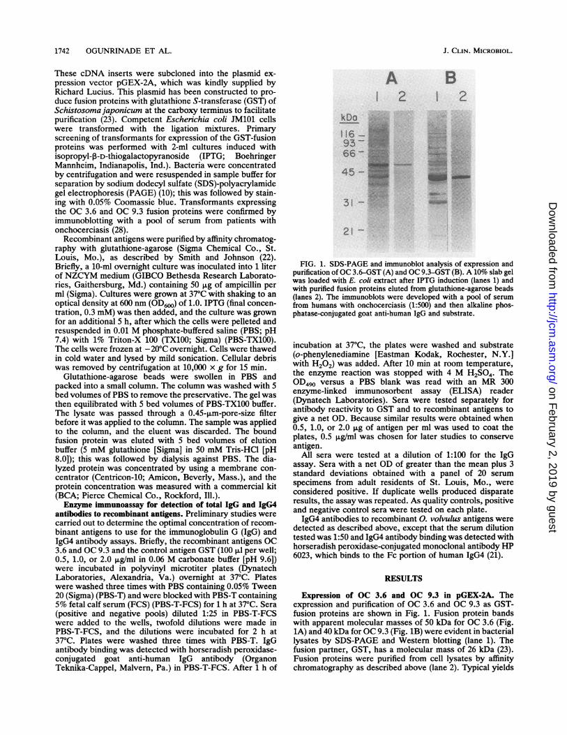
FIG. 1. SDS-PAGE and immunoblot analysis of expression andpurification of OC 3.6-GST (A) and OC 9.3-GST (B). A 10% slab gelwas loaded with E. coli extract after IPTG induction (lanes 1) andwith purified fusion proteins eluted from glutathione-agarose beads(lanes 2). The immunoblots were developed with a pool of serumfrom humans with onchocerciasis (1:500) and then alkaline phos-phatase-conjugated goat anti-human IgG and substrate.
incubation at 37°C, the plates were washed and substrate(o-phenylenediamine [Eastman Kodak, Rochester, N.Y.]with H202) was added. After 10 min at room temperature,the enzyme reaction was stopped with 4 M H2SO4. TheOD490 versus a PBS blank was read with an MR 300enzyme-linked immunosorbent assay (ELISA) reader(Dynatech Laboratories). Sera were tested separately forantibody reactivity to GST and to recombinant antigens togive a net OD. Because similar results were obtained when0.5, 1.0, or 2.0 ,ug of antigen per ml was used to coat theplates, 0.5 ,ug/ml was chosen for later studies to conserveantigen.
All sera were tested at a dilution of 1:100 for the IgGassay. Sera with a net OD of greater than the mean plus 3standard deviations obtained with a panel of 20 serumspecimens from adult residents of St. Louis, Mo., wereconsidered positive. If duplicate wells produced disparateresults, the assay was repeated. As quality controls, positiveand negative control sera were tested on each plate.IgG4 antibodies to recombinant 0. volvulus antigens were
detected as described above, except that the serum dilutiontested was 1:50 and IgG4 antibody binding was detected withhorseradish peroxidase-conjugated monoclonal antibody HP6023, which binds to the Fc portion of human IgG4 (21).
RESULTS
Expression of OC 3.6 and OC 9.3 in pGEX-2A. Theexpression and purification of OC 3.6 and OC 9.3 as GST-fusion proteins are shown in Fig. 1. Fusion protein bandswith apparent molecular masses of 50 kDa for OC 3.6 (Fig.1A) and 40 kDa for OC 9.3 (Fig. 1B) were evident in bacteriallysates by SDS-PAGE and Western blotting (lane 1). Thefusion partner, GST, has a molecular mass of 26 kDa (23).Fusion proteins were purified from cell lysates by affinitychromatography as described above (lane 2). Typical yields
J. CLIN. MICROBIOL.
on February 2, 2019 by guest
http://jcm.asm
.org/D
ownloaded from

SERODIAGNOSIS OF ONCHOCERCIASIS 1743
'3.0
2.01
1.0
a
C]0
I.
0D
-I0 1
.3.0
1.0 2.0 a 3.0
*OX**
2.0p*.
0
I.o
0
0
0 BI I
0 I.0 2.0 > 3.0
IgG (OD490)
FIG. 2. Comparison of IgG and IgG4 enzyme immunoassays forantibodies to recombinant antigens OC 3.6 (A) and OC 9.3 (B). ODcutoff values (mean plus three standard deviations) obtained with a
panel of sera from individuals in areas nonendemic for onchocercia-sis are indicated by the broken lines.
from 1-liter cultures were 0.4 to 0.6 mg for OC 3.6-GST and2 to 3 mg for OC 9.3-GST.
Relative sensitivities ofIgG and IgG4 ELISAs for antibodiesto recombinant 0. volvuls antigens. In preliminary studies,23 serum specimens from patients with onchocerciasis weretested for their IgG and IgG4 reactivities to OC 3.6 and OC9.3 by enzyme immunoassay. Although 21 of 23 serumspecimens contained antibodies to OC 3.6, the ODs in theIgG4 assay were much higher (Fig. 2A). The differencebetween the IgG and IgG4 assays was even more dramaticfor OC 9.3 (Fig. 2B). In general, human sera had little or nobackground reactivity with GST in the IgG4 assay, but IgGreactivity with GST was significant and highly variable.Thus, the best signal-to-noise ratios were obtained by mea-
suring the levels of IgG4 antibodies to the fusion proteins.Forty of 42 (95%) serum specimens from patients withonchocerciasis tested in the present study were reactive withOC 3.6; reactivity with OC 9.3 was 81% (Table 1). Assays fortotal IgG antibodies to these antigens were less sensitivethan the assays for IgG4 antibodies to the antigens (Table 1).
Reactivities of control sera with recombinant antigens. Thereactivities of various types of control sera with both recom-
binant antigens are shown in Table 1. Sera from individualsin areas of Nigeria (n = 24) and the United States (n = 20)nonendemic for onchocerciasis were uniformly nonreactivewith both antigens. Sera from patients with bancroftianfilariasis, brugian filariasis, loiasis, ascariasis, schistosomia-sis, and dracunculiasis were also nonreactive with theseantigens.
Reactivities of chimpanzee sera with recombinant antigens.
TABLE 1. Comparative sensitivities and specificities of IgG andIgG4 ELISAs for antibodies to recombinant antigens
No. of serum specimenspositive/no. tested
Serum source OC 3.6 OC 9.3
IgG IgG4 IgG IgG4
Onchocerciasis 27/32 40/42 23/33 34/42Nonendemic, normal (Nigeria) 0/24 0/24 0/24 0/24Nonendemic, normal (United States) 0/20 0/20 0/20 0/20Brugian filariasis 0/10 0/10 0/10 0/10Bancroftian flariasis 0/10 0/10 0/10 0/10Loiasis 0/3 0/3 0/3 0/3Ascariasis 0/5 0/10 0/5 0/10Schistosomiasis 0/6 0/6 0/6 0/6Dracunculiasis (pool) 0/1 0/1 0/1 0/1
Skin snips from all five chimpanzees became positive formicrofilariae between 11 and 25 months postinoculation.Results of serology studies performed with chimpanzee sera
are shown in Fig. 3. Only the IgG results are shown in Fig.
3-c
2.(
I.C
2.C
I.c
ci
w
z
ao
2.0
1.0
I XI. I~~~~~~~~~~~~~~~.- ---o---df L5 10 IS 2
5-B5 10 15 21
- E I/ X,~~~~C_____ , ~~
5 10 15 21
MONTHS POST INFECTIONFIG. 3. IgG antibody reactivities to recombinant 0. volvulus
antigens OC 3.6 (closed circles) and OC 9.3 (open circles) in serafrom experimentally infected chimpanzees. Arrows indicate whenmicrofiladermia was first detected.
.0000 400
0
00--t--- A_ I I I
VOL. 31, 1993
3.C on February 2, 2019 by guest
http://jcm.asm
.org/D
ownloaded from

1744 OGUNRINADE ET AL.
3, because the IgG4 conjugate did not work well withchimpanzee sera. Antibodies to OC 3.6 were first detected 4to 6 months after infection, during the prepatent period, andthey persisted throughout the period of observation. Incontrast, antibodies to OC 9.3 were first detected much later,around the time of onset of microfilarial patency.
DISCUSSION
Prior efforts to develop serodiagnostic assays for oncho-cerciasis have been hampered by the high degree of cross-reactivity observed between 0. volvulus and other commonparasitic nematodes (1). Numerous attempts have beenmade to improve the specificity of onchocerciasis antibodyassays by measuring isotype-specific antibodies to crudeworm extracts or by using partially purified antigens that areenriched for relatively Onchocerca-specific low-molecular-mass antigens (3, 25, 31). However, none of these assays hasbeen shown to be sensitive and specific for 0. volvulusinfection. Recently, the field has turned to recombinantDNA technology in hopes of producing sensitive and specificassays that are not dependent on scarce parasite materialsharvested from patients (2, 4, 12-15). In the present study,we evaluated the diagnostic utility of two recombinantantigens, OC 3.6 and OC 9.3, which were expressed asGST-fusion proteins.
Preliminary studies were performed with sera from pa-tients with onchocerciasis to optimize the assays. Bettersignal-to-noise ratios were obtained by measuring IgG4antibodies to the recombinant fusion proteins. There wasalmost no background IgG4 reactivity with GST, and IgG4reactivity to the recombinant antigens was much strongerthan IgG reactivity. Several groups have shown that mea-surement of IgG4 antibodies to filarial antigens improvesdiagnostic specificity (9, 11, 14, 17, 29). IgG4 is a minor IgGsubclass that comprises only 5 to 7% of total IgG in U.S.adults (8). Increased levels of IgG4 are produced in responseto certain types of chronic antigenic stimuli (e.g., infectionswith tissue-dwelling helminth parasites), and prominentIgG4 antibody responses have been reported in humans withfilariasis (18).The IgG4 enzyme immunoassays with OC 3.6 and OC 9.3
were evaluated with sera from patients with onchocerciasisand various types of control sera. The sensitivities of theseassays (95% for OC 3.6; 81% for OC 9.3) were significantlybetter than those obtained in prior studies (4, 16), which useddifferent forms of these antigens (different fusion partners) indot-immunoblot and Western blot assays. Collaborativestudies are in progress to evaluate these assays with serafrom individuals in other areas of Africa and Latin Americaendemic for onchocerciasis. Preliminary results are consis-tent with those obtained in the present study with sera fromindividuals in Nigeria (1Sa). Additional studies are alsoneeded to determine whether there is any correlation be-tween total or isotype-specific antibody responses and theage and/or clinical status of patients. The chimpanzee anti-body data are interesting in this regard. They suggest thatOC 3.6 might be useful for detecting early (prepatent) andmature infections, and that only mature infections would bedetected with OC 9.3.Antibody assays with OC 3.6 and OC 9.3 were entirely
specific for 0. volvulus. Recent studies of a recombinantantigen that is related to OC 3.6 (0v33) found significantnonspecific IgM and total IgG antibody reactivities to thatantigen, while IgG4 was much more specific (14). It ispossible that the epitopes responsible for false-positive an-
tibody reactivity to Ov33 are in the amino-terminal portionof the molecule that is missing from the cDNA sequence ofOC 3.6 (4).
Efforts are under way to cleave the GST-fusion partner toproduce nonfusion recombinant OC 3.6 and OC 9.3 proteinsin hopes of further improving these assays. Several otherresearch groups have described recombinant Onchocercaantigens with diagnostic potential (2, 4, 6, 12-15), andcooperative studies sponsored by the Filariasis SteeringCommittee of the Special Program for Research and Trainingon Tropical Diseases of the World Health Organization arein progress to determine the relative diagnostic values ofthese antigens (20). This cooperative approach should accel-erate the development of a sensitive, specific, and practicalantibody diagnostic test that will be useful for the diagnosisof onchocerciasis in countries endemic for the disease.
ACKNOWLEDGMENTS
This work was supported in part by grant Al-22488 from theNational Institutes of Health and by the Edna McConnell ClarkFoundation. A. F. Ogunrinade is a recipient of a BiotechnologyResearch Fellowship from the Rockefeller Foundation.We thank Fanya Liftis for excellent technical assistance.
REFERENCES1. Ambroise-Thomas, P. 1980. Filariasis, p. 84-103. In V. Houba
(ed.), Immunological investigations of tropical parasitic dis-eases. Churchill-Livingstone, Ltd., Edinburgh.
2. Bradley, J. E., R. Helm, M. Lahaise, and R. M. Maizels. 1991.cDNA clones of Onchocerca volvulus low molecular weightantigens provide immunologically specific diagnostic probes.Mol. Biochem. Parasitol. 46:219-228.
3. Cabrera, Z., and R. M. E. Parkhouse. 1987. Isolation of anantigenic fraction for diagnosis of onchocerciasis. Parasite Im-munol. 40:261-267.
4. Chandrashekar, R., K. Masood, R. M. Alvarez, A. F. Ogunri-nade, R. Lujan, F. 0. Richards, Jr., and G. J. Weil. 1991.Molecular cloning and characterization of recombinant parasiteantigens for immunodiagnosis of onchocerciasis. J. Clin. Invest.88:1460-1466.
5. Eberhard, M. L., J. W. Dickerson, A. E. Boyer, V. C. W. Tsang,R. Zea-Flores, E. M. Walker, F. 0. Richards, G. Zea-Flores, andE. Strobert. 1991. Experimental Onchocerca volvulus infectionsin mangabey monkeys (Cercocebus atys) compared to infec-tions in humans and chimpanzees (Pan troglotytes). Am. J.Trop. Med. Hyg. 44:151-160.
6. Garate, T., F. J. Conrath, W. Harnett, D. W. Buttner, andR. M. E. Parkhouse. 1990. Cloning of specific diagnostic anti-gens of Onchocerca volvulus. Trop. Med. Parasitol. 41:245-250.
7. Greene, B. M. 1992. Modern medicine versus an ancientscourge: progress toward control of onchocerciasis. J. Infect.Dis. 166:15-21.
8. Hamilton, R. G. 1987. Human IgG subclass measurements in theclinical laboratory. Clin. Chem. 33:1701-1725.
9. Kwan-Lim, G. E., K. P. Forsyth, and R. M. Maizels. 1990.Filarial-specific IgG4 responses correlates with active Wuchere-na bancrofti infection. J. Immunol. 145:4298-4305.
10. Laemmli, U. K. 1970. Cleavage of structural proteins during theassembly of the head of bacteriophage T4. Nature (London)227:680-685.
11. Lal, R. B., and E. A. Ottesen. 1988. Enhanced diagnosticspecificity in human filariasis by IgG4 antibody assessment. J.Infect. Dis. 158:1034-1037.
12. Lobos, E., M. Altmann, G. Mengod, N. Weiss, R. Werner, andM. Karam. 1990. Identification of an Onchocerca volvuluscDNA encoding a low molecular weight antigen uniquely rec-ognized by onchocerciasis patient sera. Mol. Biochem. Parasi-tol. 39:1603-1605.
13. Lobos, E., N. Weiss, M. Karam, H. R. Taylor, E. A. Ottesen, and
J. CLIN. MICROBIOL.
on February 2, 2019 by guest
http://jcm.asm
.org/D
ownloaded from

SERODIAGNOSIS OF ONCHOCERCIASIS 1745
T. B. Nutman. 1991. An immunogenic Onchocerca volvulusantigen: a specific and early marker of infection. Science251:1603-1605.
14. Lucius, R., A. Kern, F. Seeber, T. Pogonka, J. Willenbucher,H. R. Taylor, M. Pinder, H. W. Ghalib, H. Shultz-Key, and P.Soboslay. 1992. Specific and sensitive IgG4 immunodiagnosis ofonchocerciasis with a recombinant 33 kDa Onchocerca volvulusprotein (0v33). Trop. Med. Parasitol. 43:139-145.
15. Lustigman, S., B. Brotman, T. Huima, and A. M. Prince. 1991.Characterization of an Onchocerca volvulus cDNA clone en-coding a genus-specific antigen present in infective larvae andadult worms. Mol. Biochem. Parasitol. 45:65-76.
15a.Ogunrinade, A. F., R. Chandrashekar, and G. J. Weil. Unpub-lished data.
16. Ogunrinade, A. F., R. Chandrashekar, G. J. Well, and 0. 0.Kale. 1992. Use of a recombinant antigen (OC 3.6 cDNA) for theserological diagnosis of onchocerciasis in exposed Nigerianchildren. J. Trop. Pediatr. (Oxford) 38:103-105.
17. Ogunrinade, A. F., 0. 0. Kale, R. Chandrashekar, and G. J.Weil. 1992. Field evaluation of IgG4 serology for the diagnosis ofonchocerciasis in children. Trop. Med. Parasitol. 43:59-61.
18. Ottesen, E. A., F. Skaravil, S. P. Tripathy, R. W. Poindexter,and R. Hussain. 1985. Prominence of IgG4 in the IgG antibodyresponse to human filariasis. J. Immunol. 134:2707-2712.
19. Prost, A. 1980. Latence parasitaire dans l'onchcoercose. Bull.W.H.O. 58:923-925.
20. Ramachandran, C. P. 1993. Improved immunodiagnostic teststo monitor onchocerciasis control programmes-a multicentereffort. Parasitol. Today 9:76-79.
21. Reimer, C. B., D. J. Phillips, C. H. Aloisio, D. D. Moore, C. G.Galland, T. W. WeUs, C. M. Black, and J. S. McDougal. 1984.Evaluation of thirty one mouse monoclonal antibodies to humanIgG epitopes. Hybridoma 3:263-275.
22. Smith, D. B., and K. S. Johnson. 1988. Single-step purification ofpoly-peptides expressed in Escherichia coli as fusions withglutathione S-transferase. Gene 67:31-40.
23. Smith, D. B., M. R. Rubira, R. J. Simpson, K. M. Davern, W. U.
Tiu, P. G. Board, and G. F. Mitchell. 1988. Expression of anenzymatically active parasite molecule in Escherichia coli:Schistosoma japonicum glutathione S-transferase. Mol. Bio-chem. Parasitol. 27:249-256.
24. Southgate, B. A. 1984. Recent advances in the epidemiology andcontrol of filarial infections including entomological aspects oftransmission. Trans. R. Soc. Trop. Med. Hyg. 78(Suppl.):19-28.
25. Tada, I., M. Korenaga, K. Shiwaku, E. 0. Ogunba, G. 0.Ufomadu, and E. W. B. Nwoke. 1987. Specific serodiagnosiswith adult Onchocerca volvulus antigen in ELISA. Am. J. Trop.Med. Hyg. 36:383-392.
26. Taylor, H. R. 1985. Report of a workshop: research priorities forimmunologic aspects of onchocerciasis. J. Infect. Dis. 152:389-392.
27. Taylor, H. R., E. Keyvan-Larijani, H. S. Newland, A. T. White,and B. M. Greene. 1987. Sensitivity of skin snips in the diagnosisof onchocerciasis. Trop. Med. Parasitol. 38:145-147.
28. Towbin, H., T. Staehelin, and J. Gordon. 1979. Electrophoretictransfer of proteins from polyacrylamide gels to nitrocellulosesheets: procedure and some applications. Proc. Natl. Acad. Sci.USA 76:4350-4354.
29. Weil, G. J., A. F. Ogunrinade, R. Chandrashekar, and 0. 0.Kale. 1990. IgG4 subclass antibody serology for onchocerciasis.J. Infect. Dis. 161:549-554.
30. Weiss, N., and M. Karam. 1987. Humoral immune responses inhuman onchocerciasis: detection of serum antibodies in earlyinfections. CIBA Found. Symp. 127:180-188.
31. Weiss, N., and M. Karam. 1989. Evaluation of a specific enzymeimmunoassay for onchocerciasis using a low molecular weightantigen fraction of Onchocerca volvulus. Am. J. Trop. Med.Hyg. 40:261-267.
32. World Health Organization Expert Committee on Epidemiologyof Onchocerciasis. 1976. Epidemiology of onchocerciasis. Re-port of a WHO expert committee. WHO Tech. Rep. Ser.597:1-193.
VOL. 31, 1993
on February 2, 2019 by guest
http://jcm.asm
.org/D
ownloaded from