Pregnancy Does Not Enhance Volatile Anesthetic...
8
PERIOPERATIVE MEDICINE Anesthesiology 2010; 113:577– 84 Copyright © 2010, the American Society of Anesthesiologists, Inc. Lippincott Williams & Wilkins Pregnancy Does Not Enhance Volatile Anesthetic Sensitivity on the Brain An Electroencephalographic Analysis Study Hiroshi Ueyama, M.D.,* Satoshi Hagihira, M.D., Ph.D.,§ Masaki Takashina, M.D.,† Aya Nakae, M.D.,§ Takashi Mashimo, M.D., Ph.D.‡ ABSTRACT Backgrounds: Parturients are thought to be more sensitive to inhalational anesthetics because their minimum alveolar concentration is decreased. However, this conventional the- ory may be wrong, because, according to recent animal stud- ies, minimum alveolar concentration indicates anesthetic ef- fect on the spinal cord but not on the brain. The aim of this electroencephalographic study was to investigate the differ- ences in the hypnotic effect of sevoflurane on parturients and nonpregnant patients. Methods: Fifteen parturients undergoing cesarean section and 15 patients undergoing elective gynecologic surgery were enrolled. Anesthesia was induced with 4 mg/kg thiopental, 2 g/kg fentanyl, and 2 mg/kg suxamethonium or 0.15 mg/kg vecuronium. Anesthesia was maintained with sevoflurane and fentanyl. The electroencephalographic signals, obtained from the bispectral index monitor, were recorded on a com- puter. We calculated 95% spectral edge frequency, ampli- tude, and bicoherence using custom software (Bispectrum Analyzer for bispectral index). After confirming that end- tidal sevoflurane had reached equilibrium, we measured elec- troencephalographic parameters of sevoflurane at 2.0 and 1.5% during surgery and at 1.0 and 0.5% after surgery. Results: With the decrease of end-tidal sevoflurane concen- tration from 2.0 to 0.5%, 95% spectral edge frequency, am- plitude, bispectral index, and bicoherence values changed dose-dependently in pregnant and nonpregnant women (P 0.0001). However, there were no significant differences in those electroencephalographic parameters in pregnant and nonpregnant women. Conclusions: This electroencephalographic study has shown that pregnancy does not enhance hypnotic effect of sevoflurane. These results suggested that the decrease in min- imum alveolar concentration during pregnancy does not mean an enhanced volatile anesthetic effect on the brain. T HE application of light general anesthesia has been en- couraged in cesarean section to avoid neonatal depres- sion and uterine atony, 1 because the supposition has been that minimum alveolar concentration (MAC) decreases dur- ing pregnancy. In 1974, Palahniuk and Shnider 2 found that MAC of halothane, methoxyflurane, and isoflurane in preg- nant ewes decreased by 25– 40% compared with that in non- pregnant ewes. From this finding, they proposed that partu- rients require a smaller amount of volatile anesthetics than do nonpregnant women. Thereafter, a 30% decrease in MAC of vol- atile anesthetics was identified in first trimester parturients. 3 The incidence of intraoperative awareness during cesarean section has been reduced by the improvement of anesthesia technique. 1 How- ever, the incidence of intraoperative awareness during cesarean sec- tion performed under general anesthesia is still 0.4%, 4 which is * Assistant Professor, Department of Anesthesiology, Osaka Uni- versity Graduate School of Medicine, Osaka, Japan. Current posi- tion: Chief Anesthesiologist, Department of Anesthesiology, Kansai Rosai Hospital, Inabaso, Amagasaki, Hyogo, Japan. † Assistant Pro- fessor, Surgical Center, Osaka University Hospital, Osaka, Japan. ‡ Professor and Chairman, § Department of Anesthesiology, Osaka University Graduate School of Medicine. Received from the Department of Anesthesiology, Osaka University Graduate School of Medicine, Osaka, Japan. Submitted for publication November 4, 2009. Accepted for publication April 14, 2010. Support was provided solely from institutional and/or departmental sources. Presented in part at the 39th Annual Meeting of the Society for Obstetric Anesthesia and Perinatology, Banff, Alberta, Canada, May 18, 2007. Address correspondence to Dr. Ueyama: Department of Anesthesi- ology, Kansai Rosai Hospital, 3-1-69, Inabaso, Amagasaki, Hyogo 6608511, Japan. [email protected]. Information on purchasing reprints may be found at www.anesthesiology.org or on the masthead page at the beginning of this issue. ANESTHESIOLOGY’s articles are made freely accessible to all readers, for personal use only, 6 months from the cover date of the issue. What We Already Know about This Topic ❖ Minimum alveolar concentration (MAC) is decreased during pregnancy ❖ The incidence of intraoperative awareness is increased during cesarean section. What This Article Tells Us That Is New ❖ No differences in electroencephalographic measures during sevoflurane anesthesia were found between end-term preg- nant and nonpregnant patients. ❖ MAC may not be a correlate of anesthetic depth. Anesthesiology, V 113 • No 3 • September 2010 577 Downloaded From: http://anesthesiology.pubs.asahq.org/pdfaccess.ashx?url=/data/journals/jasa/931099/ on 06/03/2018
Transcript of Pregnancy Does Not Enhance Volatile Anesthetic...
PERIOPERATIVE MEDICINE Anesthesiology 2010; 113:577– 84
Copyright © 2010, the American Society of Anesthesiologists, Inc. Lippincott Williams & Wilkins
Pregnancy Does Not Enhance Volatile AnestheticSensitivity on the Brain
An Electroencephalographic Analysis StudyHiroshi Ueyama, M.D.,* Satoshi Hagihira, M.D., Ph.D.,§ Masaki Takashina, M.D.,† Aya Nakae, M.D.,§Takashi Mashimo, M.D., Ph.D.‡
ABSTRACTBackgrounds: Parturients are thought to be more sensitiveto inhalational anesthetics because their minimum alveolarconcentration is decreased. However, this conventional the-ory may be wrong, because, according to recent animal stud-ies, minimum alveolar concentration indicates anesthetic ef-fect on the spinal cord but not on the brain. The aim of thiselectroencephalographic study was to investigate the differ-ences in the hypnotic effect of sevoflurane on parturients andnonpregnant patients.Methods: Fifteen parturients undergoing cesarean sectionand 15 patients undergoing elective gynecologic surgery wereenrolled. Anesthesia was induced with 4 mg/kg thiopental, 2�g/kg fentanyl, and 2 mg/kg suxamethonium or 0.15 mg/kgvecuronium. Anesthesia was maintained with sevofluraneand fentanyl. The electroencephalographic signals, obtainedfrom the bispectral index monitor, were recorded on a com-puter. We calculated 95% spectral edge frequency, ampli-tude, and bicoherence using custom software (BispectrumAnalyzer for bispectral index). After confirming that end-tidal sevoflurane had reached equilibrium, we measured elec-troencephalographic parameters of sevoflurane at 2.0 and1.5% during surgery and at 1.0 and 0.5% after surgery.
Results: With the decrease of end-tidal sevoflurane concen-tration from 2.0 to 0.5%, 95% spectral edge frequency, am-plitude, bispectral index, and bicoherence values changeddose-dependently in pregnant and nonpregnant women(P � 0.0001). However, there were no significant differencesin those electroencephalographic parameters in pregnant andnonpregnant women.Conclusions: This electroencephalographic study hasshown that pregnancy does not enhance hypnotic effect ofsevoflurane. These results suggested that the decrease in min-imum alveolar concentration during pregnancy does notmean an enhanced volatile anesthetic effect on the brain.
THE application of light general anesthesia has been en-couraged in cesarean section to avoid neonatal depres-
sion and uterine atony,1 because the supposition has beenthat minimum alveolar concentration (MAC) decreases dur-ing pregnancy. In 1974, Palahniuk and Shnider2 found thatMAC of halothane, methoxyflurane, and isoflurane in preg-nant ewes decreased by 25–40% compared with that in non-pregnant ewes. From this finding, they proposed that partu-rients require a smaller amount of volatile anesthetics than dononpregnant women. Thereafter, a 30% decrease in MAC of vol-atile anesthetics was identified in first trimester parturients.3 Theincidence of intraoperative awareness during cesarean section hasbeen reduced by the improvement of anesthesia technique.1 How-ever, the incidence of intraoperative awareness during cesarean sec-tion performed under general anesthesia is still 0.4%,4 which is
* Assistant Professor, Department of Anesthesiology, Osaka Uni-versity Graduate School of Medicine, Osaka, Japan. Current posi-tion: Chief Anesthesiologist, Department of Anesthesiology, KansaiRosai Hospital, Inabaso, Amagasaki, Hyogo, Japan. † Assistant Pro-fessor, Surgical Center, Osaka University Hospital, Osaka, Japan.‡ Professor and Chairman, § Department of Anesthesiology, OsakaUniversity Graduate School of Medicine.
Received from the Department of Anesthesiology, Osaka UniversityGraduate School of Medicine, Osaka, Japan. Submitted for publicationNovember 4, 2009. Accepted for publication April 14, 2010. Supportwas provided solely from institutional and/or departmental sources.Presented in part at the 39th Annual Meeting of the Society for ObstetricAnesthesia and Perinatology, Banff, Alberta, Canada, May 18, 2007.
Address correspondence to Dr. Ueyama: Department of Anesthesi-ology, Kansai Rosai Hospital, 3-1-69, Inabaso, Amagasaki, Hyogo6608511, Japan. [email protected]. Information onpurchasing reprints may be found at www.anesthesiology.org or onthe masthead page at the beginning of this issue. ANESTHESIOLOGY’sarticles are made freely accessible to all readers, for personal use only,6 months from the cover date of the issue.
What We Already Know about This Topic
❖ Minimum alveolar concentration (MAC) is decreased duringpregnancy
❖ The incidence of intraoperative awareness is increased duringcesarean section.
What This Article Tells Us That Is New
❖ No differences in electroencephalographic measures duringsevoflurane anesthesia were found between end-term preg-nant and nonpregnant patients.
❖ MAC may not be a correlate of anesthetic depth.
Anesthesiology, V 113 • No 3 • September 2010 577
Downloaded From: http://anesthesiology.pubs.asahq.org/pdfaccess.ashx?url=/data/journals/jasa/931099/ on 06/03/2018
higher than the rate in nonpregnant women undergoing generalanesthesia for surgeries (0.2%).5 Thus, patients undergoing ce-sarean section may have increased risk of intraoperative aware-ness, as occurs in patients having cardiac surgery and traumasurgery. However, it remains unknown why intraoperativeawareness occurs commonly in parturients despite the fact thattheir MAC is low and anesthetic sensitivity is high.
MAC still does indicate anesthetic efficacy, but for move-ment,6 however, this established theory has recently beenchallenged. Rampil et al.7 showed no change in MAC beforeand after the removal of the forebrain in mice. Antognini etal.8,9 reported a MAC of 0.8% in goats that had been admin-istered isoflurane in the lower body using separate extracor-poreal circulation, but a MAC of 2.9% (i.e., greater than3-fold increase) when isoflurane had been selectively given tothe brain. These results suggest that the anesthetic efficacyindicated by MAC mainly reflects its effect on the spinalcord, not on the brain. Therefore, it is likely that MAC is nota good indicator of unconsciousness or amnesia. If the sen-sitivity to inhalational anesthetics on the brain is not en-hanced by pregnancy, current general anesthetic proceduresin cesarean section should be reviewed.
According to the current definition of anestheticdepth, anesthesia consists of hypnosis and analgesia.10 In-travenous and inhalational anesthetics induce hypnosis,whereas opioid and local anesthetics induce analgesia.10
Electroencephalographic monitoring techniques duringanesthesia, such as the bispectral index (BIS), are consideredan indicator of the hypnotic effect of volatile and intravenousanesthetics on the brain.10 In patients who are awake, theelectroencephalogram usually consists of low-amplitude fastwaves. Clinical doses of volatile anesthetics induce dose-de-pendent decreases in the electroencephalogram frequencyand the BIS and dose-dependent increases in the amplitudeand bicoherence as a result of phase consistency (i.e., en-hanced synchrony).11–13 In this study, electroencephalo-grams were obtained from parturients during cesarean sec-tion and from nonpregnant women during gynecologicalabdominal surgery at 2.0 to 0.5% sevoflurane expiratoryconcentrations. By comparing the electroencephalo-graphic parameters in the two groups, we investigatedwhether a decreased MAC in parturients indicates an en-hanced anesthetic effect on the brain or not.
Materials and Methods
PatientsThe subjects were 15 full-term pregnant patients (aged 23 to38 yr) who underwent a scheduled cesarean section undergeneral anesthesia (pregnant group) and 15 patients (aged 21to 41 yr) who underwent a scheduled gynecological surgery(nonpregnant group). All of the patients gave written in-formed consent and were approved by the institutional re-view board (Osaka University Hospital, Suita, Osaka, Ja-pan). All of the patients had an American Society ofAnesthesiologists physical status of I or II. Patients with a
history of mental or neurologic disorders and patients treatedwith central nervous system drugs, such as sedatives and an-tidepressants, were excluded. Patients with multiple-fetuspregnancies, placenta previa, or other complications werealso excluded. The pregnant group consisted of seven pa-tients with idiopathic thrombocytopenia purpura: five weretreated with steroids before surgery; four were given heparinfor thrombosis in the lower extremity before surgery; tworefused spinal anesthesia; and two patients has a history ofspinal surgery. The nonpregnant group consisted of sevenpatients undergoing myomectomy, five patients undergoingovarian cystectomy, and three patients undergoing hyster-ectomy. Patients who underwent laparoscopic surgerywere excluded. For intraoperative monitoring, we usedelectrocardiogram, noninvasive blood pressure, pulseoximetry, and a respiratory monitor (M2360A; HewlettPackard, Palo Alto, CA).
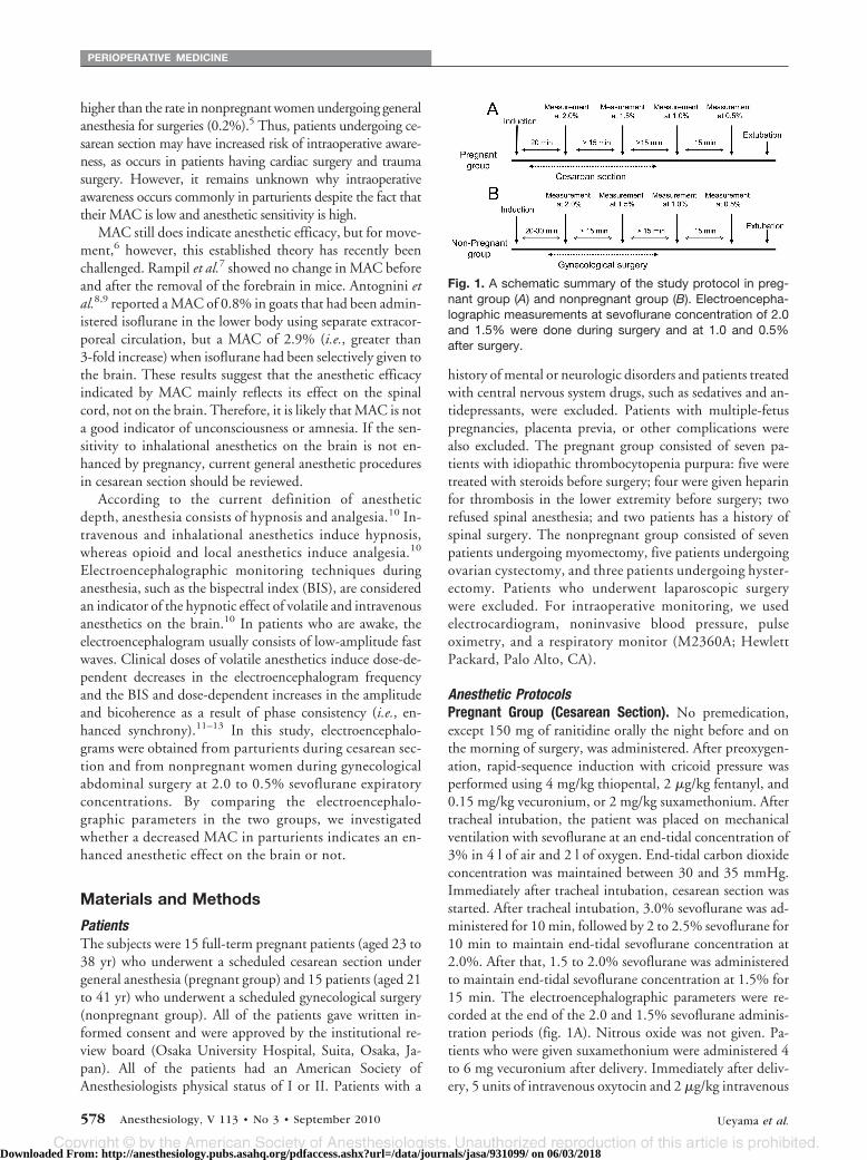
Anesthetic ProtocolsPregnant Group (Cesarean Section). No premedication,except 150 mg of ranitidine orally the night before and onthe morning of surgery, was administered. After preoxygen-ation, rapid-sequence induction with cricoid pressure wasperformed using 4 mg/kg thiopental, 2 �g/kg fentanyl, and0.15 mg/kg vecuronium, or 2 mg/kg suxamethonium. Aftertracheal intubation, the patient was placed on mechanicalventilation with sevoflurane at an end-tidal concentration of3% in 4 l of air and 2 l of oxygen. End-tidal carbon dioxideconcentration was maintained between 30 and 35 mmHg.Immediately after tracheal intubation, cesarean section wasstarted. After tracheal intubation, 3.0% sevoflurane was ad-ministered for 10 min, followed by 2 to 2.5% sevoflurane for10 min to maintain end-tidal sevoflurane concentration at2.0%. After that, 1.5 to 2.0% sevoflurane was administeredto maintain end-tidal sevoflurane concentration at 1.5% for15 min. The electroencephalographic parameters were re-corded at the end of the 2.0 and 1.5% sevoflurane adminis-tration periods (fig. 1A). Nitrous oxide was not given. Pa-tients who were given suxamethonium were administered 4to 6 mg vecuronium after delivery. Immediately after deliv-ery, 5 units of intravenous oxytocin and 2 �g/kg intravenous
Fig. 1. A schematic summary of the study protocol in preg-nant group (A) and nonpregnant group (B). Electroencepha-lographic measurements at sevoflurane concentration of 2.0and 1.5% were done during surgery and at 1.0 and 0.5%after surgery.
PERIOPERATIVE MEDICINE
578 Anesthesiology, V 113 • No 3 • September 2010 Ueyama et al.
Downloaded From: http://anesthesiology.pubs.asahq.org/pdfaccess.ashx?url=/data/journals/jasa/931099/ on 06/03/2018
fentanyl was administered for 5 min. We also administered1 �g/kg additional fentanyl every 30 min during operation.Patients with hypotension were treated with intravenous ephed-rine. After completion of surgery, we administered sevofluraneat expiratory concentrations of 1.0 and 0.5% for 15 min at eachconcentration (fig. 1A). Electroencephalographic parameterswere recorded at the end of the 1.0 and 0.5% sevoflurane ad-ministration periods. Sevoflurane administration protocol wasobtained from computer simulation using the software GasMan® (Med Man Simulations, Boston, MA).Nonpregnant Group (Gynecological Surgery). No premed-ication, except 150 mg of ranitidine orally the night beforeand on the morning of surgery, was administered. After es-tablishing an intravenous route, 4 mg/kg thiopental, 2 �g/kgfentanyl, and 0.15 mg/kg vecuronium were administered forinduction of general anesthesia. After tracheal intubation,sevoflurane, fractional inspired oxygen tension, and end-tidal carbon dioxide were maintained in the same manner asthe cesarean section group. Surgery was started 10 to 15 minafter tracheal intubation in all patients. We administered 2�g/kg fentanyl before incision, and an additional 1 �g/kgfentanyl was given every 30 min during the operation. Elec-troencephalographic parameter recording was also per-formed at the end of the 2.0 and 1.5% sevoflurane adminis-tration period (fig. 1B). Patients with hypotension duringsurgery were treated with intravenous ephedrine. After com-pleting surgery, sevoflurane was administered at an expira-tory concentration of 1.0 and 0.5% for 15 min (fig. 1B).Electroencephalographic parameters were recorded at theend of each sevoflurane administration period.
Electroencephalographic MonitoringFor electroencephalographic recordings, we used the BIS®
A-1050 monitor (Aspect Medical Systems, Natick, MA).Three-point electroencephalographic sensors were attachedto the forehead. Automatic electrode impedance check wasdone in all subjects. Raw data, including electroencephalo-graphic waveforms, BIS, and other parameters, were ob-tained from the BIS A-1050 monitor via a RS232 cableconnected to a laptop computer and analyzed with customsoftware (Bispectrum Analyzer for BIS).12,13 Using this soft-ware, we calculated the 95% spectral edge frequency(SEF95), amplitude, and bicoherence. Bicoherence is an in-dicator of electroencephalographic synchrony, and volatileanesthetics are known to increase the peak heights of bico-herence at 3–5 Hz (pBIC-low) and 5–10 Hz (pBIC-high) ina dose-dependent manner.12,13 In this study, to evaluate thedifference in response to sevoflurane, we compared thechanges in electroencephalographic parameters (SEF95, am-plitude, BIS, and bicoherence) in the pregnant and nonpreg-nant women at sevoflurane concentrations of 2.0 to 0.5%.
Statistical AnalysisA pilot study was performed in 10 patients (n � 5 for eachgroup). The pilot study showed that BIS has a larger SD(50 � 9 at 1.5% sevoflurane in nonpregnant group) than the
other parameters (SEF and amplitude). Based on BIS datafrom the pilot study, a sample size of 13.75 in each group wasconsidered to have 80% power to detect a difference inmeans of 20% (specifically, because BIS value mean in non-pregnant women was 50, the difference in mean was 10),assuming that the common SD was 9 using a two-group t testwith a 0.05 two-sided significance level. Consequently, thenumber of subjects was specified to 15 patients per group.The patient characteristics, including age, height, weight, surgi-cal time, and hemodynamic data, were compared using an un-paired t test. SEF95, amplitude, BIS value and bicoherence(pBIC-low and pBIC-high) at 2.0% to 0.5% concentrations ofsevoflurane were analyzed by a two-way analysis of variance.The model included the main effects, group (pregnant/non-pregnant), and sevoflurane concentration (0.5, 1, 1.5, or 2%),and their interaction. These electroencephalographic parame-ters in the two groups were also compared using an unpaired ttest. A P value of less than 0.05 was considered statistically sig-nificant (two-sided). All statistical analysis were performed bySAS Release 9.2 (SAS Institute Inc., Cary, NC).
ResultsThe patient’s characteristics are shown in table 1. No signif-icant differences between the pregnant and nonpregnantgroups were found in age, height, and nonpregnant weight.Maternal and neonatal data are shown in table 2. Table 3shows the hemodynamic data in each group. In the pregnantgroup, heart rate was significantly higher than in the non-pregnant group (P � 0.01) during study period. The averageephedrine doses for treatment of hypotension in the non-pregnant and pregnant groups were 6.3 � 2.5 (n � 4) and7.0 � 2.7 mg (n � 5), respectively. Seven parturients weregiven suxamethonium at induction of general anesthesia.
Typical electroencephalographic waveforms of the twogroups are shown in figure 2. There was no problem in thequality of electroencephalographic signals in the two groups.The signal quality index was 0.8 or higher. Reducing thesevoflurane concentration in 0.5 percentage point incre-ments (from 2.0 to 0.5%) decreased the electroencephalo-graphic amplitude and increased the electroencephalo-graphic frequency in both groups.
Table 1. Patient Characteristics
Characteristic
PregnantGroup
(n � 15)
NonpregnantGroup
(n � 15) P Value
Age (yr) 31 � 6 32 � 4 0.52Height (cm) 160 � 5 159 � 5 0.72Weight (kg) 64.5 � 7.0 58 � 8 0.78*(Weight before
pregnancy (kg))(59 � 8) — —
Gestational age (wk) 39 � 0.4 — —Surgical time (min) 71 � 17 77 � 23 0.43
Data are expressed as mean � SD.* P value between weight before pregnancy in pregnant groupand weight in nonpregnant group.
Volatile Anesthetic Sensitivity in Parturients
Ueyama et al. Anesthesiology, V 113 • No 3 • September 2010 579
Downloaded From: http://anesthesiology.pubs.asahq.org/pdfaccess.ashx?url=/data/journals/jasa/931099/ on 06/03/2018

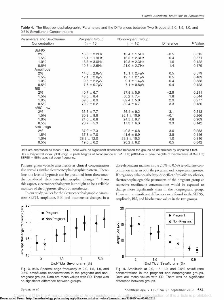
Table 4 shows the electroencephalographic parametersand the differences between the two groups at 2.0, 1.5, 1.0,and 0.5% sevoflurane concentrations. Figures 3, 4, and 5show the changes in SEF95, amplitude, and BIS value at 2.0to 0.5% sevoflurane concentrations, respectively. In thepregnant group, the reduction in sevoflurane concentrationin 0.5 percentage-point decrements from 2.0 to 0.5% causedchanges in the frequency, the BIS, and the amplitude. To bespecific, SEF95 increased from (mean � SD) 13.8 � 2.2 to19.7 � 2. 6 Hz, the BIS increased from 40.7 � 6.7 to 79.2 �6.2, and the amplitude reduced from 14.6 � 2.8 to 7.6 � 0.7�V. In addition, in the nonpregnant group, the SEF95 in-creased from 13.4 � 1.5 to 21.0 � 2.7 Hz, the BIS increasedfrom 37.8 � 5.6 to 82.4 � 6.7, and the amplitude reducedfrom 15.1 � 2.4 to 7.1 � 0.8 �V.
pBIC-low and pBIC-high (which indicate electroen-cephalographic synchrony) were 33.3 � 7.7 and 37.9 � 7.3,respectively, at 2.0% sevoflurane concentration in the preg-nant group. They decreased to 20.7 � 5.9 and 19.6 � 6.2,respectively, at 0.5%. In the nonpregnant group, pBIC-lowand pBIC-high were 36.4 � 9.2 and 40.8 � 6.8 at 2.0%sevoflurane concentration. This decreased to 17.3 � 6.3 and20.2 � 6.2, respectively, at 0.5%.
The results of two-way analysis of variance showed thatsevoflurane concentration effect was significant (P �0.0001) for each electroencephalographic parameter (table
5). However, the group effect (pregnant/nonpregnant) andthe interaction were not significant for each electroencepha-lographic parameter (table 5). The unpaired t test alsoshowed that electroencephalographic parameters in the preg-nant and nonpregnant groups at each sevoflurane concentra-tion were not significantly different (table 4). These resultsimply that each electroencephalographic parameter changeddose-dependently according to sevoflurane concentrationbut was unaffected by pregnancy.
A BIS value greater than 60 at 1.5% sevoflurane concentra-tion was observed in two patients (one each from the pregnantand nonpregnant groups). A BIS value greater than 60 at 1.0%sevoflurane concentration was observed in 10 patients from thenonpregnant group and 8 patients from the pregnant group.The patients from the two groups were interviewed on surgeryday and the following day but none had intraoperative memory.
DiscussionDuring non–rapid eye movement sleep, as the stages of sleepprogress, the electroencephalogram pattern changes from alow-amplitude fast wave to a high-amplitude slow wave.14
Table 2. Maternal and Neonatal Data
Maternal Data n � 15
Uterine Incision Delivery Time (s) 91 � 43Blood Loss with Amniotic Fluid (ml) 1,130 � 480Umbilical A
pH 7.32 � 0.07PaO2 (mmHg) 27 � 8.6PaCO2 (mmHg) 52 � 4.3Base Excess (mM) �1.1 � 1.7
Apgar scores at 1 min8–10 10�8 5
Apgar scores at 5 min8–10 13�8 2
Data are expressed as mean � SD except for Apgar scores.
Table 3. Hemodynamic Data
Groups
Sevoflurane (%)
Control 2.0 1.5 1.0 0.5
Pregnant (n � 15)MBP (mmHg) 85.8 � 8.2 95.3 � 12.0 84.3 � 10.3 81.0 � 8.5 83.4 � 12.3HR (beats/min) 88.3 � 16.7* 93.2 � 14.3* 92.2 � 13.3* 87.3 � 10.2* 88.3 � 11.2*
Nonpregnant (n � 15)MBP (mmHg) 87.2 � 11.8 97.2 � 11.5 85.3 � 12.8 80.3 � 7.8 80.2 � 10.4HR (beats/min) 73.2 � 11.3 77.3 � 13.5 72.3 � 11.7 69.5 � 10.5 71.5 � 12.0
Data are mean � SD.* Significant difference from nonpregnant group (P � 0.01).HR � heart rate; MBP � mean blood pressure.
Fig. 2. The typical electroencephalographic wave forms at2.0, 1.5, 1.0, and 0.5% sevoflurane concentration in thepregnant (A) and nonpregnant (B) groups. The reduction insevoflurane concentration from 2.0 to 0.5% changes electro-encephalograms from a high-amplitude slow wave to a low-amplitude fast wave. BIS bispectral index; SEF95 � 95%spectral edge frequency.
PERIOPERATIVE MEDICINE
580 Anesthesiology, V 113 • No 3 • September 2010 Ueyama et al.
Downloaded From: http://anesthesiology.pubs.asahq.org/pdfaccess.ashx?url=/data/journals/jasa/931099/ on 06/03/2018
Patients given volatile anesthetics at clinical concentrationalso reveal a similar electroencephalographic pattern. There-fore, the level of hypnosis can be presumed from these anes-thetic-induced electroencephalographic changes.10 Fromthis aspect, electroencephalogram is thought to be a reliablemonitor of the hypnotic effects of anesthetics.
In our study, values for the electroencephalographic param-eters SEF95, amplitude, BIS, and bicoherence changed in a
dose-dependent manner in the 2.0% to 0.5% sevoflurane con-centration range in both the pregnant and nonpregnant groups.If pregnancy enhances the hypnotic effect of volatile anesthetics,electroencephalographic parameters of the pregnant group atrespective sevoflurane concentrations would be expected tochange more significantly than in the nonpregnant group.However, no significant differences were found in the SEF95,amplitude, BIS, and bicoherence values in the two groups.
Table 4. The Electroencephalographic Parameters and the Differences between Two Groups at 2.0, 1.5, 1.0, and0.5% Sevoflurane Concentrations
Parameters and SevofluraneConcentration
Pregnant Group(n � 15)
Nonpregnant Group(n � 15) Difference P Value
SEF952% 13.8 � 2.2Hz 13.4 � 1.5Hz �0.5 0.5151.5% 16.1 � 1.9Hz 16.5 � 2.3Hz 0.4 0.5711.0% 18.3 � 3.0Hz 19.8 � 2.3Hz 1.6 0.1220.5% 19.7 � 2.6Hz 21.0 � 2.7Hz 1.4 0.179
Amplitude2% 14.6 � 2.8�V 15.1 � 2.4�V 0.5 0.5791.5% 12.1 � 2.0�V 12.7 � 2.1�V 0.5 0.4891.0% 9.5 � 2.2�V 9.1 � 1.4�V �0.4 0.5380.5% 7.6 � 0.7�V 7.1 � 0.8�V �0.4 0.123
BIS2% 40.7 � 6.7 37.8 � 5.6 �2.9 0.2111.5% 48.5 � 8.4 50.2 � 7.4 1.8 0.5441.0% 59.5 � 8.9 62.4 � 5.0 2.9 0.2770.5% 79.2 � 6.2 82.4 � 6.7 3.3 0.180
pBIC-Low2% 33.3 � 7.7 36.4 � 9.2 3.1 0.3131.5% 30.3 � 8.8 35.1 � 10.9 �0.1 0.2661.0% 24.6 � 6.8 24.5 � 9.7 4.8 0.9690.5% 20.7 � 5.9 17.3 � 6.3 �3.3 0.142
pBIC-High2% 37.9 � 7.3 40.8 � 6.8 3.0 0.2531.5% 37.8 � 7.0 41.6 � 6.9 3.8 0.1461.0% 28.3 � 12.0 29.3 � 10.3 1.0 0.8160.5% 19.6 � 6.2 20.2 � 6.2 0.5 0.842
Data are expressed as mean � SD. There were no significant differences between the groups as determined by unpaired t test.BIS � bispectral index; pBIC-high � peak heights of bicoherence at 5–10 Hz; pBIC-low � peak heights of bicoherence at 3–5 Hz;SEF95 � 95% spectral edge frequency.
Fig. 3. 95% Spectral edge frequency at 2.0, 1.5, 1.0, and0.5% sevoflurane concentrations in the pregnant and non-pregnant groups. Data are mean values with SD. There wasno significant difference between groups.
Fig. 4. Amplitude at 2.0, 1.5, 1.0, and 0.5% sevofluraneconcentrations in the pregnant and nonpregnant groups.Data are mean values with SD. There was no significantdifference between groups.
Volatile Anesthetic Sensitivity in Parturients
Ueyama et al. Anesthesiology, V 113 • No 3 • September 2010 581
Downloaded From: http://anesthesiology.pubs.asahq.org/pdfaccess.ashx?url=/data/journals/jasa/931099/ on 06/03/2018

Because sample size calculation was designed on the as-sumption of detecting 20% differences in mean value be-tween two groups, we could have missed differences under20%. In clinical studies, evaluating the difference often pro-vides more information than statistical significance testing.Actually, the observed differences between electroencephalo-graphic parameters in the two groups at each sevofluraneconcentration were all less than the SD (table 4). Therefore,we believe that there are no clinically significant differences
between the two groups. This consequently suggests thatthere is no important difference in the hypnotic effect ofsevoflurane in pregnant and nonpregnant women.
Why did the electroencephalogram not indicate a differ-ence in the hypnotic effect in the subjects in whom MAC wasthought to be different? MAC represents the alveolar con-centration of inhalational anesthetics that prevents 50% ofsubjects from moving in response to noxious stimuli.6 Be-cause such movement is considered an escape response frompainful stimuli, MAC has been considered an indicator of theeffect of anesthetics on the brain. However, as indicated inIntroduction, animal studies have shown that MAC indi-cates the effect of anesthetics on the spinal cord,7–9 whereasthe electroencephalogram shows the anesthetic effect on thebrain. Therefore, it is not surprising that hypnotic levels in-dicated by electroencephalogram are similar in pregnant andnonpregnant women, although MAC may differ between thetwo groups. The results of this study show that the decreasein MAC during pregnancy cannot validate the rationale thatparturients require less volatile anesthetics.
Various factors affect the electroencephalographic wave-forms during surgery, one being noxious stimuli. If analgesicdosage is insufficient, noxious stimuli can increase the fre-quency and decrease the amplitude of high-amplitude slowwaves induced by anesthetics.15 This phenomenon is calleddesynchronization,15 in which consciousness likely returnsby noxious stimuli reaching the brain. In this study, twotypes of operations (cesarean section in the pregnant groupand gynecological surgery in the nonpregnant group) wereperformed. It remains possible that the intensity of noxiousstimulus differed depending on the type of operation. Onemethod to evaluate the intensity of the noxious stimulus isintraoperative hemodynamic changes. In our study, becauseboth the preoperative and intraoperative heart rates werehigher in the pregnant group, the hemodynamic values arenot appropriate. Another method to evaluate the intensity ofnoxious stimuli is bicoherence, a parameter that indicates thehypnotic effect of anesthetics. Bicoherence disappears whena strong noxious stimulus is applied, because noxious stimuliinduce an arousal electroencephalographic pattern.16 It isrestored or prevented by sufficient opioid analgesia.16 In thisstudy, bicoherence was similar in the pregnant and nonpreg-nant groups at 2 and 1.5% sevoflurane. We believe thatfentanyl analgesia minimized the noxious stimuli, and thedegree of noxious stimuli was equivalent in both groups.
Some drugs administered intraoperatively also affect theelectroencephalographic waveforms. Thiopental is known toaffect electroencephalogram; however, the duration is short.According to a previous report, nonpregnant patients recov-ered consciousness 330 � 153 s after induction of 4 mg/kgthiopental.17 At the time of return of consciousness, the BISvalue was 81 � 5.17 Redistribution is the principal mecha-nism accounting for this early awakening. Approximately10–15 min after 4–6 mg/kg thiopental administration, serumlevels fall to 5 �g/ml, which is equivalent to 50% of awakeninglevel in both parturients and nonpregnant patients.18,19 Al-
Fig. 5. Bispectral index (BIS) value at 2.0, 1.5, 1.0, and 0.5%sevoflurane concentrations in the pregnant and nonpregnantgroups. Data are mean values with SD. There was no signif-icant difference between groups.
Table 5. Results of Two-way Analysis of Variance
Parameter & Effect NDF DDF F P Value
SEF95Group 1 112 2.83 0.0953Sevoflurane 3 112 49.17 � 0.0001Group�Sevoflurane 3 112 1.16 0.3265
AmplitudeGroup 1 112 0.02 0.8777Sevoflurane 3 112 88.71 � 0.0001Group�Sevoflurane 3 112 0.61 0.6076
BISGroup 1 112 0.99 0.3224Sevoflurane 3 112 195.89 � 0.0001Group�Sevoflurane 3 112 1.25 0.2968
BIC-lowGroup 1 112 0.55 0.4593Sevoflurane 3 112 23.4 � 0.0001Group�Sevoflurane 3 112 1.42 0.2396
BIC-highGroup 1 112 1.93 0.1672Sevoflurane 3 112 41.7 � 0.0001Group�Sevoflurane 3 112 0.29 0.8293
The group effect (pregnant/nonpregnant), sevoflurane concentra-tion effect, and group�sevoflurane interaction for each electroen-cephalographic parameter.BIS � bispectral index; DDF � denominator degrees of freedom;Group � pregnant vs. nonpregnant group; Group�Sevoflurane �interaction between group and sevoflurane; NDF � numeratordegrees of freedom; pBIC-high � peak heights of bicoherence at5–10 Hz; pBIC-low � peak heights of bicoherence at 3–5 Hz;SEF95 � 95% spectral edge frequency; Sevoflurane � sevoflu-rane concentration (0.5, 1.0, 1.5, and 2.0%).
PERIOPERATIVE MEDICINE
582 Anesthesiology, V 113 • No 3 • September 2010 Ueyama et al.
Downloaded From: http://anesthesiology.pubs.asahq.org/pdfaccess.ashx?url=/data/journals/jasa/931099/ on 06/03/2018
though the dose requirement of thiopental in parturients is ap-proximately 18% lower compared with nonpregnant women,20
the blood concentration of thiopental at our study period (20–150 min after induction) is thought to be sufficiently low toaffect the electroencephalogram. A muscle relaxant itself doesnot have an effect on electroencephalogram. However, electro-myogram artifacts greatly change the electroencephalographicwaveform. If an electromyogram is combined with the electro-encephalogram, the electroencephalographic frequency in-creases, and it comes to show an arousal pattern. To prevent this,we administered muscle relaxant in all cases.
Some factors affect not only the electroencephalogram butalso MAC. Drugs such as ephedrine increase MAC by increas-ing the catecholamine in the brain.21 Such drugs are usuallydose-related and require a very high dose.22 The amount ofephedrine necessary to raise MAC by 50% has been reported tobe as high as 0.04 mg/kg/min in a dog study.21 Because thedosage of the ephedrine in our study was small (0.01 mg/kg,bolus), it was probably not enough to influence the MAC.
Hormonal changes associated with delivery also affect theMAC. According to previous human studies, the postpartumchanges in MAC were as follows. During the first 1–12 hpostpartum, the MAC of isoflurane was similar to the0.775% measured in pregnant patients of 8–12 weeks’ ges-tation.3 MAC increased to 0.825% during the next 36 h, toreach normal values (1.125%) by 72 h postpartum.23 Al-though MAC in the third trimester in parturients has notbeen examined because of ethical considerations, these re-sults suggested that changes in MAC at the postpartum pe-riod are very slow. Therefore, we believe that rapid changes inMAC did not occur during our study period.
So how much volatile anesthetic should we administer dur-ing general anesthesia for cesarean section? It has been shownthat the volatile anesthetic requirement for general anesthesia islower than that required for prevention of body movements.24
The results of this study showed that the BIS ranged from 40 to60 in most patients in both groups at 1.5% sevoflurane concentra-tion. By contrast, the BIS exceeded 60 in almost half of the patientsin both groups at 1.0% sevoflurane concentration. The highest BISvalue at 1.0% was 80. These results are almost the same as the BISvalues for sevofluraneconcentrationdeterminedbyKatoh etal.25 innonpregnant patients and by Chin et al.26 in pregnant patients.
Under the assumption that the BIS value for appropriatehypnosis during operation is less than 60,10,27 approximatelyhalf of patients are insufficiently anesthetized at 1.0%sevoflurane concentration. Therefore, in cases without bothelectroencephalographic monitoring and nitrous oxide ad-ministration, at least 1.5% sevoflurane may be needed duringmaintenance of general anesthesia.
The dose requirements of inhalational anesthetics has beenbelieved to be less for parturients because MAC is significantlydecreased by pregnancy. To prevent undesired outcomes, suchas neonatal suppression or uterine atony, parturients have rou-tinely been administered volatile anesthetics at a lower concen-tration compared with nonpregnant women. However, ourelectroencephalographic study indicates that there is no differ-
ence between pregnant and nonpregnant women in the hyp-notic effects of sevoflurane, despite the groups supposedly hav-ing different MAC values. These findings suggest that a decreasein MAC during pregnancy does not mean an enhanced volatileanesthetic effect on the brain. We believe that parturients shouldbe given the same dose of anesthetics as nonpregnant women forprevention of intraoperative awareness. Thus, anesthesiologistsshould reconsider using MAC as an indicator of efficacy of vol-atile anesthetics.
The authors are grateful to Dr. Sawami Kiyonaka (Resident Anes-thesiologist, Department of Anesthesiology, Kansai Rosai Hospital,Amagasaki, Hyogo, Japan) for her advice and assistance and Mr.Tadashi Koga (Director, Biometrics Department, Shin Nippon Bio-medical Laboratories, Ltd., Kagoshima, Japan) for his assistance withstatistical analysis.
References1. Kuczkowski K, Reisner L, Lin D: Anesthesia for cesarean
section, Obstetric Anesthesia, 3rd edition. Edited by Chest-nut DH. Philadelphia, Elsevier Mosby, 2004, pp 421– 46
2. Palahniuk RJ, Shnider SM, Eger EI 2nd: Pregnancy de-creases the requirement for inhaled anesthetic agents.ANESTHESIOLOGY 1974; 41:82–3
3. Gin T, Chan MT: Decreased minimum alveolar concentra-tion of isoflurane in pregnant humans. ANESTHESIOLOGY
1994; 81:829 –32
4. Lyons G, Macdonald R: Awareness during caesarean sec-tion. Anaesthesia 1991; 46:62– 4
5. Liu WH, Thorp TA, Graham SG, Aitkenhead AR: Incidenceof awareness with recall during general anaesthesia. An-aesthesia 1991; 46:435–7
6. Eger EI 2nd, Saidman LJ, Brandstater B: Minimum alveolaranesthetic concentration: A standard of anesthetic po-tency. ANESTHESIOLOGY 1965; 26:756 – 63
7. Rampil IJ, Mason P, Singh H: Anesthetic potency (MAC) isindependent of forebrain structures in the rat. ANESTHESIOLOGY
1993; 78:707–12
8. Antognini JF, Schwartz K: Exaggerated anesthetic require-ments in the preferentially anesthetized brain. ANESTHESI-OLOGY 1993; 79:1244 –9
9. Borges M, Antognini JF: Does the brain influence somaticresponses to noxious stimuli during isoflurane anesthesia?ANESTHESIOLOGY 1994; 81:1511–5
10. Stanski DR, Shafer SL: Measuring depth of anesthesia,Anesthesia, 6th edition. Edited by Miller RD. Philadelphia,Elsevier, 2005, pp 1227– 61
11. Morimoto Y, Hagihira S, Koizumi Y, Ishida K, MatsumotoM, Sakabe T: The relationship between bispectral indexand electroencephalographic parameters during isofluraneanesthesia. Anesth Analg 2004; 98:1336 – 40
12. Hagihira S, Takashina M, Mori T, Mashimo T, Yoshiya I:Changes of electroencephalographic bicoherence duringisoflurane anesthesia combined with epidural anesthesia.ANESTHESIOLOGY 2002; 97:1409 –15
13. Hagihira S, Takashina M, Mori T, Mashimo T, Yoshiya I:Practical issues in bispectral analysis of electroencephalo-graphic signals. Anesth Analg 2001; 93:966 –70
14. Keifer J: Sleep and anesthesia, Neural Mechanisms of An-esthesia. Edited by Antognini JF. Totowa, NJ, HumanaPress, 2003, pp 65–74
15. Kochs E, Bischoff P, Pichlmeier U, Schulte am Esch J: Surgicalstimulation induces changes in brain electrical activity duringisoflurane/nitrous oxide anesthesia. A topographic electroen-cephalographic analysis. ANESTHESIOLOGY 1994; 80:1026–34
16. Hagihira S, Takashina M, Mori T, Ueyama H, Mashimo T:Electroencephalographic bicoherence is sensitive to nox-
Volatile Anesthetic Sensitivity in Parturients
Ueyama et al. Anesthesiology, V 113 • No 3 • September 2010 583
Downloaded From: http://anesthesiology.pubs.asahq.org/pdfaccess.ashx?url=/data/journals/jasa/931099/ on 06/03/2018

ious stimuli during isoflurane or sevoflurane anesthesia.ANESTHESIOLOGY 2004; 100:818 –25
17. Flaishon R, Windsor A, Sigl J, Sebel PS: Recovery of conscious-ness after thiopental or propofol. Bispectral index and theisolated forearm technique. ANESTHESIOLOGY 1997; 86:613–9
18. Kosaka Y, Takahashi T, Mark LC: Intravenous thiobarbiturateanesthesia for cesarean section. ANESTHESIOLOGY 1969; 31:613–9
19. Burch PG, Stanski DR: The role of metabolism and protein bindingin thiopental anesthesia. ANESTHESIOLOGY 1983; 58:146–52
20. Gin T, Mainland P, Chan MT, Short TG: Decreased thio-pental requirements in early pregnancy. ANESTHESIOLOGY
1997; 86:73– 8
21. Steffey EP, Eger EI: The effect of seven vasopressors ofhalothane MAC in dogs. Br J Anaesth 1975; 47:435– 8
22. Eger EI 2nd: MAC, Anesthetic Uptake and Action. Edited byEger EI 2nd. Baltimore, Williams & Wilkins, 1975, pp 1–25
23. Chan MT, Gin T: Postpartum changes in the minimumalveolar concentration of isoflurane. ANESTHESIOLOGY 1995;82:1360 –3
24. Campagna JA, Miller KW, Forman SA: Mechanisms of ac-tions of inhaled anesthetics. N Engl J Med 2003; 348:2110 –24
25. Katoh T, Suzuki A, Ikeda K: Electroencephalographic de-rivatives as a tool for predicting the depth of sedation andanesthesia induced by sevoflurane. ANESTHESIOLOGY 1998;88:642–50
26. Chin KJ, Yeo SW: Bispectral index values at sevofluraneconcentrations of 1% and 1.5% in lower segment cesareandelivery. Anesth Analg 2004; 98:1140 – 4
27. Jameson LC, Sloan TB: Using EEG to monitor anesthesiadrug effects during surgery. J Clin Monit Comput 2006;20:445–72
ANESTHESIOLOGY REFLECTIONS
The Schneider Brain Wave Synchronizer
After observing how some radar technicians had become “transfixed” by rhythmic flashing dots on theirradar screens, inventor Sidney Schneider designed his Brain Wave Synchronizer (BWS) to hypnotize byvisually stimulating subjects at frequencies mimicking those of their alpha, beta, or delta brainwaves. In1959 Schneider and hypnotist-obstetrician William Kroger, M.D., published their use of the BWS inprenatal classes for thousands of women prior to its use as an “electronic aid for hypnotic induction”during labor and delivery. Four years later, Chicago anesthesiologist Max S. Sadove, M.D., published hiswork on how BWS-induced hypnosis could reduce anesthetic agent requirements during general anes-thesia. By 1994 the BWS would be cited for causing epileptic seizures in a patient. (Copyright © theAmerican Society of Anesthesiologists, Inc. This image appears in color in the Anesthesiology Reflectionsonline collection available at www.anesthesiology.org.)
George S. Bause, M.D., M.P.H., Honorary Curator, ASA’s Wood Library-Museum of Anesthesi-ology, Park Ridge, Illinois, and Clinical Associate Professor, Case Western Reserve University,Cleveland, Ohio. [email protected].
PERIOPERATIVE MEDICINE
584 Anesthesiology, V 113 • No 3 • September 2010 Ueyama et al.
Downloaded From: http://anesthesiology.pubs.asahq.org/pdfaccess.ashx?url=/data/journals/jasa/931099/ on 06/03/2018