Preface - buecher.deUreteral Stones: Retrograde Ureteral Stenting vs Nephrostomy Tube Drainage...
25
Preface vii Endourology is one of the most important subspecialties in the field of urology because of the widespread use of endoscopy for the diagnosis and treatment of a variety of upper genitourinary tract pathologies. Although most clinical urologists incorporate some basic endourology into their practices, complex upper tract pathology and anatomy require more advanced endoscopic skills and instrumentation. Advanced Endourology: The Complete Clinical Guide is intended as a resource guide for all aspects of clinical endourology, particularly the more advanced procedures. This volume encompasses endourological applications for upper urinary tract calculi, stric- tures, and urothelial cancer. It will also serve as a comprehensive overview of available endoscopes and instrumentation. Advanced Endourology: The Complete Clinical Guide is unique in that most of its individual chapters include videos that clearly illustrate critical portions of the techniques and provide tips and tricks from the experts. Every practicing urologist should have this book in his or her library, with the accompanying DVD kept near a DVD player, for quick access to detailed procedural instruction and immediate review of the videos. Stephen Y. Nakada, MD Margaret S. Pearle, MD, PhD
Transcript of Preface - buecher.deUreteral Stones: Retrograde Ureteral Stenting vs Nephrostomy Tube Drainage...
Preface
vii
Endourology is one of the most important subspecialties in the field of urology becauseof the widespread use of endoscopy for the diagnosis and treatment of a variety of uppergenitourinary tract pathologies. Although most clinical urologists incorporate some basicendourology into their practices, complex upper tract pathology and anatomy requiremore advanced endoscopic skills and instrumentation.
Advanced Endourology: The Complete Clinical Guide is intended as a resource guidefor all aspects of clinical endourology, particularly the more advanced procedures. Thisvolume encompasses endourological applications for upper urinary tract calculi, stric-tures, and urothelial cancer. It will also serve as a comprehensive overview of availableendoscopes and instrumentation.
Advanced Endourology: The Complete Clinical Guide is unique in that most of itsindividual chapters include videos that clearly illustrate critical portions of the techniquesand provide tips and tricks from the experts. Every practicing urologist should have thisbook in his or her library, with the accompanying DVD kept near a DVD player, for quickaccess to detailed procedural instruction and immediate review of the videos.
Stephen Y. Nakada, MD
Margaret S. Pearle, MD, PhD
Access, Stents, and Urinary Drainage
Ben H. Chew, MSc, MD, FRCSC
and John D. Denstedt, MD, FRCSC
CONTENTS
INTRODUCTION
ACCESS TECHNIQUES
STENTING TECHNIQUE
STENT COMFORT, INFECTION, AND ENCRUSTATION:THE ROLE OF NEW BIOMATERIALS AND COATINGS
TIPS AND TRICKS
CONCLUSION
REFERENCES
2
SUMMARY
Ureteral access is necessary in many endourological procedures includingureteroscopy and ureteral stenting. Technologies such as ureteral access sheaths, bal-loon dilators, and coaxial dilators may be helpful in facilitating ureteral access in diffi-cult cases. This chapter describes a stenting technique that relies on fluoroscopicguidance once the initial guidewire is placed and the cystoscope is removed.
Key Words: Ureter; stent; calculi; ureteroscopy; nephrostomy tube; shockwavelithotripsy.
INTRODUCTION
Ureteral stents are a mainstay in the urological armamentarium and are utilized in thetreatment of urolithiasis including postureteroscopy, preshockwave lithotripsy, and to relievesymptomatic renal colic. Routine stenting postureteroscopy and intracorporeal lithotripsy,once the standard of care, have been shown to be unnecessary following uncomplicatedureteroscopy and stone manipulation. Advances such as laser lithotripsy and smaller uretero-scopes have minimized the potential morbidity of ureteroscopy to the point that theindwelling stent has become the most morbid part of the procedure. Ureteral stents maycause considerable side effects ranging from dysuria, urgency and frequency to hematuria
From: Advanced Endourology: The Complete Clinical GuideEdited by: S. Y. Nakada and M. S. Pearle © Humana Press Inc., Totowa, NJ
19
02_Denstedt 6/22/05 4:53 PM Page 19
and suprapubic pain. There is an emerging body of literature that routine stenting pos-tureteroscopy is not necessary and that the need for stenting should be determined on a caseby case basis.
Stents are also used to provide urinary drainage in nongenitourinary causes ofureteral obstruction, such as pregnancy and malignant ureteral obstruction. An alterna-tive and effective method of urinary drainage is the percutaneous nephrostomy tubewhich is easily placed in patients with significant hydronephrosis and may be even moresuccessful than retrograde ureteral stenting when urinary drainage is required as a resultof obstruction of the distal ureter. Incompressible stents incorporating metal into thestent material have been used to provide urinary drainage to patients with malignantureteral obstruction. Conversely, biodegradable stents have been developed to provideureteral drainage temporarily following an endourological procedure before degradingand being excreted in the urine, thus obviating the need for cystoscopic stent removal.Other stent advancements will see coatings, new materials, and drugs loaded directlyinto the stent material or coated on the stent surface to improve comfort and reducebiofilm formation, infection, and encrustation.
Access to the ureter is required any time closed endoscopic ureteral procedures are tobe carried out including during ureteral stenting and in association with diagnostic andtherapeutic ureteroscopy for urolithiasis. More detail will be provided in other chaptersregarding procedure specific aspects of ureteroscopy and percutaneous procedures; thischapter will focus on initially gaining retrograde access to the ureter, aspects relatedto ureteral stenting and a comparative analysis of alternative methods of urinary drainage.A brief summary of new stent technologies and biomaterials will also be presented.
Indications to Access the Ureter
Achievement of ureteral access is necessary for performing retrograde endoscopicprocedures such as ureteroscopy, or for placing a ureteral stent. Table 1 lists commonindications for ureteral stent placement.
Stones
Urolithiasis represents one of the more common reasons to insert a ureteral stent.Clinical indications for stenting include patients with intractable pain, those withinfected pyonephrosis, or patients with impaired renal function from obstruction. Inaddition, ureteral stenting is often employed as an adjunct to shockwave lithotripsy orendoscopic procedures in patients requiring surgical stone management.
Ureteral Stones: Retrograde Ureteral Stenting vs Nephrostomy Tube Drainage
Pyonephrosis with an obstructing stone requires urgent decompression using either ret-rograde ureteral stent placement or antegrade percutaneous nephrostomy tube drainage(1). Whether urinary drainage to bypass the obstruction is best accomplished via a ureteralstent or a nephrostomy tube is a subject of debate. The first randomized clinical trial tocompare these two methods in obstructed, infected patients was performed by Pearle et al.(2) in 42 patients with obstructing urolithiasis and pyonephrosis. The time to deferves-cence, length of stay in hospital, pain symptoms, and normalization of leukocytosis didnot differ between these two groups suggesting that urinary decompression by either ret-rograde ureteral stenting or antegrade percutaneous nephrostomy tube insertion are bothequally effective in treating obstructed pyonephrosis. However, patients had significantlyless fluoroscopy exposure (2.6 minutes less) when they were stented in a retrograde fashion.
20 Chew and Denstedt
02_Denstedt 6/22/05 4:53 PM Page 20
A similar study was performed by Mokhmalji and colleagues (3), who also found nodifference in relief of the presenting symptoms between patients randomized tonephrostomy tube insertion and ureteral stent placement. Percutaneous nephrostomytube placement was successful in all of the 20 patients randomized to that group, butonly 80% of the 20 patients randomized to retrograde ureteral stent placement were suc-cessfully stented. Although not statistically significant, there was a trend towards animproved quality of life in the nephrostomy tube group when pain, dysuria, frequency,and hematuria were taken into consideration.
From the standpoint of infection and the requirement for urinary decompression, itappears that nephrostomy tube drainage and ureteral stents offer equal drainage of theupper urinary tract. Symptoms of pain and irritation are also similar. Placement of anephrostomy tube or ureteral stent depends on availability of good interventional radi-ologists and the urologist’s access to the cystoscopy suite or operating room. At somehospitals, the radiology suite may be more accessible than the operating room or cys-toscopy suite or vice versa. Subsequent procedures should also be taken into account.For instance, in patients who will require a percutaneous nephrolithotomy, a percuta-neous nephrostomy tube is the preferred intervention and in patients with stonesamenable to shockwave lithotripsy (SWL), a ureteral stent is often preferable. Manyvariables must be taken into account to determine whether a percutaneous nephrostomytube or ureteral stent should be placed in patients with obstructing stones.
Ureteral Stenting Effects on Ureteral Physiology and Stone Passage
Animal studies have demonstrated that ureteral stents decrease the frequency andamplitude of ureteral contraction in animals. In an animal model of ureteral stones caus-ing obstruction, ureteral dilatation was observed proximal to the obstruction in the stented
Chapter 2 / Access, Stents, and Urinary Drainage 21
Table 1Indications for Ureteric Stent Insertion
• Stones—intractable pain, infection, hydronephrosis, acute renal failure, solitary kidney• Postureteroscopy • Pretreatment (pre-SWL)
Solitary kidney, stone >15 mm in diameter,• Steinstrasse post-SWL• Pyonephrosis (infection)• Stricture (endoureterotomy)• Trauma• Fistula• Ureteropelvic junction obstruction
To relieve symptomsPost endopyelotomy/pyeloplasty
• Hydronephrosis/calculi of pregnancy• Post reconstruction
Renal transplantUreteroneocystotomyUreteroureterostomyCystectomy and urinary diversion
• Extrinsic ureteral obstruction
SWL, shockwave lithotripsy.
02_Denstedt 6/22/05 4:53 PM Page 21
group whereas a nephrostomy tube group had no dilatation (4). Stents also impeded spon-taneous stone passage and reduced ureteral contractility when compared to the nephro-stomy group (4). This is controversial, however, as others have shown that ureteral stentsfacilitate spontaneous passage of distal ureteric stones less than 10 mm in diameter in 83%of patients studied (5). The ureter and ureteral orifice are theorized to passively dilate fromthe stent, thus facilitating stone passage. Although stents may affect ureteral peristalsis,the dilation of the ureter and orifice do facilitate spontaneous passage of smaller stones.
Stent Comfort and Quality of Life
There is an increasing awareness that stents impact patients’ quality of life. Stentsmay cause morbidity in up to 80% of patients with symptoms ranging from irritativevoiding symptoms, hematuria, flank pain, suprapubic pain, infection, and stent migra-tion to the “forgotten” encrusted stent (6–8). As a consequence of these concerns, theuse of routine stent placement is being more thoroughly considered on a case-by-casebasis when utilized as an adjunct to SWL or ureteroscopy.
In order to quantify patient morbidity from stents, Joshi et al. (7,8) have developedand validated the first questionnaire of stent symptoms, the Ureteral Stent SymptomQuestionnaire, which consists of 48 items spanning five criteria: pain, voiding symp-toms, work performance, sexual health, and overall general health. Results indicate that76% of stented patients experienced negative symptoms, 70% required analgesic, and42% had to reduce their activity by half (8). This validated tool should become a stan-dard evaluation technique of new stent technologies.
Stones: Stenting as an Adjunct to Shockwave Lithotripsy
Stenting prior to SWL is thought to preclude renal obstruction from stone fragmentsfollowing SWL (9). More recently, the prophylactic efficiency of pre-SWL stenting hasbeen called into question and is now a debated topic where some believe that stents inSWL patients not only lack efficacy to prevent renal obstruction, but may, in fact,impede the passage of stones fragments following SWL (10).
Steinstrasse, or the “street of stone” occurs with an overall rate of 3 to 6% of patientsundergoing SWL (11) and in 13 to 26% of nonstented patients with stone burdens greaterthan 25 mm in diameter (12,13). Placing stents prior to SWL in patients with stones greaterthan 20 to 30 mm in diameter significantly decreased the rate of steinstrasse to 3 to 7%(11,14–16). In patients with stones smaller than 25 mm, the rates of steinstrasse and infec-tion were unaffected by stenting (9,17–20). The reason for this latter finding is likely theresult of a significant risk decrease of steinstrasse in patients with stones less than 20 mm(21). A retrospective review by Madbouly et al. (21) has shown that there are four variablesthat are significantly correlated with an increased risk of steinstrasse: stone size greater than20 mm, stones located in the renal pelvis, a dilated renal pelvis, and shock wave energygreater than 22 kV. The risk of steinstrasse was 3.7 times less in stones smaller than 20 mmcompared with stones greater than that 20 mm. Stone location was also a factor becausedilation of the collecting system would lead to decreased amplitude of each contraction andlower intrapelvic pressures and propulsive power. Stone fragments in the ureter and anondilated renal pelvis are subjected to a higher force and rate of peristalsis which wouldlead to propulsion through the system. The risk of steinstrasse was reduced by two timesfor energies delivered at 18 to 22 kV and reduced by three times at energies of 14 to 18 kV(21). High-energy shock waves have been shown to produce larger stone fragments com-pared with more frequent lower powered shocks which result in finer stone fragments (22).
22 Chew and Denstedt
02_Denstedt 6/22/05 4:53 PM Page 22
These studies suggest that ureteral stents should be placed prior to SWL for largestones (>20-mm diameter). Some studies, particularly those treating large stones withSWL, must be considered with caution as percutaneous nephrolithotomy is usually thetreatment of choice in stones greater than 20 or 25 mm. For patients with stones lessthan 20 mm who are to be treated with SWL, there is little evidence that stenting priorto SWL reduces the rate of steinstrasse or infection.
Stones: Stenting Postureteroscopy
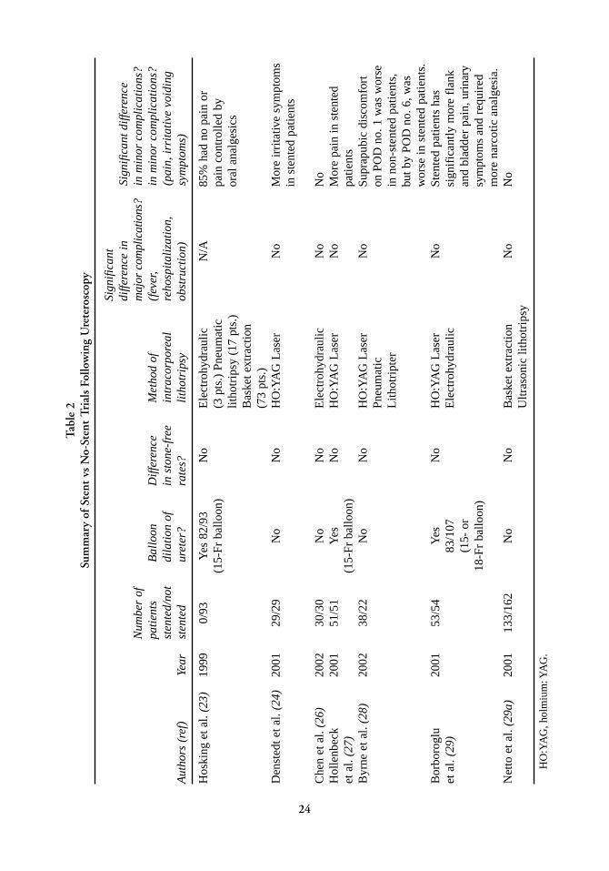
The principle of avoiding ureteral obstruction secondary to ureteral edema and stonefragments is the main driving force for routinely leaving a stent post ureteroscopy andhas traditionally been regarded as the standard of care. Technical advances includingminiaturization of ureteroscopes, utilization of the holmium:YAG laser, and softer stonebaskets have made ureteroscopy relatively atraumatic and the main morbidity followingureteroscopy originates from the use of ureteric stents. Furthermore, stents add to thecost of patient care and require an additional cystoscopy for removal unless a pull stringis used. Reducing stent use following ureteroscopy should improve patient care andsatisfaction (see Table 2).
Hosking (23) was the first to report a large series of nonstented ureteroscopy patientswho had minimal complications. Approximately half of the patients had no discomfortand the majority of those with discomfort described it as mild and easily resolved byoral analgesics. Although this report was a case series and did not have a stented con-trol group, it was the first series to suggest that ureteroscopy did not routinely requirestenting. Denstedt et al. (24) randomized 58 patients to receive either a stent or nostent after ureteroscopy. The results demonstrate that there were no differences inrehospitalization rate, analgesic use or stone free rates. At 1 week, the stented group hadsignificantly more pain and irritative voiding symptoms than the nonstented group.None of these patients underwent ureteral dilation, the holmium:YAG laser was used forintracorporeal lithotripsy, and all stones were less than 2 cm. The holmium:YAG laseris safe and has minimal effects on surrounding tissue which makes it an ideal modalityto preclude the need for a stent postoperatively (25). In addition, a randomized studyusing intracorporeal electrohydraulic lithotripsy also demonstrated that these patientscan be safely left unstented (26). Other randomized studies have found similar resultssuggesting that following uncomplicated ureteroscopy without ureteric dilation, stent-ing is not routinely required (24,26–28). Even in patients who underwent ureteral dila-tion at the time of ureteroscopy, nonstented patients had results and complication ratessimilar to stented patients (23,29). In the series by Hosking and associates (23), ureteraldilation was performed in 88% of patients who suffered minimal complications post-operatively. Borboroglu et al. (29) performed a study in 107 patients, which alsoincluded 83 patients who underwent ureteral balloon dilatation, and found that stentedpatients had more pain and analgesic requirements, but no difference in stone free orrehospitalization rates. These studies provide evidence that stenting after uncompli-cated ureteroscopy is not routinely necessary, but rather should be determined on acase-by-case basis.
Hydronephrosis/Calculi in Pregnancy
Upwards of 90% of women have hydronephrosis by the third trimester of pregnancy(30), but only 0.2–25% will become symptomatic and require medical attention
Chapter 2 / Access, Stents, and Urinary Drainage 23
02_Denstedt 6/22/05 4:53 PM Page 23
Tab
le 2
Sum
mar
y of
Ste
nt v
s N
o-St
ent
Tria
ls F
ollo
win
g U
rete
rosc
opy
Sign
ifica
nt
diffe
renc
e in
Sign
ifica
nt d
iffer
ence
Num
ber
of
maj
or c
ompl
icat
ions
?in
min
or c
ompl
icat
ions
?pa
tien
ts
Bal
loon
D
iffer
ence
M
etho
d of
(f
ever
,in
min
or c
ompl
icat
ions
?st
ente
d/no
t di
lati
on o
f in
sto
ne-f
ree
intr
acor
pore
al
reho
spit
aliz
atio
n,(p
ain,
irri
tati
ve v
oidi
ngA
utho
rs (
ref)
Year
sten
ted
uret
er?
rate
s?
lith
otri
psy
obst
ruct
ion)
sym
ptom
s)
Hos
king
et a
l. (2
3)19
990/
93Y
es 8
2/93
N
oE
lect
rohy
drau
licN
/A85
% h
ad n
o pa
in o
r (1
5-Fr
bal
loon
)(3
pts
.) P
neum
atic
pa
in c
ontr
olle
d by
litho
trip
sy (
17 p
ts.)
or
al a
nalg
esic
sB
aske
t ext
ract
ion
(73
pts.
)D
enst
edt e
t al.
(24)
2001
29/2
9N
oN
oH
O:Y
AG
Las
erN
oM
ore
irri
tativ
e sy
mpt
oms
in s
tent
ed p
atie
nts
Che
n et
al.
(26)
2002
30/3
0N
oN
oE
lect
rohy
drau
licN
oN
oH
olle
nbec
k 20
0151
/51
Yes
No
HO
:YA
G L
aser
No
Mor
e pa
in in
ste
nted
et a
l. (2
7)(1
5-Fr
bal
loon
)pa
tient
sB
yrne
et a
l. (2
8)20
0238
/22
No
No
HO
:YA
G L
aser
No
Supr
apub
ic d
isco
mfo
rt
Pneu
mat
ic
on P
OD
no.
1 w
as w
orse
Lith
otri
pter
in n
on-s
tent
ed p
atie
nts,
but b
y PO
D n
o. 6
,was
wor
se in
ste
nted
pat
ient
s.B
orbo
rogl
u 20
0153
/54
Yes
No
HO
:YA
G L
aser
No
Sten
ted
patie
nts
has
et a
l. (2
9)83
/107
Ele
ctro
hydr
aulic
sign
ific
antly
mor
e fl
ank
(15-
or
and
blad
der
pain
,uri
nary
18-F
r ba
lloon
)sy
mpt
oms
and
requ
ired
mor
e na
rcot
ic a
nalg
esia
.N
etto
et a
l. (2
9a)
2001
133/
162
No
No
Bas
ket e
xtra
ctio
nN
oN
oU
ltras
onic
lith
otri
psy
HO
:YA
G,h
olm
ium
:YA
G.
24
02_Denstedt 6/22/05 4:53 PM Page 24
Chapter 2 / Access, Stents, and Urinary Drainage 25
(30–32). The vast majority of patients respond to conservative treatment (70–93%) andfew will require ureteral stenting or nephrostomy tube insertion. Indications for stentinginclude: rising creatinine, pyelonephritis (febrile infection), and intractable pain (30–32).Although ultrasonography may be used to confirm the position of the stent during the pro-cedure (33–36), limited fluoroscopy, which most urologists are more familiar with, can beused safely and effectively, especially in the later stages of pregnancy (37,38). Shielding ofthe uterus and brief pulses of fluoroscopy should minimize the risks of radiation, but fluo-roscopy should be avoided during the early stages of pregnancy (37,39).
The incidence of urolithiasis ranges from 1 in 200 to 1 in 2500 pregnancies (40). Themajority of calculi presenting during pregnancy will pass spontaneously with conservativemanagement (41–44). If an obstructing calculus fails spontaneous passage, the options areto decompress the kidney and treat the stone after delivery, or to definitively treat the stoneduring pregnancy. Prolonged indwelling stents or nephrostomy tubes may lead to encrus-tation, biofilm formation, and infection as pregnant women have physiologic hyperurico-suria and hypercalciuria (45,46); therefore, some studies suggest that ureteral stentsshould only be placed after 22 weeks gestational age to avoid the need for multiple stentchanges (44). In pregnant women less than 22 weeks gestational age, a percutaneousnephrostomy tube can be inserted and changed multiple times with relative ease (44,47).If conservative management fails, ureteroscopy and intracorporeal lithotripsy is a reason-able treatment option and have proven to be safe in the treatment of urinary calculi in preg-nant patients (41,46,48–50). Utilization of intracorporeal methods of lithotripsy, such asthe pulsed-dye laser, pneumatic lithotripsy, and the holmium:YAG laser, have been shownto be safe with success rates greater than 90% (38,41,44,46,49). Advances in anesthesiaand ureteroscopic equipment have made intracorporeal lithotripsy safe and effective inpregnancy when conservative management fails.
Stenting Postureteropelvic Junction Obstruction Reconstruction
Endopyelotomy for ureteropelvic junction obstruction was initially described byWickham and Kellett in 1983 (51). A standard procedure following endopyelotomy is toleave a tapered 14/7-Fr endopyelotomy stent to traverse and splint the incised uretero-pelvic junction. The size of endopyelotomy stent remains controversial with one studysuggesting that a larger diameter stent (27 Fr) improves results at 2 years (52), whereasother studies in animal models find no difference between 7 and 12–14-Fr stents (53,54).Stent indwelling time is also controversial: in two studies comparing 2 vs 4 weeks ofstenting postendopyelotomy, 2 weeks were shown to have similar results to 4 weeks(55,56). Patency rates were similar between 2 and 4 weeks of stenting (92 vs 90%,respectively) and patients stented for the longer period of time had significantly higherrates of infection (56). There is also further evidence in an animal model that a longerduration of stenting results in more ureteral fibrosis and thus, a higher rate of failure (57).
These studies demonstrate that 2 and 4 weeks of stenting with a 14/7-Fr endopyelo-tomy stent result in the same success rates following endopyelotomy. Likewise, theideal stent diameter is still a matter of debate as sizes from 7- to 27-Fr have been shownto produce equal results.
Malignant Ureteric Obstruction
Malignant extramural compression of the ureter causing hydronephrosis and renal com-promise may be a consequence of many nongenitourinary cancers. When faced with this
02_Denstedt 6/22/05 4:53 PM Page 25
situation, the urologist must decide if the patient needs decompression and if so, whether itis urgent and if a stent or antegrade nephrostomy tube should be placed (58). If a stent is tobe placed, what type of stent should it be and how often should it be changed? In the deci-sion algorithm, the patient’s entire clinical picture must be taken into account including theoverall prognosis, symptoms such as flank pain, presence of infection, renal function, andintention for further treatment, such as chemotherapy (59). For instance, a terminally illpatient with bilateral hydronephrosis who is asymptomatic and free of infection may onlysuffer from the addition of urinary drainage tubes (60). The symptomatic patient (infection,flank pain, fluid overload from renal failure) should be diverted. Patients with renal com-promise from obstruction and who are about to undergo chemotherapy (palliative or cura-tive) should have their renal function optimized by urinary drainage.
Park et al. (58) reported on patients who initially had bilateral ureteral stents that failedto lower serum creatinine or relieve ureteral obstruction and subsequently required percu-taneous nephrostomy tube insertion. They suggested that percutaneous nephrostomy tubesare advantageous over ureteral stents in relieving malignant ureteral obstruction and loweringserum creatinine (58). Pappas et al. (61) evaluated 206 patients with malignant ureteralobstruction treated with percutaneous nephrostomy tubes and found that it was a safe andeffective procedure that returned normal renal function to 66% of obstructed patients. Onetheory of why nephrostomy tubes are more efficient at relieving obstruction is that becauseurine drains around a stent rather than through the lumen, extraluminal compression fromcancer prevents ureteral peristalsis and precludes peristent urinary drainage (58,62). Lastly,because stents often cause significant bladder and flank symptoms, nephrostomy tubes mayoffer a better quality of life than stents in cancer patients (60).
The percentage of successful retrograde stent placements is lower than nephrostomytube insertion which is nearly always successful in a dilated system (3,63). With verydistal ureteral obstruction owing to advanced pelvic malignancies, retrograde stentingmay be difficult because of the lack of “purchase” required to advance a guidewire orstent up the ureter (3,63).
Recently, a third method of diversion involving a silicone polytetrafluorethylenecoated tube that connects the renal pelvis to the bladder via a tunneled subcutaneousroute has been described (64–66). Metal, noncollapsible stents have also been attemptedin malignant ureteric obstruction, but the main limiting factors have been blockage ofthe stent with hyperplastic tissue and infection (67–70).
Percutaneous nephrostomy tubes offer easy placement, exchange, and good drainageof the upper urinary tract in this difficult group of patients (71). Improvements in stentmaterials and technology will increase the use of indwelling ureteral stents in managingmalignant ureteral obstruction (72).
ACCESS TECHNIQUES
Ureteral Access: Step 1—The Urethra
Retrograde approaches to the urinary tract begin at the urethra and face the potentialchallenges that are encountered in the lower urinary tract such as meatal stenosis, urethralstricture, false passage, prostatic hyperplasia, and priapism. Good urological principlesguide the management of each situation: meatotomy for stenosis, visual internal urethro-tomy or dilation for strictures, insertion of a safety guidewire to circumvent false passages,use of flexible cystoscopy as an adjunct when an enlarged prostate is encountered, andintracorporal α-agonist injection for priapism.
26 Chew and Denstedt
02_Denstedt 6/22/05 4:53 PM Page 26
Chapter 2 / Access, Stents, and Urinary Drainage 27
Ureteral Access: Step 2—Advancing a Guidewire Into the Ureter
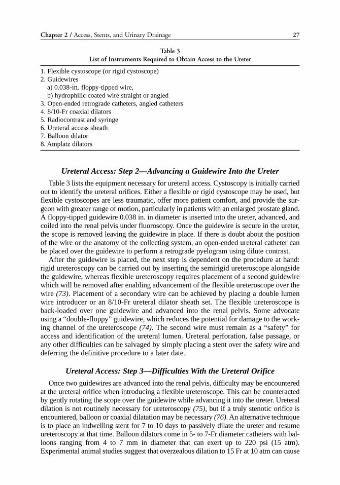
Table 3 lists the equipment necessary for ureteral access. Cystoscopy is initially carriedout to identify the ureteral orifices. Either a flexible or rigid cystoscope may be used, butflexible cystoscopes are less traumatic, offer more patient comfort, and provide the sur-geon with greater range of motion, particularly in patients with an enlarged prostate gland.A floppy-tipped guidewire 0.038 in. in diameter is inserted into the ureter, advanced, andcoiled into the renal pelvis under fluoroscopy. Once the guidewire is secure in the ureter,the scope is removed leaving the guidewire in place. If there is doubt about the positionof the wire or the anatomy of the collecting system, an open-ended ureteral catheter canbe placed over the guidewire to perform a retrograde pyelogram using dilute contrast.
After the guidewire is placed, the next step is dependent on the procedure at hand:rigid ureteroscopy can be carried out by inserting the semirigid ureteroscope alongsidethe guidewire, whereas flexible ureteroscopy requires placement of a second guidewirewhich will be removed after enabling advancement of the flexible ureteroscope over thewire (73). Placement of a secondary wire can be achieved by placing a double lumenwire introducer or an 8/10-Fr ureteral dilator sheath set. The flexible ureteroscope isback-loaded over one guidewire and advanced into the renal pelvis. Some advocateusing a “double-floppy” guidewire, which reduces the potential for damage to the work-ing channel of the ureteroscope (74). The second wire must remain as a “safety” foraccess and identification of the ureteral lumen. Ureteral perforation, false passage, orany other difficulties can be salvaged by simply placing a stent over the safety wire anddeferring the definitive procedure to a later date.
Ureteral Access: Step 3—Difficulties With the Ureteral Orifice
Once two guidewires are advanced into the renal pelvis, difficulty may be encounteredat the ureteral orifice when introducing a flexible ureteroscope. This can be counteractedby gently rotating the scope over the guidewire while advancing it into the ureter. Ureteraldilation is not routinely necessary for ureteroscopy (75), but if a truly stenotic orifice isencountered, balloon or coaxial dilatation may be necessary (76). An alternative techniqueis to place an indwelling stent for 7 to 10 days to passively dilate the ureter and resumeureteroscopy at that time. Balloon dilators come in 5- to 7-Fr diameter catheters with bal-loons ranging from 4 to 7 mm in diameter that can exert up to 220 psi (15 atm).Experimental animal studies suggest that overzealous dilation to 15 Fr at 10 atm can cause
Table 3List of Instruments Required to Obtain Access to the Ureter
1. Flexible cystoscope (or rigid cystoscope)2. Guidewires
a) 0.038-in. floppy-tipped wire,b) hydrophilic coated wire straight or angled
3. Open-ended retrograde catheters, angled catheters4. 8/10-Fr coaxial dilators5. Radiocontrast and syringe6. Ureteral access sheath 7. Balloon dilator 8. Amplatz dilators
02_Denstedt 6/22/05 4:53 PM Page 27
ureteral aperistalsis, vesicoureteric reflux, increased pressure and hydronephrosis proxi-mal to the area of dilation, and diminished ureteral contractility (77,78). Only after 6 to 7weeks of dilatation did the ureteral physiology and histology return to normal in these ani-mals (77–79). The safety and efficacy of balloon dilators in ureteroscopy have been con-firmed in humans and are in routine use (80,81). Sequential polyethylene coaxial dilatorsrange from 6 Fr and up and are more cost effective than balloon dilators (82). Care mustbe taken not to damage the urethra, ureteral orifice, or ureteral lumen. Applying the cor-rect amount of tension to the guidewire while advancing the dilators will reduce the
28 Chew and Denstedt
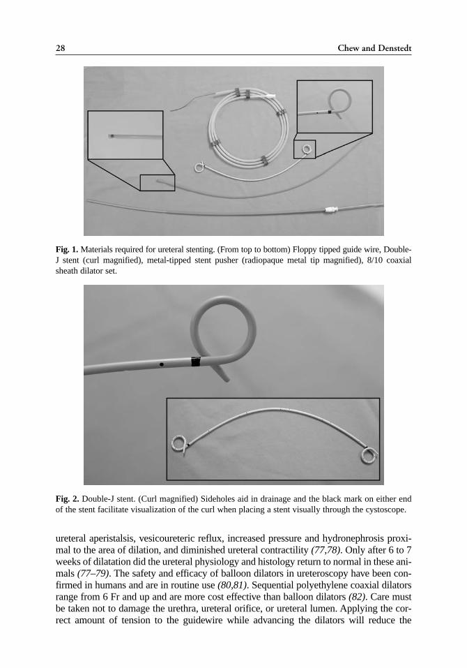
Fig. 1. Materials required for ureteral stenting. (From top to bottom) Floppy tipped guide wire, Double-J stent (curl magnified), metal-tipped stent pusher (radiopaque metal tip magnified), 8/10 coaxialsheath dilator set.
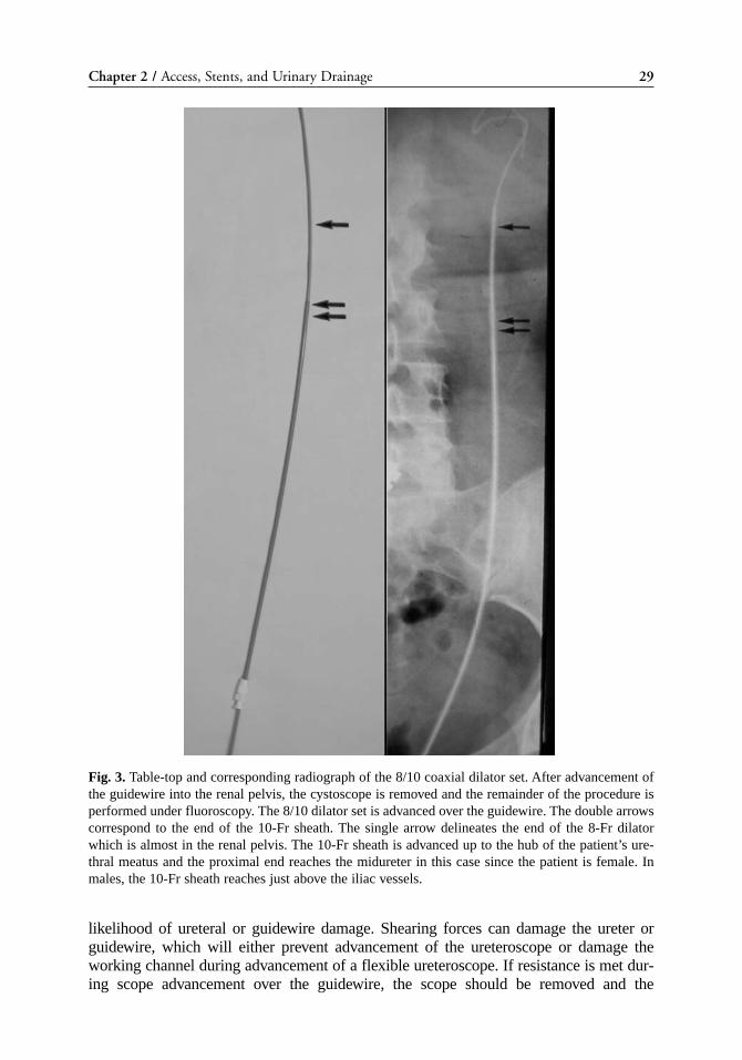
Fig. 2. Double-J stent. (Curl magnified) Sideholes aid in drainage and the black mark on either endof the stent facilitate visualization of the curl when placing a stent visually through the cystoscope.
02_Denstedt 6/22/05 4:53 PM Page 28
Chapter 2 / Access, Stents, and Urinary Drainage 29
likelihood of ureteral or guidewire damage. Shearing forces can damage the ureter orguidewire, which will either prevent advancement of the ureteroscope or damage theworking channel during advancement of a flexible ureteroscope. If resistance is met dur-ing scope advancement over the guidewire, the scope should be removed and the
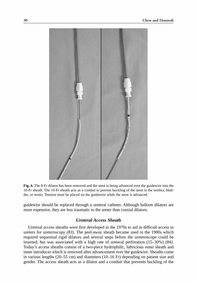
Fig. 3. Table-top and corresponding radiograph of the 8/10 coaxial dilator set. After advancement ofthe guidewire into the renal pelvis, the cystoscope is removed and the remainder of the procedure isperformed under fluoroscopy. The 8/10 dilator set is advanced over the guidewire. The double arrowscorrespond to the end of the 10-Fr sheath. The single arrow delineates the end of the 8-Fr dilatorwhich is almost in the renal pelvis. The 10-Fr sheath is advanced up to the hub of the patient’s ure-thral meatus and the proximal end reaches the midureter in this case since the patient is female. Inmales, the 10-Fr sheath reaches just above the iliac vessels.
02_Denstedt 6/22/05 4:53 PM Page 29
guidewire should be replaced through a ureteral catheter. Although balloon dilators aremore expensive, they are less traumatic to the ureter than coaxial dilators.
Ureteral Access Sheath
Ureteral access sheaths were first developed in the 1970s to aid in difficult access toureters for ureteroscopy (83). The peel-away sheath became used in the 1980s whichrequired sequential rigid dilators and several steps before the ureteroscope could beinserted, but was associated with a high rate of ureteral perforation (15–30%) (84).Today’s access sheaths consist of a two-piece hydrophilic, lubricious outer sheath andinner introducer which is removed after advancement over the guidewire. Sheaths comein various lengths (20–55 cm) and diameters (10–16 Fr) depending on patient size andgender. The access sheath acts as a dilator and a conduit that prevents buckling of the
30 Chew and Denstedt
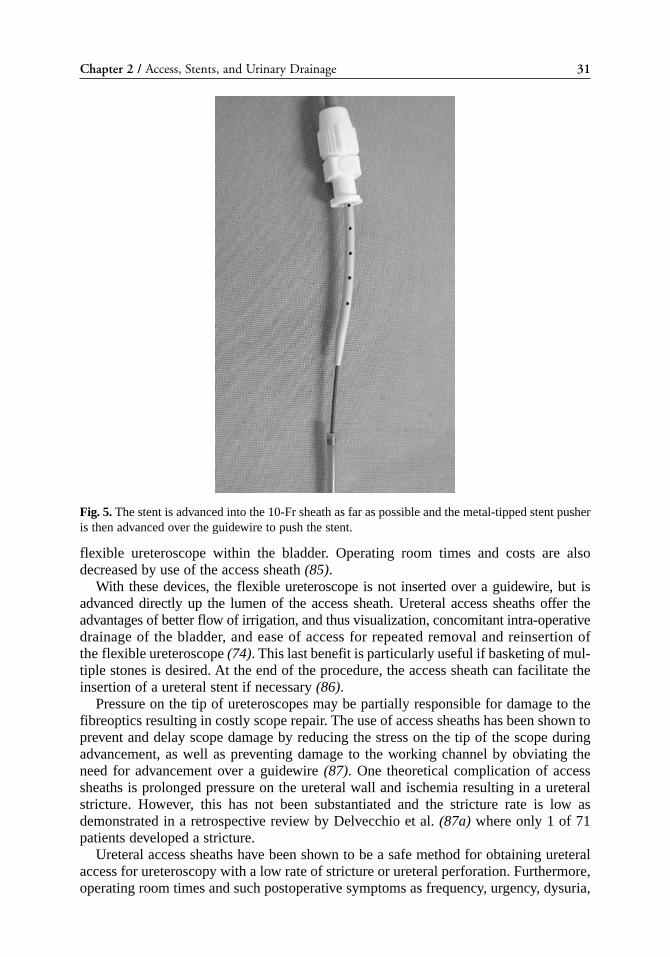
Fig. 4. The 8-Fr dilator has been removed and the stent is being advanced over the guidewire into the10-Fr sheath. The 10-Fr sheath acts as a conduit to prevent buckling of the stent in the urethra, blad-der, or ureter. Tension must be placed on the guidewire while the stent is advanced.
02_Denstedt 6/22/05 4:53 PM Page 30
Chapter 2 / Access, Stents, and Urinary Drainage 31
flexible ureteroscope within the bladder. Operating room times and costs are alsodecreased by use of the access sheath (85).
With these devices, the flexible ureteroscope is not inserted over a guidewire, but isadvanced directly up the lumen of the access sheath. Ureteral access sheaths offer theadvantages of better flow of irrigation, and thus visualization, concomitant intra-operativedrainage of the bladder, and ease of access for repeated removal and reinsertion ofthe flexible ureteroscope (74). This last benefit is particularly useful if basketing of mul-tiple stones is desired. At the end of the procedure, the access sheath can facilitate theinsertion of a ureteral stent if necessary (86).
Pressure on the tip of ureteroscopes may be partially responsible for damage to thefibreoptics resulting in costly scope repair. The use of access sheaths has been shown toprevent and delay scope damage by reducing the stress on the tip of the scope duringadvancement, as well as preventing damage to the working channel by obviating theneed for advancement over a guidewire (87). One theoretical complication of accesssheaths is prolonged pressure on the ureteral wall and ischemia resulting in a ureteralstricture. However, this has not been substantiated and the stricture rate is low asdemonstrated in a retrospective review by Delvecchio et al. (87a) where only 1 of 71patients developed a stricture.
Ureteral access sheaths have been shown to be a safe method for obtaining ureteralaccess for ureteroscopy with a low rate of stricture or ureteral perforation. Furthermore,operating room times and such postoperative symptoms as frequency, urgency, dysuria,
Fig. 5. The stent is advanced into the 10-Fr sheath as far as possible and the metal-tipped stent pusheris then advanced over the guidewire to push the stent.
02_Denstedt 6/22/05 4:53 PM Page 31
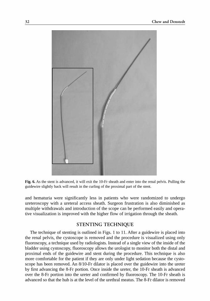
and hematuria were significantly less in patients who were randomized to undergoureteroscopy with a ureteral access sheath. Surgeon frustration is also diminished asmultiple withdrawals and introduction of the scope can be performed easily and opera-tive visualization is improved with the higher flow of irrigation through the sheath.
STENTING TECHNIQUE
The technique of stenting is outlined in Figs. 1 to 11. After a guidewire is placed intothe renal pelvis, the cystoscope is removed and the procedure is visualized using onlyfluoroscopy, a technique used by radiologists. Instead of a single view of the inside of thebladder using cystoscopy, fluoroscopy allows the urologist to monitor both the distal andproximal ends of the guidewire and stent during the procedure. This technique is alsomore comfortable for the patient if they are only under light sedation because the cysto-scope has been removed. An 8/10-Fr dilator is placed over the guidewire into the ureterby first advancing the 8-Fr portion. Once inside the ureter, the 10-Fr sheath is advancedover the 8-Fr portion into the ureter and confirmed by fluoroscopy. The 10-Fr sheath isadvanced so that the hub is at the level of the urethral meatus. The 8-Fr dilator is removed
32 Chew and Denstedt
Fig. 6. As the stent is advanced, it will exit the 10-Fr sheath and enter into the renal pelvis. Pulling theguidewire slightly back will result in the curling of the proximal part of the stent.
02_Denstedt 6/22/05 4:53 PM Page 32
Chapter 2 / Access, Stents, and Urinary Drainage 33
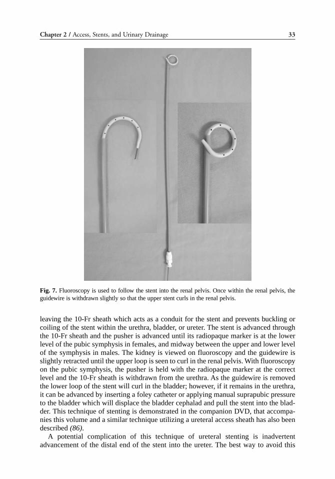
leaving the 10-Fr sheath which acts as a conduit for the stent and prevents buckling orcoiling of the stent within the urethra, bladder, or ureter. The stent is advanced throughthe 10-Fr sheath and the pusher is advanced until its radiopaque marker is at the lowerlevel of the pubic symphysis in females, and midway between the upper and lower levelof the symphysis in males. The kidney is viewed on fluoroscopy and the guidewire isslightly retracted until the upper loop is seen to curl in the renal pelvis. With fluoroscopyon the pubic symphysis, the pusher is held with the radiopaque marker at the correctlevel and the 10-Fr sheath is withdrawn from the urethra. As the guidewire is removedthe lower loop of the stent will curl in the bladder; however, if it remains in the urethra,it can be advanced by inserting a foley catheter or applying manual suprapubic pressureto the bladder which will displace the bladder cephalad and pull the stent into the blad-der. This technique of stenting is demonstrated in the companion DVD, that accompa-nies this volume and a similar technique utilizing a ureteral access sheath has also beendescribed (86).
A potential complication of this technique of ureteral stenting is inadvertentadvancement of the distal end of the stent into the ureter. The best way to avoid this
Fig. 7. Fluoroscopy is used to follow the stent into the renal pelvis. Once within the renal pelvis, theguidewire is withdrawn slightly so that the upper stent curls in the renal pelvis.
02_Denstedt 6/22/05 4:53 PM Page 33
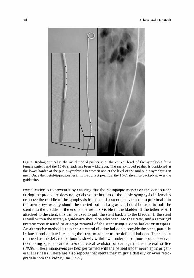
34 Chew and Denstedt
complication is to prevent it by ensuring that the radiopaque marker on the stent pusherduring the procedure does not go above the bottom of the pubic symphysis in femalesor above the middle of the symphysis in males. If a stent is advanced too proximal intothe ureter, cystoscopy should be carried out and a grasper should be used to pull thestent into the bladder if the end of the stent is visible in the bladder. If the tether is stillattached to the stent, this can be used to pull the stent back into the bladder. If the stentis well within the ureter, a guidewire should be advanced into the ureter, and a semirigidureteroscope inserted to attempt removal of the stent using a stone basket or graspers.An alternative method is to place a ureteral dilating balloon alongside the stent, partiallyinflate it and deflate it causing the stent to adhere to the deflated balloon. The stent isremoved as the deflated balloon is slowly withdrawn under close fluoroscopic observa-tion taking special care to avoid ureteral avulsion or damage to the ureteral orifice(88,89). These maneuvers are best performed with the patient under neuroleptic or gen-eral anesthesia. There are also reports that stents may migrate distally or even retro-gradely into the kidney (88,90,91).
Fig. 8. Radiographically, the metal-tipped pusher is at the correct level of the symphysis for afemale patient and the 10-Fr sheath has been withdrawn. The metal-tipped pusher is positioned atthe lower border of the pubic symphysis in women and at the level of the mid pubic symphysis inmen. Once the metal-tipped pusher is in the correct position, the 10-Fr sheath is backed-up over theguidewire.
02_Denstedt 6/22/05 4:53 PM Page 34
Chapter 2 / Access, Stents, and Urinary Drainage 35
STENT COMFORT, INFECTION, AND ENCRUSTATION: THE ROLE OF NEW BIOMATERIALS AND COATINGS
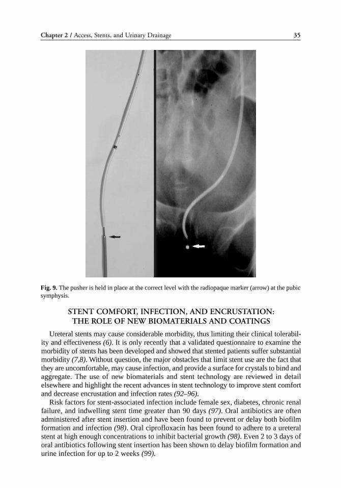
Ureteral stents may cause considerable morbidity, thus limiting their clinical tolerabil-ity and effectiveness (6). It is only recently that a validated questionnaire to examine themorbidity of stents has been developed and showed that stented patients suffer substantialmorbidity (7,8). Without question, the major obstacles that limit stent use are the fact thatthey are uncomfortable, may cause infection, and provide a surface for crystals to bind andaggregate. The use of new biomaterials and stent technology are reviewed in detailelsewhere and highlight the recent advances in stent technology to improve stent comfortand decrease encrustation and infection rates (92–96).
Risk factors for stent-associated infection include female sex, diabetes, chronic renalfailure, and indwelling stent time greater than 90 days (97). Oral antibiotics are oftenadministered after stent insertion and have been found to prevent or delay both biofilmformation and infection (98). Oral ciprofloxacin has been found to adhere to a ureteralstent at high enough concentrations to inhibit bacterial growth (98). Even 2 to 3 days oforal antibiotics following stent insertion has been shown to delay biofilm formation andurine infection for up to 2 weeks (99).
Fig. 9. The pusher is held in place at the correct level with the radiopaque marker (arrow) at the pubicsymphysis.
02_Denstedt 6/22/05 4:53 PM Page 35
In an effort to improve comfort, prevent short-term postoperative ureteral edema andpreclude cystoscopic stent removal, temporary ureteral drainage stents have beendeveloped and shown to have little or no inflammation on porcine ureters (100). Novelstent coatings, such as the enzyme oxalate decarboxylase, which breaks down oxalate,have been shown to decrease encrustation in an animal model (101), whereas silvercoatings have been shown to decrease bacterial adherence (102). Other agents, such asintravesical anti-inflammatories, have also been employed to decrease stent symptomsand may prove to be a useful stent coating (103).
A potential advance in stent technology utilizes metal in the stent material resultingin a crush resistant stent (67–70). It has been used almost exclusively in malignantureteral obstruction because of its rigidity and crush resistance. The clinical and animaltrials utilizing the metal stent all point to stent failure secondary to lumen narrowingfrom tissue hyperplasia (68). In addition, the surface of the stent is vulnerable to biofilmformation, as well as encrustation leading to infection and possible difficulty removingthe stent (68). Metal stents should be used sparingly and perhaps only in patients whohave not tolerated regular double-J stents. Further development of more rigid, uncom-pressible stents may make this modality more effective in the future.
TIPS AND TRICKS
During stenting or ureteroscopy, a large or impacted stone can often impede passageof the guidewire into the renal pelvis. Table 4 outlines a treatment algorithm for advancing
36 Chew and Denstedt
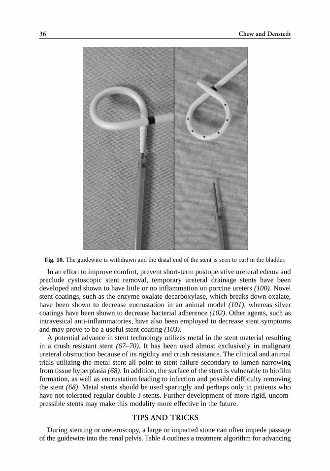
Fig. 10. The guidewire is withdrawn and the distal end of the stent is seen to curl in the bladder.
02_Denstedt 6/22/05 4:53 PM Page 36
Chapter 2 / Access, Stents, and Urinary Drainage 37
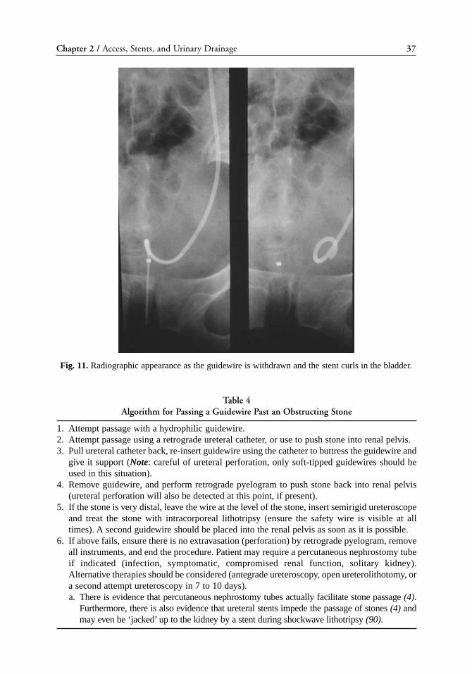
Fig. 11. Radiographic appearance as the guidewire is withdrawn and the stent curls in the bladder.
Table 4Algorithm for Passing a Guidewire Past an Obstructing Stone
1. Attempt passage with a hydrophilic guidewire. 2. Attempt passage using a retrograde ureteral catheter, or use to push stone into renal pelvis.3. Pull ureteral catheter back, re-insert guidewire using the catheter to buttress the guidewire and
give it support (Note: careful of ureteral perforation, only soft-tipped guidewires should beused in this situation).
4. Remove guidewire, and perform retrograde pyelogram to push stone back into renal pelvis(ureteral perforation will also be detected at this point, if present).
5. If the stone is very distal, leave the wire at the level of the stone, insert semirigid ureteroscopeand treat the stone with intracorporeal lithotripsy (ensure the safety wire is visible at alltimes). A second guidewire should be placed into the renal pelvis as soon as it is possible.
6. If above fails, ensure there is no extravasation (perforation) by retrograde pyelogram, removeall instruments, and end the procedure. Patient may require a percutaneous nephrostomy tubeif indicated (infection, symptomatic, compromised renal function, solitary kidney).Alternative therapies should be considered (antegrade ureteroscopy, open ureterolithotomy, ora second attempt ureteroscopy in 7 to 10 days).a. There is evidence that percutaneous nephrostomy tubes actually facilitate stone passage (4).
Furthermore, there is also evidence that ureteral stents impede the passage of stones (4) andmay even be ‘jacked’ up to the kidney by a stent during shockwave lithotripsy (90).
02_Denstedt 6/22/05 4:53 PM Page 37
38 Chew and Denstedt
a guidewire past an obstructing stone. If a regular floppy tipped guidewire cannot beinserted, a ureteral catheter can be used to exchange it for a hydrophilic guidewire. Thehydrophilic property of these wires reduces friction and allows them to slide betweenthe stone and the ureteral lumen. Hydrophilic guidewires come in both angled andstraight tips; the angled tips are often easier to manipulate around the stone. If this isunsuccessful, the next step is to reinsert the ureteral catheter over the wire and attemptto advance this past the stone. Owing to its blunt tip and greater rigidity, the ureteralcatheter will slide past the stone or push it up into the renal pelvis where it can be easilytreated. The ureteral catheter can also be used to perform a retrograde pyelogram whichmay propel the stone backwards into the renal pelvis. The retrograde pyelogram willalso detect any extravasation of contrast which indicates a ureteral perforation, a poten-tial risk in inflamed ureters with impacted stones.
When faced with difficulty advancing the guidewire past a distal ureteral stone, analternative is to leave the guidewire at the level of the stone, insert the semirigidureteroscope and treat the stone. The guidewire should remain visible at all times andas soon as it is possible, the guidewire should be advanced into the renal pelvisbeyond the obstructing lesion. The last resort is to remove all instruments and aban-don the surgical procedure, particularly if ureteral perforation has occurred. Patientswill usually require urinary drainage via a percutaneous nephrostomy if there is infec-tion, symptomatic pain, compromised renal function, solitary kidney, or ureteral per-foration. Rarely, patients may be treated conservatively and alternative methods, suchas SWL, open ureterolithotomy, or a second attempt at ureteroscopy in 7 to 14 daysmay be considered.
CONCLUSION
Retrograde access to the urinary system is the first step in many endourologic proce-dures and all urologists should be adept at dealing with the nuances of achieving access.Ureteral stents are a vital part of the urological armamentarium and play a role in thetreatment of stones, reconstructive urology, ureteropelvic junction obstruction,hydronephrosis of pregnancy, and ureteral obstruction. Development of novel ureteralstent coatings, new stent materials, and compounds loaded directly into the stent shouldimprove patient comfort and reduce the risks of infection and stent encrustation.
REFERENCES1. Camunez F, Echenagusia A, Prieto ML, Salom P, Herranz F, Hernandez C. Percutaneous nephros-
tomy in pyonephrosis. Urol Radiol 1989; 11(2): 77–81.2. Pearle MS, Pierce HL, Miller GL, et al. Optimal method of urgent decompression of the collecting
system for obstruction and infection due to ureteral calculi. J Urol 1998; 160(4): 1260–1264.3. Mokhmalji H, Braun PM, Martinez Portillo FJ, Siegsmund M, Alken P, Kohrmann KU. Percutaneous
nephrostomy versus ureteral stents for diversion of hydronephrosis caused by stones: a prospective,randomized clinical trial. J Urol 2001; 165(4): 1088–1092.
4. Lennon GM, Thornhill JA, Grainger R, McDermott TE, Butler MR. Double pigtail ureteric stent ver-sus percutaneous nephrostomy: effects on stone transit and ureteric motility. Eur Urol 1997; 31(1):24–29.
5. Leventhal EK, Rozanski TA, Crain TW, Deshon GE, Jr. Indwelling ureteral stents as definitive ther-apy for distal ureteral calculi. J Urol 1995; 153(1): 34–36.
6. Joshi HB, Okeke A, Newns N, Keeley FX, Jr., Timoney AG. Characterization of urinary symptomsin patients with ureteral stents. Urology 2002; 59(4): 511–516.
02_Denstedt 6/22/05 4:53 PM Page 38
Chapter 2 / Access, Stents, and Urinary Drainage 39
7. Joshi HB, Stainthorpe A, MacDonagh RP, Keeley FX, Jr., Timoney AG, Barry MJ. Indwelling ureteralstents: evaluation of symptoms, quality of life and utility. J Urol 2003; 169(3): 1065–1069.
8. Joshi HB, Newns N, Stainthorpe A, MacDonagh RP, Keeley FX, Jr., Timoney AG. Ureteral stentsymptom questionnaire: development and validation of a multidimensional quality of life measure.J Urol 2003; 169(3): 1060–1064.
9. Pryor JL, Jenkins AD. Use of double-pigtail stents in extracorporeal shock wave lithotripsy. J Urol1990; 143(3): 475–478.
10. Ryan PC, Lennon GM, McLean PA, Fitzpatrick JM. The effects of acute and chronic JJ stent place-ment on upper urinary tract motility and calculus transit. Br J Urol 1994; 74(4): 434–439.
11. Sayed MA, el Taher AM, Aboul-Ella HA, Shaker SE. Steinstrasse after extracorporeal shockwavelithotripsy: aetiology, prevention and management. BJU Int 2001; 88(7): 675–678.
12. Fedullo LM, Pollack HM, Banner MP, Amendola MA, Van Arsdalen KN. The development of stein-strassen after ESWL: frequency, natural history, and radiologic management. AJR Am J Roentgenol1988; 151(6): 1145–1147.
13. Libby JM, Meacham RB, Griffith DP. The role of silicone ureteral stents in extracorporeal shockwave lithotripsy of large renal calculi. J Urol 1988; 139(1): 15–17.
14. Al Awadi KA, Abdul HH, Kehinde EO, Al Tawheed A. Steinstrasse: a comparison of incidence withand without J stenting and the effect of J stenting on subsequent management. BJU Int 1999; 84(6):618–621.
15. Sulaiman MN, Buchholz NP, Clark PB. The role of ureteral stent placement in the prevention ofSteinstrasse. J Endourol 1999; 13(3): 151–155.
16. Sheir KZ, Madbouly K, Elsobky E. Prospective randomized comparative study of the effectivenessand safety of electrohydraulic and electromagnetic extracorporeal shock wave lithotriptors. J Urol2003; 170(2 Pt 1): 389–392.
17. Preminger GM, Kettelhut MC, Elkins SL, Seger J, Fetner CD. Ureteral stenting during extracorpo-real shock wave lithotripsy: help or hindrance? J Urol 1989; 142(1): 32–36.
18. Bierkens AF, Hendrikx AJ, Lemmens WA, Debruyne FM. Extracorporeal shock wave lithotripsy forlarge renal calculi: the role of ureteral stents. A randomized trial. J Urol 1991; 145(4): 699–702.
19. Kirkali Z, Esen AA, Akan G. Place of double-J stents in extracorporeal shock wave lithotripsy. EurUrol 1993; 23(4): 460–462.
20. Chandhoke PS, Barqawi AZ, Wernecke C, Chee-Awai RA. A randomized outcomes trial of ureteralstents for extracorporeal shock wave lithotripsy of solitary kidney or proximal ureteral stones. J Urol2002; 167(5): 1981–1983.
21. Madbouly K, Sheir KZ, Elsobky E, Eraky I, Kenawy M. Risk factors for the formation of a stein-strasse after extracorporeal shock wave lithotripsy: a statistical model. J Urol 2002; 167(3):1239–1242.
22. Eisenberger F, Rassweiler J. Stone Therapy in Urology. Thieme Medical Publisher, New York, 1991: 29.23. Hosking DH, McColm SE, Smith WE. Is stenting following ureteroscopy for removal of distal
ureteral calculi necessary? J Urol 1999; 161(1): 48–50.24. Denstedt JD, Wollin TA, Sofer M, Nott L, Weir M, Honey D’Art. A prospective randomized controlled
trial comparing nonstented versus stented ureteroscopic lithotripsy. J Urol 2001; 165(5): 1419–1422.25. Wollin TA, Denstedt JD. The holmium laser in urology. J Clin Laser Med Surg 1998; 16(1): 13–20.26. Chen YT, Chen J, Wong WY, Yang SS, Hsieh CH, Wang CC. Is ureteral stenting necessary after
uncomplicated ureteroscopic lithotripsy? A prospective, randomized controlled trial. J Urol 2002;167(5): 1977–1980.
27. Hollenbeck BK, Schuster TG, Faerber GJ, Wolf JS, Jr. Routine placement of ureteral stents is unnec-essary after ureteroscopy for urinary calculi. Urology 2001; 57(4): 639–643.
28. Byrne RR, Auge BK, Kourambas J, Munver R, Delvecchio F, Preminger GM. Routine ureteral stent-ing is not necessary after ureteroscopy and ureteropyeloscopy: a randomized trial. J Endourol 2002;16(1): 9–13.
29. Borboroglu PG, Amling CL, Schenkman NS, et al. Ureteral stenting after ureteroscopy for distalureteral calculi: a multi-institutional prospective randomized controlled study assessing pain, out-comes and complications. J Urol 2001; 166(5): 1651–1657.
29a. Netto NR, Jr., Ikonomidis J, Zillo C. Routine ureteral stenting after ureteroscopy for ureteral lithia-sis: is it really necessary? J Urol 2001; 166(4): 1252–1254.
02_Denstedt 6/22/05 4:53 PM Page 39
40 Chew and Denstedt
30. Puskar D, Balagovic I, Filipovic A, et al. Symptomatic physiologic hydronephrosis in pregnancy:incidence, complications and treatment. Eur Urol 2001; 39(3): 260–263.
31. Zwergel T, Lindenmeir T, Wullich B. Management of acute hydronephrosis in pregnancy by ureteralstenting. Eur Urol 1996; 29(3): 292–297.
32. Fainaru O, Almog B, Gamzu R, Lessing JB, Kupferminc M. The management of symptomatichydronephrosis in pregnancy. BJOG: 2002; 109(12): 1385–1387.
33. Wolf MC, Hollander JB, Salisz JA, Kearney DJ. A new technique for ureteral stent placement duringpregnancy using endoluminal ultrasound. Surg Gynecol Obstet 1992; 175(6): 575–576.
34. Fabrizio MD, Gray DS, Feld RI, Bagley DH. Placement of ureteral stents in pregnancy using ultra-sound guidance. Tech Urol 1996; 2(3): 121–125.
35. Sahin H, Bircan MK, Yayla M. JJS application in acute symptomatic hydronephrosis in pregnancy.BJOG 1997; 59(2): 141–142.
36. Delakas D, Karyotis I, Loumbakis P, Daskalopoulos G, Kazanis J, Cranidis A. Ureteral drainage bydouble-J-catheters during pregnancy. Clin Exp Obstet Gynecol 2000; 27(3-4): 200–202.
37. Hellawell GO, Cowan NC, Holt SJ, Mutch SJ. A radiation perspective for treating loin pain in preg-nancy by double-pigtail stents. BJU Int 2002; 90(9): 801–808.
38. Watterson JD, Girvan AR, Beiko DT, et al. Ureteroscopy and holmium:YAG laser lithotripsy: anemerging definitive management strategy for symptomatic ureteral calculi in pregnancy. Urology2002; 60(3): 383–387.
39. Roy SC, Sandison GA. Shielding for neutron scattered dose to the fetus in patients treated with 18MV X-ray beams. Med Phys 2000; 27(8): 1800–1803.
40. Gorton E, Whitfield HN. Renal calculi in pregnancy. Br J Urol 1997; 80 (Suppl 1): 4–9.41. Rodriguez PN, Klein AS. Management of urolithiasis during pregnancy. Surg Gynecol Obstet 1988;
166(2): 103–106.42. Hendricks SK, Ross SO, Krieger JN. An algorithm for diagnosis and therapy of management and
complications of urolithiasis during pregnancy. Surg, Gynecol Obstet 1991; 172(1): 49–54.43. Stothers L, Lee LM. Renal colic in pregnancy. J Urol 1992; 148(5): 1383–1387.44. Evans HJ, Wollin TA. The management of urinary calculi in pregnancy. Curr Opin Urol 2001; 11(4):
379–384.45. Goldfarb RA, Neerhut GJ, Lederer E. Management of acute hydronephrosis of pregnancy by ureteral
stenting: risk of stone formation. J Urol 1989; 141(4): 921–922.46. Denstedt JD, Razvi H. Management of urinary calculi during pregnancy. J Urol 1992; 148(3 Pt 2):
1072–1074.47. Kavoussi LR, Albala DM, Basler JW, Apte S, Clayman RV. Percutaneous management of urolithia-
sis during pregnancy. J Urol 1992; 148(3 Pt 2): 1069–1071.48. Razvi HA, Denstedt JD. Endoscopic management of ureteral injury after cesarean section. J Endourol
1994; 8(5): 345–347.49. Lifshitz DA, Lingeman JE. Ureteroscopy as a first-line intervention for ureteral calculi in pregnancy.
J Endourol 2002; 16(1): 19–22.50. Watterson JD, Girvan AR, Beiko DT, et al. Ureteroscopy and holmium:YAG laser lithotripsy: an
emerging definitive management strategy for symptomatic ureteral calculi in pregnancy. Urology2002; 60(3): 383–387.
51. Wickham JE, Kellet MJ. Percutaneous pyelolysis. Eur Urol 1983; 9(2): 122–124.52. Danuser H, Hochreiter WW, Ackermann DK, Studer UE. Influence of stent size on the success of
antegrade endopyelotomy for primary ureteropelvic junction obstruction: results of 2 consecutiveseries. J Urol 2001; 166(3): 902–909.
53. Moon YT, Kerbl K, Pearle MS, et al. Evaluation of optimal stent size after endourologic incision ofureteral strictures. J Endourol 1995; 9(1): 15–22.
54. Anidjar M, Meria P, Cochand-Priollet B, et al. Evaluation of optimal stent size after antegradeendopyelotomy: an experimental study in the porcine model. Eur Urol 1997; 32(2): 245–252.
55. Kumar R, Kapoor R, Mandhani A, Kumar A, Ahlawat R. Optimum duration of splinting after endopy-elotomy. J Endourol 1999; 13(2): 89–92.
56. Mandhani A, Kapoor R, Zaman W, Kumar A, Bhandari M, Gambhir S. Is a 2-weeks duration suffi-cient for stenting in endopyelotomy? J Urol 2003; 169(3): 886–889.
57. Kerbl K, Chandhoke PS, Figenshau RS, Stone AM, Clayman RV. Effect of stent duration on ureteralhealing following endoureterotomy in an animal model. J Urol 1993; 150(4): 1302–1305.
02_Denstedt 6/22/05 4:53 PM Page 40
Chapter 2 / Access, Stents, and Urinary Drainage 41
58. Park DS, Park JH, Lee YT. Percutaneous nephrostomy versus indwelling ureteral stents in patientswith bilateral nongenitourinary malignant extrinsic obstruction. J Endourol 2002; 16(3): 153–154.
59. Chye RW, Lickiss JN. Palliative care in bilateral malignant ureteric obstruction. Ann Acad MedSingapore 1994; 23(2): 197–203.
60. Emmert C, Rassler J, Kohler U. Survival and quality of life after percutaneous nephrostomy for malignantureteric obstruction in patients with terminal cervical cancer. Arch Gynecol Obstet 1997; 259(3): 147–151.
61. Pappas P, Stravodimos KG, Mitropoulos D, et al. Role of percutaneous urinary diversion in malignantand benign obstructive uropathy. J Endourol 2000; 14(5): 401–405.
62. Kinn AC, Lykkeskov-Andersen H. Impact on ureteral peristalsis in a stented ureter. An experimentalstudy in the pig. Urol Res 2002; 30(4): 213–218.
63. Yossepowitch O, Lifshitz DA, Dekel Y, et al. Predicting the success of retrograde stenting for manag-ing ureteral obstruction. J Urol 2001; 166(5): 1746–1749.
64. Jabbour ME, Desgrandchamps F, Angelescu E, Teillac P, Le Duc A. Percutaneous implantation ofsubcutaneous prosthetic ureters: long-term outcome. J Endourol 2001; 15(6): 611–614.
65. Minhas S, Irving HC, Lloyd SN, Eardley I, Browning AJ, Joyce AD. Extra-anatomic stents in uretericobstruction: experience and complications. BJU Int 1999; 84(7): 762–764.
66. Nissenkorn I, Gdor Y. Nephrovesical subcutaneous stent: an alternative to permanent nephrostomy.J Urol 2000; 163(2): 528–530.
67. Barbalias GA, Liatsikos EN, Kalogeropoulou C, Karnabatidis D, Siablis D. Metallic stents in gyne-cologic cancer: an approach to treat extrinsic ureteral obstruction. Eur Urol 2000; 38(1): 35–40.
68. Hekimoglu B, Men S, Pinar A, I et al. Urothelial hyperplasia complicating use of metal stents inmalignant ureteral obstruction. Eur Radiol 1996; 6(5): 675–681.
69. Lopez-Martinez RA, Singireddy S, Lang EK. The use of metallic stents to bypass ureteral stricturessecondary to metastatic prostate cancer: experience with 8 patients. J Urol 1997; 158(1): 50–53.
70. Pauer W, Lugmayr H. Metallic Wallstents: a new therapy for extrinsic ureteral obstruction. J Urol1992; 148(2 Pt 1): 281–284.
71. Little B, Ho KJ, Gawley S, Young M. Use of nephrostomy tubes in ureteric obstruction from incur-able malignancy. Int J Clin Pract 2003; 57(3): 180–181.
72. Gotman I. Characteristics of metals used in implants. J Endourol 1997; 11(6): 383–389.73. Kumar PV, Keeley FX, Timoney AG. Safe flexible ureterorenoscopy with a dual-lumen access
catheter and a safety guidewire. BJU Int 2001; 88(6): 638–639.74. Monga M, Bhayani S, Landman J, Conradie M, Sundaram CP, Clayman RV. Ureteral access for upper
urinary tract disease: the access sheath. J Endourol 2001; 15(8): 831–834.75. Stoller ML, Wolf JS, Jr., Hofmann R, Marc B. Ureteroscopy without routine balloon dilation: an out-
come assessment. J Urol 1992; 147(5): 1238–1242.76. Rodrigues NN, Jr., Caserta LG, Levi D’Ancona CA, Ikari O, Ferreira U, Francisco de Almeida CJ. Is
routine dilation of the ureter necessary for ureteroscopy? Eur Urol 1990; 17(4): 269–272.77. Boddy SA, Nimmon CC, Jones S, et al. Acute ureteric dilatation for ureteroscopy. An experimental
study. Br J Urol 1988; 61(1): 27–31.78. Lennon GM, Fitzpatrick JM. Effects of balloon dilatation on canine ureteric physiology. Eur Urol
1994; 25(3): 248–253.79. Begin LR, Selmy GI, Hassouna MM, Khalaf IM, Elhilali MM. Healing and muscular restoration of
the ureteral wall following balloon-induced rupture: an experimental animal model with light micro-scopic and ultrastructural observations. Experimental & Molecular Pathology 1993; 59(1): 58–70.
80. Ford TF, Parkinson MC, Wickham JE. Clinical and experimental evaluation of ureteric dilatation. BrJ Urol 1984; 56(5): 460–463.
81. Huffman JL, Bagley DH. Balloon dilation of the ureter for ureteroscopy. J Urol 1988; 140(5): 954–956.82. Gaylis F, Bastuba M, Bidair M, Friedel W. Ureteral dilation using a tapered dilator: a cost-effective
approach. J Endourol 2000; 14(5): 447–449.83. Takayasu H, Aso Y. Recent development for pyeloureteroscopy: guide tube method for its introduc-
tion into the ureter. J Urol 1974; 112(2): 176–178.84. Rich M, Lee WJ, Smith AD. Applications of the peel-away introducer sheath. J Urol 1987; 137(3): 452–454.85. Kourambas J, Byrne RR, Preminger GM. Does a ureteral access sheath facilitate ureteroscopy? J Urol
2001; 165(3): 789–793.86. Wu NZ, Auge BK, Preminger GM. Simplified ureteral stent placement with the assistance of a
ureteral access sheath. J Urol 2001; 166(1): 206–208.
02_Denstedt 6/22/05 4:53 PM Page 41
42 Chew and Denstedt
87. Pietrow PK, Auge BK, Delvecchio FC, et al. Techniques to maximize flexible ureteroscope longevity.Urology 2002; 60(5): 784–788.
87a. Delvecchio FC, Auge BK, Brizuela RM, et al. Assessment of structure formation with the ureteralaccess sheath. Urology 2003; 61: 518–522.
88. Chin JL, Denstedt JD. Retrieval of proximally migrated ureteral stents. J Urol 1992; 148(4): 1205–1206.89. Ilgit ET, Akpek S, Isik S. Repositioning of a misplaced ureteral stent with a balloon catheter: techni-
cal note. Eur J Radiol 1997; 24(3): 257–259.90. Ko W, Lee W, Jung J, Lee M. Spontaneous proximal double pigtail ureteral stent migration after
shock wave lithotripsy: “jack” phenomenon. J Urol 2001; 166(4): 1387.91. Maheshwari PN, Choudhri S, Parmar VP. Re: Optimal prevention and management of proximal
ureteral stent migration and remigration. J Urol 2002; 168(1): 202.92. Denstedt JD, Wollin TA, Reid G. Biomaterials used in urology: current issues of biocompatibility,
infection, and encrustation. J Endourol 1998; 12(6): 493–500.93. Denstedt JD, Reid G, Sofer M. Advances in ureteral stent technology. World J Urol 2000; 18(4):
237–242.94. Lingeman JE, Preminger GM, Berger Y, et al. Use of a temporary ureteral drainage stent after uncom-
plicated ureteroscopy: results from a phase II clinical trial. J Urol 2003; 169(5): 1682–1688.95. Beiko DT, Knudsen BE, Denstedt JD. Advances in ureteral stent design. J Endourol 2003; 17(4):
195–199.96. Beiko DT, Knudsen BE, Watterson JD, Denstedt JD. Biomaterials in urology. Curr Urol Rep 2003; 4(1):
51–55.97. Kehinde EO, Rotimi VO, Al Awadi KA, et al. Factors predisposing to urinary tract infection after
J ureteral stent insertion. J Urol 2002; 167(3): 1334–1337.98. Reid G, Habash M, Vachon D, Denstedt J, Riddell J, Beheshti M. Oral fluoroquinolone therapy results
in drug adsorption on ureteral stents and prevention of biofilm formation. Int J Antimicrob Agents2001; 17(4): 317–319.
99. Paick SH, Park HK, Oh SJ, Kim HH. Characteristics of bacterial colonization and urinary tract infec-tion after indwelling of double-J ureteral stent. Urology 2003; 62(2): 214–217.
100. Lingeman JE, Schulsinger DA, Kuo RL. Phase I trial of a temporary ureteral drainage stent. J Endourol2003; 17(3): 169–171.
101. Watterson JD, Cadieux PA, Beiko DT, et al. Oxalate-degrading enzymes from Oxalobacter formi-genes: a novel device coating to reduce urinary tract biomaterial-related encrustation. J Endourol2003; 17(5): 269–274.
102. Multanen M, Talja M, Hallanvuo S, et al. Bacterial adherence to silver nitrate coated poly-L-lacticacid urological stents in vitro. Urol Res 2000; 28(5): 327–331.
103. Beiko DT, Watterson JD, Knudsen BE, et al. A double-blinded prospective randomized controlledtrial assessing the safety and efficacy of intravesical agents for ureteral stent symptoms followingextracorporeal shockwave lithotripsy. J Endourol 2004; 18(8): 723–730.
02_Denstedt 6/22/05 4:53 PM Page 42

![Gene Therapy Based on Fragment C of Tetanus Toxin in ALS ...€¦ · rograde transport pathway and is subsequently transported to the neuronal soma in the CNS [30,31]. Once the toxin](https://static.fdocuments.net/doc/165x107/6083cabebb99f877af114933/gene-therapy-based-on-fragment-c-of-tetanus-toxin-in-als-rograde-transport-pathway.jpg)