PprA Protein Is Involved in Chromosome Segregation via Its … · PprA Protein Is Involved in...
17
PprA Protein Is Involved in Chromosome Segregation via Its Physical and Functional Interaction with DNA Gyrase in Irradiated Deinococcus radiodurans Bacteria Alice Devigne, a Philippe Guérin, b Johnny Lisboa, a * Sophie Quevillon-Cheruel, a Jean Armengaud, b Suzanne Sommer, a Claire Bouthier de la Tour, a Pascale Servant a Institute for Integrative Biology of the Cell (I2BC), CEA, CNRS, Univ Paris-Sud, Université Paris-Saclay, Gif-sur- Yvette cedex, France a ; CEA, DSV, IBiTec-S, SPI, Li2D, Laboratory Innovative Technologies for Detection and Diagnostics, Bagnols-sur-Cèze, France b * Present address: Johnny Lisboa, Institute for Molecular and Cell Biology (IBMC), Porto, Portugal. C.B.D.L.T. and P.S. contributed equally to this work as senior authors and should be considered co-last authors. ABSTRACT PprA, a radiation-induced Deinococcus-specific protein, was previously shown to be required for cell survival and accurate chromosome segregation after exposure to ionizing radiation. Here, we used an in vivo approach to determine, by shotgun proteomics, putative PprA partners coimmunoprecipitating with PprA when cells were exposed to gamma rays. Among them, we found the two subunits of DNA gyrase and, thus, chose to focus our work on characterizing the activities of the deinococcal DNA gyrase in the presence or absence of PprA. Loss of PprA rendered cells hypersensitive to novobiocin, an inhibitor of the B subunit of DNA gyrase. We showed that treatment of bacteria with novobiocin resulted in induction of the radi- ation desiccation response (RDR) regulon and in defects in chromosome segregation that were aggravated by the absence of PprA. In vitro, the deinococcal DNA gyrase, like other bacterial DNA gyrases, possesses DNA negative supercoiling and decatena- tion activities. These two activities are inhibited in vitro by novobiocin and nalidixic acid, whereas PprA specifically stimulates the decatenation activity of DNA gyrase. Together, these results suggest that PprA plays a major role in chromosome decat- enation via its interaction with the deinococcal DNA gyrase when D. radiodurans cells are recovering from exposure to ionizing radiation. IMPORTANCE D. radiodurans is one of the most radiation-resistant organisms known. This bacterium is able to cope with high levels of DNA lesions generated by exposure to extreme doses of ionizing radiation and to reconstruct a functional ge- nome from hundreds of radiation-induced chromosomal fragments. Here, we identi- fied partners of PprA, a radiation-induced Deinococcus-specific protein, previously shown to be required for radioresistance. Our study leads to three main findings: (i) PprA interacts with DNA gyrase after irradiation, (ii) treatment of cells with novobio- cin results in defects in chromosome segregation that are aggravated by the ab- sence of PprA, and (iii) PprA stimulates the decatenation activity of DNA gyrase. Our results extend the knowledge of how D. radiodurans cells survive exposure to ex- treme doses of gamma irradiation and point out the link between DNA repair, chro- mosome segregation, and DNA gyrase activities in the radioresistant D. radiodurans bacterium. KEYWORDS: Deinococcus radiodurans, PprA, DNA gyrase, DNA decatenation Received 23 October 2015 Accepted 9 December 2015 Published 13 January 2016 Citation Devigne A, Guérin P, Lisboa J, Quevillon-Cheruel S, Armengaud J, Sommer S, Bouthier de la Tour C, Servant P. 2016. PprA protein is involved in chromosome segregation via its physical and functional interaction with DNA gyrase in irradiated Deinococcus radiodurans bacteria. mSphere 1(1):e00036-15. doi:10.1128/mSphere.00036-15. Editor Grant R. Bowman, University of Wyoming Copyright © 2016 Devigne et al. This is an open-access article distributed under the terms of the Creative Commons Attribution 4.0 International license. Address correspondence to Pascale Servant, [email protected]. RESEARCH ARTICLE Molecular Biology and Physiology crossmark Volume 1 Issue 1 e00036-15 msphere.asm.org 1 on February 14, 2019 by guest http://msphere.asm.org/ Downloaded from
Transcript of PprA Protein Is Involved in Chromosome Segregation via Its … · PprA Protein Is Involved in...

PprA Protein Is Involved in ChromosomeSegregation via Its Physical andFunctional Interaction with DNA Gyrasein Irradiated Deinococcus radioduransBacteria
Alice Devigne,a Philippe Guérin,b Johnny Lisboa,a* Sophie Quevillon-Cheruel,a
Jean Armengaud,b Suzanne Sommer,a Claire Bouthier de la Tour,a
Pascale Servanta
Institute for Integrative Biology of the Cell (I2BC), CEA, CNRS, Univ Paris-Sud, Université Paris-Saclay, Gif-sur-Yvette cedex, Francea; CEA, DSV, IBiTec-S, SPI, Li2D, Laboratory Innovative Technologies for Detection andDiagnostics, Bagnols-sur-Cèze, Franceb
* Present address: Johnny Lisboa, Institute for Molecular and Cell Biology (IBMC), Porto, Portugal.
C.B.D.L.T. and P.S. contributed equally to this work as senior authors and should be considered co-last authors.
ABSTRACT PprA, a radiation-induced Deinococcus-specific protein, was previouslyshown to be required for cell survival and accurate chromosome segregation afterexposure to ionizing radiation. Here, we used an in vivo approach to determine, byshotgun proteomics, putative PprA partners coimmunoprecipitating with PprA whencells were exposed to gamma rays. Among them, we found the two subunits ofDNA gyrase and, thus, chose to focus our work on characterizing the activities of thedeinococcal DNA gyrase in the presence or absence of PprA. Loss of PprA renderedcells hypersensitive to novobiocin, an inhibitor of the B subunit of DNA gyrase. Weshowed that treatment of bacteria with novobiocin resulted in induction of the radi-ation desiccation response (RDR) regulon and in defects in chromosome segregationthat were aggravated by the absence of PprA. In vitro, the deinococcal DNA gyrase,like other bacterial DNA gyrases, possesses DNA negative supercoiling and decatena-tion activities. These two activities are inhibited in vitro by novobiocin and nalidixicacid, whereas PprA specifically stimulates the decatenation activity of DNA gyrase.Together, these results suggest that PprA plays a major role in chromosome decat-enation via its interaction with the deinococcal DNA gyrase when D. radioduranscells are recovering from exposure to ionizing radiation.
IMPORTANCE D. radiodurans is one of the most radiation-resistant organismsknown. This bacterium is able to cope with high levels of DNA lesions generated byexposure to extreme doses of ionizing radiation and to reconstruct a functional ge-nome from hundreds of radiation-induced chromosomal fragments. Here, we identi-fied partners of PprA, a radiation-induced Deinococcus-specific protein, previouslyshown to be required for radioresistance. Our study leads to three main findings: (i)PprA interacts with DNA gyrase after irradiation, (ii) treatment of cells with novobio-cin results in defects in chromosome segregation that are aggravated by the ab-sence of PprA, and (iii) PprA stimulates the decatenation activity of DNA gyrase. Ourresults extend the knowledge of how D. radiodurans cells survive exposure to ex-treme doses of gamma irradiation and point out the link between DNA repair, chro-mosome segregation, and DNA gyrase activities in the radioresistant D. radioduransbacterium.
KEYWORDS: Deinococcus radiodurans, PprA, DNA gyrase, DNA decatenation
Received 23 October 2015 Accepted 9December 2015 Published 13 January 2016
Citation Devigne A, Guérin P, Lisboa J,Quevillon-Cheruel S, Armengaud J, Sommer S,Bouthier de la Tour C, Servant P. 2016. PprAprotein is involved in chromosome segregationvia its physical and functional interaction withDNA gyrase in irradiated Deinococcus radioduransbacteria. mSphere 1(1):e00036-15.doi:10.1128/mSphere.00036-15.
Editor Grant R. Bowman, University ofWyoming
Copyright © 2016 Devigne et al. This is anopen-access article distributed under the termsof the Creative Commons Attribution 4.0International license.
Address correspondence to Pascale Servant,[email protected].
RESEARCH ARTICLEMolecular Biology and Physiology
crossmark
Volume 1 Issue 1 e00036-15 msphere.asm.org 1
on February 14, 2019 by guest
http://msphere.asm
.org/D
ownloaded from
The bacterium Deinococcus radiodurans possesses exceptional resistance to thelethal effects of DNA-damaging agents and is able to reconstruct a functional
genome from a myriad of radiation-induced chromosomal fragments. This radioresis-tance is likely the result of a combination of different mechanisms, including protectionof proteins against oxidation, efficient DNA double-strand break repair, and a compactnucleoid structure (for reviews, see references 1 to 6). Different DNA repair pathwayshave been proposed to be involved in the reconstitution of an intact genome inD. radiodurans, including extended synthesis-dependent strand annealing (ESDSA) (7),homologous recombination (HR) (8–10), single-strand annealing (SSA) (11–13), andnonhomologous end joining (14, 15).
The pprA gene (DRA_0346) is a Deinococcus-specific gene belonging to a radiationdesiccation response (RDR) regulon comprising genes that are highly induced afterDNA damage and contain a conserved motif (RDRM) upstream from their coding region(16, 17). Recently, it was shown that DdrO acts as a repressor of the RDR regulon andthat IrrE, a metalloprotease, cleaves DdrO after irradiation, leading to transcriptionalinduction of various genes belonging to the RDR regulon (18–20). A pprA mutantexhibits high sensitivity to gamma radiation and DNA-damaging agents (14, 21, 22).In vitro, PprA preferentially binds double-stranded DNA (dsDNA) carrying strand breaks,inhibits Escherichia coli exonuclease III activity, and stimulates the DNA end-joiningreaction catalyzed by ATP-dependent DNA ligases (14). It has also been shown thatPprA polymerizes along supercoiled, nicked, circular, or linear double-stranded DNA(23). After irradiation, PprA is part of a multiprotein complex containing 24 proteins,including DNA ligases, DNA topoisomerase IB (Topo IB), SSB, and DNA polymerase I andexhibiting both DNA synthesis and DNA end-processing functions (24). We recentlyreported that repair of DNA double-strand breaks (DSB) in cells devoid of PprA andexposed to gamma radiation takes place efficiently, with a delay of approximately 1 hcompared to the time for the wild type (21). All these results suggest that PprA mightfunction as a pleiotropic protein involved in the repair of DNA DSB and other radiation-induced damage (6, 14). After irradiation, the PprA protein is recruited onto thenucleoid early and localizes later through the septum of dividing cells when DNA repairis completed (21). Untreated cells devoid of PprA display a wild-type morphology, butafter gamma irradiation, the absence of PprA leads to severe defects in DNA segrega-tion and cell division (21).
In bacteria, topoisomerases play a major role in chromosome segregation aftercompletion of DNA replication. DNA topoisomerases are enzymes that resolve thetopological transitions of DNA and are associated with replication, transcription, andrecombination (for a review, see reference 25). They are divided into two types,depending on whether they operate by cleaving one strand and passing the otherstrand through the break (type I) or by cleaving both strands and passing a DNA duplexthrough the DNA double-strand break (type II). Most bacteria possess at least threeDNA topoisomerases, one type I enzyme, DNA topoisomerase I (Topo I), encoded by thetopA gene, and two type II enzymes, DNA gyrase and DNA topoisomerase IV (Topo IV),which are heterotetramers with two different subunits, encoded by the gyrA and thegyrB genes and by the parC and parE genes, respectively. DNA topoisomerase I relaxesDNA, while DNA gyrase introduces negative DNA supercoils. These opposing activitiesallow the maintenance of DNA superhelicity in the cells. DNA topoisomerase I and DNAgyrase also act in concert to resolve topological constraints during replication andtranscription. Because of these important physiological roles, DNA topoisomerase I andDNA gyrase are essential proteins for the viability of bacterial cells (26–29). Topo IV isinvolved in decatenation of intertwined DNA intermediates generated during DNAreplication and DNA recombination (30, 31) and plays a major role in decatenation ofdaughter chromosomes before cell division (for reviews, see references 25, 32, and 33).Some bacteria, such as Escherichia coli, possess, in addition to these three topoisom-erases, a DNA topoisomerase III (Topo III), which is another type IA topoisomeraseencoded by the topB gene that might play a role in the unlinking of the DNA strandsat the end of replication (34).
Devigne et al.
Volume 1 Issue 1 e00036-15 msphere.asm.org 2
on February 14, 2019 by guest
http://msphere.asm
.org/D
ownloaded from

D. radiodurans has an atypical content of DNA topoisomerases: it possesses a DNAtopoisomerase I, encoded by the DR_1374 (topA) gene, and a DNA gyrase, encoded bythe DR_1913 (gyrA) and DR_0906 (gyrB) genes. DNA gyrase is the only type II DNAtopoisomerase in this bacterium. Indeed, the sequence of the D. radiodurans genomedoes not reveal any homolog of the parC or parE genes that encode the two subunitsof Topo IV in E. coli. Like some other bacteria, D. radiodurans also possesses anothertopoisomerase, encoded by the DR_0690 gene (topIB gene) and belonging to the typeIB family, which includes eukaryotic nuclear topoisomerases and topoisomerases IB ofpoxviruses (35). D. radiodurans DNA gyrase was shown to be one of the main proteinsinvolved in the organization of the Deinococcus nucleoids (36), but its biochemicalactivities have not been extensively characterized. Recently, Kota et al. (37) showed,using a bacterial two-hybrid system, that PprA interacts with DNA Topo IB and DNAgyrase from D. radiodurans and enhances the relaxation activity of Topo IB. They alsoshowed that ΔpprA mutant bacteria are sensitive to nalidixic acid, an inhibitor of typeII bacterial DNA topoisomerases (37).
Here, in order to decipher the role of PprA in chromosome segregation, we used anin vivo approach to determine by shotgun proteomics putative PprA partners interact-ing and coimmunoprecipitating with PprA after exposure to gamma radiation. Wefound among them the two subunits of DNA gyrase, and we decided to focus our workon characterizing the activities of the deinococcal DNA gyrase in the presence orabsence of PprA. We also showed that treatment of bacteria with novobiocin, aninhibitor of DNA gyrase, resulted in defects in chromosome segregation that wereaggravated by the absence of PprA. Our results suggest that PprA plays a major role inchromosome decatenation via its interaction with the deinococcal DNA gyrase whencells are recovering from exposure to ionizing radiation.
RESULTSShotgun MS highlights gyrase subunit A as a partner of PprA protein in gamma-irradiated D. radiodurans cells. We have recently shown that PprA protein may playan important role in D. radiodurans by regulating chromosome segregation and therestart of cell division after the completion of DNA repair (21). Here, we used proteomicassays to identify possible PprA partners. The interacting proteins were trapped bycoimmunoprecipitation with PprA. Immunoprecipitation was carried out using strainGY14615, which expresses a functional PprA::HA fusion protein from the native pprApromoter (21). Prior to immunoprecipitation, the cells were exposed to 3.8 kGy gammaradiation and allowed to recover in fresh medium for a period of 110 min. Thisprocedure allowed an optimal induction of the PprA::HA protein, which is poorlyexpressed in nonirradiated cells (21). Furthermore, some partners could interact withPprA only after DNA damage.
Three independent biological replicates were performed using irradiated or nonir-radiated cells expressing a PprA::HA fusion protein, and two independent biologicalreplicates were performed using irradiated or nonirradiated ΔpprA cells as a negativecontrol. Immunoprecipitated complexes obtained using anti-HA monoclonal antibodiesfrom test and control samples were subjected to shotgun tandem mass spectrometry(MS-MS) analysis with a high-resolution orbitrap mass analyzer in order to identify andquantify the enriched proteins.
The entire set of proteins detected in the immunoprecipitated samples after irradi-ation is given in Table S1 in the supplemental material. It comprises 102 polypeptidesuncovered through the assignment of 416 unique peptide sequences, 57 being certi-fied by tandem mass spectrometry with at least two peptides detected. We firstcompared, by label-free proteomics, the protein coprecipitated with PprA::HA aftergamma irradiation and the nonirradiated control, in which PprA::HA protein is ex-pressed at its basal level (comparison 1). As expected, the PprA::HA protein wasidentified through an average of only 11 spectral counts when the cells were nonirra-diated, while an average of 93 PprA::HA spectral counts was found for irradiated cells.Comparison 1 allowed the identification of proteins that accumulate in response to the
PprA Interacts with DNA Gyrase in D. radiodurans
Volume 1 Issue 1 e00036-15 msphere.asm.org 3
on February 14, 2019 by guest
http://msphere.asm
.org/D
ownloaded from
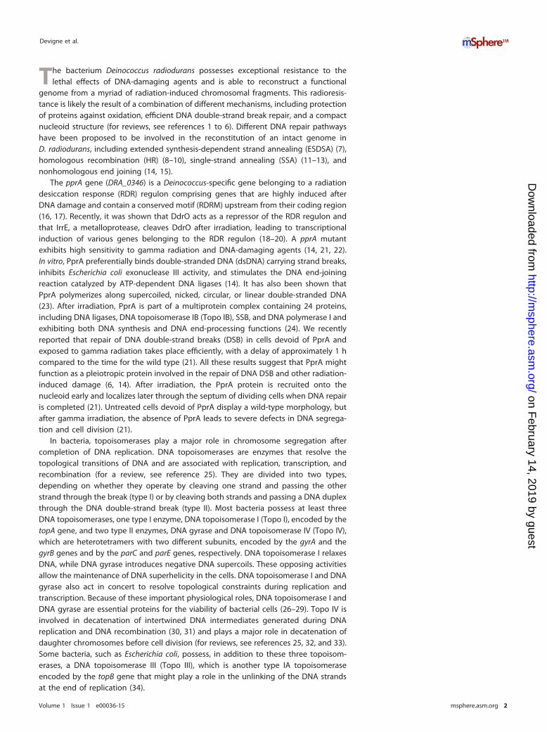
irradiation. To identify proteins interacting specifically with PprA, we compared theproteins that coprecipitated with PprA::HA protein after exposure to 3.8 kGy gammaradiation to the proteins from ΔpprA bacteria exposed to the same dose of irradiation(comparison 2). Proteins detected in both comparisons were considered putativeinteracting partners of the PprA protein. Table 1 shows the eight most relevantproteins, which had changes of twofold or more in both comparisons, pointing out thatthey interacted with PprA::HA. Among them, elongation factor Tu and ribosomalprotein S1 are implicated in translation, Hpi is involved in the maintenance andintegrity of the S layer, and RNA helicase participates in nearly all aspects of RNAmetabolism. We also found the substrate-binding subunit of a peptide ABC transporter,the chaperon protein GroEL, and the two subunits of DNA gyrase. We therefore focusedour attention on DNA gyrase as a PprA interacting partner.
In order to confirm the physical interaction between PprA protein and the GyrAsubunit highlighted by mass spectrometry, we performed a cross-immunoprecipitationexperiment using a strain expressing PprA and GyrA tagged with different epitopes(hemagglutinin [HA] and SPA [sequential peptide affinity], respectively). Both taggedproteins were functional, as shown by the viability and the wild-type radioresistance ofthe engineered strain (see Fig. S1A in the supplemental material). Immunoprecipitationwas carried out on cell extracts from nonirradiated cells or from cells exposed to 3.8 kGygamma radiation and allowed to recover for 110 min. Two different immunoprecipi-tations were performed, as follows: (i) the cell extracts were incubated with anti-HAantibodies and the bound proteins were separated by SDS-PAGE and immunoblottedwith anti-FLAG antibodies to reveal the GyrA::SPA protein, and (ii) the cell extracts wereincubated with anti-FLAG antibodies and the bound proteins were separated bySDS-PAGE and immunoblotted with anti-HA antibodies to reveal the PprA::HA protein.
To check the specificity of our system, control experiments were performed withstrains that did not express the PprA::HA protein (gyrA::SPA strain) (Fig. 1A) or theGyrA::SPA protein (pprA::HA strain) (Fig. 1B). After coimmunoprecipitation of proteinsfrom these irradiated or nonirradiated gyrA::SPA and pprA::HA control strains, no signalwas detected with anti-FLAG antibodies (Fig. 1A) or anti-HA antibodies (Fig. 1B),
TABLE 1 Proteins that coprecipitated with PprA::HA protein after gamma irradiation
Gene Function
Change in protein expression fora:
Comparison 1 (pprA::HA strain at 3.8 kGversus 0 kGy)
Comparison 2 (pprA::HA strain versus�pprA strain at 3.8kGy)
Foldchange P value
Foldchange P value
DR_0309 Elongation factor Tu 7.2 1.95 � 10�2 7.2 3.96 � 10�2
DR_2508 Hexagonally packed intermediatelayer surface protein
7.0 4.97 � 10�4 2.1 8.69 � 10�2
DR_1913 DNA gyrase subunit A 4.8 6.90 � 10�2 4.8 1.12 � 10�1
DR_1983 30S ribosomal protein S1 2.9 6.88 � 10�2 2.5 2.40 � 10�1
DR_1624 RNA helicase 2.6 2.18 � 10�1 2.6 2.90 � 10�1
DR_1955 Peptide ABC transporter substrate-binding protein
2.4 2.49 � 10�2 2.4 3.25 � 10�1
DR_0607 Chaperonin groEL 2.4 2.49 � 10�2 2.4 3.25 � 10�1
DR_0906 DNA gyrase subunit B 2.0 3.27 � 10�2 2.0 4.10 � 10�1
aD. radiodurans ΔpprA (GY12251) and pprA::HA (GY14615) bacteria were either exposed or not to gammairradiation (3.8 kGy) and allowed to recover in fresh medium for 110 min prior to performing acoimmunoprecipitation with anti-HA antibodies. Immunoprecipitated materials were analyzed by massspectrometry (see Table S1 in the supplemental material). The comparison between PprA-HA samplesexposed to gamma irradiation (3.8 kGy, five replicates) and nonirradiated samples (0 kGy, four replicates)was performed with the Tfold module. The comparison between gamma-irradiated samples (3.8 kGy) of thestrain expressing PprA::HA (five replicates) and the pprA deletion mutant (two replicates) was performedwith the ACfold module using the AC test described in Audic and Claverie (66). The eight proteins detectedby shotgun proteomics with a �twofold increase after irradiation (comparison 1) and with a �twofolddecrease after deletion of pprA (comparison 2) are listed.
Devigne et al.
Volume 1 Issue 1 e00036-15 msphere.asm.org 4
on February 14, 2019 by guest
http://msphere.asm
.org/D
ownloaded from

respectively. Our results obtained with the pprA::HA gyrA::SPA strain confirm a physicalinteraction between PprA and GyrA proteins in vivo. Indeed, the GyrA protein wasspecifically trapped by immunoprecipitation of PprA::HA (Fig. 1A), and conversely, thePprA protein was specifically trapped by immunoprecipitation of GyrA::SPA (Fig. 1B). Inthe results shown in Fig. 1A, we also observed additional bands that may correspondto GyrA::SPA cross-linked with other proteins. The interaction between the two proteinswas detected only after exposure of the cells to gamma irradiation, suggesting thatPprA is not expressed at a sufficient level in nonirradiated cells to allow the detectionof this interaction or that the interaction between the two proteins only takes placeduring recovery from DNA damage.
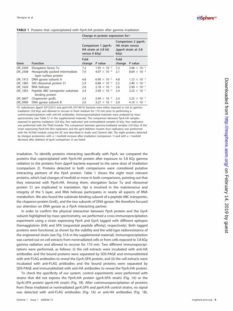
Loss of PprA renders D. radiodurans cells hypersensitive to DNA gyraseinhibitors. The above-described results suggest that PprA might act with DNA gyraseafter irradiation. Since the gyrA and gyrB genes are essential (29, 36), we decided to usedrugs that inhibit DNA gyrase activity to examine the effects of a pprA deletion in agyrase-defective context. As previously shown (37), ΔpprA cells are hypersensitive tonalidixic acid, which targets the GyrA subunit of DNA gyrase (Fig. 2). Here, we showedthat ΔpprA cells are also hypersensitive to novobiocin, a drug that targets the GyrBsubunit of DNA gyrase (Fig. 2). Kota et al. showed that PprA also interacts with Topo IBand enhances the relaxation activity of this enzyme in vitro (37). In our study, we did notfind Topo IB as an interacting partner of PprA. Moreover, ΔtopIB and ΔpprA ΔtopIBbacteria showed the same sensitivity to nalidixic acid or novobiocin as wild-type andΔpprA bacteria, respectively (Fig. 2). The ΔtopIB bacteria also showed a wild-type levelof radioresistance (see Fig. S1B in the supplemental material). Our in vivo resultssuggest that Topo IB in D. radiodurans does not play a crucial role in DNA repair.
Defects in segregation of cells exposed to novobiocin are increased in�pprA mutants. An analysis of cell morphology during the recovery of D. radioduranscells from gamma irradiation showed that a PprA deficiency results in defective
MW pprA
::HA gy
rA::S
PA
Wild ty
pe
gyrA
::SPA
1007055
kDa - - -+ + +γ-irradiation
130
A
B
pprA
::HA gy
rA::S
PA
∆pprA
gyrA
::SPA
pprA
::HA
Wild ty
pe
MW
kDa35
25
γ-irradiation - + - - -+ + +
FIG 1 The GyrA subunit of DNA gyrase interacts with the PprA protein after irradiation. GY9613(wild-type), GY13338 (gyrA::SPA), GY14641 (�pprA gyrA::SPA), GY14615 (pprA::HA), and GY14697(pprA::HA gyrA::SPA) cells were exposed to 3.8 kGy gamma radiation and grown for 110 min. Cellswere fixed and immunoprecipitation performed. (A) Anti-HA antibodies were used to captureHA-tagged PprA protein by immunoprecipitation. Eluted samples were separated on 10% SDS-PAGEgels, and primary anti-FLAG antibodies were used in Western blot analyses to reveal SPA-taggedGyrA proteins. (B) Anti-FLAG antibodies were used to capture SPA-tagged GyrA proteins by immu-noprecipitation, eluted samples were separated on 12% SDS–PAGE, and primary anti-HA antibodieswere used in Western blots to reveal HA-tagged PprA proteins.
PprA Interacts with DNA Gyrase in D. radiodurans
Volume 1 Issue 1 e00036-15 msphere.asm.org 5
on February 14, 2019 by guest
http://msphere.asm
.org/D
ownloaded from
chromosome segregation and aberrant cell division (21). In E. coli, the Topo IV enzymeplays a key role in chromosome segregation by resolving catenated chromosomesgenerated during replication (30). D. radiodurans naturally lacks Topo IV, and thus, DNAgyrase is the only type II topoisomerase present in the cells and is probably involved inthe chromosome decatenation process. We expected that inhibition of DNA gyrase bydrugs would result in defective chromosome partitioning. This defect could be exac-erbated by a PprA deficiency, accounting for the hypersensitivity of ΔpprA bacteria tonalidixic acid and novobiocin. Therefore, we analyzed the cell morphologies aftertreatment with novobiocin. For this purpose, samples of wild-type and ΔpprA cultureswere taken at different times after the addition of novobiocin and analyzed byfluorescence microscopy (Fig. 3).
No aberrant cell morphologies were observed in the absence of novobiocin foreither ΔpprA or wild-type cells (see Fig. S2 in the supplemental material). In contrast,treatment of the wild-type strain with novobiocin resulted in morphological abnormal-ities, including 9.6% of cells being anucleated (39/404) or cells having defects insegregation of the nucleoids (33/404 cells with unequal distribution of DNA betweendaughter cells or DNA blocked across the septum). The absence of the PprA proteinaggravated the morphological defects visualized after novobiocin treatment (Fig. 3),with 30.8% of cells being anucleated (136/441) or cells having defects in segregation ofnucleoids (22.2%, 98/441 cells) or defects in cell division, with cells being incompletelyseparated. Some examples of segregation and division defects are schematized inFig. 3B. Large-field images confirmed that these abnormalities were present in �80%of the ΔpprA cells treated with novobiocin (see Fig. S2).
Novobiocin treatment induces the RDR regulon. As novobiocin treatmentinduces the SOS system in Bacillus subtilis (38), we tested the effect of novobiocin onthe radiation desiccation response (RDR), the major response to DNA damage gener-ated by ionizing radiation in D. radiodurans (16, 17). The induction of the RDR regulonresults from proteolytic degradation of the DdrO repressor by an activated form of themetalloprotease IrrE (18, 19).
To test whether novobiocin treatment induced the RDR regulon, we examined, inthe presence or absence of novobiocin, the levels of three proteins encoded by genes
10-510-410-310-1 10-2 10-510-410-310-1 10-2 10-510-410-310-1 10-2 10-510-410-310-1 10-2
wild type
ΔpprA
ΔtopIB -a
ΔpprAΔtopIB -a
ΔtopIB -b
ΔpprAΔtopIB -b
0 ng/mL 5 ng/mL 10 ng/mL 20 ng/mL novobiocin concentration
10-510-410-310-1 10-2 10-510-410-310-1 10-2 10-510-410-310-1 10-210-510-410-310-1 10-2
0 ng/mL 10 μg/mL 20 μg/mL 40 μg/mL nalidixic acid concentration
wild type
ΔpprA
ΔtopIB -a
ΔpprAΔtopIB -a
ΔtopIB -b
ΔpprAΔtopIB -b
FIG 2 Sensitivity of �pprA mutant to gyrase inhibitors. Serial dilutions of cultures of GY9613(wild-type), GY14661 (�pprA), GY15987 (�topIB) (2 clones, a and b), and GY15985 (�pprA �topIB) (2clones, a and b) strains were spotted on plates in the presence or absence of novobiocin or nalidixicacid at the indicated concentrations, and plates were incubated at 30°C for 5 days.
Devigne et al.
Volume 1 Issue 1 e00036-15 msphere.asm.org 6
on February 14, 2019 by guest
http://msphere.asm
.org/D
ownloaded from

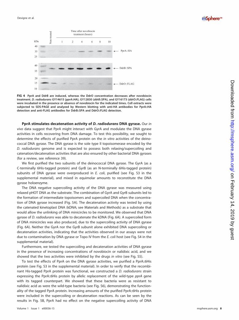
belonging to the regulon, namely, PprA and DdrB, whose expression is highly inducedduring the radiation desiccation response (RDR), and DdrO, whose level was expectedto decrease during the course of the novobiocin treatment. The PprA::HA, DdrB::SPA,and DdrO::FLAG proteins were visualized by Western blot analysis using anti-HA oranti-FLAG antibodies. As can be seen by the results in Fig. 4, a faint band correspondingto the PprA::HA protein was detected in nontreated cells, but the signal increasedmarkedly during the treatment with novobiocin. The results also clearly indicate thatthe DdrB::SPA protein was induced at early times after the addition of novobiocin(Fig. 4). In contrast, we observed a quick decrease in the level of the DdrO repressor innovobiocin-treated cells (Fig. 4). From these results, we conclude that novobiocininduces the RDR regulon in D. radiodurans.
4h 6h 8h 10h
DAPI
FM4-64
DAPI
FM4-64
A
B
wild type +
novobiocin
ΔpprA +
novobiocin
ΔpprA
wild type
- novo
ΔpprA
ΔpprA
ΔpprA
ΔpprA
+ novo
+ novo
+ novo
+ novo
- novo
FIG 3 Morphologies of wild-type and �pprA mutant cells grown in the presence of novobiocin. (A)Cells were grown in the presence of 40 ng/ml novobiocin. Samples were taken at the indicated timesafter the addition of novobiocin and examined by fluorescence microscopy. DNA was stained withDAPI, and membranes were stained with FM 4-64. (B) Various abnormal forms of �pprA nucleoidsobserved after 8 h of growth in the presence of novobiocin. The wild-type and �pprA controlsrepresent the cells grown without novobiocin. A schematic representation of the D. radioduransdividing cells is also shown.
PprA Interacts with DNA Gyrase in D. radiodurans
Volume 1 Issue 1 e00036-15 msphere.asm.org 7
on February 14, 2019 by guest
http://msphere.asm
.org/D
ownloaded from
PprA stimulates decatenation activity of D. radiodurans DNA gyrase. Our invivo data suggest that PprA might interact with GyrA and modulate the DNA gyraseactivities in cells recovering from DNA damage. To test this possibility, we sought todetermine the effects of purified PprA protein on the in vitro activities of the deino-coccal DNA gyrase. The DNA gyrase is the sole type II topoisomerase encoded by theD. radiodurans genome and is expected to possess both relaxing/supercoiling andcatenation/decatenation activities that are also ensured by other bacterial DNA gyrases(for a review, see reference 39).
We first purified the two subunits of the deinococcal DNA gyrase. The GyrA (as aC-terminally 6His-tagged protein) and GyrB (as an N-terminally 6His-tagged protein)subunits of DNA gyrase were overproduced in E. coli, purified (see Fig. S3 in thesupplemental material), and mixed in equimolar amounts to reconstitute the DNAgyrase holoenzyme.
The DNA negative supercoiling activity of the DNA gyrase was measured usingrelaxed pHOT DNA as the substrate. The combination of GyrA and GyrB subunits led tothe formation of intermediate topoisomers and supercoiled DNA when the concentra-tion of DNA gyrase increased (Fig. 5A). The decatenation activity was tested by usingthe catenated kinetoplast DNA (kDNA; see Materials and Methods) as a substrate thatwould allow the unlinking of DNA minicircles to be monitored. We observed that DNAgyrase of D. radiodurans was able to decatenate the kDNA (Fig. 6A). A supercoiled formof DNA minicircles was also produced, due to the supercoiling activity of DNA gyrase(Fig. 6A). Neither the GyrA nor the GyrB subunit alone exhibited DNA supercoiling ordecatenation activities, indicating that the activities observed in our assays were notdue to contamination by DNA gyrase or Topo IV from the E. coli host (see Fig. S4 in thesupplemental material).
Furthermore, we tested the supercoiling and decatenation activities of DNA gyrasein the presence of increasing concentrations of novobiocin or nalidixic acid, and weshowed that the two activities were inhibited by the drugs in vitro (see Fig. S5).
To test the effects of PprA on the DNA gyrase activities, we purified a PprA::6Hisprotein (see Fig. S3 in the supplemental material). In order to verify that the recombi-nant His-tagged PprA protein was functional, we constructed a D. radiodurans strainexpressing the PprA::6His protein by allelic replacement of the wild-type pprA genewith its tagged counterpart. We showed that these bacteria were as resistant tonalidixic acid as were the wild-type bacteria (see Fig. S6), demonstrating the function-ality of the tagged PprA protein. Increasing amounts of the purified PprA::6His proteinwere included in the supercoiling or decatenation reactions. As can be seen by theresults in Fig. 5B, PprA had no effect on the negative supercoiling activity of DNA
Non
-trea
ted Time after novobiocin
treatment (hours)
2 4 6 8 101
DdrO::FLAG
DdrB::SPA
PprA::HA
kDa
40
35
25
40
35
15
10
20
FIG 4 PprA and DdrB are induced, whereas the DdrO concentration decreases after novobiocintreatment. D. radiodurans GY14615 (pprA::HA), GY12830 (ddrB::SPA), and GY16173 (ddrO::FLAG) cellswere incubated in the presence or absence of novobiocin for the indicated times. Cell extracts weresubjected to SDS-PAGE and analyzed by Western blotting with anti-HA antibodies for PprA::HAdetection and anti-FLAG antibodies for DdrB::SPA and DdrO::FLAG detection.
Devigne et al.
Volume 1 Issue 1 e00036-15 msphere.asm.org 8
on February 14, 2019 by guest
http://msphere.asm
.org/D
ownloaded from
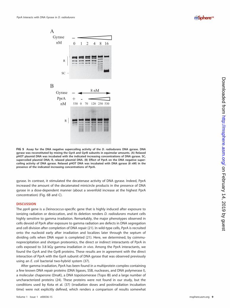
gyrase. In contrast, it stimulated the decatenase activity of DNA gyrase. Indeed, PprAincreased the amount of the decatenated minicircle products in the presence of DNAgyrase in a dose-dependent manner (about a sevenfold increase at the highest PprAconcentration) (Fig. 6B and C).
DISCUSSION
The pprA gene is a Deinococcus-specific gene that is highly induced after exposure toionizing radiation or desiccation, and its deletion renders D. radiodurans mutant cellshighly sensitive to gamma irradiation. Remarkably, the major phenotypes observed incells devoid of PprA after exposure to gamma radiation are defects in DNA segregationand cell division after completion of DNA repair (21). In wild-type cells, PprA is recruitedonto the nucleoid early after irradiation and localizes later through the septum ofdividing cells when DNA repair is completed (21). Here, we determined, by coimmu-noprecipitation and shotgun proteomics, the direct or indirect interactants of PprA incells exposed to 3.8 kGy gamma irradiation in vivo. Among the PprA interactants, wefound the GyrA and the GyrB proteins. These results are in agreement with the directinteraction of PprA with the GyrA subunit of DNA gyrase that was observed previouslyusing an E. coli bacterial two-hybrid system (37).
After gamma irradiation, PprA has been found in a multiprotein complex containinga few known DNA repair proteins (DNA ligases, SSB, nucleases, and DNA polymerase I),a molecular chaperone (DnaK), a DNA topoisomerase (Topo IB) and a large number ofuncharacterized proteins (24). These proteins were not found in our study, but theconditions used by Kota et al. (37) (irradiation doses and postirradiation incubationtime) were not explicitly defined, which renders a comparison of results somewhat
R
SC
A
R
GyrasePprA + -
0 1 2 4 8 16Gyrase
nM
_
_ 8 nM
nM 530 0 70 120 250 530
B
FIG 5 Assay for the DNA negative supercoiling activity of the D. radiodurans DNA gyrase. DNAgyrase was reconstituted by mixing the GyrA and GyrB subunits in equimolar amounts. (A) RelaxedpHOT plasmid DNA was incubated with the indicated increasing concentrations of DNA gyrase. SC,supercoiled plasmid DNA; R, relaxed plasmid DNA. (B) Effect of PprA on the DNA negative super-coiling activity of DNA gyrase. Relaxed pHOT DNA was incubated with DNA gyrase (8 nM) in thepresence of the indicated increasing concentrations of PprA.
PprA Interacts with DNA Gyrase in D. radiodurans
Volume 1 Issue 1 e00036-15 msphere.asm.org 9
on February 14, 2019 by guest
http://msphere.asm
.org/D
ownloaded from

difficult. Intriguingly, they did not find DNA gyrase in their complex. More recently, itwas shown, using a bacterial two-hybrid system, that PprA interacts with Topo IB andenhances the Topo IB-mediated relaxation activity of negative supercoiled DNA (37).The demonstration of a structural and mechanistic relationship between topoisomeraseIB and site-specific recombinases suggests a possible role of Topo IB in recombinationin D. radiodurans (35). However, a ΔtopIB mutant is not more sensitive to ionizingradiation than the wild type, suggesting that Topo IB does not play a crucial role in DNArepair.
Most bacteria possess two type II topoisomerases, DNA gyrase and Topo IV. DNAgyrase plays a major role in maintaining the global level of negative supercoiling of thechromosomes, while Topo IV is mainly a potent decatenase required for partitioning ofthe daughter chromosomes at the end of DNA replication (30, 40, 41). D. radioduranslacks Topo IV. Thus, it is expected that the deinococcal DNA gyrase will also ensure theTopo IV-mediated functions in chromosome segregation. In Mycobacterium tuberculosisand Mycobacterium smegmatis, DNA gyrase is also the unique type II topoisomerasepresent in the cells, and it was shown that it exhibits a strong decatenase activity (42).In the experiments described here, we assayed the activities of the deinococcal DNAgyrase in vitro. We showed that it possessed both DNA negative supercoiling and DNA
0 1 2 4 8 16
kDNA
SC
MC *
CI
B
kDNA
-
SC
MC
CI
AGyrase
nM
GyrasePprA + -
_ 4 nM
nM 530 0 70 120 250 530
0
2
4
6
8
10
12
0 0,2 0,4 0,6
s�m
ula�
on fa
ctor
PprA concentra�on (μM)
C
FIG 6 PprA stimulates decatenation activity of D. radiodurans DNA gyrase. (A) kDNA was incubated with theindicated increasing concentrations of DNA gyrase. kDNA was retained in the wells of the agarose gel. (B)Effect of PprA on the decatenation activity of DNA gyrase. DNA gyrase (4 nM) was incubated with kDNA in thepresence of the indicated increasing concentrations of PprA. (C) Stimulation of DNA decatenation by PprA. Theratio of the minicircles released in the presence of PprA over the minicircles released in the absence of PprAwas determined at each PprA concentration, and the average results from four independent experiments wereplotted. SC, supercoiled minicircles; MC, minicircles; CI, catenated intermediates.
Devigne et al.
Volume 1 Issue 1 e00036-15 msphere.asm.org 10
on February 14, 2019 by guest
http://msphere.asm
.org/D
ownloaded from

decatenation activities and that both activities were sensitive to nalidixic acid andnovobiocin inhibitors. Moreover, the PprA protein, shown to interact with the GyrAsubunit of DNA gyrase, was able to stimulate the DNA decatenation activity of DNAgyrase without affecting its DNA negative supercoiling activity in vitro. In vivo, theabsence of PprA increased the cellular sensitivity to nalidixic acid, a GyrA inhibitor, andto novobiocin, a GyrB inhibitor, and aggravated the morphological defects observed incells treated with novobiocin.
Furthermore, we demonstrated that novobiocin treatment leads to activation of theradiation desiccation response (RDR) in D. radiodurans by showing the downregulationof the DdrO repressor and the consequent upregulation of DdrB and PprA, two genesbelonging to the RDR regulon. Treatment of E. coli and B. subtilis with GyrB inhibitorsresulted in induction of the SOS system (38, 43) and loss of chromosomal DNAsupercoiling, associated with an inhibition of gyrase activity (38, 44). A reduction ofchromosome supercoiling parallels the rapid decline in DNA synthesis. Inhibition ofDNA gyrase activities leads to drastic topological constraints that probably result inreplication fork collapse, and DNA degradation at the stopped fork might generate anSOS signal (43). The signal that initiates RDR induction in D. radiodurans is not currentlyknown. The DdrO repressor is subject to proteolytic degradation by an activated formof the metalloprotease IrrE (18, 19). The D. radiodurans irrE gene is constitutivelyexpressed (45), suggesting that IrrE must be activated. The addition of novobiocin toD. radiodurans might result in severe modifications of DNA supercoiling and perturba-tion of DNA replication, as observed in E. coli and B. subtilis. Our recent results alsosuggest that induction of the RDR regulon may be triggered by an oxidative stressedstate of the cells (19). An oxidative stressed state may result from novobiocin treatment,since it was shown that nalidixic acid and other bactericidal antibiotics generate freeradicals responsible for cell killing in E. coli (46). An understanding of the precise modeof RDR regulon induction by novobiocin awaits further studies to identify the mecha-nism of RDR regulation.
After exposure to gamma radiation, the absence of PprA results in major defects inDNA segregation and cell division after completion of DNA repair (21). In E. coli, newlyduplicated origin regions segregate to opposite sides of the cell soon after initiation ofreplication, while segregation of the terminus region occurs very late in the cell cycle,as the daughter cells separate (47). We recently showed that D. radiodurans alsopresents prolonged colocalization of the Ter domain of chromosome 1 (48) and that thesegregation delay of the terminus is enhanced after irradiation (48). This suggests thatthe activities of the deinococcal DNA gyrase have to be regulated to control chromo-some decatenation. The GyrA and GyrB proteins are distributed largely inside thenucleoid in nonirradiated as well as in irradiated D. radiodurans cells (36, 49).
After irradiation, DNA is shattered into hundreds of fragments. Even after reconsti-tution of circular chromosomes, irradiated cells are expected to contain a large amountof relaxed DNA that might pointlessly recruit DNA gyrase. The PprA protein, by itslocalization through the septum after completion of DNA repair (21, 50), might facili-tate, by its interaction with GyrA, relocalization of the DNA gyrase at the septum. It hasbeen shown that PprA polymerizes along dsDNA (23). Thus, we can also propose thatPprA, like the MukB condensin in E. coli, may remodel the DNA and generate a preferredsubstrate for DNA gyrase and, thus, that the PprA-GyrA interaction might increase theeffective rate of DNA decatenation. In nonirradiated cells, PprA is expressed at a lowbasal level and is not detected by immunofluorescence microscopy. Moreover, itsabsence has no effect on the viability and the morphology of cells that divide normally(21). Thus, we can imagine that, like E. coli Topo IV, whose decatenation activity isregulated through a physical interaction of the ParC subunit with FtsK, MreB, or MukB(51–53), the decatenation activity of deinococcal DNA gyrase could be regulated by itsinteraction with key proteins involved in chromosome segregation or cell division. FtsKand SMC (structural maintenance of chromosome), a functional analog of MukB, arepresent in D. radiodurans, but unlike the rod-shaped Deinococcus deserti bacteria,D. radiodurans bacteria do not encode a homolog of the E. coli MreB protein. We have
PprA Interacts with DNA Gyrase in D. radiodurans
Volume 1 Issue 1 e00036-15 msphere.asm.org 11
on February 14, 2019 by guest
http://msphere.asm
.org/D
ownloaded from

previously shown that the absence of SMC in D. radiodurans does not disturb chromo-some segregation (54). Further studies will be required for a better understanding ofthe regulation of chromosome segregation and cell division in D. radiodurans.
MATERIALS AND METHODSBacterial strains, plasmids, and DNA manipulations. The bacterial strains used are listed in Table 2.The E. coli strains used were DH5� as the general cloning host and Rosetta 2(DE3)pLysS (Novagen) forprotein expression. E. coli strains were grown at 37°C in lysogeny broth (LB) or 2� YT medium (Bio101,Inc.). All D. radiodurans strains were derivatives of strain R1 (ATCC 13939). They were grown at 30°C in2� TGY (1% tryptone, 0.2% dextrose, 0.6% yeast extract) or plated on 1� TGY containing 1.5% agar.When necessary, media were supplemented with the appropriate antibiotics used at the following finalconcentrations: 6 �g/ml kanamycin and 3.5 �g/ml chloramphenicol for D. radiodurans and 30 �g/mlkanamycin, 35 �g/ml chloramphenicol, and 100 �g/ml ampicillin for E. coli.
Plasmids pET26b, pET30Ek/LIC, and pET21d were used to construct vectors for overexpression ofD. radiodurans GyrA, GyrB, and PprA, respectively (Table 3). Plasmid DNA was extracted from E. coli usingthe QIAprep spin miniprep kit (Qiagen). All constructions were verified by DNA sequencing. RelaxedpHOT-1 DNA (2.6 kb) and kinetoplast DNA (kDNA) were purchased from TopoGEN.
ΔpprA�cat, ΔtopIB�kan, and pprA::6His::kan alleles were constructed by the tripartite ligationmethod (55). Transformation of D. radiodurans with PCR products or genomic DNA was performed aspreviously described (13). The genetic structure and the purity of the mutants were checked by PCR.
Chromosomal DNA of D. radiodurans was extracted as previously described (56). PCR amplification ofDNA fragments using plasmid or genomic DNA as the template was performed using Phusion DNApolymerase (Thermo Scientific) or GoTaq DNA polymerase (Promega). Oligonucleotides used in this studywill be provided on request.
Gamma irradiation treatment of D. radiodurans. Exponential-phase cultures grown in 2� TGYwere concentrated to an A650 of 20 in 2� TGY and irradiated on ice with a 137Cs irradiation system(Institut Curie, Orsay or Paris, France) at 3.8 kGy (dose rate of 40.1 Gy/min).
TABLE 2 Bacterial strains
Bacterial strain Description Source or reference
E. coli strainsDH5� supE44 ΔlacU(�80lacZΔM15)
hsdR17 recA1 endA1 gyrA96thi-1 relA1
Laboratory stock
Rosetta 2(DE3)pLysS F� ompT hsdSB(rB� mB
�) gal dcm�(DE3 [lacI lacUV5-T7 gene 1 ind1sam-7 nin-5])pLysSRARE (cat)
Novagen
D. radiodurans strainsR1 Wild type, ATCC 13939 Laboratory stockGY12830 ddrB::SPA::cat 13GY13338 gyrA::SPA::cat 36GY14615 pprA::HA::kan 21GY14641 ΔpprA�kan gyrA::SPA::cat This workGY14661 ΔpprA�cat This workGY14697 pprA::HA::kan gyrA::SPA::cat This workGY15985 �topIB�kan �pprA�cat This workGY15987 �topIB�kan This workGY16173 ddrO::FLAG::cat 19GY16179 pprA::6His::kan This work
TABLE 3 Plasmids used in the study
Plasmid DescriptionReference orsource
pGTC101 Source of chloramphenicol cassette 67p11086 Source of kanamycin cassette in D. radiodurans Laboratory stockpET21d PET expression system, pT7lac, C-terminal 6His tag,
Ampr
Novagen
pET26b PET expression system, pT7lac, C-terminal 6His tag,Kanr
Novagen
pET21d PET expression system, pT7lac, N-terminal 6His tag,Kanr
Novagen
pET21d-pprA pET21d NcoI/XhoI � PCR fragment containing pprA This workpET26b-gyrA pET26b NdeI/XhoI � PCR fragment containing gyrA This workpET30EK/LIC-gyrB pET30Ek/LIC � PCR fragment containing gyrB This work
Devigne et al.
Volume 1 Issue 1 e00036-15 msphere.asm.org 12
on February 14, 2019 by guest
http://msphere.asm
.org/D
ownloaded from
Coimmunoprecipitation and Western blot analysis of the samples. D. radiodurans bacteria inwhich a bait protein had been tagged at its C-terminal end with a SPA or HA epitope were grown in 2�TGY and exposed or not to 3.8 kGy gamma radiation. Following irradiation, cultures were diluted in 2�TGY to an A650 of 0.4 and incubated at 30°C for 110 min. Cells (50 ml) (with or without irradiation) werecentrifuged, and the pellets were suspended in 600 �l of lysis buffer A (50 mM Tris, pH 7.5, 100 mM NaCl,5 mM MgCl2, 1 mM dithiothreitol [DTT], 0.5% Triton X-100) with 0.1 M protease inhibitor (Pefabloc;Euromedex). Cells were disrupted with a FastPrep apparatus (FP120; Bio101, Inc.), using 0.1-g amountsof glass beads (500 �m) and three 30-s pulses (speed, 4 m/s). Cell debris was removed by centrifugationat 2,000 � g for 10 min at 4°C. Amounts of approximately 500 �l of supernatant were incubated with2 �g of monoclonal antibody (mouse anti-FLAG monoclonal antibodies [Sigma-Aldrich] or mouseanti-HA monoclonal antibodies [Eurogentec]) at 10°C under gentle agitation for 2 h for immune complexformation. Amounts of 20 �l of Bio-Adembeads coated with protein G (Ademtech), washed twice in 20 �lof lysis buffer A using the Ademtech magnet, were suspended in 20 �l of lysis buffer. The washed beadswere added to the supernatants treated with antibodies, and the mixtures were incubated at 10°C undergentle agitation for 1 h. The coimmunoprecipitated complexes bound to the beads were then washedfive times with 500 �l of lysis buffer A using the Ademtech magnet before being suspended in 35 �l of1� Laemmli loading dye. Samples were heated at 95°C for 5 min and replaced on the magnet, and thesupernatants containing the enriched proteins were kept at �20°C before shotgun proteomics analyses(see below).
To prepare coimmunoprecipitated samples for Western blot analyses, cultures were treated with 2%formaldehyde for 20 min, followed by a treatment with 0.3 M glycine for 5 min before immunoprecipi-tation. Two different immunoprecipitations were performed, using a pprA::HA gyrA::SPA strain expressingthe two proteins tagged with different epitopes; the cell extracts were incubated with anti-HA antibodiesor with anti-FLAG antibodies and the bound proteins were separated by SDS-PAGE and immunoblottedwith anti-FLAG antibodies or anti-HA antibodies, respectively. For this purpose, proteins were transferredfrom the gel onto a polyvinylidene difluoride (PVDF) membrane. The membrane was blocked withTris-buffered saline (TBS) containing 0.05% Tween 20 before being incubated overnight at 4°C with a1:5,000 dilution of mouse anti-HA monoclonal antibodies (Eurogentec) or rabbit anti-FLAG monoclonalantibodies (Sigma-Aldrich) in TBS containing 3% powdered milk, 0.05% Tween 20. After extensive washesin TBS with 0.05% Tween 20, the membrane was incubated with alkaline phosphatase-conjugatedanti-mouse or anti-rabbit IgG as the secondary antibody, and bound antibodies were revealed by acolorimetric reaction. Gels were analyzed by measuring intensity profiles for each lane using Image Labsoftware (Bio-Rad).
Tandem mass spectrometry and proteomic data interpretation. The protein samples obtained bycoimmunoprecipitation with the PprA::HA protein were heated at 95°C for 5 min and then loaded ontoa 10% NuPAGE gel (Invitrogen) for a short electrophoresis in MOPS (morpholinepropanesulfonic acid)buffer. The proteins were briefly stained with Coomassie blue safe stain (Invitrogen). Polyacrylamidebands containing the whole enriched subproteomes were processed as previously described for furtherdestaining and iodoacetamide treatments (57). Samples were subjected for 4 h to proteolysis at 37°Cwith 10 ng/�l of sequencing-grade trypsin (Roche) in 50 mM NH4HCO3 and 0.01% ProteaseMAXsurfactant (Promega) to maximize peptide recovery as described previously (58). The reactions werestopped with 0.5% (final) trifluoroacetic acid. The resulting peptides (10 �l of the 40 �l generated withthe procedure) were analyzed with an LTQ-Orbitrap XL hybrid mass spectrometer (Thermo Fisher)coupled to an UltiMate 3000 NanoLC system (Dionex-LC Packings) operating a reverse-phase AcclaimPepMap100 C18 �-precolumn (5 �m, 100 Å, 300-�m inner diameter by 5 mm; Dionex) and a nanoscaleAcclaim PepMap100 C18 capillary column (3 �m, 100 Å, 75 �m-inner diameter by 15 cm; Dionex) asdescribed previously (59). Peptide mixtures (10 �l) were desalted on-line and then resolved at a flow rateof 0.3 �l per min using a 90-min gradient from 5 to 60% solvent B (0.1% HCOOH– 80% CH3CN) with 0.1%HCOOH–100% H2O as solvent A. The linear trap quadrupole (LTQ)-Orbitrap XL mass spectrometer wasrecalibrated internally in real time with polydimethylcyclosiloxane ions generated from ambient air in theelectrospray process {monoprotonated [(CH3)2SiO]6 with m/z of 445.120024} and operated in data-dependent mode using the TOP3 strategy as described previously (60). In brief, a scan cycle was initiatedwith a full scan of high mass accuracy from m/z 300 to 1,800 in the Orbitrap analyzer at a resolution of30,000, followed by MS-MS scans in the LTQ linear ion trap on the three most abundant precursor ions,with dynamic exclusion of previously selected ions. Peak lists were generated using the Mascot Daemonsoftware (Matrix Science), and MS-MS spectra were assigned with the MASCOT search engine (version2.2.04; Matrix Science) as described previously (58). The in-house D. radiodurans protein sequencedatabase (36) comprised 3,311 polypeptide sequences, totaling 1,006,757 amino acids. Peptides wereidentified with a P value threshold below 0.05. Protein spectral counts were normalized as described inLiu et al. (61) by systematically adding one spectral count to all experimental values. These normalizedvalues were then compared with the Tfold or the ACfold modules of the PatternLab software (62).
In vivo assay of D. radiodurans sensitivity to DNA gyrase inhibitors. Cultures of exponentiallygrowing cells at an A650 of 0.3 were serially diluted, and aliquots (10 �l) of each dilution were spottedon TGY agar supplemented or not with increasing concentrations of novobiocin or nalidixic acid. Plateswere incubated at 30°C for 5 days.
Fluorescence microscopy. An overnight culture was diluted to an A650 of 0.07 in fresh 2� TGYmedium with or without novobiocin (40 ng/ml) and incubated at 30°C with agitation (150 rpm). Atdifferent times, aliquots were removed and treated as previously described (29). DNA and membraneswere stained with DAPI (4[prime],6-diamidino-2-phenylindole) (2 �g/ml) and FM 4-64 (N-[3-triethylammoniumpropyl]-4-{6-[4-(diethylamino) phenyl] hexatrienyl} pyridinium dibromide) (10 �g/ml),
PprA Interacts with DNA Gyrase in D. radiodurans
Volume 1 Issue 1 e00036-15 msphere.asm.org 13
on February 14, 2019 by guest
http://msphere.asm
.org/D
ownloaded from
respectively. The stained cells were observed using a Leica DM RXA microscope, and images wereanalyzed using ImageJ software.
Western blot analysis of proteins belonging to the RDR regulon after novobiocin treatment.Novobiocin (40 ng/ml) was added or not to an exponential culture (A650 of 0.3) in 2� TGY medium. Cellswere cultivated at 30°C, and 20-ml aliquots were centrifuged at the times indicated in Fig. 4. The pelletswere suspended in 150 �l of 1� SSC (1� SSC is 0.15 M NaCl plus 0.015 M sodium citrate) buffer, and thecells were disrupted as described previously (54). After centrifugation, the protein concentration wasmeasured (Bio-Rad protein assay dye reagent), and 5 or 10 �g of proteins was subjected to electropho-resis onto acrylamide gel. For detection of PprA::HA, DdrB::SPA, and DdrO::FLAG proteins, the taggedPprA and DdrB proteins were separated onto a 12% Tris glycine SDS-PAGE gel, and the tagged DdrOproteins were separated onto a 16% Tris Tricine SDS-PAGE gel. The proteins were then transferred ontoa PVDF membrane, and the membranes were treated with anti-HA or anti-FLAG antibodies as describedabove.
Purification of the PprA::6His protein. E. coli Rosetta(DE3)pLysS was transformed with pET21d-pprA and grown in 2� YT medium (Bio101, Inc.) supplemented with ampicillin (100 �g/ml). When the cellculture reached an A600 of 1, PprA production was induced with 0.5 mM IPTG (isopropyl-�-D-thiogalac-topyranoside; Sigma) for 4 h at 37°C. Cells were harvested by centrifugation, suspended in 40 ml of bufferA (200 mM NaCl, 20 mM Tris-HCl, pH 7.5), and stored overnight at �20°C. Cell lysis was completed bysonication (probe-tip sonicator; Branson). The His-tagged PprA protein was purified on a Ni-nitrilotriacetic acid (NTA) column (Qiagen, Inc.), eluted with 200 mM imidazole in buffer A, and loadedonto a Superdex 200 column (Amersham Pharmacia Biotech) equilibrated against the same buffer. ThePprA protein was concentrated using Vivaspin centrifugal concentrators with a nominal molecularweight limit cutoff of 5,000 (Vivascience), flash frozen in liquid nitrogen, and stored at �80°C.
Purification of the GyrA::6His and 6His::GyrB proteins. Usually, to characterize the in vitrobiochemical activities of bacterial gyrases, the GyrA and GyrB subunits are expressed and purifiedseparately from pET vectors (63, 64). The A subunit is expressed as a C-terminal 6His protein and the Bsubunit as an N-terminal 6His protein. After purification of GyrA and GyrB proteins, the DNA gyraseactivity is reconstituted by mixing the two subunits.
To purify the deinococcal DNA gyrase, the gyrA (DR_1913) and gyrB (DR_0906) genes were amplifiedfrom genomic DNA and ligated with DNA of vectors pET26b and pET30Ek/LIC, respectively. The resultingplasmids were introduced into E. coli Rosetta 2(DE3)pLysS. Transformed cells were grown in 100 ml of LBmedium supplemented with 30 �g/ml kanamycin and 35 �g/ml chloramphenicol until an A650 of 0.4 to0.6 was reached. The expression of the tagged proteins was induced by IPTG at a final concentration of1 mM. Growth was continued overnight at 20°C. Cells were harvested by centrifugation, and the pelletswere suspended in binding buffer (20 mM Tris-HCl, pH 7.8, 800 mM NaCl, 5% glycerol, 20 mM imidazole)(0.1 ml for 1 OD [optical density]) containing 0.03% Triton X-100. For the purification of 6His::GyrB, anEDTA-free protease inhibitor cocktail (Sigma) was added to the solution. The cells were disrupted usingan ultrasonic cell disrupter. The disrupted suspensions were centrifuged at 4°C (13,000 � g for 20 min),and the supernatants were loaded onto 0.5-ml Ni-NTA columns (Qiagen) equilibrated with bindingbuffer. The columns were washed initially with 5 ml of binding buffer, followed by 4 elution steps with5 ml of a solution containing 20 mM Tris-HCl, pH 7.8, 200 mM NaCl, and 5% glycerol and increasingconcentrations of imidazole (40 mM, 60 mM, 100 mM, and 200 mM). The purity of the proteins wasverified by SDS-gel electrophoresis, and the protein fractions were pooled and dialyzed on a PD10column against 3.5 ml of 50 mM Tris-HCl, pH 7.8, 200 mM NaCl, and 30% glycerol according to themanufacturer’s protocol (GE Healthcare). The protein solutions were transferred to fresh precooled tubes,and DTT and EDTA were added at a final concentration of 1 mM. The GyrA::6His and 6His::GyrB proteinswere aliquoted and stored at �80°C.
DNA supercoiling and decatenation assays. DNA supercoiling activity was assayed with therecombinant D. radiodurans GyrA and GyrB proteins and relaxed pHOT DNA (TopoGEN) as the substrate.Supercoiled pHOT is a derivative of plasmid pBR322. The relaxed form is prepared by the manufacturerusing high-purity Topo I relaxation reactions, followed by inactivation and repurification of the relaxedproduct. The reaction mixture (20 �l) contained 35 mM Tris-HCl (pH 7.8), 24 mM KCl, 4 mM MgCl2, 2 mMDTT, 1 mM ATP, 1.8 mM spermidine, 100 �g/ml bovine serum albumin, 6.5% glycerol, relaxed pHOT DNA(125 ng), and GyrA and GyrB proteins in equal molar amounts. To test the effect of PprA, increasingamounts of PprA (or the equivalent volumes of buffer) were added to the reaction mixtures. The mixturewas incubated for 1 h at 37°C, and the reaction was terminated by the addition of 2 �l 10� BBSE buffer(5% SDS, 100 mM EDTA, 50% glycerol, and 0.4% bromophenol blue). The samples were loaded onto a1.2% agarose gel in TEP buffer (36 mM Tris-HCl, pH 7.8, 30 mM NaH2PO4, 1 mM EDTA) and run for 3 hat 50 V. The gel was stained with ethidium bromide (1 �g/ml) for 30 min. The bands were then visualizedand quantified using Image Lab (Bio-Rad) software.
For the decatenation assay, the reaction mixture was the same as those used in the supercoilingassay, except that the DNA substrate was replaced with kinetoplast DNA (kDNA; a network of �5,000catenated DNA minicircles and �25 maxicircles that is isolated from trypanosomatid parasites [65])(125 ng; TopoGEN), and the incubation was performed at 37°C for 3 h. The products were then analyzedas described for the DNA supercoiling assay.
The same in vitro DNA negative supercoiling and DNA decatenation assays were performed in thepresence of increasing concentrations of novobiocin or nalidixic acid.
Devigne et al.
Volume 1 Issue 1 e00036-15 msphere.asm.org 14
on February 14, 2019 by guest
http://msphere.asm
.org/D
ownloaded from
SUPPLEMENTAL MATERIALSupplemental material for this article may be found at http://dx.doi.org/10.1128/mSphere.00036-15.
Figure S1, EPS file, 0.7 MB.Figure S2, EPS file, 5.1 MB.Figure S3, EPS file, 1.4 MB.Figure S4, EPS file, 2.1 MB.Figure S5, EPS file, 4.1 MB.Figure S6, EPS file, 1.2 MB.Table S1, XLSX file, 0.1 MB.
ACKNOWLEDGMENTSWe thank Adriana Bailone for valuable discussions, suggestions, and critical reading ofthe manuscript, Michael DuBow for polishing our English, Dyana Sanchez for her helpin protein purification, and the Curie Institute for the use of the 137Cs irradiation system.
This work was supported by the Université Paris-Sud, the Centre National de laRecherche Scientifique, the Agence Nationale de la Recherche (grants ANR-07-BLAN-0106-02 to J.A. and S.S. and ANR 11BSV3-01701 to S.S.), IFR115 to P.S. and S.Q.-C.(Génomes, Transcriptomes, Protéomes), and Electricité de France (RB2011-17 to P.S. and S.S.).
FUNDING INFORMATIONElectricite de France provided funding to Suzanne Sommer and Pascale Servant undergrant number RB2011-17. IFR115 (Genomes, Transcriptomes, Proteomes) providedfunding to Sophie Quevillon-Cheruel and Pascale Servant. Agence Nationale de laRecherche (ANR) provided funding to Jean Armengaud and Suzanne Sommer undergrant numbers ANR-07-BLAN-0106-02 and ANR 11BSV3-01701.
REFERENCES1. Cox MM, Battista JR. 2005. Deinococcus radiodurans—the consummate
survivor. Nat Rev Microbiol 3:882– 892. http://dx.doi.org/10.1038/nrmicro1264.
2. Slade D, Radman M. 2011. Oxidative stress resistance in Deinococcusradiodurans. Microbiol Mol Biol Rev 75:133–191. http://dx.doi.org/10.1128/MMBR.00015-10.
3. Krisko A, Radman M. 2013. Biology of extreme radiation resistance: theway of Deinococcus radiodurans. Cold Spring Harb Perspect J Biol5:a012765. http://dx.doi.org/10.1101/cshperspect.a012765.
4. Daly MJ. 2012. Death by protein damage in irradiated cells. DNA Repair(Amst) 11:12–21. http://dx.doi.org/10.1016/j.dnarep.2011.10.024.
5. Confalonieri F, Sommer S. 2011. Bacterial and archaeal resistance toionizing radiation. J Phys Conf Ser 261: http://dx.doi.org/10.1088/1742-6596/261/1/012005.
6. Ishino Y, Narumi I. 2015. DNA repair in hyperthermophilic and hyper-radioresistant microorganisms. Curr Opin Microbiol 25:103–112. http://dx.doi.org/10.1016/j.mib.2015.05.010.
7. Zahradka K, Slade D, Bailone A, Sommer S, Averbeck D, PetranovicM, Lindner AB, Radman M. 2006. Reassembly of shattered chromo-somes in Deinococcus radiodurans. Nature 443:569 –573. http://dx.doi.org/10.1038/nature05160.
8. Daly MJ, Minton KW. 1995. Interchromosomal recombination in theextremely radioresistant bacterium Deinococcus radiodurans. J Bacteriol177:5495–5505.
9. Slade D, Lindner AB, Paul G, Radman M. 2009. Recombination andreplication in DNA repair of heavily irradiated Deinococcus radiodurans.Cell 136:1044 –1055. http://dx.doi.org/10.1016/j.cell.2009.01.018.
10. Bentchikou E, Servant P, Coste G, Sommer S. 2010. A major role of theRecFOR pathway in DNA double-strand-break repair through ESDSA inDeinococcus radiodurans. PLoS Genet 6:e1000774. http://dx.doi.org/10.1371/journal.pgen.1000774.
11. Daly MJ, Minton KW. 1996. An alternative pathway of recombination ofchromosomal fragments precedes recA-dependent recombination in theradioresistant bacterium Deinococcus radiodurans. J Bacteriol 178:4461– 4471.
12. Xu G, Lu H, Wang L, Chen H, Xu Z, Hu Y, Tian B, Hua Y. 2010. DdrBstimulates single-stranded DNA annealing and facilitates RecA-
independent DNA repair in Deinococcus radiodurans. DNA Repair (Amst)9:805– 812. http://dx.doi.org/10.1016/j.dnarep.2010.04.006.
13. Bouthier de la Tour C, Boisnard S, Norais C, Toueille M, BentchikouE, Vannier F, Cox MM, Sommer S, Servant P. 2011. The deinococcalDdrB protein is involved in an early step of DNA double strand breakrepair and in plasmid transformation through its single-strand annealingactivity. DNA Repair (Amst) 10:1223–1231. http://dx.doi.org/10.1016/j.dnarep.2011.09.010.
14. Narumi I, Satoh K, Cui S, Funayama T, Kitayama S, Watanabe H.2004. PprA: a novel protein from Deinococcus radiodurans that stimu-lates DNA ligation. Mol Microbiol 54:278 –285. http://dx.doi.org/10.1111/j.1365-2958.2004.04272.x.
15. Kota S, Kamble VA, Rajpurohit YS, Misra HS. 2010. ATP-type DNAligase requires other proteins for its activity in vitro and its operoncomponents for radiation resistance in Deinococcus radiodurans in vivo.Biochem Cell Biol 88:783–790. http://dx.doi.org/10.1139/o10-075.
16. Makarova KS, Omelchenko MV, Gaidamakova EK, Matrosova VY,Vasilenko A, Zhai M, Lapidus A, Copeland A, Kim E, Land M, Mav-rommatis K, Pitluck S, Richardson PM, Detter C, Brettin T, SaundersE, Lai B, Ravel B, Kemner KM, Wolf YI, Sorokin A, Gerasimova AV,Gelfand MS, Fredrickson JK, Koonin EV, Daly MJ. 2007. Deinococcusgeothermalis: the pool of extreme radiation resistance genes shrinks.PLoS One 2:e955. http://dx.doi.org/10.1371/journal.pone.0000955.
17. Tanaka M, Earl AM, Howell HA, Park MJ, Eisen JA, Peterson SN,Battista JR. 2004. Analysis of Deinococcus radiodurans’s transcriptionalresponse to ionizing radiation and desiccation reveals novel proteinsthat contribute to extreme radioresistance. Genetics 168:21–33. http://dx.doi.org/10.1534/genetics.104.029249.
18. Ludanyi M, Blanchard L, Dulermo R, Brandelet G, Bellanger L, PignolD, Lemaire D, de Groot A. 2014. Radiation response in Deinococcusdeserti: IrrE is a metalloprotease that cleaves repressor protein DdrO. MolMicrobiol 94:434 – 449. http://dx.doi.org/10.1111/mmi.12774.
19. Devigne A, Ithurbide S, Bouthier de la Tour C, Passot F, Mathieu M,Sommer S, Servant P. 2015. DdrO is an essential protein that regulatesthe radiation desiccation response and the apoptotic-like cell death inthe radioresistant Deinococcus radiodurans bacterium. Mol Microbiol96:1069 –1084. http://dx.doi.org/10.1111/mmi.12991.
PprA Interacts with DNA Gyrase in D. radiodurans
Volume 1 Issue 1 e00036-15 msphere.asm.org 15
on February 14, 2019 by guest
http://msphere.asm
.org/D
ownloaded from

20. Wang Y, Xu Q, Lu H, Lin L, Wang L, Xu H, Cui X, Zhang H, Li T, HuaY. 2015. Protease activity of PprI facilitates DNA damage response:Mn2�-dependence and substrate sequence-specificity of the proteo-lytic reaction. PLoS One 10:e0122071. http://dx.doi.org/10.1371/journal.pone.0122071.
21. Devigne A, Mersaoui S, Bouthier-de-la-Tour C, Sommer S, Servant P.2013. The PprA protein is required for accurate cell division of gamma-irradiated Deinococcus radiodurans bacteria. DNA Repair (Amst) 12:265–272. http://dx.doi.org/10.1016/j.dnarep.2013.01.004.
22. Selvam K, Duncan JR, Tanaka M, Battista JR. 2013. DdrA, DdrD, andPprA: components of UV and mitomycin C resistance in Deinococcusradiodurans R1. PLoS One 8:e69007. http://dx.doi.org/10.1371/journal.pone.0069007.
23. Adachi M, Hirayama H, Shimizu R, Satoh K, Narumi I, Kuroki R. 2014.Interaction of double-stranded DNA with polymerized PprA protein fromDeinococcus radiodurans. Protein Sci 23:1349 –1358. http://dx.doi.org/10.1002/pro.2519.
24. Kota S, Misra HS. 2008. Identification of a DNA processing complexfrom Deinococcus radiodurans. Biochem Cell Biol 86:448 – 458. http://dx.doi.org/10.1139/o08-122.
25. Champoux JJ. 2001. DNA topoisomerases: structure, function, andmechanism. Annu Rev Biochem 70:369 – 413. http://dx.doi.org/10.1146/annurev.biochem.70.1.369.
26. DiNardo S, Voelkel KA, Sternglanz R, Reynolds AE, Wright A. 1982.Escherichia coli DNA topoisomerase I mutants have compensatory mu-tations in DNA gyrase genes. Cell 31:43–51. http://dx.doi.org/10.1016/0092-8674(82)90403-2.
27. Drlica K, Zhao X. 1997. DNA gyrase, topoisomerase IV, and the4-quinolones. Microbiol Mol Biol Rev 61:377–392.
28. Lecointe F, Coste G, Sommer S, Bailone A. 2004. Vectors for regulatedgene expression in the radioresistant bacterium Deinococcus radio-durans. Gene 336:25–35. http://dx.doi.org/10.1016/j.gene.2004.04.006.
29. Nguyen HH, de la Tour CB, Toueille M, Vannier F, Sommer S, ServantP. 2009. The essential histone-like protein HU plays a major role inDeinococcus radiodurans nucleoid compaction. Mol Microbiol 73:240 –252. http://dx.doi.org/10.1111/j.1365-2958.2009.06766.x.
30. Adams DE, Shekhtman EM, Zechiedrich EL, Schmid MB, CozzarelliNR. 1992. The role of topoisomerase IV in partitioning bacterial repliconsand the structure of catenated intermediates in DNA replication. Cell71:277–288. http://dx.doi.org/10.1016/0092-8674(92)90356-H.
31. Zechiedrich EL, Cozzarelli NR. 1995. Roles of topoisomerase IV andDNA gyrase in DNA unlinking during replication in Escherichia coli.Genes Dev 9:2859 –2869. http://dx.doi.org/10.1101/gad.9.22.2859.
32. Forterre P, Gribaldo S, Gadelle D, Serre MC. 2007. Origin and evolu-tion of DNA topoisomerases. Biochimie 89:427– 446. http://dx.doi.org/10.1016/j.biochi.2006.12.009.
33. Khodursky AB, Peter BJ, Schmid MB, DeRisi J, Botstein D, Brown PO,Cozzarelli NR. 2000. Analysis of topoisomerase function in bacterialreplication fork movement: use of DNA microarrays. Proc Natl Acad SciU S A 97:9419 –9424. http://dx.doi.org/10.1073/pnas.97.17.9419.
34. Hiasa H, Marians KJ. 1994. Topoisomerase III, but not topoisomerase I,can support nascent chain elongation during theta-type DNA replica-tion. J Biol Chem 269:32655–32659.
35. Krogh BO, Shuman S. 2002. A poxvirus-like type IB topoisomerasefamily in bacteria. Proc Natl Acad Sci U S A 99:1853–1858. http://dx.doi.org/10.1073/pnas.032613199.
36. Toueille M, Mirabella B, Guérin P, Bouthier de la Tour C, Boisnard S,Nguyen HH, Blanchard L, Servant P, de Groot A, Sommer S, Armen-gaud J. 2012. A comparative proteomic approach to better defineDeinococcus nucleoid specificities. J Proteomics 75:2588 –2600. http://dx.doi.org/10.1016/j.jprot.2012.03.002.
37. Kota S, Charaka VK, Ringgaard S, Waldor MK, Misra HS. 2014. PprAcontributes to Deinococcus radiodurans resistance to nalidixic acid, ge-nome maintenance after DNA damage and interacts with deinococcaltopoisomerases. PLoS One 9:e85288. http://dx.doi.org/10.1371/journal.pone.0085288.
38. Osburne MS, Zavodny SM, Peterson GA. 1988. Drug-induced relax-ation of supercoiled plasmid DNA in Bacillus subtilis and induction of theSOS response. J Bacteriol 170:442– 445.
39. Collin F, Karkare S, Maxwell A. 2011. Exploiting bacterial DNA gyraseas a drug target: current state and perspectives. Appl Microbiol Biotech-nol 92:479 – 497. http://dx.doi.org/10.1007/s00253-011-3557-z.
40. Kato J, Nishimura Y, Imamura R, Niki H, Hiraga S, Suzuki H. 1990.
New topoisomerase essential for chromosome segregation in E. coli. Cell63:393– 404. http://dx.doi.org/10.1016/0092-8674(90)90172-B.
41. Peng H, Marians KJ. 1993. Decatenation activity of topoisomerase IVduring oriC and pBR322 DNA replication in vitro. Proc Natl Acad Sci U S A90:8571– 8575. http://dx.doi.org/10.1073/pnas.90.18.8571.
42. Manjunatha UH, Dalal M, Chatterji M, Radha DR, Visweswariah SS,Nagaraja V. 2002. Functional characterisation of mycobacterial DNAgyrase: an efficient decatenase. Nucleic Acids Res 30:2144 –2153. http://dx.doi.org/10.1093/nar/30.10.2144.
43. Smith CL. 1983. recF-dependent induction of recA synthesis by coumer-mycin, a specific inhibitor of the B subunit of DNA gyrase. Proc Natl AcadSci U S A 80:2510 –2513. http://dx.doi.org/10.1073/pnas.80.9.2510.
44. Drlica K, Snyder M. 1978. Superhelical Escherichia coli DNA: relaxationby coumermycin. J Mol Biol 120:145–154. http://dx.doi.org/10.1016/0022-2836(78)90061-X.
45. Gao G, Le D, Huang L, Lu H, Narumi I, Hua Y. 2006. Internal promotercharacterization and expression of the Deinococcus radiodurans pprI-folPgene cluster. FEMS Microbiol Lett 257:195–201. http://dx.doi.org/10.1111/j.1574-6968.2006.00169.x.
46. Kohanski MA, Dwyer DJ, Hayete B, Lawrence CA, Collins JJ. 2007. Acommon mechanism of cellular death induced by bactericidal antibiot-ics. Cell 130:797– 810. http://dx.doi.org/10.1016/j.cell.2007.06.049.
47. Li Y, Sergueev K, Austin S. 2002. The segregation of the Escherichia coliorigin and terminus of replication. Mol Microbiol 46:985–996. http://dx.doi.org/10.1046/j.1365-2958.2002.03234.x.
48. Passot FM, Nguyen HH, Dard-Dascot C, Thermes C, Servant P, EspéliO, Sommer S. 2015. Nucleoid organization in the radioresistant bacte-rium Deinococcus radiodurans. Mol Microbiol 97:759 –774 http://dx.doi.org/10.1111/mmi.13064.
49. de la Tour CB, Passot FM, Toueille M, Mirabella B, Guérin P,Blanchard L, Servant P, de Groot A, Sommer S, Armengaud J. 2013.Comparative proteomics reveals key proteins recruited at the nucleoidof Deinococcus after irradiation-induced DNA damage. Proteomics 13:3457–3469. http://dx.doi.org/10.1002/pmic.201300249.
50. Kota S, Charaka VK, Misra HS. 2014. PprA, a pleiotropic protein forradioresistance, works through DNA gyrase and shows cellular dynamicsduring postirradiation recovery in Deinococcus radiodurans. J Genet93:349 –354. http://dx.doi.org/10.1007/s12041-014-0382-z.
51. Espeli O, Lee C, Marians KJ. 2003. A physical and functional interactionbetween Escherichia coli FtsK and topoisomerase IV. J Biol Chem 278:44639 – 44644. http://dx.doi.org/10.1074/jbc.M308926200.
52. Madabhushi R, Marians KJ. 2009. Actin homolog MreB affects chro-mosome segregation by regulating topoisomerase IV in Escherichia coli.Mol Cell 33:171–180. http://dx.doi.org/10.1016/j.molcel.2009.01.001.
53. Hayama R, Marians KJ. 2010. Physical and functional interaction be-tween the condensin MukB and the decatenase topoisomerase IV inEscherichia coli. Proc Natl Acad Sci U S A 107:18826 –18831. http://dx.doi.org/10.1073/pnas.1008140107.
54. Bouthier de la Tour C, Toueille M, Jolivet E, Nguyen HH, Servant P,Vannier F, Sommer S. 2009. The Deinococcus radiodurans SMC proteinis dispensable for cell viability yet plays a role in DNA folding. Extremo-philes 13:827– 837. http://dx.doi.org/10.1007/s00792-009-0270-2.
55. Mennecier S, Coste G, Servant P, Bailone A, Sommer S. 2004. Mis-match repair ensures fidelity of replication and recombination in theradioresistant organism Deinococcus radiodurans. Mol Genet Genomics272:460 – 469. http://dx.doi.org/10.1007/s00438-004-1077-6.
56. Norais C, Servant P, Bouthier-de-la-Tour C, Coureux PD, Ithurbide S,Vannier F, Guerin PP, Dulberger CL, Satyshur KA, Keck JL, Armen-gaud J, Cox MM, Sommer S. 2013. The Deinococcus radioduransDR1245 protein, a DdrB partner homologous to YbjN proteins andreminiscent of type III secretion system chaperones. PLoS One 8:e56558.http://dx.doi.org/10.1371/journal.pone.0056558.
57. de Groot A, Dulermo R, Ortet P, Blanchard L, Guérin P, Fernandez B,Vacherie B, Dossat C, Jolivet E, Siguier P, Chandler M, Barakat M,Dedieu A, Barbe V, Heulin T, Sommer S, Achouak W, Armengaud J.2009. Alliance of proteomics and genomics to unravel the specificities ofSahara bacterium Deinococcus deserti. PLoS Genet 5:e1000434. http://dx.doi.org/10.1371/journal.pgen.1000434.
58. Hartmann EM, Allain F, Gaillard JC, Pible O, Armengaud J. 2014.Taking the shortcut for high-throughput shotgun proteomic analysis ofbacteria. Methods Mol Biol 1197:275–285. http://dx.doi.org/10.1007/978-1-4939-1261-2_16.
59. Clair G, Armengaud J, Duport C. 2012. Restricting fermentative poten-tial by proteome remodeling: an adaptive strategy evidenced in Bacillus
Devigne et al.
Volume 1 Issue 1 e00036-15 msphere.asm.org 16
on February 14, 2019 by guest
http://msphere.asm
.org/D
ownloaded from
cereus. Mol Cell Proteomics 11:M111.013102. http://dx.doi.org/10.1074/mcp.M111.013102.
60. Yang YS, Fernandez B, Lagorce A, Aloin V, De Guillen KM, Boyer JB,Dedieu A, Confalonieri F, Armengaud J, Roumestand C. 2015. Prior-itizing targets for structural biology through the lens of proteomics: thearchaeal protein TGAM_1934 from Thermococcus gammatolerans. Pro-teomics 15:114 –123. http://dx.doi.org/10.1002/pmic.201300535.
61. Liu H, Sadygov RG, Yates JR, III. 2004. A model for random samplingand estimation of relative protein abundance in shotgun proteomics.Anal Chem 76:4193– 4201. http://dx.doi.org/10.1021/ac0498563.
62. Carvalho PC, Hewel J, Barbosa VC, Yates JR, III. 2008. Identifyingdifferences in protein expression levels by spectral counting and featureselection. Genet Mol Res 7:342–356. http://dx.doi.org/10.4238/vol7-2gmr426.
63. Huang YY, Deng JY, Gu J, Zhang ZP, Maxwell A, Bi LJ, Chen YY, ZhouYF, Yu ZN, Zhang XE. 2006. The key DNA-binding residues in the
C-terminal domain of Mycobacterium tuberculosis DNA gyrase A subunit(GyrA). Nucleic Acids Res 34:5650 –5659. http://dx.doi.org/10.1093/nar/gkl695.
64. Pan XS, Fisher LM. 1999. Streptococcus pneumoniae DNA gyrase andtopoisomerase IV: overexpression, purification, and differential inhi-bition by fluoroquinolones. Antimicrob Agents Chemother 43:1129 –1136.
65. Chen J, Rauch CA, White JH, Englund PT, Cozzarelli NR. 1995. Thetopology of the kinetoplast DNA network. Cell 80:61– 69. http://dx.doi.org/10.1016/0092-8674(95)90451-4.
66. Audic S, Claverie JM. 1997. The significance of digital gene expressionprofiles. Genome Res 7:986 –995.
67. Earl AM, Rankin SK, Kim KP, Lamendola ON, Battista JR. 2002.Genetic evidence that the uvsE gene product of Deinococcus radioduransR1 is a UV damage endonuclease. J Bacteriol 184:1003–1009. http://dx.doi.org/10.1128/jb.184.4.1003-1009.2002.
PprA Interacts with DNA Gyrase in D. radiodurans
Volume 1 Issue 1 e00036-15 msphere.asm.org 17
on February 14, 2019 by guest
http://msphere.asm
.org/D
ownloaded from