POSTER PRESENTATIONS Sepsis 2011 - Springer · Critical Care 2011, 15(Suppl 3):P3 (doi:...
24
P1 Thrombin-activatable fibrinolysis inhibitor and organ dysfunction in disseminated intravascular coagulation associated with sepsis M Hayakawa 1 , A Sawamura 1 , M Sugano 1 , S Uegaki 1 , N Kubota 1 , S Gando 1 , S Jesmin 2 1 Department of Anesthesiology and Critical Care Medicine, Hokkaido University Graduate School of Medicine, Sapporo, Japan; 2 Division of Gene Therapeutics, National Center for Global Health and Medicine, Tokyo, Japan Critical Care 2011, 15(Suppl 3):P1 (doi: 10.1186/cc10370) Introduction Fibrinolytic shutdown plays a pivotalrole in the pathogenesis of multiple organ dysfunction syndrome (MODS) in disseminated intravascular coagulation (DIC). We tested the hypothesis that the levels of thrombin activatable fibrinolysis inhibitor (TAFI) are not sufficient to overcome fibrinolytic shutdown, thus contributing to MODS and the poor prognosis in sepsis-induced DIC. Methods Fifty patients with sepsis, severe sepsis, or septic shock were enrolled in the study. The DIC was diagnosed based on the Japanese Association for Acute Medicine (JAAM) DIC criteria. The overt DIC scores based on the International Society on Thrombosis and Haemostasis (ISTH) were also calculated. On the day of sepsis diagnosis (day 1), and days 3 and 5, we measured TAFI, soluble fibrin, and global coagulation and fibrinolysis markers. Results The JAAM DIC scores on day 1 and maximum JAAM DIC scores were independent predictors of patient death and MODS, respectively. The JAAM DIC patients, especially those who simultaneously met the ISTH overt DIC criteria, showed lower TAFI antigen levels and activity, and higher levels of soluble fibrin in comparison with non-DIC patients. There were differences in the levels of soluble fibrin and TAFI activity between the patients with and without MODS. The findings of stepwise logistic regression and multiple regression analyses suggested that low TAFI activity is an independent predictor of patient death and MODS. A multiple regression analysis also indicated that soluble fibrin negatively correlated with the TAFI activity in DIC patients. Conclusion Thrombin activation results in the consumption of TAFI. Low TAFI activity is involved in the pathogenesis of DIC-induced MODS and poor prognosis. P2 Anti-endotoxin immunity in abdominal sepsis patients O Butyrsky, V Starosek Department of Surgery, Crimean Medical University, Simferopol, Ukraine Critical Care 2011, 15(Suppl 3):P2 (doi: 10.1186/cc10371) Introduction Anti-endotoxin immunity (AEI) has many biological effects but the problem of conjugation and elimination of lipopolysaccharides (LPS) in peritonitis patients is not discussed. We investigated the role of IgA, IgM and IgG in peritonitis and their association with humoral immunity (HI). Methods We investigated 33 patients (male:female = 25:8) with ab- dominal sepsis (total peritonitis in appendicitis, perforated duodenal ulcer, pancreonecrosis). Anti-endotoxin (AE) antibodies (anti-LPS-IgA, anti-LPS-IgM, anti-LPS-IgG) were determined by original modification of hard-phase immunoenzyme analysis. Escherichia coli K30 LPS was used as antigen for AE antibody detection. The level of general immunoglobulin was determined by the microturbidimetric method with human monospecific sera to IgG, IgA and IgM. All data were compared with healthy donors (99 patients). Results A high level of AEI and HI was determined in 24% of patients who recovered rapidly without complication after surgery, discharged in 9 to 10 days. This was confirmed by clinical data (normalization of body temperature, peristalsis, spontaneous stool) by 4 to 5 days. A low level of AEI and HI was found in 42% of patients who recovered slowly; in a favorable course of peritonitis, the increase of parameters was marked by 8 to 10 days; in several with suppuration of wounds, discharge was in 14 to 16 days. A few patients with a low level of immunity against the background of abdominal sepsis required therapy with sandoglobulin H that was accompanied with a sharp positive change of a postoperative course of peritonitis and an increase of immunity indices. See Table 1. An evident decrease of AE antibodies may be a background for translocation of endotoxin from the intestine to the portal and systemic circulation. Disorder of AE mechanisms of endotoxin conjugation may activate other mechanisms of neutralization (endotoxin-conjugating protein) that stimulate CD14-receptor structures and mechanisms of active production of proinflammatory cytokines and starting systemic inflammatory response syndrome. Conclusion Abdominal sepsis patients are determined dysfunction of AEI (decrease of AE IgM and IgG). Successful treatment of peritonitis is accompanied with normalization of the IgM and IgG concentration and an increase of IgA above standard. Dynamics of AE antibodies may be a marker of the clinical course and forecast of abdominal sepsis. Comparative analysis of HI and AEI demonstrates parallelism of the dynamic concentration of immunoglobulins during treatment. Sepsis 2011 Beijing, China. 26–28 October 2011 Published: 27 October 2011 POSTER PRESENTATIONS Table 1 (abstract P2) AEI HI IgA IgG IgM IgA IgG IgM Before After Before After Before After Before After Before After Before After surgery surgery surgery surgery surgery surgery surgery surgery surgery surgery surgery surgery Peritonitis 0.28 ± 0.01, 0.45 ± 0.02 0.12 ± 0.01, 0.13 ± 0.02 0.21 ± 0.03, 0.29 ± 0.04 2.26 ± 0.16, 2.77 ± 0.18 10.1 ± 0.47, 10.98 ± 0.5 1.39 ± 0.11, 1.56 ± 0.12 patients P <0.05 P <0/01 P <0/05 P >0.5 P >0.5 P <0.05 Donors 0.35 ± 0.05 0.16 ± 0.01 0.33 ± 0.05 2.21 ± 0.08 10.54 ± 0.242 1.66 ± 0.06 Critical Care 2011, Volume 15 Suppl 3 http://ccforum.com/supplements/15/S3 © 2011 BioMed Central Ltd
Transcript of POSTER PRESENTATIONS Sepsis 2011 - Springer · Critical Care 2011, 15(Suppl 3):P3 (doi:...

P1
Thrombin-activatable fi brinolysis inhibitor and organ dysfunction in disseminated intravascular coagulation associated with sepsisM Hayakawa1, A Sawamura1, M Sugano1, S Uegaki1, N Kubota1, S Gando1,
S Jesmin2
1Department of Anesthesiology and Critical Care Medicine, Hokkaido
University Graduate School of Medicine, Sapporo, Japan; 2Division of Gene
Therapeutics, National Center for Global Health and Medicine, Tokyo, Japan
Critical Care 2011, 15(Suppl 3):P1 (doi: 10.1186/cc10370)
Introduction Fibrinolytic shutdown plays a pivotalrole in the pathogenesis of multiple organ dysfunction syndrome (MODS) in disseminated intravascular coagulation (DIC). We tested the hypothesis that the levels of thrombin activatable fi brinolysis inhibitor (TAFI) are not suffi cient to overcome fi brinolytic shutdown, thus contributing to MODS and the poor prognosis in sepsis-induced DIC.Methods Fifty patients with sepsis, severe sepsis, or septic shock were enrolled in the study. The DIC was diagnosed based on the Japanese Association for Acute Medicine (JAAM) DIC criteria. The overt DIC scores based on the International Society on Thrombosis and Haemostasis (ISTH) were also calculated. On the day of sepsis diagnosis (day 1), and days 3 and 5, we measured TAFI, soluble fi brin, and global coagulation and fi brinolysis markers.Results The JAAM DIC scores on day 1 and maximum JAAM DIC scores were independent predictors of patient death and MODS, respectively. The JAAM DIC patients, especially those who simultaneously met the ISTH overt DIC criteria, showed lower TAFI antigen levels and activity, and higher levels of soluble fi brin in comparison with non-DIC patients. There were diff erences in the levels of soluble fi brin and TAFI activity between the patients with and without MODS. The fi ndings of stepwise logistic regression and multiple regression analyses suggested that low TAFI activity is an independent predictor of patient death and MODS. A multiple regression analysis also indicated that soluble fi brin negatively correlated with the TAFI activity in DIC patients.Conclusion Thrombin activation results in the consumption of TAFI. Low TAFI activity is involved in the pathogenesis of DIC-induced MODS and poor prognosis.
P2
Anti-endotoxin immunity in abdominal sepsis patientsO Butyrsky, V Starosek
Department of Surgery, Crimean Medical University, Simferopol, Ukraine
Critical Care 2011, 15(Suppl 3):P2 (doi: 10.1186/cc10371)
Introduction Anti-endotoxin immunity (AEI) has many biological eff ects but the problem of conjugation and elimination of lipopolysaccharides (LPS) in peritonitis patients is not discussed. We investigated the role of IgA, IgM and IgG in peritonitis and their association with humoral immunity (HI).Methods We investigated 33 patients (male:female = 25:8) with ab-domi nal sepsis (total peritonitis in appendicitis, perforated duodenal ulcer, pancreonecrosis). Anti-endotoxin (AE) antibodies (anti-LPS-IgA, anti-LPS-IgM, anti-LPS-IgG) were determined by original modifi cation of hard-phase immunoenzyme analysis. Escherichia coli K30 LPS was used as antigen for AE antibody detection. The level of general immunoglobulin was determined by the microturbidimetric method with human monospecifi c sera to IgG, IgA and IgM. All data were compared with healthy donors (99 patients).Results A high level of AEI and HI was determined in 24% of patients who recovered rapidly without complication after surgery, discharged in 9 to 10 days. This was confi rmed by clinical data (normalization of body temperature, peristalsis, spontaneous stool) by 4 to 5 days. A low level of AEI and HI was found in 42% of patients who recovered slowly; in a favorable course of peritonitis, the increase of parameters was marked by 8 to 10 days; in several with suppuration of wounds, discharge was in 14 to 16 days. A few patients with a low level of immunity against the background of abdominal sepsis required therapy with sandoglobulin H that was accompanied with a sharp positive change of a postoperative course of peritonitis and an increase of immunity indices. See Table 1. An evident decrease of AE antibodies may be a background for translocation of endotoxin from the intestine to the portal and systemic circulation. Disorder of AE mechanisms of endotoxin conjugation may activate other mechanisms of neutralization (endotoxin-conjugating protein) that stimulate CD14-receptor structures and mechanisms of active production of proinfl ammatory cytokines and starting systemic infl ammatory response syndrome.Conclusion Abdominal sepsis patients are determined dysfunction of AEI (decrease of AE IgM and IgG). Successful treatment of peritonitis is accompanied with normalization of the IgM and IgG concentration and an increase of IgA above standard. Dynamics of AE antibodies may be a marker of the clinical course and forecast of abdominal sepsis. Comparative analysis of HI and AEI demonstrates parallelism of the dynamic concentration of immunoglobulins during treatment.
© 2010 BioMed Central Ltd
Sepsis 2011Beijing, China. 26–28 October 2011
Published: 27 October 2011
P O S T E R P R E S E N TAT I O N S
Table 1 (abstract P2)
AEI HI
IgA IgG IgM IgA IgG IgM
Before After Before After Before After Before After Before After Before After
surgery surgery surgery surgery surgery surgery surgery surgery surgery surgery surgery surgery
Peritonitis 0.28 ± 0.01, 0.45 ± 0.02 0.12 ± 0.01, 0.13 ± 0.02 0.21 ± 0.03, 0.29 ± 0.04 2.26 ± 0.16, 2.77 ± 0.18 10.1 ± 0.47, 10.98 ± 0.5 1.39 ± 0.11, 1.56 ± 0.12
patients P <0.05 P <0/01 P <0/05 P >0.5 P >0.5 P <0.05
Donors 0.35 ± 0.05 0.16 ± 0.01 0.33 ± 0.05 2.21 ± 0.08 10.54 ± 0.242 1.66 ± 0.06
Critical Care 2011, Volume 15 Suppl 3 http://ccforum.com/supplements/15/S3
© 2011 BioMed Central Ltd
P3
Presepsin (sCD14-ST) as a new diagnostic biomarker of sepsis: development of diagnostic tools using the whole bloodT Shozushima
Department of Critical Care Medicine, Iwate Medical University, School of
Medicine, Morika, Japan
Critical Care 2011, 15(Suppl 3):P3 (doi: 10.1186/cc10372)
Introduction CD14 is present in macrophage, monocyte, and granulocyte cells and their cell membranes, and its soluble fraction is present in blood and is thought to be produced in association with infections. It is called the soluble CD14 subtype, and its generic name is presepsin. Presepsin is a novel marker for the diagnosis of sepsis, and the results of previous study in which an ELISA kit was used showed a specifi c increase in sepsis in the early stage that also correlated well with severity. In the present study we developed a new rapid measurement method for whole blood that use a chemiluminescence enzyme immunoassay. We assessed the usefulness of presepsin values in sepsis.Methods The period of the study was the 10 months from August 2009 to June 2010. The subjects were 41 in-patients, age 62 ± 19 years old, who had been brought to the Critical Care and Emergency Center of Iwate Medical University and who fulfi lled at least two of the diagnostic criteria for systemic infl ammatory response syndrome (SIRS) on arrival. Blood specimens were collected a total of six times; that is, on admission, and 12 and 24 hours and 3, 5, and 7 days later. Presepsin values were measured by immunoassay analyzer (PATHFAST; Mitsubishi Chemical Medience Corporation, Japan). The sepsis marker PCT, IL-6, and CRP were also measured for comparison. In addition, 128 healthy subjects were assessed as controls.Results The mean presepsin level in the 128 healthy subjects in the control group was 190 pg/ml. The corresponding presepsin levels were normal (non-infection), 294.2 ± 121.4 pg/ml; local infection, 721.0 ± 611.3 pg/ml; SIRS, 333.5 ± 130.6 pg/ml; sepsis, 817.9 ± 572.7 pg/ml; and severe sepsis, 1,992.9 ± 1,509.2 pg/ml; the patients with local infection or sepsis had signifi cantly higher presepsin levels than the patients who did not have infection as a complication. In addition, the presepsin levels in SIRS that was not complicated by infection were signifi cantly lower than in sepsis. When we divided the patients into an infection group and a no infection group and plotted the ROC curves of each of the markers to compare presepsin with other markers, the results showed that presepsin was the best.Conclusion We were able to obtain results similar to those obtained with the conventional ELISA method, and it was possible to diagnose sepsis more rapidly and conveniently using the immunoassay analyzer.
P4
Investigation into problems associated with the endotoxin activity assayN Matsumoto, G Takahashi, M Kojika, Y Ishibe, S Tatsuyori, S Yasushi,
SK Inada, S Endo
Department of Critical Care Medicine, School of Medicine, Iwate Medical
University, Morioka, Japan
Critical Care 2011, 15(Suppl 3):P4 (doi: 10.1186/cc10373)
Introduction Endotoxin activity assay (EAA) levels were compared with endotoxin levels determined by the turbidimetric kinetic method.Methods A specifi c method for the measurement of endotoxin, in the blood of patients under various conditions, and the infl uence of steroids on EAA levels and contamination of tubes used for the measurements were investigated.Results EAA levels increased in patients with injuries and acute pancreatitis. EAA levels did not increase in patients infected with Gram-positive bacteria. Endotoxin levels determined by the turbidimetric kinetic method did not increase in patients with injuries and acute pancreatitis and Gram-positive bacteria. When patients with long-term steroid use developed shock due to infection with Gram-negative bacteria, EAA levels did not increase but endotoxin levels determined by the turbidimetric kinetic method increased. EAA levels, but not endotoxin levels determined by the turbidimetric kinetic method, were suppressed by giving steroids in vitro. Endotoxin was detected
in the tubes used for the measurements. This was suppressed by the addition of polymyxin B and anti-factor C antibody. EAA levels tended to increase immediately after direct hemoperfusion using a polymyxin-B-immobilized fi ber column (PMX-DHP).Conclusion Our fi ndings suggest that the EAA had limitations as a method to measure endotoxin.
P5
Yale insulin protocol infusion in sepsis patientsS Oliveira
Albert Schweitzer and Hospital Bangu Hospital, Rio de Janeiro – RJ, Brazil
Critical Care 2011, 15(Suppl 3):P5 (doi: 10.1186/cc10374)
Introduction Tight glycemic control is a major concern in critical care. The objective of this study in the ICU was to evaluate the eff ectiveness and safety of the Yale insulin infusion protocol in sepsis patients.Methods A retrospective, before–after cohort study. Selected end-points were mean blood glucose levels, time to reach the target range of 100 to 150 mg/dl, percentage of blood glucose in the target range, and hypoglycemia incidence.Results Were studied 78 patients: 42 in the control group (CG) and 36 in the protocol group (PG). Bedside blood glucose was measured 3,755 times for a mean value of 134.1 ± 15.4 mg/dl in the PG versus 1,730 times for a mean value of 172.7 ± 33.6 mg/dl in the CG. Blood glucose values were in the target range 63% and 37% of the times, respectively, for the PG and the CG (P <0.001). The median time to reach the glucose target range was 8 hours (range 5 to 17 hours) for the PG and 53 hours (range 23 to 218 hours) for the CG (P <0.001). The incidence of severe hypoglycemia reached a statistically signifi cant diff erence: one patient in the PG versus four patients in the CG (P <0.01). All patients reached the target in 72 hours of insulin infusion in the PG while only 29 patients in the CG reached this target.Conclusion The Yale insulin infusion protocol was eff ective and safe in sepsis patients admitted to the ICU.
P6
Early vasopressin application in shockS Oliveira
Albert Schweitzer and Hospital Bangu Hospital, Rio de Janeiro – RJ, Brazil
Critical Care 2011, 15(Suppl 3):P6 (doi: 10.1186/cc10375)
Introduction Vasopressin is frequently used to maintain blood pressure in refractory septic shock. We hypothesized that early infusion of vasopressin compared with norepinephrine would decrease the mortality rate and severity of septic status.Methods In this randomized, double-blind study, we assigned patients who need vasopressors and randomized to receive norepinephrine (0.05 to 2.0 μg/kg/minute) or vasopressin (0.01 to 0.03 U/minute) with norepinephrine. Both groups had the vasoactive drug infusions titrated and tapered to maintain a mean blood pressure between 65 and 75 mmHg.Results A total of 387 patients underwent randomization with 191 patients receiving vasopressin and 196 receiving norepinephrine. There was no signifi cant heterogeneity between these two study groups. There was a signifi cant diff erence between the vasopressin and norepinephrine groups in the mortality rate of 14 days (29.3% vs. 36.7%, respectively, P = 0.05) and 28 days (34% and 42.3%, respectively, P = 0.03); however, in 7-day mortality there were no signifi cant diff erences in the overall rates (21.2% vs. 23.9%, respectively; P = 1.1). Also note a reduction in the incidence of single organ dysfunction (37.7% vs. 49.2%, respectively, P = 0.02) and multiple organ dysfunction using vasopressin and norepinephrine (17.8% vs. 26%, P = 0.05; P = 0.03). The length of stay in the ICU was 14 and 17 days (P = 0.29) and the time of hospitalization was 23 and 28 days (P = 0.11), respectively, in the vasopressin and norepinephrine groups.Conclusion Early application of vasopressin reduced mortality rates in 14 and 28 days as compared with norepinephrine alone, and also a diff erence in incidence of organ dysfunction. This observed diff erence can be attributed to early restoration of tissue perfusion and vascular smooth muscle responsiveness that directly infl uenced patient survival.
Critical Care 2011, Volume 15 Suppl 3 http://ccforum.com/supplements/15/S3
S2

P7
Endotoxin removal by hemoperfusion in septic shockS Zielinski
Department of Anesthesia and Intensive Care, Wroclaw University of
Medicine, Wroclaw, Poland
Critical Care 2011, 15(Suppl 3):P7 (doi: 10.1186/cc10376)
Introduction Many symptoms of septic shock are due to the presence of endotoxin in the bloodstream. The biological activity of endotoxins is associated with lipopolysaccharide (LPS). LPS induces systemic infl ammatory response and a high level of endotoxin in blood is associated with worse clinical outcome. Reduction of the level of circulating endotoxins with hemoperfusion through the fi lter with high affi nity for LPS could potentially interrupt the biological cascade of sepsis. The aim of the study was to evaluate the effi ciency of extracorporeal endotoxin elimination in patients with Gram-negative septic shock.Methods The study was conducted at the Department of Anesthes-iology and Intensive Therapy, Wroclaw Medical University, Poland. Patients with septic shock, documented or suspected Gram-negative infection, and with high endotoxin activity (EA >0.6 units) were eligible for the study. The endotoxin activity in blood was measured with chemiluminescent activity assay. Based on the enrolment criteria and EA level, patients were assigned to the conventional treatment group (Group 1) or the conventional plus hemoperfusion therapy with LPS adsorber (Alteco Medical AB, Lund, Sweden) group (Group 2). Hemoperfusion was performed for 2 hours with blood fl ow maintained at 150 ml/minute.Results Seventeen patients with low EA (0.42 ± 0.14, Group 1) and 12 patients with high EA (0.76 ± 0.13, Group 2) (P <0.05) were included. There were no signifi cant diff erences between Group 1 and 2 regarding age (63 ± 2 and 61 ± 21), APACHE II score (22.7 ± 8.6 and 24.5 ± 7.2), SOFA score (9.8 ± 3.0 and 11.3 ± 4.1), mean arterial pressure (MAP, 66.2 ± 8.1 mmHg and 71.5 ± 7.3 mmHg), and PaO2/FiO2 (255 ± 59 and 216 ± 105) at entry to the study. In the hemoperfusion group, nine patients had Gram-negative and three had Gram-positive infection; seven patients survived to the 28-day follow-up. High endotoxin activity at baseline decreased signifi cantly 24 hours after hemoperfusion to 0.5 ± 0.1 (P <0.01) in those who survived, but remained high (0.7 ± 0.1) in nonsurvivors. At 24 hours after hemoperfusion, MAP signifi cantly increased (78.8 ± 20.8 to 89.2 ± 19.8 mm Hg, P <0.05) and vasopressor requirements decreased in survivors but not in those who died (MAP, 64.2 ± 9.6 to 71.8 ± 15.3 mm Hg, P = nonsignifi cant).Conclusion Hemoperfusion with LPS adsorber added to standard treatment improved the hemodynamic status of patients with septic shock. The chemiluminescence assay for measurement of LPS activity was a valuable diagnostic tool for rapid detection of endotoxemia.Acknowledgements The authors declare no confl ict of interest related to this work. The study was supported by the Wroclaw Medical University. LPS adsorbers were kindly provided by Alteco Medical AB, Lund, Sweden.
P8
Urinary hepcidin is potentially a marker of systemic infection rather than infl ammation, in the setting of preserved renal functionNJ Glassford1, AG Schneider1, G Eastwood1, L Peck1, H Young1,
M Westerman2, V Ostland2, R Bellomo1
1Department of Intensive Care, Austin Hospital, VIC, Australia; 2Intrinsic LifeSciences LLC, La Jolla, CA, USA
Critical Care 2011, 15(Suppl 3):P8 (doi: 10.1186/cc10377)
Introduction Urinary proteomics have recently identifi ed hepcidin, a key regulator of iron homeostasis, as a potential marker of tubular stress [1]. It appears to be released in response to situations that predispose to acute kidney injury (AKI), and greater concentrations of hepcidin in the blood and in the urine have been associated with reduced risk of AKI [2]. Catalytic iron is a biologically plausible mechanism for the development of AKI as a consequence of tubular oxidative stress [3]. The relationship between serum creatinine, urinary hepcidin and CRP may help defi ne whether urinary hepcidin is more likely to refl ect systemic infl ammation or renal events. The relationship in septic
patients has not yet been described. Patients with SIRS, oliguria and a 25 μmol/l increase from baseline creatinine are known to be at an increased risk of AKI [4]. We sought to determine if hepcidin correlated more strongly with CRP or creatinine in these patients with a diagnosis of sepsis and those without.Methods Patients meeting the inclusion criteria within 48 hours of admission had their CRP, urinary hepcidin, and serum and urinary creatinine measured. The strength of the relationship between serum creatinine or CRP and urinary hepcidin corrected for urinary creatinine was determined using Spearman’s rank correlation coeffi cient.Results We enrolled 103 patients between 31 August 2010 and 17 November 2010; 22 of whom had an APACHE III diagnosis of sepsis. Serum creatinine only correlated weakly with direct and inverse urinary hepcidin measurements in septic and nonseptic patients alike. However, there was a moderately strong correlation between CRP and urinary hepcidin in septic patients, a relationship not demonstrated in the nonseptic group (Table 1).
Table 1 (abstract P8). Relationships between hepcidin, creatinine and CRP
Correlation
Nonseptic (n = 81) Septic (n = 22)
Variable Serum Cr CRP Serum Cr CRP
Urinary hepcidin –0.272 0.204 –0.225 0.506
(P = 0.013) (P = 0.064) (P = 0.314) (P = 0.016)
1 / urinary hepcidin 0.287 –0.19 0.225 –0.506
(P = 0.009) (P = 0.087) (P = 0.314) (P = 0.016)
Urinary hepcidin –0.146 0.241 –0.276 0.418
corrected for (P = 0.191) (P = 0.029) (P = 0.227) (P = 0.06)
urinary creatinine
1 / urinary hepcidin 0.158 –0.228 0.276 –0.418
corrected for (P = 0.159) (P = 0.041) (P = 0.227) (P = 0.06)
urinary creatinine
Conclusion Hepcidin is only weakly inversely correlated with serum creatinine. A stronger relationship exists between hepcidin and CRP in septic patients, suggesting that hepcidin may primarily be a marker of infection that is fi ltered in the urine when the glomerular fi ltration rate (GFR) is preserved and fi ltered in lower amounts when the GFR is lost. That this relationship is not replicated in nonseptic patients with clinical evidence of SIRS suggests that the underlying pathophysiological processes are diff erent. Further investigation of the natural history of AKI and biomarker release is warranted.References1. Ho J, Lucy M, Krokhin O, Hayglass K, Pascoe E, Darroch G, Rush D, Nickerson P,
Rigatto C, Reslerova M: Mass spectrometry-based proteomic analysis of urine in acute kidney injury following cardiopulmonary bypass: a nested case–control study. Am J Kidney Dis 2009, 53:584-595.
2. Haase M, Bellomo R, Haase-Fielitz A: Novel biomarkers, oxidative stress, and the role of labile iron toxicity in cardiopulmonary bypass-associated acute kidney injury. J Am Coll Cardiol 2010, 55:2024-2033.
3. Prowle JR, Westerman M, Bellomo R: Urinary hepcidin: an inverse biomarker of acute kidney injury after cardiopulmonary bypass? Curr Opin Crit Care
2010, 16:540-544.
4. Ricci Z, Ronco C: Pathogenesis of acute kidney injury during sepsis. Curr
Drug Targets 2009, 10:1179-1183.
P9
Neutrophil gelatinase-associated lipocalin has a stronger association with serum creatinine than C-reactive protein in patients without sepsis; this relationship is lost in septic patientsNJ Glassford, AG Schneider, G Eastwood, L Peck, H Young, R Bellomo
Department of Intensive Care, Austin Hospital, Heidelberg, VIC, Australia
Critical Care 2011, 15(Suppl 3):P9 (doi: 10.1186/cc10378)
Introduction Neutrophil gelatinase-associated lipocalin (NGAL) predicts the development of acute kidney injury (AKI) amongst critically ill
Critical Care 2011, Volume 15 Suppl 3 http://ccforum.com/supplements/15/S3
S3

patients [1]. Serum and urinary NGAL have been shown to be elevated in patients with SIRS, sepsis and septic shock [2], and the predictive ability of NGAL in these patients is not so certain [3]. It is unclear, however, whether this predictive relationship is due to the fact that NGAL is produced by neutrophils and is, therefore, a biomarker of infl ammation and infection, or whether NGAL in blood and/or urine mostly refl ects tubular release. It is also unclear if the type of AKI that develops in SIRS is diff erent from that developing in septic patients.Methods To test these hypotheses, we studied ICU patients with SIRS and oliguria or a 25 μmol/l increase in serum creatinine. We sought to determine whether blood and urine NGAL correlated more closely with CRP or creatinine at the time of enrolment. The strength of the relationship between serum creatinine or CRP and urine and serum NGAL, as well as urinary NGAL corrected for urinary creatinine, was determined using Spearman’s rank correlation coeffi cient.Results We recruited 105 patients between 31 August 2010 and 17 November 2010; 22 of these had an APACHE III diagnosis of sepsis. In nonseptic patients NGAL in blood or urine correlated only weakly with CRP, but a stronger and statistically signifi cant relationship was observed between serum and/or urine NGAL and serum creatinine. A similar strength of relationship was observed between creatinine and NGAL and CRP and NGAL in septic patients, although it failed to reach signifi cance. See Table 1.
Table 1 (abstract P9). Relationships between NGAL, creatinine and CRP in
patients with and without sepsis
Correlation
Sepsis (n = 22) No sepsis (n = 83)
Variable Serum Cr CRP Serum Cr CRP
Urinary NGAL 0.345 0.302 0.391 0.057
(P = 0.116) (P = 0.173) (P <0.001) (P = 0.61)
Urinary NGAL 0.311 0.235 0.514 0.070
corrected for (P = 0.171) (P = 0.305) (P <0.001) (P = 0.532)
urinary creatinine
Serum NGAL 0.244 0.411 0.661 –0.034
(P = 0.274) (P = 0.057) (P <0.001) (P = 0.763)
Conclusion In patients without a diagnosis of sepsis, NGAL is only weakly correlated with CRP and a stronger relationship is observed between NGAL and serum creatinine. This suggests that NGAL is more likely a biomarker of tubular injury or stress than systemic infl ammation in these patients. Similar relationships of moderate strength are observed between NGAL in blood/urine and both serum creatinine and CRP in patients with a diagnosis of sepsis. This suggests that diff erent pathophysiological processes may exist in the genesis of septic AKI when compared with infl ammatory AKI. Further investigation regarding the natural history of AKI and the clinical and biochemical association of renal biomarkers is warranted.References1. Haase M, Bellomo R, Devarajan P, Schlattmann P, Haase-Fielitz A, NGAL Meta-
analysis Investigator Group: Accuracy of neutrophil gelatinase-associated lipocalin (NGAL) in diagnosis and prognosis in acute kidney injury: a
systematic review and meta-analysis. Am J Kidney Dis 2009, 54:1012-1024.
2. Bagshaw SM, Bennett M, Haase M, Haase-Fielitz A, Egi M, Morimatsu H,
D’amico G, Goldsmith D, Devarajan P, Bellomo R: Plasma and urine neutrophil gelatinase-associated lipocalin in septic versus non-septic acute kidney injury in critical illness. Intensive Care Med 2010, 36:452-461.
3. Mårtensson J, Bell M, Oldner A, Xu S, Venge P, Martling CR: Neutrophil gelatinase-associated lipocalin in adult septic patients with and without acute kidney injury. Intensive Care Med 2010, 36:1333-1340.
P10
Neutrophil gelatinase-associated lipocalin as a marker of tubular damage appears to be unrelated to fractional excretion of sodium as a marker of tubular function in septic patients, with or without AKINJ Glassford, AG Schneider, G Eastwood, L Peck, H Young, R Bellomo
Department of Intensive Care, Austin Hospital, Heidelberg, VIC, Australia
Critical Care 2011, 15(Suppl 3):P10 (doi: 10.1186/cc10379)
Introduction The utility of urinary biochemistry has recently been challenged [1], while there is emerging evidence that renal biomarkers may accurately quantify the risk of development of acute kidney injury (AKI) [2]. Neutrophil gelatinase-associated lipocalin (NGAL) is a marker of renal tubular damage [3]. Fractional excretion of sodium (FENa) is a marker of renal tubular function, and is a signifi cantly cheaper investigation [4]. Insults damaging the tubules and resulting in AKI should both stimulate NGAL production and prevent resorption of sodium. Given the diff erent pathological mechanisms underlying septic and nonseptic AKI, it is plausible that the relationship between these variables could be diff erent in these two groups of patients [5].Methods To test this hypothesis, we studied ICU patients developing SIRS and oliguria or a 25 μmol/l increase in serum creatinine within 48 hours of ICU admission. We sought to determine if a relationship existed between FENa and NGAL in patients, with and without sepsis, developing AKI. We measured the serum and urinary NGAL, creatinine and sodium of patients with SIRS and either oliguria or an increase in creatinine within 48 hours of admission to a tertiary referral ICU. Point-of-care creatinine measurements were used to identify the maximum RIFLE category of AKI developed within the fi rst 5 days of admission. The strength of the relationship between variables was determined using Spearman’s rank correlation coeffi cient.Results We enrolled 93 patients between 31 August 2010 and 17 November 2010; 17 had an APACHE III diagnosis of sepsis. Serum NGAL and urinary NGAL when corrected for urinary creatinine were found to correlate moderately well with FENa in patients without sepsis, a relationship that weakens with the progression of AKI in this group. No other correlation showed a signifi cant relationship (Table 1).Conclusion The lack of a strong correlation FENa and NGAL in patients developing RIFLE I and F AKI suggests that changes in NGAL and changes in sodium resorption occur as a consequence of diff erent stimuli in the pathogenesis of the syndrome. The absence of any observed relationship between NGAL and FENa in septic patients suggests a pathological process diff erent from that underlying nonseptic AKI. The small sample size may be a confounding factor.References1. Bagshaw SM, Langenberg C, Wan L, May CN, Bellomo R: A systematic review
of urinary fi ndings in experimental septic acute renal failure. Crit Care Med
Table 1 (abstract P10). Relationships between NGAL, FENa and AKI
Urinary NGAL
corrected for
Urinary NGAL urinary creatinine Serum NGAL Urine:serum NGAL ratio
FENa n Spearman P value Spearman P value Spearman P value Spearman P value
Nonseptic No AKI 43 0.153 0.328 0.587 <0.0001 0.450 0.002 –0.009 0.953
AKI 33 –0.039 0.830 0.438 0.011 0.258 0.148 –0.235 0.188
RIFLE R–F 14 0.015 0.958 0.235 0.418 –0.077 0.793 –0.068 0.817
Septic No AKI 12 –0.168 0.602 –0.007 0.983 0.232 0.467 –0.427 0.167
RIFLE R–F 5 –0.300 0.624 0.500 0.391 0.112 0.858 –0.400 0.505
Critical Care 2011, Volume 15 Suppl 3 http://ccforum.com/supplements/15/S3
S4
2007, 35:1592-1598.
2. Siew ED, Ware LB, Ikizler TA: Biological markers of acute kidney injury. J Am
Soc Nephrol 2011, 22:810-820.
3. Cruz DN, de Cal M, Garzotto F, Perazella MA, Lentini P, Corradi V, Piccinni P,
Ronco C: Plasma neutrophil gelatinase-associated lipocalin is an early biomarker for acute kidney injury in an adult ICU population. Intensive Care
Med 2010, 36:444-451.
4. Kanbay M, Kasapoglu B, Perazella MA: Acute tubular necrosis and pre-renal acute kidney injury: utility of urine microscopy in their evaluation – a systematic review. Int Urol Nephrol 2010, 42:425-433.
5. Ishikawa K, May CN, Gobe G, Langenberg C, Bellomo R: Pathophysiology of septic acute kidney injury: a diff erent view of tubular injury. Contrib
Nephrol 2010, 165:18-27.
P11
Renal biomarkers are less useful at predicting acute kidney injury in patients with sepsis than those withoutN Glassford1, A Schneider1, G Eastwood1, L Peck1, H Young1,
M Westerman2, V Ostland2, R Bellomo1
1Department of Intensive Care, Austin Hospital, Heidelberg, VIC, Australia; 2Intrinsic LifeSciences LLC, La Jolla, CA, USA
Critical Care 2011, 15(Suppl 3):P11 (doi: 10.1186/cc10380)
Introduction Multiple biomarkers have been proposed for identifying patients at risk of developing the syndrome of acute kidney injury (AKI) [1]. These biomarkers include urine and serum NGAL, and urinary hepcidin. The pathophysiology of AKI in sepsis appears to be primarily mediated by immunological, toxic and infl ammatory factors as opposed to renal ischaemia [2]. Diff erent aetiologies of AKI are likely to lead to diff erential release of serum and urinary biomarkers. We sought to determine if the predictive ability of several renal biomarkers for predicting AKI varied in the presence of sepsis in the context of routine ICU practice.Methods We measured serum and urinary NGAL and urinary hepcidin in patients admitted to the ICU of a tertiary referral hospital with SIRS and either oliguria or a 25 μmol/l serum creatinine increase within 48 hours of admission. We used point-of-care creatinine measurements to identify the maximum RIFLE category of AKI within the fi rst 5 days of enrolment. We corrected both urinary biomarkers for urinary creatinine. We calculated the reciprocal of hepcidin measurement and noted if serum NGAL was greater than the upper limit of normal (149 ng/ml). We derived the area under the curve (AUC) for the receiver operating characteristic curve (ROC) for all biomarkers.Results Between 31 August 2010 and 17 November 2010, we enrolled 92 patients; 17 of these patients had APACHE II diagnoses of sepsis. In patients with a diagnosis of sepsis, the predictive ability of all of the biomarkers measured was worse than in those without (Table 1).Conclusion Although the sample size is limited, there is a marked diff erence in the predictive ability of the measured biomarkers to predict AKI between septic and nonseptic patients. All patients admitted met
the criteria for a diagnosis of SIRS, suggesting that infl ammation and sepsis contribute to the development of AKI via diff erent pathways. The ability of these biomarkers to predict AKI in patients with a diagnosis of sepsis in our cohort is limited. Further investigation is needed into whether the combination of specifi c biomarker patterns and clinical features can better identify patients at risk, particularly in the setting of sepsis. In addition, further work examining the relationship between the various biomarkers and the aetiology and natural history of AKI is required.References1. Ho E, Fard A, Maisel A: Evolving use of biomarkers for kidney injury in acute
care settings. Curr Opin Crit Care 2010, 16:399-407.
2. Ishikawa K, May CN, Gobe G, Langenberg C, Bellomo R: Pathophysiology of septic acute kidney injury: a diff erent view of tubular injury. Contrib
Nephrol 2010, 165:18-27.
P12
Cancer patients with sepsis admitted to a specialized onco-medical ICU: incidence, ICU course and outcomeD Juneja, O Singh, Y Javeri, P Nasa, P Kumar
Department of Critical Care Medicine, Max Super Speciality Hospital, Saket,
New Delhi, India
Critical Care 2011, 15(Suppl 3):P12 (doi: 10.1186/cc10381)
Introduction While cancer patients are known to be at higher risk for infection and subsequent complications, there is a scarcity of data regarding incidence and outcome of septic cancer patients admitted to ICUs. Hence, we aimed to assess the incidence of cancer patients admitted with sepsis to an onco-medical ICU and study their ICU course and outcome.Methods Data were collected prospectively from all cancer patients admitted to a specialized onco-medical ICU of a tertiary care hospital, over a period of 6 months. Sepsis was defi ned as per the international guidelines. Cancer patients were divided into two groups on the basis of presence of sepsis on ICU admission and compared with regard to their need for organ support, length of ICU stay and mortality. Severity of illness was assessed by APACHE II score and organ failure by SOFA score. Qualitative data were analyzed using the chi-squared test or Fisher exact test as appropriate and quantitative data were analyzed using Student’s t test. P <0.05 was considered signifi cant.Results Out of 104 cancer patients admitted during the study period, 43 (41.3%) patients were admitted with sepsis. Even though there was no diff erence in age (P = 0.13), sex (P = 0.382) and presence of metastasis (P = 0.314) among the septic and nonseptic groups of patients, septic patients had comparatively higher admission APACHE II (21.4 ± 7 vs. 18.21 ± 6.9; P = 0.023) and SOFA (6.4 ± 3.8 vs. 4.56 ± 3; P = 0.007) scores, required invasive mechanical ventilation (65.1% vs. 19.7%; P = 0.000) and vasopressor support (74.4% vs. 19.7%; P = 0.000) more often, had a longer ICU stay (10.77 ± 8.4 vs. 7.44 ± 5.5; P = 0.017) and had a higher ICU mortality (62.8% vs. 18%; P = 0.000). The odds ratio and relative risk
Table 1 (abstract P11). AUC ROC for the prediction of AKI
ROC AUC
RIFLE R, I or F RIFLE I or F
Septic Nonseptic Septic Nonseptic
Test result variable Area SE Area SE Area SE Area SE
Urinary NGAL 0.367 0.136 0.561 0.068 0.367 0.136 0.633 0.090
Urinary NGAL corrected for urinary creatinine 0.417 0.136 0.578 0.066 0.417 0.136 0.670 0.082
Serum NGAL 0.375 0.162 0.639 0.065 0.375 0.162 0.685 0.087
Serum NGAL positivity 0.492 0.158 0.611 0.066 0.492 0.158 0.674 0.082
Urine:serum NGAL ratio 0.483 0.140 0.498 0.068 0.483 0.140 0.543 0.081
1 / urinary hepcidin 0.508 0.153 0.624 0.066 0.508 0.153 0.611 0.080
1 / urinary hepcidin corrected for urinary creatinine 0.483 0.156 0.598 0.067 0.483 0.156 0.578 0.083
SE, standard error.
Critical Care 2011, Volume 15 Suppl 3 http://ccforum.com/supplements/15/S3
S5

of death for septic cancer patients were 7.67 (95% CI = 3.121 to 18.85) and 3.482 (95% CI = 1.945 to 6.234).Conclusion A signifi cant proportion of cancer patients are admitted with evidence of sepsis. These patients were generally sicker, required more intensive organ support, and had a longer ICU stay and a higher ICU mortality than those cancer patients admitted to ICU with other acute problems.
P13
Use of plasma protein fraction in patients with septic shock admitted to the ICUD Juneja, O Singh, P Nasa, Y Javeri, R Dang
Department of Critical Care Medicine, Max Super Speciality Hospital, Saket,
New Delhi, India
Critical Care 2011, 15(Suppl 3):P13 (doi: 10.1186/cc10382)
Introduction Certain colloids like albumin and plasma protein fraction (PPF) have been derived from human plasma and they are used as plasma expanders to treat patients with shock. PPF, which more closely resembles plasma in its constituents, contains albumin plus α and β globulins. We conducted this study to assess the eff ect of PPF on need for vasopressors, organ support and ICU mortality in patients with septic shock.Methods A retrospective study was conducted and data were collected from the records of patients admitted to a 16-bed neuro and medical ICU over a 1.5-year period. All adult patients admitted with septic shock and requiring vasopressor support (for more than 6 hours) in spite of aggressive fl uid resuscitation were enrolled. Patients who were transferred from some other ICU or ward and those who developed shock during their ICU course were excluded from the analysis. Patients were divided into two groups: patients in whom PPF was used along with resuscitative fl uids comprised the study group, whereas others formed the control group. Patients in these groups were compared according to need for organ support, ICU mortality and time taken to stop vasopressor agents. PPF (Plasmanate®) was administered in a protocolized way at the dosage of 10 to 20 ml/hour for the fi rst 48 hours. Development of any complication like allergy or hypotension associated with PPF was also noted.Results There was no signifi cant diff erence in the baseline characteristics of patients in both groups in terms of age (P = 0.154), sex (P = 0.479), severity of illness (APACHE II score, P = 0.356), and presence of organ failure (SOFA score, P = 0.105). Among the outcome parameters there was no signifi cant diff erence in terms of need for renal support (P = 0.814), mechanical ventilation (P = 0.776), ICU stay (P = 0.122), hospital stay (P = 0.054) and ICU mortality (P = 0.091). However, there was a signifi cant diff erence in
time taken to stop the vasopressors (P = 0.030) (Table 1). There were no incidences of any complications or side eff ects in any group.Conclusion PPF may be used safely and eff ectively for initial resuscitation of patients with septic shock requiring vasopressor support. It may lead to early termination of vasopressor support; however, it did not translate to lesser need for organ support or reduced ICU mortality in our patient cohort. To demonstrate such benefi ts, larger multicenter trials are warranted.
P14
ICU scoring systems: which one to use in patients with sepsis?D Juneja, O Singh, P Nasa, Y Javeri, M Mathur, R Dang
Department of Critical Care Medicine, Max Super Speciality Hospital, Saket,
New Delhi, India
Critical Care 2011, 15(Suppl 3):P14 (doi: 10.1186/cc10383)
Introduction Disease-severity scoring systems have been developed for stratifi cation of ICU patients. These systems have been tested and validated in various general medical and surgical ICU patients. However, the validity and effi cacy of these systems, especially the newer generation, has not been assessed in patients with sepsis, which is the commonest indication for admission to a medical ICU. Hence, we conducted this study to assess the performance of various ICU scoring systems – Acute Physiology and Chronic Health Evaluation (APACHE) II, III, IV; Simplifi ed Acute Physiology Score (SAPS) II, III; Mortality Prediction Model (MPM) II0, III0; and Sequential Organ Failure Assessment (SOFA) scores – in septic patients admitted to a medical ICU.Methods A prospective, observational study was conducted in a tertiary care medical ICU and consecutive patients fulfi lling the diagnostic criteria for sepsis during the fi rst 24 hours of ICU admission were included over a 2-year period. Data related to patient demographics and that required to compute various scores were recorded. Predicted mortality was calculated using original regression formulas. The standardized mortality ratio (SMR) was computed for mortality prediction. Calibration was assessed by calculating the Lemeshow–Hosmer goodness-of-fi t C-statistic. Discrimination was assessed by calculating the area under the receiver operating characteristic (AUROC) curves. ICU mortality was the primary outcome measure.
Table 1 (abstract P13). Comparison between patient characteristics and ICU
course among patients in control and PPF groups
Parameter Control group PPF group
of interest (n = 87) (n = 99) P value
Age (years) 64.11 ± 15.4 67.28 ± 14.8 0.154
Sex, male 50 (57.5%) 63 (63.6%) 0.479
APACHE II score 20.69 ± 6.2 21.64 ± 7.6 0.356
PDR 39.51 ± 19.2 42.47 ± 22.5 0.341
SOFA score 9.53 ± 3.5 10.38 ± 3.7 0.105
RBC transfusions 32 (36.8%) 33 (33.3%) 0.735
Renal support 31 (35.6%) 38 (38.4%) 0.814
Mechanical ventilation 50 (57.5%) 60 (60.1%) 0.776
Time taken to stop 70.69 ± 55.2 51.29 ± 64.5 0.030*
vasopressors (hours)
ICU stay (days) 10.38 ± 11.4 13.42 ± 14.8 0.122
Hospital stay (days) 12.91 ± 11.8 16.83 ± 15.3 0.054
ICU mortality 34 (39.1%) 52 (52.5%) 0.091
*P <0.05. Bold text indicates statistically signifi cant.
Table 1 (abstract P14). Comparison of the actual and predicted mortality
rates for the various scoring systems
Variable Actual mortality Predicted mortality SMR
APACHE II 0.244 0.296 0.824
APACHE IV 0.244 0.206 1.18
SAPS II 0.244 0.297 0.822
SAPS III 0.244 0.249 0.98
MPM II0 0.244 0.314 0.777
MPM III0
0.244
0.216 1.13
Table 2 (abstract P14). Area under the curve for predicting ICU mortality for
various scoring systems
Scoring Sensitivity Specifi city
system AUC 95% CI Cut-off (%) (%)
APACHE II 0.880 0.845 to 0.914 >18.5 86.9 75.8
APACHE III 0.880 0.847 to 0.914 >63.5 82.2 70.4
APACHE IV 0.882 0.848 to 0.916 >17.7 83.2 74
SAPS II 0.849 0.808 to 0.890 >41.5 82.2 70.4
SAPS III 0.873 0.838 to 0.907 >53.5 85 72.2
MPM II0 0.849 0.807 to 0.891 >30.1 77.6 76.1
MPM III0
0.872 0.835 to 0.909 >18.7 81.3 77.9
SOFA 0.889 0.857 to 0.922 >5.5 86.9 79.2
Critical Care 2011, Volume 15 Suppl 3 http://ccforum.com/supplements/15/S3
S6

Results Data were analyzed for 438 septic patients. The mean age of patients was 64.5 ± 16.3 years and 301 (68.7%) were male. The mean ICU and hospital length of stay was 6.39 ± 9.7 and 9.99 ± 10.5 days, respectively. The observed ICU mortality was 107/438 (24.4%). Mortality predicted by SAPS III score was closest to that of actual mortality with a SMR of 0.98 followed by that of MPM III0 (SMR – 1.13) and APACHE IV (SMR – 1.18) scores (Table 1). APACHE IV (χ2 = 4.416; P = 0.818) had the best calibration followed by SAPS II (χ2 = 6.073; P = 0.639) and SAPS III scores (χ2 = 6.538; P = 0.587). There was no statistically signifi cant diff erence between the AUROCs of these scores; SOFA (AUROC = 0. 0.889) performed the best followed closely by APACHE IV (AUROC = 0.882) and APACHE III (AUROC = 0.880) scores (Table 2).Conclusion Overall, the newer generation of scoring systems performed better than their older counterparts and was more accurate. Older scoring systems had a tendency to overpredict mortality. However, all the scores tested had good effi cacy and the diff erence in effi cacy was not statistically signifi cant.
P15
Comparison of the value of plasma and urine cystatin-C and neutrophil gelatinase-associated lipocalin levels in prediction of acute kidney injury in sepsisG Gursel
Department of Pulmonary Critical Care Medicine, Gazi University School of
Medicine, Ankara, Turkey
Critical Care 2011, 15(Suppl 3):P15 (doi: 10.1186/cc10384)
Introduction The aim was to study the impact of infl ammation/sepsis on the concentrations of cystatin-C and neutrophil gelatinase-associated lipocalin (NGAL) in plasma and urine in adult ICU patients and to estimate the predictive properties of cystatin-C and NGAL in plasma and urine for early detection of acute kidney injury (AKI) in patients with sepsis.Methods The RIFLE class for AKI was calculated daily, while plasma and urinary Cys-C and NGAL were determined on days 0 and alternate days until ICU discharge. Test characteristics were calculated to assess the diagnostic performance of urinary and plasma Cys-C and NGAL. The diagnostic and predictive performances of the markers were assessed from the area under the receiver operator characteristic curve (AUC).Results One hundred and twenty-eight patients were studied, and three groups were defi ned: normal (n = 41); sepsis (n = 45); and sepsis and AKI (n = 42). AUCs for diagnosis of AKI using plasma and uCys-C were as follows: 0.89 (P <0.0001) and 0.91 (P <0.0001) Cut-off points for AKI for plasma and uCys-C were 1.7 mg/l (sensitivity: 83%, specifi city: 77%)and 0.11 mg/l (sensitivity = 92%, specifi city = 80%), respectively. Urinary NGAL showed fair discrimination for AKI diagnosis (AUC = 0.85). Although plasma NGAL performed less well (AUC = 0.58).Conclusion Plasma and urinary Cys-C are useful markers in predicting AKI in sepsis. pNGAL is raised in patients with sepsis, and should be used with caution as a marker of AKI in ICU patients with sepsis. uNGAL is more useful in predicting AKI as the levels are not elevated in septic patients without AKI.
P16
Clinical and biological eff ects of high-dose sodium selenite, continuously administered in septic shockX Forceville1, D Vitoux2, W Wasowicz3, M Dehoux4, P Van Antwerpeen5,
D Annane6, E Plouvier7, A Boutten4, J Gromadzinska3, B Laviolle8,
A Combes1, E Bellissant8
1Réanimation Polyvalente, CH de Meaux, France; 2Biochimie A, CHU St Louis,
Paris, France; 3Toxicology and Carcinogenesis, Nofer Institute, Lodz,
Poland; 4Biochimie métabolique et cellulaire, CHU Bichat, Paris, France; 5Chimie pharmaceutique organique, Université Libre de Bruxelles, Belgium; 6Réanimation, CHU Poincaré, Garches, France; 7Biochimie CH de Meaux,
France; 8Recherche – Santé Publique, CIC Inserm 0203, CHU Pontchaillou,
Rennes, France
Critical Care 2011, 15(Suppl 3):P16 (doi: 10.1186/cc10385)
Introduction Sodium selenite (Na2SeO3) has been proposed as an early treatment of septic shock with discrepant results [1-3]. Benefi cial action is mainly believed through improvement of major antioxidant selenoenzymes, but could on the contrary be related to a therapeutic oxidant action reducing activity of hyperactivated circulating phagocytic cells [4]. It has been suggested that the absence of benefi cial eff ect of high-dose Na2SeO3 continuously administered [2] might be related to toxicity, especially on the lung, of too much selenium (Se) as mentioned in recent parenteral nutrition guidelines in intensive care [5]. On additional clinical and biological data, our purpose was to assess if there was argument for Na2SeO3 toxicity, especially on the lung, under continuous administration of high-dose Na2SeO3 in the SERENITE study.Methods In a randomized, double-blind multicenter study performed in 60 septic shock patients [2], the effi cacy and tolerance of Na2SeO3 (4 mg Se on day 1 (D1), then 1 mg/day during 9 days or placebo) were evaluated on all components of the SOFA score measured daily, infection rate, and plasma Se, selenoprotein-P (Sel-P), glutathione peroxidase (GPx), lipid peroxidation, cytokines, and procalcitonin measured at D0, D4, D7, D10 and D14.Results No deleterious eff ect of Na2SeO3 especially on the lung was observed for any clinical or biological variables. PaO2/FiO2 was strictly identical between groups (Table 1). As compared with placebo, mean time occurrences of infections were delayed in the treated group (18 ± 24 days vs. 34 ± 28 days, respectively; P <0.0001). Plasma Se, Sel-P and GPx concentrations were increased at D4 in the treated group, achieving the high reference value for the plasma Se concentration (Figure 1).Conclusion Continuous administration of high doses of Na2SeO3 (4 mg Se D1) did not induce any deleterious eff ect in septic shock patients. We did not observe a benefi cial eff ect, contrasting with a comparable study administering Na2SeO3 in bolus, potentially more toxic [1]. In agreement with results obtained on a peritonitis sheep model [6], our data support a therapeutic oxidant action of Na2SeO3, opening a new fi eld in septic shock treatment based on oxidant selenocompounds.
Table 1 (abstract P16). PaO2/FiO
2 according to group and time
Baseline
(D0) D2 D3 D4 D7 D10 D14
Na2SeO
3 20 ± 15 23 ± 12 24 ± 10 25 ± 14 26 ± 11 30 ± 13 28 ± 15
Placebo 22 ± 13 21 ± 11 25 ± 13 26 ± 11 28 ± 14 33 ± 17 36 ± 17
P value 0.60 0.37 0.92 0.81 0.65 0.52 0.13
Data presented as mean ± SD (KPa).
Figure 1 (abstract P16). Plasma Se concentration according to groups and time. Data presented as mean ± SD (μmol/l). Reference value for plasma Se
concentration: 1 ± 0.15 μmol/l. Treated group indicated in blue and nontreated in green.
Critical Care 2011, Volume 15 Suppl 3 http://ccforum.com/supplements/15/S3
S7

Acknowledgements The authors thank all the investigators, biochemists, pharmacists and clinical research team involved in the SERENITE Study, the Minister of Health for fi nancing, and Meaux Hospital as promotor. XF is the co-inventor with DV of patent FR 98 10889, PCT N°FR 99/ 02.66 (delivered: US 6,844,012 B1, Au 760 534; EP 1107767), and has ownership of the corresponding patent. XF is the main shareholder of a small start-up named SÉRÉNITÉ-Forceville devoted to early diagnosis and treatment of septic shock especially by selenocompounds.References1. Angstwurm MW, Engelmann L, Zimmermann T, Lehmann C, Spes CH, Abel P,
Strauss R, Meier-Hellmann A, Insel R, Radke J, et al.: Selenium in Intensive Care (SIC): results of a prospective randomized, placebo-controlled, multiple-center study in patients with severe systemic infl ammatory response syndrome, sepsis, and septic shock. Crit Care Med 2007,
35:118-126.
2. Forceville X, Laviolle B, Annane D, Vitoux D, Bleichner G, Korach JM, Cantais E,
Georges H, Soubirou JL, Combes A, et al.: Eff ects of high doses of selenium, as sodium selenite, in septic shock: a placebo-controlled, randomized, double-blind, phase II study. Crit Care 2007, 11:R73.
3. Vincent JL, Forceville X: Critically elucidating the role of selenium. Curr Opin
Anesthesiol 2008, 21:148-154.
4. Forceville X: Seleno-enzymes and seleno-compounds: the two faces of selenium. Crit Care 2006, 10:180.
5. Singer P, Berger MM, Van den Berghe G, Biolo G, Calder P, Forbes A, Griffi ths R,
Kreyman G, Leverve X, Pichard C, et al.: ESPEN Guidelines on parenteral nutrition: intensive care. Clin Nutr 2009, 28:387-400.
6. Wang Z, Forceville X, Van Antwerpen P, Piagnerelli M, Ahishakiye D, Macours P,
De Backer D, Neve J, Vincent JL: A large-bolus injection, but not continuous infusion of sodium selenite improves outcome in peritonitis. Shock 2009,
32:140-146.
P17
Temperature management of patients with sepsis and infl ammation in Australian and New Zealand ICUs: a point prevalence studyNE Hammond1,2, MK Saxena1,2,3, C Taylor1,4, I Seppelt4,5, P Glass1,
J Myburgh1,2,3
1The George Institute for Global Health, Sydney, NSW, Australia; 3St George
Hospital, Sydney, NSW, Australia; 2St George Clinical School, University of NSW,
Sydney, NSW, Australia; 4Sydney Medical School, University of Sydney, NSW,
Australia
Critical Care 2011, 15(Suppl 3):P17 (doi: 10.1186/cc10386)
Introduction The use of pharmacological and physical antipyretic therapies to reduce fever in febrile patients is common in hospital settings. Actual evidence on the frequency of antipyretic use is limited, however, both in general hospital populations and, more
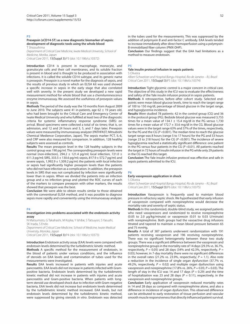
specifi cally, in adult intensive care [1-3]. We undertook a prospective point prevalence study with the aim of identifying the prevalence of physical and pharmacological antipyretic therapies in intensive care patients with sepsis and infl ammation. We also recorded the indication for antipyretic therapies, temperature measurement site, and mean temperatures on the study day.Methods We conducted a single-day observational point prevalence study in 38 ICUs in Australia and New Zealand. All patients in participating ICUs at a 10:00 am census point were studied. Data were collected for the 24-hour study day that included the 10:00 am time point.Results We studied 506 patients, with a mean age 59 years (SD = 17 years); 65% male; APACHE II score 17 (SD = 7), 28-day mortality 14%. Eighty percent of the ICU admissions were unplanned. Of the 506 patients, 311 patients had sepsis and infl ammation with mean peak temperature of 37.3°C (SD = 0.8°C). Of these, 35% (n = 100/311) had a mean peak temperature above 38°C. In the 24-hour period, paracetamol was used 50% (n = 152/311) of the time, nonsteroidal anti-infl ammatory drugs (NSAIDs) 0.6% (n = 2/311) and physical cooling 1% (n = 3/311) (Figure 1). Of patients that had an indication for paracetamol recorded, 64% was for pain (n = 92/152), 18% for both pain and fever (n = 26/152); and 10% for fever alone (n = 14/152) (Figure 2). Sixty-four percent (n = 92/152) of the patients who had paracetamol were prescribed regular paracetamol and 36% (n = 51/143) had a PRN order. Of the 40 patients who received paracetamol for an indication of fever, the mean peak temperature was 38.3°C (SD = 0.8°C; range 36.1 to 40.2°C). Of the three patients who received physical cooling, the mean peak temperature was 39.2°C (SD = 0.9°C; range 38.5 to 40.2°C). Temperature measurement sites were mainly noncore (n = 251/311) with axillary (37%; n = 116/311) and tympanic (35%; n = 110/311) most common (Figure 3).Conclusion This point prevalence study of intensive care patients with sepsis and infl ammation identifi ed pharmacological antipyretics are used regularly for pain management rather than fever management, with paracetamol the most common therapy. The use of physical cooling was rare, and noncore temperature measurements were common. These results are important in understanding current temperature management practice in intensive care and will aid in designing future clinical trials on the subject.Acknowledgements This study was undertaken as part of the Australian and New Zealand Intensive Care Society (ANZICS) Clinical Trials Group (CTG) Point Prevalence Program. The authors would like to thank all participating sites.References1. Lamb SA, Henry RL: Paracetamol pro re nata orders: an audi t. J Paediatr
Child Health 2004, 40:213-216.
2. Isaacs S, Axelrod P, Lorber B: Antipyretic orders in a uni versity hospital. Am J
Med 1990, 88:31-35.
3. Albertson T, Walker V, Stebbins M, Ashton E, Owen K, Sutte r M: A population study of the frequency of high-dose acetaminophen prescribing and dispensing. Ann Pharmacother 2010, 44:1191-1195.
Figure 1 (abstract P17). Type of antipyretic and physical cooling used on the study day (n = 311).
Critical Care 2011, Volume 15 Suppl 3 http://ccforum.com/supplements/15/S3
S8

P18
A survey of fever management in febrile intensive care patients without neurological injuryMK Saxena1,2,3, NE Hammond1, C Taylor1,4, P Young5, MC Reade6,
R Bellomo6, J Myburgh1,2,3
1The George Institute for Global Health, Sydney, NSW, Australia; 2St George
Hospital, Sydney, NSW, Australia; 3St George Clinical School, University of
New South Wales, Sydney, NSW, Australia; 4Sydney Medical School, University
of Sydney, NSW, Australia; 5Medical Research Institute of New Zealand,
Wellington, New Zealand; 6Austin Hospital & University of Melbourne, VIC,
Australia
Critical Care 2011, 15(Suppl 3):P18 (doi: 10.1186/cc10387)
Introduction Fever is a common observation during critical illness [1,2] and may be due to many possible causes such as infection, sterile infl ammation and neurological injury. Clinical trials of fever management lack suffi cient methodological quality to answer the question of whether attempts at reduction in temperature improves patient-centred outcomes in patients with sepsis, infl ammation or neurological injury [3-7]. We undertook a survey to describe the attitudes of critical care clinicians in Australia and New Zealand towards fever management in critically ill patients without neurological injury or hyperthermic syndromes.Methods An online scenario-based questionnaire survey was distributed to medical and nursing members of the Australian and New Zealand Intensive Care Society Clinical Trials Group (ANZICS-CTG) and their intensive care colleagues. Main outcome measures: the choice of drug and preferred threshold temperature for intervention with antipyretics in clinical practice and in a clinical trial.Results There were 588 email invitations distributed through the ANZICS-CTG and Research Coordinator mailing list. Four hundred and forty-seven responses were received from 308 nurses (69%), 137 doctors (31%), and two others (0.5%). The majority of respondents having more than 8 years of experience (62%) worked in mixed medical and
surgical units (84%) in a metropolitan or tertiary hospital setting (77%). The primary fi ndings of our survey suggest that fever management is highly variable. Most clinicians administer an intervention to reduce temperature at or below 39°C (Figure 1); and initially use a combination of both pharmacological and physical interventions, with an increase in intensity of physical interventions for persistent fever (Figure 2). There were diff erences between the professions, with doctors choosing higher temperature thresholds for intervention and nurses generally using more physical cooling (Figure 1 and Table 1); fourthly, temperature thresholds for a clinical trial were 39.0°C (SD = 0.7°C) for a permissive strategy and 38.0°C (SD = 0.75°C) for an intensive strategy; fi nally, there was broad support for a clinical trial of fever management.Conclusion This survey suggests there is considerable clinical variability in fever management in patients with sepsis and without neurological injury or hyperthermic syndromes. At present, no particular management strategy is known to be superior to any other and it remains possible that current practice may be harming substantial numbers of patients. A temperature threshold of up to 40°C may be acceptable to clinicians for the design of a future randomized controlled trial. Further observational data may be informative for the design of such clinical trials.
Figure 2 (abstract P17). Indication for paracetamol administration (n = 152).
Figure 3 (abstract P17). Temperature measurement site for patients with sepsis and infl ammation (n = 311). PAC, pulmonary artery catheter.
Table 1 (abstract P18). Preference of fi rst-line and second-line interventional
category of antipyretic by profession
First line (n = 418) Second line (n = 409)
Nurse Doctor P Nurse Doctor P (%) (%) value (%) (%) value
Pharmacological only 23 40 0.0002 5 5 0.87
Physical only 13 5 0.0087 58 62 0.36
Pharmacological and physical 64 55 0.077 38 33 0.39
Critical Care 2011, Volume 15 Suppl 3 http://ccforum.com/supplements/15/S3
S9
References1. Laupland KB, Shahpori R, Kirkpatrick AW, Ross T, Gregson DB, Stelfox HT:
Occurrence and outcome of fever in critically ill adults. Crit Care Med 2008,
36:1531-1535.
2. Ryan M, Levy M. Clinical review: fever in intensive care unit patients. Crit
Care 2003, 7:221-225.
3. Bernard GR, Wheeler AP, Russell JA, et al.: The eff ects of ibuprofen on the physiology and survival of patients with sepsis. The Ibuprofen in Sepsis Study Group. N Engl J Med 1997, 336:912-918.
4. Hammond NE, Boyle M: Pharmacological versus non-pharmacological antipyretic treatments in febrile critically ill patients: a systematic review and meta-analysis. Aust Crit Care 2011, 24:4-17.
5. Schulman CI, Namias N, Doherty J, et al.: The eff ect of antipyre tic therapy upon outcomes in critically ill patients: a randomized, prospective study. Surg Infect 2005, 6:369-375.
6. Saxena M, Andrews P, Cheng A: Modest cooling therapies (35°C to 37.5°C) for traumatic brain injury. Cochrane Database Syst Rev 2008, 3:CD006811.
7. Jeff eries S, Weatherall M, Young P, et al.: The eff ect of antipyre tic medications on mortality in critically ill patients with infection: a systematic review and meta-analysis. Crit Care Resusc 2011, 13:125-131.
P19
Assessment of the usefulness of presepsin (soluble CD14 subtype) in s eptic patientsT Nishida1, H Ishikura1, A Murai1, Y Irie1, T Ume mura1, T Kamitani1, S Endo2
1Depar tment of Emer gency and Critical Care Medicin e, Fukuoka Un iversi ty
Hospital, Fukuoka, Japan; 2Department of Critical Care Medicine, School of
Medicine, Iwate Medi cal University, Iwate, Japan
Critical Care 2011, 15(Suppl 3):P19 (doi: 10.1186/cc10388)
Introduction Sepsis is a life-threatening condition characterized by a whole-body infl ammatory state. The early diagnosis and treatments of sepsis will improve the outcome of patients. The aim of this study was to compare blood levels of presepsin (renamed from soluble CD14
subtype), procalcitonin (PCT), IL-6 and C-reactive protein (CRP) and to investigate the most useful biomarker for early diagnosis of sepsis.Methods A single-center, prospective, observational study. Patients who had one or more systemic infl ammatory response syndrome criteria were included in this study. The blood samples for measuring the biomarkers were collected and the severity of sepsis was evaluated at the time of admission and every other day for a week. Forty-two patients were enrolled for the prospective study from June 2010 to December 2010.Results Twenty-three patients were diagnosed with sepsis and 19 patients were without sepsis. In the receiver operating characteristics (ROC) curve analysis, the area under the curve (AUC) to distinguish sepsis was the largest for presepsin (0.930) followed by IL-6 (0.896), PCT (0.854) and CRP (0.840). Presepsin may be able to discriminate between patient groups with or without sepsis. From the ROC curve analysis, a cut-off value of presepsin was 929 pg/ml with sensi tivity and specifi city of 76% and 81%, respectively, with odds ratios and 95% CIs of 0.996 (0.992 to 0.998) and 3.376 (1.497 to 6.094). And the presepsin values were signifi cantly higher in the patients with the more severe septic condition (for example, sepsis, severe sepsis, septic shock). In addition, a signifi cant correlation was found between the Sepsis-related Organ Failure Assessment scores and the presepsin values (r2 = 0.320; P = 0.0003). But there was a no signifi cant correlation between APACHE II scores and the presepsin values.Conclusion In this study, presepsin is the most valuable predictor about sepsis compared with PCT, IL-6 and CRP. Moreover, the results suggest that presepsin values can serve as a parameter that closely refl ects the pathology. So we strongly suggest that the presepsin will be not only a very useful new biomarker of the diagnosis of sepsis, but also useful for monitoring the severity of the disease in the near future.
P20
An audit of awareness about maternal sepsis in a UK district general hospitalC Donohue, E Teh, M Kitching
Department of Anaesthesia, Lister Hospital, Stevenage, UK
Critical Care 2011, 15(Suppl 3):P20 (doi: 10.1186/cc10389)
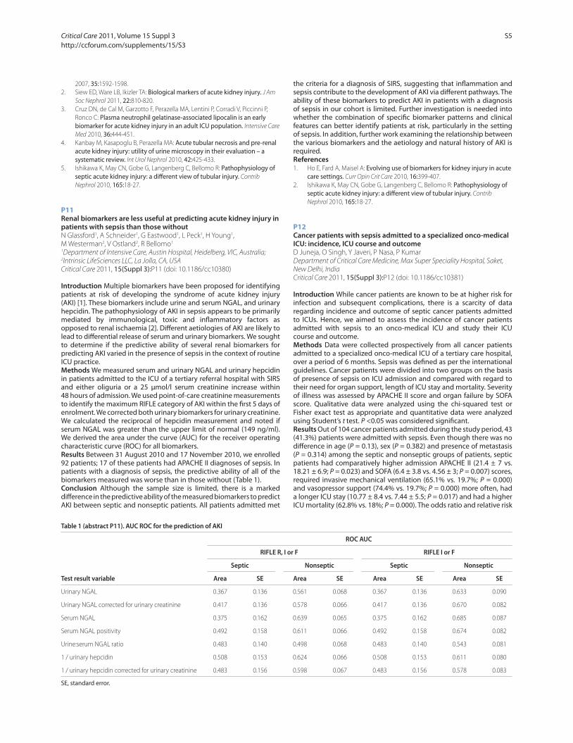
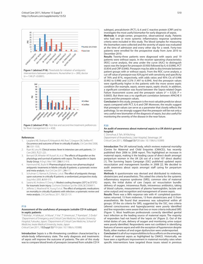
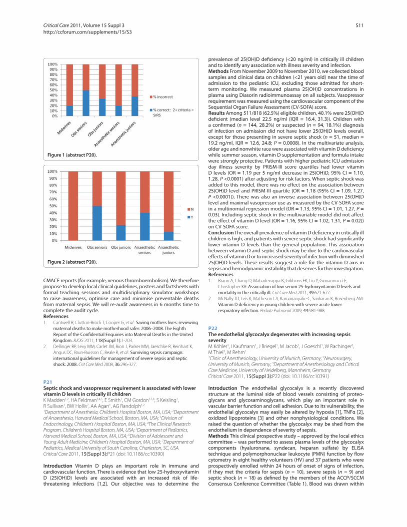
Introduction The UK national body, which reviews maternal mortality (Centre for Maternal and Child Enquiries (CMACE)), has recently published their 2006 to 2008 report. This highlighted an increase in maternal sepsis, making it the leading cause of direct death amongst peripartum women in the UK (26 out of a total 107 direct deaths) [1]. The Surviving Sepsis Campaign (SSC) published updated sepsis resuscitation and management bundles in 2008 [2]. We decided to audit awareness about sepsis amongst staff caring for perpartum women.Methods A questionnaire was devised and distributed to midwives, obstetricians and anaesthetists. This asked the criteria for the systemic infl ammatory response syndrome (SIRS), common sites of maternal sepsis, the initial duties of care (‘sepsis six’ resuscitation bundle: delivery of oxygen, intravenous fl uids, intravenous antibiotics, taking of blood cultures, measurement of plasma haemoglobin, lactate and urine output) and recognition and management of severe sepsis.Results There was a 98% response rate with 41 completed question-naires returned, 15 from midwives, 13 from obstetricians and 13 from anaesthetists. We found that awareness was suboptimal within all groups. Of the six criteria for SIRS, suggested by the SSC, two criteria (altered consciousness and hyperglycaemia) were poorly identifi ed and few responders were aware that two or more criteria indicated SIRS (Figure 1). Most healthcare professionals correctly identifi ed genital tract infection as the leading source of maternal sepsis. The majority of responders had not heard of the ‘sepsis six’ (Figure 2). Out of the initial duties of care, delivery of oxygen and monitoring urine output were poorly identifi ed. Respondents were not confi dent in identifying features of severe sepsis and with the exception of hypotension despite fl uids, other markers of end organ dysfunction were underreported.Conclusion It is all of our responsibilities to focus eff orts on the emerging threat of maternal sepsis as highlighted by CMACE. Historically, we have seen a signifi cant improvement in maternal mortality rates when specifi c interventions have targeted those issues raised in previous
Figure 1 (abstract P18). Thresholds for initiation of antipyretic
interventions between professions. Nurse/other (n = 289); doctor
(n = 134) (P <0.0001).
Figure 2 (abstract P18). First-line and second-line treatment preferences
for fever management (n = 458).
Critical Care 2011, Volume 15 Suppl 3 http://ccforum.com/supplements/15/S3
S10
CMACE reports (for example, venous thromboembolism). We therefore propose to develop local clinical guidelines, posters and factsheets with formal teaching sessions and multidisciplinary simulator workshops to raise awareness, optimise care and minimise preventable deaths from maternal sepsis. We will re-audit awareness in 6 months time to complete the audit cycle.References1. Cantwell R, Clutton-Brock T, Cooper G, et al.: Saving mothers lives: reviewing
maternal deaths to make motherhood safer: 2006–2008. The Eighth Report of the Confi dential Enquiries into Maternal Deaths in the United Kingdom. BJOG 2011, 118(Suppl 1):1-203.
2. Dellinger RP, Levy MM, Carlet JM, Bion J, Parker MM, Jaeschke R, Reinhart K,
Angus DC, Brun-Buisson C, Beale R, et al.: Surviving sepsis campaign: international guidelines for management of severe sepsis and septic shock: 2008. Crit Care Med 2008, 36:296-327.
P21
Septic shock and vasopressor requirement is associated with lower vitamin D levels in critically ill childrenK Madden1,2, HA Feldman3,4,5, E Smith1, CM Gordon3,5,6, S Keisling1,
R Sullivan1, BW Hollis7, AA Agan1, AG Randolph1,2
1Department of Anesthesia, Children’s Hospital Boston, MA, USA; 2Department
of Anaesthesia, Harvard Medical School, Boston, MA, USA; 3Division of
Endocrinology, Children’s Hospital Boston, MA, USA; 4The Clinical Research
Program, Children’s Hospital Boston, MA, USA; 5Department of Pediatrics,
Harvard Medical School, Boston, MA, USA; 6Division of Adolescent and
Young Adult Medicine, Children’s Hospital Boston, MA, USA; 7Department of
Pediatrics, Medical University of South Carolina, Charleston, SC, USA
Critical Care 2011, 15(Suppl 3):P21 (doi: 10.1186/cc10390)
Introduction Vitamin D plays an important role in immune and cardiovascular function. There is evidence that low 25-hydroxyvitamin D (25(OH)D) levels are associated with an increased risk of life-threatening infections [1,2]. Our objective was to determine the
prevalence of 25(OH)D defi ciency (<20 ng/ml) in critically ill children and to identify any association with illness severity and infection.Methods From November 2009 to November 2010, we collected blood samples and clinical data on children (<21 years old) near the time of admission to the pediatric ICU, excluding those admitted for short-term monitoring. We measured plasma 25(OH)D concentrations in plasma using Diasorin radioimmunoassay on all subjects. Vasopressor requirement was measured using the cardiovascular component of the Sequential Organ Failure Assessment (CV-SOFA) score.Results Among 511/818 (62.5%) eligible children, 40.1% were 25(OH)D defi cient (median level 22.5 ng/ml (IQR = 16.4, 31.3)). Children with a confi rmed (n = 144, 28.2%) or suspected (n = 94, 18.1%) diagnosis of infection on admission did not have lower 25(OH)D levels overall, except for those presenting in severe septic shock (n = 51, median = 19.2 ng/ml, IQR = 12.6, 24.8; P = 0.0008). In the multivariate analysis, older age and nonwhite race were associated with vitamin D defi ciency while summer season, vitamin D supplementation and formula intake were strongly protective. Patients with higher pediatric ICU admission day illness severity by PRISM-III score quartiles had lower vitamin D levels (OR = 1.19 per 5 ng/ml decrease in 25(OH)D, 95% CI = 1.10, 1.28, P <0.0001) after adjusting for risk factors. When septic shock was added to this model, there was no eff ect on the association between 25(OH)D level and PRISM-III quartile (OR = 1.18 (95% CI = 1.09, 1.27, P <0.0001)). There was also an inverse association between 25(OH)D level and maximal vasopressor use as measured by the CV-SOFA score in a multinomial regression model (OR = 1.13, 95% CI = 1.01, 1.27, P = 0.03). Including septic shock in the multivariable model did not aff ect the eff ect of vitamin D level (OR = 1.16, 95% CI = 1.02, 1.31, P = 0.02)) on CV-SOFA score.Conclusion The overall prevalence of vitamin D defi ciency in critically ill children is high, and patients with severe septic shock had signifi cantly lower vitamin D levels than the general population. This association between vitamin D and septic shock may be due to the cardiovascular eff ects of vitamin D or to increased severity of infection with diminished 25(OH)D levels. These results suggest a role for the vitamin D axis in sepsis and hemodynamic instability that deserves further investigation.References1. Braun A, Chang D, Mahadevappa K, Gibbons FK, Liu Y, Giovannucci E,
Christopher KB: Association of low serum 25-hydroxyvitamin D levels and mortality in the critically ill. Crit Care Med 2011, 39:671-677.
2. McNally JD, Leis K, Matheson LA, Karuananyake C, Sankaran K, Rosenberg AM:
Vitamin D defi ciency in young children with severe acute lower respiratory infection. Pediatr Pulmonol 2009, 44:981-988.
P22
The endothelial glycocalyx degenerates with increasing sepsis severityM Köhler1, I Kaufmann1, J Briegel1, M Jacob1, J Goeschl1, W Rachinger2,
M Thiel3, M Rehm1
1Clinic of Anesthesiology, University of Munich, Germany; 2Neurosurgery,
University of Munich, Germany; 3Department of Anesthesiology and Critical
Care Medicine, University of Heidelberg, Mannheim, Germany
Critical Care 2011, 15(Suppl 3):P22 (doi: 10.1186/cc10391)
Introduction The endothelial glycocalyx is a recently discovered structure at the luminal side of blood vessels consisting of proteo-glycans and glycosaminoglycans, which play an important role in vascular barrier function and cell adhesion. Due to its vulnerability, the endothelial glycocalyx may easily be altered by hypoxia [1], TNFα [2], oxidized lipoproteins [3] and other nonphysiological conditions. We raised the question of whether the glycocalyx may be shed from the endothelium in dependence of severity of sepsis.Methods This clinical prospective study – approved by the local ethics committee – was performed to assess plasma levels of the glycocalyx components (hyaluronane, syndecan, heparan sulfate) by ELISA technique and polymorphonuclear leukocyte (PMN) function by fl ow cytometry in eight healthy volunteers (HV) and 37 patients who were prospectively enrolled within 24 hours of onset of signs of infection, if they met the criteria for sepsis (n = 10), severe sepsis (n = 9) and septic shock (n = 18) as defi ned by the members of the ACCP/SCCM Consensus Conference Committee (Table 1). Blood was drawn within
Figure 1 (abstract P20).
Figure 2 (abstract P20).
Critical Care 2011, Volume 15 Suppl 3 http://ccforum.com/supplements/15/S3
S11

24 hours after onset of sepsis. Informed consent was obtained from all patients or their legal representatives, respectively.Results Plasma levels of the glycocalyx components were signifi cantly higher in septic patients than in healthy volunteers and even more pronounced in patients with severe sepsis and septic shock (all P <0.05; Figure 1). Hyaluronan and syndecan plasma levels correlated positively with the APACHE II, SOFA and MOD scores (Figure 1 and Table 2). Hyaluronan displayed a positive correlation with the C-reactive protein, procalcitonin and IL-6 in plasma (Table 3). The PMN dysfunction – characterized by an increase in cytotoxic capability and a decrease in microbicidity – showed a parallel course to the heparan sulfate plasma levels.Conclusion Elevated plasma levels of hyaluronan, syndecan and heparan sulfate are suggestive of a glycocalyx shedding from endothelium with increasing sepsis severity. This process might contribute to the vascular dysfunction and development of edema in septic patients.References1. Rehm M, Bruegger D, Christ F, et al.: Shedding of the endothelial glycocalyx
in patients undergoing major vascular surgery with global and regional ischemia. Circulation 2007, 116:1896-1906.
2. Henry CB, Duling BR: TNFα increases entry of macromolecules into luminal endothelial cell glycocalyx. Am J Physiol Heart Circ Physiol 2000,
279:H2815-H2823.
3. Vink H, Constantinescu AA, Spaan JA: Oxidized lipoproteins degrade the endothelial surface layer: implications for platelet-endothelial cell adhesion. Circulation 2000, 101:1500-1502.
P23
Abstract withdrawn
Figure 1 (abstract P22). (a) to (c) Increase in the glycocalyx components in plasma of healthy volunteers (HV) and of patients with increasing sepsis severity.
(d) Correlation between APACHE II score of septic patients and hyaluronan plasma concentrations.
Table 1 (abstract P22). Demographic data
Healthy Severe Septic
volunteers Sepsis sepsis shock
(n = 8) (n = 10) (n = 9) (n = 18)
Age (years) 29.1 ± 2.9 51.6 ± 19.7 63.3 ± 23.5 63.3 ± 21.4
APACHE II n.b. 7.6 ± 3.9 17.8 ± 6.9 27.9 ± 5.3
MOD n.b. 2.1 ± 1.6 6.9 ± 3.2 9.4 ± 3.6
SOFA n.b. 4.1 ± 2.8 9.0 ± 3.0 13.3 ± 3.4
Table 2 (abstract P22). Correlation between the glycocalyx components
(hyaluronan, syndecan) and the APACHE II, SOFA and MOD score of septic
patients
APACHE II SOFA MOD
Hyaluronan r2 = 0.583, r2 = 0.529, r2 = 0.435,
P = 0.000 P = 0.001 P = 0.008
Syndecan r2 = 0.425, r2 =0.476, r2 = 0.529,
P = 0.010 P = 0.003 P = 0.001
Table 3 (abstract P22). Correlation between the glycocalyx components
(heparan sulfate, hyaluronan) and the C-reactive protein, procalcitonin and
IL-6 in plasma of septic patients
CRP PCT IL-6
Heparan sulfate r2 = –0.63, r2 = 0.20, r2 = 0.505,
P = 0.714 P = 0.928 P = 0.012
Hyaluronan r2 = 0.398, r2 = 0.723, r2 = 0.468,
P = 0.016 P = 0.000 P = 0.021
Critical Care 2011, Volume 15 Suppl 3 http://ccforum.com/supplements/15/S3
S12

P24
Early peak temperature and mortality in critically ill patients with or without infectionM Saxena1,2, P Young3,4, R Beasley3,4, M Bailey5, R Bellomo5, D Pilcher6,
S Finfer1,7, D Harrison8, J Myburgh1,2, K Rowan8
1George Institute for Global Health, Sydney, NSW, Australia; 2St George Clinical
School, University of New South Wales, Sydney, NSW, Australia; 3Intensive
Care Unit, Wellington Regional Hospital, Capital and Coast District Health
Board, Wellington, New Zealand; 4Medical Research Institute of New Zealand,
Wellington, New Zealand; 5Australian and New Zealand Intensive Care
Research Centre, School of Public Health and Preventive Medicine, Monash
University, Melbourne, VIC, Australia; 6Australian and New Zealand Intensive
Care Society Centre for Outcome and Resource Evaluation, Melbourne, VIC,
Australia; 7Sydney Medical School, University of Sydney, NSW, Australia; 8Intensive Care National Audit and Research Centre, London, UK
Critical Care 2011, 15(Suppl 3):P24 (doi: 10.1186/cc10393)
Introduction The febrile response in the context of infection may be linked to a protective host response through enhanced immune function at elevated body temperatures [1-9]. Alternatively the use of antipyretics may reduce metabolic expense, patient discomfort, or protect against neurological injury.Objective To determine whether fever is associated with reduced risk of death in patients admitted to an ICU with infection compared with other patients.Methods A retrospective cohort study using a database of Australian and New Zealand (ANZ) ICU admissions as a development cohort and a database of UK ICU admissions as a validation cohort. The sample included 129 ICUs in ANZ and 201 ICUs in the UK. The ANZ development cohort consisted of 269,078 patients and the UK validation cohort consisted of 366,973 patients. All patients were admitted to an ICU between 2005 and 2009. A total of 29,083/269,078 (10.8%) ANZ patients and 103,191/366,973 (28.1%) UK patients were categorised as having an infection at the time of ICU admission. The main outcome measures were the association between peak temperature in the fi rst 24 hours after ICU admission and in-hospital mortality in patients admitted with or without infection.
Results In the ANZ cohort, adjusted in-hospital mortality risk progressively decreased with increasing peak temperature in patients with infection. Relative to 36.5 to 36.9°C, the lowest risk was at 39 to 39.4°C (adjusted OR = 0.56; 95% CI = 0.48 to 0.66). In patients without infection, the adjusted mortality risk progressively increased above 39.0°C (adjusted OR = 2.07 at ≥40.0°C; 95% CI = 1.68 to 2.55). In the UK cohort, fi ndings were similar with adjusted odds ratios at corresponding temperatures of 0.77 (95% CI = 0.71 to 0.85) and 1.94 (95% CI = 1.60 to 2.34) for the infection and non-infection groups, respectively. See Figures 1 and 2.Conclusion Peak temperature in the fi rst 24 hours in the ICU is associated with decreased in-hospital mortality in critically ill patients with an infection; randomised trials are needed to compare the eff ect on mortality of controlling fever against a permissive approach to fever management in such patients.References1. Kluger MJ: Fever: Its Biology, Evolution and Function. Princeton, NJ: Princeton
University Press; 1979:224 pp.
2. Mackowiak PA: Fever: blessing or curse? A unifying hypothesis. Ann Int Med
1994, 120:1037-1040.
3. Hench PS, Slocumb CH, Popp WC: Fever therapy: results for gonorrheal arthritis, chronic infectious (atrophic) arthritis, and other forms of ‘rheumatism’. J Am Med Assoc 1935, 104:1779-1790.
4 Wagner-Jauregg J: Uber die Einwirkung der Malaria auf die progressive Paralyse. Psychiatrisch-neurologische Wochenschrift 1919, 20:251-255.
5. Eyers S, Weatherall M, Shirtcliff e P, Perrin K, Beasley R: The eff ect on mortality of antipyretics in the treatment of infl uenza infection: Systematic review and meta-analysis. J R Soc Med 2010, 103:403-411.
6. Shann F: Antipyretics in severe sepsis. Lancet 1995, 345:338.
7. Doran T, de Angelis C, Baugardner R, Mellits E: Acetaminophen: more harm than good for chickenpox? J Pediatr 1989, 114:1045-1048.
8. Brandts CH, Ndjave M, Graninger W, Kremsner PG: Eff ect of paracetamol on parasite clearance time in plasmodium falciparum malaria. Lancet 1997,
350:704-709.
9. Stanley E, Jackson G, Panusarn C, Rubenis M, Dirda V: Increased virus shedding with aspirin treatment of rhinovirus infection. JAMA 1975,
231:1248-1251.
Figure 1 (abstract P24). Adjusted* odds ratios for in-hospital mortality
versus peak temperature in the fi rst 24 hours in the ICU for patients in
the infection group. *Odds ratios adjusted for illness severity using the
ICNARC (2009) model predicted log odds of acute hospital mortality with
the temperature component removed for the UK data and the APACHE
III predicted log odds risk of death with the temperature component
removed for the ANZ data.
Figure 2 (abstract P24). Adjusted* odds ratios for in-hospital mortality
versus peak temperature in the fi rst 24 hours in the ICU for patients in the
non-infection group. *Odds ratios adjusted for illness severity using the
ICNARC (2009) model predicted log odds of acute hospital mortality with
the temperature component removed for the UK data and the APACHE
III predicted log odds risk of death with the temperature component
removed for the ANZ data.
Critical Care 2011, Volume 15 Suppl 3 http://ccforum.com/supplements/15/S3
S13

P25
Role of procalcitonin and high-sensitivity C-reactive protein in sepsis: a prospective studyS Das1, D Anand1, S Ray2, S Bhargava1, A Manocha1, M Kankra1,
LM Srivastava1
1Department of Biochemistry, Sir Ganga Ram Hospital, New Delhi, India; 2Department of Critical Care Medicine, Sir Ganga Ram Hospital, New Delhi, India
Critical Care 2011, 15(Suppl 3):P25 (doi: 10.1186/cc10394)
Introduction Sepsis is the most common cause of morbidity and mortality in ICU patients. The clinical signs of infection and routine laboratory tests are not specifi c and at times misleading. Even the bacteriological evidence of infection is not sensitive enough. In view of this diagnostic and therapeutic dilemma, an eff ective and specifi c marker is needed that can support or exclude the diagnosis of infection. Hence we evaluated the usefulness of biochemical markers such as procalcitonin (PCT) and high-sensitivity C reactive protein (hsCRP) in monitoring the therapeutic response to treatment in patients with sepsis.Methods Fifty-seven patients admitted to the ICU of Sir Ganga Ram Hospital, New Delhi, India from July 2010 to April 2011 with a fresh episode of sepsis were included in the study. PCT and hsCRP were analyzed in serum samples at 0 hours, 24 hours and 72 hours. Blood cultures were performed on the day of admission (0 hours) and the patients were categorized as culture positive or culture negative. Patients were followed up for 28 days and were then grouped as survivors and nonsurvivors.Results During the observation period of 28 days, 44 patients survived and 13 expired. Over a period of 0 to 72 hours the PCT level decreased in 86% survivors as compared with 46% nonsurvivors (P =0.001), whereas it increased in 13.6% survivors as compared with 53.8% nonsurvivors (P = 0.001). The change in the levels of hsCRP in both surviving and nonsurviving patients was not signifi cant. The 0-hour blood culture was positive in 24 out of 57 patients. The PCT levels at 0 hours was signifi cantly high in culture-positive patients (P = 0.002) as compared with culture-negative patients, whereas there was no signifi cant diff erence in the hsCRP levels in the above-mentioned groups. The area under the curve of PCT and hsCRP in culture-positive and culture-negative patients was 0.743 (95% CI = 0.608 to 0.878, P = 0.002) and 0.564 (95% CI = 0.414 to 0.715, P = 0.405), respectively.Conclusion These observations indicate that PCT is a better prognostic indicator than hsCRP and is also a better predictor of bacteremia in the newly admitted critically ill patients of sepsis. Hence, routine use of PCT as a monitoring tool may aid in appropriate therapeutic intervention in patients with sepsis.Acknowledgements The authors thank the Indian Council of Medical Research, New Delhi, India for fi nancial support.
P26
Prehospital identifi cation of sepsis patients and alerting of receiving hospitals: impact on early goal-directed therapyK Halimi1, J Freeman-Garrick2, C Agcaoili1, K Choy1, F Claridge3, M Jacobs3,
M Taigman4
1Department of Emergency Medicine and Intensive Care, Washington
Hospital, Fremont, CA, USA; 2Highland Hospital, Oakland, CA, USA; 3Alameda
County Emergency Medical Services, San Leandro, CA, USA; 4American
Medical Response, Alameda County, CA, USA
Critical Care 2011, 15(Suppl 3):P26 (doi: 10.1186/cc10395)
Introduction Over the past several years, the early identifi cation and aggressive treatment of sepsis patients has become a standard of care in the hospital setting. A relatively small number of emergency medical service (EMS) systems have started programs to screen for sepsis; an even smaller number provide treatment based on that screening process in the prehospital setting.Objective The purpose of this study is twofold. First, the study aims to determine how eff ectively paramedics working in the county EMS system can use a screening tool to identify potential sepsis patients and provide an alert to the receiving hospital. Second, the study will examine whether or not an early identifi cation process in the fi eld leads to improved treatment of sepsis. The end goal is to reduce morbidity and mortality of sepsis patients in the hospital setting.
Methods This is a multi-site prospective observational study with comparison to retrospective cohort. Patient data will be collected to determine whether or not the alert process leads to early obtaining of a serum lactate measurement and early goal-directed therapy.Results Data points being analyzed from prehospital care reports: criteria from the sepsis screening tool include evidence of infection, temperature, heart rate, respiratory rate; and EMS fi eld clinical impression (for comparison with emergency department (ED) admitting diagnosis). Data points being analyzed in the hospitals include the following: ED admitting diagnosis; serum lactate values, blood culture, timestamps; evidence of early goal-directed therapy – timestamps/values for fl uid and antibiotic administration; hospital admitting diagnosis; and discharge diagnosis. The results of the study will be available by 2012/2013.Conclusion Preliminary anecdotal and early data analysis reports from EMS and hospital staff suggest that patients are being identifi ed as septic prior to ED arrival and have lower lactate levels. Patients are also treated more timely on arrival to the ED for sepsis. Final data analysis will shed more light on our hypothesis. Early identifi cation of septic patients has implications for further research both in the fi eld and hospital settings.
P27
Abstract withdrawn
P28
Thalidomide in combination with augmentin (amoxicillin with clavulanic acid) protects BALB/c mice suff ering from Klebsiella pneumoniae B5055-induced sepsisV Kumar, S Chhibber
Department of Microbiology, Panjab Univeristy, Chandigarh, India
Critical Care 2011, 15(Suppl 3):P28 (doi: 10.1186/cc10397)
Introduction Despite extensive research in the fi eld of sepsis pathogenesis and its management, mortality associated with sepsis in hospitals remains very high. For example, more than 18 million people are aff ected by sepsis worldwide and have an expected 1% increase annually in ICUs. Sepsis is the outcome of a deregulated immune system occurring during systemic bacterial (that is, Gram-negative or Gram-positive) infection. So modulating the immune system by an immunomodulatory approach may prove benefi cial to sepsis patients. In the present study, we evaluated the protective immunomodulatory eff ect of thalidomide alone or with augmentin in Klebsiella pneumoniae B5055-induced sepsis in BALB/c mice.Methods The mouse model of sepsis was developed by placing K. pneumoniae B5055 entrapped in fi brin and thrombin clots in the peritoneal cavity of mice. The septic mice were treated with thalidomide alone (30 mg/kg/day p.o.), with augmentin alone (20 μg/ml i.p.) and with their combination. The bacterial load in blood was estimated by blood culture on MacConkey’s agar plates along with measuring the other systemic infl ammatory parameters. For example, lipid peroxidation was measured in terms of malondialdehyde (MDA) and nitric oxide (NO) levels in serum by biochemical methods. Levels of proinfl ammatory cytokines (that is, TNFα and IL-1α) and anti-infl ammatory cytokine (that is, IL-10) levels in serum were measured by ELISA.Results The thalidomide-alone-treated mice showed 75% survival whereas 60% of the augmentin-alone-treated group survived. However, their combination (thalidomide + augmentin) treatment provided 100% survival. Treatment with thalidomide alone signifi cantly (P <0.05) decreased TNFα, IL-1α, NO and MDA levels in the serum without signifi cantly (P <0.05) decreasing the bacterial count in blood. However, levels of IL-10 in serum were found to be signifi cantly (P <0.05) elevated upon thalidomide treatment. Augmentin alone only decreased the bacterial load in blood signifi cantly (P <0.05), while no signifi cant decrease was observed on infl ammatory mediators studied. However, a combination thalidomide with augmentin signifi cantly (P <0.05) decreased both the bacterial count as well as infl ammatory mediators (that is, TNFα, IL-1α, NO and MDA) and provided 100% protection to animals.Conclusion Thalidomide can be used as an immunomodulatory agent along with antibiotics for sepsis management.
Critical Care 2011, Volume 15 Suppl 3 http://ccforum.com/supplements/15/S3
S14

P29
A novel DDAH-1 inhibitor improved sepsis-induced impairment in vasoreactivity to noradrenaline in a rat endotoxaemia modelZ Wang, V Taylor, R Stidwill, J Leiper, M Singer
Bloomsbury Institute of Intensive Care Medicine, Department of Medicine,
University College London, UK
Critical Care 2011, 15(Suppl 3):P29 (doi: 10.1186/cc10398)
Introduction In septic shock, iNOS activation and nitric oxide (NO) overproduction contribute to vascular hyporeactivity to adrenergic vasopressors. The consequent hypotension often necessitates high doses of catecholamine administration. However, this may lead to detrimental eff ects on tissue perfusion, immune function and myo-cardial function. Asymmetric dimethlyarginine (ADMA), an endo ge nous inhibitor of NO synthase, is extensively metabolised by dimethylarginine dimethylaminohydrolase (DDAH). Competitive inhi bi tion of the DDAH-1 isoform should thus reverse hypotension but, as this isoform is absent in immune cells, it should not compromise the immune eff ects of NO. Hence, we investigated whether L257, a novel DDAH-1 inhibitor, could spare norepinephrine dosing in a rat endotoxic shock model.Methods Anaesthetised, spontaneously breathing male Wistar rats (body weight 270 to 330 g) had their left carotid artery and right internal jugular vein cannulated for arterial pressure monitoring and fl uid infusion, respectively. Then 40 mg/kg Klebsiella pneumoniae lipopolysaccharide was administered intravenously over 30 minutes followed by fl uid resuscitation at a rate of 10 ml/kg/hour thereafter. When the mean arterial pressure fell over 20% below baseline, they received norepinephrine titrated to maintain arterial pressure at ±10% baseline. Thirty minutes post commencement of norepinephrine, animals were randomized to receive either L-257 (3 mg/kg bolus then infusion of 125 μg/hour) or, in controls, an equivalent volume of saline. Experiments were terminated 3 hours post commencement of norepinephrine titration, before which echocardiography was performed and serum samples were collected for biochemistry.Results L-257-treated animals (n = 8) required a signifi cantly lower total dose of noradrenaline over 3 hours compared with the eight control animals (38 ± 9 vs. 48 ± 4 μg, P <0.05). The heart rate was signifi cantly
lower in the treatment group (P <0.05), which associated with a trend of increased stroke volume and cardiac output. Serum BUN and urea were also signifi cantly lower in the treatment group (P <0.05, Table 1).Conclusion In this acute endotoxic rat model, we demonstrate that DDAH-1 inhibition by L-257 could reduce norepinephrine dosage and ameliorate its harmful eff ects. This agent warrants further study as a putative therapy for septic shock.Acknowledgements This study was funded by Wellcome Trust in the UK.
P30
A novel DDAH-1 inhibitor improved cardiovascular function in a short-term anesthetized rat model of sepsisZ Wang, V Taylor, R Stidwill, J Leiper, M Singer
Bloomsbury Institute of Intensive Care Medicine, Department of Medicine,
University College London, UK
Critical Care 2011, 15(Suppl 3):P30 (doi: 10.1186/cc10399)
Introduction Excessive NOS activity and NO overproduction are believed to play an important role in sepsis-induced macrocirculatory and microcirculatory dysfunction. Asymmetric dimethylarginine (ADMA), an endogenous inhibitor of NO synthesis, is extensively metabolised by dimethylarginine dimethylaminohydrolase (DDAH).
Table 1 (abstract P29).
Variable NE NE + L-257 P value
SV (ml/minute) 0.18 ± 0.04 0.23 ± 0.04 0.07
HR (beats/minute) 500 ± 15 449 ± 37 0.03
CO (ml) 90 ± 18 102 ± 19 0.31
U (mM) 16.6 ± 1.2 12.8 ± 1.7 0.01
BUN (mg/dl) 44.8 ± 3.7 35.9 ± 4.8 0.03
Cr (μM) 51.3 ± 15.2 31.8 ± 4.3 0.08
ALT (IU/l) 98 ± 79.8 71.3 ± 48.3 0.6
Figure 1 (abstract P30). (a) The eff ect of bolus doses of L-257 at 0 mg/kg, 3 mg/kg, 30 mg/kg and 300 mg/kg on the mean arterial pressure in the short-
term organ function study on anaesthetized Wistar rats (n = 6 in the each group). The bolus L-257 was given at 0 minutes for 10-minute infusion. Animals
treated with 300 mg/kg L-257, ×; animals treated with 30 mg/kg L-257, open triangle; animals treated with 3 mg/kg, open square; control animals, solid circle.
(b) The initial change of mean arterial pressure 30 minutes after bolus injections among the four groups. *P <0.05, 30 mg/kg vs. control; #P <0.05, 30 mg/kg
vs. 3 mg/kg; †P <0.05, 300 mg/kg vs. control; ‡P <0.05, 300 mg/kg vs. 3 m/kg. (c), (d) The eff ects of bolus doses of L-257 at 0 mg/kg, 3 mg/kg and 30 mg/kg on
perfused capillary density and microcirculatory index (n = 4 in each group). *P <0.05, 30 mg/kg vs. control; #P <0.05, 30 mg/kg vs. 3 m/kg.
Critical Care 2011, Volume 15 Suppl 3 http://ccforum.com/supplements/15/S3
S15

The DDAH-1 isoform is present in vascular smooth muscle so its inhibition should theoretically reverse sepsis-induced hypotension. We thus investigated the dose-dependent cardiovascular eff ects of a novel DDAH-1 competitive inhibitor, L-257, in experimental sepsis.Methods Anaesthetised, spontaneously breathing male Wistar rats (body weight 270 to 330 g) had their left carotid artery and right internal jugular vein cannulated for arterial pressure monitoring and fl uid infusion, respectively. Then 40 mg/kg Klebsiella pneumoniae lipopolysaccharide was administered intravenously over 30 minutes followed by fl uid resuscitation at a rate of 10 ml/kg/hour thereafter. When the mean arterial pressure fell over 20% below baseline, groups (n = 6) were randomized to receive a bolus dose of L-257 of 0 (control), 3, 30 or 300 mg/kg. Animals were sacrifi ced 2 hours later with prior measurement of gastrocnemius muscle microcirculatory perfusion and with collection of plasma samples for biochemistry, arginine, ADMA and nitrate/nitrite measurements.Results The bolus doses of L-257 were given after approximately 60 to 90 minutes post endotoxin when the mean BP fell over 20%. Arterial pressure, perfused capillary density and microcirculatory fl ow index were better maintained than in controls, especially at higher doses (Figure 1, P <0.05). Signifi cantly higher plasma ADMA concentrations and ADMA/arginine ratios were seen in the 30 mg/kg bolus group (Figure 2, P <0.05). Plasma nitrate/nitrite levels in the treated animals were signifi cantly lower compared with those in controls (Figure 2, P <0.05).Conclusion In this short-term rat model of endotoxaemia, we demonstrated protective dose-dependent eff ects of a novel DDAH-1 inhibitor, L-257, on cardiovascular function. This was associated with an elevation of plasma ADMA level and a resultant reduction of plasma nitrate/nitrite level.Acknowledgements This study was funded by Wellcome Trust in the UK.
P31
Early detection of serum enteric bacterial DNA with real-time PCR in patients with SIRSJM-C Yang
Department of Surgery, Kaohsiung Veterans General Hospital, Taiwan, China
Critical Care 2011, 15(Suppl 3):P31 (doi: 10.1186/cc10400)
Introduction Sepsis remains a major and increasing healthcare problem with a mortality exceeding 25%. The early detection of infection is
important in treating sepsis. Nucleic acid amplification methods have the potential to improve the timeliness, sensitivity, and accuracy of the tests used to detect respiratory pathogens. We used a quantitative real-time PCR (rt-PCR) to detect the enteric bacterial counts in blood from patients in the emergency room.Methods EDTA samples were collected from patients with systemic infl ammatory response syndrome (SIRS) presenting to the emergency room after obtaining informed consent. Enteric bacterial loads in blood samples were assayed by rt-PCR to quantitate the bacterial 23S rDNA and EB rDNA loads. Descriptive and clinical data were collected from the medical records and compared with 23S and EB rDNA results.Results From January 2011 to April 2011, 39 patients (mean age 71.15 ± 17.12, range 22 to 93) were enrolled in the study. There was no correlation between serum lactate and enteric bacterial load in patients with SIRS. However, in a subgroup comprising patients presenting with respiratory distress and abnormal blood white cell count, the enteric bacterial rDNA load was higher and showed correlation with serum lactate level. The serum enteric bacterial rDNA loads were signifi cantly higher in patients with positive cultures and in patients presenting with higher serum lactate. Correlations between serum lactate and enteric bacterial rDNA load were also signifi cant in the patients with positive culture results.Conclusion The quantitative assay for enteric bacterial rDNA could be a useful tool to detect early enteric bacterial translocation in patients presenting to the emergency room with elevated serum lactate level or with respiratory distress and abnormal white blood cell counts.
P32
Direct eff ects of esmolol, ultra-short-acting β-blockers, on cardiac function, ion channels, and coronary arteries in guinea pigsS Shibata
Akita University Graduate School of Medicine, Department of Cell Physiology,
Akita, Japan
Critical Care 2011, 15(Suppl 3):P32 (doi: 10.1186/cc10401)
Introduction β1-adrenergic antagonists have been recently used in septic patients to improve sepsis-induced immune, cardiovascular and coagulation dysfunction. But it is diffi cult and one is hesitant to use these drugs in septic shock patients who have already had hypotension
Figure 2 (abstract P30). The eff ect of bolus doses of L-257 at 0 mg/kg, 3 mg/kg and 30 mg/kg on (a) plasma arginine and (b) ADMA concentrations, (c) the
ADMA/arginine ratio and (d) plasma nitrate/nitrite concentrations in the short-term organ function study on anaesthetized Wistar rats (n = 6 in each group).
*P <0.05, 30 mg/kg vs. control; #P <0.05, 30 mg/kg vs. 3 m/kg; †P <0.05, 3 mg/kg vs. control.
Critical Care 2011, Volume 15 Suppl 3 http://ccforum.com/supplements/15/S3
S16
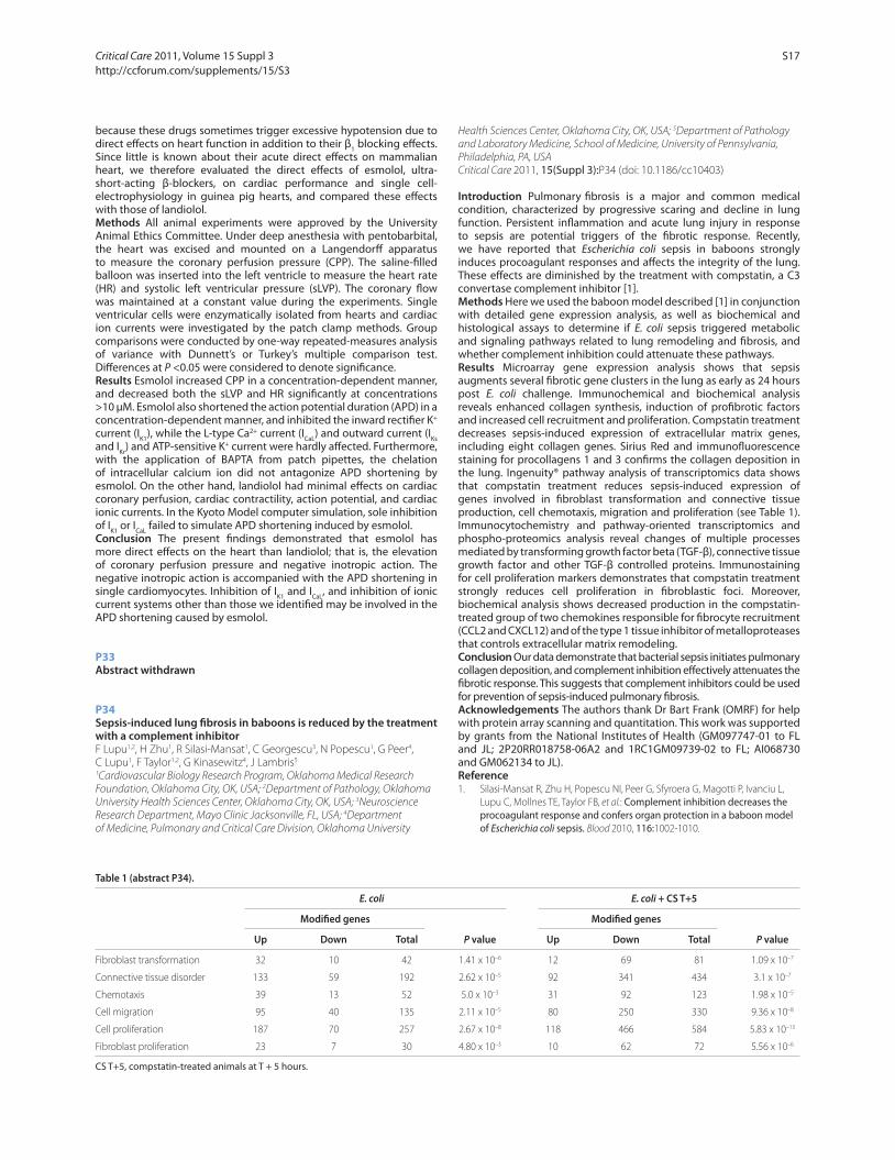
because these drugs sometimes trigger excessive hypotension due to direct eff ects on heart function in addition to their β1 blocking eff ects. Since little is known about their acute direct eff ects on mammalian heart, we therefore evaluated the direct eff ects of esmolol, ultra-short-acting β-blockers, on cardiac performance and single cell-electrophysiology in guinea pig hearts, and compared these eff ects with those of landiolol.Methods All animal experiments were approved by the University Animal Ethics Committee. Under deep anesthesia with pentobarbital, the heart was excised and mounted on a Langendorff apparatus to measure the coronary perfusion pressure (CPP). The saline-fi lled balloon was inserted into the left ventricle to measure the heart rate (HR) and systolic left ventricular pressure (sLVP). The coronary fl ow was maintained at a constant value during the experiments. Single ventricular cells were enzymatically isolated from hearts and cardiac ion currents were investigated by the patch clamp methods. Group comparisons were conducted by one-way repeated-measures analysis of variance with Dunnett’s or Turkey’s multiple comparison test. Diff erences at P <0.05 were considered to denote signifi cance.Results Esmolol increased CPP in a concentration-dependent manner, and decreased both the sLVP and HR signifi cantly at concentrations >10 μM. Esmolol also shortened the action potential duration (APD) in a concentration-dependent manner, and inhibited the inward rectifi er K+ current (IK1), while the L-type Ca2+ current (ICaL) and outward current (IKs and IKr) and ATP-sensitive K+ current were hardly aff ected. Furthermore, with the application of BAPTA from patch pipettes, the chelation of intracellular calcium ion did not antagonize APD shortening by esmolol. On the other hand, landiolol had minimal eff ects on cardiac coronary perfusion, cardiac contractility, action potential, and cardiac ionic currents. In the Kyoto Model computer simulation, sole inhibition of IK1 or ICaL failed to simulate APD shortening induced by esmolol.Conclusion The present fi ndings demonstrated that esmolol has more direct eff ects on the heart than landiolol; that is, the elevation of coronary perfusion pressure and negative inotropic action. The negative inotropic action is accompanied with the APD shortening in single cardiomyocytes. Inhibition of IK1 and ICaL, and inhibition of ionic current systems other than those we identifi ed may be involved in the APD shortening caused by esmolol.
P33
Abstract withdrawn
P34
Sepsis-induced lung fi brosis in baboons is reduced by the treatment with a complement inhibitorF Lupu1,2, H Zhu1, R Silasi-Mansat1, C Georgescu3, N Popescu1, G Peer4,
C Lupu1, F Taylor1,2, G Kinasewitz4, J Lambris5
1Cardiovascular Biology Research Program, Oklahoma Medical Research
Foundation, Oklahoma City, OK, USA; 2Department of Pathology, Oklahoma
University Health Sciences Center, Oklahoma City, OK, USA; 3Neuroscience
Research Department, Mayo Clinic Jacksonville, FL, USA; 4Department
of Medicine, Pulmonary and Critical Care Division, Oklahoma University
Health Sciences Center, Oklahoma City, OK, USA; 5Department of Pathology
and Laboratory Medicine, School of Medicine, University of Pennsylvania,
Philadelphia, PA, USA
Critical Care 2011, 15(Suppl 3):P34 (doi: 10.1186/cc10403)
Introduction Pulmonary fi brosis is a major and common medical condition, characterized by progressive scaring and decline in lung function. Persistent infl ammation and acute lung injury in response to sepsis are potential triggers of the fi brotic response. Recently, we have reported that Escherichia coli sepsis in baboons strongly induces procoagulant responses and aff ects the integrity of the lung. These eff ects are diminished by the treatment with compstatin, a C3 convertase complement inhibitor [1].Methods Here we used the baboon model described [1] in conjunction with detailed gene expression analysis, as well as biochemical and histological assays to determine if E. coli sepsis triggered metabolic and signaling pathways related to lung remodeling and fi brosis, and whether complement inhibition could attenuate these pathways.Results Microarray gene expression analysis shows that sepsis augments several fi brotic gene clusters in the lung as early as 24 hours post E. coli challenge. Immunochemical and biochemical analysis reveals enhanced collagen synthesis, induction of profi brotic factors and increased cell recruitment and proliferation. Compstatin treatment decreases sepsis-induced expression of extracellular matrix genes, including eight collagen genes. Sirius Red and immunofl uorescence staining for procollagens 1 and 3 confi rms the collagen deposition in the lung. Ingenuity® pathway analysis of transcriptomics data shows that compstatin treatment reduces sepsis-induced expression of genes involved in fi broblast transformation and connective tissue production, cell chemotaxis, migration and proliferation (see Table 1). Immunocytochemistry and pathway-oriented transcriptomics and phospho-proteomics analysis reveal changes of multiple processes mediated by transforming growth factor beta (TGF-β), connective tissue growth factor and other TGF-β controlled proteins. Immunostaining for cell proliferation markers demonstrates that compstatin treatment strongly reduces cell proliferation in fi broblastic foci. Moreover, biochemical analysis shows decreased production in the compstatin-treated group of two chemokines responsible for fi brocyte recruitment (CCL2 and CXCL12) and of the type 1 tissue inhibitor of metalloproteases that controls extracellular matrix remodeling.Conclusion Our data demonstrate that bacterial sepsis initiates pulmonary collagen deposition, and complement inhibition eff ectively attenuates the fi brotic response. This suggests that complement inhibitors could be used for prevention of sepsis-induced pulmonary fi brosis.Acknowledgements The authors thank Dr Bart Frank (OMRF) for help with protein array scanning and quantitation. This work was supported by grants from the National Institutes of Health (GM097747-01 to FL and JL; 2P20RR018758-06A2 and 1RC1GM09739-02 to FL; AI068730
and GM062134 to JL).Reference1. Silasi-Mansat R, Zhu H, Popescu NI, Peer G, Sfyroera G, Magotti P, Ivanciu L,
Lupu C, Mollnes TE, Taylor FB, et al.: Complement inhibition decreases the procoagulant response and confers organ protection in a baboon model of Escherichia coli sepsis. Blood 2010, 116:1002-1010.
Table 1 (abstract P34).
E. coli E. coli + CS T+5
Modifi ed genes Modifi ed genes
Up Down Total P value Up Down Total P value
Fibroblast transformation 32 10 42 1.41 x 10⁻⁶ 12 69 81 1.09 x 10⁻⁷
Connective tissue disorder 133 59 192 2.62 x 10⁻⁵ 92 341 434 3.1 x 10⁻⁷
Chemotaxis 39 13 52 5.0 x 10⁻³ 31 92 123 1.98 x 10⁻⁵
Cell migration 95 40 135 2.11 x 10⁻⁵ 80 250 330 9.36 x 10⁻⁸
Cell proliferation 187 70 257 2.67 x 10⁻⁸ 118 466 584 5.83 x 10⁻¹³
Fibroblast proliferation 23 7 30 4.80 x 10⁻³ 10 62 72 5.56 x 10⁻⁶
CS T+5, compstatin-treated animals at T + 5 hours.
Critical Care 2011, Volume 15 Suppl 3 http://ccforum.com/supplements/15/S3
S17

P35
AB103, a CD28 antagonist peptide: a new therapeutic agent in a model of severe sepsisD Teleman1, C-S Chung2, A Ayala2, S Opal3, A Chahin3, A Shirvan1
1Atox Bio, Ness Ziona, Israel; 2Alpert Medical School at Brown University,
Rhode Island Hospital, Providence, RI, USA; 3Memorial Hospital of Rhode
Island, Pawtucket, RI, USA
Critical Care 2011, 15(Suppl 3):P35 (doi: 10.1186/cc10404)
Introduction AB103 is a novel CD28 antagonist peptide currently in clinical development that modulates CD28 signaling in T cells, without aff ecting the normal humoral immune response. In experimental models of Gram-positive, Gram-negative and polymicrobial sepsis, AB103 demonstrated signifi cant activity, increasing overall survival.Methods The AB103 activity and mode of action (MOA) were evaluated in a murine model of cecal ligation and puncture (CLP). AB103 (5 mg/kg) was administered to mice (Balb/c) at various times points following CLP (2 to 24 hours), together with moxifl oxacin.Results A single dose of AB103, given at 12 or 24 hours post CLP, rescued 100% and 62.2% of the animals (respectively) from sepsis-induced mortality, whereas moxifl oxacin alone (LD90, given at 12 hours) rescued only 25% (P <0.05) of the animals. In a separate set of experiments investigating the MOA, AB103 administration (5 mg/kg, given without antibiotics 2 hours post CLP) was associated with: a reduction in Th-1 cytokine levels in peritoneum (TNFα, IL-3, IL-17 and Rantes) and plasma (IL-3 and IL-6); a reduction in splenocyte proliferation, stimulated ex vivo with anti-CD3 and anti-CD28 antibodies; a reduction in neutrophil recruitment to the spleen, liver and kidney, as determined by MPO activity; and a reduced bacterial load in peritoneum, blood and tissues (kidney, liver, spleen).Conclusion These data demonstrate that attenuation of CD28 signaling is a viable therapeutic approach to the treatment of sepsis. Due to its robust activity and good safety profi le in humans already established in a phase 1 study, AB103 should be clinically evaluated in sepsis patients.
P36
Cardiovascular eff ects of β-blockade in a sheep model of severe sepsisP Calzavacca1,2,3, R Ramchandra1, L Booth1, R Bellomo3, CN May1
1Howard Florey Institute, University of Melbourne, Parkville, VIC, Australia; 2Department of Anaesthesia and Intensive Care, AO Melegnano, PO Uboldo,
Cernusco sul Naviglio, Italy; 3Department of Intensive Care and Department
of Medicine, Austin Health, Melbourne, VIC, Australia
Critical Care 2011, 15(Suppl 3):P36 (doi: 10.1186/cc10405)
Introduction In sepsis, sympathetic nerve activity is diff erentially increased in individual organs. The increased cardiac sympathetic nerve activity is partly responsible for the increase in heart rate (HR) and cardiac output (CO) opposing the development of hypotension [1]. Recently, in a rat septic model, β-blockade appeared safe and decreased the infl ammatory response and mortality [2]. Accordingly, we sought to investigate the cardiovascular eff ects of selective β1-receptor blockade in a sheep model of sepsis.Methods Eight merino ewes were studied in a university-affi liated research institute in Melbourne. The study design was a prospective interventional crossover animal study. The animals had renal and cardiac fl ow probes implanted to continuously measure CO and renal blood fl ow (RBF). Every animal was randomly allocated to receive sepsis and atenolol (atenolol group, AG) or sepsis alone (control group, CG) and then crossed over. After 24 hours of baseline period, sepsis was induced through a bolus of live Escherichia coli by a continuous infusion for a total 24 hours of sepsis. After the fi rst 8 hours of sepsis (development sepsis period, DS), a bolus of atenolol (10 mg bolus) was given followed by a continuous infusion of 0.125 mg/kg/hours for 16 hours. Two-way repeated-measure ANOVA was performed to compare the average of periods and group interaction. P <0.05 was considered signifi cant (not signifi cant (NS), P >0.05).Results Animals in the AG and CG had similar baseline values and developed a similar hyperdynamic state in the DS (Figure 1 and Table 1). Atenolol reduced CO and HR without changes in stroke volume. Hypotension was slightly greater in the AG than in the CG
(MAP: 81.5 vs. 86.1 mmHg) with a greater decrease in total peripheral conductance (16.8 vs. 22.1 l/minute/mmHg). Changes in lactate level were similar. Similar increases in RBF and in renal vascular conductance (RVC) were observed in the AG and CG and after an initial increase in diuresis in the DS, oliguria similarly subsequently developed in both groups. Creatinine clearance decreased in a similar way in the AG and CG from 59.2 (± 2.8) to 32 (± 5.7) ml/minute and from 65.2 (± 9.9) to 36 (± 7) ml/minute, respectively (P = 0.381). One animal in the AG and two in the CG died in the 24 hours after the end of sepsis.Conclusion β-blockade in hyperdynamic sepsis appears safe. It results in only limited decreases in mean arterial pressure, and does not increase lactate levels or worsen renal function.References1. Ramchandra R, Wan L, Hood SG, Frithiof R, Bellomo R, May CN: Septic shock
induces distinct changes in sympathetic nerve activity to the heart and kidney in conscious sheep. Am J Physiol Regul Integr Comp Physiol 2009,
297:R1247-R1253.
2. Ackland GL, Yao ST, Rudiger A, et al.: Cardioprotection, attenuated systemic infl ammation, and survival benefi t of β1-adrenoceptor blockade in severe sepsis in rats. Crit Care Med 2010, 38:388-394.
P37
A sepsis progression model in humans: characterization of biomarkers descriptive of sepsis progressionBW Footer1,2, C-B Hsiao1,3, DM Parish2, P Wickremasingha4, A-B Halim4,
G Senaldi4, R Scheyer4, JJ Schentag1,2
1State University of New York at Buff alo, Buff alo, NY, USA; 2CPL Associates,
LLC, Buff alo, NY, USA; 3Erie County Medical Center, Buff alo, NY, USA; 4Daiichi
Sankyo Pharma Development, Edison, NJ, USA
Critical Care 2011, 15(Suppl 3):P37 (doi: 10.1186/cc10406)
Introduction Previously our group has developed neural net progression models to characterize the development of organ failure in an ovine only as well as an integrated human/ovine model of acute lung injury using early clinical information. The goal of this study was to expand our model of disease progression using clinically available
Table 1 (abstract P36). Hemodynamic and renal fi ndings during baseline,
development (DS) and intervention sepsis periods in the CG and AG groups
Development Sepsis
Baseline sepsis intervention
Group period period (DS) period P value
CO CG 3.68 (0.29) 3.68 (0.29) 4.12 (0.29) <0.001
AG 3.21 (0.22) 3.21 (0.22) 5.63 (0.53)
HR CG 66.0 (6.5) 66.0 (6.5) 108.8 (8.6) <0.001
AG 59.7 (6.4) 59.7 (6.4) 111.6 (8.4)
MAP CG 93.7 (5.1) 102.5 (3.6) 86.1 (4.1) 0.035
AG 96.3 (5.0) 102.5 (3.8) 81.5 (4.3)
RBF CG 217.3 (14.8) 217.3 (14.8) 324.8 (19.7) 0.194
AG 214.7 (19.9) 214.7 (19.9) 292.3 (27.4)
UO CG 31.1 (7.1) 84.1 (20.4) 22.1 (4.8) 0.097
AG 34.8 (7.0) 52.3 (17.4) 16.8 (11.0)
TPC CG 38.2 (3.4) 38.9 (3.4) 63.9 (6.8) 0.084
AG 34.3 (3.1) 37.3 (3.2) 51.4 (6.8) <0.001
SV CG 51.4 (3.7) 39.1 (3.6) 35.5 (8.2) 0.147
AG 51.2 (4.6) 36.7 (3.5) 39.6 (3.6)
RVC CG 2.39 (0.18) 3.26 (0.26) 3.74 (0.41) 0.55
AG 2.23 (0.16) 2.88 (0.26) 3.53 (0.44)
Values are the mean (± standard error). P value: two-way repeated-measures ANOVA interaction between treatment group and time (see text for defi nitions).
Critical Care 2011, Volume 15 Suppl 3 http://ccforum.com/supplements/15/S3
S18
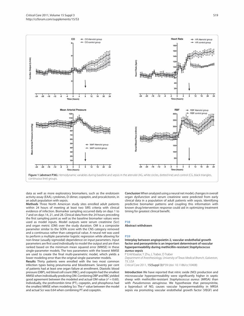
data as well as more exploratory biomarkers, such as the endotoxin activity assay (EAA), cytokines, D-dimer, copeptin, and procalcitonin, in an adult population with sepsis.Methods Three North American study sites enrolled adult patients within 24 hours of meeting at least two SIRS criteria with clinical evidence of infection. Biomarker sampling occurred daily on days 1 to 7 and on days 14, 21, and 28. Clinical data from the 24 hours preceding the fi rst sampling point as well as the baseline biomarker values were used as model inputs. Model outputs were serum creatinine (Scr) and organ metric (OM) over the study duration. OM is a composite parameter similar to the SOFA score with the CNS category removed and a continuous rather than categorical value. A neural net was used to perform a multiple parameter logistic regression while allowing for non-linear (usually sigmoidal) dependence on input parameters. Input parameters are fi rst used individually to model the output and are then ranked based on the minimum mean squared error (MMSE) in these single-parameter models. The two parameters with the lowest MMSE are used to create the fi nal multi-parametric model, which yields a lower modeling error than the original single-parameter models.Results Thirty patients were enrolled with the two most common infection types being pneumonia and bloodstream. Seventy per cent of patients had at least one organ failure at enrollment. Diastolic blood pressure (DBP), red blood cell count (RBC), and copeptin had the smallest MMSE when individually predicting OM. Combining DBP and RBC yielded good agreement between the modeled and actual OM value (r2 = 0.60). Individually, the prothrombin time (PT), copeptin, and phosphorus had the smallest MMSE when modeling Scr. The r2 value between the model and actual Scr was 0.64 when combining PT and copeptin.
Conclusion When analyzed using a neural net model, changes in overall organ dysfunction and serum creatinine were predicted from early clinical data in a population of adult patients with sepsis. Identifying predictive biomarker patterns and coupling this information with known drug/intervention response could aid in optimizing treatment timing for greatest clinical benefi t.
P38
Abstract withdrawn
P39
Interplay between angiopoietin-2, vascular endothelial growth factor and peroxynitrite is an important determinant of vascular hyperpermeability during methicillin-resistant Staphylococcus aureus sepsisP Enkhbaatar, Y Zhu, L Traber, D Traber
Department of Anesthesiology, University of Texas Medical Branch, Galveston,
TX, USA
Critical Care 2011, 15(Suppl 3):P39 (doi: 10.1186/cc10408)
Introduction We have reported that nitric oxide (NO) production and microvascular hyperpermeability were signifi cantly higher in septic sheep with methicillin-resistant Staphylococcus aureus (MRSA) than with Pseudomonas aeruginosa. We hypothesize that peroxynitrite, a byproduct of NO, causes vascular hyperpermeability in MRSA sepsis via promoting vascular endothelial growth factor (VEGF) and
Figure 1 (abstract P36). Hemodynamic variables during baseline and sepsis in the atenolol (AG, white circles, dotted line) and control (CG, black triangles,
continuous line) groups.
Critical Care 2011, Volume 15 Suppl 3 http://ccforum.com/supplements/15/S3
S19
angiopoietin-2 (Ang-2). The hypothesis was tested, using both a well-established ovine sepsis model and cultured human umbilical endothelial cells (HUVECs).Methods Female ewes were chronically instrumented with multiple catheters and live MRSA (USA300, 1011 CFU) was instilled into the both lungs by bronchoscope under deep isofl urane anesthesia. The sheep were then randomly allocated to control and treated (nonspecifi c NOS inhibitor L-NAME, 25 mg/kg, i.v., every 12 hours) groups and monitored for 24 hours for cardiopulmonary hemodynamics. The cells were challenged with 105 CFU of live MRSA or 50 μM peroxynitrite and co-incubated with or without L-NAME, peroxynitrite scavenger FeTMPyP, Tie-2 and Ang-2 antibody, and VEGF and its antibody. At diff erent times after the treatment, the permeability was measured by quantifying the amount of FITC-Dextran that passed through the confl uent HUVEC monolayer (n = 4). Ang-2 mRNA was determined by RT-PCR in those cells with or without treatment as well (n = 4). Statistical analysis: one-way ANOVA (Bonferroni).Results In vivo, L-NAME signifi cantly reduced MRSA-induced fl uid accumulation and requirement, as well as expression of VEGF. HUVEC permeability was time-dependently increased following MRSA co-incubation, reaching a plateau at 2 and 4 hours. These permeability changes (73 ± 4 RFUs) were signifi cantly (P <0.001) inhibited by 1 mM L-NAME (28 ± 1), 5 μM FeTMPy (34 ± 2), 5 μg/ml Tie-2 antibody (32 ± 2), and 5 μg/ml Ang-2 antibody (30 ± 1). In HUVECs, the Ang-2 mRNA was time-dependently (picks at 30 minutes) and dose-dependently increased by peroxynitrite (highest at 50 μM). Treatment of HUVECs with 5 μM VEGF augmented the MRSA-induced Ang-2 mRNA increases. The latter was reversed with FeTMPyP and 5 μM VEGF antibody.Conclusion Ang-2 and VEGF, Tie-2 receptor, NO and its byproduct peroxynitrite play an important role in MRSA-induced vascular hyper-permeability. The results strongly suggest that peroxynitrite increases vascular hyper-permeability by promoting Ang-2 release through stimulating the VEGF expression during MRSA-induced Gram-positive sepsis.
P40
Clinical characteristics, management, and outcomes of sepsis in Lusaka, ZambiaSM Chimese1, B Andrews1,2, S Lakhi1,3
1Department of Internal Medicine, University of Zambia, Lusaka, Zambia; 2Institute for Global Health, Vanderbilt University, Nashville, TN, USA; 3Department of Internal Medicine, University Teaching Hospital, Lusaka,
Zambia
Critical Care 2011, 15(Suppl 3):P40 (doi: 10.1186/cc10409)
Introduction Although infectious diseases are the leading causes of death in sub-Saharan Africa, there are few studies describing sepsis in the region. Available data suggest that HIV prevalence is disproportionately high among septic patients and that treatment, particularly fl uid administration, may be suboptimal [1]. Our study evaluated the clinical characteristics, management, and hospital outcomes of patients admitted with sepsis in Zambia. We hypothesized that patients with bacteremia have higher in-hospital mortality than those without.Methods We conducted a prospective observational study of patients admitted with sepsis to the Adult Filter Clinic (medical ER) of the University Teaching Hospital (UTH) in Lusaka Zambia. Sepsis was defi ned as two or more SIRS criteria and clinically suspected infection. Baseline characteristics and laboratory results were recorded, as was the timing of antibiotics and fl uid administration. Patients were followed until discharge or death.Results In 3 months, 161 septic patients were enrolled. One hundred and ten (68%) patients were HIV positive; 23 (14%) had unknown HIV status. Ninety-one (57%) had severe sepsis. Organ dysfunction included altered mentation (31%), renal dysfunction (16%), severe respiratory distress (respiratory rate ≥40) (11%), thrombocytopenia (11%), and hepatic dysfunction (7%). Multiple organ dysfunction occurred in 26%. After excluding contaminants, blood cultures were positive in 29 (18%) patients. Staphyloccoccus aureus, salmonella species, Streptococcus pneumoniae, and Klebsiella pneumoniae were the most common pathogens. Only 29% of patients received intravenous fl uids within
1 hour of presentation. Eighty-four percent of patients received ≤1 l within the fi rst 6 hours of presentation, and 55% received ≤1 l in the fi rst 24 hours. Overall in-hospital mortality was 40.4% (65/161). In-hospital mortality for severe sepsis was 54.9% (50/91). Important predictors for in-patient mortality (Table 1) were low Glasgow Coma Scale on admission (adjusted odds ratio (AOR) 16.0 (2.9 to 87.1)), positive blood culture (AOR 4.8 (1.5 to 15.0)), and positive and unknown HIV status (AOR 4.20 (1.0 to 17.0) and AOR 7.7 (1.2 to 47.7), respectively).Conclusion In-hospital mortality due to sepsis is higher in Zambia than in most studies from the developed world. Low Glasgow Coma Scale and positive blood cultures are associated with increased in-hospital mortality. Insuffi cient i.v. fl uid administration probably contributes to the high overall mortality. Standardized management including early fl uids and antibiotics might improve outcomes of sepsis and severe sepsis in sub-Saharan Africa.Reference1. Jacob S, et al.: Severe sepsis in two Ugandan hospitals: a prospective
observational study of management and outcomes in a predominantly HIV-1 infected population. PLoS One 2009, 4:e7782.
P41
AZD9773, a novel anti-TNFα immune Fab in development for severe sepsis and septic shock: demonstration of safety and effi cacy in a murine CLP sepsis modelP Newham1, P Ceuppens2, S Das3, JWT Yates4, R Knight1, JS McKay1
1Global Safety Assessment, AstraZeneca, Macclesfi eld, Cheshire, UK; 2Discovery
Enabling Capabilities and Sciences, AstraZeneca, Macclesfi eld, Cheshire, UK; 3Clinical Pharmacology & DMPK, AstraZeneca, Macclesfi eld, Cheshire, UK; 4Oncology iMed DMPK, AstraZeneca, Macclesfi eld, Cheshire, UK
Critical Care 2011, 15(Suppl 3):P41 (doi: 10.1186/cc10410)
Introduction TNFα is thought to play a central role in the pathogenesis of sepsis and septic shock. AZD9773 is an ovine polyclonal anti-human TNFα immune Fab comprising TNFα-directed and nonspecifi c Fab populations. AZD9773 potency and pharmacokinetic attributes such as a shorter half-life distinguish it from anti-TNF monoclonal antibodies, which have been assessed previously in clinical sepsis models. Here we explore the preclinical safety/effi cacy of AZD9773 in a mouse cecal ligation puncture (CLP) model. There are currently no reports of anti-TNF agent effi cacy in mouse CLP; rather, TNFα neutralization
Table 1 (abstract P40). Risk factors for in-hospital death
Variable Adjusted OR Crude OR
Anemia, Hb <9 g/dl 0.91 (0.74 to 4.93) 1.05 (0.56 to 1.80)
Blood culture positive 4.8 (1.50 to 15.0) 2.38 (1.14 to 4.95)
GCS
≥13 1 1
9 to 12 5.50 (1.90 to 16.20) 3.60 (1.60 to 8.11)
<9 16.00 (2.90 to 87.10) 11.2 (3.50 to 36.4)
HIV
Negative 1 1
Positive 4.20 (1.00 to 17.00) 2.35 (0.88 to 6.28)
Unknown 7.70 (1.20 to 47.70) 8.38 (2.36 to 29.7)
<1 hour to IVF 0.40 (0.10 to 1.10) 0.86 (0.43 to 1.72)
MAP <65 2.10 (0.70 to 6.80) 1.25 (0.56 to 2.81)
IVF in fi rst 6 hours
0 1 1
1 0.80 (0.30 to 2.00) 0.81 (0.41 to 1.61)
2 or more 0.30 (0.10 to 1.10) 0.60 (0.23 to 1.58)
Statistically signifi cant ORs in bold.
Critical Care 2011, Volume 15 Suppl 3 http://ccforum.com/supplements/15/S3
S20
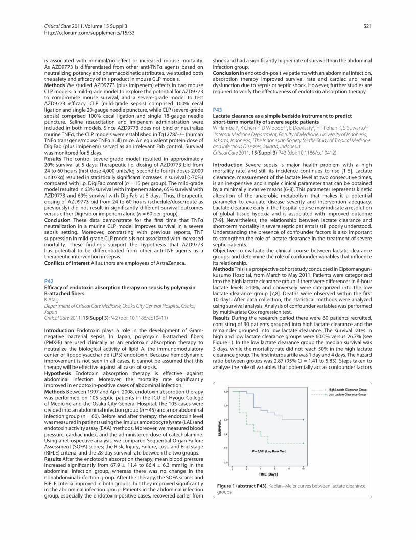
is associated with minimal/no eff ect or increased mouse mortality. As AZD9773 is diff erentiated from other anti-TNFα agents based on neutralizing potency and pharmacokinetic attributes, we studied both the safety and effi cacy of this product in mouse CLP models.Methods We studied AZD9773 (plus imipenem) eff ects in two mouse CLP models: a mild-grade model to explore the potential for AZD9773 to compromise mouse survival, and a severe-grade model to test AZD9773 effi cacy. CLP (mild-grade sepsis) comprised 100% cecal ligation and single 20-gauge needle puncture, while CLP (severe-grade sepsis) comprised 100% cecal ligation and single 18-gauge needle puncture. Saline resuscitation and imipenem administration were included in both models. Since AZD9773 does not bind or neutralize murine TNFα, the CLP models were established in Tg1278/–/– (human TNFα transgene/mouse TNFα null) mice. An equivalent protein dose of DigiFab (plus imipenem) served as an irrelevant Fab control. Survival was monitored for 5 days.Results The control severe-grade model resulted in approximately 20% survival at 5 days. Therapeutic i.p. dosing of AZD9773 bid from 24 to 60 hours (fi rst dose 4,000 units/kg, second to fourth doses 2,000 units/kg) resulted in statistically signifi cant increases in survival (>70%) compared with i.p. DigiFab control (n = 15 per group). The mild-grade model resulted in 63% survival with imipenem alone, 65% survival with AZD9773 and 69% survival with DigiFab at 5 days. Thus, therapeutic dosing of AZD9773 bid from 24 to 60 hours (schedule/dose/route as previously) did not result in signifi cantly diff erent survival outcomes versus either DigiFab or imipenem alone (n = 60 per group).Conclusion These data demonstrate for the fi rst time that TNFα neutralization in a murine CLP model improves survival in a severe sepsis setting. Moreover, contrasting with previous reports, TNF suppression in mild-grade CLP models is not associated with increased mortality. These fi ndings support the hypothesis that AZD9773 has potential to be diff erentiated from other anti-TNF agents as a therapeutic intervention in sepsis.Confl icts of interest All authors are employees of AstraZeneca.
P42
Effi cacy of endotoxin absorption therapy on sepsis by polymyxin B-attached fi bersK Atagi
Department of Critical Care Medicine, Osaka City General Hospital, Osaka,
Japan
Critical Care 2011, 15(Suppl 3):P42 (doi: 10.1186/cc10411)
Introduction Endotoxin plays a role in the development of Gram-negative bacterial sepsis. In Japan, polymyxin B-attached fi bers (PMX-B) are used clinically as an endotoxin absorption therapy to neutralize the biological activity of lipid A, the immunomodulatory center of lipopolysaccharide (LPS) endotoxin. Because hemodynamic improvement is not seen in all cases, it cannot be assumed that this therapy will be eff ective against all cases of sepsis.Hypothesis Endotoxin absorption therapy is eff ective against abdominal infection. Moreover, the mortality rate signifi cantly improved in endotoxin-positive cases of abdominal infection.Methods Between 1997 and April 2008, endotoxin absorption therapy was performed on 105 septic patients in the ICU of Hyogo College of Medicine and the Osaka City General Hospital. The 105 cases were divided into an abdominal infection group (n = 45) and a nonabdominal infection group (n = 60). Before and after therapy, the endotoxin level was measured in patients using the limulus amoebocyte lysate (LAL) and endotoxin activity assay (EAA) methods. Moreover, we measured blood pressure, cardiac index, and the administered dose of catecholamine. Using a retrospective analysis, we compared Sequential Organ Failure Assessment (SOFA) scores; the Risk, Injury, Failure, Loss, and End stage (RIFLE) criteria; and the 28-day survival rate between the two groups.Results After the endotoxin absorption therapy, mean blood pressure increased signifi cantly from 67.9 ± 11.4 to 86.4 ± 6.3 mmHg in the abdominal infection group, whereas there was no change in the nonabdominal infection group. After the therapy, the SOFA scores and RIFLE criteria improved in both groups, but they improved signifi cantly in the abdominal infection group. Patients in the abdominal infection group, especially the endotoxin-positive cases, recovered earlier from
shock and had a signifi cantly higher rate of survival than the abdominal infection group.Conclusion In endotoxin-positive patients with an abdominal infection, absorption therapy improved survival rate and cardiac and renal dysfunction due to sepsis or septic shock. However, further studies are required to verify the eff ectiveness of endotoxin absorption therapy.
P43
Lactate clearance as a simple bedside instrument to predict short-term mortality of severe septic patientsW Hambali1, K Chen1,2, D Widodo1,2, E Dewiasty1, HT Pohan1,2, S Suwarto1,2
1Internal Medicine Department, Faculty of Medicine, University of Indonesia,
Jakarta, Indonesia; 2The Indonesian Society for the Study of Tropical Medicine
and Infectious Diseases, Jakarta, Indonesia
Critical Care 2011, 15(Suppl 3):P43 (doi: 10.1186/cc10412)
Introduction Severe sepsis is major health problem with a high mortality rate, and still its incidence continues to rise [1-5]. Lactate clearance, measurement of the lactate level at two consecutive times, is an inexpensive and simple clinical parameter that can be obtained by a minimally invasive means [6-8]. This parameter represents kinetic alteration of the anaerobic metabolism that makes it a potential parameter to evaluate disease severity and intervention adequacy. Lactate clearance early in the hospital course may indicate a resolution of global tissue hypoxia and is associated with improved outcome [7-9]. Nevertheless, the relationship between lactate clearance and short-term mortality in severe septic patients is still poorly understood. Understanding the presence of confounder factors is also important to strengthen the role of lactate clearance in the treatment of severe septic patients.Objective To evaluate the clinical course between lactate clearance groups, and determine the role of confounder variables that infl uence its relationship.Methods This is a prospective cohort study conducted in Ciptomangun-kusumo Hospital, from March to May 2011. Patients were categorized into the high lactate clearance group if there were diff erences in 6-hour lactate levels ≥10%, and conversely were categorized into the low lactate clearance group [7,8]. Deaths were observed within the fi rst 10 days. After data collection, the statistical methods were analyzed using survival analysis. Analysis of confounder variables was performed by multivariate Cox regression test.Results During the research period there were 60 patients recruited, consisting of 30 patients grouped into high lactate clearance and the remainder grouped into low lactate clearance. The survival rates in high and low lactate clearance groups were 60.0% versus 26.7% (see Figure 1). In the low lactate clearance group the median survival was 3 days, while the mortality rate did not reach 50% in the high lactate clearance group. The fi rst interquartile was 1 day and 4 days. The hazard ratio between groups was 2.87 (95% CI = 1.41 to 5.83). Steps taken to analyze the role of variables that potentially act as confounder factors
Figure 1 (abstract P43). Kaplan–Meier curves between lactate clearance
groups.
Critical Care 2011, Volume 15 Suppl 3 http://ccforum.com/supplements/15/S3
S21

were by using bivariate analysis, in which variables that infl uenced the occurrence of deaths (indicated by P <0.25) underwent multivariate analysis subsequently. On multivariate analysis the presence of septic shock, degree of organ dysfunction, vasoactive drug usage, blood transfusion, and fl uid resuscitation change the hazard ratio by no more than 10% (Table 1). For that reason, these parameters were not considered as confounders.Conclusion Severe septic patients with high lactate clearance have a better survival rate compared with the low lactate clearance group, and its relationship is not infl uenced by the presence of confounder variables.Acknowledgments The authors thank the nurses and administrative staff in the Division of Tropical Medicine and Infectious Diseases, Department of Internal Medicine, Faculty Medicine, University of Indonesia for their assistance in this study.References1. Moss M: Epidemiology of sepsis: race, sex, and chronic alcohol abuse. CID
2005, 41(Suppl 7):S490-S497.
2. Guidet B, Aegerter P, Gauzit R, Meshaka P, Dreyfuss D: Incidence and impact of organ dysfunctions associated with sepsis. Chest 2005, 127:942-951.
3. O’brien JM Jr, Ali NA, Abraham E: Year in review in critical care, 2004: sepsis and multi-organ failure. Crit Care 2005, 9:409-413.
4. Ely EW, Kleinpell RM, Goyette RE: Advances in the understanding of clinical manifestations and therapy of severe sepsis: an update for critical care nurses. Am J Crit Care 2003, 12:120-133.
5. Angus DC, Linde-Zwirble WT, Lidicker J, Clermont G, Carcillo J, Pinsky MR:
Epidemiology of severe sepsis in the united states: analysis of incidence, outcome, and associated costs of care. Crit Care Med 2001, 29:1303-1310.
6. Nguyen HB, Loomba M, Yang JJ, Jacobsen G, Shah K, Otero RM, Suarez A,
Parekh H, Jaehne A, Rivers EP: Early lactate clearance is associated with biomarkers of infl ammation, coagulation, apoptosis, organ dysfunction and mortality in severe sepsis and septic shock. J Infl amm 2010, 7:6-17.
7. Nguyen HB, Rivers EP, Knoblich BP, Jacobsen G, Muzzin A, Ressler JA,
Tomlanovich MC: Early lactate clearance is associated with improved outcome in severe sepsis & septic shock. Crit Care Med 2004, 32:1637-1642.
8. Arnold RC, Shapiro NI, Jones AE, Schorr C, Pope J, Casner E, Parrillo JE,
Dellinger P, Trzeciak S: Multicenter study of early lactate clearance as a determinant of survival in patients with presumed sepsis. Shock 2009,
32:35-39.
9. Jones AE. Shapiro NI, Trzeciak S, Arnold RC, Claremont HA, Kline JA: Lactate clearance vs central venous oxygen saturation as goals of early sepsis therapy. JAMA 2010, 303:739-746.
P44
Eff ect of low-dose steroid on NF-κB and caspase-3 intestinal expression in a sepsis mouse modelHA Guntur1,2, HP Diding1,2, HT Pohan3, D Widodo3
1Indonesian Society for the Study of Tropical Medicine and Infectious
Diseases, Surakarta, Indonesia; 2Faculty of Medicine, Sebelas Maret University,
Surakarta, Indonesia; 3Indonesian Society for the Study of Tropical Medicine
and Infectious Diseases, Jakarta, Indonesia
Critical Care 2011, 15(Suppl 3):P44 (doi: 10.1186/cc10413)
Introduction The use of low-dose corticosteroids in sepsis early stages is still debated. The association of LPS–LBP complexes to CD14 receptors and will interact with TLR4 to induce NF-κB as a signal and transcription of proinfl ammatory cytokines [1,2]. Excessive production of infl ammatory cytokines will cause activation of SIRS, especially in gut-associated lymphoid tissues [3], which induces metabolic changes leading to apoptosis network, MOF, septic shock and death [3-5]. Changes in apoptosis are mediated by caspases, including caspase-3 that acts as an eff ector caspase [6,7]. Low-dose corticosteroids can inhibit the production of proinfl ammatory cytokines, production of infl ammatory mediators, and lower adhesion of leukocytes to the endothelium [8].Objective The aim of this study was to analyse NF-κB and caspase-3 intestinal expression, and also survival from use of low-dose steroid in the early stages of sepsis in the Balb/C mouse model of sepsis.Methods Male Balb/C mice were inoculated with lipopolysaccharide for the sepsis mouse model. Sepsis mouse model grouping was to a sepsis group (Group I) and to sepsis with steroid (methylprednisolone 1 to 1.5 mg/kg BW/day) (Group II). Detection of intestinal NF-κB and caspase-3 expression used the immunohistochemistry technique on days 1, 3, 5 and 7. Survival was seen until the 7th day. The two-tailed Fisher exact test for the analysis of mortality, independent-sample t test for intestinal NF-κB and caspase-3 expression, and P <0.05 were used to determine signifi cant diff erences.Results Acute infl ammatory response occurs in the early stages of sepsis (the fi rst 5 days of exposure) and the process of death occurs in advanced stages of sepsis (after the fi rst 5 days of exposure) [9]. This study shows that the use of low-dose corticosteroids in sepsis early stages (fi rst 5 days) signifi cantly inhibited the expression of NF-κB (see Table 1), so cytokine production of proinfl ammatory cytokines was not excessive. Reduced product proinfl ammatory cytokines would reduce the expression of intestinal caspase-3 (see Table 2), which will reduce
Table 1 (abstract P43). Variables that potentially act as confounder factors
Variable Nonsurvivors Survivors P value Adjusted HR (95% CI) HR change (%)
Septic shock within 6 hours
With septic shock 11 4 0.081 3.083 (1.505 to 6.317) 7.4
Without septic shock 23 22
Initial SOFA score
>9 11 3 0.038 2.919 (1.388 to 6.138) 1.7
≤9 19 21
Vasoactive drugs within 6 hours
Without vasoactive drugs 22 23 0.013 2.988 (1.462 to 6.106) 4.1
With vasoactive drugs 12 3
Invasive ventilation within 6 hoursa
Without mechanical ventilation 31 23 0.777 – –
With mechanical ventilation 3 3
PRC transfusion within 6 hours
Without transfusion 29 25 0.069 3.077 (1.493 to 6.340) 7.2
With transfusion 5 1
Fluid resuscitation within 6 hours
<1,000 cm3 15 18 0.166 2.942 (1.444 to 5.994) 2.5
≥1,000 cm3 19 8
HR, hazard ratio; SOFA, Sequential Organ Failure Assessment; PRC, packed red cells. aInvasive ventilation parameter not included in multivariate analysis, because P <0.25.
Critical Care 2011, Volume 15 Suppl 3 http://ccforum.com/supplements/15/S3
S22

the excessive apoptosis in the intestinal tract. Decreased expression of NF-κB and caspase-3 in the intestinal tract would further reduce the excessive mucosal cell death. This situation will block the destruction and disruption of mucosal defense function of the digestive tract, thereby increasing immune response. The end result will be seen that low-dose corticosteroids can reduce mortality. This study found dead animals for Group I were 70%, while Group II were 10% (P = 0.020).Conclusion Low-dose steroids can reduce NF-κB and caspase-3 intestinal expression and also mortality in early sepsis.Acknowledgements The authors thank the Faculty of Medicine, University of Sebelas Maret for their fi nancial support and assistance given in this study.References1. Hongwei Q, Cynthia AW, Sun JL, Xueyan Z, Etty NB: LPS induces CD40 gene
expression through the activation of NF-κB and STAT-1α in macrophages and microglia. Blood 2005, 106:3114-3122.
2. Kristine MJ, Sarah BL, Anncatrine LP, Jesper EO, Thomas B: Common TNF-α, IL-1β, PAI-1, uPA, CD14 and TLR4 polymorphisms are not associated with disease severity or outcome from gram negative sepsis. BMC Infect Dis
2007, 7:108.
3. Arul MC, Markus HL, Chandan KS, Terrence RB, Sunita SS, Vidya JS, Vaishalee
AP, Peter AW: Molecular signatures of sepsis multiorgan gene expression profi les of systemic infl ammation. Am J Pathol 2001, 159:1199-1209.
4. Elena GR, Alejo C, Gema R, Mario D: Cortistatin, a new antiinfl ammatory peptide with therapeutic eff ect on lethal endotoxemia. J Exp Med 2006,
203:563-571.
5. Javier C, José Y, David HE, Yolanda M, Ruben M, Isabel A, Antonia M, Pascual P,
Vicente V: Role of lipopolysaccharide and cecal ligation and puncture on blood coagulation and infl ammation in sensitive and resistant mice models. Am J Pathol 2005, 166:1089-1098.
6. Chung CS, Chaudry IH, Ayala A: The apoptotic response of the lymphoid immune system to trauma, shock and sepsis. In Yearbook of Intensive Care
and Emergency Medicine. Editted by Vincent JL. Berlin: Spinger-Verlag;
2000:27-40.
7. Chung CS, Song GY, Lomas J, Simms HH, Chaudry IH, Ayala A: Inhibition of Fas/Fas ligand signaling improves septic survival: diff erential eff ects on macrophage apoptotic and functional capacity. J Leukoc Biol 2003,
74:344-351.
8. Annane D, Caillon DM: Corticosteroid in sepsis: from bench to bedside. Shock 2003, 20:197-207.
9. Xiao H, Siddiqui J, Remick DG: Mechanisms of mortality in early and late sepsis. Infection Immunity 2006, 74:5227-5235.
P45
Etiological agents of bacterial sepsis in a newly constructed medical center in Saint Petersburg, RussiaE Barantsevich, N Barantsevich, N Rybkova, I Churkina, N Pestova,
M Karpenko
Almazov Federal Center of Heart, Blood and Endocrinology, Saint Petersburg,
Russia
Critical Care 2011, 15(Suppl 3):P45 (doi: 10.1186/cc10414)
Introduction Sepsis is one of the factors of high mortality in ICUs in critically ill patients. Annual mortality from this condition is estimated at 30 to 50 deaths per 100,000 population [1,2,4,5]. The aim of the present study was to reveal the spectrum and resistance to antibiotics of microorganisms, causing sepsis in hospitalized patients of the multidisciplinary medical center, that accumulates patients from all regions of Russia, during the fi rst year from its foundation, with low possibility of local nosocomial strains formation.Methods The diagnosis of sepsis was verifi ed by isolation of bacteria from blood (only two or more positive results were considered) and the presence of two or more criteria of systemic infl ammatory response syndrome [3]. The cultures were isolated from blood with BactAlert (BioMerieux, France). The identifi cation was performed by routine phenotypic methods and sequencing of the 16sRNA gene (ABI Prism 3130, MicroSeq ID v2.0 Software, MicroSeq ID 16s rDNA500 Library v2.0). Resistance to routinely used antibiotics was investigated with the disc diff usion method and by dilution techniques for MIC determination on Muller–Hinton agar (Oxoid, UK).Results Sepsis, associated with bloodstream infections, was revealed in 89 cases – Gram-positive cocci predominated. Staphylococcus spp. were responsible for 35 (39.3%) cases: Staphylococcus aureus was the causative agent in eight (8.9%), coagulase-negative staphylococci (Staphylococcus epidermidis, Staphylococcus haemolyticus, Staphylo-coccus hominis novobiosepticus) in 27 (30.3%) infections. Other Gram-positive cocci were Enterococcus faecalis in eight (8.9%), Enterococcus faecium in six (6.7%). Gram-negative microorganisms included Acineto-bacter baumannii that was found in 11 (12.4%), Klebsiella spp. (Klebsiella pneumonia, Klebsiella rhinoscleromatis, Klebsiella oxytoca) in fi ve (5.6%), Escherichia coli in eight (8.9%), Enterobacter spp. (Enterobacter cloaceae, Enterobacter aerogenes, Enterobacter hormaechea) in three (3.4%), Pseudomonas aeruginosa in two (2.2%) patients with sepsis. Rarely isolated bacteria were Bacillus thuringiensis in one (1.1%), Stenotrophomonas maltophylia in one (1.1%), Pantoeae agglomerans in one (1.1%), Corynebacterium mucifaciensis in one (1.1%) case. Two and more species were isolated from blood in seven (7.9%) patients. In total, 105 strains were isolated in sepsis cases. Resistance to antibiotics was observed in 96 (91.4%) bacterial isolates; 56 (53.3%) were multidrug-resistant strains. All E. faecium, and nine (81.8%) strains of A. baumannii were resistant to seven or more antibiotics. All E. faecium strains were susceptible to linezolide, A. baumannii to tigecycline. Methicillin resistance was detected in two (15.4%) strains of S. aureus and 18 (60.0%) strains of coagulase-negative staphylococci; four (66.7%) strains of E. faecium were vancomycin resistant.Conclusion Gram-positive bacteria were the leading causative agents of sepsis, associated with bloodstream infections in the newly-constructed hospital in Saint Petersburg. The majority of strains (91.4%) were resistant to antibiotics, and more than half of the isolates were multidrug resistant. Methicillin resistance was observed predominantly in coagulase-negative staphylococci, vancomycin resistance in E. faecium. All polyresistant E. faecium strains were susceptible to linezolide, A. baumannii to tigecycline.References1. Antonacci Carvalho PR, Trotta EA: Advances in sepsis diagnosis and
treatment. J Pediatria 2003, 79:S195-S204.
2. Braun L, Riedel A A, Cooper LM: Severe sepsis in managed care: analysis of incidence, one-year mortality, and associated costs of care. J Managed Care
Pharmacy 2004, 10:521-530.
3. Bone RC, Balk RA, Cerra FB, Dellinger RP, Fein AM, Knaus WA, Schein RM,
Sibbald WJ: ACCP/SCCM consensus conference: defi nitions for sepsis and organ failure and guidelines for the use of innovative therapies in sepsis. Chest 1992, 101:1644-1655.
4. Dellinger RP, Carlet JM, Masur H, Gerlac H, Calandra T, Cohen J, Gea-
Banacloche J, Keh D, Marchall JC, Parker MM, et al.: Surviving Sepsis
Table 1 (abstract P44). NF-κB intestinal expression
Amount of cell expression
Day Group I Group II P value
1 17.5 ± 4.7 10.8 ± 3.6 0.019
3 26.5 ± 4.4 15.8 ± 3.6 0.001
5 39.3 ± 4.1 31.2 ± 7.0 0.033
7 54.8 ± 9.6 50.5 ± 10.7 0.476
Data presented as mean ± standard deviation.
Table 2 (abstract P44). Caspase-3 intestinal expression
Amount of cell expression
Day Group I Group II P value
1 8.5 ± 2.9 4.3 ± 1.9 0.014
3 14.7 ± 3.1 9.8 ± 2.2 0.012
5 33.2 ± 8.3 12.8 ± 4.5 0.000
7 42.3 ± 3.2 37.7 ± 8.2 0.336
Data presented as mean ± standard deviation.
Critical Care 2011, Volume 15 Suppl 3 http://ccforum.com/supplements/15/S3
S23

Campaign guidelines for management of severe sepsis and septic shock. Crit Care Med 2004, 32:858-873.
5. Lever A, Mackenzie I: Sepsis: defi nition, epidemiology, and diagnosis. BMJ
2007, 335:879-883.
P46
Effi cacy and safety of gelatin for fl uid therapy in hypovolemia: a systematic review and meta-analysisCS Hartog1,2, V Vlasakov1, DO Thomas-Rueddel1,2, H Rueddel1,2,
R Hutagalung1, K Reinhart1,2
1Department of Anesthesiology and Intensive Care Medicine, Jena University
Hospital, Jena, Germany; 2Center for Sepsis Control & Care, Jena University
Hospital, Jena, Germany
Critical Care 2011, 15(Suppl 3):P46 (doi: 10.1186/cc10415)
Introduction Gelatin is frequently used as volume expander. There are growing concerns about safety.Objective To systematically assess clinical evidence concerning mortality, coagulation and renal function.Methods Systematic review of randomised controlled trials (RCT) on gelatin in hypovolemia in comparison to any other fl uid with a comprehensive search strategy (Ovid Medline (1948 to May 2011), EMBASE (1947 to May 2011), Cochrane Library). Data were independently extracted and risk of bias assessed using the 2010 Cochrane tool. Primary outcome was overall mortality. Secondary outcomes were the number of patients exposed to allogeneic transfusion, frequency of renal replacement therapy (RRT) or acute renal failure (ARF). Albumin and crystalloid solutions were defi ned as suitable, and other synthetic colloids as unsuitable control fl uids since they carry similar risk of side eff ects. Relative risks (RR) and weighted mean diff erences with 95% CIs were calculated. Data were pooled using a random-eff ects model (RevMan 5.1, Cochrane Collaboration).
Results The search yielded 1,288 citations, 210 reports were read in full. The fi nal sample contained 72 RCT in English, German, French and Italian, published between 1975 and 2010, with 5,915 patients overall, 2,523 of which received gelatin. The median sample size in the gelatin groups was 20 patients (range 10 to 249). In 53 RCT (74%), the study period was ≤24.0 hours. Total gelatin dose was 20 ml/kg (median, range 6 to 62). Only 38 RCT (53%) used suitable control fl uids. Forty-nine RCT (68%) investigated elective surgical patients, mostly from cardiac surgery (32 RCT, 44%). Nine RCT (13%) investigated critically ill patients, six RCT (8%) were in emergency patients and seven RCT (10%) were in children. The RR for mortality was 1.02 (CI 0.87 to 1.19, data from 23 RCT with 2,694 patients which reported mortality). Numbers of patients exposed to allogeneic transfusions were provided in 11 RCT, n = 1,148 patients and the RR was 1.16 (0.94 to 1.44). When only studies with suitable control fl uids were included, the RR for mortality was 1.13 (0.88 to 1.46, 10 RCT, 1,392 patients) and risk for transfusion exposure was 1.35 (0.88 to 2.08, seven RCT, n = 672), tending towards control. Only six RCT (n = 662 patients) reported the occurrence of RRT or ARF, fi ve of them in comparison with HES solutions. Three RCT reported anaphylactoid events.Conclusion Most published studies on gelatin are small and short-time, use unsuitable control fl uids and report too few events to reliably assess the safety of gelatin.
Cite abstracts in this supplement using the relevant abstract number, e.g.:
Hartog CS, et al.: Effi cacy and safety of gelatin for fl uid therapy in hypovolemia: a systematic review and meta-analysis [abstract]. Critical Care
2011, 15(Suppl 3):P46.
Critical Care 2011, Volume 15 Suppl 3 http://ccforum.com/supplements/15/S3
S24