Clonal Expression 'the Tn Antigen Erythroid Granulocyte...
11
Clonal Expression of 'the Tn Antigen in Erythroid and Granulocyte Colonies and Its Application to Determination of the Clonality of the Human Megakaryocyte Colony Assay WILLIAM VAINCHENKER, UGO TESTA, JEANNE FRANQOISE DESCHAMPS, ANNIE HENRI, MONIQUE TITEUX, JANINE BRETON-GORIUS, HENRI ROCHANT, DOUGLAS LEE, and JEAN-PIERRE CARTRON,Unite de Recherches sur les Anemies, Institut National de la Sante et de la Recherche Medicale U91, Hopital Henri Mondor 94010 Creteil, France; National Blood Transfusion Service, Lancaster LA1 3JP, England; Unite de Recherches sur les groupes sanguins, Institut National de la Sante et de la Recherche Medicale U76, Centre National de Transfusion Sanguine, 75739 Paris Cedex 15, France A B S T R A C T To evaluate whether exposure of Tn determinants at the surface of human erythrocytes, platelets, and granulocytes could arise from a somatic mutation in a hemopoietic stem cell, burst-forming unit erythroid (BFU-E) colonies, colony-forming unit granulocyte-macrophage (CFU-GM), and colony- forming unit-eosinophil (CFU-Eo) were grown from a blood group 0 patient with a typical Tn syndrome displaying two distinct populations (Tn+ and Tn-) of platelets, granulocytes, and erythrocytes. A large num- ber of colonies was observed. Individual colonies were studied with a fluorescent conjugate of Helix pomatia agglutinin (HPA). A sizeable fraction of each of the erythroid and granulocytic colonies appeared to con- sist exclusively of either HPA-positive or HPA-nega- tive cells, thereby demonstrating the clonal origin of those exhibiting the Tn marker. Similar results were obtained from a second patient. These findings estab- lish that the HPA labeling of Tn cells is an accurate marker permitting assessment of the clonality of the human megakaryocyte (MK) colony assay. For the study of MK cultures a double-staining procedure us- ing the HPA lectin and a monoclonal antiplatelet an- tibody (J-15) was applied in situ to identify all MK constituting a colony. Our results, obtained in studies of 133 MK colonies, provide definitive evidence that the human MK colony assay is clonal because all MK colonies were exclusively composed of Tn+ and Tn- Received for publication 24 September 1981 and in re- vised form 28 December 1981. MK. Furthermore, the distribution of MK within a single colony was shown to be seminormal with a mean at 6 MK, isolated MK typically being absent in culture. Comparison of the proportion of mature Tn+ cells in blood with their respective Tn+ progenitors has also shown that no proliferative advantage occurs after the commitment; because Tn polyagglutinability is an ac- quired disorder, then the expansion of the Tn+ clone must occur either during the proliferative stage of the pluripotent stem cell or during the commitment itself. This study therefore affords evidence that a blood group antigen plays a role in the differentiation of a pluripotent stem cell. INTRODUCTION Tn polyagglutinability is an acquired disorder char- acterized by exposure at the erythrocyte surface of receptors that are normally hidden and become avail- able to natural antibodies occurring in most human adult sera (1). Increasing interest in such markers is due to their frequent association with a mild hemolytic anemia or leucopenia and thrombopenia, as defined under the Tn syndrome (2). However, the Tn receptors may also be found in apparently healthy individuals. More recently, a Tn activation was described as pre- ceding acute leukemia or myeloproliferative disorders, suggesting its possible role in the development of the neoplastic process (3-5). Serological and biochemical evidence shows that N-acetyl-D-galactosamine, when covalently bound to erythrocyte glycoproteins by an J. Clin. Invest. © The American Society for Clinical Investigation, Inc. * 0021-9738/82/05/1081/11 $1.00 Volume 69 May 1982 1081-1091 1081
-
Upload
trankhuong -
Category
Documents
-
view
217 -
download
0
Transcript of Clonal Expression 'the Tn Antigen Erythroid Granulocyte...

Clonal Expression of 'the Tn Antigen in Erythroid andGranulocyte Colonies and Its Application toDetermination of the Clonality of the Human
Megakaryocyte Colony Assay
WILLIAM VAINCHENKER, UGOTESTA, JEANNEFRANQOISEDESCHAMPS,ANNIE HENRI, MONIQUETITEUX, JANINE BRETON-GORIUS, HENRI ROCHANT,DOUGLASLEE, and JEAN-PIERRE CARTRON,Unite de Recherches sur les Anemies,Institut National de la Sante et de la Recherche Medicale U91, HopitalHenri Mondor 94010 Creteil, France; National Blood Transfusion Service,Lancaster LA1 3JP, England; Unite de Recherches sur les groupes sanguins,Institut National de la Sante et de la Recherche Medicale U76, CentreNational de Transfusion Sanguine, 75739 Paris Cedex 15, France
A B S T R A C T To evaluate whether exposure of Tndeterminants at the surface of human erythrocytes,platelets, and granulocytes could arise from a somaticmutation in a hemopoietic stem cell, burst-formingunit erythroid (BFU-E) colonies, colony-forming unitgranulocyte-macrophage (CFU-GM), and colony-forming unit-eosinophil (CFU-Eo) were grown froma blood group 0 patient with a typical Tn syndromedisplaying two distinct populations (Tn+ and Tn-) ofplatelets, granulocytes, and erythrocytes. A large num-ber of colonies was observed. Individual colonies werestudied with a fluorescent conjugate of Helix pomatiaagglutinin (HPA). A sizeable fraction of each of theerythroid and granulocytic colonies appeared to con-sist exclusively of either HPA-positive or HPA-nega-tive cells, thereby demonstrating the clonal origin ofthose exhibiting the Tn marker. Similar results wereobtained from a second patient. These findings estab-lish that the HPA labeling of Tn cells is an accuratemarker permitting assessment of the clonality of thehuman megakaryocyte (MK) colony assay. For thestudy of MKcultures a double-staining procedure us-ing the HPA lectin and a monoclonal antiplatelet an-tibody (J-15) was applied in situ to identify all MKconstituting a colony. Our results, obtained in studiesof 133 MKcolonies, provide definitive evidence thatthe human MKcolony assay is clonal because all MKcolonies were exclusively composed of Tn+ and Tn-
Received for publication 24 September 1981 and in re-vised form 28 December 1981.
MK. Furthermore, the distribution of MK within asingle colony was shown to be seminormal with a meanat 6 MK, isolated MKtypically being absent in culture.
Comparison of the proportion of mature Tn+ cellsin blood with their respective Tn+ progenitors has alsoshown that no proliferative advantage occurs after thecommitment; because Tn polyagglutinability is an ac-quired disorder, then the expansion of the Tn+ clonemust occur either during the proliferative stage of thepluripotent stem cell or during the commitment itself.This study therefore affords evidence that a bloodgroup antigen plays a role in the differentiation of apluripotent stem cell.
INTRODUCTION
Tn polyagglutinability is an acquired disorder char-acterized by exposure at the erythrocyte surface ofreceptors that are normally hidden and become avail-able to natural antibodies occurring in most humanadult sera (1). Increasing interest in such markers isdue to their frequent association with a mild hemolyticanemia or leucopenia and thrombopenia, as definedunder the Tn syndrome (2). However, the Tn receptorsmay also be found in apparently healthy individuals.
More recently, a Tn activation was described as pre-ceding acute leukemia or myeloproliferative disorders,suggesting its possible role in the development of theneoplastic process (3-5). Serological and biochemicalevidence shows that N-acetyl-D-galactosamine, whencovalently bound to erythrocyte glycoproteins by an
J. Clin. Invest. © The American Society for Clinical Investigation, Inc. * 0021-9738/82/05/1081/11 $1.00Volume 69 May 1982 1081-1091
1081

alkali-labile 0 glycosidic linkage, is the chief structuraldeterminant of Tn specificity (6, 7). These "A-like"determinants exposed at the surface of Tn erythrocytesare recognized by several lectins such as Salvia sclarea,Dolichos biflorus, or Helix pomatia, but not by puri-fied human anti-A antibodies (8). Investigations car-ried out to further characterize Tn determinants at themolecular level have shown that the erythrocytes,platelets, and granulocytes of these patients have ab-normal surface glycoproteins with a low content ofsialic acid and galactose (9-12). These modificationsare associated with the defect of a single galactosyl-transf erase involved in the biosynthesis of sugar chainsOglycosidically linked to cell membrane glycoproteins(10-14). On the basis of these data, it was suggestedthat Tn activation may originate in a mutation of apluripotent stem cell of the bone marrow (11, 15). Thishypothesis is further supported by the finding that onlya sizeable fraction of erythrocytes or platelets exhibitthe Tn determinants because Tn+ and Tn- subpopu-lations are identifiable in the blood of the same indi-vidual (10, 11).
In an attempt to establish the hematopoietic originof the Tn activation, we have investigated the expres-sion of the Tn antigen in single colonies derived re-spectively from burst-forming unit-erythroid colonies(BFU-E)l, colony-forming unit-granulocyte-macro-phages (CFU-GM), and colony-forming unit-mega-karyocytes (CFU-MK). For this purpose we have useda fluorescent derivative of the Helix pomatia (HPA)lectin, which is N-acetylgalactosamine specific andcapable of recognizing Tn determinants. We wereaware that only the Salvia sclarea lectin is Tn specific(16) but that HPAcould be safely used providing thatTn individuals of blood group 0 or B were selected.The results have clearly shown that expression of theTn antigen in all erythroid (BFU-E) and granulocytic(CFU-GM) colonies is clonal. Indeed, the colonies wereeither completely positive or negative for the HPAmarker. Because Tn was found to be a clonal marker,it was subsequently used to demonstrate the clonalityof the megakaryocyte (CFU-MK) colony assay.
METHODS
Case report. The patient, P.L., is 27 yr old and an ap-parently healthy individual. His hematological abnormali-ties were discovered during a routine examination in July1980. The leucocyte and platelet counts were respectively2,900 and 45,000/mm3, without erythrocyte anemia. The
I Abbreviations used in this paper: MK, megakaryocyte;BFU-E, burst forming unit-erythroid; CFU-Eo, colony form-ing unit-eosinophil; CFU-GM, colony forming unit-granu-locyte-monocyte; CFU-MK, colony-forming unit-megakar-yocyte; Epo, erythropoietin; FITC, fluorescein isothiocyanate;GM-CSF, granulocyte macrophage-eosinophil-colony-stim-
patient is a blood group 0 donor with a typical Tn phenotype(1). The erythrocytes were polyagglutinable (as seen by theiragglutination with most ABOcompatible human sera) andstrongly agglutinated by Salvia sclarea, Helix pomatia, Dol-ichos biflorus, and Glycine soja lectins but unreactive withhexadimethrine bromide (Polybrene, Aldrich Chemicals,Beerse, Belgium) and Arachis hypogea (peanut agglutinin)lectin. As shown with other Tn samples (10, 11) a severereduction of the UDPGal: GalNAc-j8-3-D-galactosyltransfer-ase was found in the patient's erythrocyte and platelet ly-sates.
The direct antiglobulin test was negative. Clinical ex-amination was unremarkable. Under examination in April1981, the leucopenia and thrombopenia were confirmed.There was no anemia but the reticulocyte count and indirectbilirubin level were slightly elevated, whereas haptoglobinwas markedly reduced (<0.1 g/liter), suggesting a mild com-pensated hemolysis. A bone marrow specimen showed a nor-mal cellularity with some increase in erythroid lineage.
Blood (20 ml) was obtained on three different occasions.Bone marrow cells for culture were collected by sternumpuncture. Blood from two group 0 donors and from twogroup 0 newborns was also obtained for control purposes.A second patient, B.A., exhibiting a typical Tn syndrome(9-11) was also investigated, but few colonies could be ob-tained due to small amount of blood available.
Cell culture. Light density cells from blood or bone mar-row were isolated by Ficoll-metrizoate (Lymphoprep-Nye-gaard, Oslo, d = 1.077) density centrifugation at 400 g. Thecells were washed three times in cold a-medium (Eurobio,France) and plated by two different culture techniques togrow the different commited progenitors, BFU-E, CFU-GM,and CFU-MK.
(a) The growth of BFU-E and CFU-GMcolonies was per-formed by the methylcellulose procedure (17). Dulbecco'sMinimum Essential Medium (Gibco Laboratories, Grand Is-land Biological Co., Grand Island, N. Y.), as modified byGuilbert and Iscove (18), was used. The source of granulocytemacrophage-eosinophil-colony-stimulating factor (GM-CSF)was 10% Mo-conditioned medium (19). 2 U/ml of humanurinary erythropoietin (Epo, 100 U/mg protein), obtainedafter DEAE-cellulose chromatography (20), was used togrow BFU-E colonies. The activity of this Epo was deter-mined both by the mouse polycythemic assay and BFU-Ecultures, using as reference the Connaught step III sheeperythropoietin (batch 3034.1). 2 U Epo/ml were chosen be-cause it gives an optimal number of erythroid colonies andsatisfactory erythroid maturation. 4.2 X 10⁵ blood light den-sity cells per milliliter were plated under these conditions.At least six cultures of each were run and incubated for 12-18 d in a fully humidified atmosphere with 5% CO2. Colonieswere scored at day 14 under an inverted microscope andwere processed for immunofluorescence after having beenindividually picked off. After fluorescence, the type of cellconstituting the colonies was determined by May-GrunwaldGiemsa staining. (b) The culture technique for human MKcolonies in plasma clot (21) was used with slight modification(22). The stimulating factor was a conditioned medium fromleukocytes stimulated by phytohemagglutinin (PHA-LCM)(22, 23). 2 X 10⁵, 4 X 10⁵, and 6 X 10⁵ bone marrow cells
ulating factor; HPA, Helix pomatia agglutinin; PBS, phos-phate-buffered saline (10 mMphosphate buffer pH 7.2 con-taining 150 mMNaCl); PHA-LCM, conditionned mediumfrom leucocytes stimulated by phytohemagglutinin; TRITC,tetramethylrhodamin isothiocyanate.
1082 Vainchenker et al.
were respectively plated, after adherence, in plasma clot.The cultures were studied at day 12, a time at which MKcultures reach their optimum. They were screened by im-munofluorescence (24, 25). In addition to MKcolonies thismethod also permits the growth of a high number of gran-ulocyte colonies. The granulocyte colonies from bone mar-row cultures were only investigated by this technique.
In contrast to the methylcellulose procedure, the plasmaclot technique permits the in situ study of colonies. In ad-dition, bone marrow BFU-E were grown in plasma clot usingEpo as stimulating factor and were studied by immunoflu-orescence.
Immunofluorescent studies. Peripheral blood erythro-cytes and granulocytes were isolated after sedimentation ofthe blood at 1 g for 3 h. Erythrocytes were obtained at thebottom of this tube and granulocytes in the buff y coat. Bloodgranulocytes were purified by Ficoll-metrizoate density cen-trifugation. The cells were extensively washed and cyto-spanned for staining studies. BFU-E and CFU-GMcolonieswere individually picked off under an inverted microscope,washed, and studied. CFU-GMcolonies were studied at day12 or 14, whereas BFU-E were investigated at days 9, 14,and 18.
For methylcellulose cultures, 46 and 45 of BFU-E andCFU-GMcolonies were respectively studied. These colonieswere selected on the basis that they were distinctly separatedfrom neighboring colonies. Indeed, at the colony density atwhich CFU-GMwere grown (70 colonies per dish), 35% donot arise from a single cell origin (26). This difficulty couldbe overcome by selecting well separated colonies. In addi-tion, BFU-E colonies were recognized under an invertedmicroscope by their red or pink color (hemoglobinized cells).Immunofluorescent studies were performed after fixation ofthe cells for 10 min in pure methanol. The smears wereextensively washed in cold PBS and 100 ul of 10-2 dilutedHelix pomatia FITC conjugate (Industrie BiologiqueFranqaise, France 1 mg/ml) was added. The incubation wascontinued for 30 min at 4°C, and the smears were extensivelywashed in cold phosphate-buffered saline (PBS). Before ex-amination under the microscope the slides were mountedwith a coverslip in glycerin-buffered PBS. Similar conditionsof assay were used for immunofluorescent studies of plateletsand MKcolonies. Therefore, platelets were first isolated fromperipheral blood, washed, and resuspended in the culturemedium. Aliquots of 5_106 platelets were distributed in 35-mmpetri dishes, and the medium was allowed to clot. Fromthis step, a similar procedure was used for staining plateletsor MKcolonies. A double immunofluorescence staining wasperformed using an HPA-tetramethylrhodamin isothiocyan-ate (TRITC) conjugate to visualize the Tn antigen and amonoclonal antiplatelet antibody-fluorescein isothiocyanate(FITC) conjugate (J-15) to identify the MK. The monoclonalantibody (J-15) binds to normal human platelets but not toGlanzman's thrombasthenic platelets,2 suggesting that theantigenic structure recognized is tightly linked to the gly-coprotein lIb-IIIa complex. As shown (25), the monoclonalantibody, J-15, recognizes all the mature and immature MKwithout staining other cell types. Thus, by combination ofthe fluorescein- and rhodamin-labeled reagents, it is possibleto specifically identify in situ all the MK colonies and tocheck them for the presence or the absence of the Tn antigen.
For this purpose, the clots were dehydrated in the petridish with filter papers and then fixed for 5 min with 1%
2 Kieffer, N, G. Tobelem, A. MacMichael, J. Bastin, L.Degos, C. Ruan, and J. P. Caen. Submitted for publication.
paraformaldehyde in PBS. The clot was washed with PBS,and 100 ul of a 10-2 dilution of HPA-TRITC conjugate (IBF1 mg/ml) was applied for 90 min. After extensive washingwith PBS, 100 Al of ascitic fluid (J-15 antibody) was sub-sequently applied at a 10-2 dilution for 60 min. After twoadditional washes in PBS, 100 ,ul of a rabbit anti-mouse Fab'2FITC conjugate (Nordic Immunology, The Netherlands,distributed by TEBU-France, 28000 Versailles), was appliedat a 10-2 dilution for 30 min. The preparations were finallywashed in PBS before examination under a Zeiss fluorescentmicroscope (Carl Zeiss, Inc., New York) equipped with epi-illumination and appropriate filter for fluorescein and rho-damin.
Photographs were taken with an Ektachrome ASA 200film push-pulled to ASA 800. After fluorescent labeling, allthe smears or petri dishes were respectively stained with theMay-Grunwald-Giemsa or with Harris hematoxylin to iden-tify the cells and colonies morphologically.
RESULTS
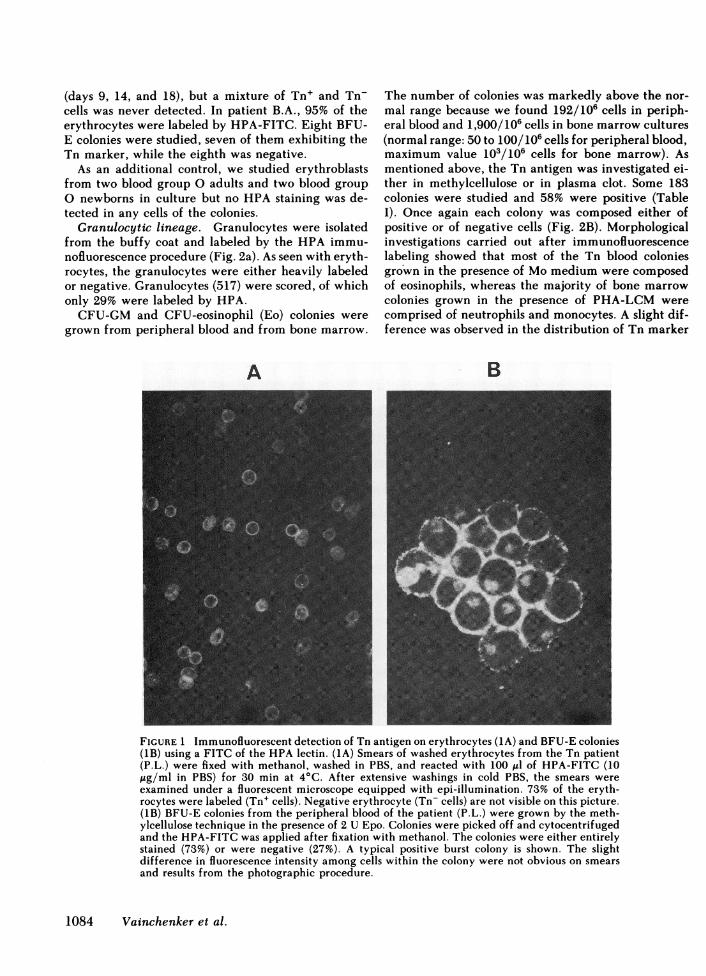
Erythroid lineage. Peripheral erythrocytes fromthe Tn patient (P.L., a group 0 individual) were firstinvestigated by HPA-labeling with the fluorescein orrhodamin conjugate of the lectin. The erythrocyteswere either strongly labeled or entirely negative, al-lowing an accurate scoring of each population (Fig.1A). 572 erythrocytes were counted, 73% of whichexhibited the Tn antigen (Table I). Control group 0erythrocytes from adult and newborns were included,but no HPAstaining was observed, confirming that thelectin recognizes only the Tn determinant, providingthat blood group 0 individuals are selected.
BFU-E colonies were grown from the blood andbone marrow of the patient. The number of BFU-Efor 1.106 plated cells was slightly above the normalrange because we obtained 364/106 cells from periph-eral blood and 1,445/106 cells from bone marrow cul-tures (normal range 40 to 200/106 cells for peripheralblood; 200 to 1,000/106 for bone marrow). IndividualBFU-E colonies grown in methylcellulose were pickedoff and the Tn antigen was investigated by HPA la-beling. The Tn marker was expressed in 34 of 46 (73%)colonies studied (Table I). In each colony, erythroblastswere either positive or negative with no intermediatecells (Fig. 1B). However, in three colonies of 46, aminority of cells (<5%) of one type (Tn+ or Tn-) weremixed with those of other types.
In methylcellulose, the cells can move more easilythan in plasma clot, and may thus migrate from onecolony to another during the pipetting procedure. Thisprompted us to perform the same investigation inplasma clot, in which case the colonies can be studiedin situ. In this case, no mixture of Tn+ and Tn- cellscould be demonstrated. The same proportion of Tn+and Tn- colonies was found in cultures of peripheraland bone marrow precursors. In addition, BFU-E col-onies were screened at different periods of culture
Hematopoietic Progenitors of Tn Blood 1083
(days 9, 14, and 18), but a mixture of Tn+ and Tn-cells was never detected. In patient B.A., 95% of theerythrocytes were labeled by HPA-FITC. Eight BFU-E colonies were studied, seven of them exhibiting theTn marker, while the eighth was negative.
As an additional control, we studied erythroblastsfrom two blood group 0 adults and two blood groupO newborns in culture but no HPA staining was de-tected in any cells of the colonies.
Granulocytic lineage. Granulocytes were isolatedfrom the buffy coat and labeled by the HPA immu-nofluorescence procedure (Fig. 2a). As seen with eryth-rocytes, the granulocytes were either heavily labeledor negative. Granulocytes (517) were scored, of whichonly 29% were labeled by HPA.
CFU-GM and CFU-eosinophil (Eo) colonies weregrown from peripheral blood and from bone marrow.
A
The number of colonies was markedly above the nor-mal range because we found 192/106 cells in periph-eral blood and 1,900/106 cells in bone marrow cultures(normal range: 50 to 100/106 cells for peripheral blood,maximum value 103/106 cells for bone marrow). Asmentioned above, the Tn antigen was investigated ei-ther in methylcellulose or in plasma clot. Some 183colonies were studied and 58% were positive (TableI). Once again each colony was composed either ofpositive or of negative cells (Fig. 2B). Morphologicalinvestigations carried out after immunofluorescencelabeling showed that most of the Tn blood coloniesgrown in the presence of Mo medium were composedof eosinophils, whereas the majority of bone marrowcolonies grown in the presence of PHA-LCM werecomprised of neutrophils and monocytes. A slight dif-ference was observed in the distribution of Tn marker
B
FIGURE 1 Immunofluorescent detection of Tn antigen on erythrocytes (1A) and BFU-E colonies(1B) using a FITC of the HPA lectin. (1A) Smears of washed erythrocytes from the Tn patient(P.L.) were fixed with methanol, washed in PBS, and reacted with 100 ul of HPA-FITC (10ug/ml in PBS) for 30 min at 4°C. After extensive washings in cold PBS, the smears wereexamined under a fluorescent microscope equipped with epi-illumination. 73% of the eryth-rocytes were labeled (Tn+ cells). Negative erythrocyte (Tn- cells) are not visible on this picture.(1B) BFU-E colonies from the peripheral blood of the patient (P.L.) were grown by the meth-ylcellulose technique in the presence of 2 U Epo. Colonies were picked off and cytocentrifugedand the HPA-FITC was applied after fixation with methanol. The colonies were either entirelystained (73%) or were negative (27%). A typical positive burst colony is shown. The slightdifference in fluorescence intensity among cells within the colony were not obvious on smearsand results from the photographic procedure.
1084 Vainchenker et al.

TABLE IHPALabeling of Peripheral Blood Cells and Hematopoietic Progenitors from a Tn Patient (P.L.)
Peripheral blood Culture
Total number of cells Proportion of HPAstained Total number of Proportion of HPA stainedCell lineage examined cells colonies examined colonies
Erythroid 572 (Erythrocytes) 154 pegative (27%) 46 (BFU-E) 34 positive (73%)1549 positive (29%) 121 positive (60.%)
Granulocytic 517 (Granulocytes) 368 pegative (71%) 200 (CFU-GM) 121 positive (60.5%)
Megakaryocytic 500 (Platelets) 251 positive (50.2%) 133 (CFU-MK) 65 positive (49%)
Quantitative determination of HPA fluorescent cells in the peripheral blood and in vitro cultures of hematopoietic progenitors derivedfrom a blood group 0 patient (P.L.) with a typical Tn syndrome. Only the Tn+ cells are labeled by the lectin. No fluorescence couldbe detected with blood from control 0 or B donors.
because 75% of the 32 eosinophilic colonies were pos-itive, while 59% of the 168 neutrophilic-monocyticcolonies were Tn positive. Bone marrow granulocytecolonies were only studied by the plasma clot tech-nique because the colony density was too high to allowfor correct pipetting when the methylcellulose tech-nique was used. In patient B.A., 82% of the peripheralgranulocytes were stained by HPA-FITC. Six CFU-GMcolonies were examined. All of them were Tnpositive.
Megacaryocytic lineage. Platelets were isolatedfrom peripheral blood; 52% were labeled by the HPAlectin (not shown). CFU-MK were grown in plasmaclot from bone marrow and their number was in thenormal range (90 CFU-MK/106 cells).
To assess the clonality of the human MKcolony as-say, double staining by HPA-TRITC and monoclonalplatelet antibody (J-15) was performed. This proce-dure permitted an easier identification of all the MKcolonies and accurate recognition of all the MKwithineach colony. Indeed, the monoclonal J-15 antibody ishighly specific for the MK lineage and recognizesmature as well as immature MK, allowing eliminationof other types of cells that may be intermingled withMKin the colony. Some 133 colonies were investigatedat different cellular concentrations of platelet cells(from 2 X 10⁵ to 6 X 10⁵ cells); 49 colonies were Tnpositive as in the other three myeloid lineages andagain, all the colonies consisted exclusively of eitherpositive or negative MK (Table I, Fig. 3).
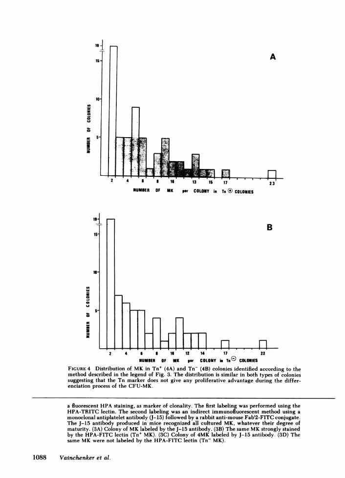
It was also possible to demonstrate by this techniquethat the average number of MKper colony was six andthat there was no difference in the distribution of MKper colony in Tn+ and Tn- colonies (Fig. 4, Student'st test = 0.2, P > 0.10). One third of the colonies were
constituted of two MK. However, these are true col-onies because they have a homogenous Tn type (ei-ther Tn+ or Tn-), and we have shown (25) that no MKcan be detected in culture before day 5, therefore dem-onstrating that such colonies really arise as a result ofthe differentiation of a progenitor. With patient B.A.,only the platelets were studied, and 81% were Tnpositive.
DISCUSSION
Because the presence of a dual population of cells iswell established within Tn bloods (12), we have in-vestigated the level of cell differentiation at which theTn antigen might be expressed. The presence of Tndeterminants was therefore explored by HPA labelingof BFU-E, CFU-GM, and CFU-MK colonies derivedfrom blood and/or bone marrow progenitors from agroup 0, Tn patient. Two possible results were ex-pected. Firstly, a mixture of Tn+ and Tn- cells mightcoexist within each colony. Secondly, a dual populationof colonies might be found, one entirely positive andthe other entirely negative for the Tn marker.
The present study clearly demonstrates that the sec-ond eventuality occurred, indicating that two differentpopulations of stem cells are present in the bone mar-row of the two Tn patients investigated: one is normal(Tn- progenitors), and the other exhibits the somaticmutation (Tn+ progenitors). The expression of the Tnantigen on the cells contained in each colony was in-dependent of their stage of maturation. In fact, inBFU-E colonies, the maturation of erythroblasts is notentirely synchronous, whereas the Tn antigen was sim-ilarly expressed on all cells of the colony. The Tnmarker was found both at early periods of culture
Hematopoietic Progenitors of Tn Blood 1085

when the erythroblasts are mostly immature and atlate periods, when they are fully mature. These resultsclearly establish that Tn cannot be considered as anantigen of maturation.
The clonal expression of Tn antigen in erythroid andgranulocytic colony assays was confirmed with a bloodsample from a second 0 patient (B.A.). Wefound thatthis patient had numerous Tn+ circulating cells be-cause 95, 81, and 82% of peripheral erythrocytes,
A ElilliB _
platelets, and granulocytes, respectively, were labeledby HPA-FITC.
As compared with the results obtained with the firstpatient, P.L., (Table I), it is obvious that the proportionof Tn+ cells in peripheral blood varies over a ratherwide range from one individual to another. The natureof the mechanism that controls the phenomenon isunknown.
Other blood group antigens are present in erythroid
FIGURE 2 Immunofluorescent detection of Tn antigen on granulocytes (2A, B) and CFU-GMcolonies (2C) using HPA-FITC labeling. (2A). An EDTAanticoagulated blood sample (patientP.L.) was sedimented at 1 g for 3 h. The buffy coat was carefully aspirated, washed in PBS,and processed for immunofluorescence. The smears were fixed with methanol, washed in coldPBS, and reacted for 30 min at 4°C with 100 pA of HPA-FITC (10 ,ug/ml in PBS). (2B). Thegranulocytes were purified by Ficoll-metrizoate (d = 1.077) density centrifugation for 25 minat 1,600 g. The polymorphonuclears were collected at the bottom of the tubes and the residualerythrocytes were eliminated by hypotonic lysis in 0.2% (wt/vol) NaCl. The isolated granu-locytes were treated for 5 min at 20°C with 1% (wt/vol) paraformaldehyde in PBS, washed,and further incubated with 0.1 ml HPA-FITC (10 jAg/ml in PBS) for 30 min at room tem-perature. After extensive washings in PBS, the smears (2A) or free erythrocyte suspensions (2B)were mounted in glycerine-buffered PBS and examined for fluorescence. As shown from thispicture, cells fixed with methanol exhibit a cytoplasmic fluorescence (2A), while a peripherallabeling was observed with free cells in suspension (2B). Both patterns of labeling were specificbecause control group 0 cells were not labeled and identical percentage of Tn+ granulocytes(29%) were found for a same patient. The Tn- granulocytes are not visible on these preparationsas they are not labeled. (2C) Blood BFU-GMand CFU-Eo were grown in methylcellulose usingMo-medium as stimulating factor. The colonies were individually picked off, cytocentrifuged,and stained with HPA-FITC after fixation in methanol. Colonies were made of either Tn+granulocytes (60%) or Tn- granulocytes (40%). A Tn+ eosinophil-granulocyte colony, with atypical cytoplasmic fluorescence, is shown.
1086 Vainchenker et al.

A B
C DFIGuRE 3 Detection of Tn antigen on CFU-MK ciolonies using a double immunofluorescentlabeling. The colonies of MKwere grown from bone marrow aspirate of the patient (P.L.) inplasma clot using PHA-LCM as stimulating factor. Because MK colonies are indeed clustersof loosely aggregated cells, a double immunofluorescent assay was applied in situ. The aimwas to demonstrate the clonality of the MKcolony assay, using the Tn antigen, revealed by
Hematopoietic Progenitors of Tn Blood 1087
A
F
.1f, "f= 1 *&J"4 5 I 16
NUMSER OF MK
i ElZ !SI I I I.
13 15 17
per COLONY in To (@ COLONIES
B
2 4 6 I 16 12 14 17 22
NUMBER OF MK per COLONY in Tm® COLONIES
FIGURE 4 Distribution of MK in Tn+ (4A) and Tn- (4B) colonies identified according to themethod described in the legend of Fig. 3. The distribution is similar in both types of coloniessuggesting that the Tn marker does not give any proliferative advantage during the differ-enciation process of the CFU-MK.
a fluorescent HPA staining, as marker of clonality. The first labeling was performed using theHPA-TRITC lectin. The second labeling was an indirect immunofluorescent method using a
monoclonal antiplatelet antibody (J-15) followed by a rabbit anti-mouse Fab'2-FITC conjugate.The J-15 antibody produced in mice recognized all cultured MK, whatever their degree ofmaturity. (3A) Colony of MKlabeled by the J-15 antibody. (3B) The same MKstrongly stainedby the HPA-FITC lectin (Tn+ MK). (3C) Colony of 4MK labeled by J-15 antibody. (3D) Thesame MKwere not labeled by the HPA-FITC lectin (Tn- MK).
1088 Vainchenker et al.
toV
a
a
M
co
U
a
!2
a=U,UU6
colonies but are expressed in a different way. For in-stance, the i antigen is heterogeneously found fromone BFU-E colony to another in adult cells. However,within the same colony, i+ and i- erythroblasts coexistin different proportions (27). Thus, the i antigen dis-plays the characteristics of a differentiation marker,whereas the Tn antigen is the consequence of a somaticmutation. As Tn abnormality is expressed in BFU-E,CFU-GM, and CFU-MK colonies, this mutation hasto be considered to have occurred at the level of apluripotent stem cell common to these lineages.
It has been extensively demonstrated that eacherythroid or granulocyte-monocyte colony is derivedfrom one cell (26, 28, 29). However, direct evidenceof the clonal origin of MKcolonies in man, and evenin the mouse, is lacking. Assessment of the clonalityof MKcolonies is critical, because most MKcoloniesare clusters of loosely aggregated MK and are thusdistinct in their aspect from the granulocytic anderythroid colonies. It could be argued that megakar-yocytic colonies merely arise from the coalescence oftwo or more MK. In addition, because each colonyincludes 2-20 MK, their content of G6PDisoenzymes,which is the most reliable marker of clonality (30),cannot be evaluated.
Because two populations of hemopoietic cells doexist even at the level of BFU-E and CFU-GMin Tnindividuals, the Tn antigen can be used as a reliablemarker for assessment of the clonality of the humanMKcolony assay. Double immunofluorescent labelingusing the HPA lectin and a monoclonal antiplateletantibody was used. This latter antibody (J-15) permitsthe specific identification in situ of all mature or im-mature MK as well as normal and leukemic pro-megakaryoblasts (25).
The results of this study have unequivocally demon-strated the clonal nature of this assay. Indeed, eachcolony was composed of either Tn- or Tn+ MK. Nosignificant coalescence was observed up to 6-105 platedmarrow cells. In addition, the Tn marker has allowedus to accurately delimit the size of MKcolonies. Onaverage, a colony was made of 6 MK. This result is inagreement with that obtained in cultures of murinebone marrow cells in plasma clot (31), but is differentfrom those found in agar (32). However, we have alsoobserved that the number of human MK per colonyis highly variable from one colony to another, with asemi-normal distribution ranging from 2 to 23 (Fig.4). In contrast with murine cultures (31), no singleisolated MKwere observed. The present study has thusafforded clear evidence that the mutation responsiblefor Tn reactivation has occurred in a pluripotent stemcell, and also that this mutation does not bring anysignificant proliferative advantage during the pathwayfrom the commited progenitors to the mature cells.However, this study does not permit direct demon-
stration of the occurrence of the Tn antigen on stemcells. For the erythroid and MK lineages, the sameproportion of Tn+ cells was observed on the erythro-cytes and BFU-E colonies on one hand, and in plateletand MKcolonies on the other. As far as the granulo-cytic lineage is concerned, an even higher proportionof Tn+ CFU-GMor CFU-Eo colonies was observed ascompared with mature granulocytes (patient P.L.).This result might be explained by a peripheral de-struction of Tn granulocytes by natural antibodies.
A slight difference in the proportion of Tn+ and Tn-colonies was observed between each hematopoietic lin-eage, which cannot be explained by this study. It couldbe tentatively ascribed either to differences in thecommitment of hematopoietic precursors from thepluripotent stem cells or to changes in the rate of self-renewal from one type of commited stem cell to an-other.
Thus, it must be assumed that the Tn mutation hasoccurred in a single pluripotent stem cell followed bya clonal expansion. As no proliferative advantage wasobserved after the commitment, this advantage shouldtake place either in the proliferative compartment ofthe pluripotent stem cell or during the process of com-mitment. In the near future, it will be possible to in-vestigate these phenomena with the development oflong-term bone marrow culture techniques (33) andpluripotent stem cell assays in man (34).
At the present time, we are of the opinion that Tnactivation results from clonal expansion and for thisreason may be considered as being equivalent to apreleukemic state. As also noticed by others (35), theTn transformation may be compared to paroxysmalnoctural hemoglobinuria because both disorders de-pend on a membrane abnormality, which itself resultsfrom a mutation occurring in a pluripotent stem cell(12, 36, 37). In both conditions, two subpopulations ofmyeloid cells coexist (12, 38), and the development ofacute leukemia can be occasionally observed (3-5, 39,40). Tn polyagglutinability is now a well characterizedmutation occurring in a pluripotent stem cell, and itwill be a useful model for future studies of the roleof blood group antigens in the proliferation of pluri-potent stem cells.
ACKNOWLEDGMENTS
The authors are indebted to Dr. Hainault (Paris, France) forsupplying blood samples from the Tn patient, P.L.
This work was supported by a grant CRL N'811030 froml'Institut National de la Sante et de la Recherche Medicale.
REFERENCES1. Bird, G. W. G. 1977. Erythrocyte polyagglutination. In
Clinical Laboratory Science. T. J. Greenwalt and E. A.Steane, editors. CRCPress, Inc., Boca Raton, FL. 1: 443-454.
Hematopoietic Progenitors of Tn Blood 1089
2. Bird, G. W. G., N. K. Shinton, and J. Wingham. 1971.Persistent mixed-field polyagglutination. Br. J. Hae-matol. 21: 443-453.
3. Bird, G. W. G., J. Wingham, M. J. Pippard, J. G. Hoult,and V. Melikian. 1976. Erythrocyte membrane modifi-cation in malignant diseases of myeloid and lymphore-ticular tissues. Br. J. Haematol. 33: 289-294.
4. Baldwin, M. L., C. Barrasso, and R. L. Ridolfi. 1979. Tn-polyagglutinability associated with acute myelomono-cytic leukemia. Am. J. Clin. Pathol. 72: 1024-1027.
5. Ness, P. M., G. Garraty, P. A. Morel, and H. A. Perkins.1979. Tn-polyagglutination preceding acute leukemia.Blood. 54: 30-34.
6. Dahr, W., G. Uhlenbruck, and G. W. G. Bird. 1974.Cryptic A-like receptor sites in human erythrocyte gly-coproteins: proposed nature of the Tn antigen. Vox Sang.27: 29-42.
7. Springer, G. F., and P. R. Desai. 1975. Human bloodgroup MN and precursor specificities: structural andbiological aspects. Carbohydr. Res. 40: 183-192.
8. Bird, G. W. G. 1978. Significant advances in lectins andpolyagglutinable red cells. In XVth Congress of the In-ternational Society of Blood Transfusion. Librairie Ar-nette. Paris. 87-93.
9. Dahr, W., G. Uhlenbruck, H. H. Gunson, and M. VanDer Hart. 1975. Molecular basis of Tn-polyagglutin-ability. Vox Sang. 29: 36-50.
10. Cartron, J. P., G. Andreu, J. Cartron, G. W. G. Bird, C.Salmon, and A. Gerbal. 1978. Demonstration of T-trans-ferase deficiency in Tn-polyagglutinable blood samples.Eur. J. Biochem. 92: 111-119.
11. Cartron, J. P., and A. T. Nurden. 1979. Galactosyltrans-ferase and membrane glycoprotein abnormality in hu-man platelets from Tn-syndrome donors. Nature (Lond.).282: 621-623.
12. Cartron, J. P., D. Blanchard, A. T. Nurden, J. Cartron,C. Rahuel, D. Lee, W. Vainchenker, U. Testa, and H.Rochant. 1981. Tn syndrome: a disorder affecting redcell, platelet and granulocyte cell surface components.In Blood groups and other red cell surface markers inhealth and disease. C. Salmon, editor. Masson PublishingU. S. A., Inc. In press.
13. Cartron, J. P., J. Cartron, G. Andreu, C. Salmon, andG. W. G. Bird. 1978. Selective deficiency of 3-p-D-ga-lactosyltransferase (T-transferase) in Tn-polyagglutina-ble erythrocytes. Lancet. I: 856-857.
14. Berger, E. G. and I. Kozdrowski 1978. Permanentmixed-field polyagglutinable erythrocytes lack galacto-syltransferase activity. FEBS (Fed. Eur. Biochem. Soc.)Lett. 93: 105-108.
15. Haynes, C. R., I. Dorner, G. L. Leonard, W. R. Ar-rowsmith, and H. Chaplin. 1970. Persistent polyagglu-tinability in vivo unrelated to T-antigen activation.Transfusion (Phila.). 10: 43-51.
16. Bird, G. W. G., and J. Wingham. 1974. The M, N, andNvg receptors of Tn erythrocytes. Vox Sang. 26: 171-175.
17. Aye, M. T., J. A. Seguin, and J. P. McBurney. 1979.Erythroid and granulocytic colony growth in culturessupplemented with human serum lipoprotein. J. Cell.Physiol. 99: 233-238.
18. Guilbert, L. J., and N. N. Iscove. 1976. Partial replace-ment of serum by selenite, transferrin, albumin and lec-ithin in hemopoietic cell cultures. Nature (Lond.). 263:594-595.
19. Lusis, A. J., D. H. Quon, and D. W. Golde. 1981. Pu-rification and characterization of a human T-lympho-cyte derived granulocyte macrophage colony stimulat-ing factor. Blood. 57: 13-21.
20. Iscove, N. N., L. J. Guilbert, and C. Weyman. 1980.Complete replacement of serum in primary cultures oferythropoietin-dependent red cell precursors (CFU-E)by albumin, transferrin, iron, unsaturated fatty acid, lec-ithin and cholesterol. Exp. Cell. Res. 126: 121-126.
21. Vainchenker, W., J. Bouguet, J. Guichard, and J. Breton-Gorius. 1979. Megakaryocyte colony formation fromhuman bone-marrow precursors. Blood. 54: 940-945.
22. Vainchenker, W., J. Breton-Gorius, and J. Chapman.1981. Induction of human megakaryocyte colonies invitro and ultrastructural aspects of their maturation. InMegakaryocyte biology and precursors. In vitro cloningand cellular properties. R. Levine, N. Williams, and B.Evalt, editors. Elsevier North-Holland, Inc., New York.139-157.
23. Aye, M. T., J. E. Till, and E. A. McCulloch. 1975. In-teracting populations affecting proliferation of leukemiccells in culture. Blood. 45: 485-493.
24. Mazur, E. M., R. Hoffman, J. Chasis, S. Marchesi, andE. Bruno. 1981. Immunofluorescent identification of hu-man megakaryocyte colonies using an antiplatelet gly-coprotein anti-serum. Blood. 57: 277-286.
25. Vainchenker, W., J. F. Deschamps, J. M. Bastin, J. Gui-chard, M. Titeux, J. Breton-Gorius, and A. J. McMichael.1982. Two monoclonal anti-platelet antibodies as mark-ers of human megakaryocyte maturation. Immunoflu-orescence staining and platelet peroxidase detection onmegakaryocyte colonies and on in vivo cells from normaland leukemic patients. Blood. In press.
26. Singer, J. W., P. J. Fialkow, L. W. Dow, C. Ernst, andL. Steinmann. 1979. Unicellular or multicellular originof human granulocyte macrophage colonies in vitro.Blood. 54: 1395-1399.
27. Vainchenker, W., U. Testa, H. Rochant, M. Titeux, A.Henri, J. Bouguet, and J. Breton-Gorius. 1981. Cellularregulation of i and I antigens expression in human eryth-roblasts grown in vitro. Stem Cell. In press.
28. Prchal, J. F., J. W. Adamson, L. Steinmann, and P. J.Fialkow. 1976. Human erythroid colony formation invitro: evidence for clonal origin. J. Cell. Physiol. 89:489-492.
29. Strome, J. E., D. L. McLeod, and M. M. Shreeve. 1978.Evidence for the clonal nature of erythropoietic bursts.Application of an in situ method for demonstrating het-erochromatin in plasma cultures. Exp. Hematol. (OakRidge). 6: 461-467.
30. Fialkow, P. J. 1980. Clonal and stem cell origin of bloodcell neoplasmes. In Contemporary hematology/oncol-ogy. J. LeBue, A. S. Gordon, R. Silber, and F. M. Muggia,editors. Plenum Publishing Corporation, New York. 1-46.
31. Dukes, P. P., P. Izadi, J. A. Ortega, N. A. Shore, and E.Gomperts. 1980. Inhibitory effects of interferon onmouse megakaryocytic progenitor cells in culture Exp.Hematol. (Oak Ridge). 8: 1048-1056.
32. Levin, J., F. C. Levin, D. G. Penington, and D. Metcalf.1981. Measurements of ploidy distribution in megakar-yocyte colonies obtained from cultures with studies ofthe effects of thrombocytopenia. Blood. 57: 287-297.
33. Dexter, T. M., T. D. Allen, and L. G. Lajtha. 1977. Con-
1090 Vainchenker et al.

ditions controlling the proliferation of haemopoieticstem cells in vitro. J. Cell. Physiol. 91: 335-344.
34. Fauser, A. A., and H. A. Messner. 1979. Identificationof megakaryocytes, macrophages and eosinophils in col-onies of human bone-marrow containing neutrophilicgranulocytes and erythroblasts. Blood. 53: 1023-1027.
35. Bird, G. W. G. 1979. Erythrocytes serology of somemalignant diseases. Blut. 35: 165-169.
36. Oni, S. B., B. 0. Osunkoya, and L. Luzzatto. 1970. Parox-ysmal nocturnal hemoglobinuria evidence for mono-clonal origin of abnormal blood cells. Blood. 36: 145-152.
37. Rosse, W. F., and J. V. Dacie. 1966. Immune lysis ofnormal human and paroxysmal nocturnal hemoglobi-
nuria (PNH) red blood cells. I. The sensitivity of PNHred cells to lysis by complement and specific antibody.J. Clin. Invest. 45: 736-748.
38. Stern, M., and W. F. Rosse. 1979. Two populations ofgranulocytes in paroxysmal nocturnal hemoglobinuria.Blood. 53: 928-934.
39. Luzzatto, L., J. B. Familusi, C. K. Williams, T. A. Junaid,B. Rotoli, and F. Alfinito. 1979. The PNHabnormalityin myeloproliferative disorders: association of PNHandacute erythremic myelosis in two children. Haemato-logica. 64: 13-30.
40. Jiji, R. M. 1979. Correspondance to Editor. Blood. 54:1451.
Hematopoietic Progenitors of Tn Blood 1091