Role of Granulocyte Colony-stimulating Factor Therapy in...
13
Review Article Role of Granulocyte Colony-stimulating Factor Therapy in Cirrhosis, ‘Inside Any Deep Asking Is the Answering’ Cyriac Abby Philips* 1 , Philip Augustine 2 , Rizwan Ahamed 2 , Sasidharan Rajesh 3 , Tom George 3 , Gopakumar C. Valiathan 4 and Solomon K. John 4 1 The Liver Unit and Monarch Liver Lab, Cochin Gastroenterology Group, Ernakulam Medical Centre, Kochi, Kerala, India; 2 Department of Gastroenterology, Cochin Gastroenterology Group, Ernakulam Medical Centre, Kochi, Kerala, India; 3 Interventional Radiology, Hepatobiliary Division, Cochin Gastroenterology Group, Ernakulam Medical Centre, Kochi, Kerala, India; 4 Department of Hepatobiliary and Transplant Surgery, Cochin Gastroenterology Group, Ernakulam Medical Centre, Kochi, Kerala, India Abstract Liver cirrhosis progresses through multiple clinical stages which culminate in either death or liver transplantation. Availability of organs, timely listing and prompt receipt of donor-livers pose difficulties in improving transplant-listed and transplant outcomes. In this regard, regenerative therapies, particularly with granulocyte colony-stimulating factor (GCSF), has become a lucrative option for improving transplant-free survival. However, the literature is confusing with regards to patient selection and real outcomes. In this exhaustive review, we describe the basics of liver fibrosis and cirrhosis through novel insights from a therapeutic point of view, discuss preclinical studies on GCSF in advanced liver disease to improve on clinical utility, shed light on the pertinent literature of GCSF in advanced cirrhosis, and provide astute inputs on growth factor therapy in decom- pensated cirrhosis. Citation of this article: Philips CA, Augustine P, Ahamed R, Rajesh S, George T, Valiathan GC, et al. Role of granulocyte colony-stimulating factor therapy in cirrhosis, ‘inside any deep asking is the answering’ . J Clin Transl Hepatol 2019;7(4):371–383. doi: 10.14218/JCTH.2019.00034. Introduction Cirrhosis is the final common pathological pathway of ongoing liver injury that arises due to multiple etiologies of insults that vary geographically. Even though the etiologies differ, there are multiple drivers and progression factors, which culminate in common pathological characteristics that lead to cirrhosis. These include hepatic necrosis, degeneration, and replace- ment of hepatic parenchyma by scar tissue surrounding failed regeneration in the form of hepatic nodules that ultimately lead to portal hypertension and liver failure. Fibrosis is the most crucial precursor that drives the central pathological process in cirrhosis. Currently, strategies in the treatment of cirrhosis are aimed at management of complications of cirrhosis and portal hypertension, and direct treatment strategies for cir- rhosis, except treatment for known causes for cirrhosis progression (such as autoimmune hepatitis, and chronic viral hepatitis B and C), are lacking. Treatment of cirrhosis and restoration or replacement of functional regenerative potential can only happen once the molecular mechanisms that drive progression to cirrhosis are better understood. These molecular mechanisms, even though generally under- stood, lack clarity in the current literature. 1 In the current review, we discuss pertinent mechanisms that lead to cirrhosis, and regenerative or restorative therapeutic strategies aimed at prevention and reversal of cirrhosis based on the currently known molecular mechanisms of cirrhosis. We also explore the current literature on clinical trials of regener- ative strategies in cirrhosis and provide a critical appraisal of the same with particular emphasis on granulocyte-colony- stimulating-factor (GCSF)-based treatments. Pathogenesis of cirrhosis and complexities associated with ideal therapeutic implications Chronic liver injury results in progressive accumulation of extracellular matrix (ECM) with distortion of hepatic paren- chymal architecture, an event that is spearheaded by myofi- broblasts that form due to activation of hepatic stellate cells (HSCs) and multiple other cell types. This fibril-forming collagen deposition that replaces the low density, basement membrane-like interstitial matrix, along with the accumula- tion of other matrix proteins such as hyaluronan, elastin, fibronectin and proteoglycans, is central to fibrosis and its Journal of Clinical and Translational Hepatology 2019 vol. 7 | 371–383 371 Copyright: © 2019 Authors. This article has been published under the terms of Creative Commons Attribution-NonCommercial 4.0 International (CC BY-NC 4.0), which permits noncommercial unrestricted use, distribution, and reproduction in any medium, provided that the following statement is provided. “This article has been published in Journal of Clinical and Translational Hepatology at DOI: 10.14218/JCTH.2019.00034 and can also be viewed on the Journal’s website at http://www.jcthnet.com” . Keywords: GCSF; Growth factor; Portal hypertension; Hepatocellular carcinoma; Fibrosis. Abbreviations: ACLF, acute-on-chronic liver failure; C/EBP-a, CCAAT enhancer- binding protein-alpha; CTGF, connective tissue growth factor; CTP, Child-Turcotte- Pugh; ECM, extracellular matrix; EpCAM, epithelial cell adhesion molecule; FGF, fibroblast growth factor; GCSF, granulocyte-colony-stimulating-factor; GCSF-R, GCSF receptor; HBV, hepatitis B virus; HGF, hepatocyte growth factor; HSCs, hepatic stellate cells; IL, interleukin; JAK, Janus kinase; LSECs, liver sinusoidal endothelial cells; MAPK, mitogen-activated protein kinase; MCP, macrophage colony-stimulating factor; MELD, model for end-stage liver disease; MMPs, matrix metalloproteinases; MSCs, mesenchymal stem cells; NF-kB, nuclear factor kappa-light-chain-enhancer of activated B cells; PDGF, platelet-derived growth factor; PPAR, peroxisome proliferator-activated receptor; SMT, standard medical care; STAT, signal transducer and activator of transcription; TGF-b, trans- forming growth factor-beta; TIMPs, tissue inhibitors of MMPs; TNF-a, tumor necrosis factor-alpha; VEGF, vascular endothelial growth factor. Received: 11 August 2019; Revised: 20 September 2019; Accepted: 5 October 2019 *Correspondence to: Cyriac Abby Philips, The Liver Unit and Monarch Liver Lab, Cochin Gastroenterology Group, Ernakulam Medical Centre, Automobile Road, Palarivattom, Kochi, 682025, Kerala, India. Tel: +91-484-2907000, Fax: +91-484-2907000, E-mail: [email protected]
Transcript of Role of Granulocyte Colony-stimulating Factor Therapy in...
Review Article
Role of Granulocyte Colony-stimulating Factor Therapy inCirrhosis, ‘Inside Any Deep Asking Is the Answering’
Cyriac Abby Philips*1, Philip Augustine2, Rizwan Ahamed2, Sasidharan Rajesh3, Tom George3,Gopakumar C. Valiathan4 and Solomon K. John4
1The Liver Unit and Monarch Liver Lab, Cochin Gastroenterology Group, Ernakulam Medical Centre, Kochi, Kerala, India;2Department of Gastroenterology, Cochin Gastroenterology Group, Ernakulam Medical Centre, Kochi, Kerala, India;
3Interventional Radiology, Hepatobiliary Division, Cochin Gastroenterology Group, Ernakulam Medical Centre, Kochi, Kerala, India;4Department of Hepatobiliary and Transplant Surgery, Cochin Gastroenterology Group, Ernakulam Medical Centre,
Kochi, Kerala, India
Abstract
Liver cirrhosis progresses through multiple clinical stageswhich culminate in either death or liver transplantation.Availability of organs, timely listing and prompt receipt ofdonor-livers pose difficulties in improving transplant-listedand transplant outcomes. In this regard, regenerativetherapies, particularly with granulocyte colony-stimulatingfactor (GCSF), has become a lucrative option for improvingtransplant-free survival. However, the literature is confusingwith regards to patient selection and real outcomes. In thisexhaustive review, we describe the basics of liver fibrosis andcirrhosis through novel insights from a therapeutic point ofview, discuss preclinical studies on GCSF in advanced liverdisease to improve on clinical utility, shed light on thepertinent literature of GCSF in advanced cirrhosis, andprovide astute inputs on growth factor therapy in decom-pensated cirrhosis.Citation of this article: Philips CA, Augustine P, Ahamed R,Rajesh S, George T, Valiathan GC, et al. Role of granulocytecolony-stimulating factor therapy in cirrhosis, ‘inside anydeep asking is the answering’. J Clin Transl Hepatol2019;7(4):371–383. doi: 10.14218/JCTH.2019.00034.
Introduction
Cirrhosis is the final common pathological pathway of ongoingliver injury that arises due to multiple etiologies of insults thatvary geographically. Even though the etiologies differ, thereare multiple drivers and progression factors, which culminatein common pathological characteristics that lead to cirrhosis.These include hepatic necrosis, degeneration, and replace-ment of hepatic parenchyma by scar tissue surrounding failedregeneration in the form of hepatic nodules that ultimatelylead to portal hypertension and liver failure. Fibrosis is themost crucial precursor that drives the central pathologicalprocess in cirrhosis.
Currently, strategies in the treatment of cirrhosis areaimed at management of complications of cirrhosis andportal hypertension, and direct treatment strategies for cir-rhosis, except treatment for known causes for cirrhosisprogression (such as autoimmune hepatitis, and chronicviral hepatitis B and C), are lacking. Treatment of cirrhosisand restoration or replacement of functional regenerativepotential can only happen once the molecular mechanismsthat drive progression to cirrhosis are better understood.These molecular mechanisms, even though generally under-stood, lack clarity in the current literature.1
In the current review, we discuss pertinentmechanisms thatlead to cirrhosis, and regenerative or restorative therapeuticstrategies aimed at prevention and reversal of cirrhosis basedon the currently known molecular mechanisms of cirrhosis. Wealso explore the current literature on clinical trials of regener-ative strategies in cirrhosis and provide a critical appraisal ofthe same with particular emphasis on granulocyte-colony-stimulating-factor (GCSF)-based treatments.
Pathogenesis of cirrhosis and complexities associatedwith ideal therapeutic implications
Chronic liver injury results in progressive accumulation ofextracellular matrix (ECM) with distortion of hepatic paren-chymal architecture, an event that is spearheaded by myofi-broblasts that form due to activation of hepatic stellate cells(HSCs) and multiple other cell types. This fibril-formingcollagen deposition that replaces the low density, basementmembrane-like interstitial matrix, along with the accumula-tion of other matrix proteins such as hyaluronan, elastin,fibronectin and proteoglycans, is central to fibrosis and its
Journal of Clinical and Translational Hepatology 2019 vol. 7 | 371–383 371
Copyright: © 2019 Authors. This article has been published under the terms of Creative Commons Attribution-NonCommercial 4.0 International (CC BY-NC 4.0), whichpermits noncommercial unrestricted use, distribution, and reproduction in any medium, provided that the following statement is provided. “This article has been publishedin Journal of Clinical and Translational Hepatology at DOI: 10.14218/JCTH.2019.00034 and can also be viewed on the Journal’s website at http://www.jcthnet.com”.
Keywords: GCSF; Growth factor; Portal hypertension; Hepatocellular carcinoma;Fibrosis.Abbreviations: ACLF, acute-on-chronic liver failure; C/EBP-a, CCAAT enhancer-binding protein-alpha; CTGF, connective tissue growth factor; CTP, Child-Turcotte-Pugh; ECM, extracellular matrix; EpCAM, epithelial cell adhesion molecule; FGF,fibroblast growth factor; GCSF, granulocyte-colony-stimulating-factor; GCSF-R,GCSF receptor; HBV, hepatitis B virus; HGF, hepatocyte growth factor; HSCs,hepatic stellate cells; IL, interleukin; JAK, Janus kinase; LSECs, liver sinusoidalendothelial cells; MAPK, mitogen-activated protein kinase; MCP, macrophagecolony-stimulating factor; MELD, model for end-stage liver disease; MMPs,matrix metalloproteinases; MSCs, mesenchymal stem cells; NF-kB, nuclearfactor kappa-light-chain-enhancer of activated B cells; PDGF, platelet-derivedgrowth factor; PPAR, peroxisome proliferator-activated receptor; SMT, standardmedical care; STAT, signal transducer and activator of transcription; TGF-b, trans-forming growth factor-beta; TIMPs, tissue inhibitors of MMPs; TNF-a, tumornecrosis factor-alpha; VEGF, vascular endothelial growth factor.Received: 11 August 2019; Revised: 20 September 2019; Accepted: 5 October2019*Correspondence to: Cyriac Abby Philips, The Liver Unit and Monarch Liver Lab,Cochin Gastroenterology Group, Ernakulam Medical Centre, Automobile Road,Palarivattom, Kochi, 682025, Kerala, India. Tel: +91-484-2907000, Fax:+91-484-2907000, E-mail: [email protected]
progression to cirrhosis.2 The ECM has the potential tosecrete various cytokines, such as transforming growthfactor-beta (TGF-b), platelet-derived growth factor (PDGF),hepatocyte growth factor (HGF), connective tissue growthfactor (CTGF), tumor necrosis factor-alpha (TNF-a), fibroblastgrowth factor (FGF) and vascular endothelial growth factor(VEGF), that promote angiogenesis, another critical eventthat leads to exaggerated wound healing in the form offibrous tissue formation. While this injury and ECM depositionis ongoing, remodeling of the ECM to preserve healthy hepaticparenchymal structure and function becomes critical in themaintenance of liver health.3
This balance is maintained by the matrix metalloprotei-nases (MMPs; specifically, MMP-1, -2, -8 and -13) and theirinhibitors, the tissue inhibitors of MMPs (TIMPs). When theinjury is chronic and the ECM deposition is overwhelming dueto the persistence of HSC activation and abnormal neo-angiogenesis, the activity of TIMPs takes the upper hand.This tipping of balance toward prolonged activity of TIMPs(mostly TIMP-1 and -2) results in anti-apoptotic effects onHSCs that further promoted fibrogenesis.4
Chronic liver inflammation leads to hepatocyte necrosis/apoptosis, paracrine stimulation, Kupffer cell activation, reac-tive oxidation and cytokine deliberation that activate HSCsthat then transform into myofibroblasts with profibrogenicpotential. HSC activation also occurs through lipid peroxiderelease, TNF-a and interferon-gamma production via theactivation of nuclear factor kappa-light-chain-enhancer ofactivated B cells (NF-kB)-interferon regulatory factor 3pathway and toll-like receptors (through lipopolysaccharideproduction by gut microbial dysbiosis).5 Stimulated HSCssecrete macrophage colony-stimulating factor (MCP)-1,interleukin (IL)-6 and chemokines that act on their respectivereceptors, leading to macrophage activation, neutrophil infil-tration, and chemotaxis, which activate mitochondrial oxida-tion in hepatocytes leading to their apoptosis; furthermore,these become a strong trigger for fibrogenesis, as phagocy-tosis of damaged hepatocytes by myofibroblasts enhancesthe fibrogenic activation through NADPH oxidase and theJanus kinase (JAK) signal transducer and activator of tran-scription (STAT) and phosphoinositide 3-kinase/Akt path-ways.6,7 The HSCs and myofibroblasts proliferate and laydown ECM, and further promote hepatic inflammationthrough enhanced TIMP expression, also under the influenceof secreted angiotensin II [via mitogen-activated proteinkinase (MAPK) signal transduction pathways], upregulatedcannabinoid receptor 1, and circulating adipokine leptin [via(JAK)-signal transduction, leading to suppression of peroxi-some proliferator-activated receptor g (PPAR-g)].8
Other cells of the liver microenvironment regulatingenhancement or reduction of fibrosis involve natural killercells, T cells, monocytes, liver sinusoidal endothelial cells(LSECs), ductular cells, cholangiocytes, and portal fibro-blasts.9 The role of monocytes in inflammation and fibrosisis important regarding GCSF therapy. Fibrogenesis is pro-moted by the subset of proinflammatory monocytes (CD14+ and CD16+ in humans). Monocytes are a source of circu-lating fibrocytes, which differentiate into collagen-producingfibroblasts, which are in turn closely related to the bonemarrow mesenchymal stem cells (MSCs).10,11 The TGF-bsecreted by myofibroblasts promotes hepatocyte apoptosisafter activation. TGF-b1 activates Smad2, and Smad3promote fibrogenesis. Reduction and clearance of activatedHSCs are central to fibrosis regression.
In clinical and experimental fibrosis models, control orelimination of the etiological agent responsible for the chronicinflammation has been shown to promote fibrosis reversal,due to the complete disappearance of myofibroblasts. Eventhen, a small subset of myofibroblasts can escape apoptosisduring liver fibrosis regression, acquiring a phenotype similarto but distinct from quiescent HSCs. These ‘fugitives’ thenrapidly reactivate into myofibroblasts in response to repeti-tion of fibrogenic stimuli and rapidly contribute to liverfibrosis.12,13 Early liver fibrosis, which lacks ECM crosslinkingand marked angiogenesis, has the best potential to revert totypical architecture, provided the chronic insult is adequatelycontrolled. Hence, the initiation, progression and reversal offibrosis is a highly complex process with multiple interactionsat the cellular and molecular levels.14,15
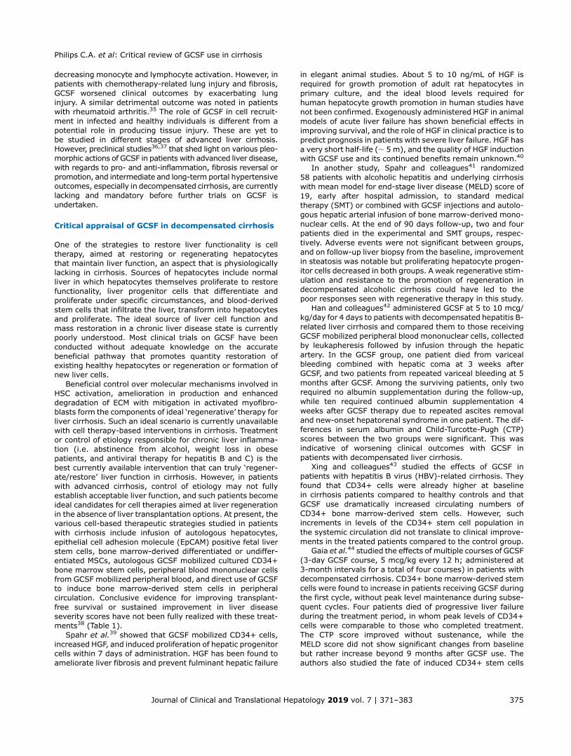
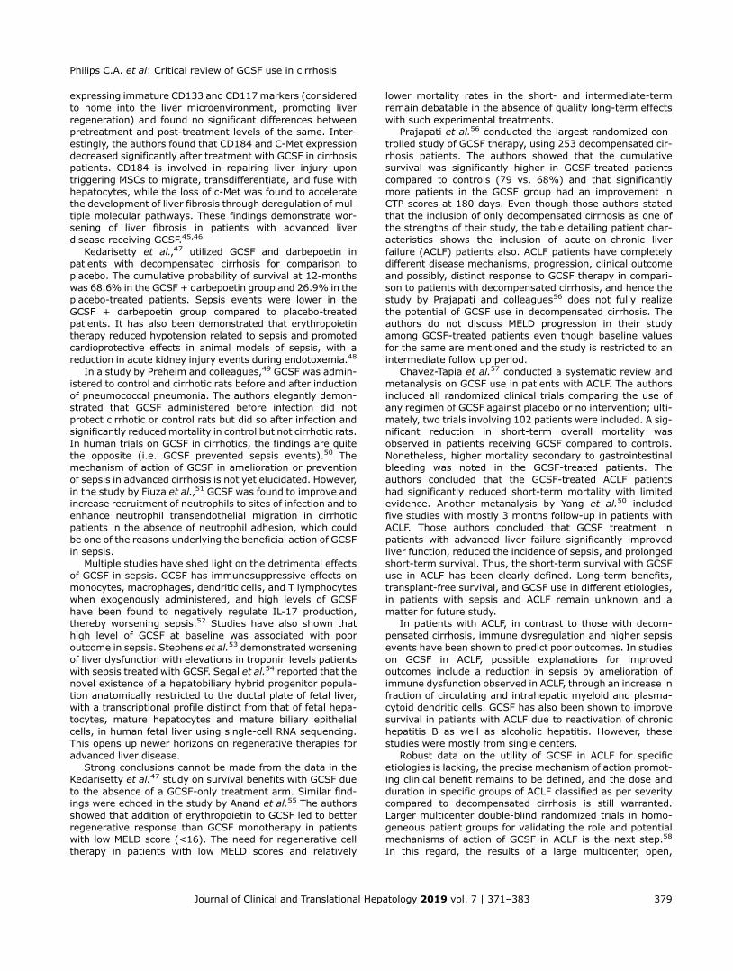
Taken together, the pleomorphic action of GCSF throughmultiple molecular mechanisms in the liver microenviron-ment could increase fibrosis and liver disease progressionapart from its beneficial effects on granulopoiesis. Hence, topromote liver regeneration or restoration, the therapeuticintervention(s) must target multiple pathways and notjust one of the ‘central’ pathways. Progression of fibrosis tocirrhosis happens through multiple ‘central’ pathways — thisis akin to a control headquarters (chronic injury) and multiple‘metro-rail-lines’ (molecular mechanisms) and major associ-ated stations (central pathways) in a large city, rather than asingle central railway station in a town. This complicated aspectof liver fibrosis progression and the complexities associatedwith treatment of fibrosis/cirrhosis is demonstrated in Fig. 1.
Pathophysiology associated with GCSF in the contextof chronic liver disease
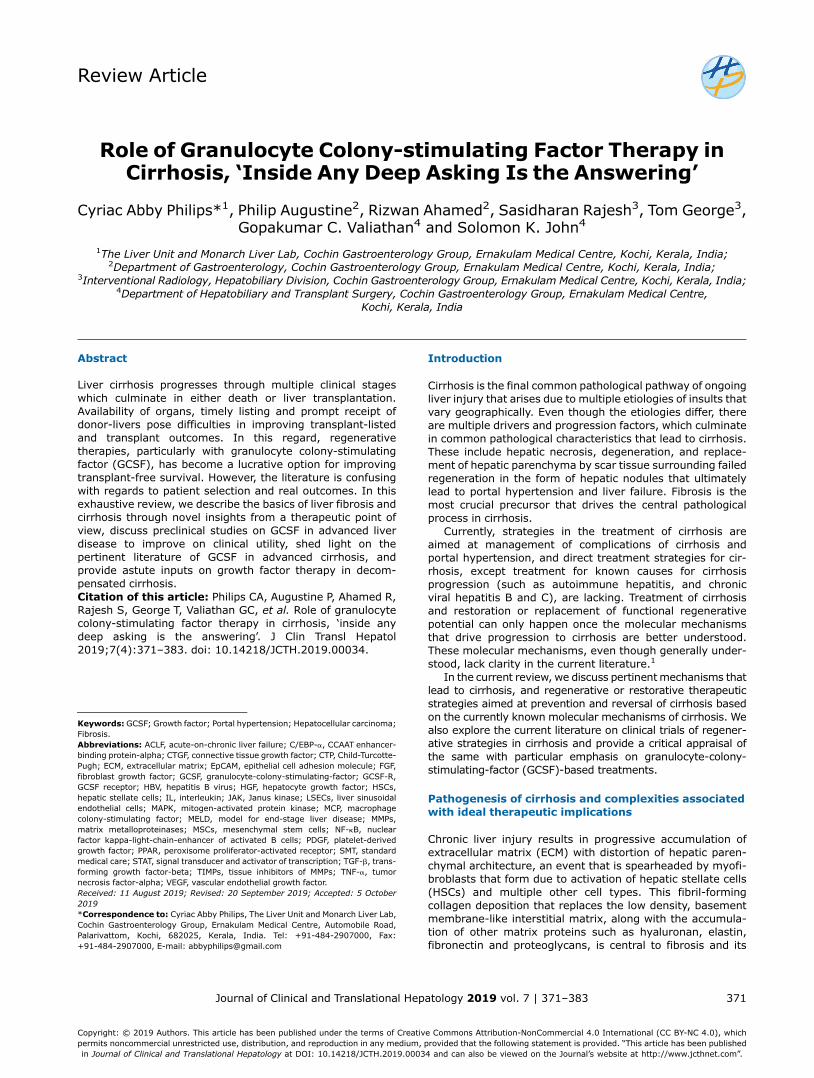
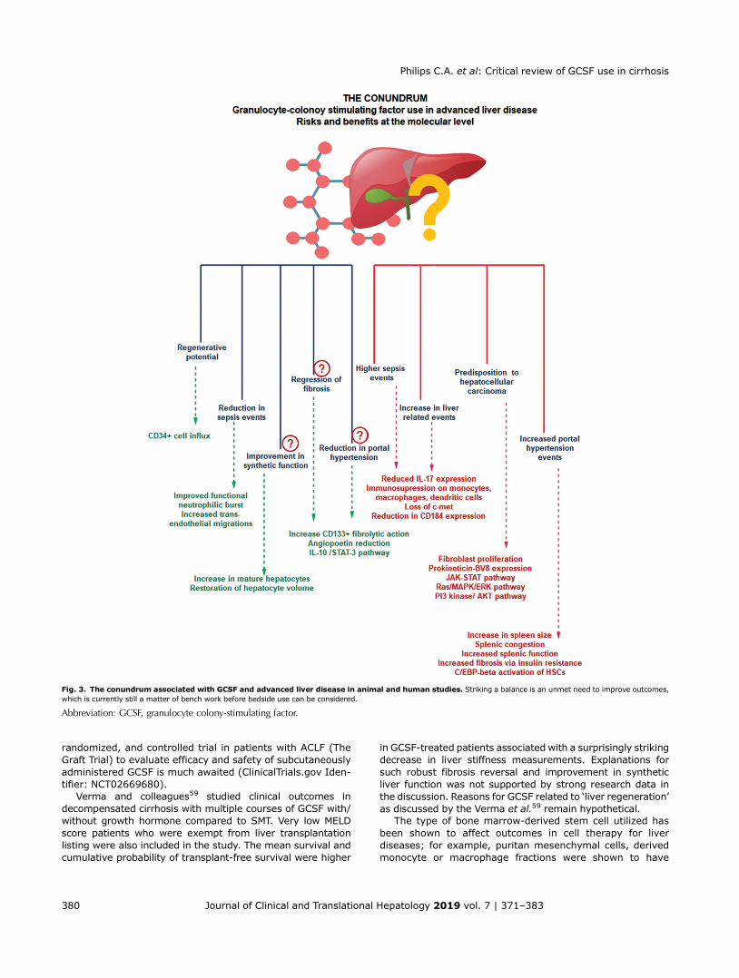
GCSF is a 25 kDa secreted glycoprotein encoded by the CSF3gene. The central physiological role played by GCSF is in theregulation of neutrophil production in health and particularlyin emergency responses to infections and bone marrowaplasia. In healthy humans, the serum concentrations ofGCSF are typically undetectable or detectable at deficientlevels, which markedly increases in the presence of aninfectious stimulus. Most of the tissues in the body secreteGCSF after stimulatory effects, such as induction of IL-1,lipopolysaccharide and TNF-a produced by the macrophages,endothelial cells, fibroblasts and related mesenchymal cells(Fig. 2).
IL-17 is a potent upstream extracellular regulator of tissueproduction of GCSF, especially in the bone marrow. Ligation ofthe extracellular domain of the GCSF receptor (GCSF-R) byGCSF results in cellular responses due to signals that arisefrom the cytoplasmic domain of the GCSF-R. The GCSF-Ris expressed by neutrophils and its precursors, such asmetamyelocytes, myelocytes, promyelocytes, myeloblasts,myeloid progenitor cells, and primitive hemopoietic stemcells. The GCSF-R signals through the JAK/STAT pathwayand through Lyn phosphorylation that activates PI3-kinase/Akt pathways, which are pertinent to the progression of liverfibrosis.
GCSF also activates Ras-MAPK through activation of tyro-sine kinases, Lyn and Hck. This is of utmost importancebecause the Ras/Raf/MEK/ERK signaling pathway has beenimplicated in the occurrence and development of hepatocel-lular carcinoma in cirrhosis. GCSF has been shown to stim-ulate tumor cell growth and migration in vitro, and to promote
372 Journal of Clinical and Translational Hepatology 2019 vol. 7 | 371–383
Philips C.A. et al: Critical review of GCSF use in cirrhosis
tumor progression in vivo by autocrine stimulation of tumorcells and paracrine activation of the tumorigenic stroma.16,17
Ordelheide et al.18 demonstrated that GCSF promoted freefatty acid-induced insulin resistance in humans and thathuman adipocytes and myotubes treated with GCSF becameinsulin-resistant. Insulin resistance is a major driver of liverfibrogenesis and carcinogenesis. In cirrhosis patients withmetabolic syndrome, obesity and insulin resistance, the useof GCSF could probably augment the disease process.19
CCAAT enhancer-binding protein-a (C/EBP-a) regulates adi-pocyte differentiation and induces apoptosis in HSCs in vivoand in vitro. Tao et al.20 showed that in the mouse liver fib-rosis model, the upregulation of C/EBP-a decreased ECM dep-osition, including collagen and hydroxyproline content, andmarkers of liver damage were reduced significantly; immuno-histochemistry showed an increase of apoptosis in HSCs,while hepatocytes were less affected.
On the other hand, C/EBPb was found to be selectivelyupregulated in granulocytic-macrophage progenitors in thepresence of GCSF. However, beneficial C/EBPa is not induced
by GCSF in hematopoietic stem cells, and hence the utility ofexogenous GSCF to decrease hepatocyte apoptosis cannot bepossible through this mechanism of action.21 Buck et al.22
showed that, in response to liver injury, activation of riboso-mal S6 kinase phosphorylation of C/EBPb in activated HSCs iscritical for the progression of liver fibrosis. Hence, in the pres-ence of GCSF, molecular mechanisms of chronic liver injurythat increases fibrosis are possibly upregulated to augmentdisease progression.
GCSF is also produced by a variety of nonhematopoieticcells, including fibroblasts and endothelial cells, and inducesthe proliferation and migration of endothelial cells, promotesangiogenesis, and upregulates inflammatory cell infiltrationinto tissues where GCSF-R is expressed.23 Shojaei et al.24
identified GCSF as a strong inducer of prokinectin-Bv8expression, both in vitro and in vivo, the latter of which pro-motes neovascularization and tumoral progression in gastro-intestinal malignancies. Hepatic angiogenesis is closelyassociated with the progression of fibrosis in chronic liver dis-eases and GCSF demonstrably induces endothelial activation
Fig. 1. The ‘Metro-Rail Concept’ to targeted therapy of the pathophysiology of cirrhosis. The development of cirrhosis follows well-coordinated steps that beginwith the etiology, leading to sustained chronic inflammation (the control or command center; black bubble) that activates and promotes multiple pathways (rail-pathways,colored lines) that feature prominent mediators of inflammation and fibrosis (central stations, grey bubbles) and parallel assisting pathway intermediaries (secondarystations, small white bubbles) that ultimately lead to the destination (red bubble). Therapies that target only few of the pathway mediators do not tend to improve outcomesas expected; however, targeting the command center (etiology) along with controlling the central-stations (central inflammatory and fibrosis pathways) would impedeprogression to destination (cirrhosis).
Abbreviations: BMDSC, bone marrow-derived stem cells; CCRs, chemokine receptors; CD, cluster of differentiation; CTGF, connective tissue growthfactor; ECM, extracellular matrix; HGF, hepatocyte growth factor; HSC, hepatic stellate cell; IL, interleukin; IRF, interferon regulatory factor; JAK/STAT, Janus kinase/signal transducers and activators of transcription; LPS, lipopolysaccharide; MAP, mitogen activated protein; MCP, monocytechemoattractant protein; MFB, myofibroblasts; NADPH, reduced form of nicotinamide adenine dinucleotide phosphate; NF-kB, nuclear factor kappaB; PDGF, platelet derived growth factor; PHT, portal hypertension; PI3K/Akt, phosphoinositide-3-kinase-protein kinase B; PPAR, peroxisomeproliferator-activated receptors; SMA, smooth muscle antibody; SMADs, homologues of the Drosophila protein, mothers against decapentaplegic(Mad) and the Caenorhabditis elegans protein Sma; TGF, transforming growth factor; TIMP, tissue inhibitors of matrix metalloproteinase; TLR, toll-likereceptor; TNF, tumor necrosis factor; VEGF, vascular endothelial growth factor.
Journal of Clinical and Translational Hepatology 2019 vol. 7 | 371–383 373
Philips C.A. et al: Critical review of GCSF use in cirrhosis

and upregulates downstream inflammatory pathways associ-ated with angiogenesis and fibrosis progression.25
The belief that GCSF improves synthetic liver function anddecreases fibrosis stems from the understanding that mobi-lization of CD34+ bone marrow-derived cells home into theliver microenvironment, transform into hepatic progenitorcells, and restore lost hepatocyte volume. Subsequently,possible reduction in expression in angiopoietin which leadsto decreased neoangiogenesis ameliorates fibrosis. Theenhancement of fibrolytic activity of CD133+ cells inducedthrough GCSF on monocyte and bone marrow activationpossibly through IL-10 mediated Stat3 regeneration pathwayhas been postulated. However, most of these processesremain clinical hypotheses, lacking proper ‘fate of cell’ tracerstudies.26–28
Several studies have shown that GCSF promoted chromo-somal changes in healthy persons associated with themodification of gene expression profile. Even though thelong-term mutagenic implications in healthy persons withsuch changes are insignificant, in cirrhosis patients, in thepresence of an inflammatory microenvironment, exogenousGCSF-associated genetic expression as well as chromosomalaberration are concerning and need further study.29 Healthypersons, when injected with GCSF, develop marked neutro-philic response within 4 h, mobilize bone marrow-activatedstem cells after 3 days, that peaks at the fifth day, which isassociated with splenic enlargement over a week’s time.Stroncek et al.30 demonstrated that the spleen lengthincreased by 20% or more in healthy subjects treated with
GCSF at 10 mcg/kg/day for 5 days. In portal hypertension,splenic congestion and splenomegaly are attributed to portalcongestion, elevated portal pressures and closely related toincreased tissue hyperplasia and fibrosis. The increase inspleen size results in increased splenic blood flow, translatingto increased portal hypertension, which may worsen withrepeated GCSF use in patients with cirrhosis.31
In the study by Nakamura et al.,32 hepatic arterial infusionof low, mid and high doses of autologous-derived CD34+ cellsmobilized by GCSF showed mild improvement in serumalbumin level, without significant sustained improvementin liver disease severity scores. The spleen size in cirrhosispatients did increase with GCSF but it was a transient phe-nomenon since the GCSF treatment was only for 5 days. Sim-ilarly, Gaia et al.33 reported significant reversible spleenenlargement with stable serum liver enzyme levels afterGCSF administration in patients with alcoholic cirrhosis, andLorenzini et al.34 demonstrated increased splenomegaly withstable levels during GCSF administration in patients with viralhepatitis. These studies concentrated on short-term use ofGCSF, and longer use or multiple dosing regimen of GCSF asseen with recent studies could be associated with greaterchances of splenic enlargement and elevation in portal pres-sures. All of these studies lacked the fate of hematopoietic cellinvestigation and, as such, conclusive evidence of liver cellrestoration or regeneration could not be ascertained.
In humans, GCSF could either be beneficial or detrimental,depending on the disease in context. For example, perioper-ative GCSF use was found to reduce postoperative morbidity by
Fig. 2. GCSF-activated molecular pathways associated with fibrosis regression and progression. GCSF is a ‘double-edged sword’ and cannot be considered a trueally in the armamentarium against fibrosis and cirrhosis.
Abbreviation: GCSF, granulocyte colony-stimulating factor.
374 Journal of Clinical and Translational Hepatology 2019 vol. 7 | 371–383
Philips C.A. et al: Critical review of GCSF use in cirrhosis
decreasing monocyte and lymphocyte activation. However, inpatients with chemotherapy-related lung injury and fibrosis,GCSF worsened clinical outcomes by exacerbating lunginjury. A similar detrimental outcome was noted in patientswith rheumatoid arthritis.35 The role of GCSF in cell recruit-ment in infected and healthy individuals is different from apotential role in producing tissue injury. These are yet tobe studied in different stages of advanced liver cirrhosis.However, preclinical studies36,37 that shed light on various pleo-morphic actions of GCSF in patients with advanced liver disease,with regards to pro- and anti-inflammation, fibrosis reversal orpromotion, and intermediate and long-term portal hypertensiveoutcomes, especially in decompensated cirrhosis, are currentlylacking and mandatory before further trials on GCSF isundertaken.
Critical appraisal of GCSF in decompensated cirrhosis
One of the strategies to restore liver functionality is celltherapy, aimed at restoring or regenerating hepatocytesthat maintain liver function, an aspect that is physiologicallylacking in cirrhosis. Sources of hepatocytes include normalliver in which hepatocytes themselves proliferate to restorefunctionality, liver progenitor cells that differentiate andproliferate under specific circumstances, and blood-derivedstem cells that infiltrate the liver, transform into hepatocytesand proliferate. The ideal source of liver cell function andmass restoration in a chronic liver disease state is currentlypoorly understood. Most clinical trials on GCSF have beenconducted without adequate knowledge on the accuratebeneficial pathway that promotes quantity restoration ofexisting healthy hepatocytes or regeneration or formation ofnew liver cells.
Beneficial control over molecular mechanisms involved inHSC activation, amelioration in production and enhanceddegradation of ECM with mitigation in activated myofibro-blasts form the components of ideal ‘regenerative’ therapy forliver cirrhosis. Such an ideal scenario is currently unavailablewith cell therapy-based interventions in cirrhosis. Treatmentor control of etiology responsible for chronic liver inflamma-tion (i.e. abstinence from alcohol, weight loss in obesepatients, and antiviral therapy for hepatitis B and C) is thebest currently available intervention that can truly ‘regener-ate/restore’ liver function in cirrhosis. However, in patientswith advanced cirrhosis, control of etiology may not fullyestablish acceptable liver function, and such patients becomeideal candidates for cell therapies aimed at liver regenerationin the absence of liver transplantation options. At present, thevarious cell-based therapeutic strategies studied in patientswith cirrhosis include infusion of autologous hepatocytes,epithelial cell adhesion molecule (EpCAM) positive fetal liverstem cells, bone marrow-derived differentiated or undiffer-entiated MSCs, autologous GCSF mobilized cultured CD34+bone marrow stem cells, peripheral blood mononuclear cellsfrom GCSF mobilized peripheral blood, and direct use of GCSFto induce bone marrow-derived stem cells in peripheralcirculation. Conclusive evidence for improving transplant-free survival or sustained improvement in liver diseaseseverity scores have not been fully realized with these treat-ments38 (Table 1).
Spahr et al.39 showed that GCSF mobilized CD34+ cells,increased HGF, and induced proliferation of hepatic progenitorcells within 7 days of administration. HGF has been found toameliorate liver fibrosis and prevent fulminant hepatic failure
in elegant animal studies. About 5 to 10 ng/mL of HGF isrequired for growth promotion of adult rat hepatocytes inprimary culture, and the ideal blood levels required forhuman hepatocyte growth promotion in human studies havenot been confirmed. Exogenously administered HGF in animalmodels of acute liver failure has shown beneficial effects inimproving survival, and the role of HGF in clinical practice is topredict prognosis in patients with severe liver failure. HGF hasa very short half-life (; 5 m), and the quality of HGF inductionwith GCSF use and its continued benefits remain unknown.40
In another study, Spahr and colleagues41 randomized58 patients with alcoholic hepatitis and underlying cirrhosiswith mean model for end-stage liver disease (MELD) score of19, early after hospital admission, to standard medicaltherapy (SMT) or combined with GCSF injections and autolo-gous hepatic arterial infusion of bone marrow-derived mono-nuclear cells. At the end of 90 days follow-up, two and fourpatients died in the experimental and SMT groups, respec-tively. Adverse events were not significant between groups,and on follow-up liver biopsy from the baseline, improvementin steatosis was notable but proliferating hepatocyte progen-itor cells decreased in both groups. A weak regenerative stim-ulation and resistance to the promotion of regeneration indecompensated alcoholic cirrhosis could have led to thepoor responses seen with regenerative therapy in this study.
Han and colleagues42 administered GCSF at 5 to 10 mcg/kg/day for 4 days to patients with decompensated hepatitis B-related liver cirrhosis and compared them to those receivingGCSF mobilized peripheral blood mononuclear cells, collectedby leukapheresis followed by infusion through the hepaticartery. In the GCSF group, one patient died from varicealbleeding combined with hepatic coma at 3 weeks afterGCSF, and two patients from repeated variceal bleeding at 5months after GCSF. Among the surviving patients, only tworequired no albumin supplementation during the follow-up,while ten required continued albumin supplementation 4weeks after GCSF therapy due to repeated ascites removaland new-onset hepatorenal syndrome in one patient. The dif-ferences in serum albumin and Child-Turcotte-Pugh (CTP)scores between the two groups were significant. This wasindicative of worsening clinical outcomes with GCSF inpatients with decompensated liver cirrhosis.
Xing and colleagues43 studied the effects of GCSF inpatients with hepatitis B virus (HBV)-related cirrhosis. Theyfound that CD34+ cells were already higher at baselinein cirrhosis patients compared to healthy controls and thatGCSF use dramatically increased circulating numbers ofCD34+ bone marrow-derived stem cells. However, suchincrements in levels of the CD34+ stem cell population inthe systemic circulation did not translate to clinical improve-ments in the treated patients compared to the control group.
Gaia et al.44 studied the effects of multiple courses of GCSF(3-day GCSF course, 5 mcg/kg every 12 h; administered at3-month intervals for a total of four courses) in patients withdecompensated cirrhosis. CD34+ bone marrow-derived stemcells were found to increase in patients receiving GCSF duringthe first cycle, without peak level maintenance during subse-quent cycles. Four patients died of progressive liver failureduring the treatment period, in whom peak levels of CD34+cells were comparable to those who completed treatment.The CTP score improved without sustenance, while theMELD score did not show significant changes from baselinebut rather increase beyond 9 months after GCSF use. Theauthors also studied the fate of induced CD34+ stem cells
Journal of Clinical and Translational Hepatology 2019 vol. 7 | 371–383 375
Philips C.A. et al: Critical review of GCSF use in cirrhosis
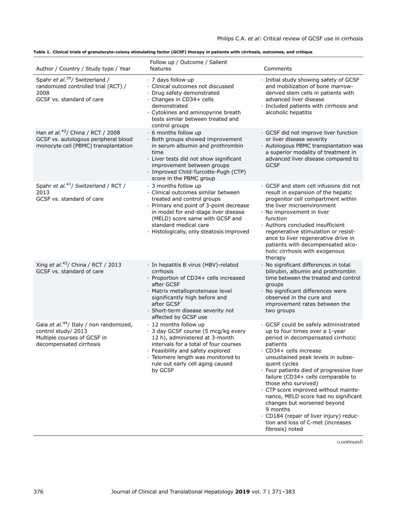
Table 1. Clinical trials of granulocyte-colony stimulating factor (GCSF) therapy in patients with cirrhosis, outcomes, and critique
Author / Country / Study type / YearFollow up / Outcome / Salientfeatures Comments
Spahr et al.39/ Switzerland /randomized controlled trial (RCT) /2008GCSF vs. standard of care
· 7 days follow up· Clinical outcomes not discussed· Drug safety demonstrated· Changes in CD34+ cellsdemonstrated
· Cytokines and aminopyrine breathtests similar between treated andcontrol groups
· Initial study showing safety of GCSFand mobilization of bone marrow-derived stem cells in patients withadvanced liver disease
· Included patients with cirrhosis andalcoholic hepatitis
Han et al.42/ China / RCT / 2008GCSF vs. autologous peripheral bloodmonocyte cell (PBMC) transplantation
· 6 months follow up· Both groups showed improvementin serum albumin and prothrombintime
· Liver tests did not show significantimprovement between groups
· Improved Child-Turcotte-Pugh (CTP)score in the PBMC group
· GCSF did not improve liver functionor liver disease severity
· Autologous PBMC transplantation wasa superior modality of treatment inadvanced liver disease compared toGCSF
Spahr et al.41/ Switzerland / RCT /2013GCSF vs. standard of care
· 3 months follow up· Clinical outcomes similar betweentreated and control groups
· Primary end point of 3-point decreasein model for end-stage liver disease(MELD) score same with GCSF andstandard medical care
· Histologically, only steatosis improved
· GCSF and stem cell infusions did notresult in expansion of the hepaticprogenitor cell compartment withinthe liver microenvironment
· No improvement in liverfunction
· Authors concluded insufficientregenerative stimulation or resist-ance to liver regenerative drive inpatients with decompensated alco-holic cirrhosis with exogenoustherapy
Xing et al.43/ China / RCT / 2013GCSF vs. standard of care
· In hepatitis B virus (HBV)-relatedcirrhosis
· Proportion of CD34+ cells increasedafter GCSF
· Matrix metalloproteinase levelsignificantly high before andafter GCSF
· Short-term disease severity notaffected by GCSF use
· No significant differences in totalbilirubin, albumin and prothrombintime between the treated and controlgroups
· No significant differences wereobserved in the cure andimprovement rates between thetwo groups
Gaia et al.44/ Italy / non randomized,control study/ 2013Multiple courses of GCSF indecompensated cirrhosis
· 12 months follow up· 3 day GCSF course (5 mcg/kg every12 h), administered at 3-monthintervals for a total of four courses
· Feasibility and safety explored· Telomere length was monitored torule out early cell aging causedby GCSF
· GCSF could be safely administratedup to four times over a 1-yearperiod in decompensated cirrhoticpatients
· CD34+ cells increaseunsustained peak levels in subse-quent cycles
· Four patients died of progressive liverfailure (CD34+ cells comparable tothose who survived)
· CTP score improved without mainte-nance, MELD score had no significantchanges but worsened beyond9 months
· CD184 (repair of liver injury) reduc-tion and loss of C-met (increasesfibrosis) noted
(continued )
376 Journal of Clinical and Translational Hepatology 2019 vol. 7 | 371–383
Philips C.A. et al: Critical review of GCSF use in cirrhosis
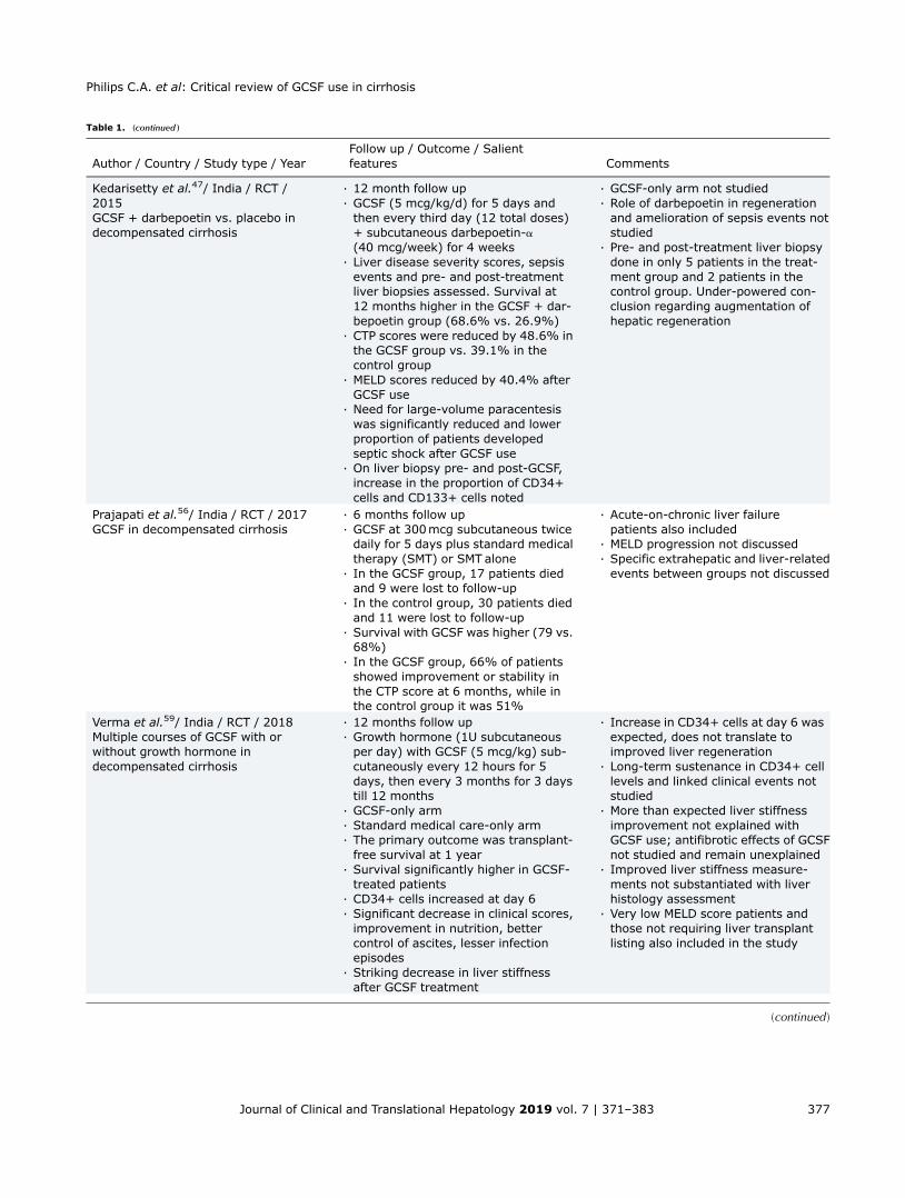
Table 1. (continued )
Author / Country / Study type / YearFollow up / Outcome / Salientfeatures Comments
Kedarisetty et al.47/ India / RCT /2015GCSF + darbepoetin vs. placebo indecompensated cirrhosis
· 12 month follow up· GCSF (5 mcg/kg/d) for 5 days andthen every third day (12 total doses)+ subcutaneous darbepoetin-a(40 mcg/week) for 4 weeks
· Liver disease severity scores, sepsisevents and pre- and post-treatmentliver biopsies assessed. Survival at12 months higher in the GCSF + dar-bepoetin group (68.6% vs. 26.9%)
· CTP scores were reduced by 48.6% inthe GCSF group vs. 39.1% in thecontrol group
· MELD scores reduced by 40.4% afterGCSF use
· Need for large-volume paracentesiswas significantly reduced and lowerproportion of patients developedseptic shock after GCSF use
· On liver biopsy pre- and post-GCSF,increase in the proportion of CD34+cells and CD133+ cells noted
· GCSF-only arm not studied· Role of darbepoetin in regenerationand amelioration of sepsis events notstudied
· Pre- and post-treatment liver biopsydone in only 5 patients in the treat-ment group and 2 patients in thecontrol group. Under-powered con-clusion regarding augmentation ofhepatic regeneration
Prajapati et al.56/ India / RCT / 2017GCSF in decompensated cirrhosis
· 6 months follow up· GCSF at 300mcg subcutaneous twicedaily for 5 days plus standard medicaltherapy (SMT) or SMT alone
· In the GCSF group, 17 patients diedand 9 were lost to follow-up
· In the control group, 30 patients diedand 11 were lost to follow-up
· Survival with GCSF was higher (79 vs.68%)
· In the GCSF group, 66% of patientsshowed improvement or stability inthe CTP score at 6 months, while inthe control group it was 51%
· Acute-on-chronic liver failurepatients also included
· MELD progression not discussed· Specific extrahepatic and liver-relatedevents between groups not discussed
Verma et al.59/ India / RCT / 2018Multiple courses of GCSF with orwithout growth hormone indecompensated cirrhosis
· 12 months follow up· Growth hormone (1U subcutaneousper day) with GCSF (5 mcg/kg) sub-cutaneously every 12 hours for 5days, then every 3 months for 3 daystill 12 months
· GCSF-only arm· Standard medical care-only arm· The primary outcome was transplant-free survival at 1 year
· Survival significantly higher in GCSF-treated patients
· CD34+ cells increased at day 6· Significant decrease in clinical scores,improvement in nutrition, bettercontrol of ascites, lesser infectionepisodes
· Striking decrease in liver stiffnessafter GCSF treatment
· Increase in CD34+ cells at day 6 wasexpected, does not translate toimproved liver regeneration
· Long-term sustenance in CD34+ celllevels and linked clinical events notstudied
· More than expected liver stiffnessimprovement not explained withGCSF use; antifibrotic effects of GCSFnot studied and remain unexplained
· Improved liver stiffness measure-ments not substantiated with liverhistology assessment
· Very low MELD score patients andthose not requiring liver transplantlisting also included in the study
(continued )
Journal of Clinical and Translational Hepatology 2019 vol. 7 | 371–383 377
Philips C.A. et al: Critical review of GCSF use in cirrhosis
Table 1. (continued )
Author / Country / Study type / YearFollow up / Outcome / Salientfeatures Comments
Newsome et al.61/ United Kingdom /RCT / 2018Safety and efficacy of GCSF andhaemopoietic stem cell infusions inpatients with compensated cirrhosis
· 3 months follow up· Inclusion MELD scores of 11 to 15· Subcutaneous GCSF (lenograstim)15 mcg/kg for 5 days, or treatmentwith GCSF for 5 days followed by leu-kapheresis and intravenous infusionof three doses of CD133+ hemato-poietic stem cells (0$23106 cellsper kg per infusion)
· Co-primary outcomes includedimprovement in severity of liverdisease (change in MELD) at 3 monthsand the trend of change in MELD scoreover time
· No improvement in liver dysfunctionor markers of liver fibrosis occurredafter the administration of GCSF orGCSF + stem-cell infusions
· GCSF / GCSF + stem infusions wors-ened liver function and increasedpatient morbidity and mortality
· The first multicenter, open-label,randomized, controlled phase 2 trialon GCSF in cirrhosis
· Very rigorous high-quality trial· Sufficiently powered· Challenges findings of other similarstudies
· New onset ascites, sepsis and hepaticencephalopathy requiring multiplehospital admissions after use ofGCSF/stem cell infusions compared toplacebo
· Adverse events were greater in thetreatment groups compared tocontrols
Anand et al.55/ India / RCT / 2019GCSF / GCSF + erythropoietin indecompensated cirrhosis
· 12 months follow up· GCSF given at a dose of 5 mcg/kgsubcutaneous at days 1, 2, 3, 4, 5 andthen every third day till day 60 (total22 doses)
· Erythropoietin given subcutaneouslyat dose of 500 IU/kg twice a week for2 months (total 16 doses)
· Follow-up until end of 12 months· Combination revealed significantimprovement in CTP and MELD scorescompared to GCSF alone
· Reduction in mortality better withcombination (16.6% vs. 36.7%)
· The combination treatment showeddecreased acute kidney injury, ence-phalopathy and refilling of ascitesincidence compared to monotherapy
· Response poor in grade 3 ascites andbetter in Child B cirrhosis with MELD<16
· Lower MELD and lower CTP scorecirrhosis patients had better sur-vival; this could be true even withouttreatment intervention
· Response to treatment in patientswith higher grades of liver diseaseseverity was poor
· Role of erythropoietin alone notassessed
· Need for regenerative therapy inlower MELD scores debatable
Philips et al.62/ India / Real-worldexperience / 2019GCSF in decompensated cirrhosisneeding liver transplantation in theintermediate term
· 12 months follow up· GCSF 10 mcg/kg per day for 5 days,followed by 5 mcg/kg/day once everythird day for total 12 doses
· Per protocol analysis (n = 56) andintention to treat analysis (n = 100)
· 16%, 43% and 75% patients died at3, 6, and 12 months respectively
· Sepsis most common cause of death,in 53% patients
· 9% developed hepatocellular carci-noma at the end of follow-up
· Patients receiving GCSF had highermortality at end of 12 months com-pared to controls (75% vs. 46%)
· Non-randomized, historical controls· Included all patients who requiredshort- and intermediate-term trans-plant-free survival
· Large number of patients, clarity infollow-up, and definition and identifi-cation of events
· Provided novel data on CTP (>11) andMELD (>20) cut-off at which GCSF useneeds to be avoided
378 Journal of Clinical and Translational Hepatology 2019 vol. 7 | 371–383
Philips C.A. et al: Critical review of GCSF use in cirrhosis
expressing immature CD133 and CD117markers (consideredto home into the liver microenvironment, promoting liverregeneration) and found no significant differences betweenpretreatment and post-treatment levels of the same. Inter-estingly, the authors found that CD184 and C-Met expressiondecreased significantly after treatment with GCSF in cirrhosispatients. CD184 is involved in repairing liver injury upontriggering MSCs to migrate, transdifferentiate, and fuse withhepatocytes, while the loss of c-Met was found to acceleratethe development of liver fibrosis through deregulation of mul-tiple molecular pathways. These findings demonstrate wor-sening of liver fibrosis in patients with advanced liverdisease receiving GCSF.45,46
Kedarisetty et al.,47 utilized GCSF and darbepoetin inpatients with decompensated cirrhosis for comparison toplacebo. The cumulative probability of survival at 12-monthswas 68.6% in the GCSF + darbepoetin group and 26.9% in theplacebo-treated patients. Sepsis events were lower in theGCSF + darbepoetin group compared to placebo-treatedpatients. It has also been demonstrated that erythropoietintherapy reduced hypotension related to sepsis and promotedcardioprotective effects in animal models of sepsis, with areduction in acute kidney injury events during endotoxemia.48
In a study by Preheim and colleagues,49 GCSF was admin-istered to control and cirrhotic rats before and after inductionof pneumococcal pneumonia. The authors elegantly demon-strated that GCSF administered before infection did notprotect cirrhotic or control rats but did so after infection andsignificantly reduced mortality in control but not cirrhotic rats.In human trials on GCSF in cirrhotics, the findings are quitethe opposite (i.e. GCSF prevented sepsis events).50 Themechanism of action of GCSF in amelioration or preventionof sepsis in advanced cirrhosis is not yet elucidated. However,in the study by Fiuza et al.,51 GCSF was found to improve andincrease recruitment of neutrophils to sites of infection and toenhance neutrophil transendothelial migration in cirrhoticpatients in the absence of neutrophil adhesion, which couldbe one of the reasons underlying the beneficial action of GCSFin sepsis.
Multiple studies have shed light on the detrimental effectsof GCSF in sepsis. GCSF has immunosuppressive effects onmonocytes, macrophages, dendritic cells, and T lymphocyteswhen exogenously administered, and high levels of GCSFhave been found to negatively regulate IL-17 production,thereby worsening sepsis.52 Studies have also shown thathigh level of GCSF at baseline was associated with pooroutcome in sepsis. Stephens et al.53 demonstrated worseningof liver dysfunction with elevations in troponin levels patientswith sepsis treated with GCSF. Segal et al.54 reported that thenovel existence of a hepatobiliary hybrid progenitor popula-tion anatomically restricted to the ductal plate of fetal liver,with a transcriptional profile distinct from that of fetal hepa-tocytes, mature hepatocytes and mature biliary epithelialcells, in human fetal liver using single-cell RNA sequencing.This opens up newer horizons on regenerative therapies foradvanced liver disease.
Strong conclusions cannot be made from the data in theKedarisetty et al.47 study on survival benefits with GCSF dueto the absence of a GCSF-only treatment arm. Similar find-ings were echoed in the study by Anand et al.55 The authorsshowed that addition of erythropoietin to GCSF led to betterregenerative response than GCSF monotherapy in patientswith low MELD score (<16). The need for regenerative celltherapy in patients with low MELD scores and relatively
lower mortality rates in the short- and intermediate-termremain debatable in the absence of quality long-term effectswith such experimental treatments.
Prajapati et al.56 conducted the largest randomized con-trolled study of GCSF therapy, using 253 decompensated cir-rhosis patients. The authors showed that the cumulativesurvival was significantly higher in GCSF-treated patientscompared to controls (79 vs. 68%) and that significantlymore patients in the GCSF group had an improvement inCTP scores at 180 days. Even though those authors statedthat the inclusion of only decompensated cirrhosis as one ofthe strengths of their study, the table detailing patient char-acteristics shows the inclusion of acute-on-chronic liverfailure (ACLF) patients also. ACLF patients have completelydifferent disease mechanisms, progression, clinical outcomeand possibly, distinct response to GCSF therapy in compari-son to patients with decompensated cirrhosis, and hence thestudy by Prajapati and colleagues56 does not fully realizethe potential of GCSF use in decompensated cirrhosis. Theauthors do not discuss MELD progression in their studyamong GCSF-treated patients even though baseline valuesfor the same are mentioned and the study is restricted to anintermediate follow up period.
Chavez-Tapia et al.57 conducted a systematic review andmetanalysis on GCSF use in patients with ACLF. The authorsincluded all randomized clinical trials comparing the use ofany regimen of GCSF against placebo or no intervention; ulti-mately, two trials involving 102 patients were included. A sig-nificant reduction in short-term overall mortality wasobserved in patients receiving GCSF compared to controls.Nonetheless, higher mortality secondary to gastrointestinalbleeding was noted in the GCSF-treated patients. Theauthors concluded that the GCSF-treated ACLF patientshad significantly reduced short-term mortality with limitedevidence. Another metanalysis by Yang et al.50 includedfive studies with mostly 3 months follow-up in patients withACLF. Those authors concluded that GCSF treatment inpatients with advanced liver failure significantly improvedliver function, reduced the incidence of sepsis, and prolongedshort-term survival. Thus, the short-term survival with GCSFuse in ACLF has been clearly defined. Long-term benefits,transplant-free survival, and GCSF use in different etiologies,in patients with sepsis and ACLF remain unknown and amatter for future study.
In patients with ACLF, in contrast to those with decom-pensated cirrhosis, immune dysregulation and higher sepsisevents have been shown to predict poor outcomes. In studieson GCSF in ACLF, possible explanations for improvedoutcomes include a reduction in sepsis by amelioration ofimmune dysfunction observed in ACLF, through an increase infraction of circulating and intrahepatic myeloid and plasma-cytoid dendritic cells. GCSF has also been shown to improvesurvival in patients with ACLF due to reactivation of chronichepatitis B as well as alcoholic hepatitis. However, thesestudies were mostly from single centers.
Robust data on the utility of GCSF in ACLF for specificetiologies is lacking, the precise mechanism of action promot-ing clinical benefit remains to be defined, and the dose andduration in specific groups of ACLF classified as per severitycompared to decompensated cirrhosis is still warranted.Larger multicenter double-blind randomized trials in homo-geneous patient groups for validating the role and potentialmechanisms of action of GCSF in ACLF is the next step.58
In this regard, the results of a large multicenter, open,
Journal of Clinical and Translational Hepatology 2019 vol. 7 | 371–383 379
Philips C.A. et al: Critical review of GCSF use in cirrhosis
randomized, and controlled trial in patients with ACLF (TheGraft Trial) to evaluate efficacy and safety of subcutaneouslyadministered GCSF is much awaited (ClinicalTrials.gov Iden-tifier: NCT02669680).
Verma and colleagues59 studied clinical outcomes indecompensated cirrhosis with multiple courses of GCSF with/without growth hormone compared to SMT. Very low MELDscore patients who were exempt from liver transplantationlisting were also included in the study. The mean survival andcumulative probability of transplant-free survival were higher
in GCSF-treated patients associated with a surprisingly strikingdecrease in liver stiffness measurements. Explanations forsuch robust fibrosis reversal and improvement in syntheticliver function was not supported by strong research data inthe discussion. Reasons for GCSF related to ‘liver regeneration’as discussed by the Verma et al.59 remain hypothetical.
The type of bone marrow-derived stem cell utilized hasbeen shown to affect outcomes in cell therapy for liverdiseases; for example, puritan mesenchymal cells, derivedmonocyte or macrophage fractions were shown to have
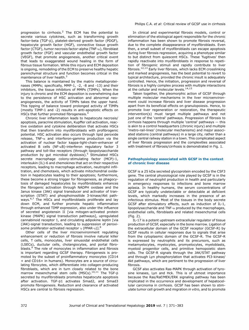
Fig. 3. The conundrum associated with GCSF and advanced liver disease in animal and human studies. Striking a balance is an unmet need to improve outcomes,which is currently still a matter of bench work before bedside use can be considered.
Abbreviation: GCSF, granulocyte colony-stimulating factor.
380 Journal of Clinical and Translational Hepatology 2019 vol. 7 | 371–383
Philips C.A. et al: Critical review of GCSF use in cirrhosis
better efficacy in amelioration of chronic liver injury inpreclinical studies. With GCSF use and an associatedgeneral increase in bone marrow-derived stem cells, themuch needed ‘fate of cell’ tracer studies to correctly identifythe probable beneficial pathway associated with this therapyremain to be performed.60
Newsome and colleagues61 assessed the safety and effi-cacy of GCSF and CD133+ hemopoietic stem-cell infusions inpatients with compensated liver cirrhosis with MELD scoresranging from 11 to 15.5 and found that liver dysfunction orfibrosis did not improve when compared to patients receivingstandard care, and furthermore patients receiving the treat-ment had a higher incidence of adverse liver-related events.This study cautioned on the use of growth factor therapy inpatients with compensated cirrhosis and moderately highMELD scores.
Recently, Philips and colleagues62 published real-worldexperience of GCSF in a large group of patients with cirrhosisand active decompensations with higher MELD scores. Cirrho-sis patients with active ascites, jaundice, or both completedGCSF treatment (10 mcg/kg/day for 5 days, followed by5 mcg/kg/day once every third day for total 12 doses).A matched historical control group was used for comparingoutcomes. Among them, 16%, 43% and 75% of patientsdied at 3, 6 and 12 months respectively, after GCSF treat-ment. Sepsis was the most frequent cause of death (in 53%of patients), followed by progressive liver failure (in 33%).Notably, a higher number of patients compared to the histor-ical control group developed hepatocellular carcinoma at theend of 12 months. Acute variceal bleeds, overt hepatic ence-phalopathy, intensive care unit admissions, and liver diseaseseverity scores were higher after GCSF use at 12 months.A CTP score of >11 and MELD-sodium score of >20 predictedworse outcomes at all time points and 12 months with GCSFuse, respectively. The modified intention to treat analysisdemonstrated poor overall survival at 6 months with GCSFtherapy compared to the historical controls (48% vs. 75%,p = 0.04). The authors concluded that survival in decompo-sition was shorter than what was expected in the naturalhistory of the disease after GCSF use in patients withadvanced cirrhosis.
In the study by Kedarisetty et al.,47 a lower proportion ofpatients developed septic shock during the follow-up periodcompared with controls, and by the end of 1 month aftertreatment the mean level of a-fetoprotein was significantlyhigher in the growth factor group (6.6 ± 3.6 ng/mL) than inthe controls (4.7 ± 2.7 ng/mL). The latter was considered tobe associated with hepatic regeneration. However, this wascontrary to findings described by Philips et al.62 and theoccurrence of sepsis as well as liver cancer was found to behigher with GCSF use. Seehofer et al.63 demonstrated, in ananimal model of chronic liver disease, that hepatic regenera-tion was slightly inhibited in the GCSF group. A study on theeffect of GCSF in liver fibrosis found that it significantlydecreased the survival rate of mice.64
Conclusions
The way forward
Liver fibrosis and its progression, and the ultimatum ofassociated portal hypertension and liver failure, is a highlycomplex disease mechanism. Furthermore, the mechanismsthat define human liver regeneration remain inadequately
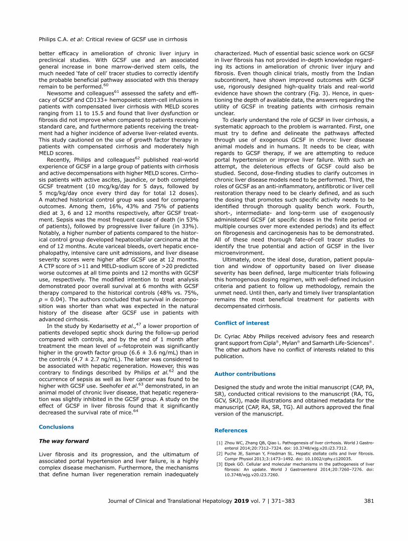
characterized. Much of essential basic science work on GCSFin liver fibrosis has not provided in-depth knowledge regard-ing its actions in amelioration of chronic liver injury andfibrosis. Even though clinical trials, mostly from the Indiansubcontinent, have shown improved outcomes with GCSFuse, rigorously designed high-quality trials and real-worldevidence have shown the contrary (Fig. 3). Hence, in ques-tioning the depth of available data, the answers regarding theutility of GCSF in treating patients with cirrhosis remainunclear.
To clearly understand the role of GCSF in liver cirrhosis, asystematic approach to the problem is warranted. First, onemust try to define and delineate the pathways affectedthrough use of exogenous GCSF in chronic liver diseaseanimal models and in humans. It needs to be clear, withregards to GCSF therapy, if we are attempting to reduceportal hypertension or improve liver failure. With such anattempt, the deleterious effects of GCSF could also bestudied. Second, dose-finding studies to clarify outcomes inchronic liver disease models need to be performed. Third, theroles of GCSF as an anti-inflammatory, antifibrotic or liver cellrestoration therapy need to be clearly defined, and as suchthe dosing that promotes such specific activity needs to beidentified through thorough quality bench work. Fourth,short-, intermediate- and long-term use of exogenouslyadministered GCSF (at specific doses in the finite period ormultiple courses over more extended periods) and its effecton fibrogenesis and carcinogenesis has to be demonstrated.All of these need thorough fate-of-cell tracer studies toidentify the true potential and action of GCSF in the livermicroenvironment.
Ultimately, once the ideal dose, duration, patient popula-tion and window of opportunity based on liver diseaseseverity has been defined, large multicenter trials followingthis homogenous dosing regimen, with well-defined inclusioncriteria and patient to follow up methodology, remain theunmet need. Until then, early and timely liver transplantationremains the most beneficial treatment for patients withdecompensated cirrhosis.
Conflict of interest
Dr. Cyriac Abby Philips received advisory fees and researchgrant support fromCipla�, Mylan� and Samarth Life-Sciences�.The other authors have no conflict of interests related to thispublication.
Author contributions
Designed the study and wrote the initial manuscript (CAP, PA,SR), conducted critical revisions to the manuscript (RA, TG,GCV, SKJ), made illustrations and obtained metadata for themanuscript (CAP, RA, SR, TG). All authors approved the finalversion of the manuscript.
References
[1] Zhou WC, Zhang QB, Qiao L. Pathogenesis of liver cirrhosis. World J Gastro-enterol 2014;20:7312–7324. doi: 10.3748/wjg.v20.i23.7312.
[2] Puche JE, Saiman Y, Friedman SL. Hepatic stellate cells and liver fibrosis.Compr Physiol 2013;3:1473–1492. doi: 10.1002/cphy.c120035.
[3] Elpek GÖ. Cellular and molecular mechanisms in the pathogenesis of liverfibrosis: An update. World J Gastroenterol 2014;20:7260–7276. doi:10.3748/wjg.v20.i23.7260.
Journal of Clinical and Translational Hepatology 2019 vol. 7 | 371–383 381
Philips C.A. et al: Critical review of GCSF use in cirrhosis
[4] Ray K. Liver: Key role for av integrins in myofibroblasts in liver fibrosis. NatRev Gastroenterol Hepatol 2014;11:4. doi: 10.1038/nrgastro.2013.227.
[5] Jiang JX, Mikami K, Venugopal S, Li Y, Török NJ. Apoptotic body engulfmentby hepatic stellate cells promotes their survival by the JAK/STAT andAkt/NF-kappaB-dependent pathways. J Hepatol 2009;51:139–148. doi:10.1016/j.jhep.2009.03.024.
[6] Schuppan D, Kim YO. Evolving therapies for liver fibrosis. J Clin Invest 2013;123:1887–1901. doi: 10.1172/JCI66028.
[7] Wasmuth HE, Tacke F, Trautwein C. Chemokines in liver inflammation andfibrosis. Semin Liver Dis 2010;30:215–225. doi: 10.1055/s-0030-1255351.
[8] Simões E Silva AC, Miranda AS, Rocha NP, Teixeira AL. Renin angiotensinsystem in liver diseases: Friend or foe? World J Gastroenterol 2017;23:3396–3406. doi: 10.3748/wjg.v23.i19.3396.
[9] Song Y, Zhao Y, Wang F, Tao L, Xiao J, Yang C. Autophagy in hepatic fibrosis.Biomed Res Int 2014;2014:436242. doi: 10.1155/2014/436242.
[10] Zimmermann HW, Seidler S, Nattermann J, Gassler N, Hellerbrand C,Zernecke A, et al. Functional contribution of elevated circulating andhepatic non-classical CD14CD16 monocytes to inflammation and humanliver fibrosis. PLoS One 2010;5:e11049. doi: 10.1371/journal.pone.0011049.
[11] Kisseleva T, Brenner DA. The phenotypic fate and functional role for bonemarrow-derived stem cells in liver fibrosis. J Hepatol 2012;56:965–972. doi:10.1016/j.jhep.2011.09.021.
[12] Dooley S, Hamzavi J, Ciuclan L, Godoy P, Ilkavets I, Ehnert S, et al. Hepato-cyte-specific Smad7 expression attenuates TGF-beta-mediated fibrogenesisand protects against liver damage. Gastroenterology 2008;135:642–659.doi: 10.1053/j.gastro.2008.04.038.
[13] Guimarães EL, Empsen C, Geerts A, van Grunsven LA. Advanced glycationend products induce production of reactive oxygen species via the activationof NADPH oxidase in murine hepatic stellate cells. J Hepatol 2010;52:389–397. doi: 10.1016/j.jhep.2009.12.007.
[14] Ellis EL, Mann DA. Clinical evidence for the regression of liver fibrosis.J Hepatol 2012;56:1171–1180. doi: 10.1016/j.jhep.2011.09.024.
[15] Jung YK, Yim HJ. Reversal of liver cirrhosis: current evidence and expect-ations. Korean J Intern Med 2017;32:213–228. doi: 10.3904/kjim.2016.268.
[16] Delire B, Stärkel P. The Ras/MAPK pathway and hepatocarcinoma: patho-genesis and therapeutic implications. Eur J Clin Invest 2015;45:609–623.doi: 10.1111/eci.12441.
[17] Li L, Zhao GD, Shi Z, Qi LL, Zhou LY, Fu ZX. The Ras/Raf/MEK/ERK signalingpathway and its role in the occurrence and development of HCC. Oncol Lett2016;12:3045–3050. doi: 10.3892/ol.2016.5110.
[18] Ordelheide AM, Gommer N, Böhm A, Hermann C, Thielker I, Machicao F, et al.Granulocyte colony-stimulating factor (G-CSF): A saturated fatty acid-induced myokine with insulin-desensitizing properties in humans. MolMetab 2016;5:305–316. doi: 10.1016/j.molmet.2016.02.001.
[19] Muzzi A, Leandro G, Rubbia-Brandt L, James R, Keiser O, Malinverni R, et al.Insulin resistance is associated with liver fibrosis in non-diabetic chronichepatitis C patients. J Hepatol 2005;42:41–46. doi: 10.1016/j.jhep.2004.09.022.
[20] Tao LL, Cheng YY, Ding D, Mei S, Xu JW, Yu J, et al. C/EBP-a ameliorates CCl(4)-induced liver fibrosis in mice through promoting apoptosis of hepaticstellate cells with little apoptotic effect on hepatocytes in vitro and in vivo.Apoptosis 2012;17:492–502. doi: 10.1007/s10495-012-0700-y.
[21] Hirai H, Zhang P, Dayaram T, Hetherington CJ, Mizuno S, Imanishi J, et al.C/EBPbeta is required for ‘emergency’ granulopoiesis. Nat Immunol 2006;7:732–739. doi: 10.1038/ni1354.
[22] Buck M, Chojkier M. A ribosomal S-6 kinase-mediated signal to C/EBP-beta iscritical for the development of liver fibrosis. PLoS One 2007;2:e1372. doi:10.1371/journal.pone.0001372.
[23] Gutschalk CM, Herold-Mende CC, Fusenig NE, Mueller MM. Granulocytecolony-stimulating factor and granulocyte-macrophage colony-stimulatingfactor promote malignant growth of cells from head and neck squamouscell carcinomas in vivo. Cancer Res 2006;66:8026–8036. doi: 10.1158/0008-5472.CAN-06-0158.
[24] Shojaei F, Wu X, Qu X, Kowanetz M, Yu L, Tan M, et al. G-CSF-initiatedmyeloid cell mobilization and angiogenesis mediate tumor refractoriness toanti-VEGF therapy in mouse models. Proc Natl Acad Sci U S A 2009;106:6742–6747. doi: 10.1073/pnas.0902280106.
[25] Elpek GÖ. Angiogenesis and liver fibrosis. World J Hepatol 2015;7:377–391.doi: 10.4254/wjh.v7.i3.377.
[26] Huebert RC, Rakela J. Cellular therapy for liver disease. Mayo Clin Proc 2014;89:414–424. doi: 10.1016/j.mayocp.2013.10.023.
[27] Serefhanoglu S, Goker H, Buyukasik Y, Turgut M, Sayinalp N, HaznedarogluIC, et al. Changes in vascular endothelial growth factor, angiopoietins, andTie-2 levels with G-CSF stimulation in healthy donors. Ann Hematol 2009;88:667–671. doi: 10.1007/s00277-008-0657-7.
[28] Cong M, Iwaisako K, Jiang C, Kisseleva T. Cell signals influencing hepaticfibrosis. Int J Hepatol 2012;2012:158547. doi: 10.1155/2012/158547.
[29] Shaw BE, Confer DL, Hwang W, Pulsipher MA. A review of the genetic andlong-term effects of G-CSF injections in healthy donors: a reassuring lack ofevidence for the development of haematological malignancies. Bone MarrowTransplant 2015;50:334–340. doi: 10.1038/bmt.2014.278.
[30] Stroncek D, Shawker T, Follmann D, Leitman SF. G-CSF-induced spleen sizechanges in peripheral blood progenitor cell donors. Transfusion 2003;43:609–613. doi: 10.1046/j.1537-2995.2003.00384.x.
[31] Bolognesi M, Merkel C, Sacerdoti D, Nava V, Gatta A. Role of spleen enlarge-ment in cirrhosis with portal hypertension. Dig Liver Dis 2002;34:144–150.doi: 10.1016/S1590-8658(02)80246-8.
[32] Nakamura T, Torimura T, Iwamoto H, Kurogi J, Inoue H, Hori Y, et al. CD34(+)cell therapy is safe and effective in slowing the decline of hepatic reservefunction in patients with decompensated liver cirrhosis. J GastroenterolHepatol 2014;29:1830–1838. doi: 10.1111/jgh.12622.
[33] Gaia S, Smedile A, Omedè P, Olivero A, Sanavio F, Balzola F, et al. Feasibilityand safety of G-CSF administration to induce bone marrow-derived cellsmobilization in patients with end stage liver disease. J Hepatol 2006;45:13–19. doi: 10.1016/j.jhep.2006.02.018.
[34] Lorenzini S, Isidori A, Catani L, Gramenzi A, Talarico S, Bonifazi F, et al. Stemcell mobilization and collection in patients with liver cirrhosis. Aliment Phar-macol Ther 2008;27:932–939. doi: 10.1111/j.1365-2036.2008.03670.x.
[35] Roberts AW. G-CSF: a key regulator of neutrophil production, but that’s notall! Growth Factors 2005;23:33–41. doi: 10.1080/08977190500055836.
[36] Gross-Weege W, Weiss M, Schneider M, Wenning M, Harms B, Dumon K,et al. Safety of a low-dosage Filgrastim (rhG-CSF) treatment in non-neutropenic surgical intensive care patients with an inflammatory process.Intensive Care Med 1997;23:16–22. doi: 10.1007/s001340050285.
[37] Lanthier N. Haemopoietic stem cell therapy in cirrhosis: the end of the story?Lancet Gastroenterol Hepatol 2018;3:3–5. doi: 10.1016/S2468-1253(17)30359-X.
[38] Nicolas CT, Hickey RD, Chen HS, Mao SA, Lopera Higuita M, Wang Y, et al.Concise review: Liver regenerative medicine: from hepatocyte transplanta-tion to bioartificial livers and bioengineered grafts. Stem Cells 2017;35:42–50. doi: 10.1002/stem.2500.
[39] Spahr L, Lambert JF, Rubbia-Brandt L, Chalandon Y, Frossard JL, Giostra E,et al. Granulocyte-colony stimulating factor induces proliferation of hepaticprogenitors in alcoholic steatohepatitis: a randomized trial. Hepatology2008;48:221–229. doi: 10.1002/hep.22317.
[40] Mizuno S, Nakamura T. Hepatocyte growth factor: a regenerative drugfor acute hepatitis and liver cirrhosis. Regen Med 2007;2:161–170. doi:10.2217/17460751.2.2.161.
[41] Spahr L, Chalandon Y, Terraz S, Kindler V, Rubbia-Brandt L, Frossard JL, et al.Autologous bone marrow mononuclear cell transplantation in patients withdecompensated alcoholic liver disease: a randomized controlled trial. PLoSOne 2013;8:e53719. doi: 10.1371/journal.pone.0053719.
[42] Han Y, Yan L, Han G, Zhou X, Hong L, Yin Z, et al. Controlled trials in hepatitis Bvirus-related decompensate liver cirrhosis: peripheral blood monocyte trans-plant versus granulocyte-colony-stimulating factor mobilization therapy.Cytotherapy 2008;10:390–396. doi: 10.1080/14653240802129901.
[43] Xing TJ, Xu HT, Xian JC, Shen ML, Li H, Ye J, et al. Mechanism and efficacy ofmobilization of granulocyte colony-stimulating factor in the treatmentof chronic hepatic failure. Hepatogastroenterology 2013;60:170–175.doi: 10.5754/hge12590
[44] Gaia S, Olivero A, Smedile A, Ruella M, Abate ML, Fadda M, et al. Multiplecourses of G-CSF in patients with decompensated cirrhosis: consistent mobi-lization of immature cells expressing hepatocyte markers and exploratoryclinical evaluation. Hepatol Int 2013;7:1075–1083. doi: 10.1007/s12072-013-9473-9.
[45] Hao NB, Li CZ, Lü MH, Tang B, Wang SM, Wu YY, et al. SDF-1/CXCR4 axispromotes MSCs to repair liver injury partially through trans-differentiationand fusion with hepatocytes. Stem Cells Int 2015;2015:960387. doi: 10.1155/2015/960387.
[46] Marquardt JU, Seo D, Gómez-Quiroz LE, Uchida K, Gillen MC, Kitade M, et al.Loss of c-Met accelerates development of liver fibrosis in response to CCl(4)exposure through deregulation of multiple molecular pathways. BiochimBiophys Acta 2012;1822:942–951. doi: 10.1016/j.bbadis.2012.02.012.
[47] Kedarisetty CK, Anand L, Bhardwaj A, Bhadoria AS, Kumar G, Vyas AK, et al.Combination of granulocyte colony-stimulating factor and erythropoietinimproves outcomes of patients with decompensated cirrhosis. Gastroenter-ology 2015;148:1362–1370.e7. doi: 10.1053/j.gastro.2015.02.054.
[48] Chousterman BG, Arnaud M. Is there a role for hematopoietic growth factorsduring sepsis? Front Immunol 2018;9:1015. doi: 10.3389/fimmu.2018.01015.
[49] Preheim LC, Snitily MU, Gentry MJ. Effects of granulocyte colony-stimulatingfactor in cirrhotic rats with pneumococcal pneumonia. J Infect Dis 1996;174:225–228. doi: 10.1093/infdis/174.1.225.
[50] Yang Q, Yang Y, Shi Y, Lv F, He J, Chen Z. Effects of granulocyte colony-stimulating factor on patients with liver failure: a meta-analysis. J ClinTransl Hepatol 2016;4:90–96. doi: 10.14218/JCTH.2016.00012.
382 Journal of Clinical and Translational Hepatology 2019 vol. 7 | 371–383
Philips C.A. et al: Critical review of GCSF use in cirrhosis
[51] Fiuza C, Salcedo M, Clemente G, Tellado JM. Granulocyte colony-stimulatingfactor improves deficient in vitro neutrophil transendothelial migrationin patients with advanced liver disease. Clin Diagn Lab Immunol 2002;9:433–439. doi: 10.1128/cdli.9.2.433-439.2002.
[52] Mohammad RA. Use of granulocyte colony-stimulating factor in patients withsevere sepsis or septic shock. Am J Health Syst Pharm 2010;67:1238–1245.doi: 10.2146/ajhp090325.
[53] Stephens DP, Thomas JH, Higgins A, Bailey M, Anstey NM, Currie BJ, et al.Randomized, double-blind, placebo-controlled trial of granulocyte colony-stimulating factor in patients with septic shock. Crit Care Med 2008;36:448–454. doi: 10.1097/01.CCM.0B013E318161E480.
[54] Segal JM, Kent D, Wesche DJ, Ng SS, Serra M, Oulès B, et al.Single cell analysis of human foetal liver captures the transcriptional profileof hepatobiliary hybrid progenitors. Nat Commun 2019;10:3350. doi:10.1038/s41467-019-11266-x.
[55] Anand L, Bihari C, Kedarisetty CK, Rooge SB, Kumar D, Shubham S, et al.Early cirrhosis and a preserved bone marrow niche favour regenerativeresponse to growth factors in decompensated cirrhosis. Liver Int 2019;39:115–126. doi: 10.1111/liv.13923.
[56] Prajapati R, Arora A, Sharma P, Bansal N, Singla V, Kumar A. Granulocytecolony-stimulating factor improves survival of patients with decompensatedcirrhosis: a randomized-controlled trial. Eur J Gastroenterol Hepatol 2017;29:448–455. doi: 10.1097/MEG.0000000000000801.
[57] Chavez-Tapia NC, Mendiola-Pastrana I, Ornelas-Arroyo VJ, Noreña-Herrera C,Vidaña-Perez D, Delgado-Sanchez G, et al. Granulocyte-colony stimulatingfactor for acute-on-chronic liver failure: systematic review and meta-analysis.Ann Hepatol 2015;14:631–641. doi: 10.1016/S1665-2681(19)30757-4.
[58] Simonetto DA, Shah VH, Kamath PS. Improving survival in ACLF: growingevidence for use of G-CSF. Hepatol Int 2017;11:473–475. doi: 10.1007/s12072-017-9834-x.
[59] Verma N, Kaur A, Sharma R, Bhalla A, Sharma N, De A, et al. Outcomes aftermultiple courses of granulocyte colony-stimulating factor and growthhormone in decompensated cirrhosis: A randomized trial. Hepatology2018;68:1559–1573. doi: 10.1002/hep.29763.
[60] Gilchrist ES, Plevris JN. Bone marrow-derived stem cells in liver repair:10 years down the line. Liver Transpl 2010;16:118–129. doi: 10.1002/lt.21965.
[61] Newsome PN, Fox R, King AL, Barton D, Than NN, Moore J, et al. Granulocytecolony-stimulating factor and autologous CD133-positive stem-cell therapyin liver cirrhosis (REALISTIC): an open-label, randomised, controlled phase 2trial. Lancet Gastroenterol Hepatol 2018;3:25–36. doi: 10.1016/S2468-1253(17)30326-6.
[62] Philips CA, Augustine P, Rajesh S, Ahamed R, George T, Padsalgi G, et al.Granulocyte colony-stimulating factor use in decompensated cirrhosis: lackof survival benefit and probable predisposition to hepatocellular carcinoma.J Clin Exp Hepatol 2019. doi: 10.1016/j.jceh.2019.05.003.
[63] Seehofer D, Neumann UP, Schirmeier A, Carter J, Cho SY, Lederer A, et al.Synergistic effect of erythropoietin but not G-CSF in combination with cur-cumin on impaired liver regeneration in rats. Langenbecks Arch Surg 2008;393:325–332. doi: 10.1007/s00423-008-0290-x.
[64] Ogiso T, Nagaki M, Takai S, Tsukada Y, Mukai T, Kimura K, et al. Granulocytecolony-stimulating factor impairs liver regeneration in mice through theup-regulation of interleukin-1beta. J Hepatol 2007;47:816–825. doi: 10.1016/j.jhep.2007.06.017.
Journal of Clinical and Translational Hepatology 2019 vol. 7 | 371–383 383
Philips C.A. et al: Critical review of GCSF use in cirrhosis


![Index [ ] immunogenicity and antiglobulin response ... See Multiple sclerosis (MS) ... – granulocyte colony-stimulating factor](https://static.fdocuments.net/doc/165x107/5a9f33d87f8b9a67178c791b/pdfindex-immunogenicity-and-antiglobulin-response-see-multiple-sclerosis.jpg)

![Granulocyte colony-stimulating factor exacerbates hematopoietic … · successfully managed by the use of hematopoietic growth factors (HGFs) [4]. However, even though some irradiated](https://static.fdocuments.net/doc/165x107/5f4fbff2a006440ac9114981/granulocyte-colony-stimulating-factor-exacerbates-hematopoietic-successfully-managed.jpg)