Pontine Glioma Extending to the Ipsilateral Cavernous ... · Pontine Glioma Extending to the...
3
Pontine Glioma Extending to the Ipsilateral Cavernous Sinus and Meckel's Cave: MR Appearance William T. C. Yuh, 1 Hoang D. Nguyen, 1 Nina A. Mayr , 1 and Kenneth A. Follete Summary: The authors describe an exophytic glioma of the pons that grew into the Meckel's cave and cavernous sinus in a 75-year-old man. Pontine gliomas should be included in the differential diagnosis of a hyperintense, complex cystic mass seen along the distribution of cranial nerve V. Index terms: Cerebellopontine angle, neoplasms; Brain stem, neoplasms; Glioma Most tumors of the cerebellopontine angle (CPA) are benign and of neuroectodermal origin. Malignant tumors of the CPA, including intrinsic brain tumors such as pontine gliomas and met- astatic carcinomas, are rare and account for less than 2% of all CPA lesions (1, 2). Exophytic extension of pontine glioma to the CPA is an occasional radiologic finding (3) . This report doc- uments extension of a pontine glioma into the Meckel's cave and cavernous sinus. Case Report A 75-year-old right-handed man presented with a 2- to 3-week history of ataxia and incoordination of his right upper extremity and bifrontal headache. He reported a several-year history of numbness over the left side of his face. Two to 3 days before admission, he developed nausea and vomiting. Neurologic examination showed decreased sensation over the ophthalmic and maxillary distributions of the left trigeminal nerve, reflexes were diffusely hyper- active, and a right Babinski sign was present. Dysmetria was present in the right arm, and he had abnormal heel- to-shin movements of both legs. His gait was ataxic. Computed tomography (CT), with and without contrast material, at another institution revealed an enhancing cystic mass involving the brain stem, suggesting a hemangioblas- toma. Magnetic resonance (MR) imaging showed a left CPA cystic complex mass that involved the brain stem and extended into the Meckel's cave and cavernous sinus (Figs. 1 and 2). The solid component of the mass showed en- hancement on the postcontrast MR study and, on precon- trast MR imaging, isointensity on the T1-weighted and hyperintensity on the T2-weighted images. The cystic com- ponent showed no enhancement on the postcontrast MR study and, on precontrast MR imaging, low signal intensity on the T1-weighted and hyperintensity on the T2-weighted images. The preoperative diagnoses, based on MR studies, included neurinoma with a cystic component and a less likely possibility of a pontine glioma. At operation, tumor had eroded through the tentorium, separating and expanding the dural leaves. It extended anteriorly within the tentorium into the left cavernous sinus. A large cystic mass was present in the left cerebral pedun- cle and extended into the interpeduncular cistern. Tumor also extended from the anterolateral aspect of the brain stem into Meckel's cave. A subtotal resection was done, and final pathologic diagnosis was malignant astrocytoma. Discussion Brain stem gliomas account for 10% of all intracranial tumors in children aged 3-9 years and 3% in adults (4). Two-thirds of all brain stem gliomas occur in children. Epstein (5) proposed a staging classification system for brain stem gliomas that included intrinsic (diffuse, focal, and cervicomedullary), exophytic, and disseminated tumors. Intrinsic tumors involve the midbrain, pons, and medulla. Most intrinsic tumors of the brain stem are pontine astrocytomas. They may extend rostrally to the midbrain and thalamus and caudally to the medulla and cervical cord. Exophytic tumors may extend anterolaterally into the CPA, posterolaterally through the brachium pontis into the cerebellar hemisphere, or poste- riorly into the fourth ventricle (4-6) . Naidich and Zimmerman (3) reported that approximately 60 % Receiv ed August 1, 1991 ; final acceptance September 16 (no revision required). 1 Department of Radiology, The University of Iowa College of Medicine, Iowa City, Iowa 52242. Address reprints toW. T. C. Yuh, The University of Iowa Hospitals and Clinics, Iowa City, lA 52242. 2 Department of Neurosurgery, Th e University of Iowa College of Medicine, Iowa City, lA 52242. AJNR 13:346-348, Jan/ Feb 1992 0195-6108/ 92/13 01-0346 © American Society of Neuroradiology 346
Transcript of Pontine Glioma Extending to the Ipsilateral Cavernous ... · Pontine Glioma Extending to the...
Pontine Glioma Extending to the Ipsilateral Cavernous Sinus and Meckel's Cave: MR Appearance
William T. C. Yuh, 1 Hoang D. Nguyen, 1 Nina A. Mayr, 1 and Kenneth A. Follete
Summary: The authors describe an exophytic glioma of the pons that grew into the Meckel's cave and cavernous sinus in a 75-year-old man. Pontine gliomas should be included in the
differential diagnosis of a hyperintense, complex cystic mass seen along the distribution of cranial nerve V.
Index terms: Cerebellopontine angle, neoplasms; Brain stem, neoplasms; Glioma
Most tumors of the cerebellopontine angle (CPA) are benign and of neuroectodermal origin. Malignant tumors of the CPA, including intrinsic brain tumors such as pontine gliomas and metastatic carcinomas, are rare and account for less than 2% of all CPA lesions (1, 2). Exophytic extension of pontine glioma to the CPA is an occasional radiologic finding (3). This report documents extension of a pontine glioma into the Meckel's cave and cavernous sinus.
Case Report
A 75-year-old right-handed man presented with a 2- to 3-week history of ataxia and incoordination of his right upper extremity and bifrontal headache. He reported a several-year history of numbness over the left side of his face. Two to 3 days before admission, he developed nausea and vomiting. Neurologic examination showed decreased sensation over the ophthalmic and maxillary distributions of the left trigeminal nerve, reflexes were diffusely hyperactive, and a right Babinski sign was present. Dysmetria was present in the right arm, and he had abnormal heelto-shin movements of both legs. His gait was ataxic.
Computed tomography (CT), with and without contrast material , at another institution revealed an enhancing cystic mass involving the brain stem, suggesting a hemangioblastoma. Magnetic resonance (MR) imaging showed a left CPA cystic complex mass that involved the brain stem and extended into the Meckel's cave and cavernous sinus (Figs.
1 and 2). The solid component of the mass showed enhancement on the postcontrast MR study and, on precontrast MR imaging, isointensity on the T1-weighted and hyperintensity on the T2-weighted images. The cystic component showed no enhancement on the postcontrast MR study and, on precontrast MR imaging, low signal intensity on the T1-weighted and hyperintensity on the T2-weighted images. The preoperative diagnoses, based on MR studies, included neurinoma with a cystic component and a less likely possibility of a pontine glioma.
At operation, tumor had eroded through the tentorium, separating and expanding the dural leaves. It extended anteriorly within the tentorium into the left cavernous sinus. A large cystic mass was present in the left cerebral peduncle and extended into the interpeduncular cistern. Tumor also extended from the anterolateral aspect of the brain stem into Meckel's cave. A subtotal resection was done, and final pathologic diagnosis was malignant astrocytoma.
Discussion
Brain stem gliomas account for 10% of all intracranial tumors in children aged 3-9 years and 3% in adults (4). Two-thirds of all brain stem gliomas occur in children. Epstein (5) proposed a staging classification system for brain stem gliomas that included intrinsic (diffuse, focal, and cervicomedullary), exophytic, and disseminated tumors. Intrinsic tumors involve the midbrain, pons, and medulla. Most intrinsic tumors of the brain stem are pontine astrocytomas. They may extend rostrally to the midbrain and thalamus and caudally to the medulla and cervical cord. Exophytic tumors may extend anterolaterally into the CPA, posterolaterally through the brachium pontis into the cerebellar hemisphere, or posteriorly into the fourth ventricle (4-6). Naidich and Zimmerman (3) reported that approximately 60%
Received August 1, 1991 ; final acceptance September 16 (no revision required). 1 Department of Radiology, The University of Iowa College of Medicine, Iowa City, Iowa 52242. Address reprints toW. T. C. Yuh , The University of
Iowa Hospitals and Clin ics, Iowa City, lA 52242. 2 Department of Neurosurgery, The University of Iowa College of Medicine, Iowa City, lA 52242.
AJNR 13:346-348, Jan/ Feb 1992 0195-6108/ 92/1301-0346 © American Society of Neuroradiology
346
AJNR: 13, January/ February 1992
A B
of all brain stem gliomas have exophytic extension into the CPA. Brain stem tumors that disseminate by invading the meninges or seeding the subarachnoid space via the cerebrospinal fluid (CSF) have been documented in 25% of brain stem tumors between diagnosis and death (4). Involvement of the basal leptomeninges, third ventricle, cerebellum, and spinal cord has been reported (4, 7). These reports do not mention
347
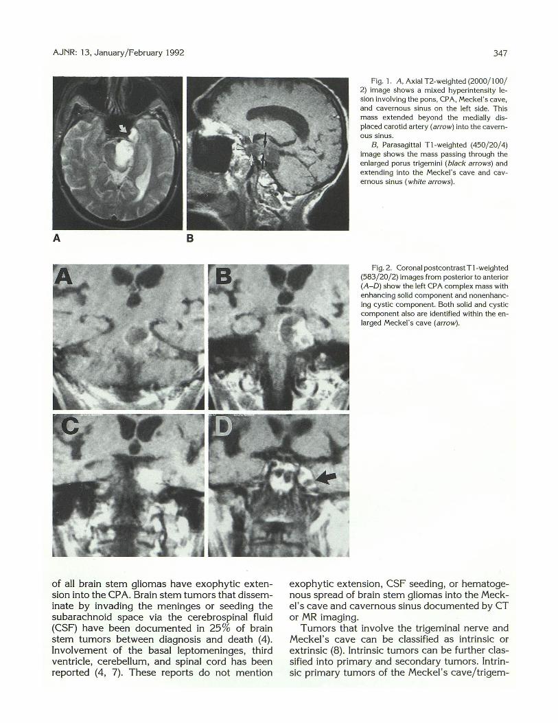
Fig. 1. A, A xial T 2-weighted (2000/ 100/ 2) image shows a mixed hyperintensity lesion involving the pons, CPA, Meckel 's cave, and cavernous sinus on the left side. This mass extended beyond the medially displaced carotid artery (arrow) into the cavernous sinus.
B, Parasagittal Tl -weighted (450/ 20/ 4) image shows the mass passing through the enlarged porus trigemini (black arrows) and extending into the Meckel's cave and cavernous sinus (white arrows) .
Fig. 2. Coronal postcontrast Tl-weighted (583/ 20/ 2) images from posterior to anterior (A-D) show the left CPA complex mass with enhancing solid component and nonenhancing cystic component. Both solid and cystic component also are identified within the enlarged Meckel 's cave (arrow) .
exophytic extension , CSF seeding, or hematogenous spread of brain stem gliomas into the Meckel's cave and cavernous sinus documented by CT or MR imaging.
Tumors that involve the trigeminal nerve and Meckel's cave can be classified as intrinsic or extrinsic (8). Intrinsic tumors can be further classified into primary and secondary tumors. Intrinsic primary tumors of the Meckel 's cave/trigem-
348
inal nerve are rare lesions, representing less than 0.5 % of all histologically proven tumors (9). Reported intrinsic primary lesions include trigeminal neurinomas, meningiomas, and congenital tumors-· -lipomas, epidermoids, and dermoids (8, 1 0). A literature review by Butti et al (9) showed that meningiomas and trigeminal neurinomas are the most common intrinsic Meckel's cave lesions. Intrinsic primary tumors usually do not cause bone destruction but may expand the Meckel's cave and displace the adjacent bone structures (10). Intrinsic secondary tumors can be divided into three groups: 1) distant hematogenous metastases from extracranial malignancies, most commonly from lung primaries; 2) subarachnoid dissemination from spinal cord and brain tumors and from tumors outside the central nervous system that theoretically may seed in the Meckel's cave (4, 8); and 3) retrograde intracranial extension of nasopharyngeal tumor via the branches of the trigeminal nerve (8). The latter category occurs most often. Most extrinsic lesions of Meckel's cave are the result of direct invasion or distortion from lesions in the adjacent structures, such as bone metastases to the skull base and primary nasopharyngeal tumors that extend into the cranial cavity by erosion of the skull bone (8).
We encountered one case report of CT findings of a glioma that extended into Meckel's cave without mention of cavernous sinus involvement in a 68-year-old man with a 4-week history of progressive left facial numbness (8). At surgery, an invasive high-grade glioma was found in the trigeminal nerve, extending from the pons to the Meckel's cave. Involvement of the cavernous sinus was not mentioned in the CT results or surgical report. As in our case, the MR imaging demonstrated the exophytic extension of a brain stem glioma (malignant astrocytoma) into the Meckel's cave, which was not reported by prior CT examinations. It is interesting to note that
AJNR: 13, January / February 1992
both cases with involvement of the Meckel's cave were seen- in older patients (75 and 68 years), although brain stem gliomas are more common in younger patients. Whether this results from the late onset of symptoms, which might allow sufficient time to grow into Meckel's cave, or from a different growth pattern of pontine glioma in the elderly remains unknown. Finally, although trigeminal neurinomas and meningiomas occur more frequently, pontine glioma should be included in the differential diagnosis of a hyperintense, complex cystic mass along the trigeminal nerve distribution, especially when there are signs of trigeminal nerve dysfunction , as was manifested by the decreased sensation and numbness in the ophthalmic and maxillary distributions of the trigeminal nerve in this case report.
References
1. Mafee MF, Meyer DH, Hill JH. Neuroradiographic evaluation of pa
tients with central auditory lesions. Otolaryngol Clin North A m
1 985; 18:223- 239 2. Martuza RL, Parker SW , Nadol JB Jr, Davis KR, Ojemann RG.
Diagnosis of cerebellopontine angle tumors. Clin Neurosurg
1984;32:177- 213
3. Naidich TP, Zimmerman RA. Primary brain tumors in children. Semin
Roentgenol 1 984; 19: 100-1 14
4. Russell DS, Rubinstein LJ . Pathology of tumours of the ner vous
system. 5th ed. Baltimore·: Williams & Wilkins, 1989
5. Epstein F . A staging system for bra in stem gliomas. Cancer
1 985;56: 1804- 1806
6. Lassman LP. Tumours of the pons and medulla. In: Vinken PJ, Bruyn
PW, eds. Handbook of clinical neurology. Vol 1 7. New York : American
Elsevier, 1974:693- 706
7. Nishio S, Fukui M , Tateishi J . Brain stem gliomas: a cl inicopatholog
ical analysis of 23 histologically proven cases. J Neuroncol
1 988;6:215- 250
8. Kapila A , Chakeres DW, Blanco E. The Meckel cave: computed
tomographic study . I. Normal anatomy . II . Pathology. Radiology
1 984; 152:425-433
9. Butti G, Gaetani P, Giordana MT, Paoletti P. Meningiomas of Meckel's
cave. Surg Neural 1 983;20:305- 309
10. Yuh WTC, Wright DC, Barloon T J , Schultz DH, Sato Y, Cervantes
CA. MR imaging of primary tumors of trigeminal nerve and Meckel's
cave. AJNR 1 988;9:665- 670