
POLYZOA ENTOPROCTA AND CTENOSTOMATA. - CORE · Some polyps in my specimens possess unripe gonads in...
30
Mem. Asiat. Soc. Bengal, Vo1. \f1. ZOOLOGIOAL RESULTS OF A TOUR IN THE FAR EAST. POLYZOA ENTOPROCTA AND CTENOSTOMATA. By ANNANDAI,:E;, D.5c., F.A.S.B.
Transcript of POLYZOA ENTOPROCTA AND CTENOSTOMATA. - CORE · Some polyps in my specimens possess unripe gonads in...

Mem. Asiat. Soc. Bengal, Vo1. \f1.
ZOOLOGIOAL RESULTS OF A TOUR IN THE FAR EAST.
POLYZOA ENTOPROCTA AND CTENOSTOMATA.
By N~ ANNANDAI,:E;, D.5c., F.A.S.B.

Introduction
Entoprocta-. Key to the family Urnatellidae
Ckitaspis, gen. novo
Ectoprocta-
CONTENTS.
Preparation of specimens of Ctenostomata for microscopic examination Classification of the Ctenostomata Family Alcyonidiidae Family Tritice11idae Family Vesiculariidae Family Paludice11idae PaludiceUa pentagonatis, sp. novo Family Victonillidae Family Hislopiidae Key to the Genus H islopia H islopia malayensis, sp. nov.
Page
I5
If)
2CJ
22
23
24 28 30 3 I
33 33 35

ZOOLOGICAL RESUL1'S OF. A TOUR IN THE FAR EAST.
POLYZOA ENTOPROCTA AND CTENOSTOMATA.
(Plate I ; plate II) figs. I} Ta.)
By N. ANNANDALE. D.Sc., F.A.S.B. (Zoological Survey of Ind1:a).
The Polyzoa discussed or described in this paper are all from fresh or brackish water .. The majority are from the Tale Sap in the north-eastern part of the Malay Peninsula} but a few come from the Tai-Hu in the Kiangsu Province of China. I have also included notes on one Indian form. The following species are to be considered :-
ENTOPROCTA.
Chitaspis athleticus, gen. et sp. nov., from the Tale Sap.
CTENOSTOMATA.
-Alcyonidium mytili, Dalyell, from Indian estuaries, etc. Triticella pediceltata (Alder), from the Tale Sap. Bowerbankiacaudata, Hincks, from the Tale Sap and Perak. PaZudicella elongata, Leidy) from the Tai-Hu. Paludicell.a pentagonalis J sp. nov., from the Tale Sap. Victorelta bengalensis, Annandale, from the Tale Sap. Hislopia cambodgiensis (J u1Hen), from the Tai-Hu. Hislopia malayensis. sp. nov.,from Jalor in the Malay Peninsula.
It will be as well to defer consideration of the biology and distribution of these species until I have been able to deal systematically with the Phylactolaemata and Chei10stomata collected on my tour. All that need be said here is that while the species of Paludicella and Hislopia are from fresh water, the others. on the list are from brackish water.
ENTOPROCT A.
The only species of Entoproctous Polyzoon represented in my collection was found in brackish water in the Tale Sap on the Gu1f of Siam. It represents I an \:1.1n-described species and genus of the family Urnatellidae. . .
I take this opportunity to state that my identification l of a species of Barentsia from the Mutlah R. in the Gangetic delta as B. discreta (Busk) was incorrect. The
i Annaudale, Rec. Ind. Mus. VII, p. 205 (I912).

r6 ZOOLOGY OF THE FAR EAST.
specimens represent B. gracilis t (Sars), as is apparent from Harmer's detailed des
cription and figures. Family URNATELLIDAE.
1915. Annandale, NJem. Ind. Mus. V, p. I27· In the paper cited I discussed the limits of this family) which I restricted provi-
sionally to the genera Urnatella, Leidy and Loxosomatoides, Annandale The discovery of a new genus that is evidently allied closely to the latter but yet has certain affinities with 1lrlyosoma, Robertson, makes it at any rate probable that Myos01na should also be included. The following key shows the more striking differences be .
tween these genera :-1. Stalk segmented, each segment heavily chitinized and
capable of functioning as a resting bud Urnatelta.
n. Stalk not segmented. A. . Aboral surface of both stalk and capitulum bear
ing scattered chitinous spines; no chitinous shield on capitulum. Muscles of stalk entering capitulum M yosoma.
B. A chitinous shield, sometimes spiniferous, on aboral surface of capitulum only.
1. Muscles of stalk nearly straight, completely surrounding it, not entering capitulum Loxosomato£des.
2. Muscles of stalk directed down wards and outwards from the capitulum, confined to oral and lateral surfaces of the stalk, meeting in the lower part of the capitulum with well-defined oblique capitular muscle-bands at an angle .. Chitaspis (nov.)
With the exception of LVI yosoma," the species of these genera have been found only in fresh or brackish water. Urnatelta is fluviatile and is only known from the neighbourhood of Philadelphia! U.S.A. Two species of Loxosomatoides 3 occur in lagoons and deltaic tracts on the east coast of India,.while thenew genus Chitaspis is represented by a species from a lagoon connected with the Gulf of Siam. A1yosoma was described from a species found in the sea on the Pacific Coast of North America.
Chitaspis, gen. novo
This genus consists of UrnatelHdae with unsegmented stalks and capitular shields like those of Loxosomatoides. The muscles of the stalk however ·emei-ge , , from the capitulum; they are directed outwards and downwards and are confined
1 See Harmer, Siboga-Exp., mOll. XXVIIIa, p. 27'(I915). " Robertson, Proc. California Acad. Sci. (Zoo1.) II (3), p. 324 (1900).
3 Alll1alldale, ReD. Ind. M'us. Il, p. 14 (I90S) and Mem. Ind. Nlus. V, p. 12;'; (19 15)'

Polyzoa Elltoprocta and Ctenostomata.
to the oral and lateral surfaces of the stalk; within the capitulnm they meet at an angle with a pair of well-defined oblique muscular bands on each side.
TYPE-SPECIES.-Chitaspis athleticus, sp. nov.} from the Tale Sap, Gulf of Siam. Closely allied as is the type-species to Loxosom:ttoides, the arrangement of its
musculature is so conspicuously different that a new genus is necessary for its reception. In this point it is different from all other Entoprocta as yet known. In lYf yosoma the muscles of the stalk emerge from the capitulum in much the same way} but the oblique body-muscles are much less highly differentiated. 1 In the resting buds of UrnateUa 2 and Loxosomatoides.) oblique strands of muscles occur, but no' such structures have been detected in the normll capitulum. The boly-muscles described by Ehlers" in Barentsia (Ascopodaria) are by no means highly specialized and the stalkmuscles do not enter the capitulum. The Lox030matidae are so different in other respects that no confusion is possible and discussion as to resemblances and differences in the musculature is unnecessary here.
Chitaspis athleticus, sp. novo
(PI. I, fig. L)
Colon,y. In the type-specimens the colony consists of a segmented, entirely adherent stolon that branches sparingly on the surface of a stone and gives rise at considerable intervals to single upright polyps. Polypiferous and non-polypiferous segments alternate with some regularity, the latter being by far the longer of the two. The lateral branches are given off, as a rule singly, from polypiferous segments of the stolon. The stoion is flattened below and evenly arched above; it varies somewhat in diameter, but does not exceed 0'082 mm. Both stolon and polyps are covered with a rather thick chitinous investment which varies somewhat in thickness, but is not more than 0'004I mm. thick; on the ab::>ral surface of the PJlyps this ectocyst is modified to form the aboral shield characteristic of the genus and of Loxosomatoides.
Polyp. Each polyp consists of a short stalk bearing a relatively large capitUlum. The stalk is rarely if ever longer than, and as a rule rather shorter than, the capitulum. It is relatively very stout and does not. taper much above; there is no defined swelling at its base. The capitulum is rather broadly oval as seen from in front or behind; it is not much compressed. Large capitula are about 0'374 mm. high and 0'272 mm. broad. The diameter of the stalk may be as much as o'I7 mm. at the base.
The normal number of tentacles is 18. The aboral shield varies considerably in extent but never encroaches on the oral
surface. vVhen fully developed it covers the whole of the aboral surface, and has well-defined limits. It never bears spines but is ornamented with a minute network of fine ridges that encloses polygonal depressions of somewhat variable size and outline but never more than 0'05I mm, in greatest diameter. The ridges are slightly
1 Robertsoll, Ptoc. California A cad. Sci. (Zool.) II (3), p. 324, pI. xvi. figs. 1-12 (I900). :2 Davenport, Bull. Mus. Camp Zool. Harvard XXIV, p. 24, pl.vi, fig. 57 ( 1893). S Annandale, Mem. Ind. A/us. V, p. 130, fig. 2 (19I5). ~ Ehlers, Abh. Rong. Gesells. Wiss. Gottingen (Math.-Naturw. Kt) XXXVI, p. 64, pI. Hi, figs. 40, 43 (1890).

IS ZOOLOGY OF THE FAR EAST.
elevated at the nodes of the reticulation. The whole structure is very thin; in opaque specimens it has a pale golden colour, which contrasts well with the tran~lucent white of the soft parts, though a yellowish tinge is given to the whole orgallls111 by the ectocyst. In specimens mounted in Canada balsam it is difficult to see details of the structure of the shield because of its transparency.
The oral surface of the capitulum and the whole surface of the stalk is qnite
smooth. The general anatomy, both in the stalk and in the capitulum, closely resembles
that of Loxosomatoides, except in respect to the muscnlatnre. Some polyps in my specimens possess unripe gonads in the form of a broad transverse band interrupted before and behind and lying in the upper half of the capitulum.
Musculature. The spincter of the orifice consists of a coilsiderable number (at least 6) of circular strands. The strands that lie externally are more or less inter-
s:------
-----So m.
A
FIG. L-CMtaspis athleticus, gen. et sp. nov., x 62.
A. Oral view of a polypide. B. Obliqne lateral view of another polypicle with a bud and part of the stolon.
b.m. = capitular ml1scies. s. = stomach. s.m. = stalk muscles.
rupted. The muscles of the lophophore apparently resemble those of Loxosomatoides and there is a well-marked retractor running along the centre of e.ach tentacle.
The body-muscles lie mainly in the body-wall. Possibly the outer strand (see fig. I) is.entirely superficial, but the inner strand certainly bends inwards above and its upper end is probably attached to the outer wall of the stomach.
The muscles of the stalk are directed outwards and downwards from the capitulum on the oral surface. They usnally form two somewhat divergent groups arranged symmetrically j but this is more clearly the case in some polyps than in others. 'rhe lower end of the muscle is situated distinctly above the base of the stalk. I have not been able to detect any trace of muscle-fibres on the aboral face of the stalk or in the rhizome.
Type-specimen. No. 715717 Z E.V., Ind. Mus. lZool. Survey of Il1:1ia): in alcohol. Locality, etc. The island of Koh Yaw; outer part of the Tale Sap (Great Lake)

Po1yzoa Entoprocta anci Ctenostomata. 1:9
on the Gulf of Siam; at the edge of the lake in water of very variable salinity but having a specific gravity (corrected to a stand'1rd temperature of IS°C) of 1'00625 at the time when the specimens were taken. The type-specimen was attached to a stone that had been built into a sea-wall. It was accompanied by colonies of Bowerba1~kia caudata and of the Cheilostomes 1\1 embranipora hippo pus and M. bengalensis.
ECTOPROCT A. Order Gymnolaemata.
Suborder Ctenostomata.
Harmer's recent account of the Ctenostomata of the 'Siboga' Expedition l has done much to elucidate the internal relationships of this very difficult group, and although I have not been able to accept all his conclusions on the families of fresh and· brackish water (which naturally do not come fully into view in the consideration of the results of a naval expedition) I must here expres; my indebtedness to this admirable work, which has done for the seas of the Malay Archipelago almost as much as Hincks' British Marine Polyzoa did for those of Great Britain. In saying this I do not of course mean to infer that the Polyzoa of that vast area in the East are as well known as those of British seas were even in Hincks' time; but there is now a solid foundation on which further study can be based.
The suborder is well represented among the Polyzoa of fresh and brackish water in eastern lakes and ponds) but until recently our knowledge of the· anatomy of critical genera has been very scanty, mainly owing to the fact that a number of the more important forms, though easily preserved in formalin or alcohol, colhpse and become valueless if transferred to oil of cloves or cedar. It is thus very difficult to examine stained specimens under a high power of the miclOscope, without the aid of which I find it impossible to ascertain details with certainty. Harmer (op. cit., p. 4I) gives elaborate directions, based on the methods put forward by Rousselet, for the mounting of specimens in formalin for microscopic examination, but specimens so prepared, though often both beautiful and useful, cannot be satisfactorily used under really powerful objectives. Moreover, the methods are so elaborate, tedious and costly that it is difficult to mount a sufficiently large number of preparations in the case of variable forms. I find it necessary, in the case of species like those of Victorelta and Bowerbankza, to examine not several but many preparations and to search in all for details that cannot be seen unless the organisms are stained and rendered transparent, and also, if not flattened, at any rate rendered as flat as may be possible without distortion. For this purpose a simple modification of Rousselet and Harmer's technique is sufficient, though I cannot say if it is permanent; the preparations will last, in a tropical climate, at least for two or even three years) and possibly for longer.
I place the specimens to be mounted, after staining with borax carmine and cleaning in acid alcohol, in a 50 % solution of glycerine in 70 % alcohol and leave
1 Siboga-E;rpeditie. Monograph XXVIIla. The Palyzoa at the Siboga E:.:jJedit£on. Pt. 1. EntopYocta, Ctenosto11lata
and Cyclostomata (Leiden, 1915).

20 ZOOLOGY OF THE FAR EAST.
them exposed in a shallow dish for 24 hours. They are then transferred to a drop of pure glycerine on a slide, and, if there is any danger of crushing, fragments of a broken cover· slip are arranged round the drop. A complete cover-slip of relatively large size is cleaned and a square or circle of rather thick Canada balsam solution painted round it to the requisite thickness. It is then dropped from a pair of forceps over the glycerine on the slide, the painted side of c.ourse being downwards. The glycerine and the balsam are pressed together without mixing.
It is always as well at the same time to attempt to mount some specimens ill Canada balsam after clearing them in the ordinary way. Ninety per c.ent or more of such attempts will, in the case of the more delicate tubular species, result· in failure j but the few zooecia that do not collapse will prove particularly valuable. I am of course presuming that abundant material is available, and'this is usually the case ·if the investigator be also the collector.
In the species of Hislopia and PaludiceUa (i.e. in the most abundant of the true freshwater Ctenostomata) there is as a rule no difficulty in clearing preparations with oil of cloves, the ectocyst being relatively thick and at the same time more permeable to oils.
To understand the Ctenostomata and their classification it is necessary above ail things to study the general anatomy of the polypide and in particular of that part of the alimentary canal that lies between the mouth and the stomach. Some confusion exists in the terminology of this system, more particularly in reference to the terms (( oesophagus') and {{ gizzard." The former has been applied in two entirely different senses, while the latter has been used indifferently in a morphological and in a physiological sense.
It is in the Division Alcyonellea or Carnosa that the simplest and probably the most primitive condition is to be found.
In Alcyonidium the mouth opens into a comparatively short funnel-shaped "oesophagus." The walls of this organ are very thick above and become gradually thinner towards its base, which is defined by a circular valve, the, so-called ,( cardia" or J as I prefer to call it, the oesophageal va1 ve. \Vhen this valve is open the lumen of the oesophagus is practically continuous with that of the stomach, at any rate when the p01ypide is expanded. The region that intervenes between the valve and the stomach proper or "pylorus" takes the form of a rather stout tube, the walls of which do not differ in essential histological characters fro111 those of the latter. There are apparently no circular muscles in the wall of this region, which may be known as the cardiac region.
In the Stolonifera the structure of this part of the alimentary canal seems to be essentially the same as in the AlcyoneIlea, but in the Paludicellea a progressive differentiation is found iu the different families. In the Paludicellidae (fig. 2, A) the only marked changes that occur are that the oesophagus is greatly lengthened and more or less distinctly differentiated into an external thick-walled' funnel shaped {( pharynx" and a thin-walled oesophagus proper, and that scattered circular musclefibres appear in the wa11 of the cardiac region.

Polyzoa Entoprocta and Ctellostomata. 2!
In the Victorellidae (fig. 2, B) this region is much more highly specialized and consists of three parts. Immediately below the valve there is a comparatively large oval chamber without muscle-fibres, but lined internally with a fine layer of horny substance. Below this there is a short muscular tube, the external wall of which is composed of close-set circular fibres, and finally a relatively long thick-walled glandular tube connects the muscular region with the pylorus.
In the Hislopiidae (fig. 2, C) still further specialization occurs. Three parts can again be distinguished, but their arrangement and structure are very different from
m.
B.
8.
FIG. 2.-Diagram of the oesophageal and cardiac regions of the alimentary canal ill certaiu families of Ctenostomata.
A.-Paludicellidae. B.-Victorellidae. C.-Hislopiidae. D.-Vesiculariidae. c. = oesophageal valve. m. =- mouth. o. = oesophagns proper. p. = pharynx. s. = stOIn ach.
Cilia are represented by depending sinuous lines, circular muscles by minute circles with a dot ill the ceutre of each, and horny structures in solid black.
the corresponding parts in Victorella. The outermost part> immediately below the valve, is a conical thick-walled but non-muscular "proventriculus." This opens directly into a spherical chamber of large size in which the outer wall is composed of very stout circL1lar mU3cle-fibres, while the lining consists of a thick layer of horny substance that has in longitndinal section the appearance of a sharp ridge. The inner surface of this horny lining is perfectly smooth. In preserved specimens the spherical chamber has the appearance of opening directly into the pylorus, but if the living animal be examined in an expanded conditionJ it will be seen that a narrow ring intervenes, bearing very long and powerful cilia. It is this ring that I regard as a

ZOOLOGY of THE FAR EAST.
third part of the cardiac region. In the Vesicularina, or at any rate in Bowerbankia (fig. 2, D), the general structure resembles that found in Hislopia, except that the horny lining of the spherical chamber is broken up into a number of sharp teeth I and that there are no cilia on the narrow ring that separates the spherical chamber from the pylorus.
~rhus, in three families, belonging to two different divisions of the suborder, we ::find a chamber lined with chitin in the cardiac region of the alimentary canal. In the Hislopiidae and the Vesiculariidae this chamber occupies the same position and is probably homologous, though, as we shall see in a moment, it is not anologous. In the Victorellidae it differs both in position and in function and seems to be homologous rather with the proventriculus of the other families than with their spherical chamber. If this be so, the spherical chamber of these forms is homologous with the narrow muscular part in Victo1!eZla. In Bowel'banl~ia the function of the chamber with the horny teeth is that of a true gizzard. It crtlshes the food. In Hisloj;ia the function is rather that of a store-chamber; the chitinous lining has very little crtlshing power and its function is merely to maintain the spherical form of the chamber in a position of rest, without preventing a change of shape and consequent diminution of the lumen in muscular contraction.2 In Victoretla the fllllction of the horny region seems to that of retaining hard particles of irregular slulpe which might injure the delicate walls of the stomach, the natural food consisting of diatoms with a smooth surface.s
In this summary description I have taken the oesophageal valve as a ::fixed point, as seems to be justified by a comparative study of the alimentary canal in different groups of Ctenostomata; but the term oesophagus has been applied in Alcyonidiwm by others not only to that region to which I have confined -it) but also to the whole of the alimentary canal between the mouth and _the stomach proper. The term {( gizzard" is applicable) in a physiological sense, only to forms like Bowei'bankia and Cryptozoon, and it is perhaps best not to use it either for the homologous, but not analogolls, structure in BOW61!bankia) or for the superficially similar, but neither homologous nor analogous, structure in VictoreUa.
Division ALCYONELLEA.
Harmer" in his recent report on the Ctenostomata of the (Siboga J has revived Gray's name Carnosa (r84I) for this division) 011 the ground that Ehrenberg's name Halcyonellea (1839) included Phylactolaemata as well as Ctenostomata.
Family ALCYONIDIIDAE.
Genus Alcyonidium, Lamx. I9I 5· Alcyo1t'idittm, Harmer, op. elt., p. 36.
1 In the anomalous genus Oryptozoon, Deudy, which pcrhaps belongs to this divisiou, the horny layer takes the form of a,palr of stout qLiadrangular masses. See Dendy, Proc. Roy. Soc. V~ctoria (n.s, j, I, pp. 1-12, pIs. i-Li (1889). -
11 Annandale, Fam~. Brit. Ind., Freshw. Sponges, etc., pp. 200-202 (19II). 8 Id., ibid., p. 197. 4 Siboga-ENp., 1110n. XXVIIla, p. 35 (1915).

Polyzoa Entoprocta and Ctenostomata.
Alcyonidium mytiH, Dalyell.
(PI. I, .fig. 2.)
1848. Alcyonidhtm mytili, Dalyell, Rare and Remark. Anim. Scotland Il, p. 36, pi. xi, figs. I-4. 1906. Alcyonidim1-Z mytili, Silbenllan, Arch. /. Naturg., Jahr. 72, I, p. 265, pIs. xix, xx. I91 5· Alcyonidium mytili, Annanclale, l11em. Incl. !llus. V, p. I27. I9I 5· Alcyonidium polyott1n, Harmer, op. cit., p. 37, pt Hi, fig. To
Harmer says, on the synonymy of this species: "It is probable that Sarcochitum polyoum J Hassall, 1841, is the form assumed by old colonies of A. mytili." But mere probability (which in this case is by no means strengthened by an examination of Indian specimens) is a poor excuse for discarding well-known specific names in favour of others much less well known.
I did not take A. mytili on my recent tour J but as I have fairly abundant material from India it will be well to give in this paper a description of Oriental specimens from brackish water.
My specimens- are on the shells of Gastropod molluscs (Purpura or Thais ca?'inifera and Putamides fhtviatilis) from the ChiUm Lake and on the skin of a sea-snake (Enhydrina valakadien) from the estuary of the R Hughli. In both cases the colony is extremely thin and transparent and when living was barely visible to the naked eye. In our survey of the Chilka Lake' we saw no thickened examples, though we found the extremely inconspicuous films of the typical A. mytili not uncommonly. On shells the outlines of the colonies are obscured by the irregularity of the surface of attachment, but on the sea snake, to which a large number of colonies were attached, they were almost exactly circular. None were more than 2 cm. in diameter. It is probable, however, that these latter colonies were young. The zooecia and polypides agree with Harmer's figure, except that, at any rate in the central part of the colony, the zooecia are much more variable in size and shape, some being very much smaller than others. I figure a single polypide (pI. i, fig. 2) for comparison with that of other species discussed in this paper.
In living specimens from the Chilka Lake I found the number of tentacles to be I2 or I4J but in polypides dissected out from a colony from the Gangetic delta it is certainly I6, as Silberman fonnd to be the case in European specimens.
Division STOLONIFERA.
Family TRITICELI.,IDAE.
Genus T riticella, DalyelL
19I5. Triticella, Harmer, op. cit., p. 90.
Harmer may be consulted for other references. Several, if not all, of the species are probably cosmopolitan, but only two records from Indo-Pacific seas have hitherto been published, viz. Harmer's (loe. cit.) of T. boeekii, Sars from Algoa Bay and my own of T. korenii,Sars from Japan (Ree. Ind. M1,tS. VII, p. I24).

ZOOLOGY OF THE FAR EAS'f.
Triticella pedicellata (Alder).
(PI. I" fig. 3.) I857. Farella pediceUata, Alder, Qvtart. Jmtr1~. Micr. Sci. V, p. 24, pt xiv, figs. 1-3· I880. Triticella pedicellata, Hincks, Brit. 111(11'. Polyz?a, p. 547. pt lxxx, figs. 1-3· 1893. Tr£#ceUa perlicellata, Dnerclell, ProG. Roy. Irish Acad. (3) Ill, p. 133, pt v,figs. ;l, 5·
I found my colonies of this species on the tail of a sea-snake (Enhydris ha1'd7fJ,zchii) and on the carapace of Lim~tlus moI1Itccam,tS, taken in both cases in fishing-nets off
Singgora near the mouth of the Tale Sap in Jannary, 1916. The w-ater had at the time a specific gravity (corrected) of 1'0085. In both cases the colonies accompanied and partly grew over those of the Cheilostome Iv.Iembranipora hippOPHS .
.My specimens agree closely in 1110st respects with Ducrclcn' s description and fignres of Irish examples. 'I'he rhizome (pt i, fig. 3) is in an intermediate condition, forming lle:ther a simple branching structure nor a flat plate but having a modified crucifonn afl'angement. Pairs of opposed lateral branches are given off at irregular intervals and at the meeting place of the four arms thus formed small polygonal flattened plates are budded off from the lateral branches and the main stem in the same plane. It is from these plates that the upright stalks of the zooeda arise. This formation is not mentioned by Duerc1ell,
FIG. 3·-Triticella pedi- but is shown in his plate (fig. 5). The only point.in which I cellata, x go.
find any actual discrepancy is in the form of the base of the zooecium and in the manner of its attlch31ellt to the stalk (see text-figure 3), but the stalk is so delicate that it is liabl= to be distorted; in many of my specimens it has much the same appearance as in Duerden's fig. 3. My fignre was drawn from a particularly well-preserved zooecium.
So far as I am aware, T. pedicellata has not hitherto been recorded frotn tropical waters, but only from the North Sea and the west of Ireland, where it occurs 011 shells in moderately deep water. As it was fOllnd in the Tale Sap attached to marine animals possessing considerable power of progression, we may suppose that it is not a permanent inhabitant of the lake J but enters brackish water occasionally.
Division VESICULARINA.
Family VESICULARIIDAE.
Genus Bowel'bankia, Farre.
Bowel'bankia caudata, Hincks.
(PI. I, figs. IO, II).
1880. Bowerbankia caudata and B. gracl:Uima, Hincks, Brit. Mar. Polyzoa, pp. 521 ,525. pI. lxxv, figs. 6-8.
1908 . Bowerbankia caudata race bengalensis, Annandale, Rec. lnd. Mus. n, p. I3.
Polyzoa Entoprocta and Ctenostomata. 25 .. I9II . Bozverbankia cal/data subsp. bengalensis, id., Fmm. Brit. bill., Fl'cshw. S/Jonges, etc.,
p. I89· I91 5· Bowerbankia caudata, £d., j\([em. Ind. Mus. V, p. 126.
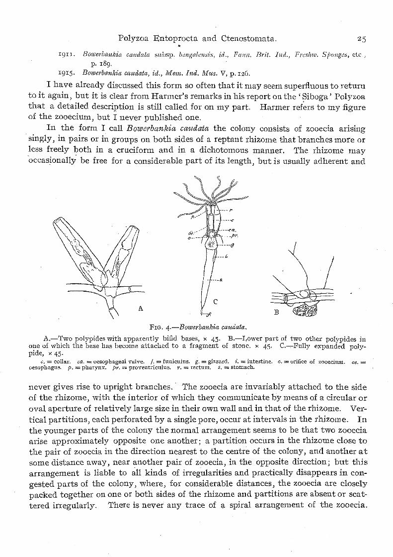
I have already discussed this form so often that it may seem superfluous to return . to it again J but it is clear from Harmer's remarks in his report on the (Siboga' Polyzoa that a detailed description is still called for on my part. Harmer ref~rs to my figure of the zooecium J but I never published one.
In the form I call Bowerbankia caudata the colony consists of zooecia arising . singly, in pairs or in groups on both sides of a reptant rhizome that branches more or less freely both in a cruciform and in a dichotomous manner. The rhizome may 'occasionally be free for a considerable part of its length J but is usually adherent and
FIG. 4.-Bowerbankia caudata.
A.-'l'wo polypides with apparently bifid bases, x 45. B.-Lower part of two other polypides in one of which the base has become attached to a fragment of stone, x 45. C.-,-Fully expanded polypide, x 45.
c. = collar. ca. = oesophageal valve. t. = funiculus. g. = gizzard. i. = intestine. o. = orifice of zooecinm. De. = oesopbagus. p. = pharynx. pr. = proventriculus. r. = rectum. s. = stomach.
never gives rise to upright branches.' The zooecia are invariably attached to the side of the rhizome) with the interior of which they communicate by means of a circular or oval aperture of relatively large size in their own wall and in that of the rhizome. Vertical partitions, each perforated by a single pore, occur at intervals in the rhizome. In the younger parts of the colony the normal arrangement seems to be that two zooecia arise approximately opposite one another; a partition occurs in the rhizome close to the pair of zooecia in the direction nearest to the centre of the colony, and another at some distance away, near another pair of zooecia, in the opposite direction; but this arrangement is liable to all kinds of irregularities and practically disappears in congested parts of the colony) where, for considerable distances~ the zooecia are closely packed together on one or both sides of the rhizome and partitions are absent or scattered irregularly. There is never any trace of a spiral arrangement of the zooecia.
ZOOLOGY OF THE FAR EAST.
Lateral branches are usually given off in the neighbourhood of groups of zooecia, but the tips of these branches divide dichotomously in front of the last zooeciulll (pl. i,
ng. 10). The size of individual zooeda varies greatly both in the same colony and in colo-
nies from different localities or growing under different. conditions. If the organism is threatened by the deposition of mud in its interstices, as often happens if it is attached to the roots of reeds in muddy estuarine waters J some zooecia are often of very great length without attaining more trian normal girth, The following table gives, in millemetres, the length and greatest transverse diameter in the longest zooecium discoverable in four colonies from different localities, the first two of which are situated on different sides of the Malay Peninsula, while the two latter are in the
Gangetic delta :-
Length Breadth
Port Weld.
1'78 0'204
Tale Sap. 1'02
O'IIg
Calc'Vttta.
2'55 0'255
Port Canning.
0'775 O'II9
The zooeda are always more or less spindle-shaped, tapering both above and at the base, which is usually prolonged below the point of attachment to the rhizome in the form of a pointed process or " tai1." If this tail comes in contact with a hard object it is often expanded into a funnel-like body, concave at the tip, which attaches itself to the object. Its position is sometimes a little eccentric so that it is situated at one side of rather than in the middle line of the main body of the zooeciutll, the base of which then grows out into a lateral pocket, thus giving the whole structure a bifid appearance (text-fig. 4, A); but the tail never forms a branching radicle. 'l'he .distal region of the zooecium is subcircular in cross-section. Its ~ctocyst is faintly and minutely striated transversely, but the striae are often obsolete. 'rhe tint of the ectocyst varies greatly; often it is colourless but sometimes it is stained with yellow or brown. It is always transparent.
In both arrangement and number the parietal muscles vary considerably. Sometimes they are practically confined to the upper part of the zooecia, while in some zooecia they extend almost to the base (cf. figs. 10 and loa, pl. i).
There are always 8 tentacles, which are armed with a sensory bristle at the base, with several horizontal hairs on the outer margin and a bunch of finer hairs at the tip. The alimentary canal resembles that of other species. The diameter of the gizzard varies with the size of the zooecium (cf. pl. i, figs. 10, roa and II).
B. caudata, therefore J differs from the form described by Waters 1 and by Harmer 'l. as B. imbricata in the following characters :-
(I) The zooecia are more slender and less cylindrical; their base never forms a binding. radicle.
(2) They are joined to the rhizome by a distinctly lateral communication and never exhibit any approach to a spiral arrangement.
1 Journ. Lin. Sac. XXXI, p. 248, pI. xxv, figs 6-10 (1910). 2 Siboga-Exp., mOll. XXVIIla, p. 70, pl. vii, figs. 15, 16 (1915).

Polyzoa Entoprocta and Ctenostomata. 27
(3) The rhizome, although it is not always adherent, never gives rise to vertical branches.
(4) The number of tentacles is always eight. I find these characters constant in a large senes of specimens from Bengal,
Madras, Perak and the Tale Sap. In eastern waters B. caudata is characteristic of estuarine tracts in which the
water has a lower salinity than that of the open sea. I found the species abundant at Koh Yaw in water of a specific gravity (corrected) of from roo4 to 1'0085. It occurred (often 'with Victorella bengalensis) on sticks and stones. I also took a specimen on a worm-eaten fragment of a wooden pier at Port \Veld on the coast of Perak. This place is situated up a creek, some distance fro111 the open sea (Straits of Malacca), but the water is probably almost if not quite as salt as that of the Straits.
Division PALUDICELLEA.
I9II. Palludicellina, Alluandale, Faun. Brit. Ind., Frcslm). Sponges, etc., p. I86. I9 I 5. Palndicellea, Harmer, Siboga·Exp., mOll. XXVlIIa, p. 43.
Harmer (loc. cit.) includes in this division the following families: Paludicellidae, Victorellidae, Arachnidiidae and Nolellidae (= Cylindroeciidae, a2tct.); whereas I have hitherto included only the Paludicellidae, Victorellidae and Hislopiidae-the last a freshwater family referred to by Harmer only in a foot-11ote. He supports his views as to the inclusion of certain marine genera with abundarlt evidence and clears up several anatomical points hitherto obscure J in particular by means of his excellent figures. These show that there is practically no difference in the general structure of the polypide between Cylindroecium and Victorella. In fig. I9 of his plate iv, for example, it is quite clear that the polypi de of Cylindroeci'U'ln (or Nolella) papuensis possesses a cardiac store-chamber and a well-defined single funiculus. Indeed, now that this evidence on anatomy is available, the grounds on which the family Cylindroecidae is separated from the family Victorellidae become rather flimsy.
It is somewhat otherwise with the Arachnidiidae, in which Harmer follows Loppens 1 in placing the freshwater genus Arachnoidea, Moore. His figure of the polypi de of Arachnidi'llt'ln ir1'egulare (op. cit., pI. ill, fig. 6) shows quite clearly that there is neither a proventriculus, nor a spherical chamber, nor a funiculus. This, of course, does not rule Arachnidiu'ln out of the division-the alimentary canal is merely in a simple and probably primitive condition; but it does prove that Arachnoidea is by no means closely related to Arachn,idiu'ln. Arachnoidea I would still" retain in the family Hislopiidae on' anatomical grounds, for although its anatomy is still imperfectly knowll, it certainly possesses a spherical chamber closely resembling that of Hislopia. This structure is llot clearly indicated either in Moore's original sketch 2 or in ROtlsselet's more elaborate figure/ but I have seen it without a doubt in specimens mounted by the latter author and in Hislopia the horny lining of the gizzard remains
] A1I11. BioI. /acust. Ill, p. 150 (I908). ~ The Tallganyilill Problem, p. 296 , fig. (1903)·
3 ['roc. Zool. Soc. Londott 1907 I), p1. xiv, figs. 5.6.

28 ZOOLOGY OF THE FAR EAS1'.
as a fairly conspicuous object even in very badly preserved specimens. There is some reason, therefore, to doubt whether Harmer's marine species Arachnoidea pro" tecta (op. cif., p. 50, pl. Hi, figs. 7-11) is really co-generic, notwithstanding the very close extelnal resemblances, with A. ray-lankesteri from Lake Tanganyika.
I would therefore arrange the families of the Division Paludicellea as follows, , , basing their classification on the structure of the polypide as well as the form of the zooecillm :-
1. Alimentary canal of simple structure, cardiac limb of stomach undifferentiated.
A. Zooecia broad, flattened, adherent, with the orifice situated on a tubercle or short upright tubule; no funiculus. . ARACHNIDIID An.
B. Zooecia relatively narrow, either entirely verticalor bearing a comparatively long, vertical orificial tubule; two funiculi P AI, UDICTI;I,I,I])AI~.
n. Alimentary canal more highly specialized in the cardiac region.
A. Cardiac region of the alinientary canal with an antechamber (always ?) lined with chitin; no proventriculus; adult zooecia vertical and
, tubular; a single funiculus. 1. Base of zooecia swollen or slipper-shaped Vrc'tOREI,LIDAE. 2. Base of zooecium sharply constricted off
from the false rhizomes by which it is connected with other zooecia CYLINDROECUDAE.
B. Cardiac region of alimentary canal with a ptoventriculus and a spherical chamber lined with thick chitin; no funiculus. .
Zo'oecia flattened and adherent with or . , without a high orificial tubule HISI,OPIIDA:b:.
FamilyP ALUDICELLIDAE.
Genus Paludicella, Gervais. 1887. PaludiceUa, Kraepelin, Deutsch. Siisswasserbryozoen I, p. 96. 191 3. Paludicetla, Harmer, Proc. Zool. Soc. London TII, p. 441. 19I 4· Paludicella, Braem, Arch. f. Hydrobiol. IX, p. 456 .
. Rece~t authors have recognized a single species in this genus, llamely Pahtdicdla artwutata (Ehrenberg) =P. ehrenbergii, v. Beneden. I have here, however) to revive a second usually relegated to the synonomy of that species and to describe a thirdthe ~atter a very distinct form. A fourth species, or what I believe to be a fonrth specles, occurs in Japan and will shortly be described by Prof. A. Oka.
The genus is probably cosmopolitan but has not yet been fonnd in India l unless

Volyzoa Entoprocta and Cteno;.,tomata.
we accept Carter's somewhat inconclusive record.' Personally I am of the OptnlO11
that this record refers to a species of Victorella.
Paludicella elongata t Leidy.
(Pl. I, fig. 4.)
r852. FllludieeUa elongata, Leic1y, ProG . .'lead. Nat. Sei. Philadelphia V, p. 32r, pI. -, figs. t, 2.
Specimens in my collection from China agree precisely with Leidy's figures, which, however, show only the outlines of zooecia. The species differs from P. articulatu in the following characters :-
I. The ectocyst is colourless and very thin, liable to collapse in spirit. 2. The proximal part of the zooecia is much elongated and attenuated, while
the distal part, as viewed' in profile J is not in uch deeper than the proximal; the orificial tubule is relatively short.
3. Young buds reach the full length of an adult zooeciul11. and assume a somewhat clavate form before the orifice is developed.
4. The whole of the alimentary canal is stonter than in the common European form, the stomach in particular being much larger; when fully developed, the phyloric part has a broadly elliptical form.
The last of these differences I consider the most important. It becomes very clear if fig. 4 011 pI. i be compared with the fignres already published by Alhnap./ Kraepelin/ Hancock" or myself." In young polypides the stomach is more slender than in those that are. fully adult and the main or pyloric portion is slightly contracted in the middle and somewhat pointed at the free extremity, but even in such polypides the organ is relatively more bulky than in European specimens.
The only examples of P. elongata I have seen were growing, with the Hydroid Cordylophora lacustris, on the roots of a willm'\') on shells of Modiolu laoustris attached to them in large numbers, and on living shells of a Unionid mollusc (A nodonta woodiana) . In these specimens there is no trace of vertical branches) but in the colonies on Toots many of the zooecia are free and floated loosely in the water In December none of the z')oecia containeclmature gonads, though immature testis and ovary were found in one. They occupied the same position as in P. articulata. A single free resting-bud was observed. It was flattened and FIG., 5.-Paludicella elongata.
Part of a colony, x r6. polygonal and had a thinner shell than is usual in P. articuluta.
I Ann. JlIlag. Nat. Hist. IH (:)l, p. 333 (1859). ~ M~au, Fresh-Watep Polyzoa, pI. x (1856 ). 3 Deutsch. Silsswasserbryo:;oell I, pI. Hi, fig. 104 (1887). .j. Ann. Nat. H~·st. V (z), pi. iv (1 850 ).
& Rec. Ind. JJ.lus. VI, pI. xii, fig. I (19 1 I).

ZOOLOGY OF THE FAR EAs'r.
Localities.-'rhe species was originally described, with Urnatella gracilis J from the Delaware and Schuylkill rivers near Philadelphia, U.S.A. My specimens were taken in a few feet of water at the mouth of the Moo-Too creek and in the northwest corner of the Tai-Hu (Great Lake) in the Kiangsu Province of China: December,
19I 5· Paludicella pentagonalis t sp. novo
(PI. I, fig. 5.)
The type-specimen of this peculiar little Polyzool1 was attached to a piece of stick and was rather deeply buried in crevices between the ridges on the bark. It consisted of a single small colony apparently in a degenerate condition) and only a few of its zooecia and polypides are at all well preserved. I found it imposs~ble, moreover) to gain more than a very general idea of the structure of the organism ht situ and only succeeded in extracting and mounting two consecutive zooecia·-evidelltly the two oldest zooecia in the colony-in such a condition as to illustrate their 11fttural relationship one to another. Fortunately these two zooecia, and the polypic1es they contain, are well pre~erved, fully mature and in one case about to produce a resting
FrG. 6.-Palttdicella pentagonalis, sp. nov., x 35.
Part of the type-colony seen in oblique lateral view. b. = bb.se of lateral bud. It. = resting bud.
bud. Their peculiarities are so well marked that I do not hesitate to accept them as the type of a new species.
Colony. The colony as observed consisted of a linear series of zooeda without lateral branches, but it is evident that lateral branches must have existed at some period in the history of the organism as the bases of the lateral buds can still he detected in mounted zooecia. Not more than half a dozen zooecia in all were present .. The colony s~ems to have arisen from an embryo or bud that gave rise to two zooeCla that were onentated in opposite directions (fig. 6).
Z ooe~ia.. The ectocyst of. the. z~oecia is perfectly colourless and hyaline except on th~ onficlal tubules, on which It 1S yellowish and considerably thickened. 1'hc zooeCla are variable in shape and proportions but always flattened, relatively broad ~nd more or less produced and narrowed proximally. They do not exceed 1'2 111111.
In length. !he ori~ce is distinc:ly pentagonal. The orificial tubule is relatively long and
SUb?lrcular In .cross-sectiOn below the orifice. Its ectocyst sometimes exhibits ~l tendency to flake 111 such a way as to produce slender irregular processes that stand up

Polyzoa Entop.rocta and Ctenostomata. 31
vertically above the orifice when the polypide is retracted. Fig. 5a J pI. i, shows the structure of the orifice so far as it can be made out In the material at my disposal.
PoZypide. The pol~pide has the structure normal in the genus, but is remarkable for the great length of the slender-walled oesophagus and for the broadly pearshaped outline of the stomach, which occupies a relatively larger part of the space available in the zooecium. The tentacles are long and slender and probably number r6. The intestine is bulky. Funiculi cannot be seen in my specimen and I have not been able to detect the collar precisely.
}1,1 usculature. All the muscle-fibres are remarkably stout, especially those of the retractor muscles. The parietal nlUscles are short and entirely lateral in position. They are variable in number and arrangement. The" pyramidal" muscles connected with the orifice are attached to the retractile part of the ectocyst very low down and are arranged in three groups, two anterior (distal) and one posterior (proximal).
Gonads. One of my mounted zooecia contains a ripe testis. It consists of rather discrete groups of cells situated on the floor of the zooecium proximad of the stomach and some distance from the proximal end of the zooecium (pI. i, fig. S).
Buds. The position of the primary lateral buds seems to be variable; sometimes they are situated much nearer the proximal end of the zooecium than is usual in P. ehrenbergi or P. elongata.
In one zooecium a young resting-bud occurs in the distal part of the zooecium. It consists of a broadly oval mass of rounded cells densely packed with food-granules. The upper surface is smoothly rounded, but below the outline seems to be irregular. A thin chitinous investment has already been deposited round it. The length is o'I477 mm. and the greatest transverse diameter 0'I02 mm. The polypi de in this zooecium is not markedly degenerate.
Type. No. 7I94/7 Z. E. V. in the register of the Indian Museum (Zoo!. Survey of India): mounted in Canada balsam on a slide.
Locality. Lampam, at the edge of Patalung R near its entry into the Tale Sap, Singgora Province, Peninsular Siam: January, I916: in permanently fresh water.
The most striking feature of this new species is its pentagonal orifice, in which it resembles PotsieUa erecta, Leidy. From that species, however) it differs entirely in the form of the zooecium) and, so far as can be seen at present, there is no reason for separating it from the genus PaludiceUa.
family VICTORELLIDAE.
Genus Victorella r Kent.
19II. VictoreUa, Annandale, Rec. Ind. 1I1'Us. IV, p. I93· I91 5. Vict01'eUa., Harmer, 'Siboga J -Exp., mOll. XXVIIIa, p. 44.
Most species of the genus are found habitually in brackish water on or near the coast, but the genus has been recorded fro111 Lake Tanganyika in Central Africa, the Birket-el-Qihun in Egypt and Issyk-kul in Central Asia. Loppens found the common European form (V. pavida, Kent) in marine oyster-beds on the coast of Belgium and

32 ZOOLOGY OF THE FAR EAST.
Harmer (ap. cd., p. 45) has ascribed to the genns a marine species (V sibogae) from a depth of 0 and 32 metres in the Malay Archipelago.
All brackish-water species as yet examined have eight tentacles) hut V. sibogac has probably more than twenty. Its generic position seems to me doubtful.
The genus is evidently cosmopolitan in distribution, but has not as yet been found in America. Definit~ records now exist fromllorthern Europe, Egypt, Central Africa, Central Asia and India; a specimen vvas recently taken ill the Main Island of Japan by Dr. A. Oka and myself.
Victo:rella bengalensls J Annandale.
(PI. I, figs. 6, 7.)
I907. Victorella pavida, Annundale(nee Kent), Ree. lnd. Mu.';. I, p. 200, figs. I-4-I908. Victorella bengaZensis, id., Ree. Ind. M1,[,8. Il, p. 12, fig. I.
I9II. VictoreUa bengalensis, id., Falm. Brit. Ind., Freshw. SIJOnges, etc., pp. H)l, Hp, llg. :1'7
!t-t; p. I70, fig. 31. 19II. VictoreUa continentali8, Braem. Trans. SOl;. Nat. St. Pc!h'sb.LXIl, p .. )0, figs. lH-:n.
I9II. Vieto1'ella bengalensis, AUllandale, Rec, Ind. lVIl/s. VI, p. I<)7, pl. xiii, figs .. 1, ?, R. 19II. Victorella symMotica, id. (? nee ROllsselet), iMd., p. I97, pI. xiii, fig. h.
I915. VictoreUa bengalensis, id., Mem. Ind. lV[us. V, p. 125.
This species was abundant on sticks in the Tale Sap off Roh Yaw in J aUl1ary, 19I6, in water that varied in specific gravity (corrected) from 1'00625 to 1'008. I can see no specific difference between specimens from India and Siam and others from the saltlake Birket-el-Qurun in Egypt. The latter seem to me to agree well enough\~vith Rotlsse1et's figure of V. symbiotica fr0111. L. Tanganyika, but Braem, who has examined examples from both African 10calitiesJ states that there is a difference (which he refrains from describing) in the alimentary canal between the true V. symbiatica and
- ---po
c.---- ----
FIG. 7.-Victorella bengalensis.
Central region of the alimentary canal of a .retracte(~ polypide in lateral view (slightly dlugramatlc).
a = oval chamber with horny lining. c. = thick walled glandular tube. m. = circular lUuscle. oe. =:
oesophagus. p: = pharynx.
the Egyptian form. As I have not examined specimens from Tanganyika and as Rouf1selet does not discuss or.figure the anatomy in detail, I can express no opinion 011 this point. but mnst content myself with reproducing a drawing of the alimentary canal of V. bengatens1:s (pl. i, fig. 7).
V. bengale'nsis, as I have pointed out elsewhere, is a very variable form; som.e colonies have larger zooecia and a thicker ectocyst than others) while envil'On111en t appears to exert a direct effect on the growth and appearance of the colony, With the thickness of the zooecia the development of the parietal muscles is to some extent correlated. Specimens fro111 Birketel-Qurl1n have very small and delicate zoo
eCIa. My Siamese examples on the other hand are particularly well developed in all

Polyzoa Entoprocta and Ctenostomata,
cases; the length and greatest diameter of the largest zooecia are 2'55 and U"2,72 mm. In a colony from the neighbourhood of Calcutta the largest zooecia are, however, only I'615 long by 0'22I in diameter, while in one from Port Canning, some 30 miles distant, the measurements are 0935 and 0'153. These differences appear to be considerable if individual colonies are compared, but they disappear com.pletely in a long series of specimens.
In all my Siamese specimens the ectocyst is rather thick and has a slight yellowish tinge. The parietal muscles, though well developed in some zooecia, are not invariably stronger or more numerous than in specimens from India or Egypt. In some Siamese zooecia, however> they extend further up the zooecia than is usual in Indian examples.
Family HISLOPHDAE.
IgII, Hislopiic1ae, Allnallc1ale, Faun. Brit. lud., Freshw. Sponges, etc., p. 199. 19II. Hislopiidae, id., Rec. lnd. Mus. VI, p. 199,
Genus Hislopia, Carter.
I have now been able to examine ample material of all the forms hitherto described in this genus with the exception of H. placoides (Korotneff), and on my recent tour was fortunate in discovering a new species which, owing to the transparency of its ectocyst, the study of the anatomy was peculiarly easy. The following key to the species may, therefore, be of some value:-
1. Orifice armed with four very long spines H. placoides. H. Orifice unarmed or bearing four short spines.
A. ZQoecia in uncongested parts of the col<?ny almost circular, slightly truncated proximally and distally. Ectocyst yellowish, orifice quadrate or· subquadrate, usually with four short spines H. monilifonnis.
B. Zooecia in uncongested parts of colony oval or ovoid. 1. Ectocyst perfectly hyaline and colourless;
terminal zooecia assuming a fan-like outline before becoming oval; no orificial spines. H. malayensis.
ii. Ectocyst yellowish; terminal zooecia not passing through a fan-shaped stage.
a. Zooecia (at any rate in peripherial parts of the colony) constricted and produced at the proximal end; the margins not noticeably thick-ened ; orifice as a rule without spines H. cam.bodgiensis.
b. Zooecia not or very rarely pedunculate; their margins thickened and chitinized ; four short orificial spines frequently present H. lacustl'is.

34 ZOOLOGY OF THE FAR EAST.
All these species are closely related and in order to identify specimens satisfactorily it is necessary to examine the peripherial parts of the colony; the older zooecia, which of course occur towards the centre, are often distorted owing to congestion.
The genus Hislopia occurs over a great part of Asia. H. placoides is only known from Lake Baikal and H. moniliformis (originally described in my volume in the (' Fauna '.' as a variety of H. lacustris) from ponds at Calcutta. H. lacustris is widely distributed in northern India and Burma and H. cambodgiensis in Indo-China, Siam and China; while the new species H.111Alayensis has been found as yet only in a small1ake in the Siamese province of Patani in the north-east of the Malay Peninsula. r have recently observed what I take to be remains of a species of H islopia on shells of the genus Aetheria from tropical Africa, probablY from the Upper Nile; but these cannot be identified specifically.
Hislopia cambodgiensis (Jullien).
(PI. I, fig. 8.) r880. Norodonia cambod~iensis and H. sinensis, Jul1iell, Bull. Soc. zool. Fm.nee V, pp. 77-7'),
figs.
I found in Chinese specimens, attached (like the types) to shells of freshwater molluscs, that the two forms described by J ullien in the paper cited passed insensibly one into the other, his cambodgiensis representing in fact merely older colonies, 01'
the central congested part of old colonies, of his sinensis. I· can find no difference between these forms and H.lacustris, the type-species of Hislopia, that would justify generic separation. Indeed, I have long hesitated whether to regard the differences that do exist as specific or as merely racial. In the collection of the Indian Museum there are specimens of H.lacustris on the shells of Unionidae and Viviparidae from ihils (swamps or shallow lakes) in northern Bengal the central or oldest zooeda of which agree almost exactly with those of the same kind in colonies from China. Moreover, the form of the orifice and the development of spines in connection with it are extremely variable characters in both the Indian and the Chinese forms. But while in the former the young zooecia are rarely if ever pedunculate, in the latter they are invariably so, thus having a vel-y characteristic appearance (see pt. i, fig. 8). Other less important differences are the following:-
1. The colony of H. lac%stris invariably forms, when fully developed, owing to profuse lateral budding, a solid pavement or layer, whereas 111 H. cambodgiensis lateral buds are produced much less sparingly, so that the colony consists of visibly radiating and separate branches.
2. In H. lacustris (and alsoin Ho moniliformis) the margin of the zooecia is thickened and chitinized, whereas in H. cambodgiensis this is not at all or very indistinctly the case.
3· In H. lacustris some at any rate of the zooecia in each colony possess four well-dev~loped but short spines at the four corners of the quadrate orificeJ whereas in H. cambodgiensis the' orifice is usually subcircular and spines are only occasionally developed in connection with it.

Polyzoa Entoprocta and Ctenostomata. 35
4· The chitinous lining of the gizzard is usually rather thicker in H. cambodgiensis than in H. lacu,stris and the thin-walled oesophagus perhaps rather longer.
5· In H. cambodgiensis the parietal muscles are, at any rate in the older zooecia, more powerful, consisting of more numerOl1S and thicker muscle-fibres.
Jullien's description of Norodonia was apparently based on dried specimens, in which the central part of the roof of the zooecia, especially if they be young, as a rule collapses, giving a somewhat false idea of the natural appearance. The ectocyst becomes considerably thicker and darker in old zooecia than in young ones.
The orifice in this species is as a rule rather small and the orificial tubercle very low. The former is in most cases surrounded by an incomplete circular or subcircular horny ring, which is interrupted posteriorly. Occasional zooecia l11.ay be found in the older parts of colonies in which the ring is complete and subquadrate. l\10re rarely it bears spines at its four corners, but one or more of the spines is usually abortive and I have not seen a case in which four well-developed spines were present ..
Zooecia developing in the depressions between ridges on the epidermis of Unionid shells are frequently assymmetrical, as in the branch figured on pI. i.
The number and the arrangement of the parietal muscles are very variable, as is apparently the case in all species of the genus. The fibres seem to become stouter and more numerous as the ectocyst thickens with age. These mnsc1es do not run parallel to the walls of the ectocyst as in PaludiceUa, VictoreUa, Bowerbankia and other more or less tubular forms, but directly from the floor to the roof at some distance from the sides of the zooecium, as in flattened forms such as ALcyonidium. In some cases they form a rather dense mass on either side of the polypideJ but in young zooecia they are always very difficult to detect. '
Localilies, etc. J u1Hen found the two forms here regarded as synonymous on Lamellibranch shells from an island in the Mekong River on the borders of Siam and Cambodia, from the interior of Cambodia and from the neighbourhood of Canton and the province of Ngan-Honi in China. My own examples of the species are on shells from the south-east corner of the Tai-Hu (Great Lake) in the Kiangsu Province of China. 1'hey were taken in the channel west of the island of Tong-Dong-Ding and in the Moo-Too creek, on a muddy bottom in from 6 to 10 feet of water, in December, I9 I6.
All specimens as yet found have been on the shells of molluscs; my own were on those of A nodonta woodiana (Lea) the animals in which were alive.
Hislopia malayensis t sp. novo
(PI. I, fig. 9; pt Il, figs. I, la.)
The species may be distinguished by the following diagnostic characters. The colony is entirely adherent and of a more or less circular form, consisting of
numerous primary branches that radiate from a common centre and give rise occa-:sionally at an acute angle to lateral branches in the typical cruciform manner.

ZOOLOGY OF THE FAR EAST.
Adult zooecia are flattened, oval or ovoid and not or but very slightly pedunculate, Voung terminal zooecia arise in the form of slender, pointed, flattened cylinders, which reach almost their full length and then expand fan-wise at the tip until they take the form of a comparatively long and narrow peduncle supporting a triangular or pentagonal head. The whole structure then gradually expands from in front backwards until the adult oval or ovoid outline is assumed. The ectocyst is hyaline and colourless except round the orifice; the margins of the zooecium are not thickened but are surrounded by a narrow 'rim of flat membrane. The orificial tubercle is low. The orifice is surrounde.d 011 three sides by a brownish chitinotls rim) which does not bear spines) though it is usually quadrate or subqnadrate, and is broadly interrupted posteriorly or proximally.
I can find no definite diagnostic characters in the polypide or musculature. Both are admirably displayed in stained and mounted specimens (pI. i, fig, 9)·
Type':'specimens. No. JI5z/7 Z.E.V., Ind. Mus. (Zool. Survey of India); in alcohol.
Locality, etc. Small lake at the base of a limestone hill (Bukit J alor) in the in.land state of J alor or Yala in the Siamese province of Pa tal1i in the eastern part of the Malay Peninsula. The specim.ens, which were taken in the first week of February, I9I6, were growing on a dead palm-leaf .that had fallen into the water. Specimens were also taken by Mr. H. C. Robinson and myself at the same place in IgOI. They met with an accident that caused thenl to dry up and I identified them provisionally as H. lacustris.
" The colony is as a rule less congested than in H. lacustris and H. camhotigiensis, if more luxuriant in its growth than ·H. moniliformis. This is mainly because lateral branches are sparingly, but not very sparingly, produced. The successive forms assumed by the young terminal zooecia are most characteristic. In the other species I have seen the buds often attain a considerable length as flattened cylinders, but seem to assume the adult form gradually. Even in H. cambodgiensis, in which adult zooecia are normally pedunculate, I cannot find any state comparable to those marked c, d and e on fig. Ia, pt ii.
The parietal 11luscles are fully developed in this species and vary greatly ill number of fibres and in arrangement. In the' zooecium figured on pI. i they consist of a number of imperfectly grouped or solitary fibres scattered chiefly on the outer side of the polypide.
In specimens preserved in alcohol the roof of the zooedum usually collapses to some extent and consequently these rl1uscles are somewhat distorted or displaced.
The homologues of the pyramidal muscles of the orificial tubule in such genera as Palud1:cella consist of separate fibres grouped in a somewhat indefinite manner 011
each side of the orifice. It is not uncommoJ?- for them to be, as in the figure, markedlyassymmetrical.
The polypide, as I have already stated, agrees closely with that of H. lacustris, the general structure of which is discussed and figured in theU Fauna." There is considerable variation in the proportions of the different parts of the alimentary

Polyzoa Entoprocta and Ctcnostomata. 37 canaL . This is due partly to the different states of contraction in which different polypides are killed and partly to changes induced by growth and other physiological conditions.
There are apparently about 16 tentacles. In contracted specimens I cannot see the collar.
The star-shaped aperture by means of which a zooecium commnnicates with its parent and daughter zooecia are clearly defined and easily seen in this species (fig. 8A). There is no trace of a funiculus in connection \yith them. They are always surrounded by wandering cells) which may sometimes be seen actually over the apertnre. The rays of the star-like aperture frequently bifurcate so as to produce a
B A
FIG. 'S.-Hislopia malayensis, Sp.llOV.
A.-Orifice between two zooecia as seen. from within distal zooeciulll, showing wandering cells (x 542). B.-Optical sectlOll of wall of circular chamber (x 542).
1 = circular muscle-fibre 2 = internal cellular layer. 3 = horny lining of chamber.
somewhat complicated figure. At the points at which buds are given off or a daughter zooecium is attached to its parent, the flat marginal membrane is interrupted and a short peduncle is developed to form the actual linking structure. \Vhen adjacent zooecia not originally connected are pressed together by the growth of the colony, as in the preparation figured on plate i) fig. 9) the membrane of the older or more vigorous zooecium often grows over the membrane or over a part of the roof of the younger or less vigorous.
The ovaries are scattered rounel the periphery of the zooecial chamber and each produces several ova. The testes probably occur similarly but are not developed in the preparations I have examined. It is possible that the colonies are unisexual.

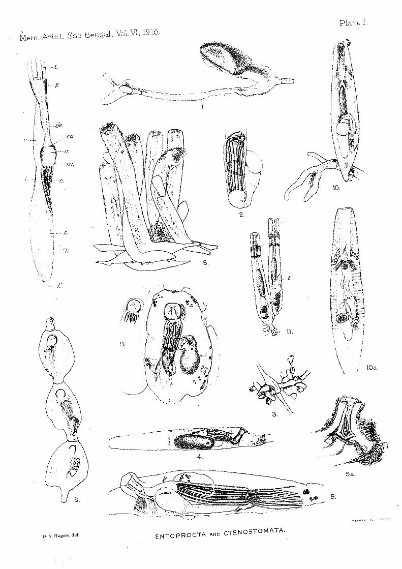
EXPLANATION OF PLATE 1.
CHI'l'ASPIS A THLETICUS, gen. et sp. novo FIG. I.-Lateral view of a part of the type-specimen, showing two polypiferous
and parts or the whole of three non-polypiferOllS segments. One of the polyps has lost its capitulum. X 46'5.
ALCYONIDIUM MYTILI, Dalyell. FIG. 2.-Retracted polypide from a specimen on a shell of Potamides /luviatilis
from the Chilka Lake in Orissa. X ca. 84.
FIG.
FIG.
FIG.
) )
FIG.
FIG.
TRITICELLA pEDICELLATA (Alder). 3.-Part of the rhizome of a specimen from the tail of the sea-snake Enhy
dris hardwickii from the Tale Sap, Peninsular Siam. x 101.
The stalks of the zooecia have been cut off near the base and have probably been somewhat compressed in the process.
PALUDICE~LLA ELONGATA, Leidy. +--H.ight lateral view of a zooecium from the roots of a willow tree at the
edge of the Moo-Too creek, Tai-Hu, Kiangsll Province, China. (Stained with borax carmine). 'x ca. 35.
PALUDICELLA PENTAGONALIS, sp. novo 5.-Left lateral view (slightly oblique) of a ZOOecill111 of the type-specimen.
(Stained with borax carmine). X 67'5. sa.-Orifice and adjacent parts of another zooecia from the same colony.
(Stained with borax carmine). X ca. 79.
VIC-TORELLA BENGALENSIS, Annandale. 6.-A group of zooecia from a colony growing on a stick in the Tale Sap J
Peninsular Siam. (Stained with borax carmine). x ca. 34. 7·-Polypide (partly expanded) from colony from Port Canning) Gangetic
delta. a = Cardiac antechamber. c = Cardiac limb of stomach. ca = Oesophageal valve. f = Funiculus. i = Intestine. 111 = Cardiac muscle. oe = Oesophagus proper. p = Pharynx. r = Rectum. s = Pyloric limb of stomach. t = Tentacles.
Hr SLOP lA CAMBODGIENSIS (J ullien). 8.-Terminal part of a branch of a specimen growing on a living shell of
. A nodonta from the south-east part of the Tai-Hu, Kiangsn Province, China. x 22'5.
HISLOPIA MALA YENSIS, sp. nov. FIG. 9·-Zooedum from type-specimen. (Stained with. borax carmine). x
ca. 34. BOWERBANKIA CAUDATA, Hil1cl~s.
FIG. Io.-Terminal zooedum of a colony from Port Weld, west coast of the Malay Peninsula. x ca. 34-
)J Ioa.-Another zooeciuni from the same colony, showing parietal muscles, etc. (Stained with borax carmine). X ca. 34.
" XL-Two zooecia from a cOlony from the Tale Sap, Peninsular Siam, showing the expansion of the C ( tail') to form an organ of adhesion in one zooecium . and its almost complete absence in another. x ca. 34 .
. t=Testes. .
· Mem. A::"iat. Sac I3engQl, \ro~.VL 1916.
, \ , ,
·,\tt~·t. r ,'-P
\. " I, 1
j !
7.
-- f.'
4.
5a.
U 8.
D N. Bagchi. del ENTOPROCTA AND CTENOSTOMATA.

Plate 1. Mem. Ac,i':Lt. Soc. Bengal. Vol.Vl. 1916.
Ill.
s.
7.
4.
5a.
n. N. Bag~hi. dA ENTOPROCTA AND CTENOSTOMATA.
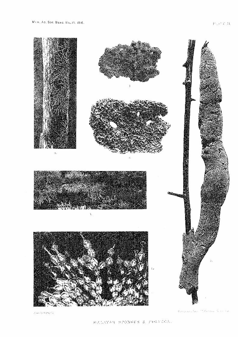
EX PLAN ATION OF PLA'rE n.
Photographs of Polyzoa and Sponges from Fresh and Brackish Water in the Malay Peninsula.
FIG.
"
HISLOPIA MALA YE;NSIS, sp. nov.
r.-Type-specimen (nat. size). ra.-Part of the same enlarged. a, b, c, d, e = young zooecia in different
stages of development.
(The other figures in this plate will be explained in subsequent reports).
MEt!.. As. Soc. BENe. VOL.VI. 1916. '; .:\ i< } I.
I.
,0
:-' I' Cl N (: .i::l L'< ,) [) I \ U\.


![RESEARCH Open Access Development of the nervous system in ... · Entoprocta and the Mollusca form a monophyletic taxon termed Lacunifera or Tetraneuralia [21-27]. Others, on the contrary,](https://static.fdocuments.net/doc/165x107/5f360355599fa60ca3094ce3/research-open-access-development-of-the-nervous-system-in-entoprocta-and-the.jpg)


