Cultivation of Pleurotus Ostreatus and Other Edibile Mushrooms
2006200620062006年度年度年度年度 2 2 2 2月月月月
博士學位論文博士學位論文博士學位論文博士學位論文
MMMMolecular Cloningolecular Cloningolecular Cloningolecular Cloning Expression Expression Expression Expression and and and and
Purification ofPurification ofPurification ofPurification of Fibrinolytic Enzyme Fibrinolytic Enzyme Fibrinolytic Enzyme Fibrinolytic Enzyme
from from from from Pleurotus ostreatusPleurotus ostreatusPleurotus ostreatusPleurotus ostreatus
朝朝朝朝 鮮鮮鮮鮮 大大大大 學學學學 校校校校 大大大大 學學學學 院院院院
遺遺遺遺 傳傳傳傳 子子子子 科科科科 學學學學 科科科科
沈沈沈沈 明明明明 花花花花
MMMMolecular Cloningolecular Cloningolecular Cloningolecular Cloning Expression Expression Expression Expression and and and and
PPPPurification ofurification ofurification ofurification of Fibrinolytic Enzyme Fibrinolytic Enzyme Fibrinolytic Enzyme Fibrinolytic Enzyme
from from from from Pleurotus ostPleurotus ostPleurotus ostPleurotus ostreatusreatusreatusreatus
느타리버섯으로부터느타리버섯으로부터느타리버섯으로부터느타리버섯으로부터 혈전분해효소혈전분해효소혈전분해효소혈전분해효소 정제정제정제정제 및및및및 특성특성특성특성
분석과분석과분석과분석과 유전자유전자유전자유전자 발현발현발현발현
2005200520052005年年年年 12121212月月月月 28 28 28 28日日日日
朝朝朝朝 鮮鮮鮮鮮 大大大大 學學學學 校校校校 大大大大 學學學學 院院院院
遺遺遺遺 傳傳傳傳 子子子子 科科科科 學學學學 科科科科
沈沈沈沈 明明明明 花花花花
MMMMolecular Cloningolecular Cloningolecular Cloningolecular Cloning Expression Expression Expression Expression and and and and
PPPPurification ofurification ofurification ofurification of Fibrinolytic Enzyme Fibrinolytic Enzyme Fibrinolytic Enzyme Fibrinolytic Enzyme
from from from from Pleurotus ostreatusPleurotus ostreatusPleurotus ostreatusPleurotus ostreatus
指導敎授指導敎授指導敎授指導敎授 金金金金 成成成成 俊俊俊俊
이이이이 論文論文論文論文을을을을 理學博士學位理學博士學位理學博士學位理學博士學位 申請論文申請論文申請論文申請論文으로으로으로으로 제출함제출함제출함제출함
2005200520052005年年年年 12121212月月月月 28282828日日日日
朝朝朝朝 鮮鮮鮮鮮 大大大大 學學學學 校校校校 大大大大 學學學學 院院院院
遺遺遺遺 傳傳傳傳 子子子子 科科科科 學學學學 科科科科
沈沈沈沈 明明明明 花花花花
沈明花沈明花沈明花沈明花의의의의 博士學位論文博士學位論文博士學位論文博士學位論文을을을을 認准認准認准認准함함함함
委員長委員長委員長委員長 朝鮮大學校朝鮮大學校朝鮮大學校朝鮮大學校 敎授敎授敎授敎授 印印印印
委委委委 員員員員 朝鮮大學校朝鮮大學校朝鮮大學校朝鮮大學校 敎授敎授敎授敎授 印印印印
委委委委 員員員員 益益益益 山山山山 大大大大 學學學學 敎授敎授敎授敎授 印印印印
委委委委 員員員員 東新大學校東新大學校東新大學校東新大學校 敎授敎授敎授敎授 印印印印
委委委委 員員員員 朝鮮大學校朝鮮大學校朝鮮大學校朝鮮大學校 敎授敎授敎授敎授 印印印印
2005200520052005年年年年 12 12 12 12月月月月 2 2 2 28888日日日日
朝朝朝朝 鮮鮮鮮鮮 大大大大 學學學學 校校校校 大大大大 學學學學 院院院院
i
목목목목 차차차차
LIST OF TABLELIST OF TABLELIST OF TABLELIST OF TABLESSSS ⅤⅤⅤⅤ
LIST OF FIGURELIST OF FIGURELIST OF FIGURELIST OF FIGURESSSS ⅥⅥⅥⅥ
ABBREVIATIONSABBREVIATIONSABBREVIATIONSABBREVIATIONS ⅨⅨⅨⅨ
ABSTRACT ABSTRACT ABSTRACT ABSTRACT XIXIXIXI
ⅠⅠⅠⅠ 서론서론서론서론 1111
ⅡⅡⅡⅡ 재료재료재료재료 및및및및 방법방법방법방법 9999
Ⅱ-1 재료 9
Ⅱ-1-1 균주 9
Ⅱ-1-2 시약 9
Ⅱ-1-3 느타리버섯 균사체 배양 10
Ⅱ-2 혈전분해효소의 분리정제 10
Ⅱ-2-1 조추출물 조제10
Ⅱ-2-2 단백질분해활성 및 혈전분해활성 검색 11
Ⅱ-2-3 조단백질 분리11
Ⅱ-2-4 단백질 정량12
Ⅱ-2-5 단백질분해활성 측정12
Ⅱ-2-6 Cation-exchange chromatography 12
Ⅱ-2-7 Anion-exchange chromatography 12
ii
Ⅱ-2-8 Gel-filtration chromatography13
Ⅱ-2- Fast protein liquid chromatography13
Ⅱ-2-10 혈전분해효소의 분리 13
Ⅱ-3 분자량 측정13
Ⅱ-3-1 FPLC를 이용한 분자량 측정(size exclusion)14
Ⅱ-3-2 SDS-PAGE를 이용한 분자량 측정 14
Ⅱ-3-3 SDS-fibrin zymography를 이용한 분자량 측정 14
Ⅱ-4 N-말단 아미노산 서열분석 15
Ⅱ-5 정제효소의 특성 분석 15
Ⅱ-5-1 정제효소의 기질특이성 분석 15
Ⅱ-5-2 효소활성에 대한 금속이온 및 단백질분해효소 억제제의 영향16
Ⅱ-5-3 효소활성에 대한 pH의 영향 16
Ⅱ-5-4 효소활성에 대한 온도의 영향 18
Ⅱ-5-5 Fibrin 및 Fibrinogen에 대한 분해특성 분석 18
Ⅱ-6 Total RNA isolation 18
Ⅱ-7 혈전분해효소 유전자에 대한 특이적 primer의 제작 19
Ⅱ-8 RT-PCR19
Ⅱ-9 Cloning을 위한 형질전환20
Ⅱ-10 DNA 염기서열 분석 20
Ⅱ-11 Construction of expression vector for recombinant fibrinolytic
enzyme gene 21
Ⅱ-12 Expression and purification of fibrinolytic enzyme22
Ⅱ-13 SDS-PAGE25
Ⅱ-14 Western blot analysis25
iii
ⅢⅢⅢⅢ 결과결과결과결과 26262626
Ⅲ-1 단백질분해활성 및 혈전분해활성 26
Ⅲ-2 느타리버섯 균사체로부터 혈전분해효소의 정제 26
Ⅲ-2-1 조단백질 분리26
Ⅲ-2-2 혈전분해효소의 정제28
Ⅲ-3 혈전분해효소의 분자량 측정 28
Ⅲ-3-1 FPLC 를 이용한 분자량 측정(size exclusion) 28
Ⅲ-3-2 SDS-PAGE 및 fibrin zymography를 이용한 분자량 측정 29
Ⅲ-4 혈전분해효소의 N-말단 아미노산 서열분석 29
Ⅲ-5 혈전분해효소의 특성분석 38
Ⅲ-5-1 기질특이성 분석38
Ⅲ-5-2 효소활성에 대한 금속이온의 영향 38
Ⅲ-5-3 효소활성에 대한 Protease inhibitor의 영향 38
Ⅲ-5-4 효소활성에 대한 pH의 영향 39
Ⅲ-5-5 효소활성에 대한 온도의 영향 39
Ⅲ-5-6 Fibrinolysis와 Fibrinogenolysis pattern 분석 39
Ⅲ-5-6-1 Fibrinogenolysis 39
Ⅲ-5-6-2 Fibrinolysis 45
Ⅲ-6 Cloning and sequence analysis of pofe45
Ⅲ-7 Analysis of deduce amino acid sequence of PoFE48
Ⅲ-8 Gene expression of pofe49
ⅣⅣⅣⅣ 고찰고찰고찰고찰 66662222
ⅤⅤⅤⅤ 적요적요적요적요 66667777
iv
ⅥⅥⅥⅥ 참고문헌참고문헌참고문헌참고문헌 66669999
감사의감사의감사의감사의 글글글글 78787878
v
LIST OF TABLELIST OF TABLELIST OF TABLELIST OF TABLESSSS
Table Table Table Table 1111 List of synthetic cList of synthetic cList of synthetic cList of synthetic chromogenic substrateshromogenic substrateshromogenic substrateshromogenic substrates 11117777
Table Table Table Table 2222 Purification Purification Purification Purification yields of fibrinolytic enzyme fromyields of fibrinolytic enzyme fromyields of fibrinolytic enzyme fromyields of fibrinolytic enzyme from P ostreaP ostreaP ostreaP ostreatustustustus
myceliummyceliummyceliummycelium 33334444
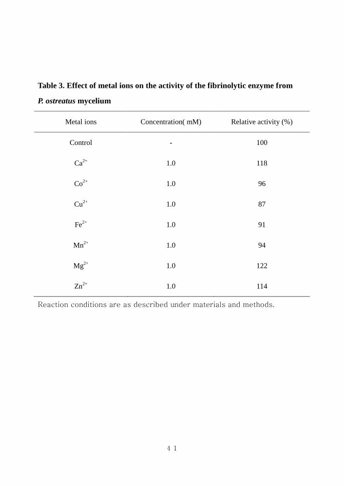
Table 3Table 3Table 3Table 3 Effect of Effect of Effect of Effect of metal ions on tmetal ions on tmetal ions on tmetal ions on the activity of the fibrinolytiche activity of the fibrinolytiche activity of the fibrinolytiche activity of the fibrinolytic
enzyme from enzyme from enzyme from enzyme from PPPP o o o ostreatusstreatusstreatusstreatus mycelium mycelium mycelium mycelium 41414141
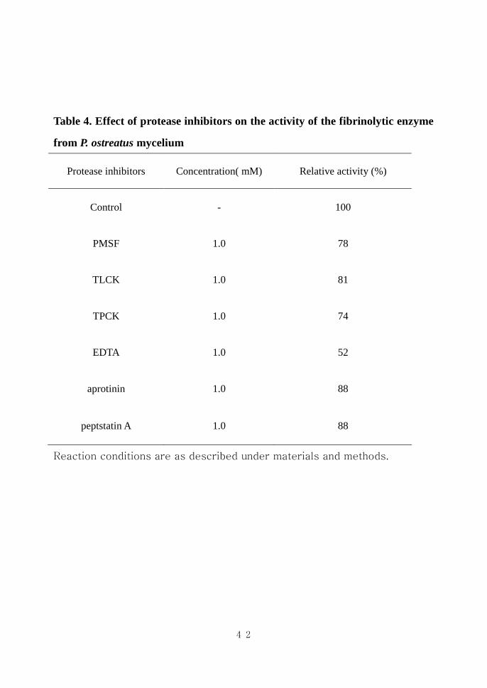
Table 4Table 4Table 4Table 4 Effect of protease inhibitors on the activity of the Effect of protease inhibitors on the activity of the Effect of protease inhibitors on the activity of the Effect of protease inhibitors on the activity of the
fibrinolyticfibrinolyticfibrinolyticfibrinolytic enzyme from enzyme from enzyme from enzyme from P ostreatusP ostreatusP ostreatusP ostreatus mycelium mycelium mycelium mycelium 42424242
vi
LIST OF FIGURELIST OF FIGURELIST OF FIGURELIST OF FIGURESSSS
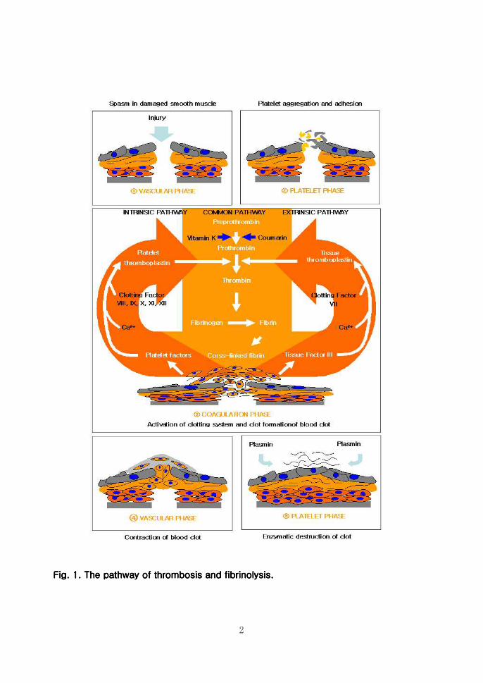
Fig l The pathway of thrombosis and fibrinolysisFig l The pathway of thrombosis and fibrinolysisFig l The pathway of thrombosis and fibrinolysisFig l The pathway of thrombosis and fibrinolysis 2222
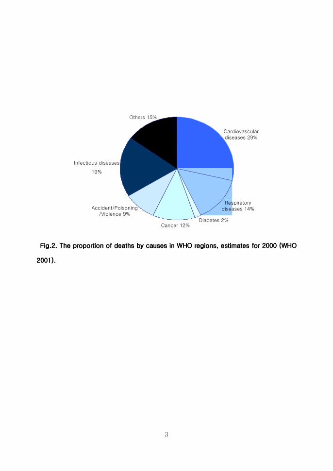
Fig 2 Fig 2 Fig 2 Fig 2 The proportion of d The proportion of d The proportion of d The proportion of deaths by causes in WHO regionseaths by causes in WHO regionseaths by causes in WHO regionseaths by causes in WHO regions
estimates for 2000 (WHO 2001)estimates for 2000 (WHO 2001)estimates for 2000 (WHO 2001)estimates for 2000 (WHO 2001) 3333
Fig 3 Fig 3 Fig 3 Fig 3 Mass formation of Mass formation of Mass formation of Mass formation of Pleurotus ostreatusPleurotus ostreatusPleurotus ostreatusPleurotus ostreatus f f f fruiting body and ruiting body and ruiting body and ruiting body and
culturing mycelia of culturing mycelia of culturing mycelia of culturing mycelia of PPPPleurotus ostreatusleurotus ostreatusleurotus ostreatusleurotus ostreatus 7777
Fig 4 Construction of cloning vector for recombinant Fig 4 Construction of cloning vector for recombinant Fig 4 Construction of cloning vector for recombinant Fig 4 Construction of cloning vector for recombinant pofepofepofepofe 22223333
Fig 5 Construction of expression vector for recombinant Fig 5 Construction of expression vector for recombinant Fig 5 Construction of expression vector for recombinant Fig 5 Construction of expression vector for recombinant pofepofepofepofe 22224444
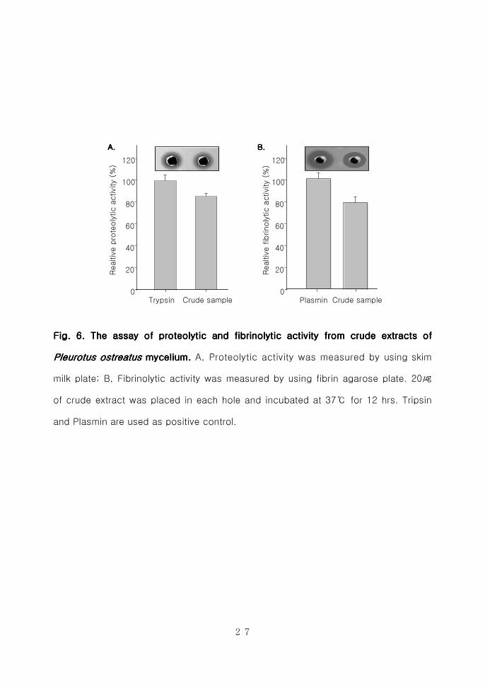
Fig 6 The assay of proteolytic and fibrinolytic activity from Fig 6 The assay of proteolytic and fibrinolytic activity from Fig 6 The assay of proteolytic and fibrinolytic activity from Fig 6 The assay of proteolytic and fibrinolytic activity from crude crude crude crude
extracts of extracts of extracts of extracts of Pleurotus ostreatusPleurotus ostreatusPleurotus ostreatusPleurotus ostreatus mmmmyceliumyceliumyceliumycelium 22227777
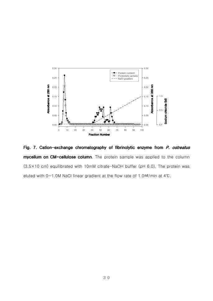
Fig 7 CationFig 7 CationFig 7 CationFig 7 Cation----exchange chromatography of fibrinolytic enzyme from exchange chromatography of fibrinolytic enzyme from exchange chromatography of fibrinolytic enzyme from exchange chromatography of fibrinolytic enzyme from
P ostP ostP ostP ostrrrreatus eatus eatus eatus mycelium on CMmycelium on CMmycelium on CMmycelium on CM----cellulosecellulosecellulosecellulose column column column column 30303030
Fig 8 AnionFig 8 AnionFig 8 AnionFig 8 Anion----exchange chromatography of fibrinolytic enzyme from exchange chromatography of fibrinolytic enzyme from exchange chromatography of fibrinolytic enzyme from exchange chromatography of fibrinolytic enzyme from
P ostP ostP ostP ostrrrreatus eatus eatus eatus mycelium on DEAEmycelium on DEAEmycelium on DEAEmycelium on DEAE----Sephadex ASephadex ASephadex ASephadex A----50505050 31313131
Fig 9 GelFig 9 GelFig 9 GelFig 9 Gel----ffffiltration chromatography of fibrinolytic enzyme from iltration chromatography of fibrinolytic enzyme from iltration chromatography of fibrinolytic enzyme from iltration chromatography of fibrinolytic enzyme from P P P P
ostostostostrrrreatuseatuseatuseatus mycelium on Sephadex G mycelium on Sephadex G mycelium on Sephadex G mycelium on Sephadex G----75757575 columncolumncolumncolumn 33332222
Fig 10 GelFig 10 GelFig 10 GelFig 10 Gel----filtration chromatografiltration chromatografiltration chromatografiltration chromatography of fibrinolytic enzyme phy of fibrinolytic enzyme phy of fibrinolytic enzyme phy of fibrinolytic enzyme from from from from P P P P
ostostostostrrrreatus eatus eatus eatus mycelium on HiLoadmycelium on HiLoadmycelium on HiLoadmycelium on HiLoad 1660 1660 1660 1660 Superdex Superdex Superdex Superdex 75757575 pg pg pg pg columncolumncolumncolumn
33333333
Fig 11 Molecular wFig 11 Molecular wFig 11 Molecular wFig 11 Molecular weight deight deight deight determination of PoFE using sizeetermination of PoFE using sizeetermination of PoFE using sizeetermination of PoFE using size----
exclusionexclusionexclusionexclusion on on on on HiLoadHiLoadHiLoadHiLoad 1660 1660 1660 1660 Superdex Superdex Superdex Superdex 75757575 pgpgpgpg column column column column 35353535
vii
Fig 12 Molecular weight determination ofFig 12 Molecular weight determination ofFig 12 Molecular weight determination ofFig 12 Molecular weight determination of P P P PoFEoFEoFEoFE using SDS using SDS using SDS using SDS----PAGEPAGEPAGEPAGE
and and and and fibrin fibrin fibrin fibrin zymographyzymographyzymographyzymography 33336666
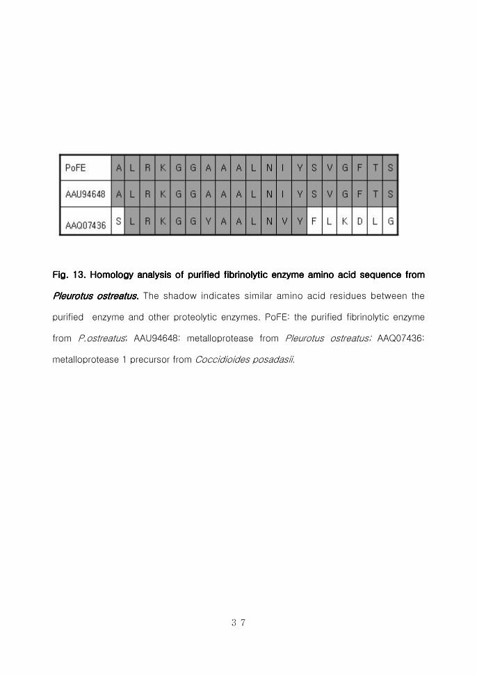
Fig 13 Homology analysis of purified fibrinolytic enzyme amino acid Fig 13 Homology analysis of purified fibrinolytic enzyme amino acid Fig 13 Homology analysis of purified fibrinolytic enzyme amino acid Fig 13 Homology analysis of purified fibrinolytic enzyme amino acid
sequsequsequsequence fromence fromence fromence from Pleurotus ostreatus Pleurotus ostreatus Pleurotus ostreatus Pleurotus ostreatus 33337777
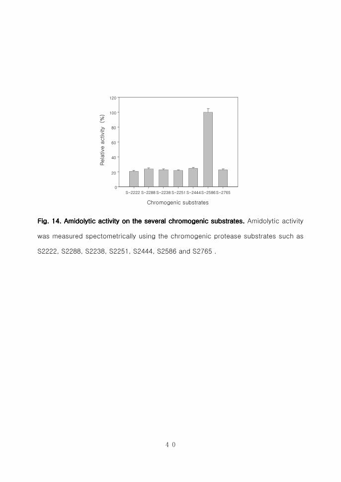
Fig 14Fig 14Fig 14Fig 14 Amidolytic activity on the several chromogenic substrateAmidolytic activity on the several chromogenic substrateAmidolytic activity on the several chromogenic substrateAmidolytic activity on the several chromogenic substratessss
40404040
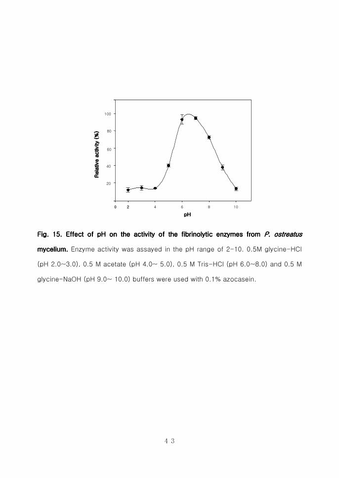
Fig 15 Effect of pH on the activity of the fibrinolyticFig 15 Effect of pH on the activity of the fibrinolyticFig 15 Effect of pH on the activity of the fibrinolyticFig 15 Effect of pH on the activity of the fibrinolytic enzymeenzymeenzymeenzymessss from from from from
P ostP ostP ostP ostrrrreatuseatuseatuseatus mycelium mycelium mycelium mycelium 44443333
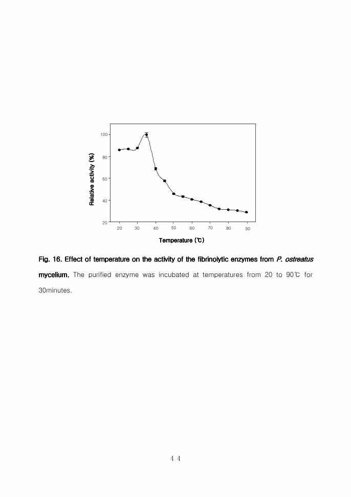
Fig 16 Fig 16 Fig 16 Fig 16 Effect of temperature on the activity of the Effect of temperature on the activity of the Effect of temperature on the activity of the Effect of temperature on the activity of the fibrinol fibrinol fibrinol fibrinolytic ytic ytic ytic
enzymesenzymesenzymesenzymes from from from from P ostreatus P ostreatus P ostreatus P ostreatus mycelium mycelium mycelium mycelium 44444444
Fig 17Fig 17Fig 17Fig 17 F F F Fibrinogenolysis anibrinogenolysis anibrinogenolysis anibrinogenolysis and fibrinolysis d fibrinolysis d fibrinolysis d fibrinolysis pattern pattern pattern pattern exhibited by the exhibited by the exhibited by the exhibited by the
pupupupurified fibrinolytic enzyme fromrified fibrinolytic enzyme fromrified fibrinolytic enzyme fromrified fibrinolytic enzyme from P ostreatusP ostreatusP ostreatusP ostreatus myceliummyceliummyceliummycelium
44446666
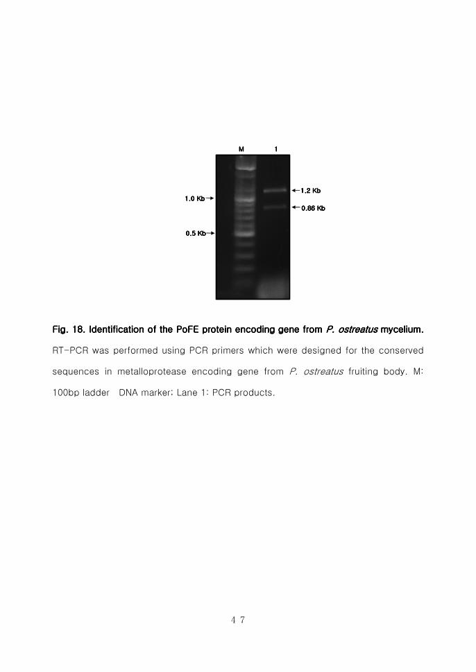
Fig 18Fig 18Fig 18Fig 18 Identification of the PoFE protein encoding gene from Identification of the PoFE protein encoding gene from Identification of the PoFE protein encoding gene from Identification of the PoFE protein encoding gene from
PPPPostreatusostreatusostreatusostreatus mycelium mycelium mycelium mycelium 44447777
Fig 19Fig 19Fig 19Fig 19 Identification of cloned ~12Kb PCR productIdentification of cloned ~12Kb PCR productIdentification of cloned ~12Kb PCR productIdentification of cloned ~12Kb PCR product 51515151
Fig 20 Fig 20 Fig 20 Fig 20 Identification of cloned ~860bp PCR productIdentification of cloned ~860bp PCR productIdentification of cloned ~860bp PCR productIdentification of cloned ~860bp PCR product 52525252
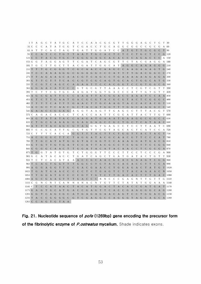
Fig 21Fig 21Fig 21Fig 21 Nucleotide sequence of Nucleotide sequence of Nucleotide sequence of Nucleotide sequence of pofe pofe pofe pofe (1269bp) gene encoding the (1269bp) gene encoding the (1269bp) gene encoding the (1269bp) gene encoding the
precursor form of the fibrinolytic enzyme of precursor form of the fibrinolytic enzyme of precursor form of the fibrinolytic enzyme of precursor form of the fibrinolytic enzyme of PostreatusPostreatusPostreatusPostreatus
mycelimycelimycelimyceliumumumum 53535353
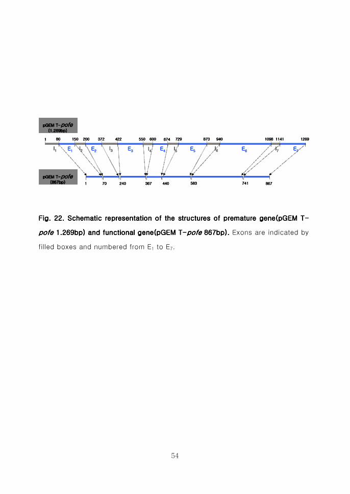
Fig 22 Fig 22 Fig 22 Fig 22 Schematic representation of the structures of premature Schematic representation of the structures of premature Schematic representation of the structures of premature Schematic representation of the structures of premature
gene(pGEM Tgene(pGEM Tgene(pGEM Tgene(pGEM T----pofepofepofepofe 1269bp) and functional gene(pGEM T 1269bp) and functional gene(pGEM T 1269bp) and functional gene(pGEM T 1269bp) and functional gene(pGEM T----
viii
pofepofepofepofe 867bp 867bp 867bp 867bp)))) 55554444
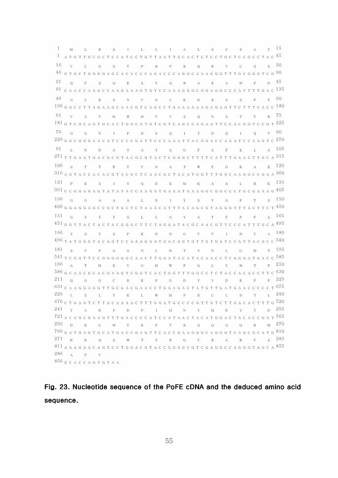
Fig 23 Nucleotide sequence Fig 23 Nucleotide sequence Fig 23 Nucleotide sequence Fig 23 Nucleotide sequence of the PoFE of the PoFE of the PoFE of the PoFE cDNAcDNAcDNAcDNA and deduced amino and deduced amino and deduced amino and deduced amino
acid sequenceacid sequenceacid sequenceacid sequence 55555555
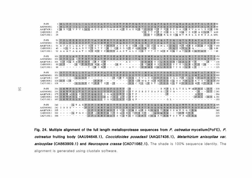
Fig 24Fig 24Fig 24Fig 24 Multiple alignment of thMultiple alignment of thMultiple alignment of thMultiple alignment of the full length metalloproteasee full length metalloproteasee full length metalloproteasee full length metalloprotease
sequences from sequences from sequences from sequences from P ostreatusP ostreatusP ostreatusP ostreatus mycelium mycelium mycelium mycelium PPPP ostreatus ostreatus ostreatus ostreatus fruiting fruiting fruiting fruiting
body body body body Coccidioides posadasiiCoccidioides posadasiiCoccidioides posadasiiCoccidioides posadasii Metarhizium anisopliae var Metarhizium anisopliae var Metarhizium anisopliae var Metarhizium anisopliae var
anisopliaeanisopliaeanisopliaeanisopliae andandandand Neurospora crassaNeurospora crassaNeurospora crassaNeurospora crassa 55556666
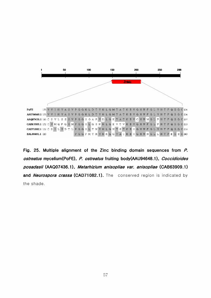
Fig 25 Fig 25 Fig 25 Fig 25 Multiple alignment of the Zinc binding domain sequences Multiple alignment of the Zinc binding domain sequences Multiple alignment of the Zinc binding domain sequences Multiple alignment of the Zinc binding domain sequences
from from from from P ostreatusP ostreatusP ostreatusP ostreatus mycelium mycelium mycelium mycelium P ostreatusP ostreatusP ostreatusP ostreatus fruiting body fruiting body fruiting body fruiting body
Coccidioides Coccidioides Coccidioides Coccidioides posadasiiposadasiiposadasiiposadasii Metarhizium anisopliae var anisopliaeMetarhizium anisopliae var anisopliaeMetarhizium anisopliae var anisopliaeMetarhizium anisopliae var anisopliae
andandandand Neurospora crassaNeurospora crassaNeurospora crassaNeurospora crassa 55557777
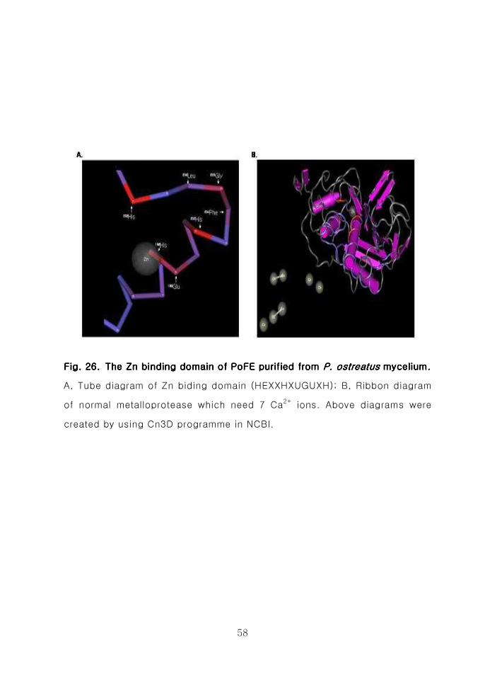
Fig 26 The Zn binding domain of PoFE purified from Fig 26 The Zn binding domain of PoFE purified from Fig 26 The Zn binding domain of PoFE purified from Fig 26 The Zn binding domain of PoFE purified from P ostreatusP ostreatusP ostreatusP ostreatus
myceliummyceliummyceliummycelium 55558888
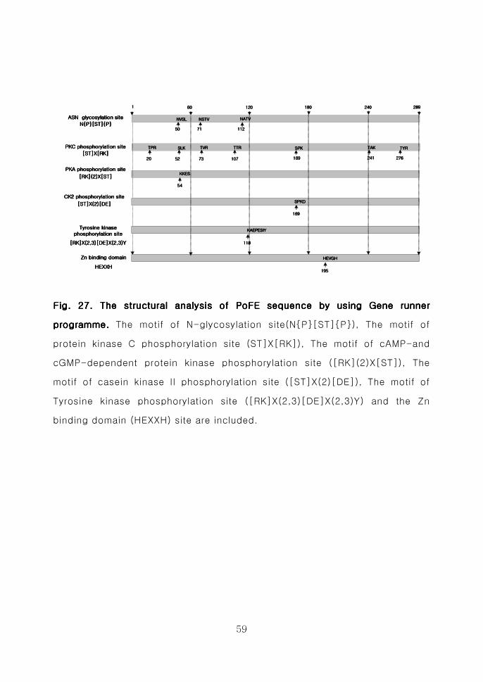
Fig 27 Fig 27 Fig 27 Fig 27 The structural analysis of P The structural analysis of P The structural analysis of P The structural analysis of PoFE sequence by using Gene oFE sequence by using Gene oFE sequence by using Gene oFE sequence by using Gene
runner programmerunner programmerunner programmerunner programme 55559999
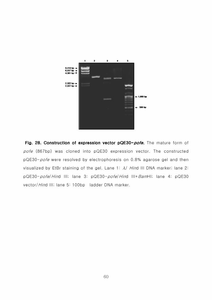
Fig 28 Construction of expression vector pQE30Fig 28 Construction of expression vector pQE30Fig 28 Construction of expression vector pQE30Fig 28 Construction of expression vector pQE30----pofepofepofepofe 60606060
Fig 29 Fig 29 Fig 29 Fig 29 Induction of recombinant PoFE in Induction of recombinant PoFE in Induction of recombinant PoFE in Induction of recombinant PoFE in E coliE coliE coliE coli M15 (pREP4) strain M15 (pREP4) strain M15 (pREP4) strain M15 (pREP4) strain
66661111
ix
ABBREVIATIONS
BSA bovine serum albumin
CM carboxymethyl cellulose
CBB coomassie brilliant blue
DEAE Diethylaminoethyl
DEPC diethyl-pyrocarbonate
E coli Escherichia coli
EDTA ethylenediamine tetraacetic acid
EtBr ethidium bromide
FPLC fast protein liquid chromatography
IPTG Isopropyl-β-D-thiogalactopyranoside
NCBI National Center for Biotechnology
OD optical density
PA plasminogen activator
PAGE polyacrylamide gel electrophoresis
PCR polymerase chain reaction
PDA potato dextrose agar
PDB potato dextrose broth
PMSF phenylmethylsulfonyl fluoride
P ostreatus Pleurotus ostreatus
Pofe Pleurotus ostreatus fibrinolytic enzyme
x
PVDF polyvinylidine difluoride
RT-PCR Reverse transcriptase-polymerase chain
SDS sodium dodecyl sulfate
SK streptokinase
TCA trichloroacetic acid
TEMED N N Nrsquo Nrsquo-tetramethylethylenediamine
TLCK N-α-tosyl-l-lysine chloromethyl ketone
tPA tissue-type plasminogen activator
TPCK N-α-tosyl-l-phenylalanine chloromethyl
UK urokinase
uPA urokinase-type plasminogen activator
WHO World Health Organization
xi
ABSTRACT
Molecular Cloning Expression and Purification of Fibrinolytic Enzyme from Pleurotus ostreatus
Shen Ming-Hua
Advisor Prof Kim Sung-Jun Ph D
Department of Genetic Science
Graduate School of Chosun University
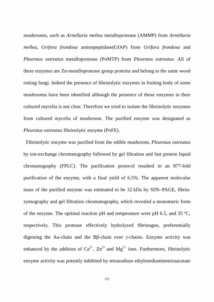
Fibrinolytic enzymes dissolve the blood clots which are formed by the
conversion of fibrinogen into fibrin via the proteolytic action of thrombin When
clots are not lysed they accumulate in blood vessels and cause thrombosis leading
to myocardial infarction and other cardiovascular diseases The major thrombolytic
agents are classified into two types The plasminogen activators such as urokinase
tPA (tissue type plasminogen activator) and streptokinase which activate
plasminogen to plasmin and the plasmin-like proteins such as nattokinase and
lumbrokinase which can directly degrade the fibrin
Mushrooms constitute an important source of thrombolytic agents Many
fibrinolytic enzymes were identified in fruiting body of different medicinal
xii
mushrooms such as Armillaria mellea metalloprotease (AMMP) from Armillaria
mellea Grifora frondosa aminopeptidase(GfAP) from Grifora frondosa and
Pleurotus ostreatus metalloprotease (PoMTP) from Pleurotus ostreatus All of
these enzymes are Zn-metalloprotease group proteins and belong to the same wood
rotting fungi Indeed the presence of fibrinolytic enzymes in fruiting body of some
mushrooms have been identified although the presence of those enzymes in their
cultured mycelia is not clear Therefore we tried to isolate the fibrinolytic enzymes
from cultured mycelia of mushroom The purified enzyme was designated as
Pleurotus ostreatus fibrinolytic enzyme (PoFE)
Fibrinolytic enzyme was purified from the edible mushroom Pleurotus ostreatus
by ion-exchange chromatography followed by gel filtration and fast protein liquid
chromatography (FPLC) The purification protocol resulted in an 877-fold
purification of the enzyme with a final yield of 65 The apparent molecular
mass of the purified enzyme was estimated to be 32 kDa by SDSndashPAGE fibrin-
zymography and gel filtration chromatography which revealed a monomeric form
of the enzyme The optimal reaction pH and temperature were pH 65 and 35 degC
respectively This protease effectively hydrolyzed fibrinogen preferentially
digesting the Aα-chain and the Bβ-chain over γ-chains Enzyme activity was
enhanced by the addition of Ca2+ Zn2+ and Mg2+ ions Furthermore fibrinolytic
enzyme activity was potently inhibited by tetrasodium ethylenediaminetetraacetate
xiii
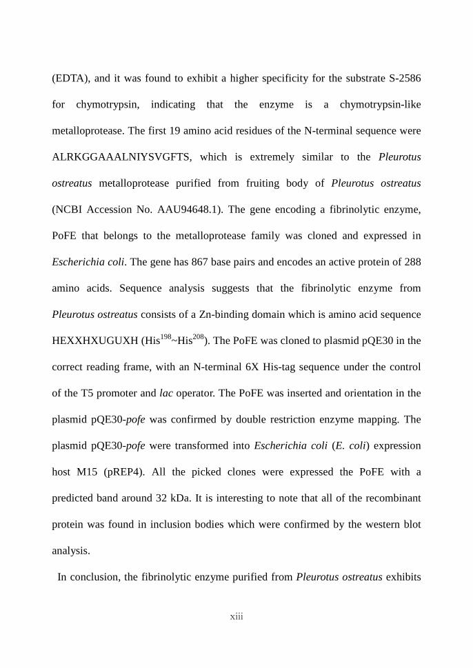
(EDTA) and it was found to exhibit a higher specificity for the substrate S-2586
for chymotrypsin indicating that the enzyme is a chymotrypsin-like
metalloprotease The first 19 amino acid residues of the N-terminal sequence were
ALRKGGAAALNIYSVGFTS which is extremely similar to the Pleurotus
ostreatus metalloprotease purified from fruiting body of Pleurotus ostreatus
(NCBI Accession No AAU946481) The gene encoding a fibrinolytic enzyme
PoFE that belongs to the metalloprotease family was cloned and expressed in
Escherichia coli The gene has 867 base pairs and encodes an active protein of 288
amino acids Sequence analysis suggests that the fibrinolytic enzyme from
Pleurotus ostreatus consists of a Zn-binding domain which is amino acid sequence
HEXXHXUGUXH (His198~His208) The PoFE was cloned to plasmid pQE30 in the
correct reading frame with an N-terminal 6X His-tag sequence under the control
of the T5 promoter and lac operator The PoFE was inserted and orientation in the
plasmid pQE30-pofe was confirmed by double restriction enzyme mapping The
plasmid pQE30-pofe were transformed into Escherichia coli (E coli) expression
host M15 (pREP4) All the picked clones were expressed the PoFE with a
predicted band around 32 kDa It is interesting to note that all of the recombinant
protein was found in inclusion bodies which were confirmed by the western blot
analysis
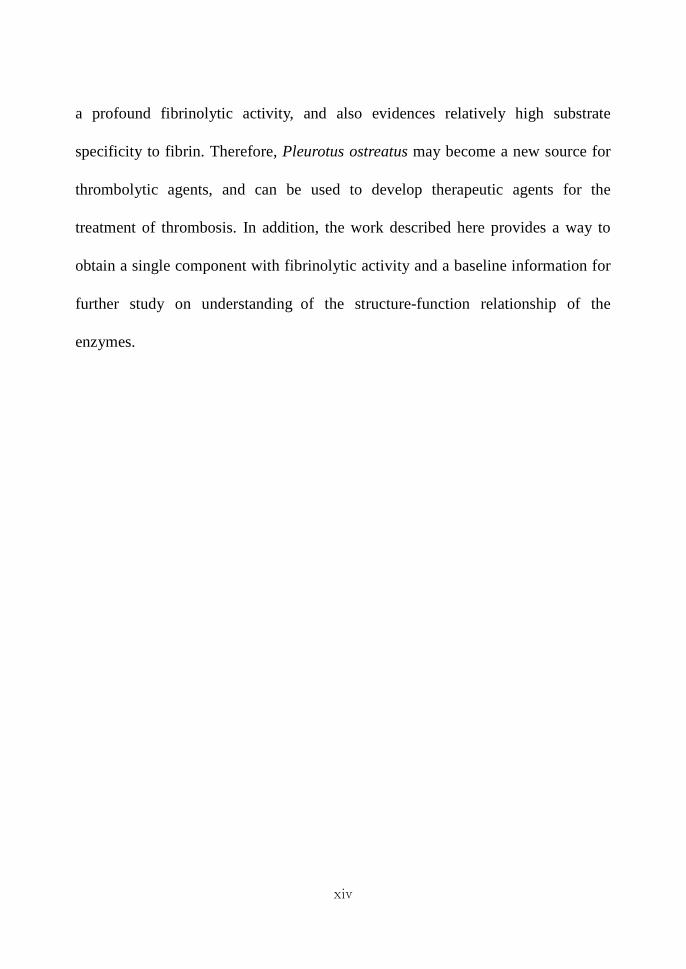
In conclusion the fibrinolytic enzyme purified from Pleurotus ostreatus exhibits
xiv
a profound fibrinolytic activity and also evidences relatively high substrate
specificity to fibrin Therefore Pleurotus ostreatus may become a new source for
thrombolytic agents and can be used to develop therapeutic agents for the
treatment of thrombosis In addition the work described here provides a way to
obtain a single component with fibrinolytic activity and a baseline information for
further study on understanding of the structure-function relationship of the
enzymes
1
ⅠⅠⅠⅠ 서서서서 론론론론
생체내의 세포는 혈액으로부터 영양물질과 산소를 공급받아 대사를 진행한
다 만일 이러한 혈액순환에 장애가 생기면 여러 가지 질병을 초래하게 된다
체내에는 혈액을 응고시키는 blood clotting system과 응고된 혈전을 용해시키
는 fibrinolytic system이 존재하는데 이러한 혈전의 생성과 용해과정은 체내의
조절계에 의하여 균형을 유지하고 있다 혈관이 손상되어 출혈이 일어나면 혈
액응고 기전이 작동하게 되는데 이 과정에서 활성화 된 thrombin에 의해
fibrinogen이 fibrin으로 전환되어 서로 중합체를 형성함으로써 생성된다[1] 형
성된 혈전(fibrin clot)은 상처 회복 후 fibrinolysis라는 체내 혈전용해작용에 의
해 plasmin등과 같은 혈전용해효소(fibrinolytic enzyme)가 활성화되어 혈전을
용해시킴으로써 혈관의 혈액은 다시 정상상태를 유지하게 된다 (Fig 1) 이러
한 혈전형성과 혈전용해계가 불균형을 이루게 되면 협심증 뇌경색 뇌출혈
심근경색과 같은 심혈관계질환을 초래하게 되며 이러한 질환은 경우에 따라서
는 심각한 후유증 및 사망을 일으키게 되는데 이는 전세계 사망원인의 약
29를 차지하고 있다[2](Fig 2) 특히 혈전에 의해 유발되는 혈전증은 체내에
생성된 혈전이 과도하게 축적되거나 혈전용해기작이 원활하게 작동하지 못할
경우 혈관을 따라 흐르면서 여러 가지 혈관계 질환을 유발한다 이러한 혈전형
성의 원인은 혈관내막의 손상 혈액응고능의 항진 정맥혈류 속도의 감소 등
다양한 요인이 작용하고 있다 또한 서구화된 식생활 노령인구의 증가 및 스
트레스의 증가에 따라 이러한 질환은 증가추세를 보이고 있다 따라서 혈관계
질환의 주원인 중 하나인 혈전을 용해하는 혈전용해제에 관한 관심이 점점 더
높아지고 있다
혈전치료에 이용되는 혈전용해효소는 혈전성분인 섬유소(fibrin)를 용해시
시키는 물질로서 작용 기전에 따라 두 가지로 나눌수 있다 즉 혈전용해효소의
2
Fig 1 The pathway of tFig 1 The pathway of tFig 1 The pathway of tFig 1 The pathway of thrombosis and fibrinolysishrombosis and fibrinolysishrombosis and fibrinolysishrombosis and fibrinolysis
3
Cardiovascular diseases
29
Respiratory diseases
14
Diabetes 2
AccidentPoisoningViolence 9
Others 15
Cardiovascular diseases 29
Respiratory diseases 14
Diabetes 2Cancer 12
AccidentPoisoningViolence 9
Infectious diseases
19
Others 15
Fig2 Fig2 Fig2 Fig2 The proportion of deaths by causes in WHO regions estimates The proportion of deaths by causes in WHO regions estimates The proportion of deaths by causes in WHO regions estimates The proportion of deaths by causes in WHO regions estimates for 2000for 2000for 2000for 2000 (WHO (WHO (WHO (WHO
2001)2001)2001)2001)
4
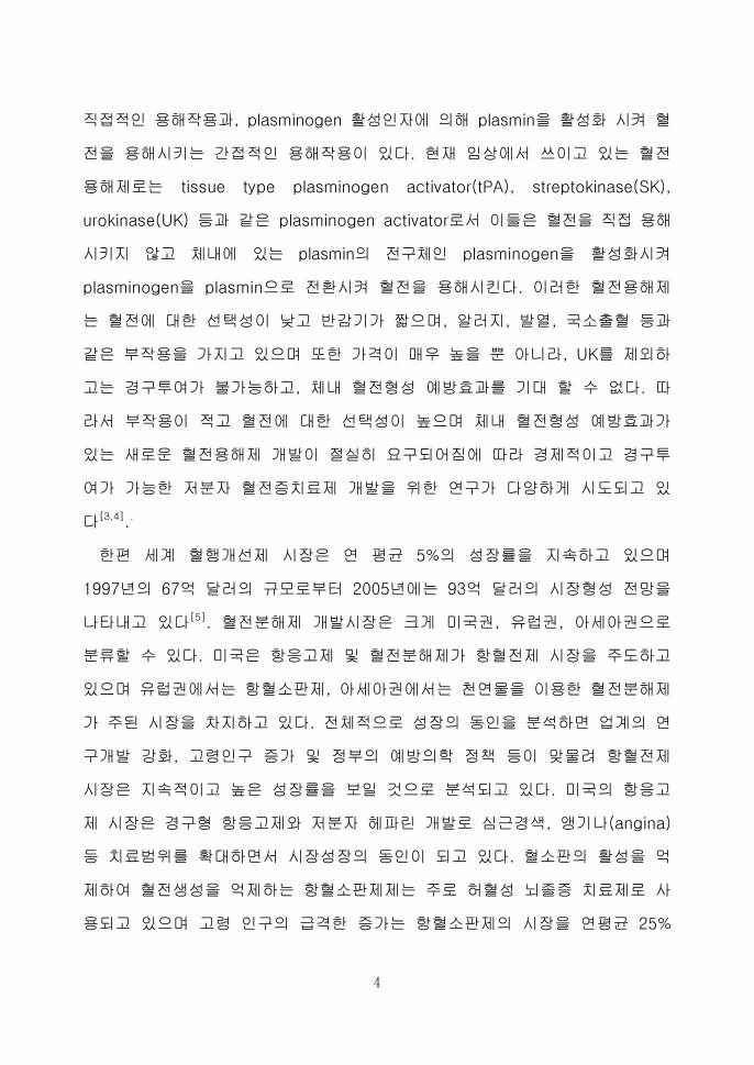
직접적인 용해작용과 plasminogen 활성인자에 의해 plasmin을 활성화 시켜 혈
전을 용해시키는 간접적인 용해작용이 있다 현재 임상에서 쓰이고 있는 혈전
용해제로는 tissue type plasminogen activator(tPA) streptokinase(SK)
urokinase(UK) 등과 같은 plasminogen activator로서 이들은 혈전을 직접 용해
시키지 않고 체내에 있는 plasmin의 전구체인 plasminogen을 활성화시켜
plasminogen을 plasmin으로 전환시켜 혈전을 용해시킨다 이러한 혈전용해제
는 혈전에 대한 선택성이 낮고 반감기가 짧으며 알러지 발열 국소출혈 등과
같은 부작용을 가지고 있으며 또한 가격이 매우 높을 뿐 아니라 UK를 제외하
고는 경구투여가 불가능하고 체내 혈전형성 예방효과를 기대 할 수 없다 따
라서 부작용이 적고 혈전에 대한 선택성이 높으며 체내 혈전형성 예방효과가
있는 새로운 혈전용해제 개발이 절실히 요구되어짐에 따라 경제적이고 경구투
여가 가능한 저분자 혈전증치료제 개발을 위한 연구가 다양하게 시도되고 있
다[34]
한편 세계 혈행개선제 시장은 연 평균 5의 성장률을 지속하고 있으며
1997년의 67억 달러의 규모로부터 2005년에는 93억 달러의 시장형성 전망을
나타내고 있다[5] 혈전분해제 개발시장은 크게 미국권 유럽권 아세아권으로
분류할 수 있다 미국은 항응고제 및 혈전분해제가 항혈전제 시장을 주도하고
있으며 유럽권에서는 항혈소판제 아세아권에서는 천연물을 이용한 혈전분해제
가 주된 시장을 차지하고 있다 전체적으로 성장의 동인을 분석하면 업계의 연
구개발 강화 고령인구 증가 및 정부의 예방의학 정책 등이 맞물려 항혈전제
시장은 지속적이고 높은 성장률을 보일 것으로 분석되고 있다 미국의 항응고
제 시장은 경구형 항응고제와 저분자 헤파린 개발로 심근경색 앵기나(angina)
등 치료범위를 확대하면서 시장성장의 동인이 되고 있다 혈소판의 활성을 억
제하여 혈전생성을 억제하는 항혈소판제제는 주로 허혈성 뇌졸증 치료제로 사
용되고 있으며 고령 인구의 급격한 증가는 항혈소판제의 시장을 연평균 25
5
의 성장률을 보일 것으로 분석하고 있다 특히 일본 한국 중국과 같이 전통
발효식품이 발달된 곳에서는 Natto[67] 청국장[89] 된장[10-12] 젓갈[13-15] 김
치[16] Douchi[1718]
등으로부터 혈전분해물질을 만들어내는 미생물인 Bacillus
sp를 분리하였으며 또한 다양한 버섯[19-23] 지렁이로부터 혈전분해효소를 분리
정제하였다[24-26]
오늘날 많은 종류의 버섯들이 식용 약용 건강음료 기호식품 화장품 및 기
타 산업분야에 이용되고 있다 현재 지구상에는 약 1500종의 버섯이 분류 동
정되어 있는데 그 중 약 50가 식용 가능한 것으로 알려져 있다 버섯은 생
태계에서 유기물을 분해하여 자연계의 평형을 조절할 뿐만 아니라 그 맛과 향
이 좋아 예로부터 인기가 높은 식품중의 하나이다 버섯은 단백질 당질 비타
민 무기질 섬유소 등과 인체에 유용한 여러가지 물질을 함유하고 있으며 광범
위한 약리작용도 나타내므로 성인병 치료에도 많이 이용되고 있다 최근에는 버
섯의 여러 가지 생리활성에 대한 연구 결과가 발표되면서 영양학적인 면은 물론
건강식품으로서의 관심이 높아지고 있다 버섯은 항산화[27-29] 항암[3031] 면역증
강효과[32] 및 콜레스테롤 저하 효과[31]
가 있으며 다양한 버섯류의 균사체 및 균
사체 배양여액으로부터 혈전분해활성을 나타내는 물질을 분리함으로써 기능성
식품 및 의약품 소재로 이용하고자 하는 시도가 활발히 진행되고 있다[1934]
느타리버섯(Pleurotus ostreatus)은 담자균류 주름버섯목 느타리과의 버섯
으로 활엽수 또는 침엽수의 고목에 군생하며 특히 늦가을에 많이 발생한다
갓은 나비 5sim15cm로 반원형 또는 약간 부채꼴이며 가로로 짧은 줄기가
달린다 표면은 어릴 때는 푸른 빛을 띤 검은 색이지만 차차 퇴색하여
잿빛에서 흰빛으로 되며 매끄럽고 습기가 있다 살은 두텁고 탄력이 있으며
흰색이다 주름은 흰색이고 줄기에 길게 늘어져 달려있다 자루는 길이
1sim3cm 굵기 1sim25cm로 흰색이며 밑부분에 흰색 털이 빽빽이 나있다
자루는 옆이나 중심에서 나며 자루가 없는 경우도 있다 포자는 무색의 원기둥
6
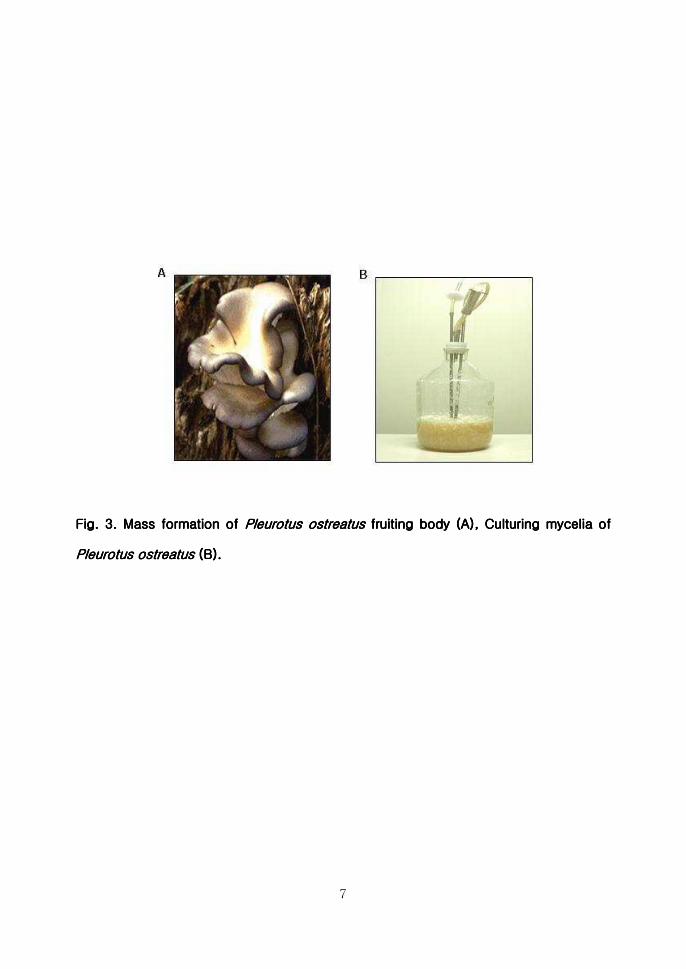
모양이고 포자무늬는 연분홍색을 띤다(Fig 3) 느타리버섯은 잘 알려진 식용
버섯으로 인공재배도 많이 하며 거의 세계적으로 분포한다 또한 우리나라
에서 가장 많이 생산되는 버섯으로서 높은 영양가와 향미를 가지고 있으며
지금까지 알려진 약리작용으로는 항산화작용[35] 항암작용[3637] 콜레스테롤
저하 효과[38] 고혈압 및 당뇨병 예방효과[39] 등이 있으며 또한 느타리버섯
자실체로부터 혈전분해물질을 분리정제한 논문이 보고된 바 있다[40] 이러한
생리활성물질은 모두 느타리버섯 자실체로부터 얻어진 것으로서 본 연구
에서는 자실체보다 훨씬 배양이 용이하고 대량 생산이 가능한 균사체로부터
혈전분해효소를 분리정제하고 그 특성을 연구하였다
또한 최근에는 유전자 조작기술 단백질공학기술 및 기타 생물공학기술의
획기적 발전으로 인하여 종래에 가격적인 차원에서 사용 불가능했던 효소의
산업적 대량생산이 가능하게 되어 사용이 가능하게 되었다 이러한 새로운 기
술에 의해 생산되는 효소는 효소 이용 공정자체가 인류가 요구하는 무공해 저
에너지공정을 실현시킬 수 있을 뿐만 아니라 가격면에서 화학적 및 물리적 방
법으로 생산한 효소보다 상대적으로 저렴한 잇점을 가지고 있기에 이에 따른
기술의 활용 범위도 확대되고 있다 따라서 유전공학기법을 이용하여 효소의
기능성을 기준으로 여러 가지 치료용 약품으로서의 효소의 개발이 많이 연구
되어지고 있다 유전공학 단백질공학 및 효소화학 등 기초학문분야의 눈부신
발전과 생물공정 분야의 기술개발로 효소공학 분야의 응용 가능성이 새로운
각도에서 재조명되고 있다
현재 임상에서 사용되어지는 혈전분해제의 여러 가지 단점을 보완하여 최근
에는 생명공학기술을 이용하여 혈전분해효소들이 생산되고 있는 바 bacteria
또는 yeast등 숙주를 이용하여 된장[41] 지렁이[42-44] Douchi[45] 등으로부터
혈전분해효소를 cloning 하고 expression한 결과가 많이 발표되고 있다 본 연
구에서는 유전공학 기법을 이용하여 고대로부터 식용버섯으로 애용되고 있는
7
Fig Fig Fig Fig 3333 MMMMass formation ass formation ass formation ass formation ofofofof Pleurotus ostreatus Pleurotus ostreatus Pleurotus ostreatus Pleurotus ostreatus fruiting body fruiting body fruiting body fruiting body (A) Culturing mycelia of (A) Culturing mycelia of (A) Culturing mycelia of (A) Culturing mycelia of
Pleurotus ostreatusPleurotus ostreatusPleurotus ostreatusPleurotus ostreatus (B)(B)(B)(B)
8
느타리버섯의 균사체로부터 혈전분해효소를 분리정제하고 그 유전자를 크로닝
하여 발현시켰다
9
ⅡⅡⅡⅡ 재료재료재료재료 및및및및 방법방법방법방법
ⅡⅡⅡⅡ----1 1 1 1 재료재료재료재료
ⅡⅡⅡⅡ----1111----1 1 1 1 균주균주균주균주
본 실험에 사용한 느타리버섯 (Pleurotus ostreatus) 균주는 국립익산대학 특
용작물가공과 균이학 실험실로부터 분양받아 사용하였다
ⅡⅡⅡⅡ----1111----2 2 2 2 시약시약시약시약
Azocasein Trichloroacetic acid (TCA) CM-cellulose Fibrinogen Aprotinin
Pepstatin A Sodium dodecyl sulfate (SDS) Phenylmethylsulfonyl fluoride(PMSF)
Glycine 1-chloro-3-tosylamido-7-amido-2-heptanone (TLCK) N-tosyl-L-
phenylalanyl chloromethyl ketone (TPCK) Trizma base Plasmin Tetrasodium
Ethylenediaminetetraacetate (EDTA) Thrombin Isopropanol Chloroform
Isopropyl-β-D-thiogalactopyranoside (IPTG) Ethanol Ampicillin Kanamycin
등은 Sigma Co(USA) 제품을 Agarose pGEM-T easy vector cloning system
Kit DNA ligase RNase DNase와 Hind III BamHⅠ등과 같은 제한효소는
Promega Co(USA) 제품을 TrizolTM은 Invitrogen Co(USA) 제품을 Ex Taq
polymerase는 TAKARA Co(Japan) 제품을 pQE expression kit Plasmid DNA
isolation kit Ni-NTA spin kit와 DNA gel elution kit는 Qiagen Co(USA) 제품을
Acrylamide Sodium chloride는 Amresco Co(USA)제품을 구입하여 사용하
였으며 lle-Glu-(-OR)-Gly-Arg-pNA(S-2222) H-D-Phe-Pip-Arg-pNA(S-
2238) H-D-Val-Leu-Lys-pNA(2251) H-D-lle-Pro-Arg-pNA (S-2288)
pyro-Glu-Gly-Arg-pNA(S-24444) MeO-Suc-Arg-Pro-Arg-Pro-Tyr-pNA(S-
2586) Z-D-Arg-Gly-Arg-pNA(S-2765)등 chromogenic substrates는
10
Chromogenix Co(Sweden) 제품을 cDNA를 합성하기 위하여 사용한 MMLV
dNTPs RNase inhibitor Taq polymerase DNA size marker와 primer는
Bioneer Co(Korea) 제품을 사용하였다 Protein assay kit와 PVDF membrane
은 Bio-Rad(USA) 제품을 혈전분해효소 정제에 사용되어진 DEAE Sephadex
A-50 Sephadex G-75 HiLoadTM SuperdexTM 75 column은 Amersham
Pharmacia Biotech Co(Sweden) 제품을 구입하여 사용하였으며 기타 시약은
특급을 사용하였다
ⅡⅡⅡⅡ----1111----3 3 3 3 느타리버섯느타리버섯느타리버섯느타리버섯 균사체균사체균사체균사체 배양배양배양배양
균사체 고체배양은 PDB(potato extract 20 dextrose 2) 배지에 한천을
2(wv) 첨가하여 121 15기압에서 20분간 가압살균 후 Petridish (Φ 85
)에 20씩 분주하여 배지를 조제하였다 수집된 균사체 선단을 내경 6의
cork borer로 취하여 조제된 고체배지 중앙에 접종하여 25plusmn1의 항온배양기
에서 배양하였다 균사체 액체배양은 PDB를 조제하여 100 씩 삼각플라스크
에 분주한 후 사용하였다 접종원은 고체배지상의 균사체의 선단을 내경 6
의 cork borer로 취하여 사용하였다 접종 후 진탕배양기에서 25plusmn1 120
rpm으로 7일간 1차 배양(seed culture)한 후 10 liter fermentor에 옮겨서 2차
배양(main culture)을 실시하였다
ⅡⅡⅡⅡ----2 2 2 2 혈전분해효소의혈전분해효소의혈전분해효소의혈전분해효소의 분리분리분리분리 정제정제정제정제
ⅡⅡⅡⅡ----2222----1 1 1 1 조추출물조추출물조추출물조추출물 조제조제조제조제
느타리버섯 균사체를 멸균수로 수차례 세척한 후 액체질소를 이용하여 세포
벽을 파쇄한 후 Ultrasonicator(Sonics amp Materials lnc USA)로 균질화하여
6000x g 에서 원심분리한 후 상층액을 회수하여 조추출물로 사용하였다
11
ⅡⅡⅡⅡ----2222----2 2 2 2 단백질분해활성단백질분해활성단백질분해활성단백질분해활성 및및및및 혈전분해활성혈전분해활성혈전분해활성혈전분해활성 검색검색검색검색
03 skim milk 용액 5에 동량의 2 agarose 용액 5을 첨가하여 혼합
하고 1시간 동안 실온에서 고화시켜 skim milk plate를 제조하였다 단백질 분
해작용을 검색하기 위하여 skim milk plate에 지름 6의 구멍을 만들어 버섯
조추출물 10을 침적하였다 37에서 12시간 반응시켜 생성된 투명대로 단
백질분해활성을 확인하였다
혈전분해활성은 Astrup와 Mullertz[46]의 Fibrin agarose plate방법을 이용하
여 측정하였다 fibrinogen을 03가 되도록 5 완충용액(50mM Tris-HCl
015 M NaCl pH 74)에 용해시킨 후 동량의 2 agarose 용액을 첨가하여 혼
합 시킨 후 thrombin 1 unit를 첨가하여 15 두께로 fibrin agarose plate를
제조하였다 fibrin agarose plate는 1시간 동안 실온에서 고화시킨 후 혈전분해
활성을 측정하기 위해 fibrin plate에 내경 6의 구멍을 만들어 여기에 균사체
조추출액 10을 침적하고 37에서 12시간 반응하여 형성된 투명대로 혈전
분해활성을 확인하였다
ⅡⅡⅡⅡ----2222----3 3 3 3 조단백질조단백질조단백질조단백질 분리분리분리분리
회수된 버섯 균사체에 동량의 멸균수를 가하여 Ultrasonicator을 사용하여
10분간 파쇄 후 4 6000x g 30분 동안 원심분리시켜 상층액을 회수하였다
회수된 상층액에 -70로 전처리한 100 에탄올을 천천히 가하여 최종적으로
에탄올 농도를 50로 조정한 후 1시간 동안 4에서 교반하여 12000x g 30
분 동안 원심분리 후 상층액을 회수하였다 회수된 상층액에 -70로 처리된
동량의 100 에탄올을 천천히 가하여 최종적으로 에탄올 농도를 75로 조정
하였고 1시간 동안 4에서 교반한 후 4 12000x g 30분 동안 원심분리 후
형성된 침전물을 10mM Citrate-NaOH (pH 60)완충용액에 현탁하였다 또한
12
불순물을 제거하기 위하여 microcentrifuge를 이용하여 10000rpm 4 10분
간 원심분리하였고 상층액을 회수한 후 시료로 사용하였다
ⅡⅡⅡⅡ----2222----4444 단백질단백질단백질단백질 정량정량정량정량
효소의 단백질 정량은 Lowry[47]등의 방법에 의한 BCA단백질정량 kit (Pierce
Co USA)를 사용하여 측정하였으며 표준단백질은 Bovine Serum Albumin
(BSA)을 사용하였다
ⅡⅡⅡⅡ----2222----5555 단백질분해활성단백질분해활성단백질분해활성단백질분해활성 측정측정측정측정
단백질분해효소 활성은 Azocasein assay법[48]을 이용하였다 Azocasein
assay법은 azocasein이 분해되어 나오는 acid soluble material양을 측정
함으로서 단백질분해활성을 판정하였다 01 Azocasein용액 (50mM Tris-HCl
pH 70) 300에 단백질분획 50를 첨가하여 37에서 20분간 반응시킨 후
미리차게 한 10(wv) trichloroacetic acid (TCA) 600를 넣고 즉시 혼합
시켰다 이 시료들을 10분 동안 얼음에 방치하여 반응을 정지시킨 후
microcentrifuge에서 12000rpm 4로 15분간 원심분리하였다 그리고 상층
액을 366nm에서 OD값을 측정하여 단백질분해활성을 측정하였다
ⅡⅡⅡⅡ----2222----6666 CationCationCationCation----exchange chromatographyexchange chromatographyexchange chromatographyexchange chromatography
CM-Cellulose column (35times10)을 01N NaOH로 활성화시킨 후 10mM
citrate-NaOH pH 60 완충용액으로 평형화시켰다 이 column에 조단백질을
주입하여 0~10M NaCl linear gradient를 형성시켜 단백질을 용출하였다
용출속도는 10min으로 수행하였으며 분획당 15을 회수하였다
ⅡⅡⅡⅡ----2222----7777 AnionAnionAnionAnion----exchange chromatographyexchange chromatographyexchange chromatographyexchange chromatography
13
DEAE Sephadex A-50 column (35times10)을 20 mM Tris-HCl(PH 80) 완충
용액으로 평형화시킨 후 cation-exchange chromatography에서 얻은 활성분
획을 주입하여 anion-exchange chromatography를 수행하였다 0~10 M NaCl
linear gradient를 형성시켜 10min 속도로 단백질을 용출하였으며 분획당
15을 회수하였다
ⅡⅡⅡⅡ----2222----8888 GelGelGelGel----filtration filtration filtration filtration chromatographychromatographychromatographychromatography
Sephadex G-75 column(15times130)을 015M NaCl를 함유한 005M
phosphate(pH 72) 완충용액으로 평형화시킨 후 anion-exchange chromato-
graphy에서 얻은 활성분획을 농축 투석하여 주입하였다 동일한 완충용액으로
01min의 속도로 단백질을 용출하였으며 15씩 분획을 회수하였다
ⅡⅡⅡⅡ----2222----9 Fast protein liquid chromatography (FPLC)9 Fast protein liquid chromatography (FPLC)9 Fast protein liquid chromatography (FPLC)9 Fast protein liquid chromatography (FPLC)
HiLoad 1660 Superdex 75 pg column을 이용하여 005M phosphate015M
NaCl pH 72 완충용액을 이용하여 bed volume의 2배를 흘려 보낸 후 Gel-
fitration chromatography에서 얻은 활성분획을 주입하여 10min의 속도로
ACTA FPLC (Pharmacia Co)를 수행하여 혈전분해효소를 분리정제하였다
ⅡⅡⅡⅡ----2222----10101010 혈전분해효소의혈전분해효소의혈전분해효소의혈전분해효소의 분리분리분리분리
혈전분해활성 측정법과 단백질분해활성 측정법에 의하여 단백질활성을 측정
함으로써 혈전분해효소를 분리하였다 먼저 분획들을 fibrin agarose plate에
20씩 침적하여 형성된 투명환의 크기를 확인하였다 그리고 활성있는 부분
을 취하여 Azocasein assay법으로 단백질분해활성을 측정하여 수치화하였다
ⅡⅡⅡⅡ----3 3 3 3 분자량분자량분자량분자량 분석분석분석분석
14
ⅡⅡⅡⅡ----3333----1 1 1 1 FPLC FPLC FPLC FPLC를를를를 이용한이용한이용한이용한 분자량분자량분자량분자량 측정측정측정측정(size exclusion)(size exclusion)(size exclusion)(size exclusion)
정제된 혈전분해효소의 분자량을 확인하기 위하여 glyceraldehydes-3-
phosphate dehydrogenase(36kDa) carbonic anhydrase(29kDa) trypsinogen
-PMSF(24kDa) trypsin-inhibitor(201kDa) 등 standard marker를 이용하여
size exclusion을 수행하였다 정제한 혈전분해효소와 standard marker를
동일한 조건으로 용출시켜 정제한 효소의 분자량을 확인하였다
ⅡⅡⅡⅡ----3333----2222 SDS SDS SDS SDS----PAGEPAGEPAGEPAGE를를를를 이용한이용한이용한이용한 분자량분자량분자량분자량 측정측정측정측정
분리한 단백질은 SDS-PAGE[49]를 수행하여 분자량을 측정하였다 전기영
동은 12의 separating gel과 5 stacking gel로 이루어진 SDS-PAGE gel
상에서 진행하였다 단백질과 5x SDS Sample Buffer (60mM Tris-HCl pH 68
25 glycerol 2 SDS 144mM β-mercaptoethanol 01 bromophenol
blue)를 혼합하여 100에서 7분간 중탕한 후 Protein marker(Cambrex Co)와
함께 전기영동을 수행하였다 전기영동된 gel은 Coomassie brilliant blue(CBB)
R 250으로 염색하고 Destaining buffer로 탈색시킴으로써 단백질 밴드를
확인하였다 그리고 Protein marker의 이동거리(Rf)를 횡축으로 marker 분자
량의 log함수를 종축으로 standard curve를 만든 후 정제된 효소의 이동거리
에 근거하여 효소의 분자량을 측정하였다
ⅡⅡⅡⅡ----3333----3333 SDSSDSSDSSDS----fibrinfibrinfibrinfibrin zymographyzymographyzymographyzymography를를를를 이용한이용한이용한이용한 분자량분자량분자량분자량 측정측정측정측정
SDS-fibrin zymography 활성확인법은 Kim[50]등의 방법에 의하여 fibrinogen
농도가 012(WV)가 되게 12 polyacrylamide 용액에 혼합한 후 thrombin
(01microU)과 NNNacuteNacute-tetramethylethylenediamine(TEMED)를 첨가하여 gel
을 제조하고 전기영동을 수행하였다 단백질의 활성을 보존하고 전기영동 과정
에서 일어나는 분해반응을 방지하기 위해 4에서 90~180V로 진행하였다
15
그런 다음 SDS에 의해 불활성화된 효소를 재활성화 시키기 위하여 gel을
25 Triton X-100을 포함한 Tris-HCl (20mM pH 72)에서 30분간 진탕시켜
SDS를 제거한 후 다시 증류수로 수 차례 세척하여 Triton X-100을 제거하였
다 그리고 gel을 활성반응 완충용액 (015M NaCl 20mM Tris-HCl pH 72)
으로 15분씩 2번 세척한 후 그 완충용액에 넣어 37 항온기에서 15시간
반응을 시켰다 반응이 끝난 후 gel을 CBB R 250으로 염색하고 Destaining
buffer로 탈색시킴으로써 투명하게 나타난 fibrin 분해능을 지닌 단백질의 활성
부위를 확인하였다
ⅡⅡⅡⅡ----4444 NNNN----말단말단말단말단 아미노산아미노산아미노산아미노산 서열분석서열분석서열분석서열분석
정제된 단백질을 12 gel 상에서 SDS-PAGE 전기영동을 수행한 후
polyvinylidene difluoride(PVDF) membrane에 transfer하였고 ponceau S로
염색하여 transfer 유무를 확인하였다 활성부위를 PVDF membrane에서
잘라내어 기초과학지원연구소 광주분소에 의뢰하여 Edman 방법으로 N-
terminal sequencing analysis를 수행하였다 N-terminal sequence는 National
Center for Biotechnology information(NCBI)의 BLAST에 등록되어 있는
느타리버섯 자실체 유래의 metalloendoprotease(PoMTP) 아미노산 서열과 비교
하였다
ⅡⅡⅡⅡ----5555 정제효소의정제효소의정제효소의정제효소의 특성특성특성특성 분석분석분석분석
ⅡⅡⅡⅡ----5555----1111 정제효소의정제효소의정제효소의정제효소의 기질특이성기질특이성기질특이성기질특이성 분석분석분석분석
정제효소의 기질특이성을 분석하기 위하여 다양한 chromogenic substrate
(Table 1)를 이용하여 amidolytic activity를 측정하였다 사용되어진 chromogenic
16
substrate는 chromogen 사용법에 따라 각각 조제한 후 정제된 혈전분해효소
1을 첨가하여 37에서 5분간 반응시켰고 ELISA reader를 사용하여
405nm에서 방출되어진 ρ-nitroaniline의 양을 측정하였다
ⅡⅡⅡⅡ----5555----2 2 2 2 효소활성에효소활성에효소활성에효소활성에 대한대한대한대한 금속이온금속이온금속이온금속이온 및및및및 단백질분해효소단백질분해효소단백질분해효소단백질분해효소 억제제의억제제의억제제의억제제의 영향영향영향영향
정제한 효소의 활성에 대하여 금속이온이 미치는 영향을 알아보기 위해
CaCl2 CoCl2 CuCl2 FeCl2 MnCl2 MgCl2 ZnCl2 등 이온들이 각각 농도가
10mM이 되도록 20mM Tris-HCl(pH 74) 완충용액에 용해시켜 사용하였다
일정양의 효소용액과 혼합한 후 최종농도가 1mM이 되도록 하여 37
항온기에서 1시간 동안 반응시켰다 그리고 azocasein assay를 수행하고
control(20mM Tris-HCl pH 74)에 대한 상대적 활성을 이용하여 혈전분해
효소활성에 대한 영향을 비교하였다 또한 효소활성에 대한 단백질 저해제의
영향을 알아보기 위해 PMSF TLCK TPCK aprotinin EDTA pepstatin A 등
저해제와 효소용액을 혼합하여 37 항온기에서 1시간 동안 반응시킨 후 같은
방법으로 azocasein assay를 수행하여 효소활성에 대한 영향을 비교하였다
ⅡⅡⅡⅡ----5555----3 3 3 3 효소활성에효소활성에효소활성에효소활성에 대한대한대한대한 pH pH pH pH의의의의 영향영향영향영향
효소반응의 최적 pH조건을 검토하기 위해 정제된 혈전용해효소를 각 pH 완
충용액에 동량으로 가하여 37 항온기에서 60분간 반응시킨 후 효소활성을
비교하였다 이때 사용한 완충용액은 혈전분해활성에 대한 영향을 최소화하기
위하여 05M Glycine-HCl (pH 30) 05M Acetate (pH 40~50) 05M Tris-
HCl (pH 60~80) 05M Glycine-NaOH (pH 90~100) 완충용액을 사용하였
다 항온기에서 60분 반응시킨 후 azocasein assay를 수행하여 pH에 의한 혈
전분해효소의 활성을 측정하였다
17
Table 1 List of synthetic chromogenic substrates
Chromogenic
substrates Amino acid sequence Characteristics
S-2222 Bz-Ile-Glu-(OR)-Gly-Arg-pNA for factor Xa
S-2288 H-D-Ile-Pro-Arg-pNA for t-PA
S-2238 H-D-Phe-Pip-Arg-pNA for thrombin
S-2251 H-D-Val-Leu-Lys-pNA for plasmin and
streptokinase-activated
S-2444 pyroGlu-Gly-Arg-pNA for urokinase
S-2586 MeO-Suc-Arg-Pro-Tyr-pNAmiddotHCl for chymotrypsin
S-2765 Z-D-Arg-Gly-Arg-pNAmiddot2HCl for factor Xa
18
ⅡⅡⅡⅡ----5555----4 4 4 4 효소효소효소효소활성활성활성활성에에에에 대한대한대한대한 온도의온도의온도의온도의 영향영향영향영향
분리된 단백질의 열 안정성을 알아보기 위하여 효소액을 20~90에서 10
간격의 온도에서 30분씩 열처리시킨 후 azocasein assay를 수행하여 온도에
의한 혈전분해효소의 활성을 측정하였다
ⅡⅡⅡⅡ----5555----5 Fibrin 5 Fibrin 5 Fibrin 5 Fibrin 및및및및 FFFFibrinogenibrinogenibrinogenibrinogen에에에에 대한대한대한대한 분해분해분해분해특성특성특성특성 분석분석분석분석
반응시간에 따른 Fibrin의 분해특성은 Datta[51]등의 방법을 이용하여 확인하
였다 5 fibrinogen을 함유한 용액(20mM Tris-HCl pH 74 015M NaCl)에
thrombin(01NIH unit)을 첨가하여 상온에서 1시간동안 반응시켜 fibrin을
조제하였다 그리고 동일 양의 효소를 처리하여 37 항온기에서 시간별로
반응시킨 후 12 SDS-PAGE를 수행하여 fibrin의 분해패턴을 확인하였다
Matsubara[52]등의 방법을 이용하여 Fibrinogen에 대한 분해특성을 확인하
였다 Fibrinogen(5)에 동일 양의 효소를 처리하여 시간별로 37 항온기
에서 반응시킨 후 위와 같은 방법으로 SDS-PAGE를 수행하여 분해패턴을
확인하였다
ⅡⅡⅡⅡ----6666 Total RNA isolation Total RNA isolation Total RNA isolation Total RNA isolation
Total RNA를 분리하기 위하여 모든 시약과 초자기구류는 diethylpyrocar-
bonate (DEPC)로 처리하였고 RNA는 TriZolTM reagent를 이용하여 분리하였다
느타리버섯 균사체 100에 TriZolTM reagent 1을 첨가시키고 5분간 반응시
켜 균질화한 후 02 Chloroform을 혼합하여 실온에서 5분간 반응시킨 후
12000X g로 4에서 15분간 원심분리하였다 상층액을 새로운 eppendorf
tube에 옮긴 후 05의 isopropanol을 첨가하여 실온에서 15분간 반응시켰다
12000times g로 4에서 10분간 원심분리하여 얻은 침전물에 75 에탄올 1
19
을 첨가하여 혼합한 후 7500times g로 원심분리하여 RNA pellet를 얻었다 이
RNA pellet를 공기중에서 건조시켰으며 RNase free-water에 녹여 purity를 측
정한 후 -20에 보관하여 cDNA 합성에 이용하였다
ⅡⅡⅡⅡ----7777 혈전분해효소혈전분해효소혈전분해효소혈전분해효소 유전자에유전자에유전자에유전자에 대한대한대한대한 특이적특이적특이적특이적 primer primer primer primer의의의의 제작제작제작제작
NCBI에 등록되어 있는 유전자은행을 검색하여 정제한 느타리버섯 균사체로
부터 분리되어진 혈전분해효소의 N-말단 아미노산서열과 느타리버섯 자실체
로부터 분리되어진 metalloprotease유전자(AY640032)의 염기서열을 비교분석
하였고 start codon부위부터 stop codon부위까지 PCR을 수행 할 수 있도록
Bioneer(Korea)에 의뢰하여 primer를 제작하였다 Sense primer는 5 end에
BamHⅠ restriction site를 첨가하여 5-GGATCCATGTTGCGCTCCATCCTGTT
AATTG-3로 하였고 antisense primer는 stop codon 뒤에 Hind Ⅲ restriction
site를 첨가하여 5-AAGCTTTTACACTGGTGCTGCTACCCTGGC-3로 하였다
ⅡⅡⅡⅡ----8888 Reverse transcriptionReverse transcriptionReverse transcriptionReverse transcription----polymerase chain reaction (RTpolymerase chain reaction (RTpolymerase chain reaction (RTpolymerase chain reaction (RT----PCR)PCR)PCR)PCR)
느타리버섯 균사체로부터 추출된 Total RNA와 oligo (dT)18 및 MMLV
reverse transcriptase을 혼합하여 역전사 반응을 하였다 2 total RNA와
10pmol oligo (dT)를 65에서 10분 동안 반응시킨 후 dNTPs RNase inhibittor
MMLV reverse transcriptase등 mixture와 42에서 한 시간 동안 역전사반응
을 하였다 합성한 cDNA는 -20에 보관하면서 사용하였다
합성된 cDNA는 제작한 primer를 이용하여 940에서 5분간 반응시킨 후
denaturation은 940에서 1분 annealing은 630에서 30초 extension은
720에서 1분씩 35cycle로 하여 PCR을 수행하였다 PCR product는 12
20
agarose gel로 전기영동하였고 ethidium bromide(EtBr)로 염색한 후 확인
하였다
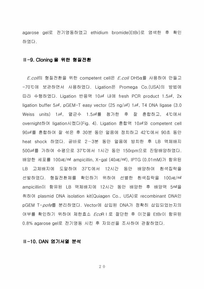
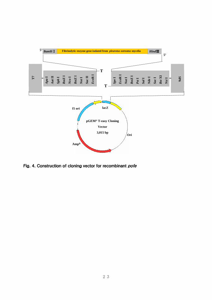
ⅡⅡⅡⅡ----9999 CCCCloningloningloningloning을을을을 위한위한위한위한 형질전환형질전환형질전환형질전환
Ecoli의 형질전환을 위한 competent cell은 Ecoli DH5α를 사용하여 만들고
-70에 보관하면서 사용하였다 Ligation은 Promega Co(USA)의 방법에
따라 수행하였다 Ligation 반응액 10 내에 fresh PCR product 15 2x
ligation buffer 5 pGEM-T easy vector (25 ng) 1 T4 DNA ligase (30
Weiss units) 1 멸균수 15를 첨가한 후 잘 혼합하고 4에서
overnight하여 ligation시켰다(Fig 4) Ligation 혼합액 10와 competent cell
90를 혼합하여 잘 섞은 후 30분 동안 얼음에 정치하고 42에서 90초 동안
heat shock 하였다 곧바로 2sim3분 동안 얼음에 방치한 후 LB 액체배지
500를 가하여 수평으로 37에서 1시간 동안 150rpm으로 진탕배양하였다
배양한 세포를 100 ampicillin X-gal (40) IPTG (001mM)가 함유된
LB 고체배지에 도말하여 37에서 12시간 동안 배양하여 흰색집락을
선발하였다 형질전환체를 확인하기 위하여 선별한 흰색집락을 100
ampicillin이 함유된 LB 액체배지에 12시간 동안 배양한 후 배양액 5을
취하여 plasmid DNA isolation kit(Quiagen Co USA)로 recombinant DNA인
pGEM T-pofe를 분리하였다 Vector에 삽입된 DNA가 정확히 삽입되었는지의
여부를 확인하기 위하여 제한효소 EcoRⅠ로 절단한 후 이것을 EtBr이 함유된
08 agarose gel로 전기영동 시킨 후 자외선을 조사하여 관찰하였다
ⅡⅡⅡⅡ----10101010 DAN DAN DAN DAN 염기서열염기서열염기서열염기서열 분석분석분석분석
21
클로닝된 vector는 SP6 primer를 사용하여 염기서열을 결정하였다 염기
서열분석은 기초과학지원연구소 광주분소에 의뢰하여 수행하였다 Automatic
DNA sequencer(model ABI PRISM 377 Perkin Elmer USA)을 이용하여
DNA 염기서열을 결정하였으며 확인된 염기서열은 National Center for
Biotechnology Information(NCBI)의 BLAST에서 느타리버섯 자실체유래의
metalloendoprotease(PoMTP)의 염기서열과 비교하여 분석하였다
ⅡⅡⅡⅡ----11111111 Construction of Construction of Construction of Construction of expression vector for recombinant fibrinolytic expression vector for recombinant fibrinolytic expression vector for recombinant fibrinolytic expression vector for recombinant fibrinolytic
emzymeemzymeemzymeemzyme
pGEM T-pofe를 BamHⅠ Hind Ⅲ로 절단하여 얻은 PoFE 유전자를 동일
한 효소로 처리한 pQE30 expression vertor에 ligation 하였다(Fig 5) Ligation
반응액 10내에 pGEM T-pofe를 제한효소 BamHⅠ Hind Ⅲ로 절단하여
얻은 insert DNA 3 2x ligation buffer (250mM Tris-HCl pH 78 100mM
MgCl2 20 mM DTT 4 mM ATP)5 pQE30 vector (25ng) 1에 T4 DNA
ligase (30 Weiss units) 1를 첨가한 후 잘 혼합하여 4에서 overnight하여
ligation시켰다 Ligation product인 pQE30-pofe를 Ecoli DH5α에 형질
전환하여 증폭시켰다 얻어진 재조합 vector들은 DNA sequence analysis를
하여 원하는 단편이 cloning 되었음을 확인하였다 발현을 위한 숙주세포는 E
coli M15[pREP4]를 사용하였다 pQE30-pofe 10와 competent cell 90를
혼합하여 잘 섞은후 30분 동안 얼음에 정치하고 42에서 90초 동안 heat
shock 하였다 곧바로 2~3분 동안 얼음에 방치하고 LB 500를 가하여
수평으로 37에서 1시간 동안 150rpm으로 진탕배양하였다 배양한 세포를
100 ampicillin 25 kanamycin이 함유된 LB 고체배지에 도말하여
37에서 12시간 동안 배양하였다 몇 개의 colony를 취하여 액체배양하여
22
plasmid를 isolation한 후 각 primer로 PCR하여 recombinant strain을
확인하였다
ⅡⅡⅡⅡ----12 Expression and purification of fibrinolytic enzyme12 Expression and purification of fibrinolytic enzyme12 Expression and purification of fibrinolytic enzyme12 Expression and purification of fibrinolytic enzyme
단일 colony를 취하여 10 배지(25 kanamycin과 100 ampicillin
을 함유)에 12시간 동안 배양한 후 500를 취하여 30 배지 (25
kanamycin과 100 ampicillin을 함유)에 접종하였다 37에 서 OD600값이
05-07이 될 때까지 진탕배양한 후 최종농도가 lmM이 되도록 IPTG를
첨가하여 4시간 동안 배양하였다 배양액을 4000x g 에서 15분 동안
원심분리하여 세포를 모은 후 1 lysis buffer (50mM NaH2PO4 300mM NaCl
10 mM imidazole pH 80)로 세포를 현탁시킨 후 lysozyme농도가 10
이 되도록 첨가하여 ice에서 30분간 반응시켰다 세포를 sonicator로 파쇄한
후 4 10000x g 에서 30분 동안 원심분리한 후 상층액을 회수하였다 lysis
buffer로 평형화시킨 Ni-NTA spin column에 상층액을 주입하여 700x g 2분
동안 원심분리하여 여과액을 수집한 후 SDS-PAGE 분석에 사용하였다 또한
700x g 2분 동안 원심분리하는 방법으로 600 washing buffer (50mM
NaH2PO4 300mM NaCl 20mM imidazol pH 80)를 사용하여 Ni-NTA spin
column을 두번 washing한 후 200 elution buffer (50mM NaH2PO4 300mM
NaCl 250mM imidazol pH 80)로 6X His-tagged protein을 함유한 column을
두차례 elution하여 분액을 수집하고 SDS-PAGE를 수행하였다 각 단계에서
얻은 분액 10에 5x SDS-PAGE sample buffer 25 를 넣고 7분 동안 끓인
후 SDS-PAGE를 수행하였다
23
AmpR
pGEMreg T-easy Cloning
Vector
3015 bpOri
f1 ori lacZ
T7
Apa
I
Aat
II
Sph
I
Bst
ZI
Nco
I
Bst
ZI
Not
I
Sac
II
Eco
RI S
P6
Spe
I
Eco
RI
Not
I
Bst
ZI
Pst
I
SalI
Nde
I
Sac
I
Bst
XI
Nsi
I
T
T
Fibrinolytic enzyme gene isolated from pleurotus ostreatus myceliaBamHⅠⅠⅠⅠ HindⅢⅢⅢⅢ5rsquo
5rsquo
Fig Fig Fig Fig 4444 Construction of Construction of Construction of Construction of cloning vcloning vcloning vcloning vector for recombinant ector for recombinant ector for recombinant ector for recombinant pofepofepofepofe
24
3-CGGTCCCATCGTCGTGGTCACATTTTCGAA-5 RE
Fibrinolytic enzyme gene isolated from pleurotus ostreatus myceliaBamHI HindⅢⅢⅢⅢ
FE 5-GGATCCATGTTGCGCTCCATCCTGTTAATTG-3
pQE-30 Expression vector
3461 bp
Col E1 Ori
AmpR
PT5 Lac O Lac O RBS ATG 6xHis MCS Stop
Bam
HI
SphI
SacL
Kpn
I
SmaI
SalI
Pst
I
Hin
d ⅢⅢ ⅢⅢ
Fig Fig Fig Fig 5555 Construction of expression vector for recombinant Construction of expression vector for recombinant Construction of expression vector for recombinant Construction of expression vector for recombinant pofepofepofepofe
25
ⅡⅡⅡⅡ----11113333 SDSSDSSDSSDS----PAGEPAGEPAGEPAGE
조제한 단백질은 SDS-PAGE[49]를 수행하여 분자량을 분석하였다 전기영
동은 12의 separating gel과 5 stacking gel로 이루어진 SDS-PAGE gel
상에서 진행하였다 단백질과 5x SDS Sample Buffer (60mM Tris-HCl pH 68
144mM β-mercaptoethanol 25 glycerol 2 SDS 01 bromophenol
blue)를 혼합하여 100에서 7분간 중탕한 후 protein marker (Cambrex co
USA)와 함께 전기영동을 수행하였다 전기영동된 gel은 CBB R 250으로
염색하고 destaining buffer로 탈색시킴으로써 단백질 밴드를 확인하였다
ⅡⅡⅡⅡ----11114444 Western blot analysisWestern blot analysisWestern blot analysisWestern blot analysis
전기영동후 단백질을 100V에서 90분간 PVDF membrane에 transfer하였다
Skim milk를 농도가 5가 되도록 TBST용액 (20mM Tris-HCl pH 75 137mM
NaCl 01 Tween20)에 녹여 blocking solution을 제조하여 membrane을
담그고 한 시간 동안 shaker 위에 올려 놓고 흔들어 주었다 한시간 후 TBST
용액으로 15분씩 4회 세척한 후 11000 비율로 TBST 용액에 희석한 6X His-
tag antibody로 4에서 12시간 처리 후 동일한 방법으로 15분씩 4회 세척한
후 11000의 비율로 TBST 용액에 희석한 alkaline phosphatase conjugated
antibody(sedondary antibody)로 실온에서 2시간 처리하였다 alkaline
phosphatase secondary antibody에 의한 반응을 확인하기 위하여
NBTBCIP를 처리하여 단백질 band를 확인하였다
26
ⅢⅢⅢⅢ 결과결과결과결과
ⅢⅢⅢⅢ----1 1 1 1 단백질분해활성단백질분해활성단백질분해활성단백질분해활성 및및및및 혈전분해활성혈전분해활성혈전분해활성혈전분해활성
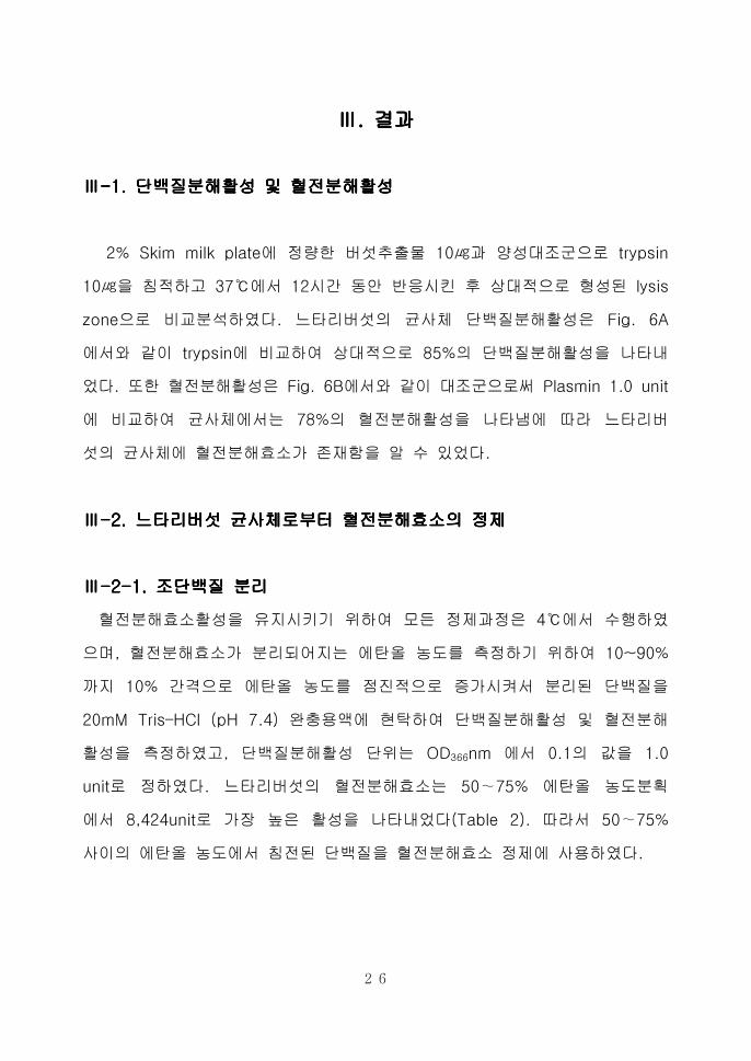
2 Skim milk plate에 정량한 버섯추출물 10과 양성대조군으로 trypsin
10을 침적하고 37에서 12시간 동안 반응시킨 후 상대적으로 형성된 lysis
zone으로 비교분석하였다 느타리버섯의 균사체 단백질분해활성은 Fig 6A
에서와 같이 trypsin에 비교하여 상대적으로 85의 단백질분해활성을 나타내
었다 또한 혈전분해활성은 Fig 6B에서와 같이 대조군으로써 Plasmin 10 unit
에 비교하여 균사체에서는 78의 혈전분해활성을 나타냄에 따라 느타리버
섯의 균사체에 혈전분해효소가 존재함을 알 수 있었다
ⅢⅢⅢⅢ----2 2 2 2 느타리버섯느타리버섯느타리버섯느타리버섯 균사체로부터균사체로부터균사체로부터균사체로부터 혈전분해효소혈전분해효소혈전분해효소혈전분해효소의의의의 정제정제정제정제
ⅢⅢⅢⅢ----2222----1111 조단백질조단백질조단백질조단백질 분리분리분리분리
혈전분해효소활성을 유지시키기 위하여 모든 정제과정은 4에서 수행하였
으며 혈전분해효소가 분리되어지는 에탄올 농도를 측정하기 위하여 10~90
까지 10 간격으로 에탄올 농도를 점진적으로 증가시켜서 분리된 단백질을
20mM TrisndashHCl (pH 74) 완충용액에 현탁하여 단백질분해활성 및 혈전분해
활성을 측정하였고 단백질분해활성 단위는 OD366nm 에서 01의 값을 10
unit로 정하였다 느타리버섯의 혈전분해효소는 50sim75 에탄올 농도분획
에서 8424unit로 가장 높은 활성을 나타내었다(Table 2) 따라서 50sim75
사이의 에탄올 농도에서 침전된 단백질을 혈전분해효소 정제에 사용하였다
27
Realtiv
epro
teoly
tic
activity (
)
0
20
40
60
80
100
120
Trypsin Crude sample
AAAA
Realtiv
efibrinoly
tic
activity (
)
0
20
40
60
80
100
120
Plasmin Crude sample
BBBB
Fig Fig Fig Fig 6666 The assay of proteolytic and fibrinolytic activity from The assay of proteolytic and fibrinolytic activity from The assay of proteolytic and fibrinolytic activity from The assay of proteolytic and fibrinolytic activity from crude crude crude crude extracts ofextracts ofextracts ofextracts of
Pleurotus ostreatusPleurotus ostreatusPleurotus ostreatusPleurotus ostreatus myceliummyceliummyceliummycelium A Proteolytic activity was measured by using skim
milk plate B Fibrinolytic activity was measured by using fibrin agarose plate 20
of crude extract was placed in each hole and incubated at 37 for 12 hrs Tripsin
and Plasmin are used as positive control
28
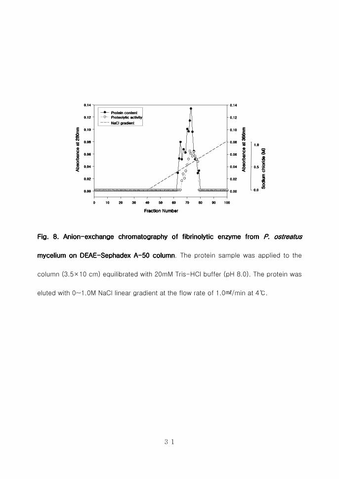
ⅢⅢⅢⅢ----2222----2 2 2 2 혈전분해효소의혈전분해효소의혈전분해효소의혈전분해효소의 정제정제정제정제
Cation-exchange chromatography를 수행하기 위하여 CM-cellulose를 이용
하여 10mM Citrate-NaOH (pH 60)으로 평형화한 후 0~10M NaCl (pH 60)로
평형화한 후 0~10M NaCl (pH 60) 농도구배를 형성시켜 10min으로
분획하였으나 결합이 이루어지지 않고 flow-through로 용출되어 anion-
exchange chromatography를 수행하였다(Fig 7) 양이온 교환수지로부터 회수
한 농축된 시료를 삽입시켜 0~10M NaCl 농도구배를 형성시켜 활성분획을
분리한 후 280nm에서 단백질함량을 측정하였고 azocasein assay를 수행하여
활성분획을 분리하였다(Fig 8) DEAE sephadex A-50 column chromato-
graphy를 수행한 결과 약 08M의 NaCl 농도에서 혈전분해효소들이 정제됨을
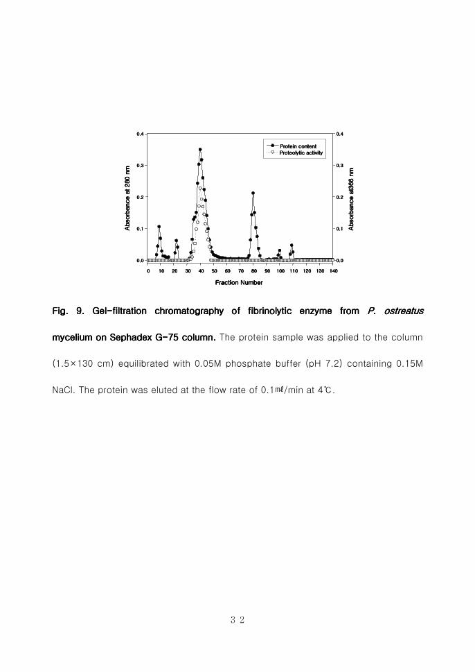
확인하였으며 활성분획을 수집하여 동결건조시킨 후 투석을 통하여 gel-
filtration을 수행하였다 Sephadex G-75 column을 이용하여 DEAE sephadex
A-50 column에서 활성을 나타낸 분획물을 01min의 용출속도로 정제하
였다(Fig 9) 활성을 나타내는 분획물들을 수집하여 동결건조로 농축 시킨 후
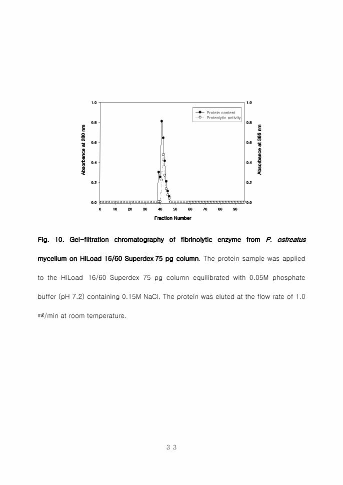
투석을 하여 FPLC를 수행하는 시료로 사용하였다 혈전분해효소를 순수정제
하기 위하여 Amersham pharmacia Co의 HiLoad 1660 Superdex 75 pg을
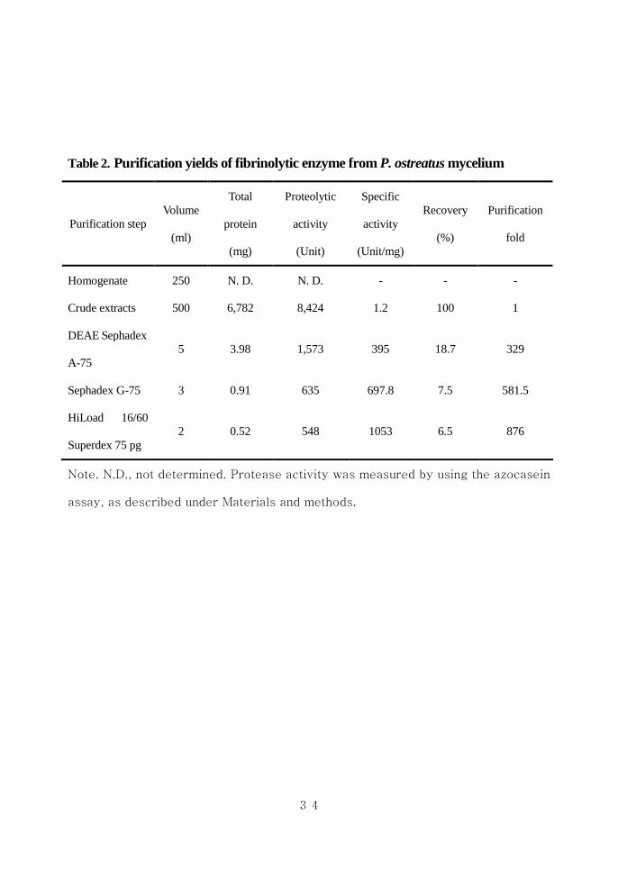
이용하여 FPLC를 수행한 결과 느타리버섯 균사체로부터 약 876배의 정제된
효소를 얻었으며 회수율은 65를 나타내었다(Fig 10 Table 2) 이와 같이
정제된 효소를 PoFE (Pleurotus ostreatus fibrinolytic enzyme)라고 명명하였다
ⅢⅢⅢⅢ----3333 혈전분해효소의혈전분해효소의혈전분해효소의혈전분해효소의 분자량분자량분자량분자량 측정측정측정측정
ⅢⅢⅢⅢ----3333----1 FPLC1 FPLC1 FPLC1 FPLC 를를를를 이용한이용한이용한이용한 분자량분자량분자량분자량 측정측정측정측정(size exclusion)(size exclusion)(size exclusion)(size exclusion)
혈전분해효소의 분자량을 측정하기 위하여 HiLoad 1660 Superdex 75 pg
column에 Gel-filtration marker로써 glyceraldehydes-3-phosphate dehydro-
29
genase (36kDa) carbonic anhydrase (29kDa) trypsinogen-PMSF (24kDa)
trypsin-inhibitor (201kDa)를 이용하여 혈전분해효소 정제시 이용된 동일한
조건으로 size exclusion을 수행하였다 size exclusion을 수행한 결과 Fig
11과 같은 Molecular mass standard curve가 작성되었으며 이를 기초로 하여
정제된 혈전분해효소의 분자량을 측정한 결과 약 32kDa으로 확인되었으며
이는 느타리버섯 자실체로부터 분리정제된 PoMTP와 유사한 분자량을 가지고
있음을 확인하였다
ⅢⅢⅢⅢ----3333----2 2 2 2 SDSSDSSDSSDS----PAGE PAGE PAGE PAGE 및및및및 ffffibrin zymography ibrin zymography ibrin zymography ibrin zymography 를를를를 이용한이용한이용한이용한 분자량분자량분자량분자량 측정측정측정측정
정제된 혈전분해효소의 분자량을 측정하기 위하여 size exclusion외에
SDS-PAGE 및 fibrin zymography를 수행하여 분자량을 확인한 결과 size
exclusion과 동일하게 약 32kDa에서 분자량을 나타냄을 확인하였다 (Fig 12)
ⅢⅢⅢⅢ----4 4 4 4 혈전분해효소의혈전분해효소의혈전분해효소의혈전분해효소의 NNNN----말단말단말단말단 아미노산아미노산아미노산아미노산 서열분석서열분석서열분석서열분석
정제효소의 N-말단 아미노산 서열은 한국기초과학지원연구소 광주분소에 의
뢰하여 결정한 결과 ALRKGGAAALNIYSVGFTS의 아미노산 서열을 확인할 수
있었으며 이에 근거하여 NCBI Blast에서 상동성이 있는 amino acid서열을 검
색하여 비교분석하였다 Fig 13에서와 같이 균사체로부터 분리정제된 혈전분
해효소는 Joh[53]등에 의해 보고되어진 느타리버섯 자실체로부터 분리정제된
metalloprotease(AAU946481)의 아미노산 서열과 N-말단 아미노산서열이 일
치함을 확인할 수 있었으며 Coccidioides posadasii[54]로부터 분리정제된
metalloprtease l precursor (AAQ07436)와 유사한 amino acid 서열을 가지고
있음을 확인하였다
30
0 10 20 30 40 50 60 70 80 90 100
Absorb
ance a
t 280 n
mAbsorb
ance a
t 280 n
mAbsorb
ance a
t 280 n
mAbsorb
ance a
t 280 n
m
000
005
010
015
020
025
030
000
005
010
015
020
025
030
Protein content
Proteolytic activity
NaCl gradient
Absorb
ance a
t 366 n
mAbsorb
ance a
t 366 n
mAbsorb
ance a
t 366 n
mAbsorb
ance a
t 366 n
m
Sodiu
m c
hlo
ride (M
)Sodiu
m c
hlo
ride (M
)Sodiu
m c
hlo
ride (M
)Sodiu
m c
hlo
ride (M
)
00
05
10
Fraction NumberFraction NumberFraction NumberFraction Number
Fig 7 CationFig 7 CationFig 7 CationFig 7 Cation----exchange chromatography of fibrinolytic enzyme from exchange chromatography of fibrinolytic enzyme from exchange chromatography of fibrinolytic enzyme from exchange chromatography of fibrinolytic enzyme from P ostP ostP ostP ostrrrreatus eatus eatus eatus
mmmmycelium on CMycelium on CMycelium on CMycelium on CM----cellulose columncellulose columncellulose columncellulose column The protein sample was applied to the column
(35times10 cm) equilibrated with 10mM citrate-NaOH buffer (pH 60) The protein was
eluted with 0~10M NaCl linear gradient at the flow rate of 10min at 4
31
Fraction NumberFraction NumberFraction NumberFraction Number
0000 10101010 20202020 30303030 40404040 50505050 60606060 70707070 80808080 90909090 100100100100
Absorb
ance a
t 280nm
Absorb
ance a
t 280nm
Absorb
ance a
t 280nm
Absorb
ance a
t 280nm
000000000000
002002002002
004004004004
006006006006
008008008008
010010010010
012012012012
014014014014
Protein contentProtein contentProtein contentProtein content
ProteolyticProteolyticProteolyticProteolytic activityactivityactivityactivity
NaClNaClNaClNaCl gradientgradientgradientgradient
Absorb
ance a
t 366nm
Absorb
ance a
t 366nm
Absorb
ance a
t 366nm
Absorb
ance a
t 366nm
000000000000
002002002002
004004004004
006006006006
008008008008
010010010010
012012012012
014014014014
Sodiu
m c
hlo
ride (M
) Sodiu
m c
hlo
ride (M
) Sodiu
m c
hlo
ride (M
) Sodiu
m c
hlo
ride (M
)
00000000
05050505
10101010
Fig 8Fig 8Fig 8Fig 8 Anion Anion Anion Anion----exchange chromatography of fibrinolytic enzyme from exchange chromatography of fibrinolytic enzyme from exchange chromatography of fibrinolytic enzyme from exchange chromatography of fibrinolytic enzyme from P ostP ostP ostP ostrrrreatus eatus eatus eatus
mycelium on DEAEmycelium on DEAEmycelium on DEAEmycelium on DEAE----Sephadex ASephadex ASephadex ASephadex A----50505050 columncolumncolumncolumn The protein sample was applied to the
column (35times10 cm) equilibrated with 20mM Tris-HCl buffer (pH 80) The protein was
eluted with 0~10M NaCl linear gradient at the flow rate of 10min at 4
32
Fraction NumberFraction NumberFraction NumberFraction Number
0000 10101010 20202020 30303030 40404040 50505050 60606060 70707070 80808080 90909090 100100100100 110110110110 120120120120 130130130130 140140140140
Absorb
ance a
t 280 n
mAbsorb
ance a
t 280 n
mAbsorb
ance a
t 280 n
mAbsorb
ance a
t 280 n
m
00000000
01010101
02020202
03030303
04040404
00000000
01010101
02020202
03030303
04040404
Protein contentProtein contentProtein contentProtein content
ProteolyticProteolyticProteolyticProteolytic activityactivityactivityactivity
Absorb
ance a
t366 n
mAbsorb
ance a
t366 n
mAbsorb
ance a
t366 n
mAbsorb
ance a
t366 n
m
Fig 9 GelFig 9 GelFig 9 GelFig 9 Gel----filtration chromatography of fibrinolytic enzyme from filtration chromatography of fibrinolytic enzyme from filtration chromatography of fibrinolytic enzyme from filtration chromatography of fibrinolytic enzyme from P ostP ostP ostP ostrrrreatuseatuseatuseatus
mycelium on Sephadex Gmycelium on Sephadex Gmycelium on Sephadex Gmycelium on Sephadex G----75757575 columncolumncolumncolumn The protein sample was applied to the column
(15times130 cm) equilibrated with 005M phosphate buffer (pH 72) containing 015M
NaCl The protein was eluted at the flow rate of 01min at 4
33
Fraction NumberFraction NumberFraction NumberFraction Number
0000 10101010 20202020 30303030 40404040 50505050 60606060 70707070 80808080 90909090
Absorb
ance a
t 280 n
mAbsorb
ance a
t 280 n
mAbsorb
ance a
t 280 n
mAbsorb
ance a
t 280 n
m
00000000
02020202
04040404
06060606
08080808
10101010
00000000
02020202
04040404
06060606
08080808
10101010
Protein content
Proteolytic activity
Absorb
ance a
t 366 n
mAbsorb
ance a
t 366 n
mAbsorb
ance a
t 366 n
mAbsorb
ance a
t 366 n
m
Fig 10 GelFig 10 GelFig 10 GelFig 10 Gel----filtration chromatography of fibrinolytic enzyme from filtration chromatography of fibrinolytic enzyme from filtration chromatography of fibrinolytic enzyme from filtration chromatography of fibrinolytic enzyme from P ostP ostP ostP ostrrrreatus eatus eatus eatus
mycelium on HiLoadmycelium on HiLoadmycelium on HiLoadmycelium on HiLoad 1660 1660 1660 1660 Superdex Superdex Superdex Superdex 75757575 pg pg pg pg columncolumncolumncolumn The protein sample was applied
to the HiLoad
1660 Superdex 75 pg column equilibrated with 005M phosphate
buffer (pH 72) containing 015M NaCl The protein was eluted at the flow rate of 10
min at room temperature
34
Table 2 Purification yields of fibrinolytic enzyme from P ostreatus mycelium
Note ND not determined Protease activity was measured by using the azocasein
assay as described under Materials and methods
Purification step Volume
(ml)
Total
protein
(mg)
Proteolytic
activity
(Unit)
Specific
activity
(Unitmg)
Recovery
()
Purification
fold
Homogenate 250 N D N D - - -
Crude extracts 500 6782 8424 12 100 1
DEAE Sephadex
A-75 5 398 1573 395 187 329
Sephadex G-75 3 091 635 6978 75 5815
HiLoad 1660
Superdex 75 pg 2 052 548 1053 65 876
35
1 Glyceraldehyde (35 kDa)
2 Carbonic anhydrase (29 kDa)
3 Trypsinogen-PMSF (24kDa)
4 Trypsin inhibitor (201 kDa)
1111
2222
3333
4444
Purified Purified Purified Purified
enzymeenzymeenzymeenzyme
Fraction NumberFraction NumberFraction NumberFraction Number
0 10 20 30 40 50 60 70 80 90
Absorb
ance a
t 280 n
mAbsorb
ance a
t 280 n
mAbsorb
ance a
t 280 n
mAbsorb
ance a
t 280 n
m
00
02
04
06
08
10
Protein Absorbance Trace
Protein Size Marker Trace
1 Glyceraldehyde-3-phosphate dehydrogenase (36 kDa)
2 Carbonic anhydrase (29 kDa)
3 Trypsinogen-PMSF (24kDa)
4 Trypsin inhibitor (201 kDa)
Purified enzyme
1111
2222
3333
4444
Fig 1Fig 1Fig 1Fig 11111 Molecular weight determination of PoFE using sizeMolecular weight determination of PoFE using sizeMolecular weight determination of PoFE using sizeMolecular weight determination of PoFE using size----exclusion on exclusion on exclusion on exclusion on HiLoad HiLoad HiLoad HiLoad
1660 Superdex1660 Superdex1660 Superdex1660 Superdex 75 pg column75 pg column75 pg column75 pg column The standard markers and purified enzyme was
eluted through a Superdex 75 equilibrated with 005mM phosphate buffer containing
015M NaCl pH 72 at a flow rate of 10min
36
Trypsin inhibitor
M 1 2
100 KDa
75 KDa
Glyceraldehyde-3-phosphate dehydrogenase
Carbonic anhydrase
Trypsinogen-PMSFPurified enzyme
201
24
29
36
10 20 30 40 50 60 70 80 900
Fraction numberFraction numberFraction numberFraction number
Mole
cula
r w
eig
ht (
Mole
cula
r w
eig
ht (
Mole
cula
r w
eig
ht (
Mole
cula
r w
eig
ht ( K
Da
KDa
KDa
KDa)) ))
50 KDa
35 KDa
25 KDa
15 KDa
32 KDa
Fig 1Fig 1Fig 1Fig 12222 Molecular weight determination of P Molecular weight determination of P Molecular weight determination of P Molecular weight determination of PoFEoFEoFEoFE using SDS using SDS using SDS using SDS----PAGE PAGE PAGE PAGE and fibrinand fibrinand fibrinand fibrin
zymography zymography zymography zymography M protein standard marker lane 1 purified PoFE on SDS-PAGE lane
2 purified PoFE on fibrin zymography
37
Fig 1Fig 1Fig 1Fig 13333 Homology analys Homology analys Homology analys Homology analysis of purified fibrinolytic enzyme amino acid sequence from is of purified fibrinolytic enzyme amino acid sequence from is of purified fibrinolytic enzyme amino acid sequence from is of purified fibrinolytic enzyme amino acid sequence from
Pleurotus ostreatusPleurotus ostreatusPleurotus ostreatusPleurotus ostreatus The shadow indicates similar amino acid residues between the
purified enzyme and other proteolytic enzymes PoFE the purified fibrinolytic enzyme
from Postreatus AAU94648 metalloprotease from Pleurotus ostreatus AAQ07436
metalloprotease 1 precursor from Coccidioides posadasii
38
ⅢⅢⅢⅢ----5 5 5 5 혈전분해효소의혈전분해효소의혈전분해효소의혈전분해효소의 특성분석특성분석특성분석특성분석
ⅢⅢⅢⅢ----5555----1 1 1 1 기질특이성기질특이성기질특이성기질특이성 분석분석분석분석
정제효소의 기질특이성을 확인하기 위하여 다양한 합성기질을 이용하여
느타리버섯 균사체로부터 분리정제된 혈전분해효소와 반응시켜 amidolytic
활성을 측정하였다 Fig 14에서 보는바와 같이 느타리버섯 균사체로부터 분리
정제된 혈전분해효소는 chymotrypsin의 기질인 S2586에서 가장 높은 활성을
나타내었다
ⅢⅢⅢⅢ----5555----2 2 2 2 효소활성에효소활성에효소활성에효소활성에 대한대한대한대한 금속이온의금속이온의금속이온의금속이온의 영향영향영향영향
금속이온에 의한 혈전분해활성을 분석하기 위하여 각각 CaCl2 CoCl2
CuCl2 FeCl2 MnCl2 MgCl2 ZnCl2 용액을 2mM 농도로 조제하여 최종농도가
1mM이 되도록 효소 100과 동일한 volume의 용액을 처리한 후 37에서
1시간 동안 반응시켜 금속에 대한 혈전분해효소활성을 측정하였다 Table 3
에서 보는바와 같이 금속이온을 첨가하지 않았을 때의 활성을 100으로 하여
그 상대적인 활성도를 측정하였을때 Zn2+ Ca2+ Mg2+에 의해 혈전분해
효소활성이 14~22 증가됨을 확인할 수 있었으며 반면에 Cu2+을 첨가한
경우에는 활성이 13정도 감소함을 볼수 있었다
ⅢⅢⅢⅢ----5555----3 3 3 3 효소활성에효소활성에효소활성에효소활성에 대한대한대한대한 Protease inhibitor Protease inhibitor Protease inhibitor Protease inhibitor 의의의의 영향영향영향영향
Protease inhibitor에 의한 혈전분해활성을 분석하기 위하여 PMSF TLCK
TPCK aprotinin EDTA pepstatin A를 효소 100 과 37에서 1시간 동안
반응시킨 후 azocasein assay를 수행하여 혈전분해활성을 측정하였다 Table 4
에서와 같이 정제된 혈전분해효소는 EDTA에 의해 활성이 억제되어 짐을 확인
39
할 수 있었으며 이는 정제된 혈전분해효소가 metalloprotease일것으로 추정
된다
ⅢⅢⅢⅢ----5555----4 4 4 4 효소활성에효소활성에효소활성에효소활성에 대한대한대한대한 pH pH pH pH 의의의의 효과효과효과효과
pH에 의한 혈전분해효소의 활성을 분석하기 위하여 pH 20 ~ pH 100
범위에서 혈전분해활성을 측정하였다 각 pH 조건의 reagent에 효소 100을
처리하고 1시간 동안 37에서 반응시킨 후 azocasein assay를 수행하여 pH
에 의한 영향을 분석하였다 그 결과 Fig 15에서 보는바와 같이 pH 60과 70
사이에서 가장 높은 활성을 나타내었으며 일반적으로 pH 60 ~ pH 70
범위를 벗어나면 활성이 급격히 감소하였다 따라서 이 효소는 중성에 가까운
pH에서 활성이 큰 효소임을 알수 있었다
ⅢⅢⅢⅢ----5555----5555 효소활성에효소활성에효소활성에효소활성에 대한대한대한대한 온도의온도의온도의온도의 영향영향영향영향
온도에 의한 혈전분해효소의 영향을 분석하기 위하여 20 ~ 90 온도
에서 효소액을 30분 동안 방치 시킨 후 분해활성 변화를 측정하였다 Fig 16
에 나타낸바와 같이 35에서 최대활성을 보였으며 35이상의 온도에서는
활성이 감소하였고 50 이상의 온도에서는 활성이 아주 낮았으며 37에서는
최적조건에서의 약 85에 해당하는 활성을 보여주었다
ⅢⅢⅢⅢ----5555----6 Fibrinolysis6 Fibrinolysis6 Fibrinolysis6 Fibrinolysis 와와와와 FFFFibrinogenolysis pattern ibrinogenolysis pattern ibrinogenolysis pattern ibrinogenolysis pattern 분석분석분석분석
ⅢⅢⅢⅢ----5555----6666----1 Fibrinogenolysi1 Fibrinogenolysi1 Fibrinogenolysi1 Fibrinogenolysis s s s
Fibrinogen (5)에 동일 양의 혈전분해효소를 처리하여 시간별로 37
항온기에서 반응시킨 후 12 SDS-PAGE를 수행하여 fibrinogen 분해패턴을
확인하였다 반응 5분 이내에 Aα Bβ chain은 완전히 분해되어졌으나 γ chain
40
Chromogenic substrates
S-2222 S-2288 S-2238 S-2251S-2444S-2586S-2765
Rela
tive a
ctivity (
)
0
20
40
60
80
100
120
Fig 14 Amidolytic activity on the several chromogenic substrateFig 14 Amidolytic activity on the several chromogenic substrateFig 14 Amidolytic activity on the several chromogenic substrateFig 14 Amidolytic activity on the several chromogenic substratessss Amidolytic activity
was measured spectometrically using the chromogenic protease substrates such as
S2222 S2288 S2238 S2251 S2444 S2586 and S2765
41
Table 3 Effect of metal ions on the activity of the fibrinolytic enzyme from
P ostreatus mycelium
Reaction conditions are as described under materials and methods
Metal ions Concentration( mM) Relative activity ()
Control - 100
Ca2+ 10 118
Co2+ 10 96
Cu2+ 10 87
Fe2+ 10 91
Mn2+ 10 94
Mg2+ 10 122
Zn2+ 10 114
42
Table 4 Effect of protease inhibitors on the activity of the fibrinolytic enzyme
from P ostreatus mycelium
Reaction conditions are as described under materials and methods
Protease inhibitors Concentration( mM) Relative activity ()
Control - 100
PMSF 10 78
TLCK 10 81
TPCK 10 74
EDTA 10 52
aprotinin 10 88
peptstatin A 10 88
43
2 4 6 8 10
pHpHpHpH
2
Rela
tive a
ctivity
()
Rela
tive a
ctivity
()
Rela
tive a
ctivity
()
Rela
tive a
ctivity
()
00
100
80
60
40
20
Fig 15 Fig 15 Fig 15 Fig 15 Effect of pH on the activity of the Effect of pH on the activity of the Effect of pH on the activity of the Effect of pH on the activity of the fibrinolyticfibrinolyticfibrinolyticfibrinolytic enzymeenzymeenzymeenzymessss from from from from P ostP ostP ostP ostrrrreatuseatuseatuseatus
myceliummyceliummyceliummycelium Enzyme activity was assayed in the pH range of 2-10 05M glycine-HCl
(pH 20~30) 05 M acetate (pH 40~ 50) 05 M Tris-HCl (pH 60~80) and 05 M
glycine-NaOH (pH 90~ 100) buffers were used with 01 azocasein
44
20 40 60 80
20
40
60
80
100
30 50 70 90
Rela
tive a
ctivity
()
Rela
tive a
ctivity
()
Rela
tive a
ctivity
()
Rela
tive a
ctivity
()
Temperature (Temperature (Temperature (Temperature ())))
Fig 16 Effect of temperature on the activity of the Fig 16 Effect of temperature on the activity of the Fig 16 Effect of temperature on the activity of the Fig 16 Effect of temperature on the activity of the fibrinolytic fibrinolytic fibrinolytic fibrinolytic enzymeenzymeenzymeenzymessss from from from from P ostreatusP ostreatusP ostreatusP ostreatus
myceliummyceliummyceliummycelium The purified enzyme was incubated at temperatures from 20 to 90 for
30minutes
45
은 반응 후 30분 부터 분해가 시작되어 α β chain에 비하여 상대적으로
혈전분해효소에 대한 저항성을 가지고 있는 것으로 판명되었다(Fig 17A)
ⅢⅢⅢⅢ----5555----6666----2 Fibrinolysis 2 Fibrinolysis 2 Fibrinolysis 2 Fibrinolysis
Fibrinogen(10)과 thrombin(01unit)을 상온에서 1시간 동안 반응시켜
fibrin을 조제한 후 동일 양의 혈전분해효소를 처리하여 시간별로 37 항온기
에서 반응시키고 나서 12 SDS-PAGE를 수행하여 fibrin 분해패턴을 확인하
였다(Fig 17B) 반응 5분 후에 α-chain이 분해되었고 β chain과 γ-γ chian은
α chain에 비하여 상대적으로 효소에 선택성이 낮은 것으로 확인되었다 β
chain과 γ-γ chian은 반응 후 5분후부터 점차 분해되어짐을 확인하였다 또한
본 실험에서 느타리버섯 균사체로부터 분리정제한 혈전분해효소는 SK UK t-
PA 등과 같이 plasminogen을 활성화시켜 혈전을 분해하는 메커니즘과는 달리
직접 혈전을 분해시킴을 확인하였다
ⅢⅢⅢⅢ----6 Cloning and sequence analysis of 6 Cloning and sequence analysis of 6 Cloning and sequence analysis of 6 Cloning and sequence analysis of pofepofepofepofe
GenBank data를 검색하여 느타리버섯 균사체로부터 정제된 혈전분해효소
의 N-terminal amino acid sequence와 느타리버섯 자실체로부터 분리한
metalloprotease유전자(AAU94648)의 염기서열을 비교분석하여 start codon부위
부터 stop condon까지 PCR를 수행할 수 있도록 primer를 제작하여 PCR를 수행하
였다 PCR 결과 약 860bp와 12Kb의 DNA 단편이 증폭됨을 확인할 수 있었
다(Fig 18) 확인된 PCR product의 염기서열을 분석하기 위하여 pGEM T-
easy cloning vector에 ligation하여 E coli DH5α competent cell에 형질전환시
킨 후 Amp+X-galIPTG가 포함된 LB plate에 도말하여 12시간 배양하였다
ampicillin에 저항성을 가지며 white colony를 나타내는 형질전환체를 분리하여
46
A αB β
γ
1 2 3 4 5 6A
γ-γ
βα
B1 2 3 4 5 6
Fig 17 Fig 17 Fig 17 Fig 17 FFFFibrinogenolysis and fibrinolysis ibrinogenolysis and fibrinolysis ibrinogenolysis and fibrinolysis ibrinogenolysis and fibrinolysis patternpatternpatternpattern exhibited by the purified fibrinolytic exhibited by the purified fibrinolytic exhibited by the purified fibrinolytic exhibited by the purified fibrinolytic
enzyme from enzyme from enzyme from enzyme from P ostreatusP ostreatusP ostreatusP ostreatus myceliummyceliummyceliummycelium The enzyme was incubated with 10μg of
fibrinogen or fibrin in 10mM of Tris-HCl buffer (pH 72) at 37degC for up to 1hr Lane
1 control without enzyme lane 2 after 5min of reaction lane 3 after 10min of
reaction lane 4 after 15min of reaction lane 5 after 30min of reaction lane 6 after
1hr of rection
47
M 1M 1M 1M 1
12 Kb12 Kb12 Kb12 Kb
086 Kb086 Kb086 Kb086 Kb
05 Kb05 Kb05 Kb05 Kb
10 Kb10 Kb10 Kb10 Kb
FigFigFigFig 11118888 Identification of the PoFE protein encoding Identification of the PoFE protein encoding Identification of the PoFE protein encoding Identification of the PoFE protein encoding gene from gene from gene from gene from P ostreatusP ostreatusP ostreatusP ostreatus mycelium mycelium mycelium mycelium
RT-PCR was performed using PCR primers which were designed for the conserved
sequences in metalloprotease encoding gene from P ostreatus fruiting body M
100bp ladder DNA marker Lane 1 PCR products
48
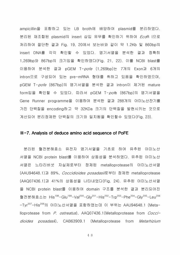
ampicillin을 포함하고 있는 LB broth에 배양하여 plasmid를 분리하였다
분리된 재조합된 plasmid의 insert 삽입 유무를 확인하기 위하여 EcoR I으로
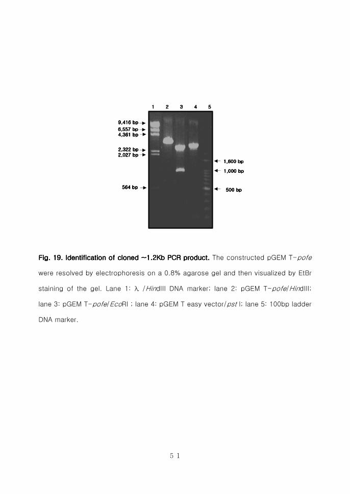
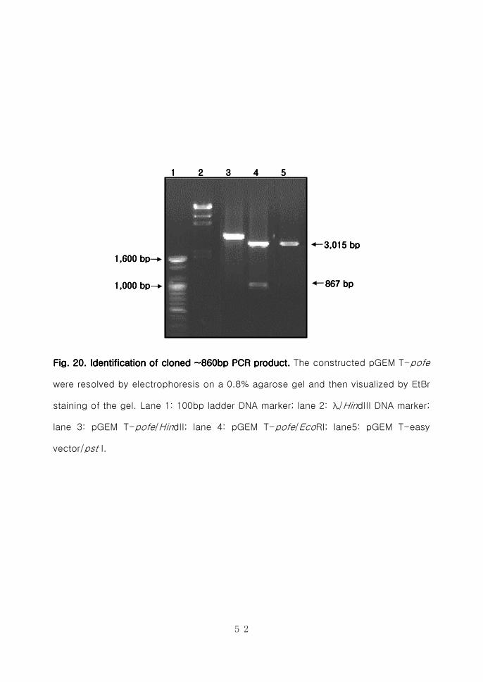
처리하여 절단한 결과 Fig 19 20에서 보는바와 같이 약 12Kb 및 860bp의
insert DNA를 각각 확인할 수 있었다 염기서열을 분석한 결과 정확히
1269bp와 867bp의 크기임을 확인하였다(Fig 21 22) 이를 NCBI blast를
이용하여 분석한 결과 pGEM T-pofe (1269bp)는 7개의 Exon과 6개의
intron으로 구성되어 있는 pre-mRNA 형태를 취하고 있음을 확인하였으며
pGEM T-pofe (867bp)의 염기서열을 분석한 결과 intron이 제거된 mature
form임을 확인할 수 있었다 따라서 pGEM T-pofe (867bp)의 염기서열을
Gene Runner programme을 이용하여 분석한 결과 288개의 아미노산잔기를
가진 단백질을 encoding하고 약 32KDa 크기의 단백질을 발현시키는 것으로
계산되어 분리정제한 단백질의 크기와 일치됨을 확인할수 있었다(Fig 23)
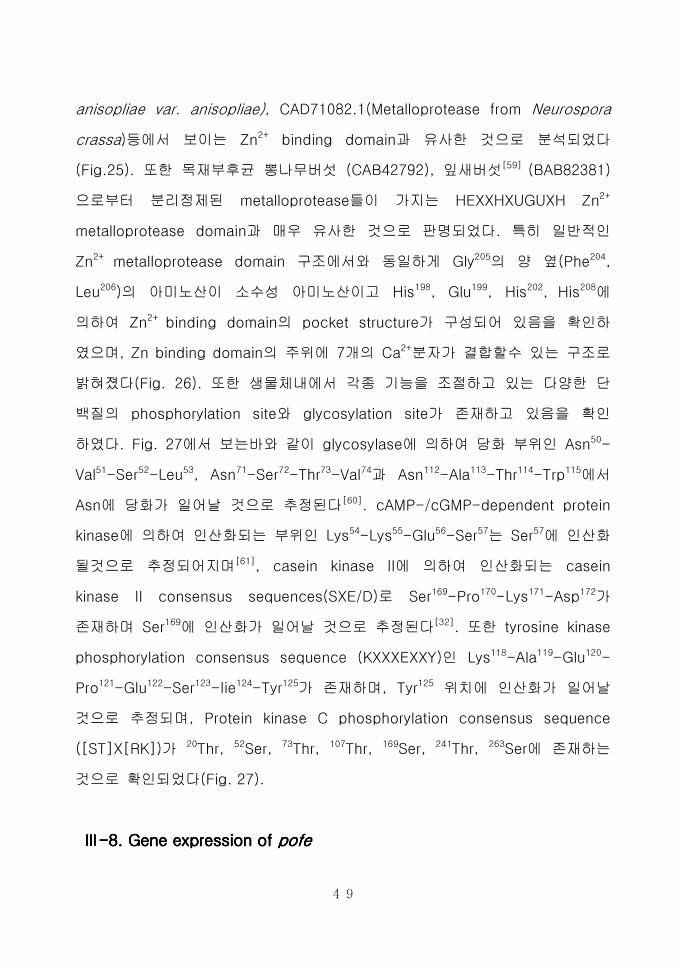
ⅢⅢⅢⅢ----7 Analysis of deduce amino acid sequence of PoFE7 Analysis of deduce amino acid sequence of PoFE7 Analysis of deduce amino acid sequence of PoFE7 Analysis of deduce amino acid sequence of PoFE
분리된 혈전분해효소 유전자 염기서열을 기초로 하여 유추된 아미노산
서열을 NCBI protein blast를 이용하여 상동성을 분석하였다 유추된 아미노산
서열은 느타리버섯 자실체로부터 정제된 metalloprotease의 아미노산서열
(AAU946481)과 89 Coccidioides posadasii로부터 정제한 metalloprotease
(AAQ074361)과 41의 상동성을 나타내었다(Fig 24) 유추된 아미노산서열
을 NCBI protein blast를 이용하여 domain 구조를 분석한 결과 분리되어진
혈전분해효소는 His198-Glu199-Val200-Gly201-His202-Trp203-Phe204-Gly205-Leu206
-Tyr207-His208의 아미노산서열을 포함하였는데 이 부위는 AAU946481 (Meta-
lloprotease from P ostreatus) AAQ074361(Metalloprotease from Cocci-
dioides posadasii) CAB639091 (Metalloprotease from Metarhizium
49
anisopliae var anisopliae) CAD710821(Metalloprotease from Neurospora
crassa)등에서 보이는 Zn2+ binding domain과 유사한 것으로 분석되었다
(Fig25) 또한 목재부후균 뽕나무버섯 (CAB42792) 잎새버섯[59] (BAB82381)
으로부터 분리정제된 metalloprotease들이 가지는 HEXXHXUGUXH Zn2+
metalloprotease domain과 매우 유사한 것으로 판명되었다 특히 일반적인
Zn2+ metalloprotease domain 구조에서와 동일하게 Gly205의 양 옆(Phe204
Leu206)의 아미노산이 소수성 아미노산이고 His198 Glu199 His202 His208에
의하여 Zn2+ binding domain의 pocket structure가 구성되어 있음을 확인하
였으며 Zn binding domain의 주위에 7개의 Ca2+분자가 결합할수 있는 구조로
밝혀졌다(Fig 26) 또한 생물체내에서 각종 기능을 조절하고 있는 다양한 단
백질의 phosphorylation site와 glycosylation site가 존재하고 있음을 확인
하였다 Fig 27에서 보는바와 같이 glycosylase에 의하여 당화 부위인 Asn50-
Val51-Ser52-Leu53 Asn71-Ser72-Thr73-Val74과 Asn112-Ala113-Thr114-Trp115
에서
Asn에 당화가 일어날 것으로 추정된다[60] cAMP-cGMP-dependent protein
kinase에 의하여 인산화되는 부위인 Lys54-Lys55-Glu56-Ser57는 Ser57
에 인산화
될것으로 추정되어지며[61] casein kinase II에 의하여 인산화되는 casein
kinase II consensus sequences(SXED)로 Ser169-Pro170-Lys171-Asp172가
존재하며 Ser169에 인산화가 일어날 것으로 추정된다[32] 또한 tyrosine kinase
phosphorylation consensus sequence (KXXXEXXY)인 Lys118-Ala119-Glu120-
Pro121-Glu122-Ser123-Iie124-Tyr125가 존재하며 Tyr125 위치에 인산화가 일어날
것으로 추정되며 Protein kinase C phosphorylation consensus sequence
([ST]X[RK])가 20Thr 52Ser 73Thr 107Thr 169Ser 241Thr 263Ser에 존재하는
것으로 확인되었다(Fig 27)
ⅢⅢⅢⅢ----8 8 8 8 Gene expression of Gene expression of Gene expression of Gene expression of pofepofepofepofe
50
느타리버섯 균사체로부터 분리된 혈전분해효소 유전자를 발현시킬 목적으로
expression vector인 pQE30 expression vector와의 reading frame을 조절하기
위하여 5-GGATCCATGTTGCGCTCCATCCTGTTAATTG-3와 5-AAGCTTTTACAC
TGGTGCTGCTACCCTGGC-3과 같이 forward와 reverse primer를 제작하여
PCR을 수행하였다 약 860bp의 PCR product를 확인하여 발현 백터인 pQE 30
에 ligation한 후 Ecoli M15(pREP4) competent cell로 transformation 하였다
재조합된 plasmid의 insert 삽입유무를 확인하기 위하여 plasmid를 분리하여
BamHⅠ Hind Ⅲ로 절단한 후 EtBr이 함유된 08 agarose gel에 전기영동한
결과 약 860bp의 insert DNA를 확인할 수 있었다(Fig 28) 형질전환된 집락을
selection하여 LB에서 12시간 배양한 후 새로운 LB에 접종하여 OD600에서 06에
이를 때 까지 배양시킨 후 10mM IPTG를 처리하여 6X His Tag이 fusion된 혈전
분해효소를 발현 유도시켰다 이에 대한 발현유무를 확인하기 위하여 형질전환된
세포로부터 조단백질을 제조한 후 20로 정량하여 전기영동을 수행하였다
Fig 29A에서 보는바와 같이 32KDa의 예상 단백질 크기로 느타리버섯 혈전분해
효소 유전자가 발현됨을 확인하였다 또한 이를 검증하기 위하여 Ni-NTA spin
column으로 affinity chromatography를 수행하여 발현된 단백질을 정제하고
Alkaine phosphatase conjugated 6X His tag primary antibody를 이용하여
western blot을 수행한 결과 Fig 29B에서와 같이 32KDa의 단백질이 발현됨을
확인하였다
51
6557 6557 6557 6557 bpbpbpbp4361 4361 4361 4361 bpbpbpbp
2322 2322 2322 2322 bpbpbpbp
2027 2027 2027 2027 bpbpbpbp
9416 9416 9416 9416 bpbpbpbp
564 564 564 564 bpbpbpbp
1 2 3 4 5 1 2 3 4 5 1 2 3 4 5 1 2 3 4 5
500 500 500 500 bpbpbpbp
1000 1000 1000 1000 bpbpbpbp
1600 1600 1600 1600 bpbpbpbp
FigFigFigFig 11119999 Identification of cloned Identification of cloned Identification of cloned Identification of cloned ~12Kb~12Kb~12Kb~12Kb PCR productPCR productPCR productPCR product The constructed pGEM T-pofe
were resolved by electrophoresis on a 08 agarose gel and then visualized by EtBr
staining of the gel Lane 1 λ HindIII DNA marker lane 2 pGEM T-pofeHindIII
lane 3 pGEM T-pofeEcoRI lane 4 pGEM T easy vectorpst I lane 5 100bp ladder
DNA marker
52
1 2 3 4 5 1 2 3 4 5 1 2 3 4 5 1 2 3 4 5
867 867 867 867 bpbpbpbp
3015 3015 3015 3015 bpbpbpbp
1000 1000 1000 1000 bpbpbpbp
1600 1600 1600 1600 bpbpbpbp
FigFigFigFig 20202020 Identification of cloned ~860bp PCR product Identification of cloned ~860bp PCR product Identification of cloned ~860bp PCR product Identification of cloned ~860bp PCR product The constructed pGEM T-pofe
were resolved by electrophoresis on a 08 agarose gel and then visualized by EtBr
staining of the gel Lane 1 100bp ladder DNA marker lane 2 λHindIII DNA marker
lane 3 pGEM T-pofeHindII lane 4 pGEM T-pofeEcoRI lane5 pGEM T-easy
vectorpst I
53
Fig 2Fig 2Fig 2Fig 21111 Nucleotide Nucleotide Nucleotide Nucleotide sequence of sequence of sequence of sequence of pofepofepofepofe (1269(1269(1269(1269bpbpbpbp) gene encoding the precursor form ) gene encoding the precursor form ) gene encoding the precursor form ) gene encoding the precursor form
of the fibrinolytic enzymeof the fibrinolytic enzymeof the fibrinolytic enzymeof the fibrinolytic enzyme of of of of PostreatusPostreatusPostreatusPostreatus mycelium mycelium mycelium mycelium Shade indicates exons
1 T A G C T A T G C A T C C A A C G C G T T G G G A G C T C T 30
31 C C C A T A T G G T C G A C C T G C A G G C G G C C G C G A 60
61 A T T C A C T A G T G A T T G G A T C C A T G T T G C G C T 90
91 C C A T C C T G T T A A T T G C A C T C T C C T G C T C C G 120
121 C C T A C G T G C T G G G G A G C A C A C C C A G A C C C A 150
151 G G T A G G A G T T C G A T C A G C G T T C T A C G A G G A 180
181 T G C T T G C T T A C G A T T G G C A T A G G C A A A G G G 210
211 T T T G C G G G T C G C A A C C A A G C C A A G A A A G T G 240
241 T C C A A A G G G C G G A G G C C C A T T T T G A C G G C C 270
271 T T A G A A G C A A C G T C A G C C T G A A A A A A G A G A 300
301 G T T C T T T C A C C G T C G C A G T G C A C T G G C A T G 330
331 T C G T C A G C C A G A A T T C C A C G G T C C G T G G C G 360
361 G G A A C A T C C C G T G C G T T A A A C C T C G T C G T T 390
391 T C T T C G T G C A A G A T G G T G A C C C A T T G T G T T 420
421 A G C G A T T C C C A A A T T A C G G A C C A A A T C C A A 450
451 G T C T T G A A T G A C G C G T A C G C G T A C T C G G G C 480
481 T T T T C A T T T G A A C T T G C A A G T A C C A C A C G T 510
511 A C G C T C A A C G C T A C A T G G T T T G G C A A G G C C 540
541 G A A C C G G A G A G G T A A G T T G A G C C T T T A C G T 570
571 C A G A C A C C C T C A C C A A T T C G G A T T A C T T G T 600
601 A G T A T A T A C C A A G A T G A A A T G A A G G C G G C C 630
631 C T G C G A A A G G G A G G A G C C G C T G C T C A T A A C 660
661 A T T T A C A G C G T A G G G T G A G C C T C C T T A A T C 690
691 T C C G C A T T G C G T G T T C G T A G C A C T C A T C C A 720
721 C T T T C A C C A G G T T T A C T T C T G G T T A C T A C T 750
751 A C G G G C T T C T A G G A T A C G C A A C G T T C C C A T 780
781 T T G C A T A T G G G T A C A G T C C A A A G G A T G A C G 810
811 G T G T T G T G A T C C A T T A C G C C T C G G T T C C G G 840
841 G G G G C A A C T T G G A T A C A T A C A A C C T C G G G A 870
871 T G G T A T G T G T C T G G G T T C C C T T C C T C C C G A 900
901 G C A T A C A C C T G A T C A G C T G A T G A T A C T G T T 930
931 T C T C A C A T A A A C C G C A A C G C A C G A A G T C G G 960
961 T C A C T G G T T T G G C C T C T A C C A C A C C T T C C A 990
991 A G G A G G T T G C G A C G A A C C T G G A G A C T A T G T 1020
1021 T G A T G A A C C C C C T C T G A G T C T T A C A A A A C N 1050
1051 T T G G A T G C C C C G T T G T C T T G A C A C T T T G A C 1080
1081 A G C G A A G T T T G A C C C N N C C C G A G N T T G T T G 1110
1111 C G N C N T C A N N A N A C N T G T T C T A T A T G G A A A 1140
1141 C T C C A T A A C T A C A T G G A C T A C A C C G A T G A T 1170
1171 G A G T G C A T G A C C G A G T T C A C C G A A G G G C A G 1200
1201 G G T C A G C G C A T G A G A G A A C A G T C C T G G A C G 1230
1231 T A C C G G G G C G T C G A G G C C A G G G T A G C A G C A 1260
1261 C C A G T G T A A
54
1111 126912691269126980808080 150150150150 200200200200 372372372372 422422422422 550550550550 600600600600 674674674674 729729729729 873873873873 940940940940 1098109810981098 1141114111411141
IIII1111
IIII2222
IIII3333 IIII
4444IIII5555
IIII6666
IIII7777
EEEE1111
EEEE2222
EEEE3333
EEEE4444
EEEE5555
EEEE6666
EEEE7777
1111 70707070 240240240240 367367367367 440440440440 583583583583 741741741741 867867867867
pGEMpGEMpGEMpGEM TTTT----pofepofepofepofe(867bp)(867bp)(867bp)(867bp)
pGEMpGEMpGEMpGEM TTTT----pofepofepofepofe(1269bp)(1269bp)(1269bp)(1269bp)
Fig 22 Schematic rFig 22 Schematic rFig 22 Schematic rFig 22 Schematic representation of epresentation of epresentation of epresentation of thethethethe structures of premature gene(pGEM T structures of premature gene(pGEM T structures of premature gene(pGEM T structures of premature gene(pGEM T----
pofepofepofepofe 1269 1269 1269 1269bpbpbpbp)))) and functional gene(pGEM T and functional gene(pGEM T and functional gene(pGEM T and functional gene(pGEM T----pofepofepofepofe 867bp) 867bp) 867bp) 867bp) Exons are indicated by
filled boxes and numbered from E1 to E7
55
1 M L R S I L L I A L S C S A Y 15
1 A T G T T G C G C T C C A T C C T G T T A A T T G C A C T C T C C T G C T C C G C C T A C 45
16 V L G S T P R P R Q R V C G S 30
46 G T G C T G G G G A G C A C A C C C A G A C C C A G G C A A A G G G T T T G C G G G T C G 90
31 Q P S Q E S V Q R A E A H F D 45
91 C A A C C A A G C C A A G A A A G T G T C C A A A G G G C G G A G G C C C A T T T T G A C 135
46 G L R S N V S L K K E S S F T 60
136 G G C C T T A G A A G C A A C G T C A G C C T G A A A A A A G A G A G T T C T T T C A C C 180
61 V A V H W H V V S Q N S T V R 75
181 G T C G C A G T G C A C T G G C A T G T C G T C A G C C A G A A T T C C A C G G T C C G T 225
76 G G N I P D S Q I T D Q I Q V 90
226 G G C G G G A A C A T C C C C G A T T C C C A A A T T A C G G A C C A A A T C C A A G T C 270
91 L N D A Y A Y S G F S F E L A 105
271 T T G A A T G A C G C G T A C G C G T A C T C G G G C T T T T C A T T T G A A C T T G C A 315
106 S T T R T V N A T W F G K A E 120
316 A G T A C C A C A C G T A C G C T C A A C G C T A C A T G G T T T G G C A A G G C C G A A 360
121 P E S I Y Q D E M K A A L R K 135
361 C C G G A G A G T A T A T A C C A A G A T G A A A T G A A G G C G G C C C T G C G A A A G 405
136 G G A A A L N I Y S V G F T S 150
406 G G A G G A G C C G C T G C T C T A A A C A T T T A C A G C G T A G G G T T T A C T T C T 450
151 G Y Y Y G L L G Y A T F P F A 165
451 G G T T A C T A C T A C G G G C T T C T A G G A T A C G C A A C G T T C C C A T T T G C A 495
166 Y G Y S P K D D G V V I H Y A 180
496 T A T G G G T A C A G T C C A A A G G A T G A C G G T G T T G T G A T C C A T T A C G C C 540
181 S V P G G N L D T Y N L G M T 195
541 T C G G T T C C G G G G G G C A A C T T G G A T A C A T A C A A C C T C G G G A T G A C C 585
196 A T H E V G H W F G L Y H T F 210
586 G C A A C G C A C G A A G T C G G T C A C T G G T T T G G C C T C T A C C A C A C C T T C 630
211 Q G G C D E P G D Y V D E P P 225
631 C A A G G A G G T T G C G A C G A A C C T G G A G A C T A T G T T G A T G A A C C C C C T 675
226 L S L T K L W M P R C L D T L 240
676 C T G A G T C T T A C A A A A C T T T G G A T G C C C C G T T G T C T T G A C A C T T T G 720
241 T A K F D P I H N Y M D Y T D 255
721 A C A G C G A A G T T T G A C C C C A T C C A T A A C T A C A T G G A C T A C A C C G A T 765
256 D E C M T E F T E G Q G Q R M 270
766 G A T G A G T G C A T G A C C G A G T T C A C C G A A G G G C A G G G T C A G C G C A T G 810
271 R E Q S W T Y R G V E A R V A 285
811 A G A G A A C A G T C C T G G A C G T A C C G G G G C G T C G A G G C C A G G G T A G C A 855
286 A P V
856 G C A C C A G T G T A A
FigFigFigFig 23 Nucleotide sequence of the PoFE cDNA and the deduced amino acid 23 Nucleotide sequence of the PoFE cDNA and the deduced amino acid 23 Nucleotide sequence of the PoFE cDNA and the deduced amino acid 23 Nucleotide sequence of the PoFE cDNA and the deduced amino acid
sequencesequencesequencesequence
56
Fig 24 Multiple alignment of the full length metalloprotFig 24 Multiple alignment of the full length metalloprotFig 24 Multiple alignment of the full length metalloprotFig 24 Multiple alignment of the full length metalloprotease sequences from ease sequences from ease sequences from ease sequences from P ostreatusP ostreatusP ostreatusP ostreatus mycelium(PoFE)mycelium(PoFE)mycelium(PoFE)mycelium(PoFE) P P P P
ostreatusostreatusostreatusostreatus fr fr fr fruiting body (uiting body (uiting body (uiting body (AAU946481) AAU946481) AAU946481) AAU946481) Coccidioides Coccidioides Coccidioides Coccidioides posadasii posadasii posadasii posadasii ((((AAQ074361)AAQ074361)AAQ074361)AAQ074361) Metarhizium anisopliae var Metarhizium anisopliae var Metarhizium anisopliae var Metarhizium anisopliae var
anisopliae anisopliae anisopliae anisopliae ((((CAB639091) andCAB639091) andCAB639091) andCAB639091) and Neurospora crassa Neurospora crassa Neurospora crassa Neurospora crassa ((((CAD710821)CAD710821)CAD710821)CAD710821) The shade is 100 sequence identity The
alignment is generated using clustalx software
57
ZnMcZnMcZnMcZnMc
1 50 100 1 50 100 1 50 100 1 50 100 150 200 150 200 150 200 150 200 250 288250 288250 288250 288
Fig 25 Multiple alignment of the Zinc binding domain sequences from Fig 25 Multiple alignment of the Zinc binding domain sequences from Fig 25 Multiple alignment of the Zinc binding domain sequences from Fig 25 Multiple alignment of the Zinc binding domain sequences from P P P P
ostreatusostreatusostreatusostreatus myceliummyceliummyceliummycelium(PoFE) (PoFE) (PoFE) (PoFE) P ostreatusP ostreatusP ostreatusP ostreatus fruiting body fruiting body fruiting body fruiting body((((AAU946481)AAU946481)AAU946481)AAU946481) CoccidiCoccidiCoccidiCoccidioides oides oides oides
posadasii posadasii posadasii posadasii ((((AAQ074361)AAQ074361)AAQ074361)AAQ074361) Metarhizium anisopliae var anisopliae Metarhizium anisopliae var anisopliae Metarhizium anisopliae var anisopliae Metarhizium anisopliae var anisopliae ((((CAB639091) CAB639091) CAB639091) CAB639091)
andandandand Neurospora crassa Neurospora crassa Neurospora crassa Neurospora crassa ((((CAD710821)CAD710821)CAD710821)CAD710821) The conserved region is indicated by
the shade
58
FigFigFigFig 22226666 The Zn binding domain of PoFE purified from The Zn binding domain of PoFE purified from The Zn binding domain of PoFE purified from The Zn binding domain of PoFE purified from PPPP ostreatus ostreatus ostreatus ostreatus mycelium mycelium mycelium mycelium
A Tube diagram of Zn biding domain (HEXXHXUGUXH) B Ribbon diagram
of normal metalloprotease which need 7 Ca2+
ions Above diagrams were
created by using Cn3D programme in NCBI
59
PKC PKC PKC PKC phosphorylationphosphorylationphosphorylationphosphorylation site site site site
[ST]X[RK][ST]X[RK][ST]X[RK][ST]X[RK]
PKA PKA PKA PKA phosphorylationphosphorylationphosphorylationphosphorylation site site site site
[RK](2)X[ST][RK](2)X[ST][RK](2)X[ST][RK](2)X[ST]
28928928928960606060 120120120120 180180180180 2402402402401111
TPRTPRTPRTPR SLKSLKSLKSLK TVRTVRTVRTVR TTRTTRTTRTTR SPKSPKSPKSPK TAKTAKTAKTAK TYRTYRTYRTYR
20202020 52525252 73737373 107107107107 169169169169 241241241241 276276276276
KKESKKESKKESKKES
54545454
CK2 CK2 CK2 CK2 phosphorylationphosphorylationphosphorylationphosphorylation site site site site
[ST]X(2)[DE][ST]X(2)[DE][ST]X(2)[DE][ST]X(2)[DE]SPKDSPKDSPKDSPKD
169169169169
Tyrosine Tyrosine Tyrosine Tyrosine kinasekinasekinasekinasephosphorylationphosphorylationphosphorylationphosphorylation site site site site
[RK]X(23)[DE]X(23)Y[RK]X(23)[DE]X(23)Y[RK]X(23)[DE]X(23)Y[RK]X(23)[DE]X(23)Y
KAEPESIYKAEPESIYKAEPESIYKAEPESIY
118118118118
Zn binding domain Zn binding domain Zn binding domain Zn binding domain
HEXXHHEXXHHEXXHHEXXH
HEVGHHEVGHHEVGHHEVGH
195195195195
ASN ASN ASN ASN glycosylationglycosylationglycosylationglycosylation sitesitesitesite
NP[ST]PNP[ST]PNP[ST]PNP[ST]P50505050 71717171 112112112112
NVSLNVSLNVSLNVSL NSTVNSTVNSTVNSTV NATVNATVNATVNATV
Fig 27 The structural analysis of PoFE sequence by using GFig 27 The structural analysis of PoFE sequence by using GFig 27 The structural analysis of PoFE sequence by using GFig 27 The structural analysis of PoFE sequence by using Gene runner ene runner ene runner ene runner
programme programme programme programme The motif of N-glycosylation site(NP[ST]P) The motif of
protein kinase C phosphorylation site (ST]X[RK]) The motif of cAMP-and
cGMP-dependent protein kinase phosphorylation site ([RK](2)X[ST]) The
motif of casein kinase II phosphorylation site ([ST]X(2)[DE]) The motif of
Tyrosine kinase phosphorylation site ([RK]X(23)[DE]X(23)Y) and the Zn
binding domain (HEXXH) site are included
60
6557 6557 6557 6557 bpbpbpbp
4361 4361 4361 4361 bpbpbpbp
2322 2322 2322 2322 bpbpbpbp
2027 2027 2027 2027 bpbpbpbp
9416 9416 9416 9416 bpbpbpbp
1 2 3 4 5 1 2 3 4 5 1 2 3 4 5 1 2 3 4 5
1000 1000 1000 1000 bpbpbpbp
500 500 500 500 bpbpbpbp
Fig 2Fig 2Fig 2Fig 28888 Construction of expression vector pQE30 Construction of expression vector pQE30 Construction of expression vector pQE30 Construction of expression vector pQE30----pofepofepofepofe The mature form of
pofe (867bp) was cloned into pQE30 expression vector The constructed
pQE30-pofe were resolved by electrophoresis on 08 agarose gel and then
visualized by EtBr staining of the gel Lane 1 λ Hind III DNA marker lane 2
pQE30-pofeHind III lane 3 pQE30-pofeHind III+BamHI lane 4 pQE30
vectorHind III lane 5 100bp ladder DNA marker
61
1 2 31 2 31 2 31 2 3
176 176 176 176 kDakDakDakDa
119 119 119 119 kDakDakDakDa
75 75 75 75 kDakDakDakDa
49 49 49 49 kDakDakDakDa
39 39 39 39 kDakDakDakDa
29 29 29 29 kDakDakDakDa32 32 32 32 kDakDakDakDa
1 2 31 2 31 2 31 2 3
32 32 32 32 kDakDakDakDa
176 176 176 176 kDakDakDakDa
119 119 119 119 kDakDakDakDa
75 75 75 75 kDakDakDakDa
49 49 49 49 kDakDakDakDa
39 39 39 39 kDakDakDakDa
29 29 29 29 kDakDakDakDa
SDSSDSSDSSDS----PAGEPAGEPAGEPAGE Western blotWestern blotWestern blotWestern blotAAAA BBBB
Fig 2Fig 2Fig 2Fig 29999 Induction of recombinant PoFE in Induction of recombinant PoFE in Induction of recombinant PoFE in Induction of recombinant PoFE in E coliE coliE coliE coli M15 (pREP4) strain M15 (pREP4) strain M15 (pREP4) strain M15 (pREP4) strain
A SDS-PAGE analysis of PoFE protein expression B Western blot analysis
of PoFE protein expression Lane 1 Protein standard marker Lane 2 Protein
sample from recombinant Ecoli without treatment of IPTG Lane 3 Protein
sample from recombinant Ecoli after treatment of IPTG for induce the PoFE
62
ⅣⅣⅣⅣ 고고고고 찰찰찰찰
생체내의 혈관계가 손상을 받게 되면 혈액은 응고현상을 일으켜 지혈
되며 이 과정에서 생성된 혈전은 체내의 혈전분해 기작에 의해 분해된다
이와 같은 혈액중의 응고-분해계는 효소의 작용에 의해 균형을 이루고
있는데 이 균형이 깨지게 되어 혈전의 분해 기능이 저하되면 이로 인해
여러 가지 증상이 유발된다 최근 서구화된 식문화와 노령인구의 증가로
인해 성인병 발생이 많아지고 있으며 그중 심혈관계 질환이 사망원인의
다수를 차지하고 있다 따라서 혈관장애의 주된 요인인 혈전의 분해에
대한 연구가 활발히 진행되어 지고 있다[63]
현재까지 많이 사용되고 있는 혈전용해제로는 SK UK t-PA등이 있다
하지만 이러한 혈전용해제는 다양한 부작용과 더불어 체내 혈전형성
예방에 사용하기에는 너무 높은 고가의 의약품으로 치료제로만 사용이
제한되어져 있다 이러한 문제점들을 해결하기 위하여 최근에는 식품의
섭취를 통해 뇌졸증 심근경색 혈전증등을 미연에 방지하거나 개선시킬
수 있는 새로운 혈전용해제 개발에 대한 연구가 활발히 진행되고 있
다[6465]
버섯은 다양한 성분을 함유하고 있어 예로부터 식용과 약용으로 널리
이용되어 왔다 최근에는 버섯이 혈관계질환 원인의 하나인 혈전을 분해
하는 성분도 함유하고 있는 것으로 나타나 혈관계질환의 예방 및 치료
에 효과가 있는 것으로 알려졌다[2358] 따라서 버섯의 자실체 및 균사체
로부터 혈전에 대한 선택성이 크고 fibrin 분해활성이 높은 새로운 혈전
분해물질을 탐색하는 연구가 많이 진행되어 오고 있다 본 연구에서는
경제성 있는 대량 액체배양이 가능하고 부작용을 최소화할수 있는 약용
및 식용버섯인 느타리버섯 균사체로부터 혈전분해효소를 정제하고 이에
대한 특성을 분석하였다
느타리버섯 균사체의 혈전분해효소는 DEAE-Sephadex A-50 column
chromatography Sephadex G-75를 이용한 gel-filtration HiLoad
63
Superdex 75를 이용한 FPLC등의 정제과정을 거쳐 분리정제한 결과
혈전분해효소의 비활성은 1053unit으로 조추출물에 비하여 876배
증가하였으며 수율은 65로 나타났다 또한 FPLC에서 얻은 활성분획을
SDS-PAGE와 fibrin zymography를 수행하여 하나의 polypeptide로 이루
어진 단일 단백질임을 알수 있었으며 분자량을 분석한 결과 약 32kDa
으로서 size-exclusion 결과와 동일한 것으로 확인되어 느타리버섯 자실
체부터 분리정제된 PoMTP와 분자량이 동일함을 알수 있었다 정제된
혈전분해효소를 N-말단 아미노산 서열을 분석한 결과 ALRKGGAAALNI
YSVGFTS의 아미노산 서열을 확인하였으며 이를 NCBI protein blast를
이용하여 분석하였다 그 결과 혈전분해효소는 Joh[53]등에 의해 보고
되어진 느타리버섯 자실체로부터 분리정제된 metalloprotease와 비 교할
때 89의 상동성을 나타내었으며 또한 Coccidioides posadasii[54]
로부터 분리된 metalloprotease와는 41의 유사한 서열을 가지고 있는
것으로 나타났다
느타리버섯 균사체로부터 분리정제된 혈전분해효소의 특성을 조사하기
위하여 온도 pH 금속이온 및 단백질분해효소 저해제 chromogenic
substrate를 이용하여 그 특이성을 분석하였다 효소의 열안정성을 알아
본 결과 정제된 혈전분해효소는 35에서 최적활성을 나타냈는 바 이는
Lee[19]등이 보고한 A mellea의 혈전분해효소가 33에서 최적활성을
나타낸 것과 유사하며 미생물로부터 분리정제된 혈전분해효소[17]와 할미
송이버섯(Tricholoma saponaceum)[31]으로부터 분리정제된 혈전분해효소
가 50~60에서 최적활성을 나타내고 있는 결과와는 차이를 나타내고
있다 따라서 이러한 결과는 느타리버섯 균사체로부터 분리정제된 혈전
분해효소는 인체투여시 온도측면에서 안정적인 효소활성을 나타낼 것으
로 생각된다 또한 효소활성에 대한 pH의 영향을 확인한 결과 pH 65에
서 최적활성을 나타내었으며 이는 대부분 버섯에서 추출한 혈전분해효소
가 산성 또는 중성에서 안정하다는 결과와 일치하였다[1920]
64
여러 가지 금속이온 및 단백질 저해제에 대한 영향을 조사한 결과
Mg2+ Ca2+ 및 Zn2+에 의해 활성이 증가됨을 확인하였고 EDTA에 의해
활성이 저하됨에 따라 분리정제된 혈전분해효소는 metalloprotease로
추정된다 이와 같은 결과는 Nonaka[55]등에 의해 보고되어진 느타리버섯
자실체로부터 분리정제된 혈전분해효소가 metalloprotease라는 사실과
일치하였다 이외에도 느타리버섯 균사체로부터 분리정제된 혈전분해효소
는 금속이온 및 단백질 저해제에 대하여 뽕나무버섯 (Armillaria mellea)[19]
잎새버섯(Grifola frondosa)[56] 할미송이버섯(Tricholoma saponaceum) [58]
등 버섯으로부터 분리정제된 metalloprotease 계열의 혈전분해효소와 매
우 유사한 특성을 나타냄을 알수 있었고 Zn2+ binding domain (HEXXH)
을 가지고 있음을 확인하였다
효소의 기질특이성을 조사한 결과 chymotrypsin에 대한 substrate인
S2586에 대하여 가장 높은 특이성을 나타냄에 따라 분리정제된 혈전분
해효소는 chymotrypsin 계열의 단백질분해효소임을 알수 있으며 이는 A
mellea[19]의 균사체로부터 분리정제된 혈전분해효소의 기질특이성과
일치하였다 혈전분해효소의 fibrinogenolysis를 분석한 결과 정제된 혈전
분해효소는 반응 5분 이내에 Aα Bβ chain을 완전히 분해하였으며 γ
chain은 반응 후 30분부터 분해가 시작되었는데 이는 fibrinogen을 Aα
Bβ 및 γ chain 순으로 분해하는 plasmin과는 다른 분해양상을 보여주
었다[66] 이러한 결과는 γ chain이 Aα Bβ chain에 비하여 상대적으로
혈전분해효소에 대한 저항성을 가지고 있는 것으로 판명되며 또한 이러
한 결과는 느타리버섯 균사체로부터 분리정제된 혈전분해효소가 fibrin의
전구체인 fibrinogen을 분해시켜 체내 혈전형성 예방효과를 가지고 있음
을 시사하였다 fibrinolysis를 확인한 결과 느타리버섯 균사체로부터
분리정제된 혈전분해효소는 α chain과 γ-γ chain β chain 순으로 fibrin
을 분해하여 plasmin 분해패턴과 다른 양상을 보였으며 반응 후 5분부터
fibrin이 분해되어 짐에 따라 급성혈전성질환 치료제로서의 사용가능성을
시사하였다 따라서 위의 결과를 종합하여 볼 때 느타리버섯 균사체로
65
부터 분리정제된 혈전분해효소는 fibrin 및 fibrinogen에 대하여 직접적인
혈전분해특이성을 가지고 있으며 자실체보다 균사체는 매우 경제적으로
대량 액체배양이 가능하기 때문에 본 연구의 목적인 경제적이고 부작용
을 최소화할 수 있는 혈전분해효소의 대량생산이 가능 하리라 사료되어
진다 하지만 인체에 사용할 목적을 위해서는 향후 정제된 혈전분해효소
에 대한 다양한 임상실험이 필요하다
최근에 생명공학기술의 도입으로 여러 가지 단백질들이 대량 생산되고
있다 현재 임상에서 사용되고 있는 혈전분해제들의 문제점들을 해결하기
위하여 bacteria 또는 yeast를 이용하여 지렁이[68] Douchi[6769] 된장
[41]
등으로부터 혈전분해효소를 cloning하고 expression한 논문이 많이 발표
되고 있다 본 연구에서는 느타리버섯의 균사체로부터 혈전분해효소 유전
자를 크로닝하여 발현시켰다 느타리버섯 균사체를 배양하여 total RNA를
분리하여 RT-PCR를 수행하였을 때 약 12Kb 860bp의 PCR product를
확인하여 각각 pGEM T vector에 ligation하여 DNA sequence를 확인한
결과 12Kb의 DNA는 intron을 함유한 premature mRNA임을 알수 있었다
Kim[70]등의 연구에 의하면 Cordyceps militaris 균사체 배양 후 자실체를
배양하기 위하여 25에서 18로 온도변환시 혈전분해효소가 분비되어
진다는 보고에 따라 느타리버섯 균사체의 배양조건을 18로 온도변환시
mature form과 premature form이 동시에 확인되어짐에 따라 균사체에서
자실체로 변환 시 온도변화에 따른 alternative splicing에 의한 결과라고
사료되어진다 하지만 이는 Joh[53]등에 의하여 보고된 metalloprotease의
유전자가 균사체보다 자실체에서 선택적으로 더 많이 발현된다는 결과와
는 다르다 pGEM T-pofe (867bp)의 염기서열을 분석한 결과 288개의
아미노산서열임을 확인 할 수 있었으며 이를 NCBI blast를 이용하여
구조분석을 진행하였다 느타리버섯 균사체의 혈전분해효소 유전자로부
터 유추된 아미노산 서열에는 zinc2+ binding domain을 가지고 있으며 또
한 Zn2+ binding domain 주위에 7개의 Ca2+분자가 결합 할 수 있는 구조
로 밝혀졌다 혈전분해효소의 활성에 대한 금속이온의 영향에서 나타난
66
바와 같이 Ca2+과 Zn2+
에 의해 효소활성이 증가되는것은 이러한 구조적
특징에 의해 결정되었을 것으로 사료된다 이외에도 glycosylation site
및 다양한 phosphorylation site가 존재하고 있음을 확인하였다 크로닝한
pofe 유전자를 Ecoli M15 숙주세포에서 발현을 유도한 결과 32KDa
크기의 단백질이 확인되였으며 또한 western blot으로도 확인되었다
발현 단백질의 활성은 fibrin agarose plate로 측정하였으나 활성이 나타
나지 않았다 본 연구에서는 진핵세포의 유전자를 원핵생물로 형질전환
시킴에 따라 post translational modification system의 차이로 protein
conformation을 취하지 못함에 따라 혈전분해활성을 나타내지 못한
것으로 추정된다 따라서 이러한 processing에 의한 conformation을
구성할 수 있는 post translation 및 host 특이성이 중요할 것으로 사료
된다 향후 이러한 post translational modification을 위해서는 yeast
vector 및 yeast cell로의 electrophorition에 의한 형질전환 및 발현 유도
가 필요하리라 사료되어진다
67
ⅤⅤⅤⅤ 적적적적 요요요요
현대사회의 식문화의 서구화와 노령인구의 증가에 따라 심혈관계 질환
으로 사망하는 인구수가 점차 증가하고 있다 이러한 심혈관계질환의 주
요원인 중 하나인 혈전은 혈액내에서 blood cascade mechanism에 의해
활성화된 thrombin에 의하여 fibrinogen이 fibrin으로 전환됨으로써 생성
된다 이러한 혈전은 plasminogen activator에 의하여 활성화된 plasmin에
의해 용해되어 혈관의 혈액은 다시 정상상태를 유지하게 된다 그러나
혈전이 과도하게 축적되거나 혈전의 용해작용이 원활하지 못할 경우에는
생성된 혈전이 혈류를 따라 이동하면서 뇌경색 뇌출혈 고혈압 심근경
색과 같은 심혈관계질환을 일으킨다 이러한 혈전에 의한 성인병을 치료
하기 위하여 최근에는 여러 가지 천연물질로부터 혈전분해활성을 가지는
물질을 분리정제하는 연구가 활발히 진행되고 있다
본 연구에서는 느타리버섯 균사체로부터 혈전분해효소를 분리정제하여
그 특성을 연구하였다 느타리버섯 균사체로부터 혈전분해효소를 분리
정제하기 위하여 ion-exchange chromatography gel-filtration chroma-
tography와 FPLC를 수행하여 876배의 활성을 나타내는 효소를 정제하였
으며 회수율은 65였다 정제된 효소의 분자량은 32KDa이었으며 N-
말단 아미노산서열을 확인한 결과 ALRKGGAAALNIYSVGFTS로서 느타리
버섯 자실체로부터 분리된 metalloprotease와 높은 상동성을 나타내었다
정제된 혈전분해효소의 특성을 연구한 결과 최적온도는 35 최적
pH는 65로서 약산성과 중성사이에서 안정한 활성을 나타내었다 정제된
혈전분해효소는 fibrinogen의 Aα Bβ chain 을 5분 이내에 분해하는 반면
γ chain은 느린 반응을 보였다 또한 fibrin에 대해 α chain은 빠르게
분해하였으나 β γ chain은 상대적으로 느린 반응을 나타내었다 이러한
결과는 인체 투여시 pH와 온도 측면에서 안정적인 효소활성을 나타낼
것으로 사료되고 혈전형성 억제 및 분해능을 가진 특이적인 기능성식품
의 재료로 적합할 것으로 예상되어 진다 금속이온인 Zn2+ Ca2+ Mg2+
68
등에 의해 활성이 14~22 증가하였으며 EDTA에 의해 활성이 48
감소되는 것으로 보아 metalloprotease인것으로 확인되었으며 또한 S-
2586에서 가장 높은 기질특이성을 나타낸 것으로 chymotrypsin계열의
효소임을 알 수 있었다
느타리버섯 균사체로부터 혈전분해효소유전자를 크로닝하여 염기서
열을 분석한 결과 이 유전자는 867bp의 염기서열을 가지고 있었으며
288개의 아미노산으로 구성된 32KDa 크기의 단백질로 발현됨을 확인하
였다 유추된 아미노산서열에 근거하여 NCBI blast에서 검색한 결과
느타리버섯 자실체로부터 분리한 metalloprotease와 89의 상동성을
나타내었으며 또한 zinc binding domain 구조를 가지고 있음을 확인하
였다 IPTG로 pofe 유전자를 Ecoli M15 숙주세포에서 발현을 유도한 결
과 32KDa 크기의 혈전분해효소가 제대로 발현되었고 western blot으로도
확인되었다 fibrin agarose plate를 이용하여 발현된 단백질의 활성을
조사하였지만 활성이 나타나지 않았다 이러한 결과는 본 연구에서 진핵
세포의 유전자를 원핵생물로 형질전환시킴에 따라 post translational
modification system의 차이로 protein conformation을 취하지 못함에
따라 혈전분해활성을 나타내지 못한 것으로 추정된다 따라서 향후 이러
한 post translational modification을 위해서는 yeast vector 및 yeast
cell로의 electrophoration에 의한 형질전환 및 발현 유도가 필요하리라
사료되어진다
69
ⅥⅥⅥⅥ 참고문헌참고문헌참고문헌참고문헌
1 D Voet and J G Voet Biochemistry Johm Wiley amp Sons New York
1990 1087-1095
2 Y Mine A H K Wong B Jiang Fibrinolytic enzyme in Asian
traditional fermented foods Food Research International 38243-250
2005
3 H Sumi H Hamada K Nakanishi H Hiratani Enhancement of the
fibrinolytic activity in plasma by oral administration of nattokinase
Acta Haematologica 84 139-143 1990
4 M Fujita K Hong Y Ito R Fujii K Kariya S Nishimuro
Thrombolytic effect of nattokinase on a chemically induced thrombosis
model in rat Biological amp pharmaceutical bulletin 18 1387-1391 1995
5 생물학정보센터 항혈전제의 세계시장 및 전망 2002 5-6
6 H Sumi H Hamada H Tsushima H Mihara and H Muraki A novel
fibrinolytic enzyme (nattokinase) in the vegetable cheese Natto a typical
and popular soybean food in the Japanese diet Experientia 43 1110ndash
1111 1987
7 M Fujita K Nomura K Hong Y Ito A Asada and S Nishimuro
Purification and characterization of a strong fibrinolytic enzyme
(nattokinase) in the vegetable cheese natto a popular soybean fermented
food in Japan Biochemical and Biophysical Research Communications
197 1340ndash1347 1993
8 Y T Kim W K Kim and H I Oh Screening and identification of the
fibrinolytic bacterial strain from ChungKookJang Korean Journal of
Applied Microbiology and Biotechnology 23 1-5 1995
9 W Kim K Choi and Y Kim Purification and characterization of a
fibrinolytic enzyme produced from Bacillus sp Strain CK 11-4 screened
70
from Chungkook-Jang Applied and Environmental Microbiology 62
2482ndash2488 1996
10 W Kim N S Choi W Y Lee J W Lee and D H Kim Isolation of
Bacillus strains secreting fibrinolytic enzymes from Doen-Jang Korean
Journal of Microbiology 34 87-90 1998
11 K W Hyun J S Lee J H Ham and S Y Choi Isolation and identify
cation of microorganism with potent fibrinolytic activity from Korean
traditional Deonjang Korean Journal of Microbiology and Biotechnology
33 24-28 2005
12 S H Kim and N S Choi Purification and characterization of subtilisin
DJ-4 secreted by Bacillus sp strain DJ-4 screened from Doen-Jang
Bioscience Biotechnology and Biochemistry 64 1722ndash1725 2000
13 H K Kim G T Kim D K Kim at al Purification and charac-
terization of novel fibrinolytic enzyme from Bacillus sp KA38
originated from fermented fish Journal of Fermentation and
Bioengineering 84 307-312 1997
14 S A Jang M H Kim M S Lee T K Oh and C B Sohn Purification
and characterization of a fibrinolytic enzyme produced by Bacillus sp
S19 from shrimp Korean Journal of Applied Microbiology and
Biotechnology 28 258-263 2000
15 A H K Wong and Y Mine A novel fibrinolytic enzyme in fermented
shrimp paste A traditional Asian fermented seasoning Journal of
Agricultural and Food Chemistry 52 980ndash986 2004
16 K A Noh D H Kim N S Choi and S H Kim Isolation of
fibrinolytic enzyme producing strains from kimchi Korean Journal of
Food Science and Technology 31 219-223 l999
17 Y Peng Q Huang RH Zhang and YZ Zhang Purification and
characterization of a fibrinolytic enzyme produced by Bacillus
71
amyloliquefaciens DC-4 screened from douchi a traditional Chinese
soybean food Comparative Biochemistry and Physiology part B
Biochemistry and Molecular Bioloty 134 45ndash52 2003
18 Y Peng Y Z Zhang Isolation and characterization of fibrinolytic
enzyme producing strain DC-4 from Chinese douchi and primary
analysis of the enzyme property Chin High Technology Letters 12
30-33 2002
19 S Y Lee J S Kim J E Kim K Sapkota M H Shen S Kim H S
Chun J C Yoo H S Choi M K Kim S J Kim Purification and
characterization of fibrinolytic enzyme from cultured mycelia of
Armillaria mellea Protein Expression and Purification 43 10-17 2005
20 J H Kim and Y S Kim Purification and characterization of
fibrinolyticenzyme from Armillaria mellea Korean Journal of Mycology
26 583-588 1998
21 J H Kim K H Yoo S J Seok and Y S Kim Screening of fibrinolytic
activities of extracts from wild mushrooms collected in Mt Chigap of
Korea Korean Journal of Mycology 33 18-21 2005
22 J H Lee J L Yang C S Jung H S Kim and J S Cho Isolation and
purification of fibrinolytic enzyme of edible mushroom Sarcodon
aspratus (Berk) S Ito Korean Journal of Life Science 11 561-567
2001
23 J S Lee H S Baik and S S Park Optimal production and
characterization of fibrinolytic enzyme from Fomitella fraxinea mycelia
Korean Journal of Microbiology and Biotechnology 30 325-331 2002
24 N Nakajima H Mihara and H Sumi Characterization of potent
fibrinolytic enzyme in earthworm Lumbricus rubellus Bioscience
Biotechnology and Biochemistry 57 1726-1730 1993
25 N Nakajima M Sugimoto and K Ishihara Earthworm serine protease
72
characterization molecular cloning and application of the catalytic
functions Journal of Molecular Catalysis B Enzymatic 23 191-212
2003
26 H Sumi N Nakajima and H Mihara A very stable and potent
fibrinolytic enzyme found in earthworm Lumbricus rubellus autolysate
Comparative Biochemistry and Physiology Part B Biochemistry and
Molecular Biology 106 763-766 1993
27 H J Kim J T Bae J W Lee at al Antioxidant Activity and Inhibitive
Effects on Human Leukemia Cells of Edible Mushrooms Extracts
Korean Journal of Food Preservation 12 80-85 2005
28 K M Lo and Peter C K Cheung Antioxidant activity of extracts from
the fruiting bodies of Agrocybe aegerita var alba Food chemistry 89
533-539 2005
29 L M Cheung Peter C K Cheung and Vincent E C Ooi Antioxidant
activity and total phenolics of edible mushroom extracts Food chemistry
81 249-255 2003
30 M Ye J K Liu Z X Lu Y Zhao S F Liu L L Li M Tan X X
Weng W Li and Y Cao Grifolin a potential antitumor natural product
from the mushroom Albatrellus confluens inhibits tumor cell growth by
inducing apoptosis in vitro FEBS Letters 579 3437-3443 2005
31 P Yoshioka T Ikikawa M Noda and F Fukuoka Studies on antitumor
activity of some fraction from Basidiomycete I An antituor acidic
polysaccharide fraction from Pleurotus ostreatus (Fr) Quel Chemical amp
pharmaceutical bulletin 20 1175-1180 1972
32 H I Kang J Y Kim K D Moon at al Effect of the crude
polysaccharide of Pleurotus eryngii on the activation of immune cell
Journal of the Korean Society of Food Science and Nutrition 33 1092-
1097 2004
73
33 M H Kweon S T Kwon S H Kwon M S Ma YI Park Lowering
Effects in Plasma Cholesterol and Body Weight by Mycelial Extracts of
Two Mushrooms Agaricus blazai and Lentinus edodes Korean Journal
of Microbiology and Biotechnology 30 402-409 2002
34 J H Kim and YS Kim A fibrinolytic metalloprotease from the fruiting
bodies of an edible mushroom Armillariella mellea Bioscience
Biotechnology and Biochemistry 63 2130ndash2136 1999
35 I C Jung J S Lee Antioxidative Effect of Mycelium-free Culture
Broth Extracts of Pleurotus ostreatus Journal of Korean Society for
Hygenic Science 5 19-24 1999
36 P Yoshioka T Ikikawa M Noda and F Fukuoka Studies on antitumor
activity of some fraction from Basidiomycete I An antitumor acidic
polysaccharide fraction from Pleurotus ostreatus (Fr) QuelChem Bull
20 1175-1180 1972
37 H X Wang J Q Gao and T B Ng A New Lectin with Highly Potent
Antihepatoma and Antisarcoma Activities from the Oyster Mushroom
Pleurotus Ostreatus Biochemical and Biophysical Research
Communications 275 810-816 2000
38 P Bobek L Ozdiacuten and S Galbavyacute Dose and Time Dependent
Hypocholesterolemic Effect of Oyster Mushroom (Pleurotus ostreatus)
in Rats Nutrition 14 282-286 1998
39 S T Chang and P G Miles Mushroom Science in ldquoEdible mushroom
and their cultivationrdquo CRC press Inc 3-251989
40 H S Choi and H H Shin Purification and partial character- rization of a
fibrinolytic protease in Pleurotus ostreatus Mycologia 90 674-679
1998
41 N S Choi K T Chang P J Maeng and S H Kim Cloning expression
and fibrin (ogen)olytic properties of a subtilisin DJ-4 gene from Bacillus
74
sp DJ-4 FEMS Microbiology Letters 236 325-331 2004
42 Y Hu X L Meng JP Xu W Lu and J Wang Cloning and expression
of earthworm fibrinolytic enzyme PM246 in Pichia pastoris Protein
Expression and Purification 43 18-25 2005
43 T Ge Z J Sun S H Fu and G D Liang Cloning of thrombolytic
enzyme (lumbrokinase) from earthworm and its expression in the yeast
Pichia pastoris Protein Expression and Purification 42 20-28 2005
44 R L Hu SF Zhang H Y Liang N Li and CC Tu Codon optimization
expression and characterization of recombinant lumbrokinase in goat
milk Protein Expression and Purification 37 83-88 2004
45 Y Peng X J Yang L Xiao and Y Z Zhang Cloning and expression of
a fibrinolytic enzyme (subtilisin DFE) gene from Bacillus amyloliquefa-
ciens DC-4 in Bacillus subtilis Research in Microbiology 155 167-
173 2004
46 Astrup and S Mullertz The fibrin plate method for estimating of
fibrinolytic activity Archives of biochemistry and biophysics 40 346ndash
351 1952
47 H Lowry N J Rosebrough A L Frarr et al Protein measurement with
the folin phenol reagent The Journal of Biological Chemistry 193 265-
275 1951
48 L M Patton D Pretzer B S Schulteis B S Saggart K D Tennant
and N K Ahmed Activity assay for characterizing the thrombolytic
protein fibrolase Journal of biochemical and biophysical methods 27
11-23 1993
49 U K Laemmli Cleavage of structural proteins during the assembly of
the head of bacteriophage T4 Nature 227 680-685 1970
50 S H Kim N S Choi W Y Lee Fibrin zymography a direct analysis
of fibrinolytic proteases on gels Analytical Biochemistry 263 115-116
75
1998
51 G Datta A Dong J Witt and AT Tu Biochemical characterization of
basilase a fibrinolytic protease from Crotalus basiliscus basiliscus
Archives of biochemistry and biophysics 317 365ndash373 1995
52 K Matsubara K Hori Y Matsuura K Miyazawa Purification and
characterization of a fibrinolytic enzyme and identification of fibrinogen
clotting enzyme in a marine green alga Codium divaricatum Comparative
biochemistry and physiology Part B Biochemistry amp molecular biology
125 137-43 2000
53 J H Joh B G Kim W S Kong at al Clong and developmental
expression of metzincin family metalloprotease cDNA from oyster
mushroom Pleurotus ostreatus Microbiology Letters 236 325-331
2004
54 Ch Y Hung K R Seshan J J Yu at al A Metalloproteinase of
Coccidioides posadasii Contributes to Evasion of Host Detection
Infection and Immunity 73 6689-6703 2005
55 T Nonaka N Dohmae Y Hashimoto and K Takio Amino acid
sequences of metalloendopeptidases specific for acyl-lysine bonds from
Grifola frondosa and Pleurotus ostreatus fruiting bodies Journal of
Biochemistry 272 30032-30039 1997
56 T Nonaka H Ishikawa Y Tsumuraya Y Hashimoto N Dohmae
Characterization of a thermostable lysine-specifiec metaloendopeptida-
se from the fruiting bodies of a basidiomycete Grifola frondosa Journal
of Biochemistry 118 1014-1020 1995
57 J H Kim Y S Kim Characterization of a metalloenzyme from a wild
mushroom Tricholoma saponaceum Bioscience Biotechnology and
Biochemistry 65 356-362 2001
58 J H Kim Pufification and characterization of fibrinolytic enzyme from
76
Tricholoma saponaceum The Korean Journal of Mycology 28 60-65
2000
59 T Saito N Dohmae M Tsujimoto K Takio PCR cloning and
heterologous expression of cDNA encoding a peptidyl-Lys metallo-
endopeptidase precursor of Grifola frondosa The Journal of general and
applied microbiology 48 287-92 2002
60 T Kato M Suzuki T Murata and E Y Park The effects of N-
glycosylation sites and the N-terminal region on the biological function
of β13-N-acetylglucosaminyltransferase 2 and its secretion Biochemical
and Biophysical Research Communications 329 699-705 2005
61 C Keicher S Gambaryan E Schulze K Marcus H E Meyer and E
Butt Phosphorylation of mouse LASP-1 on threonine 156 by cAMP-and
cGMP-dependent protein kinase Biochemical and Biophysical Research
Communications 324 308-316 2004
62 F A Saad E Salih L Wunderlich R Fluumlckiger and M J Glimcher
Prokaryotic expression of bone sialoprotein and identification of casein
kinase II phosphorylation sites Biochemical and Biophysical Research
Communications 333 443-447 2005
63 A Banerjee Y Chisti and U C Banerjee Streptokinase-a clinically
useful thrombolytic agent Biotechnology Advances 22 287-307 2004
64 H Sumi N Nakajima and C Yatagai A unique strong fibrinolytic
enzyme (katsuwokinase) in skipjack Shiokara a Japanese traditional
fermented food Comparative Biochemistry and Physiology Part B
Biochemistry and Molecular Biology 112 543-547 1995
65 Y R Jang W K Kim I B Kwon and HY Lee Screening and
identification of the fibrinolytic bacterial strain from Jeot-Gal salt-
fermented fish Korean Journal of Food Science and Technology 30
655-659 1998
77
66 이미홍 2004 팽이버섯 균사체 배양여액에서 혈전분해효소의 분
리정제 및 특성분석 조선대학교 석사학위논문
67 R H Zhang L Xiao Y Peng H Y Wang F Bai and Y Z Zhang Gene
expression and characteristics of a novel fibrinolytic enzyme (subtilisin
DFE) in Escherichia coli Letters in Applied Microbiology 41 190-195
2005
68 I HCho E S Choi H H Lee Molecular cloning sequencing and
expression of a fibrinolytic serine protease gene from the earthworm
Lumbricus rubellus Journal of biochemistry and molecular biology 30
574-581 2004
69 L Xiao R H Zhang Y Peng Y Z Zhang Highly efficient gene
expression of a fibrinolytic enzyme (subtilisin DFE) in Bacillus subtilis
mediated by the promoter of alpha-amylase gene from Bacillus
amyloliquefaciens Biotechnology Letters 26 1365-1369 2004
70 김성준 2002 밀리타리스 동충하초로부터 혈전분해효소 정제 및
생리활성분석 농림부 연구결과보고서 GA0362-0232

MMMMolecular Cloningolecular Cloningolecular Cloningolecular Cloning Expression Expression Expression Expression and and and and
PPPPurification ofurification ofurification ofurification of Fibrinolytic Enzyme Fibrinolytic Enzyme Fibrinolytic Enzyme Fibrinolytic Enzyme
from from from from Pleurotus ostPleurotus ostPleurotus ostPleurotus ostreatusreatusreatusreatus
느타리버섯으로부터느타리버섯으로부터느타리버섯으로부터느타리버섯으로부터 혈전분해효소혈전분해효소혈전분해효소혈전분해효소 정제정제정제정제 및및및및 특성특성특성특성
분석과분석과분석과분석과 유전자유전자유전자유전자 발현발현발현발현
2005200520052005年年年年 12121212月月月月 28 28 28 28日日日日
朝朝朝朝 鮮鮮鮮鮮 大大大大 學學學學 校校校校 大大大大 學學學學 院院院院
遺遺遺遺 傳傳傳傳 子子子子 科科科科 學學學學 科科科科
沈沈沈沈 明明明明 花花花花
MMMMolecular Cloningolecular Cloningolecular Cloningolecular Cloning Expression Expression Expression Expression and and and and
PPPPurification ofurification ofurification ofurification of Fibrinolytic Enzyme Fibrinolytic Enzyme Fibrinolytic Enzyme Fibrinolytic Enzyme
from from from from Pleurotus ostreatusPleurotus ostreatusPleurotus ostreatusPleurotus ostreatus
指導敎授指導敎授指導敎授指導敎授 金金金金 成成成成 俊俊俊俊
이이이이 論文論文論文論文을을을을 理學博士學位理學博士學位理學博士學位理學博士學位 申請論文申請論文申請論文申請論文으로으로으로으로 제출함제출함제출함제출함
2005200520052005年年年年 12121212月月月月 28282828日日日日
朝朝朝朝 鮮鮮鮮鮮 大大大大 學學學學 校校校校 大大大大 學學學學 院院院院
遺遺遺遺 傳傳傳傳 子子子子 科科科科 學學學學 科科科科
沈沈沈沈 明明明明 花花花花
沈明花沈明花沈明花沈明花의의의의 博士學位論文博士學位論文博士學位論文博士學位論文을을을을 認准認准認准認准함함함함
委員長委員長委員長委員長 朝鮮大學校朝鮮大學校朝鮮大學校朝鮮大學校 敎授敎授敎授敎授 印印印印
委委委委 員員員員 朝鮮大學校朝鮮大學校朝鮮大學校朝鮮大學校 敎授敎授敎授敎授 印印印印
委委委委 員員員員 益益益益 山山山山 大大大大 學學學學 敎授敎授敎授敎授 印印印印
委委委委 員員員員 東新大學校東新大學校東新大學校東新大學校 敎授敎授敎授敎授 印印印印
委委委委 員員員員 朝鮮大學校朝鮮大學校朝鮮大學校朝鮮大學校 敎授敎授敎授敎授 印印印印
2005200520052005年年年年 12 12 12 12月月月月 2 2 2 28888日日日日
朝朝朝朝 鮮鮮鮮鮮 大大大大 學學學學 校校校校 大大大大 學學學學 院院院院
i
목목목목 차차차차
LIST OF TABLELIST OF TABLELIST OF TABLELIST OF TABLESSSS ⅤⅤⅤⅤ
LIST OF FIGURELIST OF FIGURELIST OF FIGURELIST OF FIGURESSSS ⅥⅥⅥⅥ
ABBREVIATIONSABBREVIATIONSABBREVIATIONSABBREVIATIONS ⅨⅨⅨⅨ
ABSTRACT ABSTRACT ABSTRACT ABSTRACT XIXIXIXI
ⅠⅠⅠⅠ 서론서론서론서론 1111
ⅡⅡⅡⅡ 재료재료재료재료 및및및및 방법방법방법방법 9999
Ⅱ-1 재료 9
Ⅱ-1-1 균주 9
Ⅱ-1-2 시약 9
Ⅱ-1-3 느타리버섯 균사체 배양 10
Ⅱ-2 혈전분해효소의 분리정제 10
Ⅱ-2-1 조추출물 조제10
Ⅱ-2-2 단백질분해활성 및 혈전분해활성 검색 11
Ⅱ-2-3 조단백질 분리11
Ⅱ-2-4 단백질 정량12
Ⅱ-2-5 단백질분해활성 측정12
Ⅱ-2-6 Cation-exchange chromatography 12
Ⅱ-2-7 Anion-exchange chromatography 12
ii
Ⅱ-2-8 Gel-filtration chromatography13
Ⅱ-2- Fast protein liquid chromatography13
Ⅱ-2-10 혈전분해효소의 분리 13
Ⅱ-3 분자량 측정13
Ⅱ-3-1 FPLC를 이용한 분자량 측정(size exclusion)14
Ⅱ-3-2 SDS-PAGE를 이용한 분자량 측정 14
Ⅱ-3-3 SDS-fibrin zymography를 이용한 분자량 측정 14
Ⅱ-4 N-말단 아미노산 서열분석 15
Ⅱ-5 정제효소의 특성 분석 15
Ⅱ-5-1 정제효소의 기질특이성 분석 15
Ⅱ-5-2 효소활성에 대한 금속이온 및 단백질분해효소 억제제의 영향16
Ⅱ-5-3 효소활성에 대한 pH의 영향 16
Ⅱ-5-4 효소활성에 대한 온도의 영향 18
Ⅱ-5-5 Fibrin 및 Fibrinogen에 대한 분해특성 분석 18
Ⅱ-6 Total RNA isolation 18
Ⅱ-7 혈전분해효소 유전자에 대한 특이적 primer의 제작 19
Ⅱ-8 RT-PCR19
Ⅱ-9 Cloning을 위한 형질전환20
Ⅱ-10 DNA 염기서열 분석 20
Ⅱ-11 Construction of expression vector for recombinant fibrinolytic
enzyme gene 21
Ⅱ-12 Expression and purification of fibrinolytic enzyme22
Ⅱ-13 SDS-PAGE25
Ⅱ-14 Western blot analysis25
iii
ⅢⅢⅢⅢ 결과결과결과결과 26262626
Ⅲ-1 단백질분해활성 및 혈전분해활성 26
Ⅲ-2 느타리버섯 균사체로부터 혈전분해효소의 정제 26
Ⅲ-2-1 조단백질 분리26
Ⅲ-2-2 혈전분해효소의 정제28
Ⅲ-3 혈전분해효소의 분자량 측정 28
Ⅲ-3-1 FPLC 를 이용한 분자량 측정(size exclusion) 28
Ⅲ-3-2 SDS-PAGE 및 fibrin zymography를 이용한 분자량 측정 29
Ⅲ-4 혈전분해효소의 N-말단 아미노산 서열분석 29
Ⅲ-5 혈전분해효소의 특성분석 38
Ⅲ-5-1 기질특이성 분석38
Ⅲ-5-2 효소활성에 대한 금속이온의 영향 38
Ⅲ-5-3 효소활성에 대한 Protease inhibitor의 영향 38
Ⅲ-5-4 효소활성에 대한 pH의 영향 39
Ⅲ-5-5 효소활성에 대한 온도의 영향 39
Ⅲ-5-6 Fibrinolysis와 Fibrinogenolysis pattern 분석 39
Ⅲ-5-6-1 Fibrinogenolysis 39
Ⅲ-5-6-2 Fibrinolysis 45
Ⅲ-6 Cloning and sequence analysis of pofe45
Ⅲ-7 Analysis of deduce amino acid sequence of PoFE48
Ⅲ-8 Gene expression of pofe49
ⅣⅣⅣⅣ 고찰고찰고찰고찰 66662222
ⅤⅤⅤⅤ 적요적요적요적요 66667777
iv
ⅥⅥⅥⅥ 참고문헌참고문헌참고문헌참고문헌 66669999
감사의감사의감사의감사의 글글글글 78787878
v
LIST OF TABLELIST OF TABLELIST OF TABLELIST OF TABLESSSS
Table Table Table Table 1111 List of synthetic cList of synthetic cList of synthetic cList of synthetic chromogenic substrateshromogenic substrateshromogenic substrateshromogenic substrates 11117777
Table Table Table Table 2222 Purification Purification Purification Purification yields of fibrinolytic enzyme fromyields of fibrinolytic enzyme fromyields of fibrinolytic enzyme fromyields of fibrinolytic enzyme from P ostreaP ostreaP ostreaP ostreatustustustus
myceliummyceliummyceliummycelium 33334444
Table 3Table 3Table 3Table 3 Effect of Effect of Effect of Effect of metal ions on tmetal ions on tmetal ions on tmetal ions on the activity of the fibrinolytiche activity of the fibrinolytiche activity of the fibrinolytiche activity of the fibrinolytic
enzyme from enzyme from enzyme from enzyme from PPPP o o o ostreatusstreatusstreatusstreatus mycelium mycelium mycelium mycelium 41414141
Table 4Table 4Table 4Table 4 Effect of protease inhibitors on the activity of the Effect of protease inhibitors on the activity of the Effect of protease inhibitors on the activity of the Effect of protease inhibitors on the activity of the
fibrinolyticfibrinolyticfibrinolyticfibrinolytic enzyme from enzyme from enzyme from enzyme from P ostreatusP ostreatusP ostreatusP ostreatus mycelium mycelium mycelium mycelium 42424242
vi
LIST OF FIGURELIST OF FIGURELIST OF FIGURELIST OF FIGURESSSS
Fig l The pathway of thrombosis and fibrinolysisFig l The pathway of thrombosis and fibrinolysisFig l The pathway of thrombosis and fibrinolysisFig l The pathway of thrombosis and fibrinolysis 2222
Fig 2 Fig 2 Fig 2 Fig 2 The proportion of d The proportion of d The proportion of d The proportion of deaths by causes in WHO regionseaths by causes in WHO regionseaths by causes in WHO regionseaths by causes in WHO regions
estimates for 2000 (WHO 2001)estimates for 2000 (WHO 2001)estimates for 2000 (WHO 2001)estimates for 2000 (WHO 2001) 3333
Fig 3 Fig 3 Fig 3 Fig 3 Mass formation of Mass formation of Mass formation of Mass formation of Pleurotus ostreatusPleurotus ostreatusPleurotus ostreatusPleurotus ostreatus f f f fruiting body and ruiting body and ruiting body and ruiting body and
culturing mycelia of culturing mycelia of culturing mycelia of culturing mycelia of PPPPleurotus ostreatusleurotus ostreatusleurotus ostreatusleurotus ostreatus 7777
Fig 4 Construction of cloning vector for recombinant Fig 4 Construction of cloning vector for recombinant Fig 4 Construction of cloning vector for recombinant Fig 4 Construction of cloning vector for recombinant pofepofepofepofe 22223333
Fig 5 Construction of expression vector for recombinant Fig 5 Construction of expression vector for recombinant Fig 5 Construction of expression vector for recombinant Fig 5 Construction of expression vector for recombinant pofepofepofepofe 22224444
Fig 6 The assay of proteolytic and fibrinolytic activity from Fig 6 The assay of proteolytic and fibrinolytic activity from Fig 6 The assay of proteolytic and fibrinolytic activity from Fig 6 The assay of proteolytic and fibrinolytic activity from crude crude crude crude
extracts of extracts of extracts of extracts of Pleurotus ostreatusPleurotus ostreatusPleurotus ostreatusPleurotus ostreatus mmmmyceliumyceliumyceliumycelium 22227777
Fig 7 CationFig 7 CationFig 7 CationFig 7 Cation----exchange chromatography of fibrinolytic enzyme from exchange chromatography of fibrinolytic enzyme from exchange chromatography of fibrinolytic enzyme from exchange chromatography of fibrinolytic enzyme from
P ostP ostP ostP ostrrrreatus eatus eatus eatus mycelium on CMmycelium on CMmycelium on CMmycelium on CM----cellulosecellulosecellulosecellulose column column column column 30303030
Fig 8 AnionFig 8 AnionFig 8 AnionFig 8 Anion----exchange chromatography of fibrinolytic enzyme from exchange chromatography of fibrinolytic enzyme from exchange chromatography of fibrinolytic enzyme from exchange chromatography of fibrinolytic enzyme from
P ostP ostP ostP ostrrrreatus eatus eatus eatus mycelium on DEAEmycelium on DEAEmycelium on DEAEmycelium on DEAE----Sephadex ASephadex ASephadex ASephadex A----50505050 31313131
Fig 9 GelFig 9 GelFig 9 GelFig 9 Gel----ffffiltration chromatography of fibrinolytic enzyme from iltration chromatography of fibrinolytic enzyme from iltration chromatography of fibrinolytic enzyme from iltration chromatography of fibrinolytic enzyme from P P P P
ostostostostrrrreatuseatuseatuseatus mycelium on Sephadex G mycelium on Sephadex G mycelium on Sephadex G mycelium on Sephadex G----75757575 columncolumncolumncolumn 33332222
Fig 10 GelFig 10 GelFig 10 GelFig 10 Gel----filtration chromatografiltration chromatografiltration chromatografiltration chromatography of fibrinolytic enzyme phy of fibrinolytic enzyme phy of fibrinolytic enzyme phy of fibrinolytic enzyme from from from from P P P P
ostostostostrrrreatus eatus eatus eatus mycelium on HiLoadmycelium on HiLoadmycelium on HiLoadmycelium on HiLoad 1660 1660 1660 1660 Superdex Superdex Superdex Superdex 75757575 pg pg pg pg columncolumncolumncolumn
33333333
Fig 11 Molecular wFig 11 Molecular wFig 11 Molecular wFig 11 Molecular weight deight deight deight determination of PoFE using sizeetermination of PoFE using sizeetermination of PoFE using sizeetermination of PoFE using size----
exclusionexclusionexclusionexclusion on on on on HiLoadHiLoadHiLoadHiLoad 1660 1660 1660 1660 Superdex Superdex Superdex Superdex 75757575 pgpgpgpg column column column column 35353535
vii
Fig 12 Molecular weight determination ofFig 12 Molecular weight determination ofFig 12 Molecular weight determination ofFig 12 Molecular weight determination of P P P PoFEoFEoFEoFE using SDS using SDS using SDS using SDS----PAGEPAGEPAGEPAGE
and and and and fibrin fibrin fibrin fibrin zymographyzymographyzymographyzymography 33336666
Fig 13 Homology analysis of purified fibrinolytic enzyme amino acid Fig 13 Homology analysis of purified fibrinolytic enzyme amino acid Fig 13 Homology analysis of purified fibrinolytic enzyme amino acid Fig 13 Homology analysis of purified fibrinolytic enzyme amino acid
sequsequsequsequence fromence fromence fromence from Pleurotus ostreatus Pleurotus ostreatus Pleurotus ostreatus Pleurotus ostreatus 33337777
Fig 14Fig 14Fig 14Fig 14 Amidolytic activity on the several chromogenic substrateAmidolytic activity on the several chromogenic substrateAmidolytic activity on the several chromogenic substrateAmidolytic activity on the several chromogenic substratessss
40404040
Fig 15 Effect of pH on the activity of the fibrinolyticFig 15 Effect of pH on the activity of the fibrinolyticFig 15 Effect of pH on the activity of the fibrinolyticFig 15 Effect of pH on the activity of the fibrinolytic enzymeenzymeenzymeenzymessss from from from from
P ostP ostP ostP ostrrrreatuseatuseatuseatus mycelium mycelium mycelium mycelium 44443333
Fig 16 Fig 16 Fig 16 Fig 16 Effect of temperature on the activity of the Effect of temperature on the activity of the Effect of temperature on the activity of the Effect of temperature on the activity of the fibrinol fibrinol fibrinol fibrinolytic ytic ytic ytic
enzymesenzymesenzymesenzymes from from from from P ostreatus P ostreatus P ostreatus P ostreatus mycelium mycelium mycelium mycelium 44444444
Fig 17Fig 17Fig 17Fig 17 F F F Fibrinogenolysis anibrinogenolysis anibrinogenolysis anibrinogenolysis and fibrinolysis d fibrinolysis d fibrinolysis d fibrinolysis pattern pattern pattern pattern exhibited by the exhibited by the exhibited by the exhibited by the
pupupupurified fibrinolytic enzyme fromrified fibrinolytic enzyme fromrified fibrinolytic enzyme fromrified fibrinolytic enzyme from P ostreatusP ostreatusP ostreatusP ostreatus myceliummyceliummyceliummycelium
44446666
Fig 18Fig 18Fig 18Fig 18 Identification of the PoFE protein encoding gene from Identification of the PoFE protein encoding gene from Identification of the PoFE protein encoding gene from Identification of the PoFE protein encoding gene from
PPPPostreatusostreatusostreatusostreatus mycelium mycelium mycelium mycelium 44447777
Fig 19Fig 19Fig 19Fig 19 Identification of cloned ~12Kb PCR productIdentification of cloned ~12Kb PCR productIdentification of cloned ~12Kb PCR productIdentification of cloned ~12Kb PCR product 51515151
Fig 20 Fig 20 Fig 20 Fig 20 Identification of cloned ~860bp PCR productIdentification of cloned ~860bp PCR productIdentification of cloned ~860bp PCR productIdentification of cloned ~860bp PCR product 52525252
Fig 21Fig 21Fig 21Fig 21 Nucleotide sequence of Nucleotide sequence of Nucleotide sequence of Nucleotide sequence of pofe pofe pofe pofe (1269bp) gene encoding the (1269bp) gene encoding the (1269bp) gene encoding the (1269bp) gene encoding the
precursor form of the fibrinolytic enzyme of precursor form of the fibrinolytic enzyme of precursor form of the fibrinolytic enzyme of precursor form of the fibrinolytic enzyme of PostreatusPostreatusPostreatusPostreatus
mycelimycelimycelimyceliumumumum 53535353
Fig 22 Fig 22 Fig 22 Fig 22 Schematic representation of the structures of premature Schematic representation of the structures of premature Schematic representation of the structures of premature Schematic representation of the structures of premature
gene(pGEM Tgene(pGEM Tgene(pGEM Tgene(pGEM T----pofepofepofepofe 1269bp) and functional gene(pGEM T 1269bp) and functional gene(pGEM T 1269bp) and functional gene(pGEM T 1269bp) and functional gene(pGEM T----
viii
pofepofepofepofe 867bp 867bp 867bp 867bp)))) 55554444
Fig 23 Nucleotide sequence Fig 23 Nucleotide sequence Fig 23 Nucleotide sequence Fig 23 Nucleotide sequence of the PoFE of the PoFE of the PoFE of the PoFE cDNAcDNAcDNAcDNA and deduced amino and deduced amino and deduced amino and deduced amino
acid sequenceacid sequenceacid sequenceacid sequence 55555555
Fig 24Fig 24Fig 24Fig 24 Multiple alignment of thMultiple alignment of thMultiple alignment of thMultiple alignment of the full length metalloproteasee full length metalloproteasee full length metalloproteasee full length metalloprotease
sequences from sequences from sequences from sequences from P ostreatusP ostreatusP ostreatusP ostreatus mycelium mycelium mycelium mycelium PPPP ostreatus ostreatus ostreatus ostreatus fruiting fruiting fruiting fruiting
body body body body Coccidioides posadasiiCoccidioides posadasiiCoccidioides posadasiiCoccidioides posadasii Metarhizium anisopliae var Metarhizium anisopliae var Metarhizium anisopliae var Metarhizium anisopliae var
anisopliaeanisopliaeanisopliaeanisopliae andandandand Neurospora crassaNeurospora crassaNeurospora crassaNeurospora crassa 55556666
Fig 25 Fig 25 Fig 25 Fig 25 Multiple alignment of the Zinc binding domain sequences Multiple alignment of the Zinc binding domain sequences Multiple alignment of the Zinc binding domain sequences Multiple alignment of the Zinc binding domain sequences
from from from from P ostreatusP ostreatusP ostreatusP ostreatus mycelium mycelium mycelium mycelium P ostreatusP ostreatusP ostreatusP ostreatus fruiting body fruiting body fruiting body fruiting body
Coccidioides Coccidioides Coccidioides Coccidioides posadasiiposadasiiposadasiiposadasii Metarhizium anisopliae var anisopliaeMetarhizium anisopliae var anisopliaeMetarhizium anisopliae var anisopliaeMetarhizium anisopliae var anisopliae
andandandand Neurospora crassaNeurospora crassaNeurospora crassaNeurospora crassa 55557777
Fig 26 The Zn binding domain of PoFE purified from Fig 26 The Zn binding domain of PoFE purified from Fig 26 The Zn binding domain of PoFE purified from Fig 26 The Zn binding domain of PoFE purified from P ostreatusP ostreatusP ostreatusP ostreatus
myceliummyceliummyceliummycelium 55558888
Fig 27 Fig 27 Fig 27 Fig 27 The structural analysis of P The structural analysis of P The structural analysis of P The structural analysis of PoFE sequence by using Gene oFE sequence by using Gene oFE sequence by using Gene oFE sequence by using Gene
runner programmerunner programmerunner programmerunner programme 55559999
Fig 28 Construction of expression vector pQE30Fig 28 Construction of expression vector pQE30Fig 28 Construction of expression vector pQE30Fig 28 Construction of expression vector pQE30----pofepofepofepofe 60606060
Fig 29 Fig 29 Fig 29 Fig 29 Induction of recombinant PoFE in Induction of recombinant PoFE in Induction of recombinant PoFE in Induction of recombinant PoFE in E coliE coliE coliE coli M15 (pREP4) strain M15 (pREP4) strain M15 (pREP4) strain M15 (pREP4) strain
66661111
ix
ABBREVIATIONS
BSA bovine serum albumin
CM carboxymethyl cellulose
CBB coomassie brilliant blue
DEAE Diethylaminoethyl
DEPC diethyl-pyrocarbonate
E coli Escherichia coli
EDTA ethylenediamine tetraacetic acid
EtBr ethidium bromide
FPLC fast protein liquid chromatography
IPTG Isopropyl-β-D-thiogalactopyranoside
NCBI National Center for Biotechnology
OD optical density
PA plasminogen activator
PAGE polyacrylamide gel electrophoresis
PCR polymerase chain reaction
PDA potato dextrose agar
PDB potato dextrose broth
PMSF phenylmethylsulfonyl fluoride
P ostreatus Pleurotus ostreatus
Pofe Pleurotus ostreatus fibrinolytic enzyme
x
PVDF polyvinylidine difluoride
RT-PCR Reverse transcriptase-polymerase chain
SDS sodium dodecyl sulfate
SK streptokinase
TCA trichloroacetic acid
TEMED N N Nrsquo Nrsquo-tetramethylethylenediamine
TLCK N-α-tosyl-l-lysine chloromethyl ketone
tPA tissue-type plasminogen activator
TPCK N-α-tosyl-l-phenylalanine chloromethyl
UK urokinase
uPA urokinase-type plasminogen activator
WHO World Health Organization
xi
ABSTRACT
Molecular Cloning Expression and Purification of Fibrinolytic Enzyme from Pleurotus ostreatus
Shen Ming-Hua
Advisor Prof Kim Sung-Jun Ph D
Department of Genetic Science
Graduate School of Chosun University
Fibrinolytic enzymes dissolve the blood clots which are formed by the
conversion of fibrinogen into fibrin via the proteolytic action of thrombin When
clots are not lysed they accumulate in blood vessels and cause thrombosis leading
to myocardial infarction and other cardiovascular diseases The major thrombolytic
agents are classified into two types The plasminogen activators such as urokinase
tPA (tissue type plasminogen activator) and streptokinase which activate
plasminogen to plasmin and the plasmin-like proteins such as nattokinase and
lumbrokinase which can directly degrade the fibrin
Mushrooms constitute an important source of thrombolytic agents Many
fibrinolytic enzymes were identified in fruiting body of different medicinal
xii
mushrooms such as Armillaria mellea metalloprotease (AMMP) from Armillaria
mellea Grifora frondosa aminopeptidase(GfAP) from Grifora frondosa and
Pleurotus ostreatus metalloprotease (PoMTP) from Pleurotus ostreatus All of
these enzymes are Zn-metalloprotease group proteins and belong to the same wood
rotting fungi Indeed the presence of fibrinolytic enzymes in fruiting body of some
mushrooms have been identified although the presence of those enzymes in their
cultured mycelia is not clear Therefore we tried to isolate the fibrinolytic enzymes
from cultured mycelia of mushroom The purified enzyme was designated as
Pleurotus ostreatus fibrinolytic enzyme (PoFE)
Fibrinolytic enzyme was purified from the edible mushroom Pleurotus ostreatus
by ion-exchange chromatography followed by gel filtration and fast protein liquid
chromatography (FPLC) The purification protocol resulted in an 877-fold
purification of the enzyme with a final yield of 65 The apparent molecular
mass of the purified enzyme was estimated to be 32 kDa by SDSndashPAGE fibrin-
zymography and gel filtration chromatography which revealed a monomeric form
of the enzyme The optimal reaction pH and temperature were pH 65 and 35 degC
respectively This protease effectively hydrolyzed fibrinogen preferentially
digesting the Aα-chain and the Bβ-chain over γ-chains Enzyme activity was
enhanced by the addition of Ca2+ Zn2+ and Mg2+ ions Furthermore fibrinolytic
enzyme activity was potently inhibited by tetrasodium ethylenediaminetetraacetate
xiii
(EDTA) and it was found to exhibit a higher specificity for the substrate S-2586
for chymotrypsin indicating that the enzyme is a chymotrypsin-like
metalloprotease The first 19 amino acid residues of the N-terminal sequence were
ALRKGGAAALNIYSVGFTS which is extremely similar to the Pleurotus
ostreatus metalloprotease purified from fruiting body of Pleurotus ostreatus
(NCBI Accession No AAU946481) The gene encoding a fibrinolytic enzyme
PoFE that belongs to the metalloprotease family was cloned and expressed in
Escherichia coli The gene has 867 base pairs and encodes an active protein of 288
amino acids Sequence analysis suggests that the fibrinolytic enzyme from
Pleurotus ostreatus consists of a Zn-binding domain which is amino acid sequence
HEXXHXUGUXH (His198~His208) The PoFE was cloned to plasmid pQE30 in the
correct reading frame with an N-terminal 6X His-tag sequence under the control
of the T5 promoter and lac operator The PoFE was inserted and orientation in the
plasmid pQE30-pofe was confirmed by double restriction enzyme mapping The
plasmid pQE30-pofe were transformed into Escherichia coli (E coli) expression
host M15 (pREP4) All the picked clones were expressed the PoFE with a
predicted band around 32 kDa It is interesting to note that all of the recombinant
protein was found in inclusion bodies which were confirmed by the western blot
analysis
In conclusion the fibrinolytic enzyme purified from Pleurotus ostreatus exhibits
xiv
a profound fibrinolytic activity and also evidences relatively high substrate
specificity to fibrin Therefore Pleurotus ostreatus may become a new source for
thrombolytic agents and can be used to develop therapeutic agents for the
treatment of thrombosis In addition the work described here provides a way to
obtain a single component with fibrinolytic activity and a baseline information for
further study on understanding of the structure-function relationship of the
enzymes
1
ⅠⅠⅠⅠ 서서서서 론론론론
생체내의 세포는 혈액으로부터 영양물질과 산소를 공급받아 대사를 진행한
다 만일 이러한 혈액순환에 장애가 생기면 여러 가지 질병을 초래하게 된다
체내에는 혈액을 응고시키는 blood clotting system과 응고된 혈전을 용해시키
는 fibrinolytic system이 존재하는데 이러한 혈전의 생성과 용해과정은 체내의
조절계에 의하여 균형을 유지하고 있다 혈관이 손상되어 출혈이 일어나면 혈
액응고 기전이 작동하게 되는데 이 과정에서 활성화 된 thrombin에 의해
fibrinogen이 fibrin으로 전환되어 서로 중합체를 형성함으로써 생성된다[1] 형
성된 혈전(fibrin clot)은 상처 회복 후 fibrinolysis라는 체내 혈전용해작용에 의
해 plasmin등과 같은 혈전용해효소(fibrinolytic enzyme)가 활성화되어 혈전을
용해시킴으로써 혈관의 혈액은 다시 정상상태를 유지하게 된다 (Fig 1) 이러
한 혈전형성과 혈전용해계가 불균형을 이루게 되면 협심증 뇌경색 뇌출혈
심근경색과 같은 심혈관계질환을 초래하게 되며 이러한 질환은 경우에 따라서
는 심각한 후유증 및 사망을 일으키게 되는데 이는 전세계 사망원인의 약
29를 차지하고 있다[2](Fig 2) 특히 혈전에 의해 유발되는 혈전증은 체내에
생성된 혈전이 과도하게 축적되거나 혈전용해기작이 원활하게 작동하지 못할
경우 혈관을 따라 흐르면서 여러 가지 혈관계 질환을 유발한다 이러한 혈전형
성의 원인은 혈관내막의 손상 혈액응고능의 항진 정맥혈류 속도의 감소 등
다양한 요인이 작용하고 있다 또한 서구화된 식생활 노령인구의 증가 및 스
트레스의 증가에 따라 이러한 질환은 증가추세를 보이고 있다 따라서 혈관계
질환의 주원인 중 하나인 혈전을 용해하는 혈전용해제에 관한 관심이 점점 더
높아지고 있다
혈전치료에 이용되는 혈전용해효소는 혈전성분인 섬유소(fibrin)를 용해시
시키는 물질로서 작용 기전에 따라 두 가지로 나눌수 있다 즉 혈전용해효소의
2
Fig 1 The pathway of tFig 1 The pathway of tFig 1 The pathway of tFig 1 The pathway of thrombosis and fibrinolysishrombosis and fibrinolysishrombosis and fibrinolysishrombosis and fibrinolysis
3
Cardiovascular diseases
29
Respiratory diseases
14
Diabetes 2
AccidentPoisoningViolence 9
Others 15
Cardiovascular diseases 29
Respiratory diseases 14
Diabetes 2Cancer 12
AccidentPoisoningViolence 9
Infectious diseases
19
Others 15
Fig2 Fig2 Fig2 Fig2 The proportion of deaths by causes in WHO regions estimates The proportion of deaths by causes in WHO regions estimates The proportion of deaths by causes in WHO regions estimates The proportion of deaths by causes in WHO regions estimates for 2000for 2000for 2000for 2000 (WHO (WHO (WHO (WHO
2001)2001)2001)2001)
4
직접적인 용해작용과 plasminogen 활성인자에 의해 plasmin을 활성화 시켜 혈
전을 용해시키는 간접적인 용해작용이 있다 현재 임상에서 쓰이고 있는 혈전
용해제로는 tissue type plasminogen activator(tPA) streptokinase(SK)
urokinase(UK) 등과 같은 plasminogen activator로서 이들은 혈전을 직접 용해
시키지 않고 체내에 있는 plasmin의 전구체인 plasminogen을 활성화시켜
plasminogen을 plasmin으로 전환시켜 혈전을 용해시킨다 이러한 혈전용해제
는 혈전에 대한 선택성이 낮고 반감기가 짧으며 알러지 발열 국소출혈 등과
같은 부작용을 가지고 있으며 또한 가격이 매우 높을 뿐 아니라 UK를 제외하
고는 경구투여가 불가능하고 체내 혈전형성 예방효과를 기대 할 수 없다 따
라서 부작용이 적고 혈전에 대한 선택성이 높으며 체내 혈전형성 예방효과가
있는 새로운 혈전용해제 개발이 절실히 요구되어짐에 따라 경제적이고 경구투
여가 가능한 저분자 혈전증치료제 개발을 위한 연구가 다양하게 시도되고 있
다[34]
한편 세계 혈행개선제 시장은 연 평균 5의 성장률을 지속하고 있으며
1997년의 67억 달러의 규모로부터 2005년에는 93억 달러의 시장형성 전망을
나타내고 있다[5] 혈전분해제 개발시장은 크게 미국권 유럽권 아세아권으로
분류할 수 있다 미국은 항응고제 및 혈전분해제가 항혈전제 시장을 주도하고
있으며 유럽권에서는 항혈소판제 아세아권에서는 천연물을 이용한 혈전분해제
가 주된 시장을 차지하고 있다 전체적으로 성장의 동인을 분석하면 업계의 연
구개발 강화 고령인구 증가 및 정부의 예방의학 정책 등이 맞물려 항혈전제
시장은 지속적이고 높은 성장률을 보일 것으로 분석되고 있다 미국의 항응고
제 시장은 경구형 항응고제와 저분자 헤파린 개발로 심근경색 앵기나(angina)
등 치료범위를 확대하면서 시장성장의 동인이 되고 있다 혈소판의 활성을 억
제하여 혈전생성을 억제하는 항혈소판제제는 주로 허혈성 뇌졸증 치료제로 사
용되고 있으며 고령 인구의 급격한 증가는 항혈소판제의 시장을 연평균 25
5
의 성장률을 보일 것으로 분석하고 있다 특히 일본 한국 중국과 같이 전통
발효식품이 발달된 곳에서는 Natto[67] 청국장[89] 된장[10-12] 젓갈[13-15] 김
치[16] Douchi[1718]
등으로부터 혈전분해물질을 만들어내는 미생물인 Bacillus
sp를 분리하였으며 또한 다양한 버섯[19-23] 지렁이로부터 혈전분해효소를 분리
정제하였다[24-26]
오늘날 많은 종류의 버섯들이 식용 약용 건강음료 기호식품 화장품 및 기
타 산업분야에 이용되고 있다 현재 지구상에는 약 1500종의 버섯이 분류 동
정되어 있는데 그 중 약 50가 식용 가능한 것으로 알려져 있다 버섯은 생
태계에서 유기물을 분해하여 자연계의 평형을 조절할 뿐만 아니라 그 맛과 향
이 좋아 예로부터 인기가 높은 식품중의 하나이다 버섯은 단백질 당질 비타
민 무기질 섬유소 등과 인체에 유용한 여러가지 물질을 함유하고 있으며 광범
위한 약리작용도 나타내므로 성인병 치료에도 많이 이용되고 있다 최근에는 버
섯의 여러 가지 생리활성에 대한 연구 결과가 발표되면서 영양학적인 면은 물론
건강식품으로서의 관심이 높아지고 있다 버섯은 항산화[27-29] 항암[3031] 면역증
강효과[32] 및 콜레스테롤 저하 효과[31]
가 있으며 다양한 버섯류의 균사체 및 균
사체 배양여액으로부터 혈전분해활성을 나타내는 물질을 분리함으로써 기능성
식품 및 의약품 소재로 이용하고자 하는 시도가 활발히 진행되고 있다[1934]
느타리버섯(Pleurotus ostreatus)은 담자균류 주름버섯목 느타리과의 버섯
으로 활엽수 또는 침엽수의 고목에 군생하며 특히 늦가을에 많이 발생한다
갓은 나비 5sim15cm로 반원형 또는 약간 부채꼴이며 가로로 짧은 줄기가
달린다 표면은 어릴 때는 푸른 빛을 띤 검은 색이지만 차차 퇴색하여
잿빛에서 흰빛으로 되며 매끄럽고 습기가 있다 살은 두텁고 탄력이 있으며
흰색이다 주름은 흰색이고 줄기에 길게 늘어져 달려있다 자루는 길이
1sim3cm 굵기 1sim25cm로 흰색이며 밑부분에 흰색 털이 빽빽이 나있다
자루는 옆이나 중심에서 나며 자루가 없는 경우도 있다 포자는 무색의 원기둥
6
모양이고 포자무늬는 연분홍색을 띤다(Fig 3) 느타리버섯은 잘 알려진 식용
버섯으로 인공재배도 많이 하며 거의 세계적으로 분포한다 또한 우리나라
에서 가장 많이 생산되는 버섯으로서 높은 영양가와 향미를 가지고 있으며
지금까지 알려진 약리작용으로는 항산화작용[35] 항암작용[3637] 콜레스테롤
저하 효과[38] 고혈압 및 당뇨병 예방효과[39] 등이 있으며 또한 느타리버섯
자실체로부터 혈전분해물질을 분리정제한 논문이 보고된 바 있다[40] 이러한
생리활성물질은 모두 느타리버섯 자실체로부터 얻어진 것으로서 본 연구
에서는 자실체보다 훨씬 배양이 용이하고 대량 생산이 가능한 균사체로부터
혈전분해효소를 분리정제하고 그 특성을 연구하였다
또한 최근에는 유전자 조작기술 단백질공학기술 및 기타 생물공학기술의
획기적 발전으로 인하여 종래에 가격적인 차원에서 사용 불가능했던 효소의
산업적 대량생산이 가능하게 되어 사용이 가능하게 되었다 이러한 새로운 기
술에 의해 생산되는 효소는 효소 이용 공정자체가 인류가 요구하는 무공해 저
에너지공정을 실현시킬 수 있을 뿐만 아니라 가격면에서 화학적 및 물리적 방
법으로 생산한 효소보다 상대적으로 저렴한 잇점을 가지고 있기에 이에 따른
기술의 활용 범위도 확대되고 있다 따라서 유전공학기법을 이용하여 효소의
기능성을 기준으로 여러 가지 치료용 약품으로서의 효소의 개발이 많이 연구
되어지고 있다 유전공학 단백질공학 및 효소화학 등 기초학문분야의 눈부신
발전과 생물공정 분야의 기술개발로 효소공학 분야의 응용 가능성이 새로운
각도에서 재조명되고 있다
현재 임상에서 사용되어지는 혈전분해제의 여러 가지 단점을 보완하여 최근
에는 생명공학기술을 이용하여 혈전분해효소들이 생산되고 있는 바 bacteria
또는 yeast등 숙주를 이용하여 된장[41] 지렁이[42-44] Douchi[45] 등으로부터
혈전분해효소를 cloning 하고 expression한 결과가 많이 발표되고 있다 본 연
구에서는 유전공학 기법을 이용하여 고대로부터 식용버섯으로 애용되고 있는
7
Fig Fig Fig Fig 3333 MMMMass formation ass formation ass formation ass formation ofofofof Pleurotus ostreatus Pleurotus ostreatus Pleurotus ostreatus Pleurotus ostreatus fruiting body fruiting body fruiting body fruiting body (A) Culturing mycelia of (A) Culturing mycelia of (A) Culturing mycelia of (A) Culturing mycelia of
Pleurotus ostreatusPleurotus ostreatusPleurotus ostreatusPleurotus ostreatus (B)(B)(B)(B)
8
느타리버섯의 균사체로부터 혈전분해효소를 분리정제하고 그 유전자를 크로닝
하여 발현시켰다
9
ⅡⅡⅡⅡ 재료재료재료재료 및및및및 방법방법방법방법
ⅡⅡⅡⅡ----1 1 1 1 재료재료재료재료
ⅡⅡⅡⅡ----1111----1 1 1 1 균주균주균주균주
본 실험에 사용한 느타리버섯 (Pleurotus ostreatus) 균주는 국립익산대학 특
용작물가공과 균이학 실험실로부터 분양받아 사용하였다
ⅡⅡⅡⅡ----1111----2 2 2 2 시약시약시약시약
Azocasein Trichloroacetic acid (TCA) CM-cellulose Fibrinogen Aprotinin
Pepstatin A Sodium dodecyl sulfate (SDS) Phenylmethylsulfonyl fluoride(PMSF)
Glycine 1-chloro-3-tosylamido-7-amido-2-heptanone (TLCK) N-tosyl-L-
phenylalanyl chloromethyl ketone (TPCK) Trizma base Plasmin Tetrasodium
Ethylenediaminetetraacetate (EDTA) Thrombin Isopropanol Chloroform
Isopropyl-β-D-thiogalactopyranoside (IPTG) Ethanol Ampicillin Kanamycin
등은 Sigma Co(USA) 제품을 Agarose pGEM-T easy vector cloning system
Kit DNA ligase RNase DNase와 Hind III BamHⅠ등과 같은 제한효소는
Promega Co(USA) 제품을 TrizolTM은 Invitrogen Co(USA) 제품을 Ex Taq
polymerase는 TAKARA Co(Japan) 제품을 pQE expression kit Plasmid DNA
isolation kit Ni-NTA spin kit와 DNA gel elution kit는 Qiagen Co(USA) 제품을
Acrylamide Sodium chloride는 Amresco Co(USA)제품을 구입하여 사용하
였으며 lle-Glu-(-OR)-Gly-Arg-pNA(S-2222) H-D-Phe-Pip-Arg-pNA(S-
2238) H-D-Val-Leu-Lys-pNA(2251) H-D-lle-Pro-Arg-pNA (S-2288)
pyro-Glu-Gly-Arg-pNA(S-24444) MeO-Suc-Arg-Pro-Arg-Pro-Tyr-pNA(S-
2586) Z-D-Arg-Gly-Arg-pNA(S-2765)등 chromogenic substrates는
10
Chromogenix Co(Sweden) 제품을 cDNA를 합성하기 위하여 사용한 MMLV
dNTPs RNase inhibitor Taq polymerase DNA size marker와 primer는
Bioneer Co(Korea) 제품을 사용하였다 Protein assay kit와 PVDF membrane
은 Bio-Rad(USA) 제품을 혈전분해효소 정제에 사용되어진 DEAE Sephadex
A-50 Sephadex G-75 HiLoadTM SuperdexTM 75 column은 Amersham
Pharmacia Biotech Co(Sweden) 제품을 구입하여 사용하였으며 기타 시약은
특급을 사용하였다
ⅡⅡⅡⅡ----1111----3 3 3 3 느타리버섯느타리버섯느타리버섯느타리버섯 균사체균사체균사체균사체 배양배양배양배양
균사체 고체배양은 PDB(potato extract 20 dextrose 2) 배지에 한천을
2(wv) 첨가하여 121 15기압에서 20분간 가압살균 후 Petridish (Φ 85
)에 20씩 분주하여 배지를 조제하였다 수집된 균사체 선단을 내경 6의
cork borer로 취하여 조제된 고체배지 중앙에 접종하여 25plusmn1의 항온배양기
에서 배양하였다 균사체 액체배양은 PDB를 조제하여 100 씩 삼각플라스크
에 분주한 후 사용하였다 접종원은 고체배지상의 균사체의 선단을 내경 6
의 cork borer로 취하여 사용하였다 접종 후 진탕배양기에서 25plusmn1 120
rpm으로 7일간 1차 배양(seed culture)한 후 10 liter fermentor에 옮겨서 2차
배양(main culture)을 실시하였다
ⅡⅡⅡⅡ----2 2 2 2 혈전분해효소의혈전분해효소의혈전분해효소의혈전분해효소의 분리분리분리분리 정제정제정제정제
ⅡⅡⅡⅡ----2222----1 1 1 1 조추출물조추출물조추출물조추출물 조제조제조제조제
느타리버섯 균사체를 멸균수로 수차례 세척한 후 액체질소를 이용하여 세포
벽을 파쇄한 후 Ultrasonicator(Sonics amp Materials lnc USA)로 균질화하여
6000x g 에서 원심분리한 후 상층액을 회수하여 조추출물로 사용하였다
11
ⅡⅡⅡⅡ----2222----2 2 2 2 단백질분해활성단백질분해활성단백질분해활성단백질분해활성 및및및및 혈전분해활성혈전분해활성혈전분해활성혈전분해활성 검색검색검색검색
03 skim milk 용액 5에 동량의 2 agarose 용액 5을 첨가하여 혼합
하고 1시간 동안 실온에서 고화시켜 skim milk plate를 제조하였다 단백질 분
해작용을 검색하기 위하여 skim milk plate에 지름 6의 구멍을 만들어 버섯
조추출물 10을 침적하였다 37에서 12시간 반응시켜 생성된 투명대로 단
백질분해활성을 확인하였다
혈전분해활성은 Astrup와 Mullertz[46]의 Fibrin agarose plate방법을 이용하
여 측정하였다 fibrinogen을 03가 되도록 5 완충용액(50mM Tris-HCl
015 M NaCl pH 74)에 용해시킨 후 동량의 2 agarose 용액을 첨가하여 혼
합 시킨 후 thrombin 1 unit를 첨가하여 15 두께로 fibrin agarose plate를
제조하였다 fibrin agarose plate는 1시간 동안 실온에서 고화시킨 후 혈전분해
활성을 측정하기 위해 fibrin plate에 내경 6의 구멍을 만들어 여기에 균사체
조추출액 10을 침적하고 37에서 12시간 반응하여 형성된 투명대로 혈전
분해활성을 확인하였다
ⅡⅡⅡⅡ----2222----3 3 3 3 조단백질조단백질조단백질조단백질 분리분리분리분리
회수된 버섯 균사체에 동량의 멸균수를 가하여 Ultrasonicator을 사용하여
10분간 파쇄 후 4 6000x g 30분 동안 원심분리시켜 상층액을 회수하였다
회수된 상층액에 -70로 전처리한 100 에탄올을 천천히 가하여 최종적으로
에탄올 농도를 50로 조정한 후 1시간 동안 4에서 교반하여 12000x g 30
분 동안 원심분리 후 상층액을 회수하였다 회수된 상층액에 -70로 처리된
동량의 100 에탄올을 천천히 가하여 최종적으로 에탄올 농도를 75로 조정
하였고 1시간 동안 4에서 교반한 후 4 12000x g 30분 동안 원심분리 후
형성된 침전물을 10mM Citrate-NaOH (pH 60)완충용액에 현탁하였다 또한
12
불순물을 제거하기 위하여 microcentrifuge를 이용하여 10000rpm 4 10분
간 원심분리하였고 상층액을 회수한 후 시료로 사용하였다
ⅡⅡⅡⅡ----2222----4444 단백질단백질단백질단백질 정량정량정량정량
효소의 단백질 정량은 Lowry[47]등의 방법에 의한 BCA단백질정량 kit (Pierce
Co USA)를 사용하여 측정하였으며 표준단백질은 Bovine Serum Albumin
(BSA)을 사용하였다
ⅡⅡⅡⅡ----2222----5555 단백질분해활성단백질분해활성단백질분해활성단백질분해활성 측정측정측정측정
단백질분해효소 활성은 Azocasein assay법[48]을 이용하였다 Azocasein
assay법은 azocasein이 분해되어 나오는 acid soluble material양을 측정
함으로서 단백질분해활성을 판정하였다 01 Azocasein용액 (50mM Tris-HCl
pH 70) 300에 단백질분획 50를 첨가하여 37에서 20분간 반응시킨 후
미리차게 한 10(wv) trichloroacetic acid (TCA) 600를 넣고 즉시 혼합
시켰다 이 시료들을 10분 동안 얼음에 방치하여 반응을 정지시킨 후
microcentrifuge에서 12000rpm 4로 15분간 원심분리하였다 그리고 상층
액을 366nm에서 OD값을 측정하여 단백질분해활성을 측정하였다
ⅡⅡⅡⅡ----2222----6666 CationCationCationCation----exchange chromatographyexchange chromatographyexchange chromatographyexchange chromatography
CM-Cellulose column (35times10)을 01N NaOH로 활성화시킨 후 10mM
citrate-NaOH pH 60 완충용액으로 평형화시켰다 이 column에 조단백질을
주입하여 0~10M NaCl linear gradient를 형성시켜 단백질을 용출하였다
용출속도는 10min으로 수행하였으며 분획당 15을 회수하였다
ⅡⅡⅡⅡ----2222----7777 AnionAnionAnionAnion----exchange chromatographyexchange chromatographyexchange chromatographyexchange chromatography
13
DEAE Sephadex A-50 column (35times10)을 20 mM Tris-HCl(PH 80) 완충
용액으로 평형화시킨 후 cation-exchange chromatography에서 얻은 활성분
획을 주입하여 anion-exchange chromatography를 수행하였다 0~10 M NaCl
linear gradient를 형성시켜 10min 속도로 단백질을 용출하였으며 분획당
15을 회수하였다
ⅡⅡⅡⅡ----2222----8888 GelGelGelGel----filtration filtration filtration filtration chromatographychromatographychromatographychromatography
Sephadex G-75 column(15times130)을 015M NaCl를 함유한 005M
phosphate(pH 72) 완충용액으로 평형화시킨 후 anion-exchange chromato-
graphy에서 얻은 활성분획을 농축 투석하여 주입하였다 동일한 완충용액으로
01min의 속도로 단백질을 용출하였으며 15씩 분획을 회수하였다
ⅡⅡⅡⅡ----2222----9 Fast protein liquid chromatography (FPLC)9 Fast protein liquid chromatography (FPLC)9 Fast protein liquid chromatography (FPLC)9 Fast protein liquid chromatography (FPLC)
HiLoad 1660 Superdex 75 pg column을 이용하여 005M phosphate015M
NaCl pH 72 완충용액을 이용하여 bed volume의 2배를 흘려 보낸 후 Gel-
fitration chromatography에서 얻은 활성분획을 주입하여 10min의 속도로
ACTA FPLC (Pharmacia Co)를 수행하여 혈전분해효소를 분리정제하였다
ⅡⅡⅡⅡ----2222----10101010 혈전분해효소의혈전분해효소의혈전분해효소의혈전분해효소의 분리분리분리분리
혈전분해활성 측정법과 단백질분해활성 측정법에 의하여 단백질활성을 측정
함으로써 혈전분해효소를 분리하였다 먼저 분획들을 fibrin agarose plate에
20씩 침적하여 형성된 투명환의 크기를 확인하였다 그리고 활성있는 부분
을 취하여 Azocasein assay법으로 단백질분해활성을 측정하여 수치화하였다
ⅡⅡⅡⅡ----3 3 3 3 분자량분자량분자량분자량 분석분석분석분석
14
ⅡⅡⅡⅡ----3333----1 1 1 1 FPLC FPLC FPLC FPLC를를를를 이용한이용한이용한이용한 분자량분자량분자량분자량 측정측정측정측정(size exclusion)(size exclusion)(size exclusion)(size exclusion)
정제된 혈전분해효소의 분자량을 확인하기 위하여 glyceraldehydes-3-
phosphate dehydrogenase(36kDa) carbonic anhydrase(29kDa) trypsinogen
-PMSF(24kDa) trypsin-inhibitor(201kDa) 등 standard marker를 이용하여
size exclusion을 수행하였다 정제한 혈전분해효소와 standard marker를
동일한 조건으로 용출시켜 정제한 효소의 분자량을 확인하였다
ⅡⅡⅡⅡ----3333----2222 SDS SDS SDS SDS----PAGEPAGEPAGEPAGE를를를를 이용한이용한이용한이용한 분자량분자량분자량분자량 측정측정측정측정
분리한 단백질은 SDS-PAGE[49]를 수행하여 분자량을 측정하였다 전기영
동은 12의 separating gel과 5 stacking gel로 이루어진 SDS-PAGE gel
상에서 진행하였다 단백질과 5x SDS Sample Buffer (60mM Tris-HCl pH 68
25 glycerol 2 SDS 144mM β-mercaptoethanol 01 bromophenol
blue)를 혼합하여 100에서 7분간 중탕한 후 Protein marker(Cambrex Co)와
함께 전기영동을 수행하였다 전기영동된 gel은 Coomassie brilliant blue(CBB)
R 250으로 염색하고 Destaining buffer로 탈색시킴으로써 단백질 밴드를
확인하였다 그리고 Protein marker의 이동거리(Rf)를 횡축으로 marker 분자
량의 log함수를 종축으로 standard curve를 만든 후 정제된 효소의 이동거리
에 근거하여 효소의 분자량을 측정하였다
ⅡⅡⅡⅡ----3333----3333 SDSSDSSDSSDS----fibrinfibrinfibrinfibrin zymographyzymographyzymographyzymography를를를를 이용한이용한이용한이용한 분자량분자량분자량분자량 측정측정측정측정
SDS-fibrin zymography 활성확인법은 Kim[50]등의 방법에 의하여 fibrinogen
농도가 012(WV)가 되게 12 polyacrylamide 용액에 혼합한 후 thrombin
(01microU)과 NNNacuteNacute-tetramethylethylenediamine(TEMED)를 첨가하여 gel
을 제조하고 전기영동을 수행하였다 단백질의 활성을 보존하고 전기영동 과정
에서 일어나는 분해반응을 방지하기 위해 4에서 90~180V로 진행하였다
15
그런 다음 SDS에 의해 불활성화된 효소를 재활성화 시키기 위하여 gel을
25 Triton X-100을 포함한 Tris-HCl (20mM pH 72)에서 30분간 진탕시켜
SDS를 제거한 후 다시 증류수로 수 차례 세척하여 Triton X-100을 제거하였
다 그리고 gel을 활성반응 완충용액 (015M NaCl 20mM Tris-HCl pH 72)
으로 15분씩 2번 세척한 후 그 완충용액에 넣어 37 항온기에서 15시간
반응을 시켰다 반응이 끝난 후 gel을 CBB R 250으로 염색하고 Destaining
buffer로 탈색시킴으로써 투명하게 나타난 fibrin 분해능을 지닌 단백질의 활성
부위를 확인하였다
ⅡⅡⅡⅡ----4444 NNNN----말단말단말단말단 아미노산아미노산아미노산아미노산 서열분석서열분석서열분석서열분석
정제된 단백질을 12 gel 상에서 SDS-PAGE 전기영동을 수행한 후
polyvinylidene difluoride(PVDF) membrane에 transfer하였고 ponceau S로
염색하여 transfer 유무를 확인하였다 활성부위를 PVDF membrane에서
잘라내어 기초과학지원연구소 광주분소에 의뢰하여 Edman 방법으로 N-
terminal sequencing analysis를 수행하였다 N-terminal sequence는 National
Center for Biotechnology information(NCBI)의 BLAST에 등록되어 있는
느타리버섯 자실체 유래의 metalloendoprotease(PoMTP) 아미노산 서열과 비교
하였다
ⅡⅡⅡⅡ----5555 정제효소의정제효소의정제효소의정제효소의 특성특성특성특성 분석분석분석분석
ⅡⅡⅡⅡ----5555----1111 정제효소의정제효소의정제효소의정제효소의 기질특이성기질특이성기질특이성기질특이성 분석분석분석분석
정제효소의 기질특이성을 분석하기 위하여 다양한 chromogenic substrate
(Table 1)를 이용하여 amidolytic activity를 측정하였다 사용되어진 chromogenic
16
substrate는 chromogen 사용법에 따라 각각 조제한 후 정제된 혈전분해효소
1을 첨가하여 37에서 5분간 반응시켰고 ELISA reader를 사용하여
405nm에서 방출되어진 ρ-nitroaniline의 양을 측정하였다
ⅡⅡⅡⅡ----5555----2 2 2 2 효소활성에효소활성에효소활성에효소활성에 대한대한대한대한 금속이온금속이온금속이온금속이온 및및및및 단백질분해효소단백질분해효소단백질분해효소단백질분해효소 억제제의억제제의억제제의억제제의 영향영향영향영향
정제한 효소의 활성에 대하여 금속이온이 미치는 영향을 알아보기 위해
CaCl2 CoCl2 CuCl2 FeCl2 MnCl2 MgCl2 ZnCl2 등 이온들이 각각 농도가
10mM이 되도록 20mM Tris-HCl(pH 74) 완충용액에 용해시켜 사용하였다
일정양의 효소용액과 혼합한 후 최종농도가 1mM이 되도록 하여 37
항온기에서 1시간 동안 반응시켰다 그리고 azocasein assay를 수행하고
control(20mM Tris-HCl pH 74)에 대한 상대적 활성을 이용하여 혈전분해
효소활성에 대한 영향을 비교하였다 또한 효소활성에 대한 단백질 저해제의
영향을 알아보기 위해 PMSF TLCK TPCK aprotinin EDTA pepstatin A 등
저해제와 효소용액을 혼합하여 37 항온기에서 1시간 동안 반응시킨 후 같은
방법으로 azocasein assay를 수행하여 효소활성에 대한 영향을 비교하였다
ⅡⅡⅡⅡ----5555----3 3 3 3 효소활성에효소활성에효소활성에효소활성에 대한대한대한대한 pH pH pH pH의의의의 영향영향영향영향
효소반응의 최적 pH조건을 검토하기 위해 정제된 혈전용해효소를 각 pH 완
충용액에 동량으로 가하여 37 항온기에서 60분간 반응시킨 후 효소활성을
비교하였다 이때 사용한 완충용액은 혈전분해활성에 대한 영향을 최소화하기
위하여 05M Glycine-HCl (pH 30) 05M Acetate (pH 40~50) 05M Tris-
HCl (pH 60~80) 05M Glycine-NaOH (pH 90~100) 완충용액을 사용하였
다 항온기에서 60분 반응시킨 후 azocasein assay를 수행하여 pH에 의한 혈
전분해효소의 활성을 측정하였다
17
Table 1 List of synthetic chromogenic substrates
Chromogenic
substrates Amino acid sequence Characteristics
S-2222 Bz-Ile-Glu-(OR)-Gly-Arg-pNA for factor Xa
S-2288 H-D-Ile-Pro-Arg-pNA for t-PA
S-2238 H-D-Phe-Pip-Arg-pNA for thrombin
S-2251 H-D-Val-Leu-Lys-pNA for plasmin and
streptokinase-activated
S-2444 pyroGlu-Gly-Arg-pNA for urokinase
S-2586 MeO-Suc-Arg-Pro-Tyr-pNAmiddotHCl for chymotrypsin
S-2765 Z-D-Arg-Gly-Arg-pNAmiddot2HCl for factor Xa
18
ⅡⅡⅡⅡ----5555----4 4 4 4 효소효소효소효소활성활성활성활성에에에에 대한대한대한대한 온도의온도의온도의온도의 영향영향영향영향
분리된 단백질의 열 안정성을 알아보기 위하여 효소액을 20~90에서 10
간격의 온도에서 30분씩 열처리시킨 후 azocasein assay를 수행하여 온도에
의한 혈전분해효소의 활성을 측정하였다
ⅡⅡⅡⅡ----5555----5 Fibrin 5 Fibrin 5 Fibrin 5 Fibrin 및및및및 FFFFibrinogenibrinogenibrinogenibrinogen에에에에 대한대한대한대한 분해분해분해분해특성특성특성특성 분석분석분석분석
반응시간에 따른 Fibrin의 분해특성은 Datta[51]등의 방법을 이용하여 확인하
였다 5 fibrinogen을 함유한 용액(20mM Tris-HCl pH 74 015M NaCl)에
thrombin(01NIH unit)을 첨가하여 상온에서 1시간동안 반응시켜 fibrin을
조제하였다 그리고 동일 양의 효소를 처리하여 37 항온기에서 시간별로
반응시킨 후 12 SDS-PAGE를 수행하여 fibrin의 분해패턴을 확인하였다
Matsubara[52]등의 방법을 이용하여 Fibrinogen에 대한 분해특성을 확인하
였다 Fibrinogen(5)에 동일 양의 효소를 처리하여 시간별로 37 항온기
에서 반응시킨 후 위와 같은 방법으로 SDS-PAGE를 수행하여 분해패턴을
확인하였다
ⅡⅡⅡⅡ----6666 Total RNA isolation Total RNA isolation Total RNA isolation Total RNA isolation
Total RNA를 분리하기 위하여 모든 시약과 초자기구류는 diethylpyrocar-
bonate (DEPC)로 처리하였고 RNA는 TriZolTM reagent를 이용하여 분리하였다
느타리버섯 균사체 100에 TriZolTM reagent 1을 첨가시키고 5분간 반응시
켜 균질화한 후 02 Chloroform을 혼합하여 실온에서 5분간 반응시킨 후
12000X g로 4에서 15분간 원심분리하였다 상층액을 새로운 eppendorf
tube에 옮긴 후 05의 isopropanol을 첨가하여 실온에서 15분간 반응시켰다
12000times g로 4에서 10분간 원심분리하여 얻은 침전물에 75 에탄올 1
19
을 첨가하여 혼합한 후 7500times g로 원심분리하여 RNA pellet를 얻었다 이
RNA pellet를 공기중에서 건조시켰으며 RNase free-water에 녹여 purity를 측
정한 후 -20에 보관하여 cDNA 합성에 이용하였다
ⅡⅡⅡⅡ----7777 혈전분해효소혈전분해효소혈전분해효소혈전분해효소 유전자에유전자에유전자에유전자에 대한대한대한대한 특이적특이적특이적특이적 primer primer primer primer의의의의 제작제작제작제작
NCBI에 등록되어 있는 유전자은행을 검색하여 정제한 느타리버섯 균사체로
부터 분리되어진 혈전분해효소의 N-말단 아미노산서열과 느타리버섯 자실체
로부터 분리되어진 metalloprotease유전자(AY640032)의 염기서열을 비교분석
하였고 start codon부위부터 stop codon부위까지 PCR을 수행 할 수 있도록
Bioneer(Korea)에 의뢰하여 primer를 제작하였다 Sense primer는 5 end에
BamHⅠ restriction site를 첨가하여 5-GGATCCATGTTGCGCTCCATCCTGTT
AATTG-3로 하였고 antisense primer는 stop codon 뒤에 Hind Ⅲ restriction
site를 첨가하여 5-AAGCTTTTACACTGGTGCTGCTACCCTGGC-3로 하였다
ⅡⅡⅡⅡ----8888 Reverse transcriptionReverse transcriptionReverse transcriptionReverse transcription----polymerase chain reaction (RTpolymerase chain reaction (RTpolymerase chain reaction (RTpolymerase chain reaction (RT----PCR)PCR)PCR)PCR)
느타리버섯 균사체로부터 추출된 Total RNA와 oligo (dT)18 및 MMLV
reverse transcriptase을 혼합하여 역전사 반응을 하였다 2 total RNA와
10pmol oligo (dT)를 65에서 10분 동안 반응시킨 후 dNTPs RNase inhibittor
MMLV reverse transcriptase등 mixture와 42에서 한 시간 동안 역전사반응
을 하였다 합성한 cDNA는 -20에 보관하면서 사용하였다
합성된 cDNA는 제작한 primer를 이용하여 940에서 5분간 반응시킨 후
denaturation은 940에서 1분 annealing은 630에서 30초 extension은
720에서 1분씩 35cycle로 하여 PCR을 수행하였다 PCR product는 12
20
agarose gel로 전기영동하였고 ethidium bromide(EtBr)로 염색한 후 확인
하였다
ⅡⅡⅡⅡ----9999 CCCCloningloningloningloning을을을을 위한위한위한위한 형질전환형질전환형질전환형질전환
Ecoli의 형질전환을 위한 competent cell은 Ecoli DH5α를 사용하여 만들고
-70에 보관하면서 사용하였다 Ligation은 Promega Co(USA)의 방법에
따라 수행하였다 Ligation 반응액 10 내에 fresh PCR product 15 2x
ligation buffer 5 pGEM-T easy vector (25 ng) 1 T4 DNA ligase (30
Weiss units) 1 멸균수 15를 첨가한 후 잘 혼합하고 4에서
overnight하여 ligation시켰다(Fig 4) Ligation 혼합액 10와 competent cell
90를 혼합하여 잘 섞은 후 30분 동안 얼음에 정치하고 42에서 90초 동안
heat shock 하였다 곧바로 2sim3분 동안 얼음에 방치한 후 LB 액체배지
500를 가하여 수평으로 37에서 1시간 동안 150rpm으로 진탕배양하였다
배양한 세포를 100 ampicillin X-gal (40) IPTG (001mM)가 함유된
LB 고체배지에 도말하여 37에서 12시간 동안 배양하여 흰색집락을
선발하였다 형질전환체를 확인하기 위하여 선별한 흰색집락을 100
ampicillin이 함유된 LB 액체배지에 12시간 동안 배양한 후 배양액 5을
취하여 plasmid DNA isolation kit(Quiagen Co USA)로 recombinant DNA인
pGEM T-pofe를 분리하였다 Vector에 삽입된 DNA가 정확히 삽입되었는지의
여부를 확인하기 위하여 제한효소 EcoRⅠ로 절단한 후 이것을 EtBr이 함유된
08 agarose gel로 전기영동 시킨 후 자외선을 조사하여 관찰하였다
ⅡⅡⅡⅡ----10101010 DAN DAN DAN DAN 염기서열염기서열염기서열염기서열 분석분석분석분석
21
클로닝된 vector는 SP6 primer를 사용하여 염기서열을 결정하였다 염기
서열분석은 기초과학지원연구소 광주분소에 의뢰하여 수행하였다 Automatic
DNA sequencer(model ABI PRISM 377 Perkin Elmer USA)을 이용하여
DNA 염기서열을 결정하였으며 확인된 염기서열은 National Center for
Biotechnology Information(NCBI)의 BLAST에서 느타리버섯 자실체유래의
metalloendoprotease(PoMTP)의 염기서열과 비교하여 분석하였다
ⅡⅡⅡⅡ----11111111 Construction of Construction of Construction of Construction of expression vector for recombinant fibrinolytic expression vector for recombinant fibrinolytic expression vector for recombinant fibrinolytic expression vector for recombinant fibrinolytic
emzymeemzymeemzymeemzyme
pGEM T-pofe를 BamHⅠ Hind Ⅲ로 절단하여 얻은 PoFE 유전자를 동일
한 효소로 처리한 pQE30 expression vertor에 ligation 하였다(Fig 5) Ligation
반응액 10내에 pGEM T-pofe를 제한효소 BamHⅠ Hind Ⅲ로 절단하여
얻은 insert DNA 3 2x ligation buffer (250mM Tris-HCl pH 78 100mM
MgCl2 20 mM DTT 4 mM ATP)5 pQE30 vector (25ng) 1에 T4 DNA
ligase (30 Weiss units) 1를 첨가한 후 잘 혼합하여 4에서 overnight하여
ligation시켰다 Ligation product인 pQE30-pofe를 Ecoli DH5α에 형질
전환하여 증폭시켰다 얻어진 재조합 vector들은 DNA sequence analysis를
하여 원하는 단편이 cloning 되었음을 확인하였다 발현을 위한 숙주세포는 E
coli M15[pREP4]를 사용하였다 pQE30-pofe 10와 competent cell 90를
혼합하여 잘 섞은후 30분 동안 얼음에 정치하고 42에서 90초 동안 heat
shock 하였다 곧바로 2~3분 동안 얼음에 방치하고 LB 500를 가하여
수평으로 37에서 1시간 동안 150rpm으로 진탕배양하였다 배양한 세포를
100 ampicillin 25 kanamycin이 함유된 LB 고체배지에 도말하여
37에서 12시간 동안 배양하였다 몇 개의 colony를 취하여 액체배양하여
22
plasmid를 isolation한 후 각 primer로 PCR하여 recombinant strain을
확인하였다
ⅡⅡⅡⅡ----12 Expression and purification of fibrinolytic enzyme12 Expression and purification of fibrinolytic enzyme12 Expression and purification of fibrinolytic enzyme12 Expression and purification of fibrinolytic enzyme
단일 colony를 취하여 10 배지(25 kanamycin과 100 ampicillin
을 함유)에 12시간 동안 배양한 후 500를 취하여 30 배지 (25
kanamycin과 100 ampicillin을 함유)에 접종하였다 37에 서 OD600값이
05-07이 될 때까지 진탕배양한 후 최종농도가 lmM이 되도록 IPTG를
첨가하여 4시간 동안 배양하였다 배양액을 4000x g 에서 15분 동안
원심분리하여 세포를 모은 후 1 lysis buffer (50mM NaH2PO4 300mM NaCl
10 mM imidazole pH 80)로 세포를 현탁시킨 후 lysozyme농도가 10
이 되도록 첨가하여 ice에서 30분간 반응시켰다 세포를 sonicator로 파쇄한
후 4 10000x g 에서 30분 동안 원심분리한 후 상층액을 회수하였다 lysis
buffer로 평형화시킨 Ni-NTA spin column에 상층액을 주입하여 700x g 2분
동안 원심분리하여 여과액을 수집한 후 SDS-PAGE 분석에 사용하였다 또한
700x g 2분 동안 원심분리하는 방법으로 600 washing buffer (50mM
NaH2PO4 300mM NaCl 20mM imidazol pH 80)를 사용하여 Ni-NTA spin
column을 두번 washing한 후 200 elution buffer (50mM NaH2PO4 300mM
NaCl 250mM imidazol pH 80)로 6X His-tagged protein을 함유한 column을
두차례 elution하여 분액을 수집하고 SDS-PAGE를 수행하였다 각 단계에서
얻은 분액 10에 5x SDS-PAGE sample buffer 25 를 넣고 7분 동안 끓인
후 SDS-PAGE를 수행하였다
23
AmpR
pGEMreg T-easy Cloning
Vector
3015 bpOri
f1 ori lacZ
T7
Apa
I
Aat
II
Sph
I
Bst
ZI
Nco
I
Bst
ZI
Not
I
Sac
II
Eco
RI S
P6
Spe
I
Eco
RI
Not
I
Bst
ZI
Pst
I
SalI
Nde
I
Sac
I
Bst
XI
Nsi
I
T
T
Fibrinolytic enzyme gene isolated from pleurotus ostreatus myceliaBamHⅠⅠⅠⅠ HindⅢⅢⅢⅢ5rsquo
5rsquo
Fig Fig Fig Fig 4444 Construction of Construction of Construction of Construction of cloning vcloning vcloning vcloning vector for recombinant ector for recombinant ector for recombinant ector for recombinant pofepofepofepofe
24
3-CGGTCCCATCGTCGTGGTCACATTTTCGAA-5 RE
Fibrinolytic enzyme gene isolated from pleurotus ostreatus myceliaBamHI HindⅢⅢⅢⅢ
FE 5-GGATCCATGTTGCGCTCCATCCTGTTAATTG-3
pQE-30 Expression vector
3461 bp
Col E1 Ori
AmpR
PT5 Lac O Lac O RBS ATG 6xHis MCS Stop
Bam
HI
SphI
SacL
Kpn
I
SmaI
SalI
Pst
I
Hin
d ⅢⅢ ⅢⅢ
Fig Fig Fig Fig 5555 Construction of expression vector for recombinant Construction of expression vector for recombinant Construction of expression vector for recombinant Construction of expression vector for recombinant pofepofepofepofe
25
ⅡⅡⅡⅡ----11113333 SDSSDSSDSSDS----PAGEPAGEPAGEPAGE
조제한 단백질은 SDS-PAGE[49]를 수행하여 분자량을 분석하였다 전기영
동은 12의 separating gel과 5 stacking gel로 이루어진 SDS-PAGE gel
상에서 진행하였다 단백질과 5x SDS Sample Buffer (60mM Tris-HCl pH 68
144mM β-mercaptoethanol 25 glycerol 2 SDS 01 bromophenol
blue)를 혼합하여 100에서 7분간 중탕한 후 protein marker (Cambrex co
USA)와 함께 전기영동을 수행하였다 전기영동된 gel은 CBB R 250으로
염색하고 destaining buffer로 탈색시킴으로써 단백질 밴드를 확인하였다
ⅡⅡⅡⅡ----11114444 Western blot analysisWestern blot analysisWestern blot analysisWestern blot analysis
전기영동후 단백질을 100V에서 90분간 PVDF membrane에 transfer하였다
Skim milk를 농도가 5가 되도록 TBST용액 (20mM Tris-HCl pH 75 137mM
NaCl 01 Tween20)에 녹여 blocking solution을 제조하여 membrane을
담그고 한 시간 동안 shaker 위에 올려 놓고 흔들어 주었다 한시간 후 TBST
용액으로 15분씩 4회 세척한 후 11000 비율로 TBST 용액에 희석한 6X His-
tag antibody로 4에서 12시간 처리 후 동일한 방법으로 15분씩 4회 세척한
후 11000의 비율로 TBST 용액에 희석한 alkaline phosphatase conjugated
antibody(sedondary antibody)로 실온에서 2시간 처리하였다 alkaline
phosphatase secondary antibody에 의한 반응을 확인하기 위하여
NBTBCIP를 처리하여 단백질 band를 확인하였다
26
ⅢⅢⅢⅢ 결과결과결과결과
ⅢⅢⅢⅢ----1 1 1 1 단백질분해활성단백질분해활성단백질분해활성단백질분해활성 및및및및 혈전분해활성혈전분해활성혈전분해활성혈전분해활성
2 Skim milk plate에 정량한 버섯추출물 10과 양성대조군으로 trypsin
10을 침적하고 37에서 12시간 동안 반응시킨 후 상대적으로 형성된 lysis
zone으로 비교분석하였다 느타리버섯의 균사체 단백질분해활성은 Fig 6A
에서와 같이 trypsin에 비교하여 상대적으로 85의 단백질분해활성을 나타내
었다 또한 혈전분해활성은 Fig 6B에서와 같이 대조군으로써 Plasmin 10 unit
에 비교하여 균사체에서는 78의 혈전분해활성을 나타냄에 따라 느타리버
섯의 균사체에 혈전분해효소가 존재함을 알 수 있었다
ⅢⅢⅢⅢ----2 2 2 2 느타리버섯느타리버섯느타리버섯느타리버섯 균사체로부터균사체로부터균사체로부터균사체로부터 혈전분해효소혈전분해효소혈전분해효소혈전분해효소의의의의 정제정제정제정제
ⅢⅢⅢⅢ----2222----1111 조단백질조단백질조단백질조단백질 분리분리분리분리
혈전분해효소활성을 유지시키기 위하여 모든 정제과정은 4에서 수행하였
으며 혈전분해효소가 분리되어지는 에탄올 농도를 측정하기 위하여 10~90
까지 10 간격으로 에탄올 농도를 점진적으로 증가시켜서 분리된 단백질을
20mM TrisndashHCl (pH 74) 완충용액에 현탁하여 단백질분해활성 및 혈전분해
활성을 측정하였고 단백질분해활성 단위는 OD366nm 에서 01의 값을 10
unit로 정하였다 느타리버섯의 혈전분해효소는 50sim75 에탄올 농도분획
에서 8424unit로 가장 높은 활성을 나타내었다(Table 2) 따라서 50sim75
사이의 에탄올 농도에서 침전된 단백질을 혈전분해효소 정제에 사용하였다
27
Realtiv
epro
teoly
tic
activity (
)
0
20
40
60
80
100
120
Trypsin Crude sample
AAAA
Realtiv
efibrinoly
tic
activity (
)
0
20
40
60
80
100
120
Plasmin Crude sample
BBBB
Fig Fig Fig Fig 6666 The assay of proteolytic and fibrinolytic activity from The assay of proteolytic and fibrinolytic activity from The assay of proteolytic and fibrinolytic activity from The assay of proteolytic and fibrinolytic activity from crude crude crude crude extracts ofextracts ofextracts ofextracts of
Pleurotus ostreatusPleurotus ostreatusPleurotus ostreatusPleurotus ostreatus myceliummyceliummyceliummycelium A Proteolytic activity was measured by using skim
milk plate B Fibrinolytic activity was measured by using fibrin agarose plate 20
of crude extract was placed in each hole and incubated at 37 for 12 hrs Tripsin
and Plasmin are used as positive control
28
ⅢⅢⅢⅢ----2222----2 2 2 2 혈전분해효소의혈전분해효소의혈전분해효소의혈전분해효소의 정제정제정제정제
Cation-exchange chromatography를 수행하기 위하여 CM-cellulose를 이용
하여 10mM Citrate-NaOH (pH 60)으로 평형화한 후 0~10M NaCl (pH 60)로
평형화한 후 0~10M NaCl (pH 60) 농도구배를 형성시켜 10min으로
분획하였으나 결합이 이루어지지 않고 flow-through로 용출되어 anion-
exchange chromatography를 수행하였다(Fig 7) 양이온 교환수지로부터 회수
한 농축된 시료를 삽입시켜 0~10M NaCl 농도구배를 형성시켜 활성분획을
분리한 후 280nm에서 단백질함량을 측정하였고 azocasein assay를 수행하여
활성분획을 분리하였다(Fig 8) DEAE sephadex A-50 column chromato-
graphy를 수행한 결과 약 08M의 NaCl 농도에서 혈전분해효소들이 정제됨을
확인하였으며 활성분획을 수집하여 동결건조시킨 후 투석을 통하여 gel-
filtration을 수행하였다 Sephadex G-75 column을 이용하여 DEAE sephadex
A-50 column에서 활성을 나타낸 분획물을 01min의 용출속도로 정제하
였다(Fig 9) 활성을 나타내는 분획물들을 수집하여 동결건조로 농축 시킨 후
투석을 하여 FPLC를 수행하는 시료로 사용하였다 혈전분해효소를 순수정제
하기 위하여 Amersham pharmacia Co의 HiLoad 1660 Superdex 75 pg을
이용하여 FPLC를 수행한 결과 느타리버섯 균사체로부터 약 876배의 정제된
효소를 얻었으며 회수율은 65를 나타내었다(Fig 10 Table 2) 이와 같이
정제된 효소를 PoFE (Pleurotus ostreatus fibrinolytic enzyme)라고 명명하였다
ⅢⅢⅢⅢ----3333 혈전분해효소의혈전분해효소의혈전분해효소의혈전분해효소의 분자량분자량분자량분자량 측정측정측정측정
ⅢⅢⅢⅢ----3333----1 FPLC1 FPLC1 FPLC1 FPLC 를를를를 이용한이용한이용한이용한 분자량분자량분자량분자량 측정측정측정측정(size exclusion)(size exclusion)(size exclusion)(size exclusion)
혈전분해효소의 분자량을 측정하기 위하여 HiLoad 1660 Superdex 75 pg
column에 Gel-filtration marker로써 glyceraldehydes-3-phosphate dehydro-
29
genase (36kDa) carbonic anhydrase (29kDa) trypsinogen-PMSF (24kDa)
trypsin-inhibitor (201kDa)를 이용하여 혈전분해효소 정제시 이용된 동일한
조건으로 size exclusion을 수행하였다 size exclusion을 수행한 결과 Fig
11과 같은 Molecular mass standard curve가 작성되었으며 이를 기초로 하여
정제된 혈전분해효소의 분자량을 측정한 결과 약 32kDa으로 확인되었으며
이는 느타리버섯 자실체로부터 분리정제된 PoMTP와 유사한 분자량을 가지고
있음을 확인하였다
ⅢⅢⅢⅢ----3333----2 2 2 2 SDSSDSSDSSDS----PAGE PAGE PAGE PAGE 및및및및 ffffibrin zymography ibrin zymography ibrin zymography ibrin zymography 를를를를 이용한이용한이용한이용한 분자량분자량분자량분자량 측정측정측정측정
정제된 혈전분해효소의 분자량을 측정하기 위하여 size exclusion외에
SDS-PAGE 및 fibrin zymography를 수행하여 분자량을 확인한 결과 size
exclusion과 동일하게 약 32kDa에서 분자량을 나타냄을 확인하였다 (Fig 12)
ⅢⅢⅢⅢ----4 4 4 4 혈전분해효소의혈전분해효소의혈전분해효소의혈전분해효소의 NNNN----말단말단말단말단 아미노산아미노산아미노산아미노산 서열분석서열분석서열분석서열분석
정제효소의 N-말단 아미노산 서열은 한국기초과학지원연구소 광주분소에 의
뢰하여 결정한 결과 ALRKGGAAALNIYSVGFTS의 아미노산 서열을 확인할 수
있었으며 이에 근거하여 NCBI Blast에서 상동성이 있는 amino acid서열을 검
색하여 비교분석하였다 Fig 13에서와 같이 균사체로부터 분리정제된 혈전분
해효소는 Joh[53]등에 의해 보고되어진 느타리버섯 자실체로부터 분리정제된
metalloprotease(AAU946481)의 아미노산 서열과 N-말단 아미노산서열이 일
치함을 확인할 수 있었으며 Coccidioides posadasii[54]로부터 분리정제된
metalloprtease l precursor (AAQ07436)와 유사한 amino acid 서열을 가지고
있음을 확인하였다
30
0 10 20 30 40 50 60 70 80 90 100
Absorb
ance a
t 280 n
mAbsorb
ance a
t 280 n
mAbsorb
ance a
t 280 n
mAbsorb
ance a
t 280 n
m
000
005
010
015
020
025
030
000
005
010
015
020
025
030
Protein content
Proteolytic activity
NaCl gradient
Absorb
ance a
t 366 n
mAbsorb
ance a
t 366 n
mAbsorb
ance a
t 366 n
mAbsorb
ance a
t 366 n
m
Sodiu
m c
hlo
ride (M
)Sodiu
m c
hlo
ride (M
)Sodiu
m c
hlo
ride (M
)Sodiu
m c
hlo
ride (M
)
00
05
10
Fraction NumberFraction NumberFraction NumberFraction Number
Fig 7 CationFig 7 CationFig 7 CationFig 7 Cation----exchange chromatography of fibrinolytic enzyme from exchange chromatography of fibrinolytic enzyme from exchange chromatography of fibrinolytic enzyme from exchange chromatography of fibrinolytic enzyme from P ostP ostP ostP ostrrrreatus eatus eatus eatus
mmmmycelium on CMycelium on CMycelium on CMycelium on CM----cellulose columncellulose columncellulose columncellulose column The protein sample was applied to the column
(35times10 cm) equilibrated with 10mM citrate-NaOH buffer (pH 60) The protein was
eluted with 0~10M NaCl linear gradient at the flow rate of 10min at 4
31
Fraction NumberFraction NumberFraction NumberFraction Number
0000 10101010 20202020 30303030 40404040 50505050 60606060 70707070 80808080 90909090 100100100100
Absorb
ance a
t 280nm
Absorb
ance a
t 280nm
Absorb
ance a
t 280nm
Absorb
ance a
t 280nm
000000000000
002002002002
004004004004
006006006006
008008008008
010010010010
012012012012
014014014014
Protein contentProtein contentProtein contentProtein content
ProteolyticProteolyticProteolyticProteolytic activityactivityactivityactivity
NaClNaClNaClNaCl gradientgradientgradientgradient
Absorb
ance a
t 366nm
Absorb
ance a
t 366nm
Absorb
ance a
t 366nm
Absorb
ance a
t 366nm
000000000000
002002002002
004004004004
006006006006
008008008008
010010010010
012012012012
014014014014
Sodiu
m c
hlo
ride (M
) Sodiu
m c
hlo
ride (M
) Sodiu
m c
hlo
ride (M
) Sodiu
m c
hlo
ride (M
)
00000000
05050505
10101010
Fig 8Fig 8Fig 8Fig 8 Anion Anion Anion Anion----exchange chromatography of fibrinolytic enzyme from exchange chromatography of fibrinolytic enzyme from exchange chromatography of fibrinolytic enzyme from exchange chromatography of fibrinolytic enzyme from P ostP ostP ostP ostrrrreatus eatus eatus eatus
mycelium on DEAEmycelium on DEAEmycelium on DEAEmycelium on DEAE----Sephadex ASephadex ASephadex ASephadex A----50505050 columncolumncolumncolumn The protein sample was applied to the
column (35times10 cm) equilibrated with 20mM Tris-HCl buffer (pH 80) The protein was
eluted with 0~10M NaCl linear gradient at the flow rate of 10min at 4
32
Fraction NumberFraction NumberFraction NumberFraction Number
0000 10101010 20202020 30303030 40404040 50505050 60606060 70707070 80808080 90909090 100100100100 110110110110 120120120120 130130130130 140140140140
Absorb
ance a
t 280 n
mAbsorb
ance a
t 280 n
mAbsorb
ance a
t 280 n
mAbsorb
ance a
t 280 n
m
00000000
01010101
02020202
03030303
04040404
00000000
01010101
02020202
03030303
04040404
Protein contentProtein contentProtein contentProtein content
ProteolyticProteolyticProteolyticProteolytic activityactivityactivityactivity
Absorb
ance a
t366 n
mAbsorb
ance a
t366 n
mAbsorb
ance a
t366 n
mAbsorb
ance a
t366 n
m
Fig 9 GelFig 9 GelFig 9 GelFig 9 Gel----filtration chromatography of fibrinolytic enzyme from filtration chromatography of fibrinolytic enzyme from filtration chromatography of fibrinolytic enzyme from filtration chromatography of fibrinolytic enzyme from P ostP ostP ostP ostrrrreatuseatuseatuseatus
mycelium on Sephadex Gmycelium on Sephadex Gmycelium on Sephadex Gmycelium on Sephadex G----75757575 columncolumncolumncolumn The protein sample was applied to the column
(15times130 cm) equilibrated with 005M phosphate buffer (pH 72) containing 015M
NaCl The protein was eluted at the flow rate of 01min at 4
33
Fraction NumberFraction NumberFraction NumberFraction Number
0000 10101010 20202020 30303030 40404040 50505050 60606060 70707070 80808080 90909090
Absorb
ance a
t 280 n
mAbsorb
ance a
t 280 n
mAbsorb
ance a
t 280 n
mAbsorb
ance a
t 280 n
m
00000000
02020202
04040404
06060606
08080808
10101010
00000000
02020202
04040404
06060606
08080808
10101010
Protein content
Proteolytic activity
Absorb
ance a
t 366 n
mAbsorb
ance a
t 366 n
mAbsorb
ance a
t 366 n
mAbsorb
ance a
t 366 n
m
Fig 10 GelFig 10 GelFig 10 GelFig 10 Gel----filtration chromatography of fibrinolytic enzyme from filtration chromatography of fibrinolytic enzyme from filtration chromatography of fibrinolytic enzyme from filtration chromatography of fibrinolytic enzyme from P ostP ostP ostP ostrrrreatus eatus eatus eatus
mycelium on HiLoadmycelium on HiLoadmycelium on HiLoadmycelium on HiLoad 1660 1660 1660 1660 Superdex Superdex Superdex Superdex 75757575 pg pg pg pg columncolumncolumncolumn The protein sample was applied
to the HiLoad
1660 Superdex 75 pg column equilibrated with 005M phosphate
buffer (pH 72) containing 015M NaCl The protein was eluted at the flow rate of 10
min at room temperature
34
Table 2 Purification yields of fibrinolytic enzyme from P ostreatus mycelium
Note ND not determined Protease activity was measured by using the azocasein
assay as described under Materials and methods
Purification step Volume
(ml)
Total
protein
(mg)
Proteolytic
activity
(Unit)
Specific
activity
(Unitmg)
Recovery
()
Purification
fold
Homogenate 250 N D N D - - -
Crude extracts 500 6782 8424 12 100 1
DEAE Sephadex
A-75 5 398 1573 395 187 329
Sephadex G-75 3 091 635 6978 75 5815
HiLoad 1660
Superdex 75 pg 2 052 548 1053 65 876
35
1 Glyceraldehyde (35 kDa)
2 Carbonic anhydrase (29 kDa)
3 Trypsinogen-PMSF (24kDa)
4 Trypsin inhibitor (201 kDa)
1111
2222
3333
4444
Purified Purified Purified Purified
enzymeenzymeenzymeenzyme
Fraction NumberFraction NumberFraction NumberFraction Number
0 10 20 30 40 50 60 70 80 90
Absorb
ance a
t 280 n
mAbsorb
ance a
t 280 n
mAbsorb
ance a
t 280 n
mAbsorb
ance a
t 280 n
m
00
02
04
06
08
10
Protein Absorbance Trace
Protein Size Marker Trace
1 Glyceraldehyde-3-phosphate dehydrogenase (36 kDa)
2 Carbonic anhydrase (29 kDa)
3 Trypsinogen-PMSF (24kDa)
4 Trypsin inhibitor (201 kDa)
Purified enzyme
1111
2222
3333
4444
Fig 1Fig 1Fig 1Fig 11111 Molecular weight determination of PoFE using sizeMolecular weight determination of PoFE using sizeMolecular weight determination of PoFE using sizeMolecular weight determination of PoFE using size----exclusion on exclusion on exclusion on exclusion on HiLoad HiLoad HiLoad HiLoad
1660 Superdex1660 Superdex1660 Superdex1660 Superdex 75 pg column75 pg column75 pg column75 pg column The standard markers and purified enzyme was
eluted through a Superdex 75 equilibrated with 005mM phosphate buffer containing
015M NaCl pH 72 at a flow rate of 10min
36
Trypsin inhibitor
M 1 2
100 KDa
75 KDa
Glyceraldehyde-3-phosphate dehydrogenase
Carbonic anhydrase
Trypsinogen-PMSFPurified enzyme
201
24
29
36
10 20 30 40 50 60 70 80 900
Fraction numberFraction numberFraction numberFraction number
Mole
cula
r w
eig
ht (
Mole
cula
r w
eig
ht (
Mole
cula
r w
eig
ht (
Mole
cula
r w
eig
ht ( K
Da
KDa
KDa
KDa)) ))
50 KDa
35 KDa
25 KDa
15 KDa
32 KDa
Fig 1Fig 1Fig 1Fig 12222 Molecular weight determination of P Molecular weight determination of P Molecular weight determination of P Molecular weight determination of PoFEoFEoFEoFE using SDS using SDS using SDS using SDS----PAGE PAGE PAGE PAGE and fibrinand fibrinand fibrinand fibrin
zymography zymography zymography zymography M protein standard marker lane 1 purified PoFE on SDS-PAGE lane
2 purified PoFE on fibrin zymography
37
Fig 1Fig 1Fig 1Fig 13333 Homology analys Homology analys Homology analys Homology analysis of purified fibrinolytic enzyme amino acid sequence from is of purified fibrinolytic enzyme amino acid sequence from is of purified fibrinolytic enzyme amino acid sequence from is of purified fibrinolytic enzyme amino acid sequence from
Pleurotus ostreatusPleurotus ostreatusPleurotus ostreatusPleurotus ostreatus The shadow indicates similar amino acid residues between the
purified enzyme and other proteolytic enzymes PoFE the purified fibrinolytic enzyme
from Postreatus AAU94648 metalloprotease from Pleurotus ostreatus AAQ07436
metalloprotease 1 precursor from Coccidioides posadasii
38
ⅢⅢⅢⅢ----5 5 5 5 혈전분해효소의혈전분해효소의혈전분해효소의혈전분해효소의 특성분석특성분석특성분석특성분석
ⅢⅢⅢⅢ----5555----1 1 1 1 기질특이성기질특이성기질특이성기질특이성 분석분석분석분석
정제효소의 기질특이성을 확인하기 위하여 다양한 합성기질을 이용하여
느타리버섯 균사체로부터 분리정제된 혈전분해효소와 반응시켜 amidolytic
활성을 측정하였다 Fig 14에서 보는바와 같이 느타리버섯 균사체로부터 분리
정제된 혈전분해효소는 chymotrypsin의 기질인 S2586에서 가장 높은 활성을
나타내었다
ⅢⅢⅢⅢ----5555----2 2 2 2 효소활성에효소활성에효소활성에효소활성에 대한대한대한대한 금속이온의금속이온의금속이온의금속이온의 영향영향영향영향
금속이온에 의한 혈전분해활성을 분석하기 위하여 각각 CaCl2 CoCl2
CuCl2 FeCl2 MnCl2 MgCl2 ZnCl2 용액을 2mM 농도로 조제하여 최종농도가
1mM이 되도록 효소 100과 동일한 volume의 용액을 처리한 후 37에서
1시간 동안 반응시켜 금속에 대한 혈전분해효소활성을 측정하였다 Table 3
에서 보는바와 같이 금속이온을 첨가하지 않았을 때의 활성을 100으로 하여
그 상대적인 활성도를 측정하였을때 Zn2+ Ca2+ Mg2+에 의해 혈전분해
효소활성이 14~22 증가됨을 확인할 수 있었으며 반면에 Cu2+을 첨가한
경우에는 활성이 13정도 감소함을 볼수 있었다
ⅢⅢⅢⅢ----5555----3 3 3 3 효소활성에효소활성에효소활성에효소활성에 대한대한대한대한 Protease inhibitor Protease inhibitor Protease inhibitor Protease inhibitor 의의의의 영향영향영향영향
Protease inhibitor에 의한 혈전분해활성을 분석하기 위하여 PMSF TLCK
TPCK aprotinin EDTA pepstatin A를 효소 100 과 37에서 1시간 동안
반응시킨 후 azocasein assay를 수행하여 혈전분해활성을 측정하였다 Table 4
에서와 같이 정제된 혈전분해효소는 EDTA에 의해 활성이 억제되어 짐을 확인
39
할 수 있었으며 이는 정제된 혈전분해효소가 metalloprotease일것으로 추정
된다
ⅢⅢⅢⅢ----5555----4 4 4 4 효소활성에효소활성에효소활성에효소활성에 대한대한대한대한 pH pH pH pH 의의의의 효과효과효과효과
pH에 의한 혈전분해효소의 활성을 분석하기 위하여 pH 20 ~ pH 100
범위에서 혈전분해활성을 측정하였다 각 pH 조건의 reagent에 효소 100을
처리하고 1시간 동안 37에서 반응시킨 후 azocasein assay를 수행하여 pH
에 의한 영향을 분석하였다 그 결과 Fig 15에서 보는바와 같이 pH 60과 70
사이에서 가장 높은 활성을 나타내었으며 일반적으로 pH 60 ~ pH 70
범위를 벗어나면 활성이 급격히 감소하였다 따라서 이 효소는 중성에 가까운
pH에서 활성이 큰 효소임을 알수 있었다
ⅢⅢⅢⅢ----5555----5555 효소활성에효소활성에효소활성에효소활성에 대한대한대한대한 온도의온도의온도의온도의 영향영향영향영향
온도에 의한 혈전분해효소의 영향을 분석하기 위하여 20 ~ 90 온도
에서 효소액을 30분 동안 방치 시킨 후 분해활성 변화를 측정하였다 Fig 16
에 나타낸바와 같이 35에서 최대활성을 보였으며 35이상의 온도에서는
활성이 감소하였고 50 이상의 온도에서는 활성이 아주 낮았으며 37에서는
최적조건에서의 약 85에 해당하는 활성을 보여주었다
ⅢⅢⅢⅢ----5555----6 Fibrinolysis6 Fibrinolysis6 Fibrinolysis6 Fibrinolysis 와와와와 FFFFibrinogenolysis pattern ibrinogenolysis pattern ibrinogenolysis pattern ibrinogenolysis pattern 분석분석분석분석
ⅢⅢⅢⅢ----5555----6666----1 Fibrinogenolysi1 Fibrinogenolysi1 Fibrinogenolysi1 Fibrinogenolysis s s s
Fibrinogen (5)에 동일 양의 혈전분해효소를 처리하여 시간별로 37
항온기에서 반응시킨 후 12 SDS-PAGE를 수행하여 fibrinogen 분해패턴을
확인하였다 반응 5분 이내에 Aα Bβ chain은 완전히 분해되어졌으나 γ chain
40
Chromogenic substrates
S-2222 S-2288 S-2238 S-2251S-2444S-2586S-2765
Rela
tive a
ctivity (
)
0
20
40
60
80
100
120
Fig 14 Amidolytic activity on the several chromogenic substrateFig 14 Amidolytic activity on the several chromogenic substrateFig 14 Amidolytic activity on the several chromogenic substrateFig 14 Amidolytic activity on the several chromogenic substratessss Amidolytic activity
was measured spectometrically using the chromogenic protease substrates such as
S2222 S2288 S2238 S2251 S2444 S2586 and S2765
41
Table 3 Effect of metal ions on the activity of the fibrinolytic enzyme from
P ostreatus mycelium
Reaction conditions are as described under materials and methods
Metal ions Concentration( mM) Relative activity ()
Control - 100
Ca2+ 10 118
Co2+ 10 96
Cu2+ 10 87
Fe2+ 10 91
Mn2+ 10 94
Mg2+ 10 122
Zn2+ 10 114
42
Table 4 Effect of protease inhibitors on the activity of the fibrinolytic enzyme
from P ostreatus mycelium
Reaction conditions are as described under materials and methods
Protease inhibitors Concentration( mM) Relative activity ()
Control - 100
PMSF 10 78
TLCK 10 81
TPCK 10 74
EDTA 10 52
aprotinin 10 88
peptstatin A 10 88
43
2 4 6 8 10
pHpHpHpH
2
Rela
tive a
ctivity
()
Rela
tive a
ctivity
()
Rela
tive a
ctivity
()
Rela
tive a
ctivity
()
00
100
80
60
40
20
Fig 15 Fig 15 Fig 15 Fig 15 Effect of pH on the activity of the Effect of pH on the activity of the Effect of pH on the activity of the Effect of pH on the activity of the fibrinolyticfibrinolyticfibrinolyticfibrinolytic enzymeenzymeenzymeenzymessss from from from from P ostP ostP ostP ostrrrreatuseatuseatuseatus
myceliummyceliummyceliummycelium Enzyme activity was assayed in the pH range of 2-10 05M glycine-HCl
(pH 20~30) 05 M acetate (pH 40~ 50) 05 M Tris-HCl (pH 60~80) and 05 M
glycine-NaOH (pH 90~ 100) buffers were used with 01 azocasein
44
20 40 60 80
20
40
60
80
100
30 50 70 90
Rela
tive a
ctivity
()
Rela
tive a
ctivity
()
Rela
tive a
ctivity
()
Rela
tive a
ctivity
()
Temperature (Temperature (Temperature (Temperature ())))
Fig 16 Effect of temperature on the activity of the Fig 16 Effect of temperature on the activity of the Fig 16 Effect of temperature on the activity of the Fig 16 Effect of temperature on the activity of the fibrinolytic fibrinolytic fibrinolytic fibrinolytic enzymeenzymeenzymeenzymessss from from from from P ostreatusP ostreatusP ostreatusP ostreatus
myceliummyceliummyceliummycelium The purified enzyme was incubated at temperatures from 20 to 90 for
30minutes
45
은 반응 후 30분 부터 분해가 시작되어 α β chain에 비하여 상대적으로
혈전분해효소에 대한 저항성을 가지고 있는 것으로 판명되었다(Fig 17A)
ⅢⅢⅢⅢ----5555----6666----2 Fibrinolysis 2 Fibrinolysis 2 Fibrinolysis 2 Fibrinolysis
Fibrinogen(10)과 thrombin(01unit)을 상온에서 1시간 동안 반응시켜
fibrin을 조제한 후 동일 양의 혈전분해효소를 처리하여 시간별로 37 항온기
에서 반응시키고 나서 12 SDS-PAGE를 수행하여 fibrin 분해패턴을 확인하
였다(Fig 17B) 반응 5분 후에 α-chain이 분해되었고 β chain과 γ-γ chian은
α chain에 비하여 상대적으로 효소에 선택성이 낮은 것으로 확인되었다 β
chain과 γ-γ chian은 반응 후 5분후부터 점차 분해되어짐을 확인하였다 또한
본 실험에서 느타리버섯 균사체로부터 분리정제한 혈전분해효소는 SK UK t-
PA 등과 같이 plasminogen을 활성화시켜 혈전을 분해하는 메커니즘과는 달리
직접 혈전을 분해시킴을 확인하였다
ⅢⅢⅢⅢ----6 Cloning and sequence analysis of 6 Cloning and sequence analysis of 6 Cloning and sequence analysis of 6 Cloning and sequence analysis of pofepofepofepofe
GenBank data를 검색하여 느타리버섯 균사체로부터 정제된 혈전분해효소
의 N-terminal amino acid sequence와 느타리버섯 자실체로부터 분리한
metalloprotease유전자(AAU94648)의 염기서열을 비교분석하여 start codon부위
부터 stop condon까지 PCR를 수행할 수 있도록 primer를 제작하여 PCR를 수행하
였다 PCR 결과 약 860bp와 12Kb의 DNA 단편이 증폭됨을 확인할 수 있었
다(Fig 18) 확인된 PCR product의 염기서열을 분석하기 위하여 pGEM T-
easy cloning vector에 ligation하여 E coli DH5α competent cell에 형질전환시
킨 후 Amp+X-galIPTG가 포함된 LB plate에 도말하여 12시간 배양하였다
ampicillin에 저항성을 가지며 white colony를 나타내는 형질전환체를 분리하여
46
A αB β
γ
1 2 3 4 5 6A
γ-γ
βα
B1 2 3 4 5 6
Fig 17 Fig 17 Fig 17 Fig 17 FFFFibrinogenolysis and fibrinolysis ibrinogenolysis and fibrinolysis ibrinogenolysis and fibrinolysis ibrinogenolysis and fibrinolysis patternpatternpatternpattern exhibited by the purified fibrinolytic exhibited by the purified fibrinolytic exhibited by the purified fibrinolytic exhibited by the purified fibrinolytic
enzyme from enzyme from enzyme from enzyme from P ostreatusP ostreatusP ostreatusP ostreatus myceliummyceliummyceliummycelium The enzyme was incubated with 10μg of
fibrinogen or fibrin in 10mM of Tris-HCl buffer (pH 72) at 37degC for up to 1hr Lane
1 control without enzyme lane 2 after 5min of reaction lane 3 after 10min of
reaction lane 4 after 15min of reaction lane 5 after 30min of reaction lane 6 after
1hr of rection
47
M 1M 1M 1M 1
12 Kb12 Kb12 Kb12 Kb
086 Kb086 Kb086 Kb086 Kb
05 Kb05 Kb05 Kb05 Kb
10 Kb10 Kb10 Kb10 Kb
FigFigFigFig 11118888 Identification of the PoFE protein encoding Identification of the PoFE protein encoding Identification of the PoFE protein encoding Identification of the PoFE protein encoding gene from gene from gene from gene from P ostreatusP ostreatusP ostreatusP ostreatus mycelium mycelium mycelium mycelium
RT-PCR was performed using PCR primers which were designed for the conserved
sequences in metalloprotease encoding gene from P ostreatus fruiting body M
100bp ladder DNA marker Lane 1 PCR products
48
ampicillin을 포함하고 있는 LB broth에 배양하여 plasmid를 분리하였다
분리된 재조합된 plasmid의 insert 삽입 유무를 확인하기 위하여 EcoR I으로
처리하여 절단한 결과 Fig 19 20에서 보는바와 같이 약 12Kb 및 860bp의
insert DNA를 각각 확인할 수 있었다 염기서열을 분석한 결과 정확히
1269bp와 867bp의 크기임을 확인하였다(Fig 21 22) 이를 NCBI blast를
이용하여 분석한 결과 pGEM T-pofe (1269bp)는 7개의 Exon과 6개의
intron으로 구성되어 있는 pre-mRNA 형태를 취하고 있음을 확인하였으며
pGEM T-pofe (867bp)의 염기서열을 분석한 결과 intron이 제거된 mature
form임을 확인할 수 있었다 따라서 pGEM T-pofe (867bp)의 염기서열을
Gene Runner programme을 이용하여 분석한 결과 288개의 아미노산잔기를
가진 단백질을 encoding하고 약 32KDa 크기의 단백질을 발현시키는 것으로
계산되어 분리정제한 단백질의 크기와 일치됨을 확인할수 있었다(Fig 23)
ⅢⅢⅢⅢ----7 Analysis of deduce amino acid sequence of PoFE7 Analysis of deduce amino acid sequence of PoFE7 Analysis of deduce amino acid sequence of PoFE7 Analysis of deduce amino acid sequence of PoFE
분리된 혈전분해효소 유전자 염기서열을 기초로 하여 유추된 아미노산
서열을 NCBI protein blast를 이용하여 상동성을 분석하였다 유추된 아미노산
서열은 느타리버섯 자실체로부터 정제된 metalloprotease의 아미노산서열
(AAU946481)과 89 Coccidioides posadasii로부터 정제한 metalloprotease
(AAQ074361)과 41의 상동성을 나타내었다(Fig 24) 유추된 아미노산서열
을 NCBI protein blast를 이용하여 domain 구조를 분석한 결과 분리되어진
혈전분해효소는 His198-Glu199-Val200-Gly201-His202-Trp203-Phe204-Gly205-Leu206
-Tyr207-His208의 아미노산서열을 포함하였는데 이 부위는 AAU946481 (Meta-
lloprotease from P ostreatus) AAQ074361(Metalloprotease from Cocci-
dioides posadasii) CAB639091 (Metalloprotease from Metarhizium
49
anisopliae var anisopliae) CAD710821(Metalloprotease from Neurospora
crassa)등에서 보이는 Zn2+ binding domain과 유사한 것으로 분석되었다
(Fig25) 또한 목재부후균 뽕나무버섯 (CAB42792) 잎새버섯[59] (BAB82381)
으로부터 분리정제된 metalloprotease들이 가지는 HEXXHXUGUXH Zn2+
metalloprotease domain과 매우 유사한 것으로 판명되었다 특히 일반적인
Zn2+ metalloprotease domain 구조에서와 동일하게 Gly205의 양 옆(Phe204
Leu206)의 아미노산이 소수성 아미노산이고 His198 Glu199 His202 His208에
의하여 Zn2+ binding domain의 pocket structure가 구성되어 있음을 확인하
였으며 Zn binding domain의 주위에 7개의 Ca2+분자가 결합할수 있는 구조로
밝혀졌다(Fig 26) 또한 생물체내에서 각종 기능을 조절하고 있는 다양한 단
백질의 phosphorylation site와 glycosylation site가 존재하고 있음을 확인
하였다 Fig 27에서 보는바와 같이 glycosylase에 의하여 당화 부위인 Asn50-
Val51-Ser52-Leu53 Asn71-Ser72-Thr73-Val74과 Asn112-Ala113-Thr114-Trp115
에서
Asn에 당화가 일어날 것으로 추정된다[60] cAMP-cGMP-dependent protein
kinase에 의하여 인산화되는 부위인 Lys54-Lys55-Glu56-Ser57는 Ser57
에 인산화
될것으로 추정되어지며[61] casein kinase II에 의하여 인산화되는 casein
kinase II consensus sequences(SXED)로 Ser169-Pro170-Lys171-Asp172가
존재하며 Ser169에 인산화가 일어날 것으로 추정된다[32] 또한 tyrosine kinase
phosphorylation consensus sequence (KXXXEXXY)인 Lys118-Ala119-Glu120-
Pro121-Glu122-Ser123-Iie124-Tyr125가 존재하며 Tyr125 위치에 인산화가 일어날
것으로 추정되며 Protein kinase C phosphorylation consensus sequence
([ST]X[RK])가 20Thr 52Ser 73Thr 107Thr 169Ser 241Thr 263Ser에 존재하는
것으로 확인되었다(Fig 27)
ⅢⅢⅢⅢ----8 8 8 8 Gene expression of Gene expression of Gene expression of Gene expression of pofepofepofepofe
50
느타리버섯 균사체로부터 분리된 혈전분해효소 유전자를 발현시킬 목적으로
expression vector인 pQE30 expression vector와의 reading frame을 조절하기
위하여 5-GGATCCATGTTGCGCTCCATCCTGTTAATTG-3와 5-AAGCTTTTACAC
TGGTGCTGCTACCCTGGC-3과 같이 forward와 reverse primer를 제작하여
PCR을 수행하였다 약 860bp의 PCR product를 확인하여 발현 백터인 pQE 30
에 ligation한 후 Ecoli M15(pREP4) competent cell로 transformation 하였다
재조합된 plasmid의 insert 삽입유무를 확인하기 위하여 plasmid를 분리하여
BamHⅠ Hind Ⅲ로 절단한 후 EtBr이 함유된 08 agarose gel에 전기영동한
결과 약 860bp의 insert DNA를 확인할 수 있었다(Fig 28) 형질전환된 집락을
selection하여 LB에서 12시간 배양한 후 새로운 LB에 접종하여 OD600에서 06에
이를 때 까지 배양시킨 후 10mM IPTG를 처리하여 6X His Tag이 fusion된 혈전
분해효소를 발현 유도시켰다 이에 대한 발현유무를 확인하기 위하여 형질전환된
세포로부터 조단백질을 제조한 후 20로 정량하여 전기영동을 수행하였다
Fig 29A에서 보는바와 같이 32KDa의 예상 단백질 크기로 느타리버섯 혈전분해
효소 유전자가 발현됨을 확인하였다 또한 이를 검증하기 위하여 Ni-NTA spin
column으로 affinity chromatography를 수행하여 발현된 단백질을 정제하고
Alkaine phosphatase conjugated 6X His tag primary antibody를 이용하여
western blot을 수행한 결과 Fig 29B에서와 같이 32KDa의 단백질이 발현됨을
확인하였다
51
6557 6557 6557 6557 bpbpbpbp4361 4361 4361 4361 bpbpbpbp
2322 2322 2322 2322 bpbpbpbp
2027 2027 2027 2027 bpbpbpbp
9416 9416 9416 9416 bpbpbpbp
564 564 564 564 bpbpbpbp
1 2 3 4 5 1 2 3 4 5 1 2 3 4 5 1 2 3 4 5
500 500 500 500 bpbpbpbp
1000 1000 1000 1000 bpbpbpbp
1600 1600 1600 1600 bpbpbpbp
FigFigFigFig 11119999 Identification of cloned Identification of cloned Identification of cloned Identification of cloned ~12Kb~12Kb~12Kb~12Kb PCR productPCR productPCR productPCR product The constructed pGEM T-pofe
were resolved by electrophoresis on a 08 agarose gel and then visualized by EtBr
staining of the gel Lane 1 λ HindIII DNA marker lane 2 pGEM T-pofeHindIII
lane 3 pGEM T-pofeEcoRI lane 4 pGEM T easy vectorpst I lane 5 100bp ladder
DNA marker
52
1 2 3 4 5 1 2 3 4 5 1 2 3 4 5 1 2 3 4 5
867 867 867 867 bpbpbpbp
3015 3015 3015 3015 bpbpbpbp
1000 1000 1000 1000 bpbpbpbp
1600 1600 1600 1600 bpbpbpbp
FigFigFigFig 20202020 Identification of cloned ~860bp PCR product Identification of cloned ~860bp PCR product Identification of cloned ~860bp PCR product Identification of cloned ~860bp PCR product The constructed pGEM T-pofe
were resolved by electrophoresis on a 08 agarose gel and then visualized by EtBr
staining of the gel Lane 1 100bp ladder DNA marker lane 2 λHindIII DNA marker
lane 3 pGEM T-pofeHindII lane 4 pGEM T-pofeEcoRI lane5 pGEM T-easy
vectorpst I
53
Fig 2Fig 2Fig 2Fig 21111 Nucleotide Nucleotide Nucleotide Nucleotide sequence of sequence of sequence of sequence of pofepofepofepofe (1269(1269(1269(1269bpbpbpbp) gene encoding the precursor form ) gene encoding the precursor form ) gene encoding the precursor form ) gene encoding the precursor form
of the fibrinolytic enzymeof the fibrinolytic enzymeof the fibrinolytic enzymeof the fibrinolytic enzyme of of of of PostreatusPostreatusPostreatusPostreatus mycelium mycelium mycelium mycelium Shade indicates exons
1 T A G C T A T G C A T C C A A C G C G T T G G G A G C T C T 30
31 C C C A T A T G G T C G A C C T G C A G G C G G C C G C G A 60
61 A T T C A C T A G T G A T T G G A T C C A T G T T G C G C T 90
91 C C A T C C T G T T A A T T G C A C T C T C C T G C T C C G 120
121 C C T A C G T G C T G G G G A G C A C A C C C A G A C C C A 150
151 G G T A G G A G T T C G A T C A G C G T T C T A C G A G G A 180
181 T G C T T G C T T A C G A T T G G C A T A G G C A A A G G G 210
211 T T T G C G G G T C G C A A C C A A G C C A A G A A A G T G 240
241 T C C A A A G G G C G G A G G C C C A T T T T G A C G G C C 270
271 T T A G A A G C A A C G T C A G C C T G A A A A A A G A G A 300
301 G T T C T T T C A C C G T C G C A G T G C A C T G G C A T G 330
331 T C G T C A G C C A G A A T T C C A C G G T C C G T G G C G 360
361 G G A A C A T C C C G T G C G T T A A A C C T C G T C G T T 390
391 T C T T C G T G C A A G A T G G T G A C C C A T T G T G T T 420
421 A G C G A T T C C C A A A T T A C G G A C C A A A T C C A A 450
451 G T C T T G A A T G A C G C G T A C G C G T A C T C G G G C 480
481 T T T T C A T T T G A A C T T G C A A G T A C C A C A C G T 510
511 A C G C T C A A C G C T A C A T G G T T T G G C A A G G C C 540
541 G A A C C G G A G A G G T A A G T T G A G C C T T T A C G T 570
571 C A G A C A C C C T C A C C A A T T C G G A T T A C T T G T 600
601 A G T A T A T A C C A A G A T G A A A T G A A G G C G G C C 630
631 C T G C G A A A G G G A G G A G C C G C T G C T C A T A A C 660
661 A T T T A C A G C G T A G G G T G A G C C T C C T T A A T C 690
691 T C C G C A T T G C G T G T T C G T A G C A C T C A T C C A 720
721 C T T T C A C C A G G T T T A C T T C T G G T T A C T A C T 750
751 A C G G G C T T C T A G G A T A C G C A A C G T T C C C A T 780
781 T T G C A T A T G G G T A C A G T C C A A A G G A T G A C G 810
811 G T G T T G T G A T C C A T T A C G C C T C G G T T C C G G 840
841 G G G G C A A C T T G G A T A C A T A C A A C C T C G G G A 870
871 T G G T A T G T G T C T G G G T T C C C T T C C T C C C G A 900
901 G C A T A C A C C T G A T C A G C T G A T G A T A C T G T T 930
931 T C T C A C A T A A A C C G C A A C G C A C G A A G T C G G 960
961 T C A C T G G T T T G G C C T C T A C C A C A C C T T C C A 990
991 A G G A G G T T G C G A C G A A C C T G G A G A C T A T G T 1020
1021 T G A T G A A C C C C C T C T G A G T C T T A C A A A A C N 1050
1051 T T G G A T G C C C C G T T G T C T T G A C A C T T T G A C 1080
1081 A G C G A A G T T T G A C C C N N C C C G A G N T T G T T G 1110
1111 C G N C N T C A N N A N A C N T G T T C T A T A T G G A A A 1140
1141 C T C C A T A A C T A C A T G G A C T A C A C C G A T G A T 1170
1171 G A G T G C A T G A C C G A G T T C A C C G A A G G G C A G 1200
1201 G G T C A G C G C A T G A G A G A A C A G T C C T G G A C G 1230
1231 T A C C G G G G C G T C G A G G C C A G G G T A G C A G C A 1260
1261 C C A G T G T A A
54
1111 126912691269126980808080 150150150150 200200200200 372372372372 422422422422 550550550550 600600600600 674674674674 729729729729 873873873873 940940940940 1098109810981098 1141114111411141
IIII1111
IIII2222
IIII3333 IIII
4444IIII5555
IIII6666
IIII7777
EEEE1111
EEEE2222
EEEE3333
EEEE4444
EEEE5555
EEEE6666
EEEE7777
1111 70707070 240240240240 367367367367 440440440440 583583583583 741741741741 867867867867
pGEMpGEMpGEMpGEM TTTT----pofepofepofepofe(867bp)(867bp)(867bp)(867bp)
pGEMpGEMpGEMpGEM TTTT----pofepofepofepofe(1269bp)(1269bp)(1269bp)(1269bp)
Fig 22 Schematic rFig 22 Schematic rFig 22 Schematic rFig 22 Schematic representation of epresentation of epresentation of epresentation of thethethethe structures of premature gene(pGEM T structures of premature gene(pGEM T structures of premature gene(pGEM T structures of premature gene(pGEM T----
pofepofepofepofe 1269 1269 1269 1269bpbpbpbp)))) and functional gene(pGEM T and functional gene(pGEM T and functional gene(pGEM T and functional gene(pGEM T----pofepofepofepofe 867bp) 867bp) 867bp) 867bp) Exons are indicated by
filled boxes and numbered from E1 to E7
55
1 M L R S I L L I A L S C S A Y 15
1 A T G T T G C G C T C C A T C C T G T T A A T T G C A C T C T C C T G C T C C G C C T A C 45
16 V L G S T P R P R Q R V C G S 30
46 G T G C T G G G G A G C A C A C C C A G A C C C A G G C A A A G G G T T T G C G G G T C G 90
31 Q P S Q E S V Q R A E A H F D 45
91 C A A C C A A G C C A A G A A A G T G T C C A A A G G G C G G A G G C C C A T T T T G A C 135
46 G L R S N V S L K K E S S F T 60
136 G G C C T T A G A A G C A A C G T C A G C C T G A A A A A A G A G A G T T C T T T C A C C 180
61 V A V H W H V V S Q N S T V R 75
181 G T C G C A G T G C A C T G G C A T G T C G T C A G C C A G A A T T C C A C G G T C C G T 225
76 G G N I P D S Q I T D Q I Q V 90
226 G G C G G G A A C A T C C C C G A T T C C C A A A T T A C G G A C C A A A T C C A A G T C 270
91 L N D A Y A Y S G F S F E L A 105
271 T T G A A T G A C G C G T A C G C G T A C T C G G G C T T T T C A T T T G A A C T T G C A 315
106 S T T R T V N A T W F G K A E 120
316 A G T A C C A C A C G T A C G C T C A A C G C T A C A T G G T T T G G C A A G G C C G A A 360
121 P E S I Y Q D E M K A A L R K 135
361 C C G G A G A G T A T A T A C C A A G A T G A A A T G A A G G C G G C C C T G C G A A A G 405
136 G G A A A L N I Y S V G F T S 150
406 G G A G G A G C C G C T G C T C T A A A C A T T T A C A G C G T A G G G T T T A C T T C T 450
151 G Y Y Y G L L G Y A T F P F A 165
451 G G T T A C T A C T A C G G G C T T C T A G G A T A C G C A A C G T T C C C A T T T G C A 495
166 Y G Y S P K D D G V V I H Y A 180
496 T A T G G G T A C A G T C C A A A G G A T G A C G G T G T T G T G A T C C A T T A C G C C 540
181 S V P G G N L D T Y N L G M T 195
541 T C G G T T C C G G G G G G C A A C T T G G A T A C A T A C A A C C T C G G G A T G A C C 585
196 A T H E V G H W F G L Y H T F 210
586 G C A A C G C A C G A A G T C G G T C A C T G G T T T G G C C T C T A C C A C A C C T T C 630
211 Q G G C D E P G D Y V D E P P 225
631 C A A G G A G G T T G C G A C G A A C C T G G A G A C T A T G T T G A T G A A C C C C C T 675
226 L S L T K L W M P R C L D T L 240
676 C T G A G T C T T A C A A A A C T T T G G A T G C C C C G T T G T C T T G A C A C T T T G 720
241 T A K F D P I H N Y M D Y T D 255
721 A C A G C G A A G T T T G A C C C C A T C C A T A A C T A C A T G G A C T A C A C C G A T 765
256 D E C M T E F T E G Q G Q R M 270
766 G A T G A G T G C A T G A C C G A G T T C A C C G A A G G G C A G G G T C A G C G C A T G 810
271 R E Q S W T Y R G V E A R V A 285
811 A G A G A A C A G T C C T G G A C G T A C C G G G G C G T C G A G G C C A G G G T A G C A 855
286 A P V
856 G C A C C A G T G T A A
FigFigFigFig 23 Nucleotide sequence of the PoFE cDNA and the deduced amino acid 23 Nucleotide sequence of the PoFE cDNA and the deduced amino acid 23 Nucleotide sequence of the PoFE cDNA and the deduced amino acid 23 Nucleotide sequence of the PoFE cDNA and the deduced amino acid
sequencesequencesequencesequence
56
Fig 24 Multiple alignment of the full length metalloprotFig 24 Multiple alignment of the full length metalloprotFig 24 Multiple alignment of the full length metalloprotFig 24 Multiple alignment of the full length metalloprotease sequences from ease sequences from ease sequences from ease sequences from P ostreatusP ostreatusP ostreatusP ostreatus mycelium(PoFE)mycelium(PoFE)mycelium(PoFE)mycelium(PoFE) P P P P
ostreatusostreatusostreatusostreatus fr fr fr fruiting body (uiting body (uiting body (uiting body (AAU946481) AAU946481) AAU946481) AAU946481) Coccidioides Coccidioides Coccidioides Coccidioides posadasii posadasii posadasii posadasii ((((AAQ074361)AAQ074361)AAQ074361)AAQ074361) Metarhizium anisopliae var Metarhizium anisopliae var Metarhizium anisopliae var Metarhizium anisopliae var
anisopliae anisopliae anisopliae anisopliae ((((CAB639091) andCAB639091) andCAB639091) andCAB639091) and Neurospora crassa Neurospora crassa Neurospora crassa Neurospora crassa ((((CAD710821)CAD710821)CAD710821)CAD710821) The shade is 100 sequence identity The
alignment is generated using clustalx software
57
ZnMcZnMcZnMcZnMc
1 50 100 1 50 100 1 50 100 1 50 100 150 200 150 200 150 200 150 200 250 288250 288250 288250 288
Fig 25 Multiple alignment of the Zinc binding domain sequences from Fig 25 Multiple alignment of the Zinc binding domain sequences from Fig 25 Multiple alignment of the Zinc binding domain sequences from Fig 25 Multiple alignment of the Zinc binding domain sequences from P P P P
ostreatusostreatusostreatusostreatus myceliummyceliummyceliummycelium(PoFE) (PoFE) (PoFE) (PoFE) P ostreatusP ostreatusP ostreatusP ostreatus fruiting body fruiting body fruiting body fruiting body((((AAU946481)AAU946481)AAU946481)AAU946481) CoccidiCoccidiCoccidiCoccidioides oides oides oides
posadasii posadasii posadasii posadasii ((((AAQ074361)AAQ074361)AAQ074361)AAQ074361) Metarhizium anisopliae var anisopliae Metarhizium anisopliae var anisopliae Metarhizium anisopliae var anisopliae Metarhizium anisopliae var anisopliae ((((CAB639091) CAB639091) CAB639091) CAB639091)
andandandand Neurospora crassa Neurospora crassa Neurospora crassa Neurospora crassa ((((CAD710821)CAD710821)CAD710821)CAD710821) The conserved region is indicated by
the shade
58
FigFigFigFig 22226666 The Zn binding domain of PoFE purified from The Zn binding domain of PoFE purified from The Zn binding domain of PoFE purified from The Zn binding domain of PoFE purified from PPPP ostreatus ostreatus ostreatus ostreatus mycelium mycelium mycelium mycelium
A Tube diagram of Zn biding domain (HEXXHXUGUXH) B Ribbon diagram
of normal metalloprotease which need 7 Ca2+
ions Above diagrams were
created by using Cn3D programme in NCBI
59
PKC PKC PKC PKC phosphorylationphosphorylationphosphorylationphosphorylation site site site site
[ST]X[RK][ST]X[RK][ST]X[RK][ST]X[RK]
PKA PKA PKA PKA phosphorylationphosphorylationphosphorylationphosphorylation site site site site
[RK](2)X[ST][RK](2)X[ST][RK](2)X[ST][RK](2)X[ST]
28928928928960606060 120120120120 180180180180 2402402402401111
TPRTPRTPRTPR SLKSLKSLKSLK TVRTVRTVRTVR TTRTTRTTRTTR SPKSPKSPKSPK TAKTAKTAKTAK TYRTYRTYRTYR
20202020 52525252 73737373 107107107107 169169169169 241241241241 276276276276
KKESKKESKKESKKES
54545454
CK2 CK2 CK2 CK2 phosphorylationphosphorylationphosphorylationphosphorylation site site site site
[ST]X(2)[DE][ST]X(2)[DE][ST]X(2)[DE][ST]X(2)[DE]SPKDSPKDSPKDSPKD
169169169169
Tyrosine Tyrosine Tyrosine Tyrosine kinasekinasekinasekinasephosphorylationphosphorylationphosphorylationphosphorylation site site site site
[RK]X(23)[DE]X(23)Y[RK]X(23)[DE]X(23)Y[RK]X(23)[DE]X(23)Y[RK]X(23)[DE]X(23)Y
KAEPESIYKAEPESIYKAEPESIYKAEPESIY
118118118118
Zn binding domain Zn binding domain Zn binding domain Zn binding domain
HEXXHHEXXHHEXXHHEXXH
HEVGHHEVGHHEVGHHEVGH
195195195195
ASN ASN ASN ASN glycosylationglycosylationglycosylationglycosylation sitesitesitesite
NP[ST]PNP[ST]PNP[ST]PNP[ST]P50505050 71717171 112112112112
NVSLNVSLNVSLNVSL NSTVNSTVNSTVNSTV NATVNATVNATVNATV
Fig 27 The structural analysis of PoFE sequence by using GFig 27 The structural analysis of PoFE sequence by using GFig 27 The structural analysis of PoFE sequence by using GFig 27 The structural analysis of PoFE sequence by using Gene runner ene runner ene runner ene runner
programme programme programme programme The motif of N-glycosylation site(NP[ST]P) The motif of
protein kinase C phosphorylation site (ST]X[RK]) The motif of cAMP-and
cGMP-dependent protein kinase phosphorylation site ([RK](2)X[ST]) The
motif of casein kinase II phosphorylation site ([ST]X(2)[DE]) The motif of
Tyrosine kinase phosphorylation site ([RK]X(23)[DE]X(23)Y) and the Zn
binding domain (HEXXH) site are included
60
6557 6557 6557 6557 bpbpbpbp
4361 4361 4361 4361 bpbpbpbp
2322 2322 2322 2322 bpbpbpbp
2027 2027 2027 2027 bpbpbpbp
9416 9416 9416 9416 bpbpbpbp
1 2 3 4 5 1 2 3 4 5 1 2 3 4 5 1 2 3 4 5
1000 1000 1000 1000 bpbpbpbp
500 500 500 500 bpbpbpbp
Fig 2Fig 2Fig 2Fig 28888 Construction of expression vector pQE30 Construction of expression vector pQE30 Construction of expression vector pQE30 Construction of expression vector pQE30----pofepofepofepofe The mature form of
pofe (867bp) was cloned into pQE30 expression vector The constructed
pQE30-pofe were resolved by electrophoresis on 08 agarose gel and then
visualized by EtBr staining of the gel Lane 1 λ Hind III DNA marker lane 2
pQE30-pofeHind III lane 3 pQE30-pofeHind III+BamHI lane 4 pQE30
vectorHind III lane 5 100bp ladder DNA marker
61
1 2 31 2 31 2 31 2 3
176 176 176 176 kDakDakDakDa
119 119 119 119 kDakDakDakDa
75 75 75 75 kDakDakDakDa
49 49 49 49 kDakDakDakDa
39 39 39 39 kDakDakDakDa
29 29 29 29 kDakDakDakDa32 32 32 32 kDakDakDakDa
1 2 31 2 31 2 31 2 3
32 32 32 32 kDakDakDakDa
176 176 176 176 kDakDakDakDa
119 119 119 119 kDakDakDakDa
75 75 75 75 kDakDakDakDa
49 49 49 49 kDakDakDakDa
39 39 39 39 kDakDakDakDa
29 29 29 29 kDakDakDakDa
SDSSDSSDSSDS----PAGEPAGEPAGEPAGE Western blotWestern blotWestern blotWestern blotAAAA BBBB
Fig 2Fig 2Fig 2Fig 29999 Induction of recombinant PoFE in Induction of recombinant PoFE in Induction of recombinant PoFE in Induction of recombinant PoFE in E coliE coliE coliE coli M15 (pREP4) strain M15 (pREP4) strain M15 (pREP4) strain M15 (pREP4) strain
A SDS-PAGE analysis of PoFE protein expression B Western blot analysis
of PoFE protein expression Lane 1 Protein standard marker Lane 2 Protein
sample from recombinant Ecoli without treatment of IPTG Lane 3 Protein
sample from recombinant Ecoli after treatment of IPTG for induce the PoFE
62
ⅣⅣⅣⅣ 고고고고 찰찰찰찰
생체내의 혈관계가 손상을 받게 되면 혈액은 응고현상을 일으켜 지혈
되며 이 과정에서 생성된 혈전은 체내의 혈전분해 기작에 의해 분해된다
이와 같은 혈액중의 응고-분해계는 효소의 작용에 의해 균형을 이루고
있는데 이 균형이 깨지게 되어 혈전의 분해 기능이 저하되면 이로 인해
여러 가지 증상이 유발된다 최근 서구화된 식문화와 노령인구의 증가로
인해 성인병 발생이 많아지고 있으며 그중 심혈관계 질환이 사망원인의
다수를 차지하고 있다 따라서 혈관장애의 주된 요인인 혈전의 분해에
대한 연구가 활발히 진행되어 지고 있다[63]
현재까지 많이 사용되고 있는 혈전용해제로는 SK UK t-PA등이 있다
하지만 이러한 혈전용해제는 다양한 부작용과 더불어 체내 혈전형성
예방에 사용하기에는 너무 높은 고가의 의약품으로 치료제로만 사용이
제한되어져 있다 이러한 문제점들을 해결하기 위하여 최근에는 식품의
섭취를 통해 뇌졸증 심근경색 혈전증등을 미연에 방지하거나 개선시킬
수 있는 새로운 혈전용해제 개발에 대한 연구가 활발히 진행되고 있
다[6465]
버섯은 다양한 성분을 함유하고 있어 예로부터 식용과 약용으로 널리
이용되어 왔다 최근에는 버섯이 혈관계질환 원인의 하나인 혈전을 분해
하는 성분도 함유하고 있는 것으로 나타나 혈관계질환의 예방 및 치료
에 효과가 있는 것으로 알려졌다[2358] 따라서 버섯의 자실체 및 균사체
로부터 혈전에 대한 선택성이 크고 fibrin 분해활성이 높은 새로운 혈전
분해물질을 탐색하는 연구가 많이 진행되어 오고 있다 본 연구에서는
경제성 있는 대량 액체배양이 가능하고 부작용을 최소화할수 있는 약용
및 식용버섯인 느타리버섯 균사체로부터 혈전분해효소를 정제하고 이에
대한 특성을 분석하였다
느타리버섯 균사체의 혈전분해효소는 DEAE-Sephadex A-50 column
chromatography Sephadex G-75를 이용한 gel-filtration HiLoad
63
Superdex 75를 이용한 FPLC등의 정제과정을 거쳐 분리정제한 결과
혈전분해효소의 비활성은 1053unit으로 조추출물에 비하여 876배
증가하였으며 수율은 65로 나타났다 또한 FPLC에서 얻은 활성분획을
SDS-PAGE와 fibrin zymography를 수행하여 하나의 polypeptide로 이루
어진 단일 단백질임을 알수 있었으며 분자량을 분석한 결과 약 32kDa
으로서 size-exclusion 결과와 동일한 것으로 확인되어 느타리버섯 자실
체부터 분리정제된 PoMTP와 분자량이 동일함을 알수 있었다 정제된
혈전분해효소를 N-말단 아미노산 서열을 분석한 결과 ALRKGGAAALNI
YSVGFTS의 아미노산 서열을 확인하였으며 이를 NCBI protein blast를
이용하여 분석하였다 그 결과 혈전분해효소는 Joh[53]등에 의해 보고
되어진 느타리버섯 자실체로부터 분리정제된 metalloprotease와 비 교할
때 89의 상동성을 나타내었으며 또한 Coccidioides posadasii[54]
로부터 분리된 metalloprotease와는 41의 유사한 서열을 가지고 있는
것으로 나타났다
느타리버섯 균사체로부터 분리정제된 혈전분해효소의 특성을 조사하기
위하여 온도 pH 금속이온 및 단백질분해효소 저해제 chromogenic
substrate를 이용하여 그 특이성을 분석하였다 효소의 열안정성을 알아
본 결과 정제된 혈전분해효소는 35에서 최적활성을 나타냈는 바 이는
Lee[19]등이 보고한 A mellea의 혈전분해효소가 33에서 최적활성을
나타낸 것과 유사하며 미생물로부터 분리정제된 혈전분해효소[17]와 할미
송이버섯(Tricholoma saponaceum)[31]으로부터 분리정제된 혈전분해효소
가 50~60에서 최적활성을 나타내고 있는 결과와는 차이를 나타내고
있다 따라서 이러한 결과는 느타리버섯 균사체로부터 분리정제된 혈전
분해효소는 인체투여시 온도측면에서 안정적인 효소활성을 나타낼 것으
로 생각된다 또한 효소활성에 대한 pH의 영향을 확인한 결과 pH 65에
서 최적활성을 나타내었으며 이는 대부분 버섯에서 추출한 혈전분해효소
가 산성 또는 중성에서 안정하다는 결과와 일치하였다[1920]
64
여러 가지 금속이온 및 단백질 저해제에 대한 영향을 조사한 결과
Mg2+ Ca2+ 및 Zn2+에 의해 활성이 증가됨을 확인하였고 EDTA에 의해
활성이 저하됨에 따라 분리정제된 혈전분해효소는 metalloprotease로
추정된다 이와 같은 결과는 Nonaka[55]등에 의해 보고되어진 느타리버섯
자실체로부터 분리정제된 혈전분해효소가 metalloprotease라는 사실과
일치하였다 이외에도 느타리버섯 균사체로부터 분리정제된 혈전분해효소
는 금속이온 및 단백질 저해제에 대하여 뽕나무버섯 (Armillaria mellea)[19]
잎새버섯(Grifola frondosa)[56] 할미송이버섯(Tricholoma saponaceum) [58]
등 버섯으로부터 분리정제된 metalloprotease 계열의 혈전분해효소와 매
우 유사한 특성을 나타냄을 알수 있었고 Zn2+ binding domain (HEXXH)
을 가지고 있음을 확인하였다
효소의 기질특이성을 조사한 결과 chymotrypsin에 대한 substrate인
S2586에 대하여 가장 높은 특이성을 나타냄에 따라 분리정제된 혈전분
해효소는 chymotrypsin 계열의 단백질분해효소임을 알수 있으며 이는 A
mellea[19]의 균사체로부터 분리정제된 혈전분해효소의 기질특이성과
일치하였다 혈전분해효소의 fibrinogenolysis를 분석한 결과 정제된 혈전
분해효소는 반응 5분 이내에 Aα Bβ chain을 완전히 분해하였으며 γ
chain은 반응 후 30분부터 분해가 시작되었는데 이는 fibrinogen을 Aα
Bβ 및 γ chain 순으로 분해하는 plasmin과는 다른 분해양상을 보여주
었다[66] 이러한 결과는 γ chain이 Aα Bβ chain에 비하여 상대적으로
혈전분해효소에 대한 저항성을 가지고 있는 것으로 판명되며 또한 이러
한 결과는 느타리버섯 균사체로부터 분리정제된 혈전분해효소가 fibrin의
전구체인 fibrinogen을 분해시켜 체내 혈전형성 예방효과를 가지고 있음
을 시사하였다 fibrinolysis를 확인한 결과 느타리버섯 균사체로부터
분리정제된 혈전분해효소는 α chain과 γ-γ chain β chain 순으로 fibrin
을 분해하여 plasmin 분해패턴과 다른 양상을 보였으며 반응 후 5분부터
fibrin이 분해되어 짐에 따라 급성혈전성질환 치료제로서의 사용가능성을
시사하였다 따라서 위의 결과를 종합하여 볼 때 느타리버섯 균사체로
65
부터 분리정제된 혈전분해효소는 fibrin 및 fibrinogen에 대하여 직접적인
혈전분해특이성을 가지고 있으며 자실체보다 균사체는 매우 경제적으로
대량 액체배양이 가능하기 때문에 본 연구의 목적인 경제적이고 부작용
을 최소화할 수 있는 혈전분해효소의 대량생산이 가능 하리라 사료되어
진다 하지만 인체에 사용할 목적을 위해서는 향후 정제된 혈전분해효소
에 대한 다양한 임상실험이 필요하다
최근에 생명공학기술의 도입으로 여러 가지 단백질들이 대량 생산되고
있다 현재 임상에서 사용되고 있는 혈전분해제들의 문제점들을 해결하기
위하여 bacteria 또는 yeast를 이용하여 지렁이[68] Douchi[6769] 된장
[41]
등으로부터 혈전분해효소를 cloning하고 expression한 논문이 많이 발표
되고 있다 본 연구에서는 느타리버섯의 균사체로부터 혈전분해효소 유전
자를 크로닝하여 발현시켰다 느타리버섯 균사체를 배양하여 total RNA를
분리하여 RT-PCR를 수행하였을 때 약 12Kb 860bp의 PCR product를
확인하여 각각 pGEM T vector에 ligation하여 DNA sequence를 확인한
결과 12Kb의 DNA는 intron을 함유한 premature mRNA임을 알수 있었다
Kim[70]등의 연구에 의하면 Cordyceps militaris 균사체 배양 후 자실체를
배양하기 위하여 25에서 18로 온도변환시 혈전분해효소가 분비되어
진다는 보고에 따라 느타리버섯 균사체의 배양조건을 18로 온도변환시
mature form과 premature form이 동시에 확인되어짐에 따라 균사체에서
자실체로 변환 시 온도변화에 따른 alternative splicing에 의한 결과라고
사료되어진다 하지만 이는 Joh[53]등에 의하여 보고된 metalloprotease의
유전자가 균사체보다 자실체에서 선택적으로 더 많이 발현된다는 결과와
는 다르다 pGEM T-pofe (867bp)의 염기서열을 분석한 결과 288개의
아미노산서열임을 확인 할 수 있었으며 이를 NCBI blast를 이용하여
구조분석을 진행하였다 느타리버섯 균사체의 혈전분해효소 유전자로부
터 유추된 아미노산 서열에는 zinc2+ binding domain을 가지고 있으며 또
한 Zn2+ binding domain 주위에 7개의 Ca2+분자가 결합 할 수 있는 구조
로 밝혀졌다 혈전분해효소의 활성에 대한 금속이온의 영향에서 나타난
66
바와 같이 Ca2+과 Zn2+
에 의해 효소활성이 증가되는것은 이러한 구조적
특징에 의해 결정되었을 것으로 사료된다 이외에도 glycosylation site
및 다양한 phosphorylation site가 존재하고 있음을 확인하였다 크로닝한
pofe 유전자를 Ecoli M15 숙주세포에서 발현을 유도한 결과 32KDa
크기의 단백질이 확인되였으며 또한 western blot으로도 확인되었다
발현 단백질의 활성은 fibrin agarose plate로 측정하였으나 활성이 나타
나지 않았다 본 연구에서는 진핵세포의 유전자를 원핵생물로 형질전환
시킴에 따라 post translational modification system의 차이로 protein
conformation을 취하지 못함에 따라 혈전분해활성을 나타내지 못한
것으로 추정된다 따라서 이러한 processing에 의한 conformation을
구성할 수 있는 post translation 및 host 특이성이 중요할 것으로 사료
된다 향후 이러한 post translational modification을 위해서는 yeast
vector 및 yeast cell로의 electrophorition에 의한 형질전환 및 발현 유도
가 필요하리라 사료되어진다
67
ⅤⅤⅤⅤ 적적적적 요요요요
현대사회의 식문화의 서구화와 노령인구의 증가에 따라 심혈관계 질환
으로 사망하는 인구수가 점차 증가하고 있다 이러한 심혈관계질환의 주
요원인 중 하나인 혈전은 혈액내에서 blood cascade mechanism에 의해
활성화된 thrombin에 의하여 fibrinogen이 fibrin으로 전환됨으로써 생성
된다 이러한 혈전은 plasminogen activator에 의하여 활성화된 plasmin에
의해 용해되어 혈관의 혈액은 다시 정상상태를 유지하게 된다 그러나
혈전이 과도하게 축적되거나 혈전의 용해작용이 원활하지 못할 경우에는
생성된 혈전이 혈류를 따라 이동하면서 뇌경색 뇌출혈 고혈압 심근경
색과 같은 심혈관계질환을 일으킨다 이러한 혈전에 의한 성인병을 치료
하기 위하여 최근에는 여러 가지 천연물질로부터 혈전분해활성을 가지는
물질을 분리정제하는 연구가 활발히 진행되고 있다
본 연구에서는 느타리버섯 균사체로부터 혈전분해효소를 분리정제하여
그 특성을 연구하였다 느타리버섯 균사체로부터 혈전분해효소를 분리
정제하기 위하여 ion-exchange chromatography gel-filtration chroma-
tography와 FPLC를 수행하여 876배의 활성을 나타내는 효소를 정제하였
으며 회수율은 65였다 정제된 효소의 분자량은 32KDa이었으며 N-
말단 아미노산서열을 확인한 결과 ALRKGGAAALNIYSVGFTS로서 느타리
버섯 자실체로부터 분리된 metalloprotease와 높은 상동성을 나타내었다
정제된 혈전분해효소의 특성을 연구한 결과 최적온도는 35 최적
pH는 65로서 약산성과 중성사이에서 안정한 활성을 나타내었다 정제된
혈전분해효소는 fibrinogen의 Aα Bβ chain 을 5분 이내에 분해하는 반면
γ chain은 느린 반응을 보였다 또한 fibrin에 대해 α chain은 빠르게
분해하였으나 β γ chain은 상대적으로 느린 반응을 나타내었다 이러한
결과는 인체 투여시 pH와 온도 측면에서 안정적인 효소활성을 나타낼
것으로 사료되고 혈전형성 억제 및 분해능을 가진 특이적인 기능성식품
의 재료로 적합할 것으로 예상되어 진다 금속이온인 Zn2+ Ca2+ Mg2+
68
등에 의해 활성이 14~22 증가하였으며 EDTA에 의해 활성이 48
감소되는 것으로 보아 metalloprotease인것으로 확인되었으며 또한 S-
2586에서 가장 높은 기질특이성을 나타낸 것으로 chymotrypsin계열의
효소임을 알 수 있었다
느타리버섯 균사체로부터 혈전분해효소유전자를 크로닝하여 염기서
열을 분석한 결과 이 유전자는 867bp의 염기서열을 가지고 있었으며
288개의 아미노산으로 구성된 32KDa 크기의 단백질로 발현됨을 확인하
였다 유추된 아미노산서열에 근거하여 NCBI blast에서 검색한 결과
느타리버섯 자실체로부터 분리한 metalloprotease와 89의 상동성을
나타내었으며 또한 zinc binding domain 구조를 가지고 있음을 확인하
였다 IPTG로 pofe 유전자를 Ecoli M15 숙주세포에서 발현을 유도한 결
과 32KDa 크기의 혈전분해효소가 제대로 발현되었고 western blot으로도
확인되었다 fibrin agarose plate를 이용하여 발현된 단백질의 활성을
조사하였지만 활성이 나타나지 않았다 이러한 결과는 본 연구에서 진핵
세포의 유전자를 원핵생물로 형질전환시킴에 따라 post translational
modification system의 차이로 protein conformation을 취하지 못함에
따라 혈전분해활성을 나타내지 못한 것으로 추정된다 따라서 향후 이러
한 post translational modification을 위해서는 yeast vector 및 yeast
cell로의 electrophoration에 의한 형질전환 및 발현 유도가 필요하리라
사료되어진다
69
ⅥⅥⅥⅥ 참고문헌참고문헌참고문헌참고문헌
1 D Voet and J G Voet Biochemistry Johm Wiley amp Sons New York
1990 1087-1095
2 Y Mine A H K Wong B Jiang Fibrinolytic enzyme in Asian
traditional fermented foods Food Research International 38243-250
2005
3 H Sumi H Hamada K Nakanishi H Hiratani Enhancement of the
fibrinolytic activity in plasma by oral administration of nattokinase
Acta Haematologica 84 139-143 1990
4 M Fujita K Hong Y Ito R Fujii K Kariya S Nishimuro
Thrombolytic effect of nattokinase on a chemically induced thrombosis
model in rat Biological amp pharmaceutical bulletin 18 1387-1391 1995
5 생물학정보센터 항혈전제의 세계시장 및 전망 2002 5-6
6 H Sumi H Hamada H Tsushima H Mihara and H Muraki A novel
fibrinolytic enzyme (nattokinase) in the vegetable cheese Natto a typical
and popular soybean food in the Japanese diet Experientia 43 1110ndash
1111 1987
7 M Fujita K Nomura K Hong Y Ito A Asada and S Nishimuro
Purification and characterization of a strong fibrinolytic enzyme
(nattokinase) in the vegetable cheese natto a popular soybean fermented
food in Japan Biochemical and Biophysical Research Communications
197 1340ndash1347 1993
8 Y T Kim W K Kim and H I Oh Screening and identification of the
fibrinolytic bacterial strain from ChungKookJang Korean Journal of
Applied Microbiology and Biotechnology 23 1-5 1995
9 W Kim K Choi and Y Kim Purification and characterization of a
fibrinolytic enzyme produced from Bacillus sp Strain CK 11-4 screened
70
from Chungkook-Jang Applied and Environmental Microbiology 62
2482ndash2488 1996
10 W Kim N S Choi W Y Lee J W Lee and D H Kim Isolation of
Bacillus strains secreting fibrinolytic enzymes from Doen-Jang Korean
Journal of Microbiology 34 87-90 1998
11 K W Hyun J S Lee J H Ham and S Y Choi Isolation and identify
cation of microorganism with potent fibrinolytic activity from Korean
traditional Deonjang Korean Journal of Microbiology and Biotechnology
33 24-28 2005
12 S H Kim and N S Choi Purification and characterization of subtilisin
DJ-4 secreted by Bacillus sp strain DJ-4 screened from Doen-Jang
Bioscience Biotechnology and Biochemistry 64 1722ndash1725 2000
13 H K Kim G T Kim D K Kim at al Purification and charac-
terization of novel fibrinolytic enzyme from Bacillus sp KA38
originated from fermented fish Journal of Fermentation and
Bioengineering 84 307-312 1997
14 S A Jang M H Kim M S Lee T K Oh and C B Sohn Purification
and characterization of a fibrinolytic enzyme produced by Bacillus sp
S19 from shrimp Korean Journal of Applied Microbiology and
Biotechnology 28 258-263 2000
15 A H K Wong and Y Mine A novel fibrinolytic enzyme in fermented
shrimp paste A traditional Asian fermented seasoning Journal of
Agricultural and Food Chemistry 52 980ndash986 2004
16 K A Noh D H Kim N S Choi and S H Kim Isolation of
fibrinolytic enzyme producing strains from kimchi Korean Journal of
Food Science and Technology 31 219-223 l999
17 Y Peng Q Huang RH Zhang and YZ Zhang Purification and
characterization of a fibrinolytic enzyme produced by Bacillus
71
amyloliquefaciens DC-4 screened from douchi a traditional Chinese
soybean food Comparative Biochemistry and Physiology part B
Biochemistry and Molecular Bioloty 134 45ndash52 2003
18 Y Peng Y Z Zhang Isolation and characterization of fibrinolytic
enzyme producing strain DC-4 from Chinese douchi and primary
analysis of the enzyme property Chin High Technology Letters 12
30-33 2002
19 S Y Lee J S Kim J E Kim K Sapkota M H Shen S Kim H S
Chun J C Yoo H S Choi M K Kim S J Kim Purification and
characterization of fibrinolytic enzyme from cultured mycelia of
Armillaria mellea Protein Expression and Purification 43 10-17 2005
20 J H Kim and Y S Kim Purification and characterization of
fibrinolyticenzyme from Armillaria mellea Korean Journal of Mycology
26 583-588 1998
21 J H Kim K H Yoo S J Seok and Y S Kim Screening of fibrinolytic
activities of extracts from wild mushrooms collected in Mt Chigap of
Korea Korean Journal of Mycology 33 18-21 2005
22 J H Lee J L Yang C S Jung H S Kim and J S Cho Isolation and
purification of fibrinolytic enzyme of edible mushroom Sarcodon
aspratus (Berk) S Ito Korean Journal of Life Science 11 561-567
2001
23 J S Lee H S Baik and S S Park Optimal production and
characterization of fibrinolytic enzyme from Fomitella fraxinea mycelia
Korean Journal of Microbiology and Biotechnology 30 325-331 2002
24 N Nakajima H Mihara and H Sumi Characterization of potent
fibrinolytic enzyme in earthworm Lumbricus rubellus Bioscience
Biotechnology and Biochemistry 57 1726-1730 1993
25 N Nakajima M Sugimoto and K Ishihara Earthworm serine protease
72
characterization molecular cloning and application of the catalytic
functions Journal of Molecular Catalysis B Enzymatic 23 191-212
2003
26 H Sumi N Nakajima and H Mihara A very stable and potent
fibrinolytic enzyme found in earthworm Lumbricus rubellus autolysate
Comparative Biochemistry and Physiology Part B Biochemistry and
Molecular Biology 106 763-766 1993
27 H J Kim J T Bae J W Lee at al Antioxidant Activity and Inhibitive
Effects on Human Leukemia Cells of Edible Mushrooms Extracts
Korean Journal of Food Preservation 12 80-85 2005
28 K M Lo and Peter C K Cheung Antioxidant activity of extracts from
the fruiting bodies of Agrocybe aegerita var alba Food chemistry 89
533-539 2005
29 L M Cheung Peter C K Cheung and Vincent E C Ooi Antioxidant
activity and total phenolics of edible mushroom extracts Food chemistry
81 249-255 2003
30 M Ye J K Liu Z X Lu Y Zhao S F Liu L L Li M Tan X X
Weng W Li and Y Cao Grifolin a potential antitumor natural product
from the mushroom Albatrellus confluens inhibits tumor cell growth by
inducing apoptosis in vitro FEBS Letters 579 3437-3443 2005
31 P Yoshioka T Ikikawa M Noda and F Fukuoka Studies on antitumor
activity of some fraction from Basidiomycete I An antituor acidic
polysaccharide fraction from Pleurotus ostreatus (Fr) Quel Chemical amp
pharmaceutical bulletin 20 1175-1180 1972
32 H I Kang J Y Kim K D Moon at al Effect of the crude
polysaccharide of Pleurotus eryngii on the activation of immune cell
Journal of the Korean Society of Food Science and Nutrition 33 1092-
1097 2004
73
33 M H Kweon S T Kwon S H Kwon M S Ma YI Park Lowering
Effects in Plasma Cholesterol and Body Weight by Mycelial Extracts of
Two Mushrooms Agaricus blazai and Lentinus edodes Korean Journal
of Microbiology and Biotechnology 30 402-409 2002
34 J H Kim and YS Kim A fibrinolytic metalloprotease from the fruiting
bodies of an edible mushroom Armillariella mellea Bioscience
Biotechnology and Biochemistry 63 2130ndash2136 1999
35 I C Jung J S Lee Antioxidative Effect of Mycelium-free Culture
Broth Extracts of Pleurotus ostreatus Journal of Korean Society for
Hygenic Science 5 19-24 1999
36 P Yoshioka T Ikikawa M Noda and F Fukuoka Studies on antitumor
activity of some fraction from Basidiomycete I An antitumor acidic
polysaccharide fraction from Pleurotus ostreatus (Fr) QuelChem Bull
20 1175-1180 1972
37 H X Wang J Q Gao and T B Ng A New Lectin with Highly Potent
Antihepatoma and Antisarcoma Activities from the Oyster Mushroom
Pleurotus Ostreatus Biochemical and Biophysical Research
Communications 275 810-816 2000
38 P Bobek L Ozdiacuten and S Galbavyacute Dose and Time Dependent
Hypocholesterolemic Effect of Oyster Mushroom (Pleurotus ostreatus)
in Rats Nutrition 14 282-286 1998
39 S T Chang and P G Miles Mushroom Science in ldquoEdible mushroom
and their cultivationrdquo CRC press Inc 3-251989
40 H S Choi and H H Shin Purification and partial character- rization of a
fibrinolytic protease in Pleurotus ostreatus Mycologia 90 674-679
1998
41 N S Choi K T Chang P J Maeng and S H Kim Cloning expression
and fibrin (ogen)olytic properties of a subtilisin DJ-4 gene from Bacillus
74
sp DJ-4 FEMS Microbiology Letters 236 325-331 2004
42 Y Hu X L Meng JP Xu W Lu and J Wang Cloning and expression
of earthworm fibrinolytic enzyme PM246 in Pichia pastoris Protein
Expression and Purification 43 18-25 2005
43 T Ge Z J Sun S H Fu and G D Liang Cloning of thrombolytic
enzyme (lumbrokinase) from earthworm and its expression in the yeast
Pichia pastoris Protein Expression and Purification 42 20-28 2005
44 R L Hu SF Zhang H Y Liang N Li and CC Tu Codon optimization
expression and characterization of recombinant lumbrokinase in goat
milk Protein Expression and Purification 37 83-88 2004
45 Y Peng X J Yang L Xiao and Y Z Zhang Cloning and expression of
a fibrinolytic enzyme (subtilisin DFE) gene from Bacillus amyloliquefa-
ciens DC-4 in Bacillus subtilis Research in Microbiology 155 167-
173 2004
46 Astrup and S Mullertz The fibrin plate method for estimating of
fibrinolytic activity Archives of biochemistry and biophysics 40 346ndash
351 1952
47 H Lowry N J Rosebrough A L Frarr et al Protein measurement with
the folin phenol reagent The Journal of Biological Chemistry 193 265-
275 1951
48 L M Patton D Pretzer B S Schulteis B S Saggart K D Tennant
and N K Ahmed Activity assay for characterizing the thrombolytic
protein fibrolase Journal of biochemical and biophysical methods 27
11-23 1993
49 U K Laemmli Cleavage of structural proteins during the assembly of
the head of bacteriophage T4 Nature 227 680-685 1970
50 S H Kim N S Choi W Y Lee Fibrin zymography a direct analysis
of fibrinolytic proteases on gels Analytical Biochemistry 263 115-116
75
1998
51 G Datta A Dong J Witt and AT Tu Biochemical characterization of
basilase a fibrinolytic protease from Crotalus basiliscus basiliscus
Archives of biochemistry and biophysics 317 365ndash373 1995
52 K Matsubara K Hori Y Matsuura K Miyazawa Purification and
characterization of a fibrinolytic enzyme and identification of fibrinogen
clotting enzyme in a marine green alga Codium divaricatum Comparative
biochemistry and physiology Part B Biochemistry amp molecular biology
125 137-43 2000
53 J H Joh B G Kim W S Kong at al Clong and developmental
expression of metzincin family metalloprotease cDNA from oyster
mushroom Pleurotus ostreatus Microbiology Letters 236 325-331
2004
54 Ch Y Hung K R Seshan J J Yu at al A Metalloproteinase of
Coccidioides posadasii Contributes to Evasion of Host Detection
Infection and Immunity 73 6689-6703 2005
55 T Nonaka N Dohmae Y Hashimoto and K Takio Amino acid
sequences of metalloendopeptidases specific for acyl-lysine bonds from
Grifola frondosa and Pleurotus ostreatus fruiting bodies Journal of
Biochemistry 272 30032-30039 1997
56 T Nonaka H Ishikawa Y Tsumuraya Y Hashimoto N Dohmae
Characterization of a thermostable lysine-specifiec metaloendopeptida-
se from the fruiting bodies of a basidiomycete Grifola frondosa Journal
of Biochemistry 118 1014-1020 1995
57 J H Kim Y S Kim Characterization of a metalloenzyme from a wild
mushroom Tricholoma saponaceum Bioscience Biotechnology and
Biochemistry 65 356-362 2001
58 J H Kim Pufification and characterization of fibrinolytic enzyme from
76
Tricholoma saponaceum The Korean Journal of Mycology 28 60-65
2000
59 T Saito N Dohmae M Tsujimoto K Takio PCR cloning and
heterologous expression of cDNA encoding a peptidyl-Lys metallo-
endopeptidase precursor of Grifola frondosa The Journal of general and
applied microbiology 48 287-92 2002
60 T Kato M Suzuki T Murata and E Y Park The effects of N-
glycosylation sites and the N-terminal region on the biological function
of β13-N-acetylglucosaminyltransferase 2 and its secretion Biochemical
and Biophysical Research Communications 329 699-705 2005
61 C Keicher S Gambaryan E Schulze K Marcus H E Meyer and E
Butt Phosphorylation of mouse LASP-1 on threonine 156 by cAMP-and
cGMP-dependent protein kinase Biochemical and Biophysical Research
Communications 324 308-316 2004
62 F A Saad E Salih L Wunderlich R Fluumlckiger and M J Glimcher
Prokaryotic expression of bone sialoprotein and identification of casein
kinase II phosphorylation sites Biochemical and Biophysical Research
Communications 333 443-447 2005
63 A Banerjee Y Chisti and U C Banerjee Streptokinase-a clinically
useful thrombolytic agent Biotechnology Advances 22 287-307 2004
64 H Sumi N Nakajima and C Yatagai A unique strong fibrinolytic
enzyme (katsuwokinase) in skipjack Shiokara a Japanese traditional
fermented food Comparative Biochemistry and Physiology Part B
Biochemistry and Molecular Biology 112 543-547 1995
65 Y R Jang W K Kim I B Kwon and HY Lee Screening and
identification of the fibrinolytic bacterial strain from Jeot-Gal salt-
fermented fish Korean Journal of Food Science and Technology 30
655-659 1998
77
66 이미홍 2004 팽이버섯 균사체 배양여액에서 혈전분해효소의 분
리정제 및 특성분석 조선대학교 석사학위논문
67 R H Zhang L Xiao Y Peng H Y Wang F Bai and Y Z Zhang Gene
expression and characteristics of a novel fibrinolytic enzyme (subtilisin
DFE) in Escherichia coli Letters in Applied Microbiology 41 190-195
2005
68 I HCho E S Choi H H Lee Molecular cloning sequencing and
expression of a fibrinolytic serine protease gene from the earthworm
Lumbricus rubellus Journal of biochemistry and molecular biology 30
574-581 2004
69 L Xiao R H Zhang Y Peng Y Z Zhang Highly efficient gene
expression of a fibrinolytic enzyme (subtilisin DFE) in Bacillus subtilis
mediated by the promoter of alpha-amylase gene from Bacillus
amyloliquefaciens Biotechnology Letters 26 1365-1369 2004
70 김성준 2002 밀리타리스 동충하초로부터 혈전분해효소 정제 및
생리활성분석 농림부 연구결과보고서 GA0362-0232

MMMMolecular Cloningolecular Cloningolecular Cloningolecular Cloning Expression Expression Expression Expression and and and and
PPPPurification ofurification ofurification ofurification of Fibrinolytic Enzyme Fibrinolytic Enzyme Fibrinolytic Enzyme Fibrinolytic Enzyme
from from from from Pleurotus ostreatusPleurotus ostreatusPleurotus ostreatusPleurotus ostreatus
指導敎授指導敎授指導敎授指導敎授 金金金金 成成成成 俊俊俊俊
이이이이 論文論文論文論文을을을을 理學博士學位理學博士學位理學博士學位理學博士學位 申請論文申請論文申請論文申請論文으로으로으로으로 제출함제출함제출함제출함
2005200520052005年年年年 12121212月月月月 28282828日日日日
朝朝朝朝 鮮鮮鮮鮮 大大大大 學學學學 校校校校 大大大大 學學學學 院院院院
遺遺遺遺 傳傳傳傳 子子子子 科科科科 學學學學 科科科科
沈沈沈沈 明明明明 花花花花
沈明花沈明花沈明花沈明花의의의의 博士學位論文博士學位論文博士學位論文博士學位論文을을을을 認准認准認准認准함함함함
委員長委員長委員長委員長 朝鮮大學校朝鮮大學校朝鮮大學校朝鮮大學校 敎授敎授敎授敎授 印印印印
委委委委 員員員員 朝鮮大學校朝鮮大學校朝鮮大學校朝鮮大學校 敎授敎授敎授敎授 印印印印
委委委委 員員員員 益益益益 山山山山 大大大大 學學學學 敎授敎授敎授敎授 印印印印
委委委委 員員員員 東新大學校東新大學校東新大學校東新大學校 敎授敎授敎授敎授 印印印印
委委委委 員員員員 朝鮮大學校朝鮮大學校朝鮮大學校朝鮮大學校 敎授敎授敎授敎授 印印印印
2005200520052005年年年年 12 12 12 12月月月月 2 2 2 28888日日日日
朝朝朝朝 鮮鮮鮮鮮 大大大大 學學學學 校校校校 大大大大 學學學學 院院院院
i
목목목목 차차차차
LIST OF TABLELIST OF TABLELIST OF TABLELIST OF TABLESSSS ⅤⅤⅤⅤ
LIST OF FIGURELIST OF FIGURELIST OF FIGURELIST OF FIGURESSSS ⅥⅥⅥⅥ
ABBREVIATIONSABBREVIATIONSABBREVIATIONSABBREVIATIONS ⅨⅨⅨⅨ
ABSTRACT ABSTRACT ABSTRACT ABSTRACT XIXIXIXI
ⅠⅠⅠⅠ 서론서론서론서론 1111
ⅡⅡⅡⅡ 재료재료재료재료 및및및및 방법방법방법방법 9999
Ⅱ-1 재료 9
Ⅱ-1-1 균주 9
Ⅱ-1-2 시약 9
Ⅱ-1-3 느타리버섯 균사체 배양 10
Ⅱ-2 혈전분해효소의 분리정제 10
Ⅱ-2-1 조추출물 조제10
Ⅱ-2-2 단백질분해활성 및 혈전분해활성 검색 11
Ⅱ-2-3 조단백질 분리11
Ⅱ-2-4 단백질 정량12
Ⅱ-2-5 단백질분해활성 측정12
Ⅱ-2-6 Cation-exchange chromatography 12
Ⅱ-2-7 Anion-exchange chromatography 12
ii
Ⅱ-2-8 Gel-filtration chromatography13
Ⅱ-2- Fast protein liquid chromatography13
Ⅱ-2-10 혈전분해효소의 분리 13
Ⅱ-3 분자량 측정13
Ⅱ-3-1 FPLC를 이용한 분자량 측정(size exclusion)14
Ⅱ-3-2 SDS-PAGE를 이용한 분자량 측정 14
Ⅱ-3-3 SDS-fibrin zymography를 이용한 분자량 측정 14
Ⅱ-4 N-말단 아미노산 서열분석 15
Ⅱ-5 정제효소의 특성 분석 15
Ⅱ-5-1 정제효소의 기질특이성 분석 15
Ⅱ-5-2 효소활성에 대한 금속이온 및 단백질분해효소 억제제의 영향16
Ⅱ-5-3 효소활성에 대한 pH의 영향 16
Ⅱ-5-4 효소활성에 대한 온도의 영향 18
Ⅱ-5-5 Fibrin 및 Fibrinogen에 대한 분해특성 분석 18
Ⅱ-6 Total RNA isolation 18
Ⅱ-7 혈전분해효소 유전자에 대한 특이적 primer의 제작 19
Ⅱ-8 RT-PCR19
Ⅱ-9 Cloning을 위한 형질전환20
Ⅱ-10 DNA 염기서열 분석 20
Ⅱ-11 Construction of expression vector for recombinant fibrinolytic
enzyme gene 21
Ⅱ-12 Expression and purification of fibrinolytic enzyme22
Ⅱ-13 SDS-PAGE25
Ⅱ-14 Western blot analysis25
iii
ⅢⅢⅢⅢ 결과결과결과결과 26262626
Ⅲ-1 단백질분해활성 및 혈전분해활성 26
Ⅲ-2 느타리버섯 균사체로부터 혈전분해효소의 정제 26
Ⅲ-2-1 조단백질 분리26
Ⅲ-2-2 혈전분해효소의 정제28
Ⅲ-3 혈전분해효소의 분자량 측정 28
Ⅲ-3-1 FPLC 를 이용한 분자량 측정(size exclusion) 28
Ⅲ-3-2 SDS-PAGE 및 fibrin zymography를 이용한 분자량 측정 29
Ⅲ-4 혈전분해효소의 N-말단 아미노산 서열분석 29
Ⅲ-5 혈전분해효소의 특성분석 38
Ⅲ-5-1 기질특이성 분석38
Ⅲ-5-2 효소활성에 대한 금속이온의 영향 38
Ⅲ-5-3 효소활성에 대한 Protease inhibitor의 영향 38
Ⅲ-5-4 효소활성에 대한 pH의 영향 39
Ⅲ-5-5 효소활성에 대한 온도의 영향 39
Ⅲ-5-6 Fibrinolysis와 Fibrinogenolysis pattern 분석 39
Ⅲ-5-6-1 Fibrinogenolysis 39
Ⅲ-5-6-2 Fibrinolysis 45
Ⅲ-6 Cloning and sequence analysis of pofe45
Ⅲ-7 Analysis of deduce amino acid sequence of PoFE48
Ⅲ-8 Gene expression of pofe49
ⅣⅣⅣⅣ 고찰고찰고찰고찰 66662222
ⅤⅤⅤⅤ 적요적요적요적요 66667777
iv
ⅥⅥⅥⅥ 참고문헌참고문헌참고문헌참고문헌 66669999
감사의감사의감사의감사의 글글글글 78787878
v
LIST OF TABLELIST OF TABLELIST OF TABLELIST OF TABLESSSS
Table Table Table Table 1111 List of synthetic cList of synthetic cList of synthetic cList of synthetic chromogenic substrateshromogenic substrateshromogenic substrateshromogenic substrates 11117777
Table Table Table Table 2222 Purification Purification Purification Purification yields of fibrinolytic enzyme fromyields of fibrinolytic enzyme fromyields of fibrinolytic enzyme fromyields of fibrinolytic enzyme from P ostreaP ostreaP ostreaP ostreatustustustus
myceliummyceliummyceliummycelium 33334444
Table 3Table 3Table 3Table 3 Effect of Effect of Effect of Effect of metal ions on tmetal ions on tmetal ions on tmetal ions on the activity of the fibrinolytiche activity of the fibrinolytiche activity of the fibrinolytiche activity of the fibrinolytic
enzyme from enzyme from enzyme from enzyme from PPPP o o o ostreatusstreatusstreatusstreatus mycelium mycelium mycelium mycelium 41414141
Table 4Table 4Table 4Table 4 Effect of protease inhibitors on the activity of the Effect of protease inhibitors on the activity of the Effect of protease inhibitors on the activity of the Effect of protease inhibitors on the activity of the
fibrinolyticfibrinolyticfibrinolyticfibrinolytic enzyme from enzyme from enzyme from enzyme from P ostreatusP ostreatusP ostreatusP ostreatus mycelium mycelium mycelium mycelium 42424242
vi
LIST OF FIGURELIST OF FIGURELIST OF FIGURELIST OF FIGURESSSS
Fig l The pathway of thrombosis and fibrinolysisFig l The pathway of thrombosis and fibrinolysisFig l The pathway of thrombosis and fibrinolysisFig l The pathway of thrombosis and fibrinolysis 2222
Fig 2 Fig 2 Fig 2 Fig 2 The proportion of d The proportion of d The proportion of d The proportion of deaths by causes in WHO regionseaths by causes in WHO regionseaths by causes in WHO regionseaths by causes in WHO regions
estimates for 2000 (WHO 2001)estimates for 2000 (WHO 2001)estimates for 2000 (WHO 2001)estimates for 2000 (WHO 2001) 3333
Fig 3 Fig 3 Fig 3 Fig 3 Mass formation of Mass formation of Mass formation of Mass formation of Pleurotus ostreatusPleurotus ostreatusPleurotus ostreatusPleurotus ostreatus f f f fruiting body and ruiting body and ruiting body and ruiting body and
culturing mycelia of culturing mycelia of culturing mycelia of culturing mycelia of PPPPleurotus ostreatusleurotus ostreatusleurotus ostreatusleurotus ostreatus 7777
Fig 4 Construction of cloning vector for recombinant Fig 4 Construction of cloning vector for recombinant Fig 4 Construction of cloning vector for recombinant Fig 4 Construction of cloning vector for recombinant pofepofepofepofe 22223333
Fig 5 Construction of expression vector for recombinant Fig 5 Construction of expression vector for recombinant Fig 5 Construction of expression vector for recombinant Fig 5 Construction of expression vector for recombinant pofepofepofepofe 22224444
Fig 6 The assay of proteolytic and fibrinolytic activity from Fig 6 The assay of proteolytic and fibrinolytic activity from Fig 6 The assay of proteolytic and fibrinolytic activity from Fig 6 The assay of proteolytic and fibrinolytic activity from crude crude crude crude
extracts of extracts of extracts of extracts of Pleurotus ostreatusPleurotus ostreatusPleurotus ostreatusPleurotus ostreatus mmmmyceliumyceliumyceliumycelium 22227777
Fig 7 CationFig 7 CationFig 7 CationFig 7 Cation----exchange chromatography of fibrinolytic enzyme from exchange chromatography of fibrinolytic enzyme from exchange chromatography of fibrinolytic enzyme from exchange chromatography of fibrinolytic enzyme from
P ostP ostP ostP ostrrrreatus eatus eatus eatus mycelium on CMmycelium on CMmycelium on CMmycelium on CM----cellulosecellulosecellulosecellulose column column column column 30303030
Fig 8 AnionFig 8 AnionFig 8 AnionFig 8 Anion----exchange chromatography of fibrinolytic enzyme from exchange chromatography of fibrinolytic enzyme from exchange chromatography of fibrinolytic enzyme from exchange chromatography of fibrinolytic enzyme from
P ostP ostP ostP ostrrrreatus eatus eatus eatus mycelium on DEAEmycelium on DEAEmycelium on DEAEmycelium on DEAE----Sephadex ASephadex ASephadex ASephadex A----50505050 31313131
Fig 9 GelFig 9 GelFig 9 GelFig 9 Gel----ffffiltration chromatography of fibrinolytic enzyme from iltration chromatography of fibrinolytic enzyme from iltration chromatography of fibrinolytic enzyme from iltration chromatography of fibrinolytic enzyme from P P P P
ostostostostrrrreatuseatuseatuseatus mycelium on Sephadex G mycelium on Sephadex G mycelium on Sephadex G mycelium on Sephadex G----75757575 columncolumncolumncolumn 33332222
Fig 10 GelFig 10 GelFig 10 GelFig 10 Gel----filtration chromatografiltration chromatografiltration chromatografiltration chromatography of fibrinolytic enzyme phy of fibrinolytic enzyme phy of fibrinolytic enzyme phy of fibrinolytic enzyme from from from from P P P P
ostostostostrrrreatus eatus eatus eatus mycelium on HiLoadmycelium on HiLoadmycelium on HiLoadmycelium on HiLoad 1660 1660 1660 1660 Superdex Superdex Superdex Superdex 75757575 pg pg pg pg columncolumncolumncolumn
33333333
Fig 11 Molecular wFig 11 Molecular wFig 11 Molecular wFig 11 Molecular weight deight deight deight determination of PoFE using sizeetermination of PoFE using sizeetermination of PoFE using sizeetermination of PoFE using size----
exclusionexclusionexclusionexclusion on on on on HiLoadHiLoadHiLoadHiLoad 1660 1660 1660 1660 Superdex Superdex Superdex Superdex 75757575 pgpgpgpg column column column column 35353535
vii
Fig 12 Molecular weight determination ofFig 12 Molecular weight determination ofFig 12 Molecular weight determination ofFig 12 Molecular weight determination of P P P PoFEoFEoFEoFE using SDS using SDS using SDS using SDS----PAGEPAGEPAGEPAGE
and and and and fibrin fibrin fibrin fibrin zymographyzymographyzymographyzymography 33336666
Fig 13 Homology analysis of purified fibrinolytic enzyme amino acid Fig 13 Homology analysis of purified fibrinolytic enzyme amino acid Fig 13 Homology analysis of purified fibrinolytic enzyme amino acid Fig 13 Homology analysis of purified fibrinolytic enzyme amino acid
sequsequsequsequence fromence fromence fromence from Pleurotus ostreatus Pleurotus ostreatus Pleurotus ostreatus Pleurotus ostreatus 33337777
Fig 14Fig 14Fig 14Fig 14 Amidolytic activity on the several chromogenic substrateAmidolytic activity on the several chromogenic substrateAmidolytic activity on the several chromogenic substrateAmidolytic activity on the several chromogenic substratessss
40404040
Fig 15 Effect of pH on the activity of the fibrinolyticFig 15 Effect of pH on the activity of the fibrinolyticFig 15 Effect of pH on the activity of the fibrinolyticFig 15 Effect of pH on the activity of the fibrinolytic enzymeenzymeenzymeenzymessss from from from from
P ostP ostP ostP ostrrrreatuseatuseatuseatus mycelium mycelium mycelium mycelium 44443333
Fig 16 Fig 16 Fig 16 Fig 16 Effect of temperature on the activity of the Effect of temperature on the activity of the Effect of temperature on the activity of the Effect of temperature on the activity of the fibrinol fibrinol fibrinol fibrinolytic ytic ytic ytic
enzymesenzymesenzymesenzymes from from from from P ostreatus P ostreatus P ostreatus P ostreatus mycelium mycelium mycelium mycelium 44444444
Fig 17Fig 17Fig 17Fig 17 F F F Fibrinogenolysis anibrinogenolysis anibrinogenolysis anibrinogenolysis and fibrinolysis d fibrinolysis d fibrinolysis d fibrinolysis pattern pattern pattern pattern exhibited by the exhibited by the exhibited by the exhibited by the
pupupupurified fibrinolytic enzyme fromrified fibrinolytic enzyme fromrified fibrinolytic enzyme fromrified fibrinolytic enzyme from P ostreatusP ostreatusP ostreatusP ostreatus myceliummyceliummyceliummycelium
44446666
Fig 18Fig 18Fig 18Fig 18 Identification of the PoFE protein encoding gene from Identification of the PoFE protein encoding gene from Identification of the PoFE protein encoding gene from Identification of the PoFE protein encoding gene from
PPPPostreatusostreatusostreatusostreatus mycelium mycelium mycelium mycelium 44447777
Fig 19Fig 19Fig 19Fig 19 Identification of cloned ~12Kb PCR productIdentification of cloned ~12Kb PCR productIdentification of cloned ~12Kb PCR productIdentification of cloned ~12Kb PCR product 51515151
Fig 20 Fig 20 Fig 20 Fig 20 Identification of cloned ~860bp PCR productIdentification of cloned ~860bp PCR productIdentification of cloned ~860bp PCR productIdentification of cloned ~860bp PCR product 52525252
Fig 21Fig 21Fig 21Fig 21 Nucleotide sequence of Nucleotide sequence of Nucleotide sequence of Nucleotide sequence of pofe pofe pofe pofe (1269bp) gene encoding the (1269bp) gene encoding the (1269bp) gene encoding the (1269bp) gene encoding the
precursor form of the fibrinolytic enzyme of precursor form of the fibrinolytic enzyme of precursor form of the fibrinolytic enzyme of precursor form of the fibrinolytic enzyme of PostreatusPostreatusPostreatusPostreatus
mycelimycelimycelimyceliumumumum 53535353
Fig 22 Fig 22 Fig 22 Fig 22 Schematic representation of the structures of premature Schematic representation of the structures of premature Schematic representation of the structures of premature Schematic representation of the structures of premature
gene(pGEM Tgene(pGEM Tgene(pGEM Tgene(pGEM T----pofepofepofepofe 1269bp) and functional gene(pGEM T 1269bp) and functional gene(pGEM T 1269bp) and functional gene(pGEM T 1269bp) and functional gene(pGEM T----
viii
pofepofepofepofe 867bp 867bp 867bp 867bp)))) 55554444
Fig 23 Nucleotide sequence Fig 23 Nucleotide sequence Fig 23 Nucleotide sequence Fig 23 Nucleotide sequence of the PoFE of the PoFE of the PoFE of the PoFE cDNAcDNAcDNAcDNA and deduced amino and deduced amino and deduced amino and deduced amino
acid sequenceacid sequenceacid sequenceacid sequence 55555555
Fig 24Fig 24Fig 24Fig 24 Multiple alignment of thMultiple alignment of thMultiple alignment of thMultiple alignment of the full length metalloproteasee full length metalloproteasee full length metalloproteasee full length metalloprotease
sequences from sequences from sequences from sequences from P ostreatusP ostreatusP ostreatusP ostreatus mycelium mycelium mycelium mycelium PPPP ostreatus ostreatus ostreatus ostreatus fruiting fruiting fruiting fruiting
body body body body Coccidioides posadasiiCoccidioides posadasiiCoccidioides posadasiiCoccidioides posadasii Metarhizium anisopliae var Metarhizium anisopliae var Metarhizium anisopliae var Metarhizium anisopliae var
anisopliaeanisopliaeanisopliaeanisopliae andandandand Neurospora crassaNeurospora crassaNeurospora crassaNeurospora crassa 55556666
Fig 25 Fig 25 Fig 25 Fig 25 Multiple alignment of the Zinc binding domain sequences Multiple alignment of the Zinc binding domain sequences Multiple alignment of the Zinc binding domain sequences Multiple alignment of the Zinc binding domain sequences
from from from from P ostreatusP ostreatusP ostreatusP ostreatus mycelium mycelium mycelium mycelium P ostreatusP ostreatusP ostreatusP ostreatus fruiting body fruiting body fruiting body fruiting body
Coccidioides Coccidioides Coccidioides Coccidioides posadasiiposadasiiposadasiiposadasii Metarhizium anisopliae var anisopliaeMetarhizium anisopliae var anisopliaeMetarhizium anisopliae var anisopliaeMetarhizium anisopliae var anisopliae
andandandand Neurospora crassaNeurospora crassaNeurospora crassaNeurospora crassa 55557777
Fig 26 The Zn binding domain of PoFE purified from Fig 26 The Zn binding domain of PoFE purified from Fig 26 The Zn binding domain of PoFE purified from Fig 26 The Zn binding domain of PoFE purified from P ostreatusP ostreatusP ostreatusP ostreatus
myceliummyceliummyceliummycelium 55558888
Fig 27 Fig 27 Fig 27 Fig 27 The structural analysis of P The structural analysis of P The structural analysis of P The structural analysis of PoFE sequence by using Gene oFE sequence by using Gene oFE sequence by using Gene oFE sequence by using Gene
runner programmerunner programmerunner programmerunner programme 55559999
Fig 28 Construction of expression vector pQE30Fig 28 Construction of expression vector pQE30Fig 28 Construction of expression vector pQE30Fig 28 Construction of expression vector pQE30----pofepofepofepofe 60606060
Fig 29 Fig 29 Fig 29 Fig 29 Induction of recombinant PoFE in Induction of recombinant PoFE in Induction of recombinant PoFE in Induction of recombinant PoFE in E coliE coliE coliE coli M15 (pREP4) strain M15 (pREP4) strain M15 (pREP4) strain M15 (pREP4) strain
66661111
ix
ABBREVIATIONS
BSA bovine serum albumin
CM carboxymethyl cellulose
CBB coomassie brilliant blue
DEAE Diethylaminoethyl
DEPC diethyl-pyrocarbonate
E coli Escherichia coli
EDTA ethylenediamine tetraacetic acid
EtBr ethidium bromide
FPLC fast protein liquid chromatography
IPTG Isopropyl-β-D-thiogalactopyranoside
NCBI National Center for Biotechnology
OD optical density
PA plasminogen activator
PAGE polyacrylamide gel electrophoresis
PCR polymerase chain reaction
PDA potato dextrose agar
PDB potato dextrose broth
PMSF phenylmethylsulfonyl fluoride
P ostreatus Pleurotus ostreatus
Pofe Pleurotus ostreatus fibrinolytic enzyme
x
PVDF polyvinylidine difluoride
RT-PCR Reverse transcriptase-polymerase chain
SDS sodium dodecyl sulfate
SK streptokinase
TCA trichloroacetic acid
TEMED N N Nrsquo Nrsquo-tetramethylethylenediamine
TLCK N-α-tosyl-l-lysine chloromethyl ketone
tPA tissue-type plasminogen activator
TPCK N-α-tosyl-l-phenylalanine chloromethyl
UK urokinase
uPA urokinase-type plasminogen activator
WHO World Health Organization
xi
ABSTRACT
Molecular Cloning Expression and Purification of Fibrinolytic Enzyme from Pleurotus ostreatus
Shen Ming-Hua
Advisor Prof Kim Sung-Jun Ph D
Department of Genetic Science
Graduate School of Chosun University
Fibrinolytic enzymes dissolve the blood clots which are formed by the
conversion of fibrinogen into fibrin via the proteolytic action of thrombin When
clots are not lysed they accumulate in blood vessels and cause thrombosis leading
to myocardial infarction and other cardiovascular diseases The major thrombolytic
agents are classified into two types The plasminogen activators such as urokinase
tPA (tissue type plasminogen activator) and streptokinase which activate
plasminogen to plasmin and the plasmin-like proteins such as nattokinase and
lumbrokinase which can directly degrade the fibrin
Mushrooms constitute an important source of thrombolytic agents Many
fibrinolytic enzymes were identified in fruiting body of different medicinal
xii
mushrooms such as Armillaria mellea metalloprotease (AMMP) from Armillaria
mellea Grifora frondosa aminopeptidase(GfAP) from Grifora frondosa and
Pleurotus ostreatus metalloprotease (PoMTP) from Pleurotus ostreatus All of
these enzymes are Zn-metalloprotease group proteins and belong to the same wood
rotting fungi Indeed the presence of fibrinolytic enzymes in fruiting body of some
mushrooms have been identified although the presence of those enzymes in their
cultured mycelia is not clear Therefore we tried to isolate the fibrinolytic enzymes
from cultured mycelia of mushroom The purified enzyme was designated as
Pleurotus ostreatus fibrinolytic enzyme (PoFE)
Fibrinolytic enzyme was purified from the edible mushroom Pleurotus ostreatus
by ion-exchange chromatography followed by gel filtration and fast protein liquid
chromatography (FPLC) The purification protocol resulted in an 877-fold
purification of the enzyme with a final yield of 65 The apparent molecular
mass of the purified enzyme was estimated to be 32 kDa by SDSndashPAGE fibrin-
zymography and gel filtration chromatography which revealed a monomeric form
of the enzyme The optimal reaction pH and temperature were pH 65 and 35 degC
respectively This protease effectively hydrolyzed fibrinogen preferentially
digesting the Aα-chain and the Bβ-chain over γ-chains Enzyme activity was
enhanced by the addition of Ca2+ Zn2+ and Mg2+ ions Furthermore fibrinolytic
enzyme activity was potently inhibited by tetrasodium ethylenediaminetetraacetate
xiii
(EDTA) and it was found to exhibit a higher specificity for the substrate S-2586
for chymotrypsin indicating that the enzyme is a chymotrypsin-like
metalloprotease The first 19 amino acid residues of the N-terminal sequence were
ALRKGGAAALNIYSVGFTS which is extremely similar to the Pleurotus
ostreatus metalloprotease purified from fruiting body of Pleurotus ostreatus
(NCBI Accession No AAU946481) The gene encoding a fibrinolytic enzyme
PoFE that belongs to the metalloprotease family was cloned and expressed in
Escherichia coli The gene has 867 base pairs and encodes an active protein of 288
amino acids Sequence analysis suggests that the fibrinolytic enzyme from
Pleurotus ostreatus consists of a Zn-binding domain which is amino acid sequence
HEXXHXUGUXH (His198~His208) The PoFE was cloned to plasmid pQE30 in the
correct reading frame with an N-terminal 6X His-tag sequence under the control
of the T5 promoter and lac operator The PoFE was inserted and orientation in the
plasmid pQE30-pofe was confirmed by double restriction enzyme mapping The
plasmid pQE30-pofe were transformed into Escherichia coli (E coli) expression
host M15 (pREP4) All the picked clones were expressed the PoFE with a
predicted band around 32 kDa It is interesting to note that all of the recombinant
protein was found in inclusion bodies which were confirmed by the western blot
analysis
In conclusion the fibrinolytic enzyme purified from Pleurotus ostreatus exhibits
xiv
a profound fibrinolytic activity and also evidences relatively high substrate
specificity to fibrin Therefore Pleurotus ostreatus may become a new source for
thrombolytic agents and can be used to develop therapeutic agents for the
treatment of thrombosis In addition the work described here provides a way to
obtain a single component with fibrinolytic activity and a baseline information for
further study on understanding of the structure-function relationship of the
enzymes
1
ⅠⅠⅠⅠ 서서서서 론론론론
생체내의 세포는 혈액으로부터 영양물질과 산소를 공급받아 대사를 진행한
다 만일 이러한 혈액순환에 장애가 생기면 여러 가지 질병을 초래하게 된다
체내에는 혈액을 응고시키는 blood clotting system과 응고된 혈전을 용해시키
는 fibrinolytic system이 존재하는데 이러한 혈전의 생성과 용해과정은 체내의
조절계에 의하여 균형을 유지하고 있다 혈관이 손상되어 출혈이 일어나면 혈
액응고 기전이 작동하게 되는데 이 과정에서 활성화 된 thrombin에 의해
fibrinogen이 fibrin으로 전환되어 서로 중합체를 형성함으로써 생성된다[1] 형
성된 혈전(fibrin clot)은 상처 회복 후 fibrinolysis라는 체내 혈전용해작용에 의
해 plasmin등과 같은 혈전용해효소(fibrinolytic enzyme)가 활성화되어 혈전을
용해시킴으로써 혈관의 혈액은 다시 정상상태를 유지하게 된다 (Fig 1) 이러
한 혈전형성과 혈전용해계가 불균형을 이루게 되면 협심증 뇌경색 뇌출혈
심근경색과 같은 심혈관계질환을 초래하게 되며 이러한 질환은 경우에 따라서
는 심각한 후유증 및 사망을 일으키게 되는데 이는 전세계 사망원인의 약
29를 차지하고 있다[2](Fig 2) 특히 혈전에 의해 유발되는 혈전증은 체내에
생성된 혈전이 과도하게 축적되거나 혈전용해기작이 원활하게 작동하지 못할
경우 혈관을 따라 흐르면서 여러 가지 혈관계 질환을 유발한다 이러한 혈전형
성의 원인은 혈관내막의 손상 혈액응고능의 항진 정맥혈류 속도의 감소 등
다양한 요인이 작용하고 있다 또한 서구화된 식생활 노령인구의 증가 및 스
트레스의 증가에 따라 이러한 질환은 증가추세를 보이고 있다 따라서 혈관계
질환의 주원인 중 하나인 혈전을 용해하는 혈전용해제에 관한 관심이 점점 더
높아지고 있다
혈전치료에 이용되는 혈전용해효소는 혈전성분인 섬유소(fibrin)를 용해시
시키는 물질로서 작용 기전에 따라 두 가지로 나눌수 있다 즉 혈전용해효소의
2
Fig 1 The pathway of tFig 1 The pathway of tFig 1 The pathway of tFig 1 The pathway of thrombosis and fibrinolysishrombosis and fibrinolysishrombosis and fibrinolysishrombosis and fibrinolysis
3
Cardiovascular diseases
29
Respiratory diseases
14
Diabetes 2
AccidentPoisoningViolence 9
Others 15
Cardiovascular diseases 29
Respiratory diseases 14
Diabetes 2Cancer 12
AccidentPoisoningViolence 9
Infectious diseases
19
Others 15
Fig2 Fig2 Fig2 Fig2 The proportion of deaths by causes in WHO regions estimates The proportion of deaths by causes in WHO regions estimates The proportion of deaths by causes in WHO regions estimates The proportion of deaths by causes in WHO regions estimates for 2000for 2000for 2000for 2000 (WHO (WHO (WHO (WHO
2001)2001)2001)2001)
4
직접적인 용해작용과 plasminogen 활성인자에 의해 plasmin을 활성화 시켜 혈
전을 용해시키는 간접적인 용해작용이 있다 현재 임상에서 쓰이고 있는 혈전
용해제로는 tissue type plasminogen activator(tPA) streptokinase(SK)
urokinase(UK) 등과 같은 plasminogen activator로서 이들은 혈전을 직접 용해
시키지 않고 체내에 있는 plasmin의 전구체인 plasminogen을 활성화시켜
plasminogen을 plasmin으로 전환시켜 혈전을 용해시킨다 이러한 혈전용해제
는 혈전에 대한 선택성이 낮고 반감기가 짧으며 알러지 발열 국소출혈 등과
같은 부작용을 가지고 있으며 또한 가격이 매우 높을 뿐 아니라 UK를 제외하
고는 경구투여가 불가능하고 체내 혈전형성 예방효과를 기대 할 수 없다 따
라서 부작용이 적고 혈전에 대한 선택성이 높으며 체내 혈전형성 예방효과가
있는 새로운 혈전용해제 개발이 절실히 요구되어짐에 따라 경제적이고 경구투
여가 가능한 저분자 혈전증치료제 개발을 위한 연구가 다양하게 시도되고 있
다[34]
한편 세계 혈행개선제 시장은 연 평균 5의 성장률을 지속하고 있으며
1997년의 67억 달러의 규모로부터 2005년에는 93억 달러의 시장형성 전망을
나타내고 있다[5] 혈전분해제 개발시장은 크게 미국권 유럽권 아세아권으로
분류할 수 있다 미국은 항응고제 및 혈전분해제가 항혈전제 시장을 주도하고
있으며 유럽권에서는 항혈소판제 아세아권에서는 천연물을 이용한 혈전분해제
가 주된 시장을 차지하고 있다 전체적으로 성장의 동인을 분석하면 업계의 연
구개발 강화 고령인구 증가 및 정부의 예방의학 정책 등이 맞물려 항혈전제
시장은 지속적이고 높은 성장률을 보일 것으로 분석되고 있다 미국의 항응고
제 시장은 경구형 항응고제와 저분자 헤파린 개발로 심근경색 앵기나(angina)
등 치료범위를 확대하면서 시장성장의 동인이 되고 있다 혈소판의 활성을 억
제하여 혈전생성을 억제하는 항혈소판제제는 주로 허혈성 뇌졸증 치료제로 사
용되고 있으며 고령 인구의 급격한 증가는 항혈소판제의 시장을 연평균 25
5
의 성장률을 보일 것으로 분석하고 있다 특히 일본 한국 중국과 같이 전통
발효식품이 발달된 곳에서는 Natto[67] 청국장[89] 된장[10-12] 젓갈[13-15] 김
치[16] Douchi[1718]
등으로부터 혈전분해물질을 만들어내는 미생물인 Bacillus
sp를 분리하였으며 또한 다양한 버섯[19-23] 지렁이로부터 혈전분해효소를 분리
정제하였다[24-26]
오늘날 많은 종류의 버섯들이 식용 약용 건강음료 기호식품 화장품 및 기
타 산업분야에 이용되고 있다 현재 지구상에는 약 1500종의 버섯이 분류 동
정되어 있는데 그 중 약 50가 식용 가능한 것으로 알려져 있다 버섯은 생
태계에서 유기물을 분해하여 자연계의 평형을 조절할 뿐만 아니라 그 맛과 향
이 좋아 예로부터 인기가 높은 식품중의 하나이다 버섯은 단백질 당질 비타
민 무기질 섬유소 등과 인체에 유용한 여러가지 물질을 함유하고 있으며 광범
위한 약리작용도 나타내므로 성인병 치료에도 많이 이용되고 있다 최근에는 버
섯의 여러 가지 생리활성에 대한 연구 결과가 발표되면서 영양학적인 면은 물론
건강식품으로서의 관심이 높아지고 있다 버섯은 항산화[27-29] 항암[3031] 면역증
강효과[32] 및 콜레스테롤 저하 효과[31]
가 있으며 다양한 버섯류의 균사체 및 균
사체 배양여액으로부터 혈전분해활성을 나타내는 물질을 분리함으로써 기능성
식품 및 의약품 소재로 이용하고자 하는 시도가 활발히 진행되고 있다[1934]
느타리버섯(Pleurotus ostreatus)은 담자균류 주름버섯목 느타리과의 버섯
으로 활엽수 또는 침엽수의 고목에 군생하며 특히 늦가을에 많이 발생한다
갓은 나비 5sim15cm로 반원형 또는 약간 부채꼴이며 가로로 짧은 줄기가
달린다 표면은 어릴 때는 푸른 빛을 띤 검은 색이지만 차차 퇴색하여
잿빛에서 흰빛으로 되며 매끄럽고 습기가 있다 살은 두텁고 탄력이 있으며
흰색이다 주름은 흰색이고 줄기에 길게 늘어져 달려있다 자루는 길이
1sim3cm 굵기 1sim25cm로 흰색이며 밑부분에 흰색 털이 빽빽이 나있다
자루는 옆이나 중심에서 나며 자루가 없는 경우도 있다 포자는 무색의 원기둥
6
모양이고 포자무늬는 연분홍색을 띤다(Fig 3) 느타리버섯은 잘 알려진 식용
버섯으로 인공재배도 많이 하며 거의 세계적으로 분포한다 또한 우리나라
에서 가장 많이 생산되는 버섯으로서 높은 영양가와 향미를 가지고 있으며
지금까지 알려진 약리작용으로는 항산화작용[35] 항암작용[3637] 콜레스테롤
저하 효과[38] 고혈압 및 당뇨병 예방효과[39] 등이 있으며 또한 느타리버섯
자실체로부터 혈전분해물질을 분리정제한 논문이 보고된 바 있다[40] 이러한
생리활성물질은 모두 느타리버섯 자실체로부터 얻어진 것으로서 본 연구
에서는 자실체보다 훨씬 배양이 용이하고 대량 생산이 가능한 균사체로부터
혈전분해효소를 분리정제하고 그 특성을 연구하였다
또한 최근에는 유전자 조작기술 단백질공학기술 및 기타 생물공학기술의
획기적 발전으로 인하여 종래에 가격적인 차원에서 사용 불가능했던 효소의
산업적 대량생산이 가능하게 되어 사용이 가능하게 되었다 이러한 새로운 기
술에 의해 생산되는 효소는 효소 이용 공정자체가 인류가 요구하는 무공해 저
에너지공정을 실현시킬 수 있을 뿐만 아니라 가격면에서 화학적 및 물리적 방
법으로 생산한 효소보다 상대적으로 저렴한 잇점을 가지고 있기에 이에 따른
기술의 활용 범위도 확대되고 있다 따라서 유전공학기법을 이용하여 효소의
기능성을 기준으로 여러 가지 치료용 약품으로서의 효소의 개발이 많이 연구
되어지고 있다 유전공학 단백질공학 및 효소화학 등 기초학문분야의 눈부신
발전과 생물공정 분야의 기술개발로 효소공학 분야의 응용 가능성이 새로운
각도에서 재조명되고 있다
현재 임상에서 사용되어지는 혈전분해제의 여러 가지 단점을 보완하여 최근
에는 생명공학기술을 이용하여 혈전분해효소들이 생산되고 있는 바 bacteria
또는 yeast등 숙주를 이용하여 된장[41] 지렁이[42-44] Douchi[45] 등으로부터
혈전분해효소를 cloning 하고 expression한 결과가 많이 발표되고 있다 본 연
구에서는 유전공학 기법을 이용하여 고대로부터 식용버섯으로 애용되고 있는
7
Fig Fig Fig Fig 3333 MMMMass formation ass formation ass formation ass formation ofofofof Pleurotus ostreatus Pleurotus ostreatus Pleurotus ostreatus Pleurotus ostreatus fruiting body fruiting body fruiting body fruiting body (A) Culturing mycelia of (A) Culturing mycelia of (A) Culturing mycelia of (A) Culturing mycelia of
Pleurotus ostreatusPleurotus ostreatusPleurotus ostreatusPleurotus ostreatus (B)(B)(B)(B)
8
느타리버섯의 균사체로부터 혈전분해효소를 분리정제하고 그 유전자를 크로닝
하여 발현시켰다
9
ⅡⅡⅡⅡ 재료재료재료재료 및및및및 방법방법방법방법
ⅡⅡⅡⅡ----1 1 1 1 재료재료재료재료
ⅡⅡⅡⅡ----1111----1 1 1 1 균주균주균주균주
본 실험에 사용한 느타리버섯 (Pleurotus ostreatus) 균주는 국립익산대학 특
용작물가공과 균이학 실험실로부터 분양받아 사용하였다
ⅡⅡⅡⅡ----1111----2 2 2 2 시약시약시약시약
Azocasein Trichloroacetic acid (TCA) CM-cellulose Fibrinogen Aprotinin
Pepstatin A Sodium dodecyl sulfate (SDS) Phenylmethylsulfonyl fluoride(PMSF)
Glycine 1-chloro-3-tosylamido-7-amido-2-heptanone (TLCK) N-tosyl-L-
phenylalanyl chloromethyl ketone (TPCK) Trizma base Plasmin Tetrasodium
Ethylenediaminetetraacetate (EDTA) Thrombin Isopropanol Chloroform
Isopropyl-β-D-thiogalactopyranoside (IPTG) Ethanol Ampicillin Kanamycin
등은 Sigma Co(USA) 제품을 Agarose pGEM-T easy vector cloning system
Kit DNA ligase RNase DNase와 Hind III BamHⅠ등과 같은 제한효소는
Promega Co(USA) 제품을 TrizolTM은 Invitrogen Co(USA) 제품을 Ex Taq
polymerase는 TAKARA Co(Japan) 제품을 pQE expression kit Plasmid DNA
isolation kit Ni-NTA spin kit와 DNA gel elution kit는 Qiagen Co(USA) 제품을
Acrylamide Sodium chloride는 Amresco Co(USA)제품을 구입하여 사용하
였으며 lle-Glu-(-OR)-Gly-Arg-pNA(S-2222) H-D-Phe-Pip-Arg-pNA(S-
2238) H-D-Val-Leu-Lys-pNA(2251) H-D-lle-Pro-Arg-pNA (S-2288)
pyro-Glu-Gly-Arg-pNA(S-24444) MeO-Suc-Arg-Pro-Arg-Pro-Tyr-pNA(S-
2586) Z-D-Arg-Gly-Arg-pNA(S-2765)등 chromogenic substrates는
10
Chromogenix Co(Sweden) 제품을 cDNA를 합성하기 위하여 사용한 MMLV
dNTPs RNase inhibitor Taq polymerase DNA size marker와 primer는
Bioneer Co(Korea) 제품을 사용하였다 Protein assay kit와 PVDF membrane
은 Bio-Rad(USA) 제품을 혈전분해효소 정제에 사용되어진 DEAE Sephadex
A-50 Sephadex G-75 HiLoadTM SuperdexTM 75 column은 Amersham
Pharmacia Biotech Co(Sweden) 제품을 구입하여 사용하였으며 기타 시약은
특급을 사용하였다
ⅡⅡⅡⅡ----1111----3 3 3 3 느타리버섯느타리버섯느타리버섯느타리버섯 균사체균사체균사체균사체 배양배양배양배양
균사체 고체배양은 PDB(potato extract 20 dextrose 2) 배지에 한천을
2(wv) 첨가하여 121 15기압에서 20분간 가압살균 후 Petridish (Φ 85
)에 20씩 분주하여 배지를 조제하였다 수집된 균사체 선단을 내경 6의
cork borer로 취하여 조제된 고체배지 중앙에 접종하여 25plusmn1의 항온배양기
에서 배양하였다 균사체 액체배양은 PDB를 조제하여 100 씩 삼각플라스크
에 분주한 후 사용하였다 접종원은 고체배지상의 균사체의 선단을 내경 6
의 cork borer로 취하여 사용하였다 접종 후 진탕배양기에서 25plusmn1 120
rpm으로 7일간 1차 배양(seed culture)한 후 10 liter fermentor에 옮겨서 2차
배양(main culture)을 실시하였다
ⅡⅡⅡⅡ----2 2 2 2 혈전분해효소의혈전분해효소의혈전분해효소의혈전분해효소의 분리분리분리분리 정제정제정제정제
ⅡⅡⅡⅡ----2222----1 1 1 1 조추출물조추출물조추출물조추출물 조제조제조제조제
느타리버섯 균사체를 멸균수로 수차례 세척한 후 액체질소를 이용하여 세포
벽을 파쇄한 후 Ultrasonicator(Sonics amp Materials lnc USA)로 균질화하여
6000x g 에서 원심분리한 후 상층액을 회수하여 조추출물로 사용하였다
11
ⅡⅡⅡⅡ----2222----2 2 2 2 단백질분해활성단백질분해활성단백질분해활성단백질분해활성 및및및및 혈전분해활성혈전분해활성혈전분해활성혈전분해활성 검색검색검색검색
03 skim milk 용액 5에 동량의 2 agarose 용액 5을 첨가하여 혼합
하고 1시간 동안 실온에서 고화시켜 skim milk plate를 제조하였다 단백질 분
해작용을 검색하기 위하여 skim milk plate에 지름 6의 구멍을 만들어 버섯
조추출물 10을 침적하였다 37에서 12시간 반응시켜 생성된 투명대로 단
백질분해활성을 확인하였다
혈전분해활성은 Astrup와 Mullertz[46]의 Fibrin agarose plate방법을 이용하
여 측정하였다 fibrinogen을 03가 되도록 5 완충용액(50mM Tris-HCl
015 M NaCl pH 74)에 용해시킨 후 동량의 2 agarose 용액을 첨가하여 혼
합 시킨 후 thrombin 1 unit를 첨가하여 15 두께로 fibrin agarose plate를
제조하였다 fibrin agarose plate는 1시간 동안 실온에서 고화시킨 후 혈전분해
활성을 측정하기 위해 fibrin plate에 내경 6의 구멍을 만들어 여기에 균사체
조추출액 10을 침적하고 37에서 12시간 반응하여 형성된 투명대로 혈전
분해활성을 확인하였다
ⅡⅡⅡⅡ----2222----3 3 3 3 조단백질조단백질조단백질조단백질 분리분리분리분리
회수된 버섯 균사체에 동량의 멸균수를 가하여 Ultrasonicator을 사용하여
10분간 파쇄 후 4 6000x g 30분 동안 원심분리시켜 상층액을 회수하였다
회수된 상층액에 -70로 전처리한 100 에탄올을 천천히 가하여 최종적으로
에탄올 농도를 50로 조정한 후 1시간 동안 4에서 교반하여 12000x g 30
분 동안 원심분리 후 상층액을 회수하였다 회수된 상층액에 -70로 처리된
동량의 100 에탄올을 천천히 가하여 최종적으로 에탄올 농도를 75로 조정
하였고 1시간 동안 4에서 교반한 후 4 12000x g 30분 동안 원심분리 후
형성된 침전물을 10mM Citrate-NaOH (pH 60)완충용액에 현탁하였다 또한
12
불순물을 제거하기 위하여 microcentrifuge를 이용하여 10000rpm 4 10분
간 원심분리하였고 상층액을 회수한 후 시료로 사용하였다
ⅡⅡⅡⅡ----2222----4444 단백질단백질단백질단백질 정량정량정량정량
효소의 단백질 정량은 Lowry[47]등의 방법에 의한 BCA단백질정량 kit (Pierce
Co USA)를 사용하여 측정하였으며 표준단백질은 Bovine Serum Albumin
(BSA)을 사용하였다
ⅡⅡⅡⅡ----2222----5555 단백질분해활성단백질분해활성단백질분해활성단백질분해활성 측정측정측정측정
단백질분해효소 활성은 Azocasein assay법[48]을 이용하였다 Azocasein
assay법은 azocasein이 분해되어 나오는 acid soluble material양을 측정
함으로서 단백질분해활성을 판정하였다 01 Azocasein용액 (50mM Tris-HCl
pH 70) 300에 단백질분획 50를 첨가하여 37에서 20분간 반응시킨 후
미리차게 한 10(wv) trichloroacetic acid (TCA) 600를 넣고 즉시 혼합
시켰다 이 시료들을 10분 동안 얼음에 방치하여 반응을 정지시킨 후
microcentrifuge에서 12000rpm 4로 15분간 원심분리하였다 그리고 상층
액을 366nm에서 OD값을 측정하여 단백질분해활성을 측정하였다
ⅡⅡⅡⅡ----2222----6666 CationCationCationCation----exchange chromatographyexchange chromatographyexchange chromatographyexchange chromatography
CM-Cellulose column (35times10)을 01N NaOH로 활성화시킨 후 10mM
citrate-NaOH pH 60 완충용액으로 평형화시켰다 이 column에 조단백질을
주입하여 0~10M NaCl linear gradient를 형성시켜 단백질을 용출하였다
용출속도는 10min으로 수행하였으며 분획당 15을 회수하였다
ⅡⅡⅡⅡ----2222----7777 AnionAnionAnionAnion----exchange chromatographyexchange chromatographyexchange chromatographyexchange chromatography
13
DEAE Sephadex A-50 column (35times10)을 20 mM Tris-HCl(PH 80) 완충
용액으로 평형화시킨 후 cation-exchange chromatography에서 얻은 활성분
획을 주입하여 anion-exchange chromatography를 수행하였다 0~10 M NaCl
linear gradient를 형성시켜 10min 속도로 단백질을 용출하였으며 분획당
15을 회수하였다
ⅡⅡⅡⅡ----2222----8888 GelGelGelGel----filtration filtration filtration filtration chromatographychromatographychromatographychromatography
Sephadex G-75 column(15times130)을 015M NaCl를 함유한 005M
phosphate(pH 72) 완충용액으로 평형화시킨 후 anion-exchange chromato-
graphy에서 얻은 활성분획을 농축 투석하여 주입하였다 동일한 완충용액으로
01min의 속도로 단백질을 용출하였으며 15씩 분획을 회수하였다
ⅡⅡⅡⅡ----2222----9 Fast protein liquid chromatography (FPLC)9 Fast protein liquid chromatography (FPLC)9 Fast protein liquid chromatography (FPLC)9 Fast protein liquid chromatography (FPLC)
HiLoad 1660 Superdex 75 pg column을 이용하여 005M phosphate015M
NaCl pH 72 완충용액을 이용하여 bed volume의 2배를 흘려 보낸 후 Gel-
fitration chromatography에서 얻은 활성분획을 주입하여 10min의 속도로
ACTA FPLC (Pharmacia Co)를 수행하여 혈전분해효소를 분리정제하였다
ⅡⅡⅡⅡ----2222----10101010 혈전분해효소의혈전분해효소의혈전분해효소의혈전분해효소의 분리분리분리분리
혈전분해활성 측정법과 단백질분해활성 측정법에 의하여 단백질활성을 측정
함으로써 혈전분해효소를 분리하였다 먼저 분획들을 fibrin agarose plate에
20씩 침적하여 형성된 투명환의 크기를 확인하였다 그리고 활성있는 부분
을 취하여 Azocasein assay법으로 단백질분해활성을 측정하여 수치화하였다
ⅡⅡⅡⅡ----3 3 3 3 분자량분자량분자량분자량 분석분석분석분석
14
ⅡⅡⅡⅡ----3333----1 1 1 1 FPLC FPLC FPLC FPLC를를를를 이용한이용한이용한이용한 분자량분자량분자량분자량 측정측정측정측정(size exclusion)(size exclusion)(size exclusion)(size exclusion)
정제된 혈전분해효소의 분자량을 확인하기 위하여 glyceraldehydes-3-
phosphate dehydrogenase(36kDa) carbonic anhydrase(29kDa) trypsinogen
-PMSF(24kDa) trypsin-inhibitor(201kDa) 등 standard marker를 이용하여
size exclusion을 수행하였다 정제한 혈전분해효소와 standard marker를
동일한 조건으로 용출시켜 정제한 효소의 분자량을 확인하였다
ⅡⅡⅡⅡ----3333----2222 SDS SDS SDS SDS----PAGEPAGEPAGEPAGE를를를를 이용한이용한이용한이용한 분자량분자량분자량분자량 측정측정측정측정
분리한 단백질은 SDS-PAGE[49]를 수행하여 분자량을 측정하였다 전기영
동은 12의 separating gel과 5 stacking gel로 이루어진 SDS-PAGE gel
상에서 진행하였다 단백질과 5x SDS Sample Buffer (60mM Tris-HCl pH 68
25 glycerol 2 SDS 144mM β-mercaptoethanol 01 bromophenol
blue)를 혼합하여 100에서 7분간 중탕한 후 Protein marker(Cambrex Co)와
함께 전기영동을 수행하였다 전기영동된 gel은 Coomassie brilliant blue(CBB)
R 250으로 염색하고 Destaining buffer로 탈색시킴으로써 단백질 밴드를
확인하였다 그리고 Protein marker의 이동거리(Rf)를 횡축으로 marker 분자
량의 log함수를 종축으로 standard curve를 만든 후 정제된 효소의 이동거리
에 근거하여 효소의 분자량을 측정하였다
ⅡⅡⅡⅡ----3333----3333 SDSSDSSDSSDS----fibrinfibrinfibrinfibrin zymographyzymographyzymographyzymography를를를를 이용한이용한이용한이용한 분자량분자량분자량분자량 측정측정측정측정
SDS-fibrin zymography 활성확인법은 Kim[50]등의 방법에 의하여 fibrinogen
농도가 012(WV)가 되게 12 polyacrylamide 용액에 혼합한 후 thrombin
(01microU)과 NNNacuteNacute-tetramethylethylenediamine(TEMED)를 첨가하여 gel
을 제조하고 전기영동을 수행하였다 단백질의 활성을 보존하고 전기영동 과정
에서 일어나는 분해반응을 방지하기 위해 4에서 90~180V로 진행하였다
15
그런 다음 SDS에 의해 불활성화된 효소를 재활성화 시키기 위하여 gel을
25 Triton X-100을 포함한 Tris-HCl (20mM pH 72)에서 30분간 진탕시켜
SDS를 제거한 후 다시 증류수로 수 차례 세척하여 Triton X-100을 제거하였
다 그리고 gel을 활성반응 완충용액 (015M NaCl 20mM Tris-HCl pH 72)
으로 15분씩 2번 세척한 후 그 완충용액에 넣어 37 항온기에서 15시간
반응을 시켰다 반응이 끝난 후 gel을 CBB R 250으로 염색하고 Destaining
buffer로 탈색시킴으로써 투명하게 나타난 fibrin 분해능을 지닌 단백질의 활성
부위를 확인하였다
ⅡⅡⅡⅡ----4444 NNNN----말단말단말단말단 아미노산아미노산아미노산아미노산 서열분석서열분석서열분석서열분석
정제된 단백질을 12 gel 상에서 SDS-PAGE 전기영동을 수행한 후
polyvinylidene difluoride(PVDF) membrane에 transfer하였고 ponceau S로
염색하여 transfer 유무를 확인하였다 활성부위를 PVDF membrane에서
잘라내어 기초과학지원연구소 광주분소에 의뢰하여 Edman 방법으로 N-
terminal sequencing analysis를 수행하였다 N-terminal sequence는 National
Center for Biotechnology information(NCBI)의 BLAST에 등록되어 있는
느타리버섯 자실체 유래의 metalloendoprotease(PoMTP) 아미노산 서열과 비교
하였다
ⅡⅡⅡⅡ----5555 정제효소의정제효소의정제효소의정제효소의 특성특성특성특성 분석분석분석분석
ⅡⅡⅡⅡ----5555----1111 정제효소의정제효소의정제효소의정제효소의 기질특이성기질특이성기질특이성기질특이성 분석분석분석분석
정제효소의 기질특이성을 분석하기 위하여 다양한 chromogenic substrate
(Table 1)를 이용하여 amidolytic activity를 측정하였다 사용되어진 chromogenic
16
substrate는 chromogen 사용법에 따라 각각 조제한 후 정제된 혈전분해효소
1을 첨가하여 37에서 5분간 반응시켰고 ELISA reader를 사용하여
405nm에서 방출되어진 ρ-nitroaniline의 양을 측정하였다
ⅡⅡⅡⅡ----5555----2 2 2 2 효소활성에효소활성에효소활성에효소활성에 대한대한대한대한 금속이온금속이온금속이온금속이온 및및및및 단백질분해효소단백질분해효소단백질분해효소단백질분해효소 억제제의억제제의억제제의억제제의 영향영향영향영향
정제한 효소의 활성에 대하여 금속이온이 미치는 영향을 알아보기 위해
CaCl2 CoCl2 CuCl2 FeCl2 MnCl2 MgCl2 ZnCl2 등 이온들이 각각 농도가
10mM이 되도록 20mM Tris-HCl(pH 74) 완충용액에 용해시켜 사용하였다
일정양의 효소용액과 혼합한 후 최종농도가 1mM이 되도록 하여 37
항온기에서 1시간 동안 반응시켰다 그리고 azocasein assay를 수행하고
control(20mM Tris-HCl pH 74)에 대한 상대적 활성을 이용하여 혈전분해
효소활성에 대한 영향을 비교하였다 또한 효소활성에 대한 단백질 저해제의
영향을 알아보기 위해 PMSF TLCK TPCK aprotinin EDTA pepstatin A 등
저해제와 효소용액을 혼합하여 37 항온기에서 1시간 동안 반응시킨 후 같은
방법으로 azocasein assay를 수행하여 효소활성에 대한 영향을 비교하였다
ⅡⅡⅡⅡ----5555----3 3 3 3 효소활성에효소활성에효소활성에효소활성에 대한대한대한대한 pH pH pH pH의의의의 영향영향영향영향
효소반응의 최적 pH조건을 검토하기 위해 정제된 혈전용해효소를 각 pH 완
충용액에 동량으로 가하여 37 항온기에서 60분간 반응시킨 후 효소활성을
비교하였다 이때 사용한 완충용액은 혈전분해활성에 대한 영향을 최소화하기
위하여 05M Glycine-HCl (pH 30) 05M Acetate (pH 40~50) 05M Tris-
HCl (pH 60~80) 05M Glycine-NaOH (pH 90~100) 완충용액을 사용하였
다 항온기에서 60분 반응시킨 후 azocasein assay를 수행하여 pH에 의한 혈
전분해효소의 활성을 측정하였다
17
Table 1 List of synthetic chromogenic substrates
Chromogenic
substrates Amino acid sequence Characteristics
S-2222 Bz-Ile-Glu-(OR)-Gly-Arg-pNA for factor Xa
S-2288 H-D-Ile-Pro-Arg-pNA for t-PA
S-2238 H-D-Phe-Pip-Arg-pNA for thrombin
S-2251 H-D-Val-Leu-Lys-pNA for plasmin and
streptokinase-activated
S-2444 pyroGlu-Gly-Arg-pNA for urokinase
S-2586 MeO-Suc-Arg-Pro-Tyr-pNAmiddotHCl for chymotrypsin
S-2765 Z-D-Arg-Gly-Arg-pNAmiddot2HCl for factor Xa
18
ⅡⅡⅡⅡ----5555----4 4 4 4 효소효소효소효소활성활성활성활성에에에에 대한대한대한대한 온도의온도의온도의온도의 영향영향영향영향
분리된 단백질의 열 안정성을 알아보기 위하여 효소액을 20~90에서 10
간격의 온도에서 30분씩 열처리시킨 후 azocasein assay를 수행하여 온도에
의한 혈전분해효소의 활성을 측정하였다
ⅡⅡⅡⅡ----5555----5 Fibrin 5 Fibrin 5 Fibrin 5 Fibrin 및및및및 FFFFibrinogenibrinogenibrinogenibrinogen에에에에 대한대한대한대한 분해분해분해분해특성특성특성특성 분석분석분석분석
반응시간에 따른 Fibrin의 분해특성은 Datta[51]등의 방법을 이용하여 확인하
였다 5 fibrinogen을 함유한 용액(20mM Tris-HCl pH 74 015M NaCl)에
thrombin(01NIH unit)을 첨가하여 상온에서 1시간동안 반응시켜 fibrin을
조제하였다 그리고 동일 양의 효소를 처리하여 37 항온기에서 시간별로
반응시킨 후 12 SDS-PAGE를 수행하여 fibrin의 분해패턴을 확인하였다
Matsubara[52]등의 방법을 이용하여 Fibrinogen에 대한 분해특성을 확인하
였다 Fibrinogen(5)에 동일 양의 효소를 처리하여 시간별로 37 항온기
에서 반응시킨 후 위와 같은 방법으로 SDS-PAGE를 수행하여 분해패턴을
확인하였다
ⅡⅡⅡⅡ----6666 Total RNA isolation Total RNA isolation Total RNA isolation Total RNA isolation
Total RNA를 분리하기 위하여 모든 시약과 초자기구류는 diethylpyrocar-
bonate (DEPC)로 처리하였고 RNA는 TriZolTM reagent를 이용하여 분리하였다
느타리버섯 균사체 100에 TriZolTM reagent 1을 첨가시키고 5분간 반응시
켜 균질화한 후 02 Chloroform을 혼합하여 실온에서 5분간 반응시킨 후
12000X g로 4에서 15분간 원심분리하였다 상층액을 새로운 eppendorf
tube에 옮긴 후 05의 isopropanol을 첨가하여 실온에서 15분간 반응시켰다
12000times g로 4에서 10분간 원심분리하여 얻은 침전물에 75 에탄올 1
19
을 첨가하여 혼합한 후 7500times g로 원심분리하여 RNA pellet를 얻었다 이
RNA pellet를 공기중에서 건조시켰으며 RNase free-water에 녹여 purity를 측
정한 후 -20에 보관하여 cDNA 합성에 이용하였다
ⅡⅡⅡⅡ----7777 혈전분해효소혈전분해효소혈전분해효소혈전분해효소 유전자에유전자에유전자에유전자에 대한대한대한대한 특이적특이적특이적특이적 primer primer primer primer의의의의 제작제작제작제작
NCBI에 등록되어 있는 유전자은행을 검색하여 정제한 느타리버섯 균사체로
부터 분리되어진 혈전분해효소의 N-말단 아미노산서열과 느타리버섯 자실체
로부터 분리되어진 metalloprotease유전자(AY640032)의 염기서열을 비교분석
하였고 start codon부위부터 stop codon부위까지 PCR을 수행 할 수 있도록
Bioneer(Korea)에 의뢰하여 primer를 제작하였다 Sense primer는 5 end에
BamHⅠ restriction site를 첨가하여 5-GGATCCATGTTGCGCTCCATCCTGTT
AATTG-3로 하였고 antisense primer는 stop codon 뒤에 Hind Ⅲ restriction
site를 첨가하여 5-AAGCTTTTACACTGGTGCTGCTACCCTGGC-3로 하였다
ⅡⅡⅡⅡ----8888 Reverse transcriptionReverse transcriptionReverse transcriptionReverse transcription----polymerase chain reaction (RTpolymerase chain reaction (RTpolymerase chain reaction (RTpolymerase chain reaction (RT----PCR)PCR)PCR)PCR)
느타리버섯 균사체로부터 추출된 Total RNA와 oligo (dT)18 및 MMLV
reverse transcriptase을 혼합하여 역전사 반응을 하였다 2 total RNA와
10pmol oligo (dT)를 65에서 10분 동안 반응시킨 후 dNTPs RNase inhibittor
MMLV reverse transcriptase등 mixture와 42에서 한 시간 동안 역전사반응
을 하였다 합성한 cDNA는 -20에 보관하면서 사용하였다
합성된 cDNA는 제작한 primer를 이용하여 940에서 5분간 반응시킨 후
denaturation은 940에서 1분 annealing은 630에서 30초 extension은
720에서 1분씩 35cycle로 하여 PCR을 수행하였다 PCR product는 12
20
agarose gel로 전기영동하였고 ethidium bromide(EtBr)로 염색한 후 확인
하였다
ⅡⅡⅡⅡ----9999 CCCCloningloningloningloning을을을을 위한위한위한위한 형질전환형질전환형질전환형질전환
Ecoli의 형질전환을 위한 competent cell은 Ecoli DH5α를 사용하여 만들고
-70에 보관하면서 사용하였다 Ligation은 Promega Co(USA)의 방법에
따라 수행하였다 Ligation 반응액 10 내에 fresh PCR product 15 2x
ligation buffer 5 pGEM-T easy vector (25 ng) 1 T4 DNA ligase (30
Weiss units) 1 멸균수 15를 첨가한 후 잘 혼합하고 4에서
overnight하여 ligation시켰다(Fig 4) Ligation 혼합액 10와 competent cell
90를 혼합하여 잘 섞은 후 30분 동안 얼음에 정치하고 42에서 90초 동안
heat shock 하였다 곧바로 2sim3분 동안 얼음에 방치한 후 LB 액체배지
500를 가하여 수평으로 37에서 1시간 동안 150rpm으로 진탕배양하였다
배양한 세포를 100 ampicillin X-gal (40) IPTG (001mM)가 함유된
LB 고체배지에 도말하여 37에서 12시간 동안 배양하여 흰색집락을
선발하였다 형질전환체를 확인하기 위하여 선별한 흰색집락을 100
ampicillin이 함유된 LB 액체배지에 12시간 동안 배양한 후 배양액 5을
취하여 plasmid DNA isolation kit(Quiagen Co USA)로 recombinant DNA인
pGEM T-pofe를 분리하였다 Vector에 삽입된 DNA가 정확히 삽입되었는지의
여부를 확인하기 위하여 제한효소 EcoRⅠ로 절단한 후 이것을 EtBr이 함유된
08 agarose gel로 전기영동 시킨 후 자외선을 조사하여 관찰하였다
ⅡⅡⅡⅡ----10101010 DAN DAN DAN DAN 염기서열염기서열염기서열염기서열 분석분석분석분석
21
클로닝된 vector는 SP6 primer를 사용하여 염기서열을 결정하였다 염기
서열분석은 기초과학지원연구소 광주분소에 의뢰하여 수행하였다 Automatic
DNA sequencer(model ABI PRISM 377 Perkin Elmer USA)을 이용하여
DNA 염기서열을 결정하였으며 확인된 염기서열은 National Center for
Biotechnology Information(NCBI)의 BLAST에서 느타리버섯 자실체유래의
metalloendoprotease(PoMTP)의 염기서열과 비교하여 분석하였다
ⅡⅡⅡⅡ----11111111 Construction of Construction of Construction of Construction of expression vector for recombinant fibrinolytic expression vector for recombinant fibrinolytic expression vector for recombinant fibrinolytic expression vector for recombinant fibrinolytic
emzymeemzymeemzymeemzyme
pGEM T-pofe를 BamHⅠ Hind Ⅲ로 절단하여 얻은 PoFE 유전자를 동일
한 효소로 처리한 pQE30 expression vertor에 ligation 하였다(Fig 5) Ligation
반응액 10내에 pGEM T-pofe를 제한효소 BamHⅠ Hind Ⅲ로 절단하여
얻은 insert DNA 3 2x ligation buffer (250mM Tris-HCl pH 78 100mM
MgCl2 20 mM DTT 4 mM ATP)5 pQE30 vector (25ng) 1에 T4 DNA
ligase (30 Weiss units) 1를 첨가한 후 잘 혼합하여 4에서 overnight하여
ligation시켰다 Ligation product인 pQE30-pofe를 Ecoli DH5α에 형질
전환하여 증폭시켰다 얻어진 재조합 vector들은 DNA sequence analysis를
하여 원하는 단편이 cloning 되었음을 확인하였다 발현을 위한 숙주세포는 E
coli M15[pREP4]를 사용하였다 pQE30-pofe 10와 competent cell 90를
혼합하여 잘 섞은후 30분 동안 얼음에 정치하고 42에서 90초 동안 heat
shock 하였다 곧바로 2~3분 동안 얼음에 방치하고 LB 500를 가하여
수평으로 37에서 1시간 동안 150rpm으로 진탕배양하였다 배양한 세포를
100 ampicillin 25 kanamycin이 함유된 LB 고체배지에 도말하여
37에서 12시간 동안 배양하였다 몇 개의 colony를 취하여 액체배양하여
22
plasmid를 isolation한 후 각 primer로 PCR하여 recombinant strain을
확인하였다
ⅡⅡⅡⅡ----12 Expression and purification of fibrinolytic enzyme12 Expression and purification of fibrinolytic enzyme12 Expression and purification of fibrinolytic enzyme12 Expression and purification of fibrinolytic enzyme
단일 colony를 취하여 10 배지(25 kanamycin과 100 ampicillin
을 함유)에 12시간 동안 배양한 후 500를 취하여 30 배지 (25
kanamycin과 100 ampicillin을 함유)에 접종하였다 37에 서 OD600값이
05-07이 될 때까지 진탕배양한 후 최종농도가 lmM이 되도록 IPTG를
첨가하여 4시간 동안 배양하였다 배양액을 4000x g 에서 15분 동안
원심분리하여 세포를 모은 후 1 lysis buffer (50mM NaH2PO4 300mM NaCl
10 mM imidazole pH 80)로 세포를 현탁시킨 후 lysozyme농도가 10
이 되도록 첨가하여 ice에서 30분간 반응시켰다 세포를 sonicator로 파쇄한
후 4 10000x g 에서 30분 동안 원심분리한 후 상층액을 회수하였다 lysis
buffer로 평형화시킨 Ni-NTA spin column에 상층액을 주입하여 700x g 2분
동안 원심분리하여 여과액을 수집한 후 SDS-PAGE 분석에 사용하였다 또한
700x g 2분 동안 원심분리하는 방법으로 600 washing buffer (50mM
NaH2PO4 300mM NaCl 20mM imidazol pH 80)를 사용하여 Ni-NTA spin
column을 두번 washing한 후 200 elution buffer (50mM NaH2PO4 300mM
NaCl 250mM imidazol pH 80)로 6X His-tagged protein을 함유한 column을
두차례 elution하여 분액을 수집하고 SDS-PAGE를 수행하였다 각 단계에서
얻은 분액 10에 5x SDS-PAGE sample buffer 25 를 넣고 7분 동안 끓인
후 SDS-PAGE를 수행하였다
23
AmpR
pGEMreg T-easy Cloning
Vector
3015 bpOri
f1 ori lacZ
T7
Apa
I
Aat
II
Sph
I
Bst
ZI
Nco
I
Bst
ZI
Not
I
Sac
II
Eco
RI S
P6
Spe
I
Eco
RI
Not
I
Bst
ZI
Pst
I
SalI
Nde
I
Sac
I
Bst
XI
Nsi
I
T
T
Fibrinolytic enzyme gene isolated from pleurotus ostreatus myceliaBamHⅠⅠⅠⅠ HindⅢⅢⅢⅢ5rsquo
5rsquo
Fig Fig Fig Fig 4444 Construction of Construction of Construction of Construction of cloning vcloning vcloning vcloning vector for recombinant ector for recombinant ector for recombinant ector for recombinant pofepofepofepofe
24
3-CGGTCCCATCGTCGTGGTCACATTTTCGAA-5 RE
Fibrinolytic enzyme gene isolated from pleurotus ostreatus myceliaBamHI HindⅢⅢⅢⅢ
FE 5-GGATCCATGTTGCGCTCCATCCTGTTAATTG-3
pQE-30 Expression vector
3461 bp
Col E1 Ori
AmpR
PT5 Lac O Lac O RBS ATG 6xHis MCS Stop
Bam
HI
SphI
SacL
Kpn
I
SmaI
SalI
Pst
I
Hin
d ⅢⅢ ⅢⅢ
Fig Fig Fig Fig 5555 Construction of expression vector for recombinant Construction of expression vector for recombinant Construction of expression vector for recombinant Construction of expression vector for recombinant pofepofepofepofe
25
ⅡⅡⅡⅡ----11113333 SDSSDSSDSSDS----PAGEPAGEPAGEPAGE
조제한 단백질은 SDS-PAGE[49]를 수행하여 분자량을 분석하였다 전기영
동은 12의 separating gel과 5 stacking gel로 이루어진 SDS-PAGE gel
상에서 진행하였다 단백질과 5x SDS Sample Buffer (60mM Tris-HCl pH 68
144mM β-mercaptoethanol 25 glycerol 2 SDS 01 bromophenol
blue)를 혼합하여 100에서 7분간 중탕한 후 protein marker (Cambrex co
USA)와 함께 전기영동을 수행하였다 전기영동된 gel은 CBB R 250으로
염색하고 destaining buffer로 탈색시킴으로써 단백질 밴드를 확인하였다
ⅡⅡⅡⅡ----11114444 Western blot analysisWestern blot analysisWestern blot analysisWestern blot analysis
전기영동후 단백질을 100V에서 90분간 PVDF membrane에 transfer하였다
Skim milk를 농도가 5가 되도록 TBST용액 (20mM Tris-HCl pH 75 137mM
NaCl 01 Tween20)에 녹여 blocking solution을 제조하여 membrane을
담그고 한 시간 동안 shaker 위에 올려 놓고 흔들어 주었다 한시간 후 TBST
용액으로 15분씩 4회 세척한 후 11000 비율로 TBST 용액에 희석한 6X His-
tag antibody로 4에서 12시간 처리 후 동일한 방법으로 15분씩 4회 세척한
후 11000의 비율로 TBST 용액에 희석한 alkaline phosphatase conjugated
antibody(sedondary antibody)로 실온에서 2시간 처리하였다 alkaline
phosphatase secondary antibody에 의한 반응을 확인하기 위하여
NBTBCIP를 처리하여 단백질 band를 확인하였다
26
ⅢⅢⅢⅢ 결과결과결과결과
ⅢⅢⅢⅢ----1 1 1 1 단백질분해활성단백질분해활성단백질분해활성단백질분해활성 및및및및 혈전분해활성혈전분해활성혈전분해활성혈전분해활성
2 Skim milk plate에 정량한 버섯추출물 10과 양성대조군으로 trypsin
10을 침적하고 37에서 12시간 동안 반응시킨 후 상대적으로 형성된 lysis
zone으로 비교분석하였다 느타리버섯의 균사체 단백질분해활성은 Fig 6A
에서와 같이 trypsin에 비교하여 상대적으로 85의 단백질분해활성을 나타내
었다 또한 혈전분해활성은 Fig 6B에서와 같이 대조군으로써 Plasmin 10 unit
에 비교하여 균사체에서는 78의 혈전분해활성을 나타냄에 따라 느타리버
섯의 균사체에 혈전분해효소가 존재함을 알 수 있었다
ⅢⅢⅢⅢ----2 2 2 2 느타리버섯느타리버섯느타리버섯느타리버섯 균사체로부터균사체로부터균사체로부터균사체로부터 혈전분해효소혈전분해효소혈전분해효소혈전분해효소의의의의 정제정제정제정제
ⅢⅢⅢⅢ----2222----1111 조단백질조단백질조단백질조단백질 분리분리분리분리
혈전분해효소활성을 유지시키기 위하여 모든 정제과정은 4에서 수행하였
으며 혈전분해효소가 분리되어지는 에탄올 농도를 측정하기 위하여 10~90
까지 10 간격으로 에탄올 농도를 점진적으로 증가시켜서 분리된 단백질을
20mM TrisndashHCl (pH 74) 완충용액에 현탁하여 단백질분해활성 및 혈전분해
활성을 측정하였고 단백질분해활성 단위는 OD366nm 에서 01의 값을 10
unit로 정하였다 느타리버섯의 혈전분해효소는 50sim75 에탄올 농도분획
에서 8424unit로 가장 높은 활성을 나타내었다(Table 2) 따라서 50sim75
사이의 에탄올 농도에서 침전된 단백질을 혈전분해효소 정제에 사용하였다
27
Realtiv
epro
teoly
tic
activity (
)
0
20
40
60
80
100
120
Trypsin Crude sample
AAAA
Realtiv
efibrinoly
tic
activity (
)
0
20
40
60
80
100
120
Plasmin Crude sample
BBBB
Fig Fig Fig Fig 6666 The assay of proteolytic and fibrinolytic activity from The assay of proteolytic and fibrinolytic activity from The assay of proteolytic and fibrinolytic activity from The assay of proteolytic and fibrinolytic activity from crude crude crude crude extracts ofextracts ofextracts ofextracts of
Pleurotus ostreatusPleurotus ostreatusPleurotus ostreatusPleurotus ostreatus myceliummyceliummyceliummycelium A Proteolytic activity was measured by using skim
milk plate B Fibrinolytic activity was measured by using fibrin agarose plate 20
of crude extract was placed in each hole and incubated at 37 for 12 hrs Tripsin
and Plasmin are used as positive control
28
ⅢⅢⅢⅢ----2222----2 2 2 2 혈전분해효소의혈전분해효소의혈전분해효소의혈전분해효소의 정제정제정제정제
Cation-exchange chromatography를 수행하기 위하여 CM-cellulose를 이용
하여 10mM Citrate-NaOH (pH 60)으로 평형화한 후 0~10M NaCl (pH 60)로
평형화한 후 0~10M NaCl (pH 60) 농도구배를 형성시켜 10min으로
분획하였으나 결합이 이루어지지 않고 flow-through로 용출되어 anion-
exchange chromatography를 수행하였다(Fig 7) 양이온 교환수지로부터 회수
한 농축된 시료를 삽입시켜 0~10M NaCl 농도구배를 형성시켜 활성분획을
분리한 후 280nm에서 단백질함량을 측정하였고 azocasein assay를 수행하여
활성분획을 분리하였다(Fig 8) DEAE sephadex A-50 column chromato-
graphy를 수행한 결과 약 08M의 NaCl 농도에서 혈전분해효소들이 정제됨을
확인하였으며 활성분획을 수집하여 동결건조시킨 후 투석을 통하여 gel-
filtration을 수행하였다 Sephadex G-75 column을 이용하여 DEAE sephadex
A-50 column에서 활성을 나타낸 분획물을 01min의 용출속도로 정제하
였다(Fig 9) 활성을 나타내는 분획물들을 수집하여 동결건조로 농축 시킨 후
투석을 하여 FPLC를 수행하는 시료로 사용하였다 혈전분해효소를 순수정제
하기 위하여 Amersham pharmacia Co의 HiLoad 1660 Superdex 75 pg을
이용하여 FPLC를 수행한 결과 느타리버섯 균사체로부터 약 876배의 정제된
효소를 얻었으며 회수율은 65를 나타내었다(Fig 10 Table 2) 이와 같이
정제된 효소를 PoFE (Pleurotus ostreatus fibrinolytic enzyme)라고 명명하였다
ⅢⅢⅢⅢ----3333 혈전분해효소의혈전분해효소의혈전분해효소의혈전분해효소의 분자량분자량분자량분자량 측정측정측정측정
ⅢⅢⅢⅢ----3333----1 FPLC1 FPLC1 FPLC1 FPLC 를를를를 이용한이용한이용한이용한 분자량분자량분자량분자량 측정측정측정측정(size exclusion)(size exclusion)(size exclusion)(size exclusion)
혈전분해효소의 분자량을 측정하기 위하여 HiLoad 1660 Superdex 75 pg
column에 Gel-filtration marker로써 glyceraldehydes-3-phosphate dehydro-
29
genase (36kDa) carbonic anhydrase (29kDa) trypsinogen-PMSF (24kDa)
trypsin-inhibitor (201kDa)를 이용하여 혈전분해효소 정제시 이용된 동일한
조건으로 size exclusion을 수행하였다 size exclusion을 수행한 결과 Fig
11과 같은 Molecular mass standard curve가 작성되었으며 이를 기초로 하여
정제된 혈전분해효소의 분자량을 측정한 결과 약 32kDa으로 확인되었으며
이는 느타리버섯 자실체로부터 분리정제된 PoMTP와 유사한 분자량을 가지고
있음을 확인하였다
ⅢⅢⅢⅢ----3333----2 2 2 2 SDSSDSSDSSDS----PAGE PAGE PAGE PAGE 및및및및 ffffibrin zymography ibrin zymography ibrin zymography ibrin zymography 를를를를 이용한이용한이용한이용한 분자량분자량분자량분자량 측정측정측정측정
정제된 혈전분해효소의 분자량을 측정하기 위하여 size exclusion외에
SDS-PAGE 및 fibrin zymography를 수행하여 분자량을 확인한 결과 size
exclusion과 동일하게 약 32kDa에서 분자량을 나타냄을 확인하였다 (Fig 12)
ⅢⅢⅢⅢ----4 4 4 4 혈전분해효소의혈전분해효소의혈전분해효소의혈전분해효소의 NNNN----말단말단말단말단 아미노산아미노산아미노산아미노산 서열분석서열분석서열분석서열분석
정제효소의 N-말단 아미노산 서열은 한국기초과학지원연구소 광주분소에 의
뢰하여 결정한 결과 ALRKGGAAALNIYSVGFTS의 아미노산 서열을 확인할 수
있었으며 이에 근거하여 NCBI Blast에서 상동성이 있는 amino acid서열을 검
색하여 비교분석하였다 Fig 13에서와 같이 균사체로부터 분리정제된 혈전분
해효소는 Joh[53]등에 의해 보고되어진 느타리버섯 자실체로부터 분리정제된
metalloprotease(AAU946481)의 아미노산 서열과 N-말단 아미노산서열이 일
치함을 확인할 수 있었으며 Coccidioides posadasii[54]로부터 분리정제된
metalloprtease l precursor (AAQ07436)와 유사한 amino acid 서열을 가지고
있음을 확인하였다
30
0 10 20 30 40 50 60 70 80 90 100
Absorb
ance a
t 280 n
mAbsorb
ance a
t 280 n
mAbsorb
ance a
t 280 n
mAbsorb
ance a
t 280 n
m
000
005
010
015
020
025
030
000
005
010
015
020
025
030
Protein content
Proteolytic activity
NaCl gradient
Absorb
ance a
t 366 n
mAbsorb
ance a
t 366 n
mAbsorb
ance a
t 366 n
mAbsorb
ance a
t 366 n
m
Sodiu
m c
hlo
ride (M
)Sodiu
m c
hlo
ride (M
)Sodiu
m c
hlo
ride (M
)Sodiu
m c
hlo
ride (M
)
00
05
10
Fraction NumberFraction NumberFraction NumberFraction Number
Fig 7 CationFig 7 CationFig 7 CationFig 7 Cation----exchange chromatography of fibrinolytic enzyme from exchange chromatography of fibrinolytic enzyme from exchange chromatography of fibrinolytic enzyme from exchange chromatography of fibrinolytic enzyme from P ostP ostP ostP ostrrrreatus eatus eatus eatus
mmmmycelium on CMycelium on CMycelium on CMycelium on CM----cellulose columncellulose columncellulose columncellulose column The protein sample was applied to the column
(35times10 cm) equilibrated with 10mM citrate-NaOH buffer (pH 60) The protein was
eluted with 0~10M NaCl linear gradient at the flow rate of 10min at 4
31
Fraction NumberFraction NumberFraction NumberFraction Number
0000 10101010 20202020 30303030 40404040 50505050 60606060 70707070 80808080 90909090 100100100100
Absorb
ance a
t 280nm
Absorb
ance a
t 280nm
Absorb
ance a
t 280nm
Absorb
ance a
t 280nm
000000000000
002002002002
004004004004
006006006006
008008008008
010010010010
012012012012
014014014014
Protein contentProtein contentProtein contentProtein content
ProteolyticProteolyticProteolyticProteolytic activityactivityactivityactivity
NaClNaClNaClNaCl gradientgradientgradientgradient
Absorb
ance a
t 366nm
Absorb
ance a
t 366nm
Absorb
ance a
t 366nm
Absorb
ance a
t 366nm
000000000000
002002002002
004004004004
006006006006
008008008008
010010010010
012012012012
014014014014
Sodiu
m c
hlo
ride (M
) Sodiu
m c
hlo
ride (M
) Sodiu
m c
hlo
ride (M
) Sodiu
m c
hlo
ride (M
)
00000000
05050505
10101010
Fig 8Fig 8Fig 8Fig 8 Anion Anion Anion Anion----exchange chromatography of fibrinolytic enzyme from exchange chromatography of fibrinolytic enzyme from exchange chromatography of fibrinolytic enzyme from exchange chromatography of fibrinolytic enzyme from P ostP ostP ostP ostrrrreatus eatus eatus eatus
mycelium on DEAEmycelium on DEAEmycelium on DEAEmycelium on DEAE----Sephadex ASephadex ASephadex ASephadex A----50505050 columncolumncolumncolumn The protein sample was applied to the
column (35times10 cm) equilibrated with 20mM Tris-HCl buffer (pH 80) The protein was
eluted with 0~10M NaCl linear gradient at the flow rate of 10min at 4
32
Fraction NumberFraction NumberFraction NumberFraction Number
0000 10101010 20202020 30303030 40404040 50505050 60606060 70707070 80808080 90909090 100100100100 110110110110 120120120120 130130130130 140140140140
Absorb
ance a
t 280 n
mAbsorb
ance a
t 280 n
mAbsorb
ance a
t 280 n
mAbsorb
ance a
t 280 n
m
00000000
01010101
02020202
03030303
04040404
00000000
01010101
02020202
03030303
04040404
Protein contentProtein contentProtein contentProtein content
ProteolyticProteolyticProteolyticProteolytic activityactivityactivityactivity
Absorb
ance a
t366 n
mAbsorb
ance a
t366 n
mAbsorb
ance a
t366 n
mAbsorb
ance a
t366 n
m
Fig 9 GelFig 9 GelFig 9 GelFig 9 Gel----filtration chromatography of fibrinolytic enzyme from filtration chromatography of fibrinolytic enzyme from filtration chromatography of fibrinolytic enzyme from filtration chromatography of fibrinolytic enzyme from P ostP ostP ostP ostrrrreatuseatuseatuseatus
mycelium on Sephadex Gmycelium on Sephadex Gmycelium on Sephadex Gmycelium on Sephadex G----75757575 columncolumncolumncolumn The protein sample was applied to the column
(15times130 cm) equilibrated with 005M phosphate buffer (pH 72) containing 015M
NaCl The protein was eluted at the flow rate of 01min at 4
33
Fraction NumberFraction NumberFraction NumberFraction Number
0000 10101010 20202020 30303030 40404040 50505050 60606060 70707070 80808080 90909090
Absorb
ance a
t 280 n
mAbsorb
ance a
t 280 n
mAbsorb
ance a
t 280 n
mAbsorb
ance a
t 280 n
m
00000000
02020202
04040404
06060606
08080808
10101010
00000000
02020202
04040404
06060606
08080808
10101010
Protein content
Proteolytic activity
Absorb
ance a
t 366 n
mAbsorb
ance a
t 366 n
mAbsorb
ance a
t 366 n
mAbsorb
ance a
t 366 n
m
Fig 10 GelFig 10 GelFig 10 GelFig 10 Gel----filtration chromatography of fibrinolytic enzyme from filtration chromatography of fibrinolytic enzyme from filtration chromatography of fibrinolytic enzyme from filtration chromatography of fibrinolytic enzyme from P ostP ostP ostP ostrrrreatus eatus eatus eatus
mycelium on HiLoadmycelium on HiLoadmycelium on HiLoadmycelium on HiLoad 1660 1660 1660 1660 Superdex Superdex Superdex Superdex 75757575 pg pg pg pg columncolumncolumncolumn The protein sample was applied
to the HiLoad
1660 Superdex 75 pg column equilibrated with 005M phosphate
buffer (pH 72) containing 015M NaCl The protein was eluted at the flow rate of 10
min at room temperature
34
Table 2 Purification yields of fibrinolytic enzyme from P ostreatus mycelium
Note ND not determined Protease activity was measured by using the azocasein
assay as described under Materials and methods
Purification step Volume
(ml)
Total
protein
(mg)
Proteolytic
activity
(Unit)
Specific
activity
(Unitmg)
Recovery
()
Purification
fold
Homogenate 250 N D N D - - -
Crude extracts 500 6782 8424 12 100 1
DEAE Sephadex
A-75 5 398 1573 395 187 329
Sephadex G-75 3 091 635 6978 75 5815
HiLoad 1660
Superdex 75 pg 2 052 548 1053 65 876
35
1 Glyceraldehyde (35 kDa)
2 Carbonic anhydrase (29 kDa)
3 Trypsinogen-PMSF (24kDa)
4 Trypsin inhibitor (201 kDa)
1111
2222
3333
4444
Purified Purified Purified Purified
enzymeenzymeenzymeenzyme
Fraction NumberFraction NumberFraction NumberFraction Number
0 10 20 30 40 50 60 70 80 90
Absorb
ance a
t 280 n
mAbsorb
ance a
t 280 n
mAbsorb
ance a
t 280 n
mAbsorb
ance a
t 280 n
m
00
02
04
06
08
10
Protein Absorbance Trace
Protein Size Marker Trace
1 Glyceraldehyde-3-phosphate dehydrogenase (36 kDa)
2 Carbonic anhydrase (29 kDa)
3 Trypsinogen-PMSF (24kDa)
4 Trypsin inhibitor (201 kDa)
Purified enzyme
1111
2222
3333
4444
Fig 1Fig 1Fig 1Fig 11111 Molecular weight determination of PoFE using sizeMolecular weight determination of PoFE using sizeMolecular weight determination of PoFE using sizeMolecular weight determination of PoFE using size----exclusion on exclusion on exclusion on exclusion on HiLoad HiLoad HiLoad HiLoad
1660 Superdex1660 Superdex1660 Superdex1660 Superdex 75 pg column75 pg column75 pg column75 pg column The standard markers and purified enzyme was
eluted through a Superdex 75 equilibrated with 005mM phosphate buffer containing
015M NaCl pH 72 at a flow rate of 10min
36
Trypsin inhibitor
M 1 2
100 KDa
75 KDa
Glyceraldehyde-3-phosphate dehydrogenase
Carbonic anhydrase
Trypsinogen-PMSFPurified enzyme
201
24
29
36
10 20 30 40 50 60 70 80 900
Fraction numberFraction numberFraction numberFraction number
Mole
cula
r w
eig
ht (
Mole
cula
r w
eig
ht (
Mole
cula
r w
eig
ht (
Mole
cula
r w
eig
ht ( K
Da
KDa
KDa
KDa)) ))
50 KDa
35 KDa
25 KDa
15 KDa
32 KDa
Fig 1Fig 1Fig 1Fig 12222 Molecular weight determination of P Molecular weight determination of P Molecular weight determination of P Molecular weight determination of PoFEoFEoFEoFE using SDS using SDS using SDS using SDS----PAGE PAGE PAGE PAGE and fibrinand fibrinand fibrinand fibrin
zymography zymography zymography zymography M protein standard marker lane 1 purified PoFE on SDS-PAGE lane
2 purified PoFE on fibrin zymography
37
Fig 1Fig 1Fig 1Fig 13333 Homology analys Homology analys Homology analys Homology analysis of purified fibrinolytic enzyme amino acid sequence from is of purified fibrinolytic enzyme amino acid sequence from is of purified fibrinolytic enzyme amino acid sequence from is of purified fibrinolytic enzyme amino acid sequence from
Pleurotus ostreatusPleurotus ostreatusPleurotus ostreatusPleurotus ostreatus The shadow indicates similar amino acid residues between the
purified enzyme and other proteolytic enzymes PoFE the purified fibrinolytic enzyme
from Postreatus AAU94648 metalloprotease from Pleurotus ostreatus AAQ07436
metalloprotease 1 precursor from Coccidioides posadasii
38
ⅢⅢⅢⅢ----5 5 5 5 혈전분해효소의혈전분해효소의혈전분해효소의혈전분해효소의 특성분석특성분석특성분석특성분석
ⅢⅢⅢⅢ----5555----1 1 1 1 기질특이성기질특이성기질특이성기질특이성 분석분석분석분석
정제효소의 기질특이성을 확인하기 위하여 다양한 합성기질을 이용하여
느타리버섯 균사체로부터 분리정제된 혈전분해효소와 반응시켜 amidolytic
활성을 측정하였다 Fig 14에서 보는바와 같이 느타리버섯 균사체로부터 분리
정제된 혈전분해효소는 chymotrypsin의 기질인 S2586에서 가장 높은 활성을
나타내었다
ⅢⅢⅢⅢ----5555----2 2 2 2 효소활성에효소활성에효소활성에효소활성에 대한대한대한대한 금속이온의금속이온의금속이온의금속이온의 영향영향영향영향
금속이온에 의한 혈전분해활성을 분석하기 위하여 각각 CaCl2 CoCl2
CuCl2 FeCl2 MnCl2 MgCl2 ZnCl2 용액을 2mM 농도로 조제하여 최종농도가
1mM이 되도록 효소 100과 동일한 volume의 용액을 처리한 후 37에서
1시간 동안 반응시켜 금속에 대한 혈전분해효소활성을 측정하였다 Table 3
에서 보는바와 같이 금속이온을 첨가하지 않았을 때의 활성을 100으로 하여
그 상대적인 활성도를 측정하였을때 Zn2+ Ca2+ Mg2+에 의해 혈전분해
효소활성이 14~22 증가됨을 확인할 수 있었으며 반면에 Cu2+을 첨가한
경우에는 활성이 13정도 감소함을 볼수 있었다
ⅢⅢⅢⅢ----5555----3 3 3 3 효소활성에효소활성에효소활성에효소활성에 대한대한대한대한 Protease inhibitor Protease inhibitor Protease inhibitor Protease inhibitor 의의의의 영향영향영향영향
Protease inhibitor에 의한 혈전분해활성을 분석하기 위하여 PMSF TLCK
TPCK aprotinin EDTA pepstatin A를 효소 100 과 37에서 1시간 동안
반응시킨 후 azocasein assay를 수행하여 혈전분해활성을 측정하였다 Table 4
에서와 같이 정제된 혈전분해효소는 EDTA에 의해 활성이 억제되어 짐을 확인
39
할 수 있었으며 이는 정제된 혈전분해효소가 metalloprotease일것으로 추정
된다
ⅢⅢⅢⅢ----5555----4 4 4 4 효소활성에효소활성에효소활성에효소활성에 대한대한대한대한 pH pH pH pH 의의의의 효과효과효과효과
pH에 의한 혈전분해효소의 활성을 분석하기 위하여 pH 20 ~ pH 100
범위에서 혈전분해활성을 측정하였다 각 pH 조건의 reagent에 효소 100을
처리하고 1시간 동안 37에서 반응시킨 후 azocasein assay를 수행하여 pH
에 의한 영향을 분석하였다 그 결과 Fig 15에서 보는바와 같이 pH 60과 70
사이에서 가장 높은 활성을 나타내었으며 일반적으로 pH 60 ~ pH 70
범위를 벗어나면 활성이 급격히 감소하였다 따라서 이 효소는 중성에 가까운
pH에서 활성이 큰 효소임을 알수 있었다
ⅢⅢⅢⅢ----5555----5555 효소활성에효소활성에효소활성에효소활성에 대한대한대한대한 온도의온도의온도의온도의 영향영향영향영향
온도에 의한 혈전분해효소의 영향을 분석하기 위하여 20 ~ 90 온도
에서 효소액을 30분 동안 방치 시킨 후 분해활성 변화를 측정하였다 Fig 16
에 나타낸바와 같이 35에서 최대활성을 보였으며 35이상의 온도에서는
활성이 감소하였고 50 이상의 온도에서는 활성이 아주 낮았으며 37에서는
최적조건에서의 약 85에 해당하는 활성을 보여주었다
ⅢⅢⅢⅢ----5555----6 Fibrinolysis6 Fibrinolysis6 Fibrinolysis6 Fibrinolysis 와와와와 FFFFibrinogenolysis pattern ibrinogenolysis pattern ibrinogenolysis pattern ibrinogenolysis pattern 분석분석분석분석
ⅢⅢⅢⅢ----5555----6666----1 Fibrinogenolysi1 Fibrinogenolysi1 Fibrinogenolysi1 Fibrinogenolysis s s s
Fibrinogen (5)에 동일 양의 혈전분해효소를 처리하여 시간별로 37
항온기에서 반응시킨 후 12 SDS-PAGE를 수행하여 fibrinogen 분해패턴을
확인하였다 반응 5분 이내에 Aα Bβ chain은 완전히 분해되어졌으나 γ chain
40
Chromogenic substrates
S-2222 S-2288 S-2238 S-2251S-2444S-2586S-2765
Rela
tive a
ctivity (
)
0
20
40
60
80
100
120
Fig 14 Amidolytic activity on the several chromogenic substrateFig 14 Amidolytic activity on the several chromogenic substrateFig 14 Amidolytic activity on the several chromogenic substrateFig 14 Amidolytic activity on the several chromogenic substratessss Amidolytic activity
was measured spectometrically using the chromogenic protease substrates such as
S2222 S2288 S2238 S2251 S2444 S2586 and S2765
41
Table 3 Effect of metal ions on the activity of the fibrinolytic enzyme from
P ostreatus mycelium
Reaction conditions are as described under materials and methods
Metal ions Concentration( mM) Relative activity ()
Control - 100
Ca2+ 10 118
Co2+ 10 96
Cu2+ 10 87
Fe2+ 10 91
Mn2+ 10 94
Mg2+ 10 122
Zn2+ 10 114
42
Table 4 Effect of protease inhibitors on the activity of the fibrinolytic enzyme
from P ostreatus mycelium
Reaction conditions are as described under materials and methods
Protease inhibitors Concentration( mM) Relative activity ()
Control - 100
PMSF 10 78
TLCK 10 81
TPCK 10 74
EDTA 10 52
aprotinin 10 88
peptstatin A 10 88
43
2 4 6 8 10
pHpHpHpH
2
Rela
tive a
ctivity
()
Rela
tive a
ctivity
()
Rela
tive a
ctivity
()
Rela
tive a
ctivity
()
00
100
80
60
40
20
Fig 15 Fig 15 Fig 15 Fig 15 Effect of pH on the activity of the Effect of pH on the activity of the Effect of pH on the activity of the Effect of pH on the activity of the fibrinolyticfibrinolyticfibrinolyticfibrinolytic enzymeenzymeenzymeenzymessss from from from from P ostP ostP ostP ostrrrreatuseatuseatuseatus
myceliummyceliummyceliummycelium Enzyme activity was assayed in the pH range of 2-10 05M glycine-HCl
(pH 20~30) 05 M acetate (pH 40~ 50) 05 M Tris-HCl (pH 60~80) and 05 M
glycine-NaOH (pH 90~ 100) buffers were used with 01 azocasein
44
20 40 60 80
20
40
60
80
100
30 50 70 90
Rela
tive a
ctivity
()
Rela
tive a
ctivity
()
Rela
tive a
ctivity
()
Rela
tive a
ctivity
()
Temperature (Temperature (Temperature (Temperature ())))
Fig 16 Effect of temperature on the activity of the Fig 16 Effect of temperature on the activity of the Fig 16 Effect of temperature on the activity of the Fig 16 Effect of temperature on the activity of the fibrinolytic fibrinolytic fibrinolytic fibrinolytic enzymeenzymeenzymeenzymessss from from from from P ostreatusP ostreatusP ostreatusP ostreatus
myceliummyceliummyceliummycelium The purified enzyme was incubated at temperatures from 20 to 90 for
30minutes
45
은 반응 후 30분 부터 분해가 시작되어 α β chain에 비하여 상대적으로
혈전분해효소에 대한 저항성을 가지고 있는 것으로 판명되었다(Fig 17A)
ⅢⅢⅢⅢ----5555----6666----2 Fibrinolysis 2 Fibrinolysis 2 Fibrinolysis 2 Fibrinolysis
Fibrinogen(10)과 thrombin(01unit)을 상온에서 1시간 동안 반응시켜
fibrin을 조제한 후 동일 양의 혈전분해효소를 처리하여 시간별로 37 항온기
에서 반응시키고 나서 12 SDS-PAGE를 수행하여 fibrin 분해패턴을 확인하
였다(Fig 17B) 반응 5분 후에 α-chain이 분해되었고 β chain과 γ-γ chian은
α chain에 비하여 상대적으로 효소에 선택성이 낮은 것으로 확인되었다 β
chain과 γ-γ chian은 반응 후 5분후부터 점차 분해되어짐을 확인하였다 또한
본 실험에서 느타리버섯 균사체로부터 분리정제한 혈전분해효소는 SK UK t-
PA 등과 같이 plasminogen을 활성화시켜 혈전을 분해하는 메커니즘과는 달리
직접 혈전을 분해시킴을 확인하였다
ⅢⅢⅢⅢ----6 Cloning and sequence analysis of 6 Cloning and sequence analysis of 6 Cloning and sequence analysis of 6 Cloning and sequence analysis of pofepofepofepofe
GenBank data를 검색하여 느타리버섯 균사체로부터 정제된 혈전분해효소
의 N-terminal amino acid sequence와 느타리버섯 자실체로부터 분리한
metalloprotease유전자(AAU94648)의 염기서열을 비교분석하여 start codon부위
부터 stop condon까지 PCR를 수행할 수 있도록 primer를 제작하여 PCR를 수행하
였다 PCR 결과 약 860bp와 12Kb의 DNA 단편이 증폭됨을 확인할 수 있었
다(Fig 18) 확인된 PCR product의 염기서열을 분석하기 위하여 pGEM T-
easy cloning vector에 ligation하여 E coli DH5α competent cell에 형질전환시
킨 후 Amp+X-galIPTG가 포함된 LB plate에 도말하여 12시간 배양하였다
ampicillin에 저항성을 가지며 white colony를 나타내는 형질전환체를 분리하여
46
A αB β
γ
1 2 3 4 5 6A
γ-γ
βα
B1 2 3 4 5 6
Fig 17 Fig 17 Fig 17 Fig 17 FFFFibrinogenolysis and fibrinolysis ibrinogenolysis and fibrinolysis ibrinogenolysis and fibrinolysis ibrinogenolysis and fibrinolysis patternpatternpatternpattern exhibited by the purified fibrinolytic exhibited by the purified fibrinolytic exhibited by the purified fibrinolytic exhibited by the purified fibrinolytic
enzyme from enzyme from enzyme from enzyme from P ostreatusP ostreatusP ostreatusP ostreatus myceliummyceliummyceliummycelium The enzyme was incubated with 10μg of
fibrinogen or fibrin in 10mM of Tris-HCl buffer (pH 72) at 37degC for up to 1hr Lane
1 control without enzyme lane 2 after 5min of reaction lane 3 after 10min of
reaction lane 4 after 15min of reaction lane 5 after 30min of reaction lane 6 after
1hr of rection
47
M 1M 1M 1M 1
12 Kb12 Kb12 Kb12 Kb
086 Kb086 Kb086 Kb086 Kb
05 Kb05 Kb05 Kb05 Kb
10 Kb10 Kb10 Kb10 Kb
FigFigFigFig 11118888 Identification of the PoFE protein encoding Identification of the PoFE protein encoding Identification of the PoFE protein encoding Identification of the PoFE protein encoding gene from gene from gene from gene from P ostreatusP ostreatusP ostreatusP ostreatus mycelium mycelium mycelium mycelium
RT-PCR was performed using PCR primers which were designed for the conserved
sequences in metalloprotease encoding gene from P ostreatus fruiting body M
100bp ladder DNA marker Lane 1 PCR products
48
ampicillin을 포함하고 있는 LB broth에 배양하여 plasmid를 분리하였다
분리된 재조합된 plasmid의 insert 삽입 유무를 확인하기 위하여 EcoR I으로
처리하여 절단한 결과 Fig 19 20에서 보는바와 같이 약 12Kb 및 860bp의
insert DNA를 각각 확인할 수 있었다 염기서열을 분석한 결과 정확히
1269bp와 867bp의 크기임을 확인하였다(Fig 21 22) 이를 NCBI blast를
이용하여 분석한 결과 pGEM T-pofe (1269bp)는 7개의 Exon과 6개의
intron으로 구성되어 있는 pre-mRNA 형태를 취하고 있음을 확인하였으며
pGEM T-pofe (867bp)의 염기서열을 분석한 결과 intron이 제거된 mature
form임을 확인할 수 있었다 따라서 pGEM T-pofe (867bp)의 염기서열을
Gene Runner programme을 이용하여 분석한 결과 288개의 아미노산잔기를
가진 단백질을 encoding하고 약 32KDa 크기의 단백질을 발현시키는 것으로
계산되어 분리정제한 단백질의 크기와 일치됨을 확인할수 있었다(Fig 23)
ⅢⅢⅢⅢ----7 Analysis of deduce amino acid sequence of PoFE7 Analysis of deduce amino acid sequence of PoFE7 Analysis of deduce amino acid sequence of PoFE7 Analysis of deduce amino acid sequence of PoFE
분리된 혈전분해효소 유전자 염기서열을 기초로 하여 유추된 아미노산
서열을 NCBI protein blast를 이용하여 상동성을 분석하였다 유추된 아미노산
서열은 느타리버섯 자실체로부터 정제된 metalloprotease의 아미노산서열
(AAU946481)과 89 Coccidioides posadasii로부터 정제한 metalloprotease
(AAQ074361)과 41의 상동성을 나타내었다(Fig 24) 유추된 아미노산서열
을 NCBI protein blast를 이용하여 domain 구조를 분석한 결과 분리되어진
혈전분해효소는 His198-Glu199-Val200-Gly201-His202-Trp203-Phe204-Gly205-Leu206
-Tyr207-His208의 아미노산서열을 포함하였는데 이 부위는 AAU946481 (Meta-
lloprotease from P ostreatus) AAQ074361(Metalloprotease from Cocci-
dioides posadasii) CAB639091 (Metalloprotease from Metarhizium
49
anisopliae var anisopliae) CAD710821(Metalloprotease from Neurospora
crassa)등에서 보이는 Zn2+ binding domain과 유사한 것으로 분석되었다
(Fig25) 또한 목재부후균 뽕나무버섯 (CAB42792) 잎새버섯[59] (BAB82381)
으로부터 분리정제된 metalloprotease들이 가지는 HEXXHXUGUXH Zn2+
metalloprotease domain과 매우 유사한 것으로 판명되었다 특히 일반적인
Zn2+ metalloprotease domain 구조에서와 동일하게 Gly205의 양 옆(Phe204
Leu206)의 아미노산이 소수성 아미노산이고 His198 Glu199 His202 His208에
의하여 Zn2+ binding domain의 pocket structure가 구성되어 있음을 확인하
였으며 Zn binding domain의 주위에 7개의 Ca2+분자가 결합할수 있는 구조로
밝혀졌다(Fig 26) 또한 생물체내에서 각종 기능을 조절하고 있는 다양한 단
백질의 phosphorylation site와 glycosylation site가 존재하고 있음을 확인
하였다 Fig 27에서 보는바와 같이 glycosylase에 의하여 당화 부위인 Asn50-
Val51-Ser52-Leu53 Asn71-Ser72-Thr73-Val74과 Asn112-Ala113-Thr114-Trp115
에서
Asn에 당화가 일어날 것으로 추정된다[60] cAMP-cGMP-dependent protein
kinase에 의하여 인산화되는 부위인 Lys54-Lys55-Glu56-Ser57는 Ser57
에 인산화
될것으로 추정되어지며[61] casein kinase II에 의하여 인산화되는 casein
kinase II consensus sequences(SXED)로 Ser169-Pro170-Lys171-Asp172가
존재하며 Ser169에 인산화가 일어날 것으로 추정된다[32] 또한 tyrosine kinase
phosphorylation consensus sequence (KXXXEXXY)인 Lys118-Ala119-Glu120-
Pro121-Glu122-Ser123-Iie124-Tyr125가 존재하며 Tyr125 위치에 인산화가 일어날
것으로 추정되며 Protein kinase C phosphorylation consensus sequence
([ST]X[RK])가 20Thr 52Ser 73Thr 107Thr 169Ser 241Thr 263Ser에 존재하는
것으로 확인되었다(Fig 27)
ⅢⅢⅢⅢ----8 8 8 8 Gene expression of Gene expression of Gene expression of Gene expression of pofepofepofepofe
50
느타리버섯 균사체로부터 분리된 혈전분해효소 유전자를 발현시킬 목적으로
expression vector인 pQE30 expression vector와의 reading frame을 조절하기
위하여 5-GGATCCATGTTGCGCTCCATCCTGTTAATTG-3와 5-AAGCTTTTACAC
TGGTGCTGCTACCCTGGC-3과 같이 forward와 reverse primer를 제작하여
PCR을 수행하였다 약 860bp의 PCR product를 확인하여 발현 백터인 pQE 30
에 ligation한 후 Ecoli M15(pREP4) competent cell로 transformation 하였다
재조합된 plasmid의 insert 삽입유무를 확인하기 위하여 plasmid를 분리하여
BamHⅠ Hind Ⅲ로 절단한 후 EtBr이 함유된 08 agarose gel에 전기영동한
결과 약 860bp의 insert DNA를 확인할 수 있었다(Fig 28) 형질전환된 집락을
selection하여 LB에서 12시간 배양한 후 새로운 LB에 접종하여 OD600에서 06에
이를 때 까지 배양시킨 후 10mM IPTG를 처리하여 6X His Tag이 fusion된 혈전
분해효소를 발현 유도시켰다 이에 대한 발현유무를 확인하기 위하여 형질전환된
세포로부터 조단백질을 제조한 후 20로 정량하여 전기영동을 수행하였다
Fig 29A에서 보는바와 같이 32KDa의 예상 단백질 크기로 느타리버섯 혈전분해
효소 유전자가 발현됨을 확인하였다 또한 이를 검증하기 위하여 Ni-NTA spin
column으로 affinity chromatography를 수행하여 발현된 단백질을 정제하고
Alkaine phosphatase conjugated 6X His tag primary antibody를 이용하여
western blot을 수행한 결과 Fig 29B에서와 같이 32KDa의 단백질이 발현됨을
확인하였다
51
6557 6557 6557 6557 bpbpbpbp4361 4361 4361 4361 bpbpbpbp
2322 2322 2322 2322 bpbpbpbp
2027 2027 2027 2027 bpbpbpbp
9416 9416 9416 9416 bpbpbpbp
564 564 564 564 bpbpbpbp
1 2 3 4 5 1 2 3 4 5 1 2 3 4 5 1 2 3 4 5
500 500 500 500 bpbpbpbp
1000 1000 1000 1000 bpbpbpbp
1600 1600 1600 1600 bpbpbpbp
FigFigFigFig 11119999 Identification of cloned Identification of cloned Identification of cloned Identification of cloned ~12Kb~12Kb~12Kb~12Kb PCR productPCR productPCR productPCR product The constructed pGEM T-pofe
were resolved by electrophoresis on a 08 agarose gel and then visualized by EtBr
staining of the gel Lane 1 λ HindIII DNA marker lane 2 pGEM T-pofeHindIII
lane 3 pGEM T-pofeEcoRI lane 4 pGEM T easy vectorpst I lane 5 100bp ladder
DNA marker
52
1 2 3 4 5 1 2 3 4 5 1 2 3 4 5 1 2 3 4 5
867 867 867 867 bpbpbpbp
3015 3015 3015 3015 bpbpbpbp
1000 1000 1000 1000 bpbpbpbp
1600 1600 1600 1600 bpbpbpbp
FigFigFigFig 20202020 Identification of cloned ~860bp PCR product Identification of cloned ~860bp PCR product Identification of cloned ~860bp PCR product Identification of cloned ~860bp PCR product The constructed pGEM T-pofe
were resolved by electrophoresis on a 08 agarose gel and then visualized by EtBr
staining of the gel Lane 1 100bp ladder DNA marker lane 2 λHindIII DNA marker
lane 3 pGEM T-pofeHindII lane 4 pGEM T-pofeEcoRI lane5 pGEM T-easy
vectorpst I
53
Fig 2Fig 2Fig 2Fig 21111 Nucleotide Nucleotide Nucleotide Nucleotide sequence of sequence of sequence of sequence of pofepofepofepofe (1269(1269(1269(1269bpbpbpbp) gene encoding the precursor form ) gene encoding the precursor form ) gene encoding the precursor form ) gene encoding the precursor form
of the fibrinolytic enzymeof the fibrinolytic enzymeof the fibrinolytic enzymeof the fibrinolytic enzyme of of of of PostreatusPostreatusPostreatusPostreatus mycelium mycelium mycelium mycelium Shade indicates exons
1 T A G C T A T G C A T C C A A C G C G T T G G G A G C T C T 30
31 C C C A T A T G G T C G A C C T G C A G G C G G C C G C G A 60
61 A T T C A C T A G T G A T T G G A T C C A T G T T G C G C T 90
91 C C A T C C T G T T A A T T G C A C T C T C C T G C T C C G 120
121 C C T A C G T G C T G G G G A G C A C A C C C A G A C C C A 150
151 G G T A G G A G T T C G A T C A G C G T T C T A C G A G G A 180
181 T G C T T G C T T A C G A T T G G C A T A G G C A A A G G G 210
211 T T T G C G G G T C G C A A C C A A G C C A A G A A A G T G 240
241 T C C A A A G G G C G G A G G C C C A T T T T G A C G G C C 270
271 T T A G A A G C A A C G T C A G C C T G A A A A A A G A G A 300
301 G T T C T T T C A C C G T C G C A G T G C A C T G G C A T G 330
331 T C G T C A G C C A G A A T T C C A C G G T C C G T G G C G 360
361 G G A A C A T C C C G T G C G T T A A A C C T C G T C G T T 390
391 T C T T C G T G C A A G A T G G T G A C C C A T T G T G T T 420
421 A G C G A T T C C C A A A T T A C G G A C C A A A T C C A A 450
451 G T C T T G A A T G A C G C G T A C G C G T A C T C G G G C 480
481 T T T T C A T T T G A A C T T G C A A G T A C C A C A C G T 510
511 A C G C T C A A C G C T A C A T G G T T T G G C A A G G C C 540
541 G A A C C G G A G A G G T A A G T T G A G C C T T T A C G T 570
571 C A G A C A C C C T C A C C A A T T C G G A T T A C T T G T 600
601 A G T A T A T A C C A A G A T G A A A T G A A G G C G G C C 630
631 C T G C G A A A G G G A G G A G C C G C T G C T C A T A A C 660
661 A T T T A C A G C G T A G G G T G A G C C T C C T T A A T C 690
691 T C C G C A T T G C G T G T T C G T A G C A C T C A T C C A 720
721 C T T T C A C C A G G T T T A C T T C T G G T T A C T A C T 750
751 A C G G G C T T C T A G G A T A C G C A A C G T T C C C A T 780
781 T T G C A T A T G G G T A C A G T C C A A A G G A T G A C G 810
811 G T G T T G T G A T C C A T T A C G C C T C G G T T C C G G 840
841 G G G G C A A C T T G G A T A C A T A C A A C C T C G G G A 870
871 T G G T A T G T G T C T G G G T T C C C T T C C T C C C G A 900
901 G C A T A C A C C T G A T C A G C T G A T G A T A C T G T T 930
931 T C T C A C A T A A A C C G C A A C G C A C G A A G T C G G 960
961 T C A C T G G T T T G G C C T C T A C C A C A C C T T C C A 990
991 A G G A G G T T G C G A C G A A C C T G G A G A C T A T G T 1020
1021 T G A T G A A C C C C C T C T G A G T C T T A C A A A A C N 1050
1051 T T G G A T G C C C C G T T G T C T T G A C A C T T T G A C 1080
1081 A G C G A A G T T T G A C C C N N C C C G A G N T T G T T G 1110
1111 C G N C N T C A N N A N A C N T G T T C T A T A T G G A A A 1140
1141 C T C C A T A A C T A C A T G G A C T A C A C C G A T G A T 1170
1171 G A G T G C A T G A C C G A G T T C A C C G A A G G G C A G 1200
1201 G G T C A G C G C A T G A G A G A A C A G T C C T G G A C G 1230
1231 T A C C G G G G C G T C G A G G C C A G G G T A G C A G C A 1260
1261 C C A G T G T A A
54
1111 126912691269126980808080 150150150150 200200200200 372372372372 422422422422 550550550550 600600600600 674674674674 729729729729 873873873873 940940940940 1098109810981098 1141114111411141
IIII1111
IIII2222
IIII3333 IIII
4444IIII5555
IIII6666
IIII7777
EEEE1111
EEEE2222
EEEE3333
EEEE4444
EEEE5555
EEEE6666
EEEE7777
1111 70707070 240240240240 367367367367 440440440440 583583583583 741741741741 867867867867
pGEMpGEMpGEMpGEM TTTT----pofepofepofepofe(867bp)(867bp)(867bp)(867bp)
pGEMpGEMpGEMpGEM TTTT----pofepofepofepofe(1269bp)(1269bp)(1269bp)(1269bp)
Fig 22 Schematic rFig 22 Schematic rFig 22 Schematic rFig 22 Schematic representation of epresentation of epresentation of epresentation of thethethethe structures of premature gene(pGEM T structures of premature gene(pGEM T structures of premature gene(pGEM T structures of premature gene(pGEM T----
pofepofepofepofe 1269 1269 1269 1269bpbpbpbp)))) and functional gene(pGEM T and functional gene(pGEM T and functional gene(pGEM T and functional gene(pGEM T----pofepofepofepofe 867bp) 867bp) 867bp) 867bp) Exons are indicated by
filled boxes and numbered from E1 to E7
55
1 M L R S I L L I A L S C S A Y 15
1 A T G T T G C G C T C C A T C C T G T T A A T T G C A C T C T C C T G C T C C G C C T A C 45
16 V L G S T P R P R Q R V C G S 30
46 G T G C T G G G G A G C A C A C C C A G A C C C A G G C A A A G G G T T T G C G G G T C G 90
31 Q P S Q E S V Q R A E A H F D 45
91 C A A C C A A G C C A A G A A A G T G T C C A A A G G G C G G A G G C C C A T T T T G A C 135
46 G L R S N V S L K K E S S F T 60
136 G G C C T T A G A A G C A A C G T C A G C C T G A A A A A A G A G A G T T C T T T C A C C 180
61 V A V H W H V V S Q N S T V R 75
181 G T C G C A G T G C A C T G G C A T G T C G T C A G C C A G A A T T C C A C G G T C C G T 225
76 G G N I P D S Q I T D Q I Q V 90
226 G G C G G G A A C A T C C C C G A T T C C C A A A T T A C G G A C C A A A T C C A A G T C 270
91 L N D A Y A Y S G F S F E L A 105
271 T T G A A T G A C G C G T A C G C G T A C T C G G G C T T T T C A T T T G A A C T T G C A 315
106 S T T R T V N A T W F G K A E 120
316 A G T A C C A C A C G T A C G C T C A A C G C T A C A T G G T T T G G C A A G G C C G A A 360
121 P E S I Y Q D E M K A A L R K 135
361 C C G G A G A G T A T A T A C C A A G A T G A A A T G A A G G C G G C C C T G C G A A A G 405
136 G G A A A L N I Y S V G F T S 150
406 G G A G G A G C C G C T G C T C T A A A C A T T T A C A G C G T A G G G T T T A C T T C T 450
151 G Y Y Y G L L G Y A T F P F A 165
451 G G T T A C T A C T A C G G G C T T C T A G G A T A C G C A A C G T T C C C A T T T G C A 495
166 Y G Y S P K D D G V V I H Y A 180
496 T A T G G G T A C A G T C C A A A G G A T G A C G G T G T T G T G A T C C A T T A C G C C 540
181 S V P G G N L D T Y N L G M T 195
541 T C G G T T C C G G G G G G C A A C T T G G A T A C A T A C A A C C T C G G G A T G A C C 585
196 A T H E V G H W F G L Y H T F 210
586 G C A A C G C A C G A A G T C G G T C A C T G G T T T G G C C T C T A C C A C A C C T T C 630
211 Q G G C D E P G D Y V D E P P 225
631 C A A G G A G G T T G C G A C G A A C C T G G A G A C T A T G T T G A T G A A C C C C C T 675
226 L S L T K L W M P R C L D T L 240
676 C T G A G T C T T A C A A A A C T T T G G A T G C C C C G T T G T C T T G A C A C T T T G 720
241 T A K F D P I H N Y M D Y T D 255
721 A C A G C G A A G T T T G A C C C C A T C C A T A A C T A C A T G G A C T A C A C C G A T 765
256 D E C M T E F T E G Q G Q R M 270
766 G A T G A G T G C A T G A C C G A G T T C A C C G A A G G G C A G G G T C A G C G C A T G 810
271 R E Q S W T Y R G V E A R V A 285
811 A G A G A A C A G T C C T G G A C G T A C C G G G G C G T C G A G G C C A G G G T A G C A 855
286 A P V
856 G C A C C A G T G T A A
FigFigFigFig 23 Nucleotide sequence of the PoFE cDNA and the deduced amino acid 23 Nucleotide sequence of the PoFE cDNA and the deduced amino acid 23 Nucleotide sequence of the PoFE cDNA and the deduced amino acid 23 Nucleotide sequence of the PoFE cDNA and the deduced amino acid
sequencesequencesequencesequence
56
Fig 24 Multiple alignment of the full length metalloprotFig 24 Multiple alignment of the full length metalloprotFig 24 Multiple alignment of the full length metalloprotFig 24 Multiple alignment of the full length metalloprotease sequences from ease sequences from ease sequences from ease sequences from P ostreatusP ostreatusP ostreatusP ostreatus mycelium(PoFE)mycelium(PoFE)mycelium(PoFE)mycelium(PoFE) P P P P
ostreatusostreatusostreatusostreatus fr fr fr fruiting body (uiting body (uiting body (uiting body (AAU946481) AAU946481) AAU946481) AAU946481) Coccidioides Coccidioides Coccidioides Coccidioides posadasii posadasii posadasii posadasii ((((AAQ074361)AAQ074361)AAQ074361)AAQ074361) Metarhizium anisopliae var Metarhizium anisopliae var Metarhizium anisopliae var Metarhizium anisopliae var
anisopliae anisopliae anisopliae anisopliae ((((CAB639091) andCAB639091) andCAB639091) andCAB639091) and Neurospora crassa Neurospora crassa Neurospora crassa Neurospora crassa ((((CAD710821)CAD710821)CAD710821)CAD710821) The shade is 100 sequence identity The
alignment is generated using clustalx software
57
ZnMcZnMcZnMcZnMc
1 50 100 1 50 100 1 50 100 1 50 100 150 200 150 200 150 200 150 200 250 288250 288250 288250 288
Fig 25 Multiple alignment of the Zinc binding domain sequences from Fig 25 Multiple alignment of the Zinc binding domain sequences from Fig 25 Multiple alignment of the Zinc binding domain sequences from Fig 25 Multiple alignment of the Zinc binding domain sequences from P P P P
ostreatusostreatusostreatusostreatus myceliummyceliummyceliummycelium(PoFE) (PoFE) (PoFE) (PoFE) P ostreatusP ostreatusP ostreatusP ostreatus fruiting body fruiting body fruiting body fruiting body((((AAU946481)AAU946481)AAU946481)AAU946481) CoccidiCoccidiCoccidiCoccidioides oides oides oides
posadasii posadasii posadasii posadasii ((((AAQ074361)AAQ074361)AAQ074361)AAQ074361) Metarhizium anisopliae var anisopliae Metarhizium anisopliae var anisopliae Metarhizium anisopliae var anisopliae Metarhizium anisopliae var anisopliae ((((CAB639091) CAB639091) CAB639091) CAB639091)
andandandand Neurospora crassa Neurospora crassa Neurospora crassa Neurospora crassa ((((CAD710821)CAD710821)CAD710821)CAD710821) The conserved region is indicated by
the shade
58
FigFigFigFig 22226666 The Zn binding domain of PoFE purified from The Zn binding domain of PoFE purified from The Zn binding domain of PoFE purified from The Zn binding domain of PoFE purified from PPPP ostreatus ostreatus ostreatus ostreatus mycelium mycelium mycelium mycelium
A Tube diagram of Zn biding domain (HEXXHXUGUXH) B Ribbon diagram
of normal metalloprotease which need 7 Ca2+
ions Above diagrams were
created by using Cn3D programme in NCBI
59
PKC PKC PKC PKC phosphorylationphosphorylationphosphorylationphosphorylation site site site site
[ST]X[RK][ST]X[RK][ST]X[RK][ST]X[RK]
PKA PKA PKA PKA phosphorylationphosphorylationphosphorylationphosphorylation site site site site
[RK](2)X[ST][RK](2)X[ST][RK](2)X[ST][RK](2)X[ST]
28928928928960606060 120120120120 180180180180 2402402402401111
TPRTPRTPRTPR SLKSLKSLKSLK TVRTVRTVRTVR TTRTTRTTRTTR SPKSPKSPKSPK TAKTAKTAKTAK TYRTYRTYRTYR
20202020 52525252 73737373 107107107107 169169169169 241241241241 276276276276
KKESKKESKKESKKES
54545454
CK2 CK2 CK2 CK2 phosphorylationphosphorylationphosphorylationphosphorylation site site site site
[ST]X(2)[DE][ST]X(2)[DE][ST]X(2)[DE][ST]X(2)[DE]SPKDSPKDSPKDSPKD
169169169169
Tyrosine Tyrosine Tyrosine Tyrosine kinasekinasekinasekinasephosphorylationphosphorylationphosphorylationphosphorylation site site site site
[RK]X(23)[DE]X(23)Y[RK]X(23)[DE]X(23)Y[RK]X(23)[DE]X(23)Y[RK]X(23)[DE]X(23)Y
KAEPESIYKAEPESIYKAEPESIYKAEPESIY
118118118118
Zn binding domain Zn binding domain Zn binding domain Zn binding domain
HEXXHHEXXHHEXXHHEXXH
HEVGHHEVGHHEVGHHEVGH
195195195195
ASN ASN ASN ASN glycosylationglycosylationglycosylationglycosylation sitesitesitesite
NP[ST]PNP[ST]PNP[ST]PNP[ST]P50505050 71717171 112112112112
NVSLNVSLNVSLNVSL NSTVNSTVNSTVNSTV NATVNATVNATVNATV
Fig 27 The structural analysis of PoFE sequence by using GFig 27 The structural analysis of PoFE sequence by using GFig 27 The structural analysis of PoFE sequence by using GFig 27 The structural analysis of PoFE sequence by using Gene runner ene runner ene runner ene runner
programme programme programme programme The motif of N-glycosylation site(NP[ST]P) The motif of
protein kinase C phosphorylation site (ST]X[RK]) The motif of cAMP-and
cGMP-dependent protein kinase phosphorylation site ([RK](2)X[ST]) The
motif of casein kinase II phosphorylation site ([ST]X(2)[DE]) The motif of
Tyrosine kinase phosphorylation site ([RK]X(23)[DE]X(23)Y) and the Zn
binding domain (HEXXH) site are included
60
6557 6557 6557 6557 bpbpbpbp
4361 4361 4361 4361 bpbpbpbp
2322 2322 2322 2322 bpbpbpbp
2027 2027 2027 2027 bpbpbpbp
9416 9416 9416 9416 bpbpbpbp
1 2 3 4 5 1 2 3 4 5 1 2 3 4 5 1 2 3 4 5
1000 1000 1000 1000 bpbpbpbp
500 500 500 500 bpbpbpbp
Fig 2Fig 2Fig 2Fig 28888 Construction of expression vector pQE30 Construction of expression vector pQE30 Construction of expression vector pQE30 Construction of expression vector pQE30----pofepofepofepofe The mature form of
pofe (867bp) was cloned into pQE30 expression vector The constructed
pQE30-pofe were resolved by electrophoresis on 08 agarose gel and then
visualized by EtBr staining of the gel Lane 1 λ Hind III DNA marker lane 2
pQE30-pofeHind III lane 3 pQE30-pofeHind III+BamHI lane 4 pQE30
vectorHind III lane 5 100bp ladder DNA marker
61
1 2 31 2 31 2 31 2 3
176 176 176 176 kDakDakDakDa
119 119 119 119 kDakDakDakDa
75 75 75 75 kDakDakDakDa
49 49 49 49 kDakDakDakDa
39 39 39 39 kDakDakDakDa
29 29 29 29 kDakDakDakDa32 32 32 32 kDakDakDakDa
1 2 31 2 31 2 31 2 3
32 32 32 32 kDakDakDakDa
176 176 176 176 kDakDakDakDa
119 119 119 119 kDakDakDakDa
75 75 75 75 kDakDakDakDa
49 49 49 49 kDakDakDakDa
39 39 39 39 kDakDakDakDa
29 29 29 29 kDakDakDakDa
SDSSDSSDSSDS----PAGEPAGEPAGEPAGE Western blotWestern blotWestern blotWestern blotAAAA BBBB
Fig 2Fig 2Fig 2Fig 29999 Induction of recombinant PoFE in Induction of recombinant PoFE in Induction of recombinant PoFE in Induction of recombinant PoFE in E coliE coliE coliE coli M15 (pREP4) strain M15 (pREP4) strain M15 (pREP4) strain M15 (pREP4) strain
A SDS-PAGE analysis of PoFE protein expression B Western blot analysis
of PoFE protein expression Lane 1 Protein standard marker Lane 2 Protein
sample from recombinant Ecoli without treatment of IPTG Lane 3 Protein
sample from recombinant Ecoli after treatment of IPTG for induce the PoFE
62
ⅣⅣⅣⅣ 고고고고 찰찰찰찰
생체내의 혈관계가 손상을 받게 되면 혈액은 응고현상을 일으켜 지혈
되며 이 과정에서 생성된 혈전은 체내의 혈전분해 기작에 의해 분해된다
이와 같은 혈액중의 응고-분해계는 효소의 작용에 의해 균형을 이루고
있는데 이 균형이 깨지게 되어 혈전의 분해 기능이 저하되면 이로 인해
여러 가지 증상이 유발된다 최근 서구화된 식문화와 노령인구의 증가로
인해 성인병 발생이 많아지고 있으며 그중 심혈관계 질환이 사망원인의
다수를 차지하고 있다 따라서 혈관장애의 주된 요인인 혈전의 분해에
대한 연구가 활발히 진행되어 지고 있다[63]
현재까지 많이 사용되고 있는 혈전용해제로는 SK UK t-PA등이 있다
하지만 이러한 혈전용해제는 다양한 부작용과 더불어 체내 혈전형성
예방에 사용하기에는 너무 높은 고가의 의약품으로 치료제로만 사용이
제한되어져 있다 이러한 문제점들을 해결하기 위하여 최근에는 식품의
섭취를 통해 뇌졸증 심근경색 혈전증등을 미연에 방지하거나 개선시킬
수 있는 새로운 혈전용해제 개발에 대한 연구가 활발히 진행되고 있
다[6465]
버섯은 다양한 성분을 함유하고 있어 예로부터 식용과 약용으로 널리
이용되어 왔다 최근에는 버섯이 혈관계질환 원인의 하나인 혈전을 분해
하는 성분도 함유하고 있는 것으로 나타나 혈관계질환의 예방 및 치료
에 효과가 있는 것으로 알려졌다[2358] 따라서 버섯의 자실체 및 균사체
로부터 혈전에 대한 선택성이 크고 fibrin 분해활성이 높은 새로운 혈전
분해물질을 탐색하는 연구가 많이 진행되어 오고 있다 본 연구에서는
경제성 있는 대량 액체배양이 가능하고 부작용을 최소화할수 있는 약용
및 식용버섯인 느타리버섯 균사체로부터 혈전분해효소를 정제하고 이에
대한 특성을 분석하였다
느타리버섯 균사체의 혈전분해효소는 DEAE-Sephadex A-50 column
chromatography Sephadex G-75를 이용한 gel-filtration HiLoad
63
Superdex 75를 이용한 FPLC등의 정제과정을 거쳐 분리정제한 결과
혈전분해효소의 비활성은 1053unit으로 조추출물에 비하여 876배
증가하였으며 수율은 65로 나타났다 또한 FPLC에서 얻은 활성분획을
SDS-PAGE와 fibrin zymography를 수행하여 하나의 polypeptide로 이루
어진 단일 단백질임을 알수 있었으며 분자량을 분석한 결과 약 32kDa
으로서 size-exclusion 결과와 동일한 것으로 확인되어 느타리버섯 자실
체부터 분리정제된 PoMTP와 분자량이 동일함을 알수 있었다 정제된
혈전분해효소를 N-말단 아미노산 서열을 분석한 결과 ALRKGGAAALNI
YSVGFTS의 아미노산 서열을 확인하였으며 이를 NCBI protein blast를
이용하여 분석하였다 그 결과 혈전분해효소는 Joh[53]등에 의해 보고
되어진 느타리버섯 자실체로부터 분리정제된 metalloprotease와 비 교할
때 89의 상동성을 나타내었으며 또한 Coccidioides posadasii[54]
로부터 분리된 metalloprotease와는 41의 유사한 서열을 가지고 있는
것으로 나타났다
느타리버섯 균사체로부터 분리정제된 혈전분해효소의 특성을 조사하기
위하여 온도 pH 금속이온 및 단백질분해효소 저해제 chromogenic
substrate를 이용하여 그 특이성을 분석하였다 효소의 열안정성을 알아
본 결과 정제된 혈전분해효소는 35에서 최적활성을 나타냈는 바 이는
Lee[19]등이 보고한 A mellea의 혈전분해효소가 33에서 최적활성을
나타낸 것과 유사하며 미생물로부터 분리정제된 혈전분해효소[17]와 할미
송이버섯(Tricholoma saponaceum)[31]으로부터 분리정제된 혈전분해효소
가 50~60에서 최적활성을 나타내고 있는 결과와는 차이를 나타내고
있다 따라서 이러한 결과는 느타리버섯 균사체로부터 분리정제된 혈전
분해효소는 인체투여시 온도측면에서 안정적인 효소활성을 나타낼 것으
로 생각된다 또한 효소활성에 대한 pH의 영향을 확인한 결과 pH 65에
서 최적활성을 나타내었으며 이는 대부분 버섯에서 추출한 혈전분해효소
가 산성 또는 중성에서 안정하다는 결과와 일치하였다[1920]
64
여러 가지 금속이온 및 단백질 저해제에 대한 영향을 조사한 결과
Mg2+ Ca2+ 및 Zn2+에 의해 활성이 증가됨을 확인하였고 EDTA에 의해
활성이 저하됨에 따라 분리정제된 혈전분해효소는 metalloprotease로
추정된다 이와 같은 결과는 Nonaka[55]등에 의해 보고되어진 느타리버섯
자실체로부터 분리정제된 혈전분해효소가 metalloprotease라는 사실과
일치하였다 이외에도 느타리버섯 균사체로부터 분리정제된 혈전분해효소
는 금속이온 및 단백질 저해제에 대하여 뽕나무버섯 (Armillaria mellea)[19]
잎새버섯(Grifola frondosa)[56] 할미송이버섯(Tricholoma saponaceum) [58]
등 버섯으로부터 분리정제된 metalloprotease 계열의 혈전분해효소와 매
우 유사한 특성을 나타냄을 알수 있었고 Zn2+ binding domain (HEXXH)
을 가지고 있음을 확인하였다
효소의 기질특이성을 조사한 결과 chymotrypsin에 대한 substrate인
S2586에 대하여 가장 높은 특이성을 나타냄에 따라 분리정제된 혈전분
해효소는 chymotrypsin 계열의 단백질분해효소임을 알수 있으며 이는 A
mellea[19]의 균사체로부터 분리정제된 혈전분해효소의 기질특이성과
일치하였다 혈전분해효소의 fibrinogenolysis를 분석한 결과 정제된 혈전
분해효소는 반응 5분 이내에 Aα Bβ chain을 완전히 분해하였으며 γ
chain은 반응 후 30분부터 분해가 시작되었는데 이는 fibrinogen을 Aα
Bβ 및 γ chain 순으로 분해하는 plasmin과는 다른 분해양상을 보여주
었다[66] 이러한 결과는 γ chain이 Aα Bβ chain에 비하여 상대적으로
혈전분해효소에 대한 저항성을 가지고 있는 것으로 판명되며 또한 이러
한 결과는 느타리버섯 균사체로부터 분리정제된 혈전분해효소가 fibrin의
전구체인 fibrinogen을 분해시켜 체내 혈전형성 예방효과를 가지고 있음
을 시사하였다 fibrinolysis를 확인한 결과 느타리버섯 균사체로부터
분리정제된 혈전분해효소는 α chain과 γ-γ chain β chain 순으로 fibrin
을 분해하여 plasmin 분해패턴과 다른 양상을 보였으며 반응 후 5분부터
fibrin이 분해되어 짐에 따라 급성혈전성질환 치료제로서의 사용가능성을
시사하였다 따라서 위의 결과를 종합하여 볼 때 느타리버섯 균사체로
65
부터 분리정제된 혈전분해효소는 fibrin 및 fibrinogen에 대하여 직접적인
혈전분해특이성을 가지고 있으며 자실체보다 균사체는 매우 경제적으로
대량 액체배양이 가능하기 때문에 본 연구의 목적인 경제적이고 부작용
을 최소화할 수 있는 혈전분해효소의 대량생산이 가능 하리라 사료되어
진다 하지만 인체에 사용할 목적을 위해서는 향후 정제된 혈전분해효소
에 대한 다양한 임상실험이 필요하다
최근에 생명공학기술의 도입으로 여러 가지 단백질들이 대량 생산되고
있다 현재 임상에서 사용되고 있는 혈전분해제들의 문제점들을 해결하기
위하여 bacteria 또는 yeast를 이용하여 지렁이[68] Douchi[6769] 된장
[41]
등으로부터 혈전분해효소를 cloning하고 expression한 논문이 많이 발표
되고 있다 본 연구에서는 느타리버섯의 균사체로부터 혈전분해효소 유전
자를 크로닝하여 발현시켰다 느타리버섯 균사체를 배양하여 total RNA를
분리하여 RT-PCR를 수행하였을 때 약 12Kb 860bp의 PCR product를
확인하여 각각 pGEM T vector에 ligation하여 DNA sequence를 확인한
결과 12Kb의 DNA는 intron을 함유한 premature mRNA임을 알수 있었다
Kim[70]등의 연구에 의하면 Cordyceps militaris 균사체 배양 후 자실체를
배양하기 위하여 25에서 18로 온도변환시 혈전분해효소가 분비되어
진다는 보고에 따라 느타리버섯 균사체의 배양조건을 18로 온도변환시
mature form과 premature form이 동시에 확인되어짐에 따라 균사체에서
자실체로 변환 시 온도변화에 따른 alternative splicing에 의한 결과라고
사료되어진다 하지만 이는 Joh[53]등에 의하여 보고된 metalloprotease의
유전자가 균사체보다 자실체에서 선택적으로 더 많이 발현된다는 결과와
는 다르다 pGEM T-pofe (867bp)의 염기서열을 분석한 결과 288개의
아미노산서열임을 확인 할 수 있었으며 이를 NCBI blast를 이용하여
구조분석을 진행하였다 느타리버섯 균사체의 혈전분해효소 유전자로부
터 유추된 아미노산 서열에는 zinc2+ binding domain을 가지고 있으며 또
한 Zn2+ binding domain 주위에 7개의 Ca2+분자가 결합 할 수 있는 구조
로 밝혀졌다 혈전분해효소의 활성에 대한 금속이온의 영향에서 나타난
66
바와 같이 Ca2+과 Zn2+
에 의해 효소활성이 증가되는것은 이러한 구조적
특징에 의해 결정되었을 것으로 사료된다 이외에도 glycosylation site
및 다양한 phosphorylation site가 존재하고 있음을 확인하였다 크로닝한
pofe 유전자를 Ecoli M15 숙주세포에서 발현을 유도한 결과 32KDa
크기의 단백질이 확인되였으며 또한 western blot으로도 확인되었다
발현 단백질의 활성은 fibrin agarose plate로 측정하였으나 활성이 나타
나지 않았다 본 연구에서는 진핵세포의 유전자를 원핵생물로 형질전환
시킴에 따라 post translational modification system의 차이로 protein
conformation을 취하지 못함에 따라 혈전분해활성을 나타내지 못한
것으로 추정된다 따라서 이러한 processing에 의한 conformation을
구성할 수 있는 post translation 및 host 특이성이 중요할 것으로 사료
된다 향후 이러한 post translational modification을 위해서는 yeast
vector 및 yeast cell로의 electrophorition에 의한 형질전환 및 발현 유도
가 필요하리라 사료되어진다
67
ⅤⅤⅤⅤ 적적적적 요요요요
현대사회의 식문화의 서구화와 노령인구의 증가에 따라 심혈관계 질환
으로 사망하는 인구수가 점차 증가하고 있다 이러한 심혈관계질환의 주
요원인 중 하나인 혈전은 혈액내에서 blood cascade mechanism에 의해
활성화된 thrombin에 의하여 fibrinogen이 fibrin으로 전환됨으로써 생성
된다 이러한 혈전은 plasminogen activator에 의하여 활성화된 plasmin에
의해 용해되어 혈관의 혈액은 다시 정상상태를 유지하게 된다 그러나
혈전이 과도하게 축적되거나 혈전의 용해작용이 원활하지 못할 경우에는
생성된 혈전이 혈류를 따라 이동하면서 뇌경색 뇌출혈 고혈압 심근경
색과 같은 심혈관계질환을 일으킨다 이러한 혈전에 의한 성인병을 치료
하기 위하여 최근에는 여러 가지 천연물질로부터 혈전분해활성을 가지는
물질을 분리정제하는 연구가 활발히 진행되고 있다
본 연구에서는 느타리버섯 균사체로부터 혈전분해효소를 분리정제하여
그 특성을 연구하였다 느타리버섯 균사체로부터 혈전분해효소를 분리
정제하기 위하여 ion-exchange chromatography gel-filtration chroma-
tography와 FPLC를 수행하여 876배의 활성을 나타내는 효소를 정제하였
으며 회수율은 65였다 정제된 효소의 분자량은 32KDa이었으며 N-
말단 아미노산서열을 확인한 결과 ALRKGGAAALNIYSVGFTS로서 느타리
버섯 자실체로부터 분리된 metalloprotease와 높은 상동성을 나타내었다
정제된 혈전분해효소의 특성을 연구한 결과 최적온도는 35 최적
pH는 65로서 약산성과 중성사이에서 안정한 활성을 나타내었다 정제된
혈전분해효소는 fibrinogen의 Aα Bβ chain 을 5분 이내에 분해하는 반면
γ chain은 느린 반응을 보였다 또한 fibrin에 대해 α chain은 빠르게
분해하였으나 β γ chain은 상대적으로 느린 반응을 나타내었다 이러한
결과는 인체 투여시 pH와 온도 측면에서 안정적인 효소활성을 나타낼
것으로 사료되고 혈전형성 억제 및 분해능을 가진 특이적인 기능성식품
의 재료로 적합할 것으로 예상되어 진다 금속이온인 Zn2+ Ca2+ Mg2+
68
등에 의해 활성이 14~22 증가하였으며 EDTA에 의해 활성이 48
감소되는 것으로 보아 metalloprotease인것으로 확인되었으며 또한 S-
2586에서 가장 높은 기질특이성을 나타낸 것으로 chymotrypsin계열의
효소임을 알 수 있었다
느타리버섯 균사체로부터 혈전분해효소유전자를 크로닝하여 염기서
열을 분석한 결과 이 유전자는 867bp의 염기서열을 가지고 있었으며
288개의 아미노산으로 구성된 32KDa 크기의 단백질로 발현됨을 확인하
였다 유추된 아미노산서열에 근거하여 NCBI blast에서 검색한 결과
느타리버섯 자실체로부터 분리한 metalloprotease와 89의 상동성을
나타내었으며 또한 zinc binding domain 구조를 가지고 있음을 확인하
였다 IPTG로 pofe 유전자를 Ecoli M15 숙주세포에서 발현을 유도한 결
과 32KDa 크기의 혈전분해효소가 제대로 발현되었고 western blot으로도
확인되었다 fibrin agarose plate를 이용하여 발현된 단백질의 활성을
조사하였지만 활성이 나타나지 않았다 이러한 결과는 본 연구에서 진핵
세포의 유전자를 원핵생물로 형질전환시킴에 따라 post translational
modification system의 차이로 protein conformation을 취하지 못함에
따라 혈전분해활성을 나타내지 못한 것으로 추정된다 따라서 향후 이러
한 post translational modification을 위해서는 yeast vector 및 yeast
cell로의 electrophoration에 의한 형질전환 및 발현 유도가 필요하리라
사료되어진다
69
ⅥⅥⅥⅥ 참고문헌참고문헌참고문헌참고문헌
1 D Voet and J G Voet Biochemistry Johm Wiley amp Sons New York
1990 1087-1095
2 Y Mine A H K Wong B Jiang Fibrinolytic enzyme in Asian
traditional fermented foods Food Research International 38243-250
2005
3 H Sumi H Hamada K Nakanishi H Hiratani Enhancement of the
fibrinolytic activity in plasma by oral administration of nattokinase
Acta Haematologica 84 139-143 1990
4 M Fujita K Hong Y Ito R Fujii K Kariya S Nishimuro
Thrombolytic effect of nattokinase on a chemically induced thrombosis
model in rat Biological amp pharmaceutical bulletin 18 1387-1391 1995
5 생물학정보센터 항혈전제의 세계시장 및 전망 2002 5-6
6 H Sumi H Hamada H Tsushima H Mihara and H Muraki A novel
fibrinolytic enzyme (nattokinase) in the vegetable cheese Natto a typical
and popular soybean food in the Japanese diet Experientia 43 1110ndash
1111 1987
7 M Fujita K Nomura K Hong Y Ito A Asada and S Nishimuro
Purification and characterization of a strong fibrinolytic enzyme
(nattokinase) in the vegetable cheese natto a popular soybean fermented
food in Japan Biochemical and Biophysical Research Communications
197 1340ndash1347 1993
8 Y T Kim W K Kim and H I Oh Screening and identification of the
fibrinolytic bacterial strain from ChungKookJang Korean Journal of
Applied Microbiology and Biotechnology 23 1-5 1995
9 W Kim K Choi and Y Kim Purification and characterization of a
fibrinolytic enzyme produced from Bacillus sp Strain CK 11-4 screened
70
from Chungkook-Jang Applied and Environmental Microbiology 62
2482ndash2488 1996
10 W Kim N S Choi W Y Lee J W Lee and D H Kim Isolation of
Bacillus strains secreting fibrinolytic enzymes from Doen-Jang Korean
Journal of Microbiology 34 87-90 1998
11 K W Hyun J S Lee J H Ham and S Y Choi Isolation and identify
cation of microorganism with potent fibrinolytic activity from Korean
traditional Deonjang Korean Journal of Microbiology and Biotechnology
33 24-28 2005
12 S H Kim and N S Choi Purification and characterization of subtilisin
DJ-4 secreted by Bacillus sp strain DJ-4 screened from Doen-Jang
Bioscience Biotechnology and Biochemistry 64 1722ndash1725 2000
13 H K Kim G T Kim D K Kim at al Purification and charac-
terization of novel fibrinolytic enzyme from Bacillus sp KA38
originated from fermented fish Journal of Fermentation and
Bioengineering 84 307-312 1997
14 S A Jang M H Kim M S Lee T K Oh and C B Sohn Purification
and characterization of a fibrinolytic enzyme produced by Bacillus sp
S19 from shrimp Korean Journal of Applied Microbiology and
Biotechnology 28 258-263 2000
15 A H K Wong and Y Mine A novel fibrinolytic enzyme in fermented
shrimp paste A traditional Asian fermented seasoning Journal of
Agricultural and Food Chemistry 52 980ndash986 2004
16 K A Noh D H Kim N S Choi and S H Kim Isolation of
fibrinolytic enzyme producing strains from kimchi Korean Journal of
Food Science and Technology 31 219-223 l999
17 Y Peng Q Huang RH Zhang and YZ Zhang Purification and
characterization of a fibrinolytic enzyme produced by Bacillus
71
amyloliquefaciens DC-4 screened from douchi a traditional Chinese
soybean food Comparative Biochemistry and Physiology part B
Biochemistry and Molecular Bioloty 134 45ndash52 2003
18 Y Peng Y Z Zhang Isolation and characterization of fibrinolytic
enzyme producing strain DC-4 from Chinese douchi and primary
analysis of the enzyme property Chin High Technology Letters 12
30-33 2002
19 S Y Lee J S Kim J E Kim K Sapkota M H Shen S Kim H S
Chun J C Yoo H S Choi M K Kim S J Kim Purification and
characterization of fibrinolytic enzyme from cultured mycelia of
Armillaria mellea Protein Expression and Purification 43 10-17 2005
20 J H Kim and Y S Kim Purification and characterization of
fibrinolyticenzyme from Armillaria mellea Korean Journal of Mycology
26 583-588 1998
21 J H Kim K H Yoo S J Seok and Y S Kim Screening of fibrinolytic
activities of extracts from wild mushrooms collected in Mt Chigap of
Korea Korean Journal of Mycology 33 18-21 2005
22 J H Lee J L Yang C S Jung H S Kim and J S Cho Isolation and
purification of fibrinolytic enzyme of edible mushroom Sarcodon
aspratus (Berk) S Ito Korean Journal of Life Science 11 561-567
2001
23 J S Lee H S Baik and S S Park Optimal production and
characterization of fibrinolytic enzyme from Fomitella fraxinea mycelia
Korean Journal of Microbiology and Biotechnology 30 325-331 2002
24 N Nakajima H Mihara and H Sumi Characterization of potent
fibrinolytic enzyme in earthworm Lumbricus rubellus Bioscience
Biotechnology and Biochemistry 57 1726-1730 1993
25 N Nakajima M Sugimoto and K Ishihara Earthworm serine protease
72
characterization molecular cloning and application of the catalytic
functions Journal of Molecular Catalysis B Enzymatic 23 191-212
2003
26 H Sumi N Nakajima and H Mihara A very stable and potent
fibrinolytic enzyme found in earthworm Lumbricus rubellus autolysate
Comparative Biochemistry and Physiology Part B Biochemistry and
Molecular Biology 106 763-766 1993
27 H J Kim J T Bae J W Lee at al Antioxidant Activity and Inhibitive
Effects on Human Leukemia Cells of Edible Mushrooms Extracts
Korean Journal of Food Preservation 12 80-85 2005
28 K M Lo and Peter C K Cheung Antioxidant activity of extracts from
the fruiting bodies of Agrocybe aegerita var alba Food chemistry 89
533-539 2005
29 L M Cheung Peter C K Cheung and Vincent E C Ooi Antioxidant
activity and total phenolics of edible mushroom extracts Food chemistry
81 249-255 2003
30 M Ye J K Liu Z X Lu Y Zhao S F Liu L L Li M Tan X X
Weng W Li and Y Cao Grifolin a potential antitumor natural product
from the mushroom Albatrellus confluens inhibits tumor cell growth by
inducing apoptosis in vitro FEBS Letters 579 3437-3443 2005
31 P Yoshioka T Ikikawa M Noda and F Fukuoka Studies on antitumor
activity of some fraction from Basidiomycete I An antituor acidic
polysaccharide fraction from Pleurotus ostreatus (Fr) Quel Chemical amp
pharmaceutical bulletin 20 1175-1180 1972
32 H I Kang J Y Kim K D Moon at al Effect of the crude
polysaccharide of Pleurotus eryngii on the activation of immune cell
Journal of the Korean Society of Food Science and Nutrition 33 1092-
1097 2004
73
33 M H Kweon S T Kwon S H Kwon M S Ma YI Park Lowering
Effects in Plasma Cholesterol and Body Weight by Mycelial Extracts of
Two Mushrooms Agaricus blazai and Lentinus edodes Korean Journal
of Microbiology and Biotechnology 30 402-409 2002
34 J H Kim and YS Kim A fibrinolytic metalloprotease from the fruiting
bodies of an edible mushroom Armillariella mellea Bioscience
Biotechnology and Biochemistry 63 2130ndash2136 1999
35 I C Jung J S Lee Antioxidative Effect of Mycelium-free Culture
Broth Extracts of Pleurotus ostreatus Journal of Korean Society for
Hygenic Science 5 19-24 1999
36 P Yoshioka T Ikikawa M Noda and F Fukuoka Studies on antitumor
activity of some fraction from Basidiomycete I An antitumor acidic
polysaccharide fraction from Pleurotus ostreatus (Fr) QuelChem Bull
20 1175-1180 1972
37 H X Wang J Q Gao and T B Ng A New Lectin with Highly Potent
Antihepatoma and Antisarcoma Activities from the Oyster Mushroom
Pleurotus Ostreatus Biochemical and Biophysical Research
Communications 275 810-816 2000
38 P Bobek L Ozdiacuten and S Galbavyacute Dose and Time Dependent
Hypocholesterolemic Effect of Oyster Mushroom (Pleurotus ostreatus)
in Rats Nutrition 14 282-286 1998
39 S T Chang and P G Miles Mushroom Science in ldquoEdible mushroom
and their cultivationrdquo CRC press Inc 3-251989
40 H S Choi and H H Shin Purification and partial character- rization of a
fibrinolytic protease in Pleurotus ostreatus Mycologia 90 674-679
1998
41 N S Choi K T Chang P J Maeng and S H Kim Cloning expression
and fibrin (ogen)olytic properties of a subtilisin DJ-4 gene from Bacillus
74
sp DJ-4 FEMS Microbiology Letters 236 325-331 2004
42 Y Hu X L Meng JP Xu W Lu and J Wang Cloning and expression
of earthworm fibrinolytic enzyme PM246 in Pichia pastoris Protein
Expression and Purification 43 18-25 2005
43 T Ge Z J Sun S H Fu and G D Liang Cloning of thrombolytic
enzyme (lumbrokinase) from earthworm and its expression in the yeast
Pichia pastoris Protein Expression and Purification 42 20-28 2005
44 R L Hu SF Zhang H Y Liang N Li and CC Tu Codon optimization
expression and characterization of recombinant lumbrokinase in goat
milk Protein Expression and Purification 37 83-88 2004
45 Y Peng X J Yang L Xiao and Y Z Zhang Cloning and expression of
a fibrinolytic enzyme (subtilisin DFE) gene from Bacillus amyloliquefa-
ciens DC-4 in Bacillus subtilis Research in Microbiology 155 167-
173 2004
46 Astrup and S Mullertz The fibrin plate method for estimating of
fibrinolytic activity Archives of biochemistry and biophysics 40 346ndash
351 1952
47 H Lowry N J Rosebrough A L Frarr et al Protein measurement with
the folin phenol reagent The Journal of Biological Chemistry 193 265-
275 1951
48 L M Patton D Pretzer B S Schulteis B S Saggart K D Tennant
and N K Ahmed Activity assay for characterizing the thrombolytic
protein fibrolase Journal of biochemical and biophysical methods 27
11-23 1993
49 U K Laemmli Cleavage of structural proteins during the assembly of
the head of bacteriophage T4 Nature 227 680-685 1970
50 S H Kim N S Choi W Y Lee Fibrin zymography a direct analysis
of fibrinolytic proteases on gels Analytical Biochemistry 263 115-116
75
1998
51 G Datta A Dong J Witt and AT Tu Biochemical characterization of
basilase a fibrinolytic protease from Crotalus basiliscus basiliscus
Archives of biochemistry and biophysics 317 365ndash373 1995
52 K Matsubara K Hori Y Matsuura K Miyazawa Purification and
characterization of a fibrinolytic enzyme and identification of fibrinogen
clotting enzyme in a marine green alga Codium divaricatum Comparative
biochemistry and physiology Part B Biochemistry amp molecular biology
125 137-43 2000
53 J H Joh B G Kim W S Kong at al Clong and developmental
expression of metzincin family metalloprotease cDNA from oyster
mushroom Pleurotus ostreatus Microbiology Letters 236 325-331
2004
54 Ch Y Hung K R Seshan J J Yu at al A Metalloproteinase of
Coccidioides posadasii Contributes to Evasion of Host Detection
Infection and Immunity 73 6689-6703 2005
55 T Nonaka N Dohmae Y Hashimoto and K Takio Amino acid
sequences of metalloendopeptidases specific for acyl-lysine bonds from
Grifola frondosa and Pleurotus ostreatus fruiting bodies Journal of
Biochemistry 272 30032-30039 1997
56 T Nonaka H Ishikawa Y Tsumuraya Y Hashimoto N Dohmae
Characterization of a thermostable lysine-specifiec metaloendopeptida-
se from the fruiting bodies of a basidiomycete Grifola frondosa Journal
of Biochemistry 118 1014-1020 1995
57 J H Kim Y S Kim Characterization of a metalloenzyme from a wild
mushroom Tricholoma saponaceum Bioscience Biotechnology and
Biochemistry 65 356-362 2001
58 J H Kim Pufification and characterization of fibrinolytic enzyme from
76
Tricholoma saponaceum The Korean Journal of Mycology 28 60-65
2000
59 T Saito N Dohmae M Tsujimoto K Takio PCR cloning and
heterologous expression of cDNA encoding a peptidyl-Lys metallo-
endopeptidase precursor of Grifola frondosa The Journal of general and
applied microbiology 48 287-92 2002
60 T Kato M Suzuki T Murata and E Y Park The effects of N-
glycosylation sites and the N-terminal region on the biological function
of β13-N-acetylglucosaminyltransferase 2 and its secretion Biochemical
and Biophysical Research Communications 329 699-705 2005
61 C Keicher S Gambaryan E Schulze K Marcus H E Meyer and E
Butt Phosphorylation of mouse LASP-1 on threonine 156 by cAMP-and
cGMP-dependent protein kinase Biochemical and Biophysical Research
Communications 324 308-316 2004
62 F A Saad E Salih L Wunderlich R Fluumlckiger and M J Glimcher
Prokaryotic expression of bone sialoprotein and identification of casein
kinase II phosphorylation sites Biochemical and Biophysical Research
Communications 333 443-447 2005
63 A Banerjee Y Chisti and U C Banerjee Streptokinase-a clinically
useful thrombolytic agent Biotechnology Advances 22 287-307 2004
64 H Sumi N Nakajima and C Yatagai A unique strong fibrinolytic
enzyme (katsuwokinase) in skipjack Shiokara a Japanese traditional
fermented food Comparative Biochemistry and Physiology Part B
Biochemistry and Molecular Biology 112 543-547 1995
65 Y R Jang W K Kim I B Kwon and HY Lee Screening and
identification of the fibrinolytic bacterial strain from Jeot-Gal salt-
fermented fish Korean Journal of Food Science and Technology 30
655-659 1998
77
66 이미홍 2004 팽이버섯 균사체 배양여액에서 혈전분해효소의 분
리정제 및 특성분석 조선대학교 석사학위논문
67 R H Zhang L Xiao Y Peng H Y Wang F Bai and Y Z Zhang Gene
expression and characteristics of a novel fibrinolytic enzyme (subtilisin
DFE) in Escherichia coli Letters in Applied Microbiology 41 190-195
2005
68 I HCho E S Choi H H Lee Molecular cloning sequencing and
expression of a fibrinolytic serine protease gene from the earthworm
Lumbricus rubellus Journal of biochemistry and molecular biology 30
574-581 2004
69 L Xiao R H Zhang Y Peng Y Z Zhang Highly efficient gene
expression of a fibrinolytic enzyme (subtilisin DFE) in Bacillus subtilis
mediated by the promoter of alpha-amylase gene from Bacillus
amyloliquefaciens Biotechnology Letters 26 1365-1369 2004
70 김성준 2002 밀리타리스 동충하초로부터 혈전분해효소 정제 및
생리활성분석 농림부 연구결과보고서 GA0362-0232
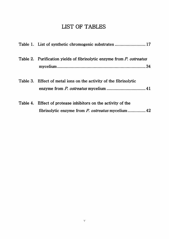
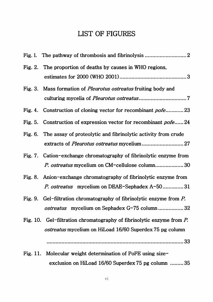
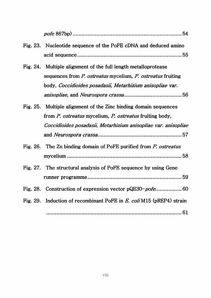