Plasma Membrane Rap. ① Phospholipids ② Cholesterol ③ Embedded Proteins ④ Glycoproteins ⑤...
43
Plasma Membrane Rap Chapter 7 Membrane Structure and Function
-
Upload
corey-nash -
Category
Documents
-
view
215 -
download
1
Transcript of Plasma Membrane Rap. ① Phospholipids ② Cholesterol ③ Embedded Proteins ④ Glycoproteins ⑤...
Plasma Membrane Rap
Chapter 7Membrane Structure and
Function

① Phospholipids② Cholesterol③ Embedded Proteins④ Glycoproteins⑤ Glycolipids
Makeup of the PM
CARBOHYDRATES
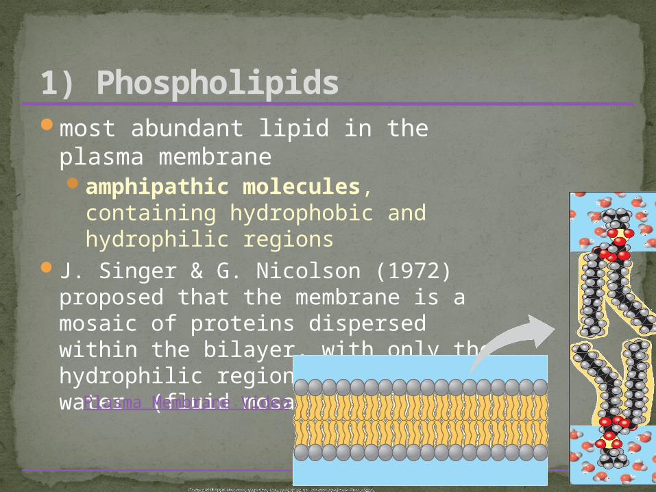
most abundant lipid in the plasma membraneamphipathic molecules, containing
hydrophobic and hydrophilic regionsJ. Singer & G. Nicolson (1972) proposed
that the membrane is a mosaic of proteins dispersed within the bilayer, with only the hydrophilic regions exposed to water (fluid mosaic model)
1) Phospholipids
Plasma Membrane Video
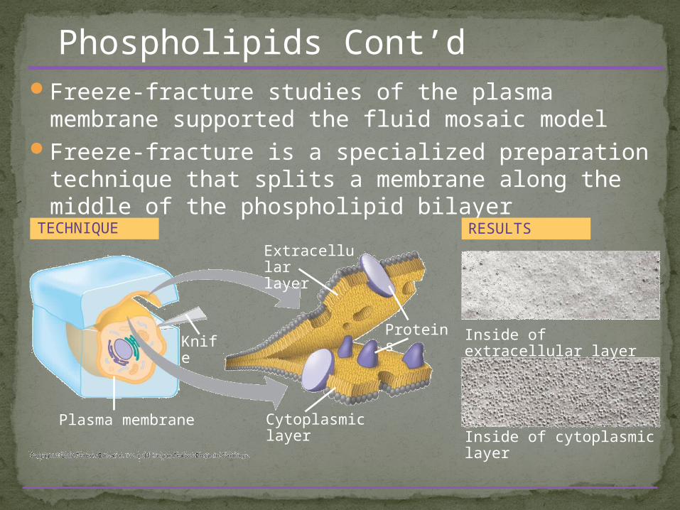
Freeze-fracture studies of the plasma membrane supported the fluid mosaic model
Freeze-fracture is a specialized preparation technique that splits a membrane along the middle of the phospholipid bilayer
Inside of cytoplasmic layer
TECHNIQUE
Extracellularlayer
KnifeProteins Inside of extracellular layer
RESULTS
Cytoplasmic layerPlasma membrane
Phospholipids Cont’d

Phospholipids can move within the bilayer
As temperatures cool, membranes become more solid
Membranes rich in unsaturated fatty acids are more fluid that those rich in saturated fatty acids
Membranes must be fluid to work properly; they are usually about as fluid as salad oil
Phospholipids Cont’d
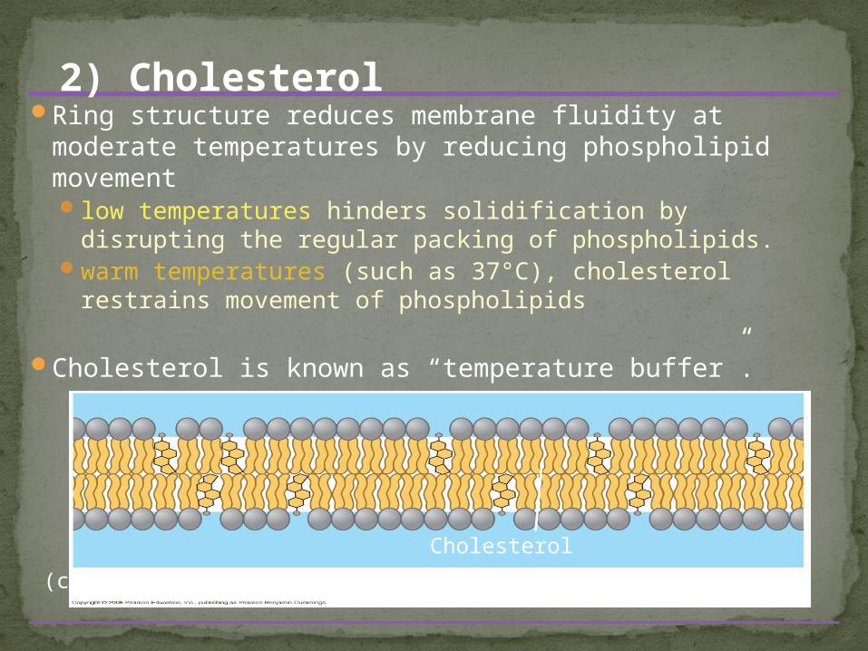
Ring structure reduces membrane fluidity at moderate temperatures by reducing phospholipid movementlow temperatures hinders solidification by disrupting the
regular packing of phospholipids.warm temperatures (such as 37°C), cholesterol restrains
movement of phospholipids
Cholesterol is known as “temperature buffer”.
Cholesterol
(c) Cholesterol within the animal cell membrane
2) Cholesterol
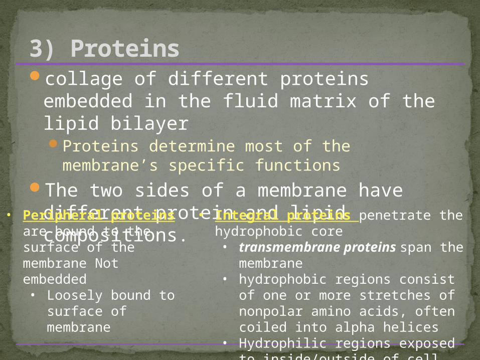
collage of different proteins embedded in the fluid matrix of the lipid bilayerProteins determine most of the membrane’s
specific functionsThe two sides of a membrane have
different protein and lipid compositions.
3) Proteins
• Peripheral proteins are bound to the surface of the membrane Not embedded• Loosely bound to
surface of membrane
• Integral proteins penetrate the hydrophobic core• transmembrane proteins span
the membrane• hydrophobic regions consist of
one or more stretches of nonpolar amino acids, often coiled into alpha helices
• Hydrophilic regions exposed to inside/outside of cell
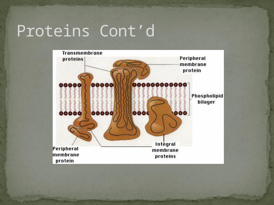
Proteins Cont’d
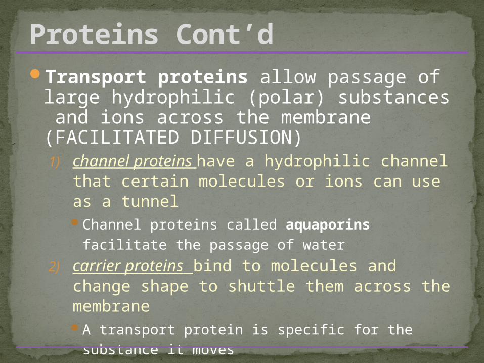
Transport proteins allow passage of large hydrophilic (polar) substances and ions across the membrane (FACILITATED DIFFUSION)1) channel proteins have a hydrophilic channel
that certain molecules or ions can use as a tunnelChannel proteins called aquaporins facilitate the
passage of water
2) carrier proteins bind to molecules and change shape to shuttle them across the membraneA transport protein is specific for the substance it
moves
Proteins Cont’d

Small, uncharged polar and nonpolar molecules can freely pass through a cell membraneHydrophobic (nonpolar) molecules, such as
hydrocarbons, can dissolve in the lipid bilayer and pass through the membrane rapidlyHydrocarbons, CO2 and O2
Polar molecules, such as sugars, do not cross the membrane easilyC6H12O6, or charged molecules
The Permeability of the Lipid Bilayer

Six major functionsTransportEnzymatic
activitySignal
transductionCell-cell
recognitionIntercellular
joiningAttachment to
the cytoskeleton and extracellular matrix (ECM)
Proteins Cont’d

The Role of Membrane Carbohydrates in Cell-Cell Recognition
Cells recognize each other by binding to surface molecules, usually carbohydrates
Membrane carbohydrates may be covalently bonded to lipids (forming glycolipids) or more commonly to proteins (forming glycoproteins)
Carbohydrates on the external side of the plasma membrane vary among species, individuals, and even cell types in an individual
Copyright © 2008 Pearson Education, Inc., publishing as Pearson Benjamin Cummings

Carbohydrates are the third major component of plasma membranes found on the exterior surface of cells
bound either to proteins (forming glycoproteins)or bound to lipids (forming glycolipids)
Chains can be either straight or branchedForm specialized sites on the cell surface that allow
cells to recognize each other. unique patterns that allow the cell to be recognized
allows the immune system to differentiate between body cells (called “self”) and foreign cells or tissues (called “non-self”).
Similar types of glycoproteins and glycolipids are found on the surfaces of viruses and may change frequently, preventing immune cells from recognizing and attacking them.
4) Glycoproteins & 5) Glycolipids

Glycoproteins & Glycolipids Cont’d

Substances diffuse down their concentration gradient (the difference in concentration of a substance from one area to another)From an area of high concentration to low
concentrationNo work must be done to move substances down
the concentration gradientO2 gets into cells this way for cellular respiration
Diffusion is the tendency for molecules to spread out evenly into the available space
At dynamic equilibrium, as many molecules cross one way as cross in the other directionThis is not static – even at equilibrium molecules are still
moving.
Passive Transport:
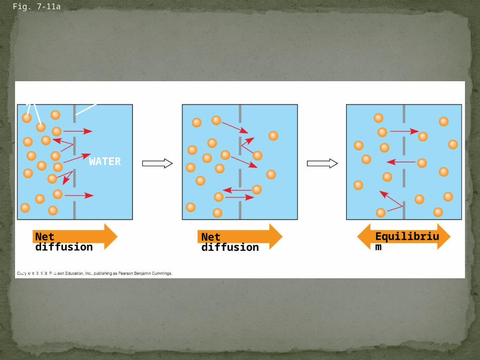
Molecules of dye
Fig. 7-11a
Membrane (cross section)
WATER
Net diffusion Net diffusion
(a) Diffusion of one solute
Equilibrium
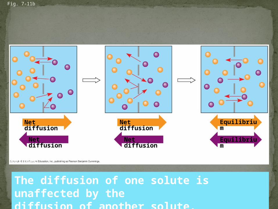
(b) Diffusion of two solutes
Fig. 7-11b
Net diffusion
Net diffusion
Net diffusion
Net diffusion
Equilibrium
Equilibrium
The diffusion of one solute is unaffected by the diffusion of another solute.

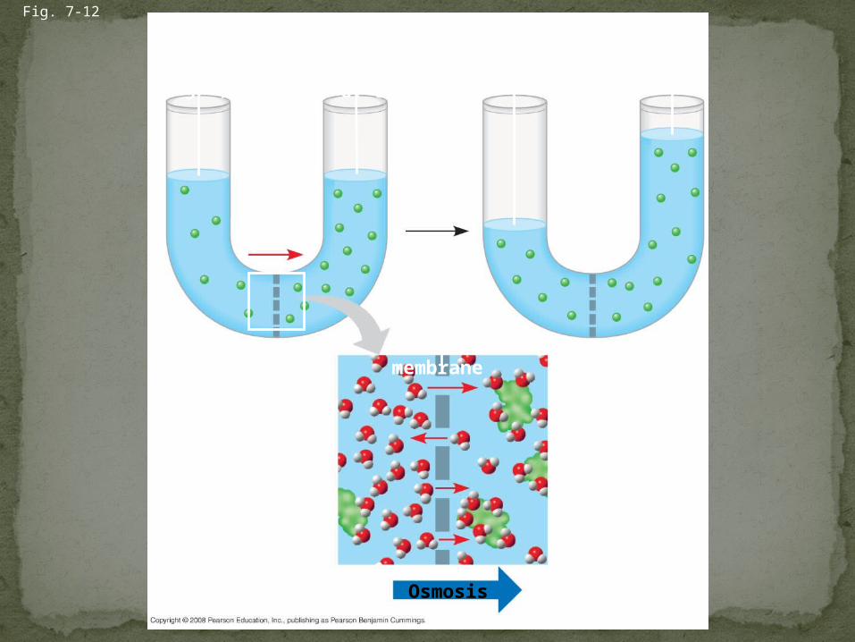
Osmosis is the diffusion of water across a selectively permeable membraneThe direction of osmosis is determined only by a
difference in total solute concentration. Water diffuses across a membrane from the
region of lower solute concentration to the region of higher solute concentrationor you can think [high water] to [low water]
Passive Transport Cont’d
Lowerconcentrationof solute (sugar)
Fig. 7-12
H2O
Higher concentrationof sugar
Selectivelypermeablemembrane
Same concentrationof sugar
Osmosis

Tonicity is the ability of a solution to cause a cell to gain or lose waterIsotonic solution: Solute concentration is the
same as that inside the cell; no net water movement across the plasma membrane
Hypertonic solution: Solute concentration is greater than that inside the cell; cell loses water
Hypotonic solution: Solute concentration is less than that inside the cell; cell gains water
Passive Transport Cont’d
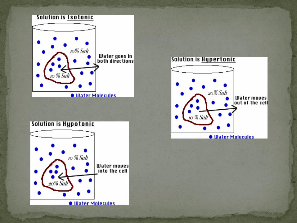

Fig. 7-13
Hypotonic solution
(a) Animal cell
(b) Plant cell
H2O
Lysed
H2O
Turgid (normal)
H2O
H2O
H2O
H2O
Normal
Isotonic solution
Flaccid
H2O
H2O
Shriveled
Plasmolyzed
Hypertonic solution
Hypertonic or hypotonic environments create osmotic problems for organisms
Osmoregulation, the control of water balance, is a necessary adaptation for life in such environmentsEx) The protist Paramecium, which is hypertonic
to its pond water environment, has a contractile vacuole that acts as a pump
Passive Transport Cont’d
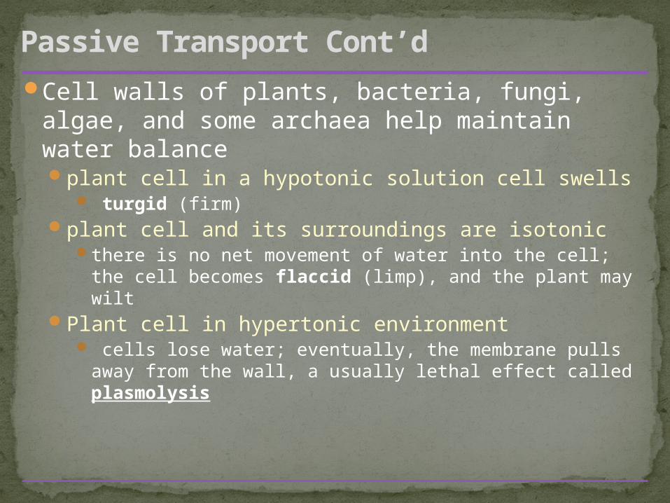
Cell walls of plants, bacteria, fungi, algae, and some archaea help maintain water balanceplant cell in a hypotonic solution cell swells
turgid (firm)plant cell and its surroundings are isotonic
there is no net movement of water into the cell; the cell becomes flaccid (limp), and the plant may wilt
Plant cell in hypertonic environment cells lose water; eventually, the membrane pulls
away from the wall, a usually lethal effect called plasmolysis
Passive Transport Cont’d
In facilitated diffusion, transport proteins speed the passive movement of molecules across the plasma membraneMost transport proteins are very specific2 types of transport proteins
Channel proteins and carrier proteins
Passive Transport Cont’d: Facilitated Diffusion

Channel proteins provide corridors that allow a specific molecule or ion to cross the membraneAquaporins, for facilitated diffusion of water –
speed up osmosisIon channels that open or close in response to
a stimulus (gated channels)
Passive Transport Cont’d: Facilitated Diffusion via Channel Proteins

Carrier proteins transport substances out of or into the cell by facilitated diffusion and active transport.Each designed to recognize only ONE substance or
ONE group of very similar substances. Diffusion of sugars, amino acids, nucleoside. Uptake of glucose. Transportation of salts, glucose, and amino acids
Passive Transport Cont’d: Facilitated Diffusion via Carrier Proteins
undergo a subtle change in shape that translocates the solute-binding site across the membrane
Substances diffuse against their concentration gradientFrom an area of low concentration to high
concentrationWork must be done to move substances from low
to highperformed by specific proteins embedded in the
membranesAllows cells to maintain concentration gradients that
differ from their surroundingsEx) sodium-potassium pump
Active Transport

Figure 5-14 Mechanism of the Na+-K+-ATPase
ADP
ATP
ATPase isphosphorylated
with Pi from ATP.
Protein changesconformation.
ICF
ECF
Protein changesconformation.
3 Na+ fromICF bind
3 Na+ releasedinto ECF
2 K+ fromECF bind
2 K+ releasedinto ICF
1
25
4 3
Membrane potential is the voltage difference across a membraneVoltage is created by differences in the
distribution of positive and negative ionsCells have a net negative internal charge
Active Transport Cont’d:Ion Pumps
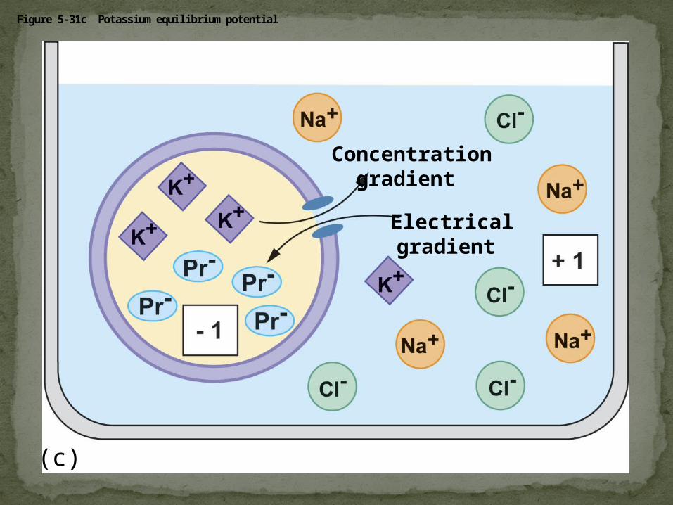
Figure 5-31c Potassium equilibrium potential
Concentrationgradient
Electricalgradient
(c)
Two combined forces, collectively called the electrochemical gradient, drive the diffusion of ions across a membrane:A chemical force (the ion’s concentration
gradient)An electrical force (the effect of the membrane
potential on the ion’s movement)

An electrogenic pump is a transport protein that generates voltage across a membraneEX: Sodium-Potassium pump – major electrogenic
pump of animals3Na+out 2K+ in = overall1 positive charge to
the extracellular fluidThe main electrogenic pump of plants, fungi,
and bacteria is a proton pumpThese pumps generate voltage across
membranes which stores energy for use for the cell
Large molecules cross the membrane in bulk via vesiclespolysaccharides and proteins
Bulk transport requires energy
Active Transport Cont’d:Bulk transport: exocytosis and endocytosis
Transport vesicles migrate to the membrane, fuse with it, and release their contents
Many secretory cells use exocytosis to export their productsPancreatic beta cells releasing insulinNeurons releasing neurotransmitersPlants making cell walls
Exocytosis

Cell takes in macromolecules by forming vesicles from the plasma membranereversal of exocytosis, involving different
proteinsThere are three types of endocytosis:
Phagocytosis (“cellular eating”)Pinocytosis (“cellular drinking”)Receptor-mediated endocytosis
Endocytosis
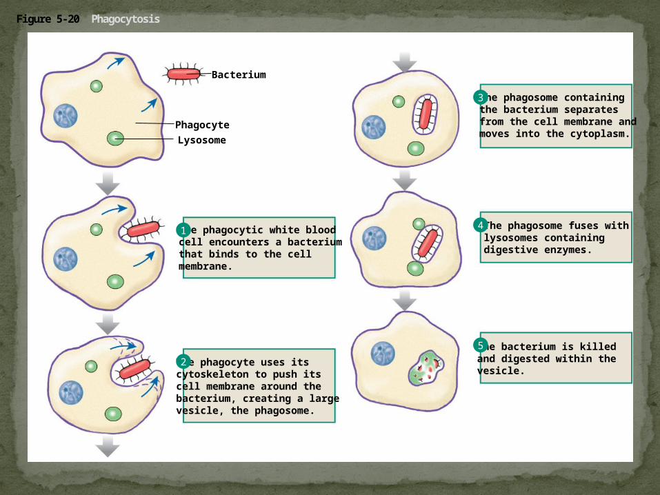
Figure 5-20 Phagocytosis
Bacterium
Lysosome
Phagocyte
The phagocytic white bloodcell encounters a bacteriumthat binds to the cellmembrane.
The phagocyte uses itscytoskeleton to push itscell membrane around thebacterium, creating a largevesicle, the phagosome.
The phagosome containingthe bacterium separatesfrom the cell membrane andmoves into the cytoplasm.
The phagosome fuses withlysosomes containing digestive enzymes.
The bacterium is killedand digested within thevesicle.
1
2
3
4
5

In phagocytosis (cellular eating) a cell engulfs a particle in a vacuoleThe vacuole fuses with a lysosome to digest the
particleIn pinocytosis,( cellular drinking) molecules are
taken up when extracellular fluid is “gulped” into tiny vesicles
In receptor-mediated endocytosis, (picky eater) binding of ligands to receptors triggers vesicle formationA ligand is any molecule that binds specifically
to a receptor site of another molecule

Figure 5-21 Receptor-mediated endocytosis and exocytosis
Ligand binds to membrane receptor.
Clathrin-coatedpit
Receptor
Extracellular fluid
Intracellular fluid
To lysosome orGolgi complex
Receptor-ligand migrates to clathrin-coated pit.
Endocytosis
Vesicle losesclathrin coat.
Ligands go to lysosomesor Golgi for processing.
Transport vesiclewith receptors moves to the cell membrane.
Transport vesicleand cell membranefuse (membranerecycling).
Exocytosis
Clathrin
Endosome
1
2
3
4
5
6
7
8
9
Receptorsand ligands separate.
1. Define the following terms: amphipathic molecules, aquaporins, diffusion
2. Explain how membrane fluidity is influenced by temperature and membrane composition
3. Distinguish between the following pairs or sets of terms: peripheral and integral membrane proteins; channel and carrier proteins; osmosis, facilitated diffusion, and active transport; hypertonic, hypotonic, and isotonic solutions
You should now be able to:
4. Explain how transport proteins facilitate diffusion
5. Explain how an electrogenic pump creates voltage across a membrane, and name two electrogenic pumps
6. Explain how large molecules are transported across a cell membrane
You should now be able to: