Glycoproteins at the rubbing interfaces of biosystemsorbit.dtu.dk/files/5156975/Proteins DTU_2010...
33
General rights Copyright and moral rights for the publications made accessible in the public portal are retained by the authors and/or other copyright owners and it is a condition of accessing publications that users recognise and abide by the legal requirements associated with these rights. • Users may download and print one copy of any publication from the public portal for the purpose of private study or research. • You may not further distribute the material or use it for any profit-making activity or commercial gain • You may freely distribute the URL identifying the publication in the public portal If you believe that this document breaches copyright please contact us providing details, and we will remove access to the work immediately and investigate your claim. Downloaded from orbit.dtu.dk on: Jun 14, 2018 Glycoproteins at the rubbing interfaces of biosystems Lee, Seunghwan Publication date: 2010 Document Version Publisher's PDF, also known as Version of record Link back to DTU Orbit Citation (APA): Lee, S. (2010). Glycoproteins at the rubbing interfaces of biosystems [Sound/Visual production (digital)]. 4th Protein.DTU Workshop, Technical University of Denmark, 01/01/2010
Transcript of Glycoproteins at the rubbing interfaces of biosystemsorbit.dtu.dk/files/5156975/Proteins DTU_2010...

General rights Copyright and moral rights for the publications made accessible in the public portal are retained by the authors and/or other copyright owners and it is a condition of accessing publications that users recognise and abide by the legal requirements associated with these rights.
• Users may download and print one copy of any publication from the public portal for the purpose of private study or research. • You may not further distribute the material or use it for any profit-making activity or commercial gain • You may freely distribute the URL identifying the publication in the public portal
If you believe that this document breaches copyright please contact us providing details, and we will remove access to the work immediately and investigate your claim.
Downloaded from orbit.dtu.dk on: Jun 14, 2018
Glycoproteins at the rubbing interfaces of biosystems
Lee, Seunghwan
Publication date:2010
Document VersionPublisher's PDF, also known as Version of record
Link back to DTU Orbit
Citation (APA):Lee, S. (2010). Glycoproteins at the rubbing interfaces of biosystems [Sound/Visual production (digital)]. 4thProtein.DTU Workshop, Technical University of Denmark, 01/01/2010

Glycoproteins at the Rubbing Interfaces of BiosystemsInterfaces of Biosystems
4th Workshop in Proteins.DTUNovember 12, 2010, DTU
Seunghwan LeeDepartment of Mechanical Engineering, DTU
Contact: [email protected]

Water as a lubricant
Oil
Nature’s primary choice of lubricant
Men’s primary choice of lubricant
Water
lubricantlubricant
Challenges in oil‐based lubrication:f h til l 0 001!
g
limited resources
environmental issue (especially additives)
µ for human cartilage: as low as 0.001!

Water as a lubricant in engineering point of view
‐ non‐toxic
‐ environmentally‐friendly
readily available and cost effective
‐ poor pressure response
low pressure‐coefficient of viscosity‐ readily available and cost effective
‐ non‐flammable
‐ high thermal capacity
p ywater: α = 0.36 GPa‐1
oil: α = 10‐20 GPa‐1
limited application temperature‐ biocompatible
‐ limited application temperature
‐ corrosion for ferrous materials
water oil

Nature’s approach to use water as lubricant
brush‐like, sugar‐basedmacromolecules
Mucins
Mucus (gel)Mucin (polymer)
P t l t
PGM (STM, 360 nm × 360 nm)
Roberts, CJ et al Proteins and Proteoglycan aggregate
• plays a key structural role in cartilage
Peptide Letters 1995 2, 409
Lubricin
• mucinous glycoprotein of the synovialsugar chains
link protein
hyaluronan
core protein
g y p yfluid (250 µg/ml, MW = 2.3 × 10⁵ g/mol)
S. Lee et al., SCIENCE 2008
y

Lubricity of mucins/mucus gels

Mucus, Mucin, and Mucin Domains
stiff , charged, hydrophilic, ca. 70% of mass
Schematic representation of the mucin
mucus (gel)2HN COOH
maybe flexible, charged, hydrophobic/‐philic
mucus (gel)
Water
Salts
IgG
polypeptide
Proteins
mucins
mucin (polymer)
Schematic representation of the Lubricin (PRG 4)
mucin (polymer)
Zappone B et al, Langmuir 2008, 24, 1495.
Roberts, CJ et al, Proteins and Peptide Letters 1995, 2, 409
PGM, STM (360 nm � 360 nm)

Mucus gels
Hattrup CL and Gendler SJ, Ann. Rev. Physol. 2008, 70, 431
S‐S S‐S
Gel‐formation (in vivo)disulfide bonding
hydrophobic interaction sugar‐sugar interaction

Monolayer of mucins at water/solid interface
(sub)monolayer surface‐coating(sub)monolayer surface‐coatingwater
solid
Hydrophilicity
L. Shi and K.D. Caldwell, J. Colloid & Interf. Sci. (2000) 224, 372‐381
Suppression of proteins and bacteria adsorptionSuppression of proteins and bacteria adsorption L. Shi, R. Ardehali, P. Valint, & K.D. Caldwell, Biotech. Letters (2001) 23, 437‐441
LubricationI.C.H. Berg, L. Lindh & T. Arnebrant, Biofouling (2004) 20, 65‐70
S. Lee, M. Müller, K. Rezwan, N.D. Spencer, Langmuir (2005) 21, 8344‐8353
h h l l
2HN COOH
Mucins as a amphiphilic copolymer
‐CH2CH2O‐ ‐CH2CH2O‐‐CH2CHO‐
m mnCH3
m mn

Model surface and pin‐on‐disk tribometry
pin‐on‐disk tribometerElastomer as model surface of biological i h l
loaddead
tissues: mimic mechanical properties
lever
Pin (PDMS)
deadweight
PGM-containingYoung´s modulus
Poly(dimethylsiloxane) (PDMS)
aqueous solutionYoung s modulus
ca. 2 MPa
Poission ratio
rotationalmotion
disk (PDMS)
L d 1 N
Poission ratio
0.5
side view
Load = 1 N
P ~ 0.5 MPa

Lubrication properties of PGM solution: pH 7

Lubrication properties of PGM solution: pH 2

Lubrication properties of PGM solution: pH 12

Optical Waveguide Lightmode Spectroscopy (OWLS)
Surface adsorption properties: OWLS
Optical Waveguide Lightmode Spectroscopy (OWLS)
Adsorption of mucins onto PDMS surface dso pt o o uc s o to S su ace
*PDMS (~30nm)
Waveguide(SiOx0.75TiOx0.25)
TM TE
incidence angle
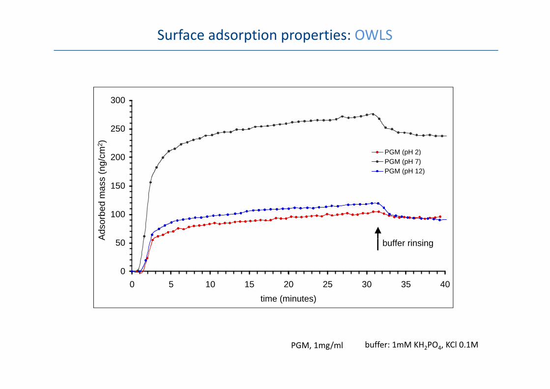
Surface adsorption properties: OWLS
300
250
PGM ( H 2)m2 )
150
200 PGM (pH 2)PGM (pH 7)PGM (pH 12)
mas
s (n
g/cm
100
Adso
rbed
m
0
50
0 5 10 15 20 25 30 35 40
buffer rinsingA
0 5 10 15 20 25 30 35 40time (minutes)
PGM, 1mg/ml buffer: 1mM KH2PO4, KCl 0.1M

Surface adsorption properties: OWLS
pH and ionic strength dependence
300pH 2pH 7
H 12ng/c
m2 )
200
pH 12
d m
ass
(n
100
Ads
orbe
d
00 0001 0 001 0 01 0 1 1 10
A
0.0001 0.001 0.01 0.1 1 10
total ionic strength (M)

Tertiary structure: Near‐UV CD spectroscopy
pH dependence
20
30pH2pH4pH7
pH 7
0
10pH10pH12
-20
-10pH 2
-30250 270 290 310 330 350
wavelength (nm)
disruption of tertiary structure of “naked” polypeptide region
a e e g ( )
PGM, 1mg/ml buffer: 1mM KH2PO4, KCl 0.1M

Tertiary structure: Near‐UV CD spectroscopy
Ionic strength dependence
20
30pH2 (buffer only)pH2 (KCl, 0.01M)pH2 (KCl, 0.1M)pH2 (KCl, 1.0M)
0
10
p ( )pH7 (buffer only)pH7 (KCl, 0.01M)pH7 (KCl, 0.1M)pH7 (KCl, 1.0M)pH12 (buffer only)pH12 (KCl, 0.01M)
-20
-10pH12 (KCl, 0.1M)pH12 (KCl, 1.0M)
-30250 270 290 310 330 350
wavelength (nm)a e e g ( )
PGM, 1mg/ml buffer: 1mM KH2PO4, KCl 0.1M

A schematic model at the sliding interface
Before sliding After sliding
1 turnpH 71
n (N
)pH 7
0,5frict
ion
pH 12
00 1250
rotation (or accumulated scan length) pH 2
pH 2

Model polysaccharides : Dextran and Hyaluronic acid
1
10 Lubrication
0 1
1
μbuffer (pH7)buffer (pH2)
Dextran
Hyaluronic acid 0.01
0.1 buffer (pH2)dextran (pH7)dextran (pH2)HA (pH7)HA (pH2)
0.011 10 100
speed (mm/sec)
Adsorption
200
250
300
Hyaluronic AcidDextran
Adsorption
0
100
150
0
50
0 5 10 15 20 25 30time (min)
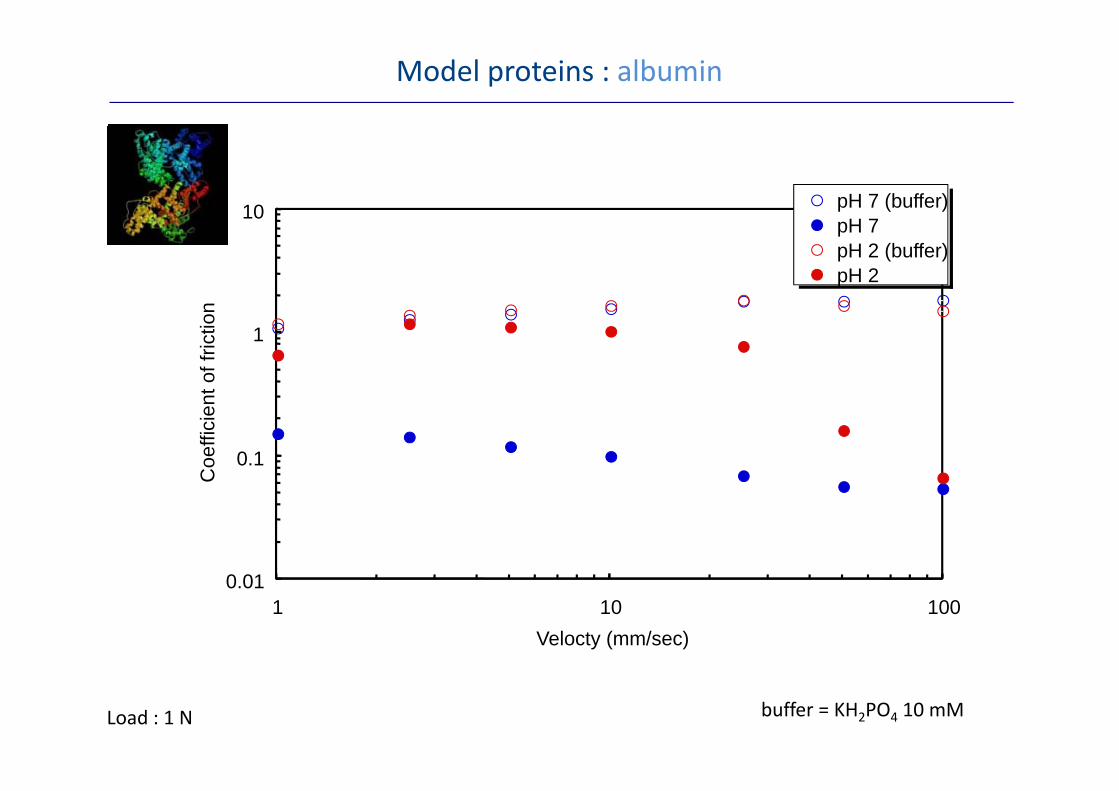
Model proteins : albumin
10 pH 7 (buffer)10n
pH 7pH 2 (buffer)pH 2
1
t of f
rictio
n
0.1
Coe
ffici
en
0 010.011 10 100
Velocty (mm/sec)
buffer = KH2PO4 10 mMLoad : 1 N

H k D E f h i
Soft Elastohydrodynamic Lubrication (soft EHL)
Hamrock, Dowson, EsfahanianHamrock, B.J. and Dowson, D., Proc. 5th Leeds‐Lyon symp. on Trib. 22‐27 (1979)
Esfahanian, M. and Hamrock, B.J., Tribol. Trans. 34, 628‐632 (1991)
H d EHL h 0 47 0 49 0 68 0 68 0 12 0 07Hard EHL
Soft EHL
hmin = 1.79 R0.47α 0.49 η0 0.68 U0.68 E‐0.12 W‐0.07
hmin = 2.8 R0. η0 0.65 U0.65 E‐0..44 W‐0.21
α : pressure coefficient of viscosity
rigidrigid elastic
300
R = 3 mm
1 N Soft contact: PDMS vs. PDMS
Rigid contact: steel vs. steel 200
250"soft" contacts
"rigid" contacts
ess (
nm)
E (PDMS) = 2 MPa
ν (PDMS) = 0.5
50
100
150fil
m th
ickn
e
E (steel) = 200 GPa
ν (steel) = 0.3 0
50
0.0001 0.001 0.01 0.1 1
f
speed (m/s)

1010
Effect of surface hydrophilicity
PDMS
PDMS1
10
1
10
0.1μ O2 plasma
ox‐PDMS
0.1μ
0.010.1 1 10 100
speed (mm/sec)
ox‐PDMS0.01
0.1 1 10 100speed (mm/sec)
No significant change in bulkNo significant change in bulk mechanical properties
Hydrophilization of surface ( OH and/or COOH groups)(‐OH and/or ‐COOH groups)

PEO‐b‐PPO‐b‐PEO (Pluronic®)
CH2CH2O CH2CH2OCH2CHO
CH3n nm
PEO‐b‐PPO‐b‐PEO
3
f PEO
% of
M.W. of PPO

PEO‐b‐PPO‐b‐PEO
Adsorption of albumin
1
10
0.1
1μ
bufferF68P105
0.010.001 0.01 0.1
speed (m/sec)J. Biomed. Mat. Res., 1998, R.J. Green et al
speed (m/sec)
F68 EO76.4 PO29 EO76.4
tribostress
P105 EO PO EO
tribostress
P105 EO36.9 PO56 EO36.9

Mucins from different organs: similarity and difference
pH 2pH 7
PDMS
PDMS
PDMS
PDMS
Bansil et al, Annu. Rev. Physiol. 1995, 57, 635.
10PGM
(Porcine Gastric Mucin)
1010
111pH 7 pH 2
0.1
μ BSM(Bovine Submaxillary Mucin)
0.1
μ0.1
μ
0 010 010 01
pH 7 pH 2
0.010.1 1 10 100
sliding speed (mm/s) S. Lee et al., unpublished
0.010.1 1 10 100
sliding speed (mm/s)
0.010.1 1 10 100
sliding speed (mm/s)

BSM vs. PGM: Adsorption behavior
200
150
g/c
m2)
100
bed
mas
s (n
g
50Adso
rb
2 2 770
1 2
PGM BSM

BSM vs. PGM: Size (Dynamic Light Scattering)
30pH 7
pH 2
pH 7
10
20 pH 2
01 10 100 1000 10000
diameter (nm)diameter (nm)
1
2
Increase in size (aggregation) at pH 2 is general for both mucins,
0 50 100 150 200Z-average (nm)
pH 2 is general for both mucins, but is more pronounced for PGMthan BSM
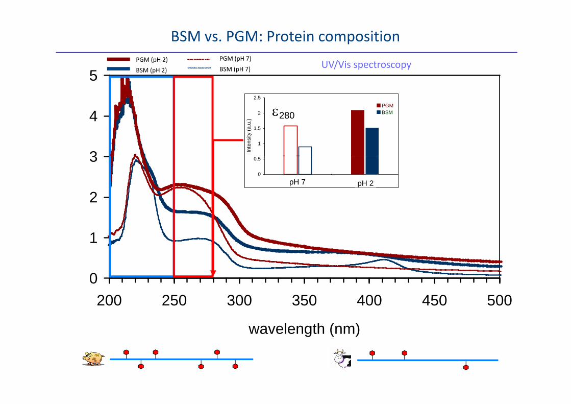
PGM (pH 2) PGM (pH 7)UV/Vis spectroscopy
BSM vs. PGM: Protein composition
4
52.5
PGMε
BSM (pH 2) BSM (pH 7) UV/Vis spectroscopy
3
41
1.5
2
Inte
nsity
(a.u
.)
BSMε280
2
30
0.5
1 2
pH 7 pH 2
1
2
0200 250 300 350 400 450 500
wavelength (nm)

Tertiary structure: Near UV CDTertiary structure: Near UV CDSecondary structure: Far UV CDSecondary structure: Far UV CD
BSM vs. PGM: Protein conformation
Tertiary structure: Near‐UV CD Tertiary structure: Near‐UV CD
20
30Secondary structure: Far‐UV CD Secondary structure: Far‐UV CD
40
60
-10
0
10
-20
0
20
-30
-20
250 300 350-60
-40
200 210 220 230 240 250 250 300 350wavelength (nm)
200 210 220 230 240 250
wavelength (nm)
300Protein unfolding: Fluorescence Spec.Protein unfolding: Fluorescence Spec.
150
200
250
50
100
150
0300 320 340 360 380 400 420 440
wavelength (nm)

A proposed model: In bulk solution

A proposed model: At liquid/solid interface
pH 7pH 7p
tribostress
pH 2pH 2
tribostress

Thank you for your attention!y y