Identification of sialylated glycoproteins from ...
13
RESEARCH Open Access Identification of sialylated glycoproteins from metabolically oligosaccharide engineered pancreatic cells Yuan Tian 1 , Ruben T Almaraz 2 , Caitlin H Choi 1 , Qing Kay Li 1 , Christopher Saeui 2 , Danni Li 1 , Punit Shah 1 , Rahul Bhattacharya 2 , Kevin J Yarema 2 and Hui Zhang 1* Abstract In this study, we investigated the use of metabolic oligosaccharide engineering and bio-orthogonal ligation reactions combined with lectin microarray and mass spectrometry to analyze sialoglycoproteins in the SW1990 human pancreatic cancer line. Specifically, cells were treated with the azido N-acetylmannosamine analog, 1,3,4-Bu 3 ManNAz, to label sialoglycoproteins with azide-modified sialic acids. The metabolically labeled sialoglyproteins were then biotinylated via the Staudinger ligation, and sialoglycopeptides containing azido-sialic acid glycans were immobilized to a solid support. The peptides linked to metabolically labeled sialylated glycans were then released from sialoglycopeptides and analyzed by mass spectrometry; in parallel, the glycans from azido-sialoglycoproteins were characterized by lectin microarrays. This method identified 75 unique N-glycosite-containing peptides from 55 different metabolically labeled sialoglycoproteins of which 42 were previously linked to cancer in the literature. A comparison of two of these glycoproteins, LAMP1 and ORP150, in histological tumor samples showed overexpression of these proteins in the cancerous tissue demonstrating that our approach constitutes a viable strategy to identify and discover sialoglycoproteins associated with cancer, which can serve as biomarkers for cancer diagnosis or targets for therapy. Keywords: Sialylated glycoproteins, Metabolic oligosaccharide engineering, Pancreatic cancer cells Introduction Altered patterns of glycosylation are a universal feature of cancer with sialic acids – which are a group of un- usual amino-modified acidic sugars ubiquitously dis- played on the outer ends of mammalian glycan chains [1] – playing an especially prominent role as tumor- associated carbohydrate antigens (TACAs) [2,3]. TACAs, in general, are involved in many biological and patho- logical processes, such as regulating cellular and molecu- lar interactions by either masking recognition sites or serving as recognition determinants [4]. More specific- ally, altered sialylation of tumor cell surfaces is associ- ated with several critical malignant properties that include invasiveness and metastatic potential [5-7]. Cell surface sialylation is controlled by several factors, which include regulation of metabolic flux of ManNAc into the sialic biosynthetic pathway [8,9], changes in the expres- sion and enzymatic activities of sialyltransferases and sialidases, and the availability of candidate penultimate glycan termini on glycoproteins that serve as acceptor sites for sialylation [5,10]. An interesting observation made about two decades ago was that cancer cells expressed the non-human “Neu5Gc” form of sialic acid at much higher levels than normal tis- sue [11]. The accumulation of Neu5Gc in tumors was at- tributed to scavenge of dietary Neu5Gc [12], which was ultimately biosynthetically incorporated into sialylated TACAs at much higher levels than into the sialoglycans of healthy cells. Building on the observation, the enhanced ability of tumors to over-express types of exogenously- supplied sialic acid other than the canonical human “Neu5Ac” form, has been proposed as a new approach to cancer therapy with Neu5Prop and Neu5Phenyl an- alogs being evaluated for immunotherapy purposes * Correspondence: [email protected] 1 Department of Pathology, Johns Hopkins University, 400 N. Broadway, Room 4011, Baltimore MD 21287, USA Full list of author information is available at the end of the article CLINICAL PROTEOMICS © 2015 Tian et al.; licensee BioMed Central. This is an Open Access article distributed under the terms of the Creative Commons Attribution License (http://creativecommons.org/licenses/by/4.0), which permits unrestricted use, distribution, and reproduction in any medium, provided the original work is properly credited. The Creative Commons Public Domain Dedication waiver (http://creativecommons.org/publicdomain/zero/1.0/) applies to the data made available in this article, unless otherwise stated. Tian et al. Clinical Proteomics (2015) 12:11 DOI 10.1186/s12014-015-9083-8
Transcript of Identification of sialylated glycoproteins from ...

CLINICALPROTEOMICS
Tian et al. Clinical Proteomics (2015) 12:11 DOI 10.1186/s12014-015-9083-8
RESEARCH Open Access
Identification of sialylated glycoproteins frommetabolically oligosaccharide engineeredpancreatic cellsYuan Tian1, Ruben T Almaraz2, Caitlin H Choi1, Qing Kay Li1, Christopher Saeui2, Danni Li1, Punit Shah1,Rahul Bhattacharya2, Kevin J Yarema2 and Hui Zhang1*
Abstract
In this study, we investigated the use of metabolic oligosaccharide engineering and bio-orthogonal ligation reactionscombined with lectin microarray and mass spectrometry to analyze sialoglycoproteins in the SW1990 humanpancreatic cancer line. Specifically, cells were treated with the azido N-acetylmannosamine analog, 1,3,4-Bu3ManNAz,to label sialoglycoproteins with azide-modified sialic acids. The metabolically labeled sialoglyproteins were thenbiotinylated via the Staudinger ligation, and sialoglycopeptides containing azido-sialic acid glycans were immobilizedto a solid support. The peptides linked to metabolically labeled sialylated glycans were then released fromsialoglycopeptides and analyzed by mass spectrometry; in parallel, the glycans from azido-sialoglycoproteinswere characterized by lectin microarrays. This method identified 75 unique N-glycosite-containing peptidesfrom 55 different metabolically labeled sialoglycoproteins of which 42 were previously linked to cancer in theliterature. A comparison of two of these glycoproteins, LAMP1 and ORP150, in histological tumor samplesshowed overexpression of these proteins in the cancerous tissue demonstrating that our approach constitutesa viable strategy to identify and discover sialoglycoproteins associated with cancer, which can serve as biomarkersfor cancer diagnosis or targets for therapy.
Keywords: Sialylated glycoproteins, Metabolic oligosaccharide engineering, Pancreatic cancer cells
IntroductionAltered patterns of glycosylation are a universal featureof cancer with sialic acids – which are a group of un-usual amino-modified acidic sugars ubiquitously dis-played on the outer ends of mammalian glycan chains[1] – playing an especially prominent role as tumor-associated carbohydrate antigens (TACAs) [2,3]. TACAs,in general, are involved in many biological and patho-logical processes, such as regulating cellular and molecu-lar interactions by either masking recognition sites orserving as recognition determinants [4]. More specific-ally, altered sialylation of tumor cell surfaces is associ-ated with several critical malignant properties thatinclude invasiveness and metastatic potential [5-7]. Cellsurface sialylation is controlled by several factors, which
* Correspondence: [email protected] of Pathology, Johns Hopkins University, 400 N. Broadway,Room 4011, Baltimore MD 21287, USAFull list of author information is available at the end of the article
© 2015 Tian et al.; licensee BioMed Central. ThCommons Attribution License (http://creativecreproduction in any medium, provided the orDedication waiver (http://creativecommons.orunless otherwise stated.
include regulation of metabolic flux of ManNAc into thesialic biosynthetic pathway [8,9], changes in the expres-sion and enzymatic activities of sialyltransferases andsialidases, and the availability of candidate penultimateglycan termini on glycoproteins that serve as acceptorsites for sialylation [5,10].An interesting observation made about two decades ago
was that cancer cells expressed the non-human “Neu5Gc”form of sialic acid at much higher levels than normal tis-sue [11]. The accumulation of Neu5Gc in tumors was at-tributed to scavenge of dietary Neu5Gc [12], which wasultimately biosynthetically incorporated into sialylatedTACAs at much higher levels than into the sialoglycans ofhealthy cells. Building on the observation, the enhancedability of tumors to over-express types of exogenously-supplied sialic acid other than the canonical human“Neu5Ac” form, has been proposed as a new approachto cancer therapy with Neu5Prop and Neu5Phenyl an-alogs being evaluated for immunotherapy purposes
is is an Open Access article distributed under the terms of the Creativeommons.org/licenses/by/4.0), which permits unrestricted use, distribution, andiginal work is properly credited. The Creative Commons Public Domaing/publicdomain/zero/1.0/) applies to the data made available in this article,
Tian et al. Clinical Proteomics (2015) 12:11 Page 2 of 13
[13]. Similarly, sialic acid precursors with chemical func-tional groups such as the ketone [14] or azide [15] notnormally found in mammalian sugars have been appliedto the preferential delivery of diagnostic or therapeuticagents to tumors. The emerging success of these strategiesis evident in the recent in vivo demonstration that azido-modified sialic acids derived from corresponding ManNAcmetabolic precursors are highly expressed in tumors com-pared to healthy tissues [16].While a variety of high-throughput approaches have
been used to study sialylated glycoproteins [17], whichinclude lectin affinity [6,10,18], titanium dioxide affinitychromatography [19], strong cation exchange columns[20], conditional hydrazide chemistry [7,18,21-24], or thecombination of diagonal chromatographic technology andneuraminidase treatments [25], the above-mentionedmetabolic oligosaccharide engineering strategy is an emer-ging chemical method to analyze glycoproteins that hasbecome increasingly used in the past decade [15,26]. Inthis approach, an unnatural monosaccharide with a bio-orthogonal functional group can be introduced into thebiosynthetic pathways of living cells and incorporatedinto cellular glycoproteins. The bio-orthogonal functionalgroup can function as a chemical handle and be taggedwith complementary reactive partners via covalent ligationreactions. An important feature of metabolic oligosacchar-ide engineering is that the labeling of glycans can occurin both living cells and animals and the method canbe used to manipulate and study glycosylation events[27]. Accordingly, this method is becoming valuable forglycoproteomics studies in both eukaryotic and bacterialsystems with investigation of cancer a particularly activearea of research [27-32].Based on the selective in vivo incorporation of exogen-
ously supplied monosaccharides into tumors, as discussedabove, we reasoned that a metabolic oligosaccharide en-gineering approach could be used to meet a present needin understanding cancer, which is to more thoroughlycharacterize the role of sialoglycoproteins in tumor pro-gression. As a caveat, the selective incorporation of non-natural sialic acids into tumors could simply be a conse-quence of the increased metabolic activity of cancer cellscompared to most normal cells found in the body. How-ever, a previous study where we treated the humanSW1990 pancreatic cancer cell line with the “high flux”1,3,4-O-Bu3ManNAc analog that increases levels of nat-ural sialic acid , showed that increased sialylation did notoccur evenly across all glycoproteins. Instead, certain gly-coproteins did not experience a measurable increase insialylation while important cancer-related markers includ-ing CD44 and integrin α6 [9] as well as EGFR [33] experi-enced increased levels of sialic acid of 2-fold or more.Increased levels of natural sialic acid, however, hold lesspotential to manipulate or study the system compared to
a similar approach where non-natural sialosides aremetabolically installed into cancer-associated glycans.Therefore, in the present study, we investigated whethertreatment of SW1990 cells with the non-natural azido-modified sialic acid precursor 1,3,4-O-Bu3ManNAz [34]would similarly preferentially label cancer-associatedsialoglycans and, by doing so, provide a method for thediscovery, isolation, and study of this important class oftumor-promoting molecules.To experimentally verify the incorporation of azido-sialic
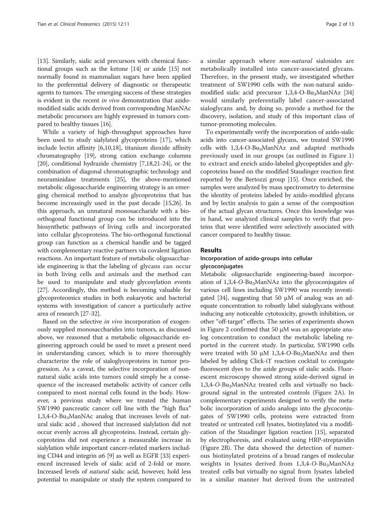
acids into cancer-associated glycans, we treated SW1990cells with 1,3,4-O-Bu3ManNAz and adapted methodspreviously used in our groups (as outlined in Figure 1)to extract and enrich azido-labeled glycopeptides and gly-coproteins based on the modified Staudinger reaction firstreported by the Bertozzi group [15]. Once enriched, thesamples were analyzed by mass spectrometry to determinethe identity of proteins labeled by azido-modified glycansand by lectin analysis to gain a sense of the compositionof the actual glycan structures. Once this knowledge wasin hand, we analyzed clinical samples to verify that pro-teins that were identified were selectively associated withcancer compared to healthy tissue.
ResultsIncorporation of azido-groups into cellularglycoconjugatesMetabolic oligosaccharide engineering-based incorpor-ation of 1,3,4-O-Bu3ManNAz into the glycoconjugates ofvarious cell lines including SW1990 was recently investi-gated [34], suggesting that 50 μM of analog was an ad-equate concentration to robustly label sialoglycans withoutinducing any noticeable cytotoxicity, growth inhibition, orother “off-target” effects. The series of experiments shownin Figure 2 confirmed that 50 μM was an appropriate ana-log concentration to conduct the metabolic labeling re-ported in the current study. In particular, SW1990 cellswere treated with 50 μM 1,3,4-O-Bu3ManNAz and thenlabeled by adding Click-iT reaction cocktail to conjugatefluorescent dyes to the azide groups of sialic acids. Fluor-escent microscopy showed strong azide-derived signal in1,3,4-O-Bu3ManNAz treated cells and virtually no back-ground signal in the untreated controls (Figure 2A). Incomplementary experiments designed to verify the meta-bolic incorporation of azido analogs into the glycoconju-gates of SW1990 cells, proteins were extracted fromtreated or untreated cell lysates, biotinylated via a modifi-cation of the Staudinger ligation reaction [15], separatedby electrophoresis, and evaluated using HRP-streptavidin(Figure 2B). The data showed the detection of numer-ous biotinylated proteins of a broad ranges of molecularweights in lysates derived from 1,3,4-O-Bu3ManNAztreated cells but virtually no signal from lysates labeledin a similar manner but derived from the untreated
Biotin-phosphine
Cell
Biotinylated azido-peptides
Streptavidinagarose
Formerly N-sialoglycopeptides
1 2
3
6
7
8
4
Streptavidinagarose
5
Biotinylated azido-proteins
Glycan information
9
Peptide identification
Cell
Azido-labeled glycoproteinsAzido-labeled cell surface glycans
Figure 1 Strategy for analyzing azide-modified sialoglycoproteins. The strategy used to analyze the samples includes multiple steps as follows:1) Cells were metabolically labeled using 1,3,4-O-Bu3ManNAz. 2) Proteins were extracted using RIPA buffer at which point samples were dividedwith one set of aliquots used for steps 4 and 5 and another set of aliquots used for steps 6 through 9. 3) Azide-labeled proteins were biotinylatedusing through the Staudinger reaction using biotin-PEG3-phosphine and excess reagent was removed by protein precipitation. 4) Biotinlabeled,azide-modified proteins were purified using monomeric avidin agarose. 5) Glycan profiles of biotin-labeled, azide-modified proteins weredetermined by lectin microarray analysis. 6) Proteins were trypsin digested after biotinylation. 7) Biotin-labeled peptides were coupled tostreptavidin agarose. 8) PNGase F was used to release the formerly N-glycosylated peptide from the agarose beads. 9) The released peptideswere analyzed by LC-MS.
Tian et al. Clinical Proteomics (2015) 12:11 Page 3 of 13
control cells. Silver staining (Figure 2C) showed that simi-lar amounts of proteins were loaded for each blot, con-firming that the biotin tag was specifically added only toazido-sugars present in the glycans of analog treated cellsvia the intended bioorthogonal ligation reaction betweenthe azide and phosphine groups.
Lectin microarray analysisTo investigate the glycosylation patterns on azido-sialicacid containing glycoproteins in pancreatic cancer cells,our previously developed lectin microarray [35] was
used to bind biotinylated glycoproteins (Figure 1, Steps 4and 5) that had first been affinity purified by mono-meric avidin agarose resin from 1,3,4-O-Bu3ManNAz-treatedand control cells (Figure 1, Steps 1–3). Streptavidin-conjugated Dylight 549 was used to detect the bio-tinylated azido-sialoglycoproteins on lectin microarray.No detectable binding signal above background wasobtained from samples from the control cells withoutManNAz analog treatment (Figure 3). In contrast, glyco-proteins obtained from treated cells showed high bind-ing intensity to 10 of the 38 lectins on the array with

(A)
M C T(B) (C)
C T
Figure 2 Assessment of metabolic incorporation of azido-modified sialic acids into cellular glycans. (A) Cell surface labeling of SW1990 cellsincubated with 1,3,4-O-Bu3ManNAz for two days. Labeling of the resulting surface azido-glycoconjugates, indicated by green fluorescence, wasachieved by click chemistry with an Alexa488 fluorophore and the cells were counterstained with DAPI (blue fluorescence) as a nucleus marker.(B) Proteins were harvested from untreated control (denoted by “C”) and treated (denoted by “T”) SW1990 cells and the specific biotinylation ofazide-modified proteins was assessed by Western blots (“M” denotes molecular weight markers). (C) Silver staining of PAGE-separated proteinsfrom control (denoted “C”) or treated (denoted “T”) SW1990 cells verified that similar amounts of protein were analyzed in both cases.
Figure 3 Glycan analysis of azido-modified glycoproteins. Glycan profiles of azido-modified glycoproteins analyzed by lectin microarray. Fourteenlectins showed significant binding to treated cells compared to control cells (*p < 0.05 and **p < 0.01). The lectin binding signals were inhibitedby the appropriate mono- and disaccharides.
Tian et al. Clinical Proteomics (2015) 12:11 Page 4 of 13

Tian et al. Clinical Proteomics (2015) 12:11 Page 5 of 13
a significant difference of p < 0.01 above the backgroundwhile binding to four additional lectins showed lowerintensity that was nonetheless significantly above back-ground levels (p < 0.05). Binding to the lectins was specif-ically inhibited by a monosaccharide or a disaccharide thatcompetes the binding to the same lectin, which confirmedthe binding signal was specific (Figure 3). Lectins with sig-nificant binding signal were DSA, PHA-E, PSA, WFA,Jacalin, MPA, ECA, UDA, GNA, HHL, NPA, Calsepa,MNA-G, and VVA (Figure 3 and Table 1). Based on theglycan-binding specificity of these lectins, among theN-type glycans, the azide-labeled sialoglycoproteinscontained α-1-6 core fucoses (PSA), Galβ1-4GlcNAcstructures (DSA and PHAE), and high mannose (NPA,GNA, and HHL) (Table 1 and Additional file 1: Table S1).For O-type glycans, the glycan structure contained Galβ1-3GalNAc (Jacalin and MPA) and GalNAcβ1-4GlcNAc(WFA) (Table 1 and Additional file 1: Table S1). The mostintensive binding signal was detected in DSA and NPA(Figure 3).
Identification and characterization of azide-labeledsialoglycoproteins by LC-MS/MSBy using the strategy outlined in Figure 1, Steps 6 to 9,75 unique formerly azido-sialic acid modified glycopep-tides were identified in samples obtained from SW1990cells treated with 1,3,4-O-Bu3ManNAz analog, represent-ing 55 different glycoproteins (Table 2 and Additionalfile 2: Table S2). The MS/MS annotation of all theidentified peptides is provided in Additional file 3:Table S3. No glycoproteins were identified from cellsthat were not treated with 1,3,4-O-Bu3ManNAc, indicat-ing the specific enrichment and release of azido-sialic acid
Table 1 Lectins showed specific binding signal to azide-modi
Lectin Full name Specificity*
DSA Datura stramonium (GlcNAcβ1-4
PHA-E Phaseolus vulgaris E Complex-typ
PSA Pisum sativum Fucα1-6GlcN
WFA Wisteria floribunda GalNAcβ1-4G
Jacalin Artocarpas integliforia Galβ1-3GalN
MPA Maclura pomifera Galβ1-3GalN
ECA Erythrina cristagalli Galβ1-4GlcN
UDA Urtica dioica GlcNAcβ1-4G
GNA Galanthus nivalis High-Man, M
HHL Hippeastrum Hybrid High-Man, M
NPA Narcissus pseudonarcissus High-Man, M
Calsepa Calystegia sepium Man, maltos
MNA-G Morus nigra G Man
VVA Vicia villosa α-Linked ter
*Lectin frontier Database; http://riodb.ibase.aist.go.jp/rcmg/glycodb/LectinSearch an
containing sialoglycoproteins. Approximately 17% of theproteins were identified by two or more unique glycopep-tides, e.g., galectin-3-binding protein (GAL3BP) was identi-fied by 5 glycopeptides, and lysosome-associated membraneglycoprotein 1 (LAMP1) identified by 4 glycopeptides.
Analysis of metabolically labeled sialoglycoproteinslabeled with azido-sialoglycansTo gain insight into the biological activities of the glyco-proteins that had been isolated and identified from the1,3,4-O-Bu3ManNAz treated SW1990 cells, GO analysiswas performed [38]. The analysis of cellular componentshows that the majority of identified proteins (83%) areextracellular proteins, including cell membrane proteinsand secreted proteins. The analysis of molecular functionshows that these glycoproteins play important roles in cellsignal transduction and cell-cell interaction includingcatalytic activity (32.7%), receptor activity (29.1%), binding(25.5%), enzyme regulator activity (5.5%), transporter ac-tivity (5.5%), and ion channel activity (1.8%). The analysisof biological process again reveals a broad range of activ-ities for the identified proteins including cellular process(15.0%), metabolic process (14.4%), cell communication(12.4%), immune system process (11.8%), cell adhesion(11.1%), response to stimulus (8.5%), and developmentalprocess (6.5%). Investigation of the identified sialoglyco-proteins using previous reported glycoproteins showedthat among the 55 identified sialoglycoproteins labeled bysialic acid analog, 42 proteins have documented correl-ation with cancer (Additional file 2: Table S2), includingthe 4F2 cell-surface antigen, CD 109 antigen, integrin β1,and carboxypeptidase D [39-42]. In addition, fifteen of theproteins have been specifically linked to pancreatic cancer
fied glycoproteins
p value
)n, Galβ1-4GlcNAc <0.01
e N-glycans with outer Gal and bisecting GlcNAc <0.05
Ac, α-D-Glc, α-D-Man <0.01
lcNAc, Galβ1-3(-6)GalNAc <0.01
Ac, GalNAc <0.01
Ac, GalNAc <0.01
Ac (Terminal) <0.05
lcNAc, Mixture of Man5 to Man9 <0.01
anα1-3Man <0.05
anα1-3Man, Manα1-6Man <0.01
anα1-6Man <0.05
e <0.01
<0.01
minal GalNAc, GalNAcα1-3Gal <0.01
d references [36,37].

Table 2 Identification of azido-sialic acid modified N-linked sialoglycoproteins
Protein group accessions Protein name Gene symbol
167466198 Intercellular adhesion molecule 1 ICAM1
296010912 Tissue factor CD142
7669492 Glyceraldehyde-3-phosphate dehydrogenase GAPD
4503143 Cathepsin D CTSD
4505467 5′-nucleotidase isoform 1 NT5E
4507483 Thrombomodulin THBD
9845238 UDP-GlcNAc:βGal β-1,3-N-acetylglucosaminyltransferase 2 B3GNT2
223468595 Integrin α-V Itgav
20150648 Chain A,Human Dipeptidyl Peptidase I (Cathepsin C) CTSC
19743823 Integrin β-1 Itgb1
29550838 Golgi membrane protein 1 GOLM1
53829379 Urokinase plasminogen activator surface receptor PLAUR
94962177 Activated leukocyte cell adhesion molecule variant 1 Alcam
38327634 ATP-dependent RNA helicase DDX18 DDX18
51247095 Chain T, Tissue Factor-Factor Viia Complex
116534898 Desmoglein-2 DSG2
4505061 Cation-dependent mannose-6-phosphate receptor M6PR
22202611 Carboxypeptidase D cpd
85544358 Chain C, A Minimal Gas6-Axl Complex
227430301 CD109 antigen CD109
4507509 Metalloproteinase inhibitor 1 TIMP1
189458817 Transferrin receptor protein 1 TFRC
4959370 Radical fringe RFNG
91199546 CD63 antigen CD63
5031863 Galectin-3-binding protein LGALS3BP
6014587 Mesothelin/megakaryocyte potentiating factor MSLN
10863927 Peptidyl-prolyl cis-trans isomerase A PPIAL3
149363636 Plexin-B2 plxnb2
157266292 Intestinal-type alkaline phosphatase ALPI
294660768 MHC class I polypeptide-related sequence A (MICA*00801) MICA
27754771 Protocadherin-1 PCDH1
222831610 Choline transporter-like protein 2 SLC44A2
47419930 Chondroitin sulfate proteoglycan 4 cspg4
32967311 Ephrin type-A receptor 2 EPHA2
4758950 Peptidyl-prolyl cis-trans isomerase B ppiB
112380628 Lysosome-associated membrane glycoprotein 1 lamp1
4504957 Lysosome-associated membrane glycoprotein 2 lamp2
38569398 Integrin α-10 ITGA10
4506113 Major prion protein PRNP
21536337 Myelin protein zero-like protein 2 MPZL2
187828910 CD59 glycoprotein CD59
10092665 Sushi domain-containing protein 2 Susd2
68163411 CD166 antigen Alcam
190194386 Transmembrane 9 superfamily member 3 TM9SF3
Tian et al. Clinical Proteomics (2015) 12:11 Page 6 of 13
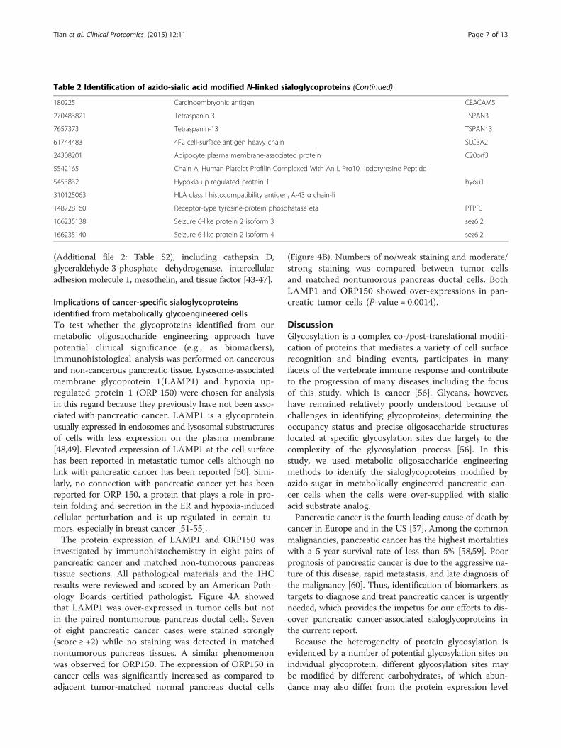
Table 2 Identification of azido-sialic acid modified N-linked sialoglycoproteins (Continued)
180225 Carcinoembryonic antigen CEACAM5
270483821 Tetraspanin-3 TSPAN3
7657373 Tetraspanin-13 TSPAN13
61744483 4F2 cell-surface antigen heavy chain SLC3A2
24308201 Adipocyte plasma membrane-associated protein C20orf3
5542165 Chain A, Human Platelet Profilin Complexed With An L-Pro10- Iodotyrosine Peptide
5453832 Hypoxia up-regulated protein 1 hyou1
310125063 HLA class I histocompatibility antigen, A-43 α chain-li
148728160 Receptor-type tyrosine-protein phosphatase eta PTPRJ
166235138 Seizure 6-like protein 2 isoform 3 sez6l2
166235140 Seizure 6-like protein 2 isoform 4 sez6l2
Tian et al. Clinical Proteomics (2015) 12:11 Page 7 of 13
(Additional file 2: Table S2), including cathepsin D,glyceraldehyde-3-phosphate dehydrogenase, intercellularadhesion molecule 1, mesothelin, and tissue factor [43-47].
Implications of cancer-specific sialoglycoproteinsidentified from metabolically glycoengineered cellsTo test whether the glycoproteins identified from ourmetabolic oligosaccharide engineering approach havepotential clinical significance (e.g., as biomarkers),immunohistological analysis was performed on cancerousand non-cancerous pancreatic tissue. Lysosome-associatedmembrane glycoprotein 1(LAMP1) and hypoxia up-regulated protein 1 (ORP 150) were chosen for analysisin this regard because they previously have not been asso-ciated with pancreatic cancer. LAMP1 is a glycoproteinusually expressed in endosomes and lysosomal substructuresof cells with less expression on the plasma membrane[48,49]. Elevated expression of LAMP1 at the cell surfacehas been reported in metastatic tumor cells although nolink with pancreatic cancer has been reported [50]. Simi-larly, no connection with pancreatic cancer yet has beenreported for ORP 150, a protein that plays a role in pro-tein folding and secretion in the ER and hypoxia-inducedcellular perturbation and is up-regulated in certain tu-mors, especially in breast cancer [51-55].The protein expression of LAMP1 and ORP150 was
investigated by immunohistochemistry in eight pairs ofpancreatic cancer and matched non-tumorous pancreastissue sections. All pathological materials and the IHCresults were reviewed and scored by an American Path-ology Boards certified pathologist. Figure 4A showedthat LAMP1 was over-expressed in tumor cells but notin the paired nontumorous pancreas ductal cells. Sevenof eight pancreatic cancer cases were stained strongly(score ≥ +2) while no staining was detected in matchednontumorous pancreas tissues. A similar phenomenonwas observed for ORP150. The expression of ORP150 incancer cells was significantly increased as compared toadjacent tumor-matched normal pancreas ductal cells
(Figure 4B). Numbers of no/weak staining and moderate/strong staining was compared between tumor cellsand matched nontumorous pancreas ductal cells. BothLAMP1 and ORP150 showed over-expressions in pan-creatic tumor cells (P-value = 0.0014).
DiscussionGlycosylation is a complex co-/post-translational modifi-cation of proteins that mediates a variety of cell surfacerecognition and binding events, participates in manyfacets of the vertebrate immune response and contributeto the progression of many diseases including the focusof this study, which is cancer [56]. Glycans, however,have remained relatively poorly understood because ofchallenges in identifying glycoproteins, determining theoccupancy status and precise oligosaccharide structureslocated at specific glycosylation sites due largely to thecomplexity of the glycosylation process [56]. In thisstudy, we used metabolic oligosaccharide engineeringmethods to identify the sialoglycoproteins modified byazido-sugar in metabolically engineered pancreatic can-cer cells when the cells were over-supplied with sialicacid substrate analog.Pancreatic cancer is the fourth leading cause of death by
cancer in Europe and in the US [57]. Among the commonmalignancies, pancreatic cancer has the highest mortalitieswith a 5-year survival rate of less than 5% [58,59]. Poorprognosis of pancreatic cancer is due to the aggressive na-ture of this disease, rapid metastasis, and late diagnosis ofthe malignancy [60]. Thus, identification of biomarkers astargets to diagnose and treat pancreatic cancer is urgentlyneeded, which provides the impetus for our efforts to dis-cover pancreatic cancer-associated sialoglycoproteins inthe current report.Because the heterogeneity of protein glycosylation is
evidenced by a number of potential glycosylation sites onindividual glycoprotein, different glycosylation sites maybe modified by different carbohydrates, of which abun-dance may also differ from the protein expression level

(i) Nontumorous tissue (H&E)
(ii) Nontumorous tissue (IHC)
(iii) Tumor (H&E) (iv) Tumor (IHC)
(A) LAMP1 (i) Nontumorous tissue (H&E)
(ii) Nontumorous vs. tumor (IHC)
(iii) Nontumorous tissue (IHC) (40X)
(iv) Tumor (IHC)(40X)
(B) ORP150
Figure 4 Verification of protein expression in pancreatic cancer and matched nontumorous pancreas tissues using IHC. (A) Increased expressionof LAMP1 in pancreatic tumor: (i) H&E staining of nontumorous pancreas duct, (ii) low expression of LAMP in matched nontumorous pancreasductal cells, (iii) H&E staining of pancreatic tumors, and (iv) overexpression of LAMP in pancreatic tumor. (B) Increased expression of ORP150 inpancreatic cancer tissue as compared to matched nontumorous pancreas duct: (i) H&E staining of pancreas tumor and adjacent nontumorouspancreas duct, (ii) IHC of nontumorous pancreas duct versus pancreatic tumor, (iii) a high power view of adjacent nontumorous pancreas duct,and (iv) A high power view of a pancreatic tumor. The blue arrows indicate the nontumorous pancreas ductal cells, and red arrows indicatepancreatic tumor cells.
Tian et al. Clinical Proteomics (2015) 12:11 Page 8 of 13
[61]. Compared to a previous strategy that used metabolicengineering to isolate sialoglycoproteins and associatedproteins in the protein mixture [29], the major advantageof the approach used in this study is the capture of azidolabeled sialoglycopeptides and enzymatic release of N-linked sialoglycopeptides for mass spectrometry iden-tification of sialoglycosylation sites. The procedure allowsthe identification of not only the isolated proteins, but alsothe specific N-glycosylation sites that may contribute tomore sensitive and specific identification of sialoglyco-proteins [29]. Another advantage of our approach comparedto previous reports is the use of synthetic azido-derivatizedManNAc analog 1,3,4-O-Bu3ManNAz, which is capable ofsupporting higher metabolic flux through the sialic acidbiosynthetic pathway at low concentrations compared toAc4ManNAz [29,34].Seventy-five unique formerly azido-sialic acid modified
glycopeptides were identified from 55 glycoproteinsusing 1,3,4-O-Bu3ManNAz analog treatment (Table 2and Additional file 2: Table S2). The number of proteinswe identified was relatively low compared to whole gly-coproteome datasets, in which hundreds or even thou-sands of glycoproteins are expected to be expressed in a
particular cell type. However we emphasize that we delib-erately did not seek to maximize detection of all possiblesialoglycans in the present study but rather conducted ourexperiments in a way that would be most conducive tothe identification of cancer-associated biomarkers. In par-ticular, we first limited the labeling period to two dayseven though evidence suggests that non-natural sialicacids can accumulate in tissue over a 6-week period uponrepeated daily dosing [62]; we consider it to be unlikelythat a diagnostic test could be conducted in a clinically-relevant manner over this extended period (instead weused a 2-day period in which a single bolus of analog hasbeen shown to resist esterase degradation [63] and further,maximize cellular labeling without the need for addeddoses or replenishment). Second, the number of cells in-volved in each experiment (<10e7) coupled with the sub-femtomolar mass spec detection sensitivity limit impliesthat only relatively abundant markers (e.g., glycoproteinsexpressed in the hundreds to thousands of copies per cell)will be detected by our methods; again, this feature is de-sired to ensure that our methods will have clinical rele-vance where sample size is likely to be limiting. Finally, asdescribed in our previous publication [9], we use stringent

Tian et al. Clinical Proteomics (2015) 12:11 Page 9 of 13
statistical cutoffs to avoid false positives. Together, thesefactors – by design – limit the number of glycoproteinsthat are identified by our method and ensure that the gly-coproteins we identify will be sufficiently abundant andmetabolically labeled with sufficient kinetics to be viablecancer biomarkers.Another matter of interest is the exact glycan struc-
tures that the non-natural azido-modified sialic acidslabel in cancer cells. In our previous experiments using1,3,4-O-Bu3ManNAc, which increased levels of naturalsialic acid by ~75% in treated cells [9], the levels of sialy-lation were sufficiently higher in the treated cells toallow characterization of the new glycoforms by usingmass spectrometry-based methods [64]. By comparison,treatment of cells with non-natural analogs tends to re-sult in the replacement of natural sialic acids with theirnon-natural counterparts rather than an overall increasein sialylation (for perspective on this issue, consult refer-ences [65-68]. In general, even though several millionnon-natural sialic acids can be incorporated on the sur-face of metabolically oligosaccharide engineered cancercells [69], the number of these moieties constitute only asmall percentage of the hundreds of millions of sialicacids typically found on each human cell [69]. Conse-quently, rigorous detection of non-natural sialic acids byunambiguous techniques such as mass spectrometry be-cause of their low abundance remains extremely challen-ging and alternative approaches such as lectin bindingassays have provided some insight.Lectins have long been used for purification and detec-
tion of glycans in many studies [70]; many lectins haveoverlapping affinity while some others have unique spe-cificity, allowing for the efficient detection of differentialexpression of glycan structure. In the current studies theuse of a previously-reported lectin array [35] showed sig-nificant binding signal from fourteen lectins (p < 0.05),indicating α-1-6 core fucoses (PSA), Galβ1-4GlcNAcstructures (DSA and PHAE), high mannose (NPA, GNA,and HHL) among the N-type glycans, and Galβ1-3GalNAc (Jacalin and MPA) and GalNAcβ1-4GlcNAc(WFA) for O-type glycans (Figure 3 and Table 1). Thisdata provided rudimentary insight into the glycan struc-tures compatible with azido-sialic acid incorporation butthe most interesting aspect of these experiments wasthe lack of. lectin binding to native sialic acid despitethe presence of the appropriate lectins (e.g., SNA) onthe array and the plentiful display of sialic acids onSW1990 cells demonstrated in our previous experi-ments [9,64]. Possible explanations for this outcomeinclude that 1) azido-sialic acid was not well recog-nized by, or had weak binding, to sialic acid specificlectins, such as SNA or that 2) the proteins modifiedby native sialic acid are not simultaneously modifiedby azido-sialic acid.
As a final part of this project, we return to the initialmotivation of this study, which was to use metabolic oligo-saccharide engineering methodology to identify cancer-associated biomarkers in pancreatic cancer. This task iscomplicated by not having a high quality “normal” pancre-atic line that readily grows in cell culture conditions forcomparison. Therefore, one way we attempted to addressthis issue was to cross-reference the list of the 55 proteinswe identified against cancer (in general) and pancreaticcancer (more specifically) and found that 42 proteins havebeen reported to be associated with cancer in generaland 15 have been specifically linked to pancreatic cancer(Additional file 2: Table S2). Although encouraging, thesignificance of this type of analysis is open to interpret-ation considering that many of the proteins are linked tocancer by as few as one reference, and further, it is not un-expected to detect cancer-related proteins by analyzingcancer cells. Therefore, we tried an alternative approachthat we consider to be more stringent by analyzing clinicalsamples for two proteins (LAMP1 and ORP150) notpreviously associated with pancreatic cancer and foundpreferential expression in tumors compared to normal tis-sue in both cases (Figure 4). Although far from definitive(for example, comprehensive analysis of all the identifiedglycoproteins is beyond the scope of the current investiga-tion), this early result strongly supports our metabolicoligosaccharide engineering approach as a viable strategyfor the identification of new cancer biomarkers.In conclusion, this paper describes a strategy to analyze
metabolically active sialoglycoproteins found in pancreaticcancer cells and shows that robust labeling of severalproteins already associated with tumors occurs. Even moresignificantly, the positive evaluation of two proteins notpreviously associated with pancreatic cancer in clinicalsamples suggests that this method may be a powerfultechnique for biomarker discovery. In summary, the re-sults showed that metabolic oligosaccharide engineeringhad broader implications by the discovery of sialoglyco-proteins in cancer cells that hold potential for the diagno-sis of cancer or as cell surface therapeutic targets [71].
Materials and methodsCell cultureThe pancreatic cell line, SW1990, was purchased from theAmerican Type Culture Collection (ATCC; Manasses,VA). Cells were grown in 1:1 DMEM medium supple-mented with 10% (v/v) FBS and 1% (v/v) of a solution of10,000 units penicillin and 10 mg streptomycin/mL, at37°C and 5% CO2.
Metabolic labeling of cells and cell surface azidevisualization via fluorescence microscopyPancreatic cells were seeded on 6-well TC plates and syn-thetic 1,3,4-O-Bu3ManNAz analog [34] from a 50 mM

Tian et al. Clinical Proteomics (2015) 12:11 Page 10 of 13
stock solution was added to each well prior seeding thecells to provide a final concentration of 50 μM. Cells wereincubated for two days with or without the analog. Priorto visualization, the cells were washed with PBS and fixedwith 3.7% formaldehyde for 20 min at room temperature.Labeling was done by adding 200 μL of a freshly preparedClick Reaction Mixture to each well followed by incuba-tion in the dark for 45 min as described previously [34].The cells were then washed 3–5 times with PBS contain-ing 5% BSA. Images were taken using a Zeiss Observermicroscope with a 40X objective lens (Zeiss, Inc., Melville,NY). Fluorescence pictures of Alexa488 (surface sialogly-coconjugates), and DAPI (nucleus) labeled cells were re-corded and overlay images were generated with the ZeissImaging System.
Biotinylation of azido-sialic acid containing glycoproteinsTo selectively biotinylate azido-sialic acid modified glyco-proteins, the modified Staudinger ligation reaction reportedby Saxon and coworkers using Biotin-PEG3-Phosphine wasperformed on proteins extracted from cell lysates [15].The protein concentration was adjusted to 2.5 mg/mL inPBS (pH 7.2), Biotin-PEG3-Phosphine was added to afinal concentration of 200 μM. The sample was incubatedovernight at room temperature. The biotinylation effi-ciency and specificity was examined by Western blot usingHRP-streptavidin at 1:5000. The non-reacted Biotin-PEG3-Phosphine was removed through protein precipita-tion. The proteins were mixed with an 8-fold volume ofpre-chilled (−20°C) acetone by vortexing and then incu-bated at −20°C for 60 min. The samples were centrifugedat 13,000 × g for 10 min. The supernatant was removedand disposed without disturbing the protein pellet. Thiswashing and precipitation process was repeated and re-sidual acetone was allowed to evaporate from the un-capped tube at room temperature for 30 min.
Affinity purification of biotinylated proteinsA 50% slurry of monomeric avidin agarose (1.4 mL)resin was sequentially washed with 3 mL PBS, 3 mL of2 mM D-biotin in PBS, 3 mL of 0.1 M glycine (pH 2.8),and 3 mL PBS. Protein (500 μg per sample, isolated fromtreated or untreated cells as described above) was added tothe pre-conditioned monomeric avidin agarose resin andincubated at room temperature for 1 h, followed by fivewashes with 6 mL PBS. The biotinylated azido-modifiedglycoproteins were eluted with 0.7 mL of 2 mM D-biotinin PBS for 10 min followed by a second elution step of0.7 mL of 0.1 M glycine (pH 2.8) for 10 min.
Characterized sialic acid glycans using lectin microarraysA lectin microarray comprised of 38 lectins as describedin our previous study was used to profile the glycanstructures of the proteins biotinylated via Staudinger
ligation [35]. Each lectin was prepared in three concen-trations (1, 0.5, and 0.25 mg/mL) on the chip. The slideswere blocked with 50 mM ethanolamine in 40 mM so-dium borate for 1 h followed by three subsequent washeswith PBS-0.1% Tween. The biotinylated proteins (100 μL)were applied to each lectin microarray. Inhibitory sugarsolutions (e.g. 100 μL of 50 mM lactose, 50 mM galactose,50 mM mannose, or 50 mM acetic acid) were mixed withsamples, and then were applied to the control wells. Afteran overnight incubation at room temperature, each wellwas washed with 100 μL of PBS-0.1% Tween 20 with5 min shaking. Rabbit IgG (20 μg) was added to eacharray followed by a 30 min incubation to mask any re-sidual lectin sites. The slides were washed three times withPBS-0.1% Tween 20. Afterwards, 100 μL of streptavidinconjugated to Dylight 549 (diluted 500-fold in PBS-0.1% Tween) was added to each well and incubatedfor 30 min in the dark before the array was washed,dried, and scanned with a GenePix 4100B scanner(Sunnyvale, CA).
Identification of formerly azide-sialic acid modifiedglycopeptidesAfter biotinylation and removal of the non-reacted Biotin-PEG3-Phosphine using protein precipitation, 1 mg of pro-tein was denatured and reduced in 8 M urea, 10 mMTCEP, and 1% SDS in 200 mM Tris buffer at 37°C for 2 h[72]. Iodoacetamide (10 mM final concentration) wasadded to the sample and incubated for 30 min at RT inthe dark. The solution was diluted 8-fold with 100 mMKH2PO4 (pH 8.0). The proteins were digested by trypsin(Promega, 1:50, w/w) at 37°C overnight with shaking.After digestion, the samples were centrifuged at 16,110 ×g for 5 min to remove particulate matter. The peptideswere cleaned by using a Sep-Pak C18 Vac cartridge(Waters) and resuspended in PBS. Six hundred microli-ters of 50% slurry of streptavidin agarose resin was washedwith 1 mL PBS (pH 7.2) three times. Protease inhibitor(Pierce) was added to the peptides at a 1:1000 dilutionprior to mixing peptides with pre-washed streptavidinagarose. The peptides were allowed to bind to streptavidinagarose at room temperature for 15 min. The unboundpeptides were removed by centrifugation. The streptavidinagarose was washed with 1 mL PBS 6 times, then resus-pended in 250 μL G7 buffer (New England Biolabs) andpeptides containing N-linked glycosylation sites withmetabolic labeled sialoglyans were released by PNGase F(New England Biolabs) [73-75]. The peptides were an-alyzed using a LTQ-Orbitrap velos (ThermoFisher,Waltham, MA) coupled with a 15 cm × 75 μm C18column (5 μm particles with 100 angstrom pore size). Thespectra were analyzed as described in our previous report[76]. Briefly, a nanoAquity UPLC at 300 nL/min with a90 min linear acetonitrile gradient (from 5-32% B over
Tian et al. Clinical Proteomics (2015) 12:11 Page 11 of 13
90 min; A = 0.1% formic acid in water, B = 0.1% formicacid in acetonitrile) was used. A top 10 data dependentMS/MS with exclusion for 20 s was set.MS/MS spectra were searched with MASCOT (version
2.2.0) using Proteome Discoverer (version 1.0) (ThermoFisher) against human subdatabase of NCBI ReferenceSequence (RefSeq) (version 40, released at April 16,2010) containing 29,704 sequences. For this databasesearch, the precursor mass tolerance and fragment masstolerance were set at 15 ppm and 0.05 Da respectively.Trypsin was specified as the protease. The fixed modifi-cation was set as carbamidomethylation (C), and otherdatabase-searching parameters were set as flexible modi-fication as follows: deamidation (NQ) and oxidation (M).Semi-tryptic end and one missed cleavage site was allowed.The False Discovery Rate was set at 0.01 to eliminate low-probability protein identifications.Identified proteins were classified according to their
main biological processes, their molecular functions andtheir cellular component using “Blast2Go” software(www.blast2go.com) [38].
Immunohistochemistry (IHC)Formalin-fixed and paraffin-embedded tumor tissue wasobtained from the Department of Pathology at JohnsHopkins Hospital, and was cut into 4-micron sections.Slides were de-paraffined and rehydrated before IHCstudy. Dako LSAB+ System-AP kit (Carpinteria, CA)used for IHC study according to the manufacturer’s in-structions. Antigen-retrieval using 10 mM sodium citrate(pH 6.0) at 100°C for 20 min was performed prior to applymouse monoclonal anti- human LAMP1 antibody at1:150 dilution. The mouse monoclonal anti- humanORP150 antibody was applied to slides at 1:200 dilutionwithout antigen retrieval. The primary antibodies were in-cubated overnight at 4°C. All pathological materials andIHC results were reviewed and evaluated by an AmericanPathology Boards certified pathologist, Dr. Qing Kay Li.The cytoplasmic immuno-staining pattern and immuno-reactive intensity were scored semi-quantitatively using a4 tier system: no staining (0), weak (1+, <10% positivity),moderate (2+, 10-50% positivity), and strong (3+, >50%positivity). The IHC staining pattern was correlated withtumors’ pathological stages and the differentiations. Pa-tients’ clinical information was also correlated. Fisher’sexact test was used to determine the statistical significanceof IHC staining in pancreas tumor and paired normal pan-creas tissues.
Additional files
Additional file 1: Table S1. Information of 38 lectins used in lectinmicroarray.
Additional file 2: Table S2. Identification of N-glycosite-containingsialoglycopeptides using LC-MS/MS.
Additional file 3: Table S3. MS/MS annotation for the identifiedsialoglycopeptides.
Competing interestsThe authors declare that they have no competing interests.
Authors’ contributionsYT participated in the study design and carried out the sialoglycopeptide/sialoglycoprotein isolation, sample preparation for MS analysis, and draftedthe manuscript. RA and CS carried out the cell metabolic labeling. CC andQL carried out the immunohistochemistry analysis supporting this study. DLhelped with lectin microarray analysis. PS carried out bioinformatics analysis.KJY and HZ participated in the study design and manuscript preparation. Allauthors read and approved the final manuscript.
AcknowledgementsThis work was supported by federal funds from the National Cancer Institute,National Institutes of Health, grants 2R01CA112315, U01CA152813,R01CA112314, and P01HL107153-01. We also gratefully acknowledge thesupport of Drs. Robert Cole and Robert O’Meally from Johns Hopkins Universityfor their assistance in mass spectrometry analysis and data processing.
Author details1Department of Pathology, Johns Hopkins University, 400 N. Broadway,Room 4011, Baltimore MD 21287, USA. 2Department of BiomedicalEngineering and the Translational Tissue Engineering Center, Johns HopkinsUniversity School of Medicine, Baltimore, MD, USA.
Received: 3 October 2014 Accepted: 23 March 2015
References1. Varki A. Glycan-based interactions involving vertebrate sialic-acid-recognizing
proteins. Nature. 2007;446(7139):1023–9.2. Hakomori S. Tumor-associated carbohydrate antigens. Annu Rev Immunol.
1984;2:103–26.3. Fukuda M. Possible roles of tumor-associated carbohydrate antigens. Cancer
Res. 1996;56(10):2237–44.4. Schauer R. Achievements and challenges of sialic acid research. Glycoconj J.
2000;17(7–9):485–99.5. Schultz MJ, Swindall AF, Bellis SL. Regulation of the metastatic cell phenotype
by sialylated glycans. Cancer Metastasis Rev. 2012;31(3–4):501–18.6. Meany DL, Zhang Z, Sokoll LJ, Zhang H, Chan DW. Glycoproteomics for
prostate cancer detection: changes in serum PSA glycosylation patterns.J Proteome Res. 2009;8(2):613–9.
7. Tian Y, Esteva FJ, Song J, Zhang H. Altered expression of sialylatedglycoproteins in breast cancer using hydrazide chemistry and massspectrometry. Mol Cell Proteomics. 2012;11(6):M111 011403.
8. Keppler OT, Hinderlich S, Langner J, Schwartz-Albiez R, Reutter W, Pawlita M.UDP-GlcNAc 2-epimerase: a regulator of cell surface sialylation. Science.1999;284(5418):1372–6.
9. Almaraz RT, Tian Y, Bhattarcharya R, Tan E, Chen SH, Dallas MR, et al.Metabolic flux increases glycoprotein sialylation: implications for cell adhesionand cancer metastasis. Mol Cell Proteomics. 2012;11(7):M112 017558.
10. Zhao J, Simeone DM, Heidt D, Anderson MA, Lubman DM. Comparativeserum glycoproteomics using lectin selected sialic acid glycoproteins withmass spectrometric analysis: application to pancreatic cancer serum.J Proteome Res. 2006;5(7):1792–802.
11. Malykh YN, Schauer R, Shaw L. N-Glycolylneuraminic acid in humantumours. Biochimie. 2001;83(7):623–34.
12. Bardor M, Nguyen DH, Diaz S, Varki A. Mechanism of uptake andincorporation of the non-human sialic acid N-glycolylneuraminic acid intohuman cells. J Biol Chem. 2005;280(6):4228–37.
13. Pan Y, Chefalo P, Nagy N, Harding C, Guo Z. Synthesis and immunologicalproperties of N-modified GM3 antigens as therapeutic cancer vaccines.J Med Chem. 2005;48(3):875–83.
14. Mahal LK, Yarema KJ, Bertozzi CR. Engineering chemical reactivity on cellsurfaces through oligosaccharide biosynthesis. Science. 1997;276(5315):1125–8.

Tian et al. Clinical Proteomics (2015) 12:11 Page 12 of 13
15. Saxon E, Bertozzi CR. Cell surface engineering by a modified Staudingerreaction. Science. 2000;287(5460):2007–10.
16. Neves AA, Stockmann H, Harmston RR, Pryor HJ, Alam IS, Ireland-Zecchini H,et al. Imaging sialylated tumor cell glycans in vivo. FASEB J.2011;25(8):2528–37.
17. Tian Y, Zhang H. Glycoproteomics and clinical applications. Proteomics: ClinAppl. 2010;4(2):124–32.
18. Li Y, Tao SC, Bova GS, Liu AY, Chan DW, Zhu H, et al. Detection and verificationof glycosylation patterns of glycoproteins from clinical specimens usinglectin microarrays and lectin-based immunosorbent assays. Anal Chem.2011;83(22):8509–16.
19. Larsen MR, Jensen SS, Jakobsen LA, Heegaard NH. Exploring the sialiomeusing titanium dioxide chromatography and mass spectrometry. Mol CellProteomics. 2007;6(10):1778–87.
20. Lewandrowski U, Zahedi RP, Moebius J, Walter U, Sickmann A. EnhancedN-glycosylation site analysis of sialoglycopeptides by strong cation exchangeprefractionation applied to platelet plasma membranes. Mol Cell Proteomics.2007;6(11):1933–41.
21. Gundry RL, Raginski K, Tarasova Y, Tchernyshyov I, Bausch-Fluck D, Elliott ST,et al. The mouse C2C12 myoblast cell surface N-linked glycoproteome:identification, glycosite occupancy, and membrane orientation. Mol CellProteomics. 2009;8(11):2555–69.
22. Wollscheid B, Bausch-Fluck D, Henderson C, O’Brien R, Bibel M, Schiess R,et al. Mass-spectrometric identification and relative quantification of N-linkedcell surface glycoproteins. Nat Biotechnol. 2009;27(4):378–86.
23. Kurogochi M, Matsushista T, Amano M, Furukawa J, Shinohara Y,Aoshima M, et al. Sialic acid-focused quantitative mouse serumglycoproteomics by multiple reaction monitoring assay. Mol Cell Proteomics.2010;9(11):2354–68.
24. Li Y, Tian Y, Rezai T, Prakash A, Lopez MF, Chan DW, et al. Simultaneousanalysis of glycosylated and sialylated prostate-specific antigen revealingdifferential distribution of glycosylated prostate-specific antigen isoforms inprostate cancer tissues. Anal Chem. 2011;83(1):240–5.
25. Ghesquiere B, Buyl L, Demol H, Van Damme J, Staes A, Timmerman E, et al.A new approach for mapping sialylated N-glycosites in serum proteomes.J Proteome Res. 2007;6(11):4304–12.
26. Kayser H, Zeitler R, Kannicht C, Grunow D, Nuck R, Reutter W. Biosynthesisof a nonphysiological sialic acid in different rat organs, using N-propanoyl-D-hexosamines as precursors. J Biol Chem. 1992;267(24):16934–8.
27. Bond MR, Kohler JJ. Chemical methods for glycoprotein discovery. CurrOpin Chem Biol. 2007;11(1):52–8.
28. Laughlin ST, Agard NJ, Baskin JM, Carrico IS, Chang PV, Ganguli AS, et al.Metabolic labeling of glycans with azido sugars for visualization andglycoproteomics. Methods Enzymol. 2006;415:230–50.
29. Yang L, Nyalwidhe JO, Guo S, Drake RR, Semmes OJ. Targeted identificationof metastasis-associated cell-surface sialoglycoproteins in prostate cancer.Mol Cell Proteomics. 2011;10(6):M110 007294.
30. Longwell SA, Dube DH. Deciphering the bacterial glycocode: recent advancesin bacterial glycoproteomics. Curr Opin Chem Biol. 2013;17(1):41–8.
31. Champasa K, Longwell SA, Eldridge AM, Stemmler EA, Dube DH. Targetedidentification of glycosylated proteins in the gastric pathogen Helicobacterpylon (Hp). Mol Cell Proteomics. 2013;12(9):2568–86.
32. Slade PG, Hajivandi M, Bartel CM, Gorfien SF. Identifying the CHO secretomeusing mucin-type O-linked glycosylation and click-chemistry. J ProteomeRes. 2012;11(12):6175–86.
33. Mathew MP, Tan E, Saeui CT, Bovonratwet P, Liu L, Bhattacharya R, et al.Metabolic glycoengineering sensitizes drug-resistant pancreatic cancer cellsto tyrosine kinase inhibitors erlotinib and gefitinib. Bioorg Med Chem Lett.2015;25(6):1223–7.
34. Almaraz RT, Aich U, Khanna HS, Tan E, Bhattacharya R, Shah S, et al.Metabolic oligosaccharide engineering with N-acyl functionalized ManNAcanalogs: cytotoxicity, metabolic flux, and glycan-display considerations.Biotechnol Bioeng. 2012;109(4):992–1006.
35. Meany DL, Hackler Jr L, Zhang H, Chan DW. Tyramide signal amplificationfor antibody-overlay lectin microarray: a strategy to improve the sensitivityof targeted glycan profiling. J Proteome Res. 2011;10(3):1425–31.
36. Hirabayashi J, Kuno A, Tateno H. Lectin-based structural glycomics: a practicalapproach to complex glycans. Electrophoresis. 2011;32(10):1118–28.
37. Fry SA, Afrough B, Lomax-Browne HJ, Timms JF, Velentzis LS, Leathem AJ.Lectin microarray profiling of metastatic breast cancers. Glycobiology.2011;21(8):1060–70.
38. Conesa A, Gotz S, Garcia-Gomez JM, Terol J, Talon M, Robles M. Blast2GO: auniversal tool for annotation, visualization and analysis in functional genomicsresearch. Bioinformatics. 2005;21(18):3674–6.
39. Hagikura M, Murakumo Y, Hasegawa M, Jijiwa M, Hagiwara S, Mii S, et al.Correlation of pathological grade and tumor stage of urothelial carcinomaswith CD109 expression. Pathol Int. 2010;60(11):735–43.
40. Nawashiro H, Otani N, Shinomiya N, Fukui S, Ooigawa H, Shima K, et al.L-type amino acid transporter 1 as a potential molecular target in humanastrocytic tumors. Int J Cancer. 2006;119(3):484–92.
41. Zhou G, Chiu D, Qin D, Niu L, Cai J, He L, et al. Detection and clinicalsignificance of CD44v6 and integrin-beta1 in pancreatic cancer patientsusing a triplex real-time RT-PCR assay. Appl Biochem Biotechnol.2012;167(8):2257–68.
42. Galamb O, Gyorffy B, Sipos F, Spisak S, Nemeth AM, Miheller P, et al.Inflammation, adenoma and cancer: objective classification of colon biopsyspecimens with gene expression signature. Dis Markers. 2008;25(1):1–16.
43. Tsume Y, Amidon GL. The feasibility of enzyme targeted activation foramino acid/dipeptide monoester prodrugs of floxuridine; cathepsin D as apotential targeted enzyme. Molecules. 2012;17(4):3672–89.
44. Menon R, Zhang Q, Zhang Y, Fermin D, Bardeesy N, DePinho RA, et al.Identification of novel alternative splice isoforms of circulating proteins in amouse model of human pancreatic cancer. Cancer Res. 2009;69(1):300–9.
45. Roland CL, Dineen SP, Toombs JE, Carbon JG, Smith CW, Brekken RA, et al.Tumor-derived intercellular adhesion molecule-1 mediates tumor-associatedleukocyte infiltration in orthotopic pancreatic xenografts. Exp Biol Med.2010;235(2):263–70.
46. Tang Z, Qian M, Ho M. The role of mesothelin in tumor progression andtargeted therapy. Anticancer Agents Med Chem. 2013;13(2):276–80.
47. Gerotziafas GT, Galea V, Mbemba E, Khaterchi A, Sassi M, Baccouche H, et al.Tissue factor over-expression by human pancreatic cancer cells BXPC3 isrelated to higher prothrombotic potential as compared to breast cancercells MCF7. Thromb Res. 2012;129(6):779–86.
48. Granger BL, Green SA, Gabel CA, Howe CL, Mellman I, Helenius A.Characterization and cloning of lgp110, a lysosomal membraneglycoprotein from mouse and rat cells. J Biol Chem. 1990;265(20):12036–43.
49. Holcombe RF, Baethge BA, Stewart RM, Betzing K, Hall VC, Fukuda M, et al.Cell surface expression of lysosome-associated membrane proteins (LAMPs)in scleroderma: relationship of lamp2 to disease duration, anti-Sc170antibodies, serum interleukin-8, and soluble interleukin-2 receptor levels.Clin Immunol Immunopathol. 1993;67(1):31–9.
50. Heffernan M, Yousefi S, Dennis JW. Molecular characterization of P2B/LAMP-1,a major protein target of a metastasis-associated oligosaccharide structure.Cancer Res. 1989;49(21):6077–84.
51. Ozawa K, Kuwabara K, Tamatani M, Takatsuji K, Tsukamoto Y, Kaneda S, et al.150-kDa oxygen-regulated protein (ORP150) suppresses hypoxia-inducedapoptotic cell death. J Biol Chem. 1999;274(10):6397–404.
52. Asahi H, Koshida K, Hori O, Ogawa S, Namiki M. Immunohistochemicaldetection of the 150-kDa oxygen-regulated protein in bladder cancer.BJU Int. 2002;90(4):462–6.
53. Namba T, Hoshino T, Tanaka K, Tsutsumi S, Ishihara T, Mima S, et al. Up-regulationof 150-kDa oxygen-regulated protein by celecoxib in human gastric carcinomacells. Mol Pharmacol. 2007;71(3):860–70.
54. Tsukamoto Y, Kuwabara K, Hirota S, Kawano K, Yoshikawa K, Ozawa K, et al.Expression of the 150-kd oxygen-regulated protein in human breast cancer.Lab Invest. 1998;78(6):699–706.
55. Stojadinovic A, Hooke JA, Shriver CD, Nissan A, Kovatich AJ, Kao TC,et al. HYOU1/Orp150 expression in breast cancer. Med Sci Monit.2007;13(11):BR231–9.
56. Hanson SR, Hsu TL, Weerapana E, Kishikawa K, Simon GM, Cravatt BF, et al.Tailored glycoproteomics and glycan site mapping using saccharide-selectivebioorthogonal probes. J Am Chem Soc. 2007;129(23):7266–7.
57. Niederhuber JE, Brennan MF, Menck HR. The national cancer data basereport on pancreatic cancer. Cancer Lett. 1995;76(9):1671–7.
58. Wray CJ, Ahmad SA, Matthews JB, Lowy AM. Surgery for pancreatic cancer:recent controversies and current practice. Gastroenterology. 2005;128(6):1626–41.
59. Jemal A, Siegel R, Ward E, Hao Y, Xu J, Thun MJ. Cancer statistics, 2009. CACancer J Clin. 2009;59(4):225–49.
60. Zhao J, Patwa TH, Qiu W, Shedden K, Hinderer R, Misek DE, et al.Glycoprotein microarrays with multi-lectin detection: unique lectinbinding patterns as a tool for classifying normal, chronic pancreatitisand pancreatic cancer sera. J Proteome Res. 2007;6(5):1864–74.

Tian et al. Clinical Proteomics (2015) 12:11 Page 13 of 13
61. Ozohanics O, Turiak L, Puerta A, Vekey K, Drahos L. High-performance liquidchromatography coupled to mass spectrometry methodology for analyzingsite-specific N-glycosylation patterns. J Chromatogr A. 2012;1259:200–12.
62. Gagiannis D, Gossrau R, Reutter W, Zimmermann-Kordmann M, HorstkorteR. Engineering the sialic acid in organs of mice using N-propanoylmannosamine. Biochim Biophys Acta. 2007;1770(2):297–306.
63. Mathew MP, Tan E, Shah S, Bhattacharya R, Adam Meledeo M, Huang J,et al. Extracellular and intracellular esterase processing of SCFA-hexosamineanalogs: implications for metabolic glycoengineering and drug delivery.Bioorg Med Chem Lett. 2012;22(22):6929–33.
64. Shah P, Yang S, Sun S, Aiyetan P, Yarema KJ, Zhang H. Mass spectrometricanalysis of sialylated glycans with use of solid-phase labeling of sialic acids.Anal Chem. 2013;85(7):3606–13.
65. Kim EJ, Sampathkumar SG, Jones MB, Rhee JK, Baskaran G, Goon S, et al.Characterization of the metabolic flux and apoptotic effects of O-hydroxyl- andN-acyl-modified N-acetylmannosamine analogs in Jurkat cells. J Biol Chem.2004;279(18):18342–52.
66. Du J, Meledeo MA, Wang Z, Khanna HS, Paruchuri VD, Yarema KJ. Metabolicglycoengineering: sialic acid and beyond. Glycobiology. 2009;19(12):1382–401.
67. Dafik L, D’Alarcao M, Kumar K. Modulation of cellular adhesion byglycoengineering. J Med Chem. 2010;53(10):4277–84.
68. Almaraz RT, Mathew MP, Tan E, Yarema KJ. Metabolic oligosaccharideengineering: implications for selectin-mediated adhesion and leukocyteextravasation. Ann Biomed Eng. 2012;40(4):806–15.
69. Yarema KJ, Bertozzi CR. Chemical approaches to glycobiology and emergingcarbohydrate-based therapeutic agents. Curr Opin Chem Biol. 1998;2(1):49–61.
70. Hirabayashi J, Yamada M, Kuno A, Tateno H. Lectin microarrays: concept,principle and applications. Chem Soc Rev. 2013;42(10):4443–58.
71. Zhang H, Chan DW. Cancer biomarker discovery in plasma using atissue-targeted proteomic approach. Cancer Epidemiol Biomarkers Prev.2007;16(10):1915–7.
72. Sun S, Zhou JY, Yang W, Zhang H. Inhibition of protein carbamylation inurea solution using ammonium-containing buffers. Anal Biochem.2014;446:76–81.
73. Zhang H, Li XJ, Martin DB, Aebersold R. Identification and quantification ofN-linked glycoproteins using hydrazide chemistry, stable isotope labelingand mass spectrometry. Nat Biotechnol. 2003;21(6):660–6.
74. Zhang H, Aebersold R. Isolation of glycoproteins and identification of theirN-linked glycosylation sites. Methods Mol Biol. 2006;328:177–85.
75. Tian Y, Zhou Y, Elliott S, Aebersold R, Zhang H. Solid-phase extraction ofN-linked glycopeptides. Nat Protoc. 2007;2(2):334–9.
76. Tian Y, Bova GS, Zhang H. Quantitative glycoproteomic analysis of optimalcutting temperature-embedded frozen tissues identifying glycoproteinsassociated with aggressive prostate cancer. Anal Chem. 2011;83(18):7013–9.
Submit your next manuscript to BioMed Centraland take full advantage of:
• Convenient online submission
• Thorough peer review
• No space constraints or color figure charges
• Immediate publication on acceptance
• Inclusion in PubMed, CAS, Scopus and Google Scholar
• Research which is freely available for redistribution
Submit your manuscript at www.biomedcentral.com/submit