PLANT CELL MORPHOGENESIS: Plasma Membrane Interactions with the
49
Annu. Rev. Cell Dev. Biol. 1997. 13:697–743 Copyright c 1997 by Annual Reviews Inc. All rights reserved PLANT CELL MORPHOGENESIS: Plasma Membrane Interactions with the Cytoskeleton and Cell Wall John E. Fowler and Ralph S. Quatrano Department of Biology, University of North Carolina, Chapel Hill, North Carolina 27599-3280; e-mail: [email protected] KEY WORDS: cell polarity, Fucus, secretion, microfilaments, asymmetric division ABSTRACT Because plants are composed of immobile cells, plant morphogenesis requires mechanisms allowing precise control of cell expansion and cell division pat- terns. Cortical domains, localized in response to directional cues, are of central importance in establishing cell polarity, orienting cell division, and determining daughter cell fates in a wide variety of prokaryotic and eukaryotic organisms. Such domains consist of localized macromolecular complexes that, in plant cells, provide spatial control of cell expansion and cell division functions. The role of the cytoskeleton, plasma membrane, and targeted secretion to the cell wall in the spatial regulation of cell morphogenesis in plants is discussed in light of recent results from model organisms, including brown algal zygotes (e.g. Fucus). A gen- eral model, emphasizing the importance of cortical sites and targeted secretion, is proposed for morphogenesis in higher plant cells based on current knowledge and principles derived from analysis of the establishment of a stable cortical asym- metry in Fucus. The model illustrates mechanisms to direct the orientation of an asymmetric division resulting in daughter cells with different fates. CONTENTS INTRODUCTION ........................................................... 698 MORPHOGENESIS IN PROKARYOTES, YEAST, AND ANIMALS .................. 699 Prokaryotes ............................................................. 699 Yeast ................................................................... 700 Animal Cells ............................................................. 701 MORPHOGENESIS IN NONVASCULAR PLANTS ............................... 703 Fucus Zygotes ........................................................... 703 Tip Growth in Moss and Asymmetric Division in Volvox .......................... 710 MORPHOGENESIS IN VASCULAR PLANTS .................................... 712 697 1081-0706/97/1115-0697$08.00 Annu. Rev. Cell. Dev. Biol. 1997.13:697-743. Downloaded from arjournals.annualreviews.org by Washington University Library on 07/13/05. For personal use only.
Transcript of PLANT CELL MORPHOGENESIS: Plasma Membrane Interactions with the
P1: ARK/dat P2: ARK
September 3, 1997 15:25 Annual Reviews AR041-20
Annu. Rev. Cell Dev. Biol. 1997. 13:697–743Copyright c© 1997 by Annual Reviews Inc. All rights reserved
PLANT CELL MORPHOGENESIS:Plasma Membrane Interactions withthe Cytoskeleton and Cell Wall
John E. Fowler and Ralph S. QuatranoDepartment of Biology, University of North Carolina, Chapel Hill, North Carolina27599-3280; e-mail: [email protected]
KEY WORDS: cell polarity,Fucus, secretion, microfilaments, asymmetric division
ABSTRACT
Because plants are composed of immobile cells, plant morphogenesis requiresmechanisms allowing precise control of cell expansion and cell division pat-terns. Cortical domains, localized in response to directional cues, are of centralimportance in establishing cell polarity, orienting cell division, and determiningdaughter cell fates in a wide variety of prokaryotic and eukaryotic organisms.Such domains consist of localized macromolecular complexes that, in plant cells,provide spatial control of cell expansion and cell division functions. The role ofthe cytoskeleton, plasma membrane, and targeted secretion to the cell wall in thespatial regulation of cell morphogenesis in plants is discussed in light of recentresults from model organisms, including brown algal zygotes (e.g.Fucus). A gen-eral model, emphasizing the importance of cortical sites and targeted secretion, isproposed for morphogenesis in higher plant cells based on current knowledge andprinciples derived from analysis of the establishment of a stable cortical asym-metry inFucus. The model illustrates mechanisms to direct the orientation of anasymmetric division resulting in daughter cells with different fates.
CONTENTS
INTRODUCTION . . . . . . . . . . . . . . . . . . . . . . . . . . . . . . . . . . . . . . . . . . . . . . . . . . . . . . . . . . . 698
MORPHOGENESIS IN PROKARYOTES, YEAST, AND ANIMALS. . . . . . . . . . . . . . . . . . 699Prokaryotes. . . . . . . . . . . . . . . . . . . . . . . . . . . . . . . . . . . . . . . . . . . . . . . . . . . . . . . . . . . . . 699Yeast. . . . . . . . . . . . . . . . . . . . . . . . . . . . . . . . . . . . . . . . . . . . . . . . . . . . . . . . . . . . . . . . . . . 700Animal Cells. . . . . . . . . . . . . . . . . . . . . . . . . . . . . . . . . . . . . . . . . . . . . . . . . . . . . . . . . . . . . 701
MORPHOGENESIS IN NONVASCULAR PLANTS. . . . . . . . . . . . . . . . . . . . . . . . . . . . . . . 703Fucus Zygotes. . . . . . . . . . . . . . . . . . . . . . . . . . . . . . . . . . . . . . . . . . . . . . . . . . . . . . . . . . . 703Tip Growth in Moss and Asymmetric Division in Volvox. . . . . . . . . . . . . . . . . . . . . . . . . . 710
MORPHOGENESIS IN VASCULAR PLANTS. . . . . . . . . . . . . . . . . . . . . . . . . . . . . . . . . . . . 712
6971081-0706/97/1115-0697$08.00
Ann
u. R
ev. C
ell.
Dev
. Bio
l. 19
97.1
3:69
7-74
3. D
ownl
oade
d fr
om a
rjou
rnal
s.an
nual
revi
ews.
org
by W
ashi
ngto
n U
nive
rsity
Lib
rary
on
07/1
3/05
. For
per
sona
l use
onl
y.

P1: ARK/dat P2: ARK
September 3, 1997 15:25 Annual Reviews AR041-20
698 FOWLER & QUATRANO
Spatial Regulation of Cell Wall Expansion. . . . . . . . . . . . . . . . . . . . . . . . . . . . . . . . . . . . . 712Spatial Regulation of Cell Division. . . . . . . . . . . . . . . . . . . . . . . . . . . . . . . . . . . . . . . . . . . 721
CONCLUSIONS . . . . . . . . . . . . . . . . . . . . . . . . . . . . . . . . . . . . . . . . . . . . . . . . . . . . . . . . . . . . 733
INTRODUCTION
The fixed, immobile nature of plant cells and tissues requires that the planeof cell division and the site of cell expansion be closely regulated to gener-ate morphological and developmental diversity (Figure 1). The mechanisms
Figure 1 Patterns of cell morphogenesis in plants. Examples of how the site of cell expansion(a-d) and the plane of cell division (e-h) can generate morphological and developmental diversityin immobile plant cells. Sites of targeted vesicle secretion and cell wall deposition (dashed line)are present in cells undergoing (a) symmetrical, (b) horizontal, (c) vertical, and (d ) localized cellwall expansion, as well as at new cell walls that partition daughter cells following (e, g) symmetricor ( f, h) asymmetric divisions. The division sites (solid circle) represent cortical domains thatinclude putative macromolecular complexes involved in orienting the division apparatus to generatedaughter cells of equal or unequal sizes. Certain asymmetric divisions in plants (e.g. in zygotes,microspores, root cortical cells) partition determinants to one daughter cell (hatched area; f, h),a process essential for generating differences in developmental fate between the daughter cells.Various morphogenetic patterns diagrammed above for plant cells also occur in other organisms; forexample, polar growth in yeast (d ), and asymmetric cell division in bacteria, flies and worms (f, h).
Ann
u. R
ev. C
ell.
Dev
. Bio
l. 19
97.1
3:69
7-74
3. D
ownl
oade
d fr
om a
rjou
rnal
s.an
nual
revi
ews.
org
by W
ashi
ngto
n U
nive
rsity
Lib
rary
on
07/1
3/05
. For
per
sona
l use
onl
y.

P1: ARK/dat P2: ARK
September 3, 1997 15:25 Annual Reviews AR041-20
PLANT CELL MORPHOGENESIS 699
that specify differences in the shape of plant cells require the localization ofthe macromolecular complexes controlling cell expansion or cell division toone or more cortical sites or domains. In some cases, the local assembly of themacromolecules required for cell division or expansion at a site is accompaniedby the localization of macromolecules that may also direct the developmentalfate of the new daughter cells (Figure 1). Coordination between the sites forcell division and expansion requires some type of communication, possiblythrough signaling cascades that are triggered by directional cues from internaland/or external sources. The ability to sense directional signals for position-ing structural complexes and the division plane is well documented in bacte-rial, yeast, and animal cells (Horvitz & Herskowitz 1992, Drubin & Nelson1996, Gonczy & Hyman 1996). Usually, a directional signal serves to producea cortex- or membrane-associated asymmetry, which then defines a corticaldomain to which macromolecules are directed and anchored by a cytoskeletal-based complex.
Although this review centers around the role of interactions between the cy-toskeleton, the plasma membrane, and the cell wall during plant cell morpho-genesis, we first use examples from several other model systems to address somegeneral questions applicable to all cell types: What are the source and natureof signals that target particular cortical sites for assembly of a macromolecularcomplex; how are these sites stabilized to direct localized morphogenesis; andwhat are the developmental consequences of these events for subsequent cellu-lar differentiation? General principles from this discussion provide a frameworkwithin which we can examine similar problems in plants.
MORPHOGENESIS IN PROKARYOTES, YEAST,AND ANIMALS
ProkaryotesIn Bacillus subtilis,an asymmetric cell division is required for the differentia-tion of the spore (Errington 1993). Initially, the bacterial cell envelope appears tocontain three potential division sites: one medial and one near each of the poles.Structural components that are localized to the polar sites early in developmentinclude the membrane-spanning protein SpoIIE (Duncan et al 1995) and FtsZ,which is a tubulin-like GTPase laid down as a ring or disc associated with theseptum (Osteryoung & Vierling 1995, Erickson et al 1996). During a normaldivision, the septum is placed at the medial site (Rothfield & Justice 1997)(D Bramhill, this volume). However, at the start of sporulation, the medialsite is apparently masked, whereas the polar sites are equally accessible to theseptation machinery. Although the FtsZ (Levin & Losick 1996) and the SpoIIE
Ann
u. R
ev. C
ell.
Dev
. Bio
l. 19
97.1
3:69
7-74
3. D
ownl
oade
d fr
om a
rjou
rnal
s.an
nual
revi
ews.
org
by W
ashi
ngto
n U
nive
rsity
Lib
rary
on
07/1
3/05
. For
per
sona
l use
onl
y.
P1: ARK/dat P2: ARK
September 3, 1997 15:25 Annual Reviews AR041-20
700 FOWLER & QUATRANO
(Arigoni et al 1995) proteins are initially directed to both polar sites, eventuallyonly one forms a septum, usually the site at the pole most distal to the precedingdivision site (Figure 1, f ). Apparently, the activated form of the transcriptionfactor SpoOA induces gene expression that suppresses FtsZ assembly at themedial site and/or activates the sites at both poles (Levin & Losick 1996).Little is known, however, of the nature of the division sites in this system or ofthe mechanisms that direct the placement of the apparatus responsible for thesporulation division.
The asymmetric site of flagellar assembly in the predivisional cell ofCaulo-bacter crescentusis another example of localized morphogenesis in a prokary-otic cell (Shapiro 1993, Gober & Marques 1995, Domian et al 1996). The dis-tribution and anchoring of the flagellar motor protein and the methyl-acceptingchemoreceptor protein (McpA) to a discrete patch of cell membrane at theswarmer pole are the first signs of the morphogenesis. Like FtsZ and SpoIIEin B. subtilis, McpA is initially localized to membrane domains at two sites; inthis case, the swarmer and stalk poles. However, it is unclear whether McpAis directionally targeted and inserted locally, or inserted at random and thentrapped at the poles by a receptor. Localization is followed by degradation atthe stalked pole (Jenal & Shapiro 1996). The initial asymmetric cue that definesthe final membrane localization site is set by the previous division site. In bothprokaryotes, a cortical domain is identified that positions a macromolecularcomplex which promotes a localized morphological change.
YeastCortical sites in eukaryotes serve to localize signaling networks or molecularswitches that redirect the cytoskeleton, the secretory apparatus, and the celldivision apparatus toward the spatial cue. For example, in the yeastSaccha-romyces cerevisiae,the site of polarized growth (i.e. bud formation or matingprojection formation; Figure 1d ) is identified by either an intrinsic cue (theprevious bud site) or an extrinsic cue (pheromone gradient), which directs thecytoskeleton (actin and septins) and secretory apparatus to the target site bya series of molecular switches comprised of ras-related GTPases (e.g. Bud1p,Cdc42p, Rho1p) (Chant & Stowers 1995). The intrinsic and extrinsic cues bothresult in local expansion of the plasma membrane and cell wall, forming, re-spectively, the bud and the mating projection toward the nearest partner of theopposite mating type (Chant 1996, Roemer et al 1996). Recently it was shownthat the enzyme complex responsible for much of the cell wall synthesis at thebud site,β glucan synthase (GS), is activated by GTP-bound Rho1p (Drgonov´aet al 1996, Qadota et al 1996). The GS complex, Rho1p and actin colocalizeat the tip of growing buds. Apparently, when Rho1p exchanges GDP for GTP,it binds to the inactive GS complex, activating GS and allowing synthesis and
Ann
u. R
ev. C
ell.
Dev
. Bio
l. 19
97.1
3:69
7-74
3. D
ownl
oade
d fr
om a
rjou
rnal
s.an
nual
revi
ews.
org
by W
ashi
ngto
n U
nive
rsity
Lib
rary
on
07/1
3/05
. For
per
sona
l use
onl
y.

P1: ARK/dat P2: ARK
September 3, 1997 15:25 Annual Reviews AR041-20
PLANT CELL MORPHOGENESIS 701
extrusion of theβ glucan into the region of cell wall expansion. GTP-boundRho1p also acts via Pkc1p (a protein kinase C) to signal for bud site organizationand remodeling, possibly through interaction with localized cytoskeletal com-ponents. How the secretory pathway and GS complex are targeted toward andanchored at the bud/growth site is not known, but it could be via cytoplasmicactin cables and myosins oriented along the growth axis.
Animal CellsA counterpart to the yeast GTPase-regulated assembly of macromolecular com-plexes linking cell wall morphogenesis with the cytoskeleton exists in mam-malian cells. Mammalian cells regulate their morphology, migratory properties,growth, and differentiation largely through their adhesive interaction with theextracellular matrix (ECM) (Machesky & Hall 1996). Cells in culture developspecialized sites of adhesion known as focal adhesions (FA), which containintegrins as their central component. Integrins are transmembrane proteins thatserve as receptors for ECM components and provide attachment domains forbundles of actin filaments (i.e. stress fibers) through actin-binding proteinssuch as vinculin (Burridge & Chrzanowska-Wodnicka 1996). Macromoleculeswithin FAs, like the complex at the yeast bud site, provide a network of struc-tural (e.g. actin) and signaling (e.g. kinases) components that alter cell shapein response to various cues. For example, GTP-bound RhoA, which is homol-ogous to the yeast Rho1p, regulates FA and stress fiber formation inducedby growth factors and phospholipids such as lysophosphatidic acid (LPA).Microinjection of constitutively active (i.e. GTP-bound) Rho into mouse fi-broblasts rapidly induces FA and stress fiber assembly (Ridley & Hall 1992).RhoA interacts with at least two distinct mammalian serine/threonine proteinkinases and regulates the affinity of certain integrins toward their ligands. Again,in response to an external cue (i.e. LPA), a localized cortical complex, composedof transmembrane structural components and molecular switches, is assembledthat controls local formation and contraction of actin stress fibers (Tapon & Hall1997). These examples illustrate that different extracellular signals interactingwith diverse receptor types, from yeast to mammals, use GTPases of the Rhofamily to initiate and control local structural and functional rearrangements,including those involving cytoskeletal components.
A clear case in which cortical sites control the orientation of the plane ofcell division is in the P1 cell of theCaenorhabditis elegansembryo at the two-celled stage. The zygote divides asymmetrically along the anterior-posterior(A-P) axis to form a larger AB cell and a smaller P1 cell. During the divisionof the P1, a 90◦ rotation of one of the P1 centrosomes, toward a site on theanterior cortex, realigns the spindle pole along the A-P axis, thereby partitioningposteriorly localized P granules exclusively to the smaller daughter cell in this
Ann
u. R
ev. C
ell.
Dev
. Bio
l. 19
97.1
3:69
7-74
3. D
ownl
oade
d fr
om a
rjou
rnal
s.an
nual
revi
ews.
org
by W
ashi
ngto
n U
nive
rsity
Lib
rary
on
07/1
3/05
. For
per
sona
l use
onl
y.

P1: ARK/dat P2: ARK
September 3, 1997 15:25 Annual Reviews AR041-20
702 FOWLER & QUATRANO
second division (White & Strome 1996) (Figure 1f ). The 90◦ rotation dependson actin and microtubules positioned between the centrosome and the actin-enriched cortical site, and also on an actin-capping protein (Waddle et al 1994).Both actin and actin-capping proteins are components of the dynactin complex,which is known to bind to the microtubule motor protein dynein and couldprovide the cortex-anchored motor for movement of the spindle apparatus.Several genes have been identified that mutate to cause defects in the asymmetryand orientation of these first divisions.par-1, apar (partition-defective) gene,encodes a serine/threonine kinase that may be part of the signaling relays inthe asymmetric cell division (Guo & Kemphues 1995). An interaction betweenthe non-kinase domain of PAR-1 and a non-muscle myosin II heavy chain(nmmII) is required for establishing the localized cortical site, as injection ofnmmIIanti-sense RNA into ovaries of adult worms causes mislocalization of thePAR-1, PAR-2, and PAR-3 proteins, as well as embryonic partitioning defects(Guo & Kemphues 1996). A similar myosin is involved in the asymmetricalbudding division ofS. cerevisiae(Jansen et al 1996), suggesting that myosinsmay have a conserved role in generating cellular asymmetry in many organisms.Thus as an alternative to the extracellular functions already described in yeastand mammalian cells, a cortical domain may influence morphogenesis throughsignaling and anchoring cytoplasmic processes, e.g. the cell division apparatusin C. elegans.
Neuroblasts of the developing ectoderm ofDrosophila melanogasteralsoundergo an asymmetric division that results in a smaller, basal ganglion mothercell (GMC) and a larger, apical neuroblast, both of which express differentsets of genes (Doe 1996, Lin & Schagat 1997). Prior to the asymmetric di-vision, one centrosome migrates 180◦ to the basal cortex, whereas the otherremains anchored at the apical pole, resulting in a spindle aligned along theA-P axis (Spana et al 1995). Localized in the basal cortex and partitioned intothe GMC are the homeodomain protein Prospero, a transcription factor thatcontrols GMC-specific gene expression, and the membrane-associated proteinNumb, required to specify the cell fate of the GMC (Knoblich et al 1995,Spana & Doe 1995). Inscuteable is a novel protein that presumably plays arole in anchoring the apical centrosome, thus resulting in a vertical spindleaxis in ectodermal cells. If Inscuteable is absent, spindle orientation in neuro-blasts is random; furthermore, Inscuteable is also required for the proper basallocalization of Prospero and Numb. Inscuteable contains both SH3-bindingand ankyrin domains, and it is transiently localized to the apical cell cor-tex (Kraut & Campos-Ortega 1996). This localization requires actin micro-filaments; however, the cytoskeletal basis by which Inscuteable anchors thecentrosome at the apical cortical site in neuroblasts is unknown. Nonetheless, itis clear that mechanisms controlling the orientation of cytokinesis, resulting in
Ann
u. R
ev. C
ell.
Dev
. Bio
l. 19
97.1
3:69
7-74
3. D
ownl
oade
d fr
om a
rjou
rnal
s.an
nual
revi
ews.
org
by W
ashi
ngto
n U
nive
rsity
Lib
rary
on
07/1
3/05
. For
per
sona
l use
onl
y.

P1: ARK/dat P2: ARK
September 3, 1997 15:25 Annual Reviews AR041-20
PLANT CELL MORPHOGENESIS 703
daughter cells with different fates, are similar, whether one examines neurob-lasts inD. melangasteror embryonic divisions inC. elegans(Lin & Schagat1997).
In view of these results from multiple organisms, a general pattern emerges;a cortical site, identified by some internal or external cue, serves as an assemblysite for a cytoskeletal complex, which then becomes anchored at that site andserves to direct local morphogenesis. To what extent can we transfer thesebasic conclusions to plant cells? Do interactions between the cytoskeleton, theplasma membrane and the cell wall follow similar mechanistic principles duringmorphogenesis in plant cells (Figure 1)?
MORPHOGENESIS IN NONVASCULAR PLANTS
Fucus ZygotesZygotes of marine brown algae from the generaFucusandPelvetiahave beenused to study how external signals direct local cell expansion and the orien-tation of cell division (Kropf 1992, 1994, Goodner & Quatrano 1993, Fowler& Quatrano 1995, Quatrano & Shaw 1997). Fusion of a motile sperm witha wall-less egg cell occurs in the open sea and results in the secretion of auniform cell wall around the fertilized zygote (Quatrano 1982). When theseapolar, spherical zygotes are subjected to unilateral light, either continuouslyor as a pulse, during the first 10 h of development, a localized growth (therhizoid tip) emerges from the shaded quadrant 14 h after fertilization. In theabsence of light or any other orienting vector, the site of sperm entry is be-lieved to become the location of polar growth (Knapp 1931). Several hoursafter rhizoid emergence, the zygote divides asymmetrically; nuclear rotationpreceding the division results in a division plane that is oriented perpendicularto the light or rhizoid/growth axis (Kropf 1994). This first division partitionsasymmetrically distributed cytoplasmic and cell wall components, establishedin the zygote, into two morphologically distinct daughters, the rhizoid and thal-lus cells (Figure 2). The smaller, elongated rhizoid cell continues to expandby tip growth, dividing only a few more times, always asymmetrically, withthe smaller cell at the elongating tip. The resulting rhizoid filament attachesthe free-living embryo to the substratum and morphologically resembles thesuspensor of higher plant embryos (Goldberg et al 1994). The spherical thalluscell, however, divides continually and symmetrically, without nuclear rotation,to form a globular embryo, similar to the proembryo of higher plants. Currentmodels to explain these events suggest that the directional light signal imposedon theFucuszygote establishes a cortical site to which the secretory apparatusis redirected (Figure 2a, b) from its initial symmetrical distribution (for theformation of a uniform cell wall following fertilization, see Figure 1a) to an
Ann
u. R
ev. C
ell.
Dev
. Bio
l. 19
97.1
3:69
7-74
3. D
ownl
oade
d fr
om a
rjou
rnal
s.an
nual
revi
ews.
org
by W
ashi
ngto
n U
nive
rsity
Lib
rary
on
07/1
3/05
. For
per
sona
l use
onl
y.

P1: ARK/dat P2: ARK
September 3, 1997 15:25 Annual Reviews AR041-20
704 FOWLER & QUATRANO
Figure 2 Stages in the establishment of a polar axis and oriented division in brown algal zygotes.Zygotes are oriented relative to a unilateral light gradient originating from the top of the page. (a) Thelight gradient is translated into a cortical asymmetry by the actin-dependent translocation of plasmamembrane molecules (e.g. ion channels/dihydropyridine (DHP) receptors) to the shaded quadrantof the zygote. Initially, this cortical asymmetry is labile and can be repositioned by altering theincident light vector. (b) The colocalization of molecules (e.g. F-actin, high levels of Ca2+) with theplasma membrane asymmetry generates a locally specialized cortical domain; this leads to targetedsecretion of a specific population of Golgi vesicles at the cortical site, depositing their contents(solid circle) into the plasma membrane and cell wall. This event stabilizes the plasma membraneasymmetries, forming the fixed site for polar growth. (c) The fixed asymmetry in the plasmamembrane and cell wall at the emerging tip continues to direct actin-mediated vesicle secretion tothe rhizoid wall. The cortical domain also signals inward to orient the asymmetric division plane,perhaps interacting with microtubules to alter spindle orientation through centrosome rotation (seeFigure 3a). Cell wall/membrane molecules, locally deposited by targeted secretion, also appear toaffect the determination of rhizoid and thallus traits of the daughter cells (see Figure 3b).
asymmetrical distribution (oriented toward the site of polar growth; Figure 1d )(Quatrano 1982). Polar secretion continues to be oriented toward the rhizoid tipthroughout development; however, additional cortical sites may be establishedin the zygote, as well as in the thallus and rhizoid cells, that will serve to orientthe cell division apparatus and to direct the vesicle transport and fusion requiredfor cytokinesis and cell plate formation (Figure 2c). The asymmetries in the zy-gote that are partitioned into separate daughter cells include not only structural
Ann
u. R
ev. C
ell.
Dev
. Bio
l. 19
97.1
3:69
7-74
3. D
ownl
oade
d fr
om a
rjou
rnal
s.an
nual
revi
ews.
org
by W
ashi
ngto
n U
nive
rsity
Lib
rary
on
07/1
3/05
. For
per
sona
l use
onl
y.

P1: ARK/dat P2: ARK
September 3, 1997 15:25 Annual Reviews AR041-20
PLANT CELL MORPHOGENESIS 705
components, but also factors that may influence the cell fate of the resultingdaughter cells (Berger et al 1994, Brownlee & Berger 1995). InFucus, what isknown about the cortical sites that stabilize the initial light-directed asymmetryand direct vesicle transport for localized expansion and cytokinesis? What arethe developmental consequences of partitioning of these asymmetries?
THE CORTICAL SITE FOR POLAR GROWTH Interactions between actin micro-filaments (MFs) and the plasma membrane appear to help transmit the direc-tional light signal that reorients polar growth of the rhizoid from the site ofsperm entry to the shaded side of the light gradient. UV/blue light photorecep-tors, which are randomly spaced within a cortical layer parallel to the surface(Jaffe 1958), perceive the light gradient and apparently transmit a signal thatstimulates redox chains in the plasma membrane of the shaded quadrant (Berger& Brownlee 1994, Robinson 1996b). Temporary disruption of MFs by cytocha-lasin B (CB) during the orienting light treatment does not prevent subsequentpolar growth, but does cause rhizoids to exhibit a random orientation relative tothe light gradient, i.e. the growth axis is not oriented by light (Quatrano 1973).MFs could play a role in stabilizing the localization of the photoreceptors, orin localizing other components in response to the photoreceptor signal, andwhen disrupted with CB, the perception and/or the transmission of the signalis blocked. Interference with the alignment of the axis relative to light has beenalso reported inPelvetiawhen zygotes are grown in calcium-free sea water ortreated with inhibitors of Ca2+ or calmodulin function (Robinson 1996a). Un-der certain treatments that alter intracellular Ca2+ gradients, the axis is alignednormally (i.e. parallel to the light axis), but surprisingly, with reversed polarity(i.e. the rhizoid is formed within the lighted quadrant, not the shaded one). Thissupports the observation in moss (see below) that establishment of the develop-mental axis is composed of two steps: alignment of the axis of symmetry relativeto light and the formation of the axis polarity (Cove et al 1996). Although theinterpretation of the data inPelvetiais not yet clear, the results suggest thatduring orientation by an external cue, MFs and a calmodulin-regulated Ca2+
signaling pathway play a key role in the perception and/or transmission of thesignal to a local cortical site.
Because alignment of the growth axis can be reversed using light from adifferent direction up to several hours before rhizoid emergence, any asymmetryrelated to this alignment process should also be reversible. Reversibility hasbeen demonstrated for two plasma membrane asymmetries, which are amongthe earliest to be localized in the shaded quadrant: an inward transmembranecurrent (carried in part by Ca2+ ions) (Nuccitelli 1978), and dihydropyridine(DHP) receptors (which bind the fluorescent probe FL-DHP in vivo) (Figure 2a)(Shaw & Quatrano 1996a). Associated with the localization of these membraneasymmetries is a local increase in free calcium (Robinson & Jaffe 1975, Berger
Ann
u. R
ev. C
ell.
Dev
. Bio
l. 19
97.1
3:69
7-74
3. D
ownl
oade
d fr
om a
rjou
rnal
s.an
nual
revi
ews.
org
by W
ashi
ngto
n U
nive
rsity
Lib
rary
on
07/1
3/05
. For
per
sona
l use
onl
y.

P1: ARK/dat P2: ARK
September 3, 1997 15:25 Annual Reviews AR041-20
706 FOWLER & QUATRANO
& Brownlee 1993, Shaw & Quatrano 1996a), although the exact timing of theseevents relative to one another is not clear. Both asymmetries can be relocated tothe opposite quadrant when the light is reversed 180◦ (Nuccitelli 1978, Shaw &Quatrano 1996a), and the localization of both can be prevented by CB treatment(Brawley & Robinson 1985, Shaw & Quatrano 1996a). Fluorescent-DHP, whichbinds Ca2+ channels in mammalian cells (Knaus et al 1992), is a vital dye inFucusand allows easy monitoring, in living cells, of the upstream events inthe response pathway, i.e. the reversible plasma membrane asymmetry that islikely an early intermediate between perception of the unilateral light gradientand the established cortical site directing polarized growth (Figure 2a). Withthis assay, one can also test if different vectors (e.g. gravity, electric fields, iongradients) that are known to align the axis (Robinson & McCaig 1980) alsocause localization of DHP receptors. Do all external gradients initially result inthe asymmetrical distribution of DHP receptors within the plasma membraneor, conversely, are there several parallel signaling pathways of which only one(the light-induced pathway) directs DHP receptor localization?
The cortical site for future polar growth includes not only DHP receptors,membrane domains that direct ion flow inward, and higher levels of free Ca2+,but also a patch of actin MFs (Brawley & Robinson 1985, Kropf et al 1989).This cortical complex, whose components accumulate several hours beforepolar growth, is the target site for directed secretion of Golgi-derived vesicles,possibly triggered by the local cortical environment (e.g. low pH, high Ca2+,MFs) (Figure 2b). The local secretion is assayed by the appearance of the highlysulfated fucan polysaccharide fucoidan (F2) in the cell wall and occurs withthe same kinetics in a population as does the final alignment, or fixation, ofthe growth axis (Shaw & Quatrano 1996b). Fixation occurs when the growthaxis is no longer reversible upon reorientation of the incident light vector. Axisfixation is dependent upon this secretory event because treatment with brefeldinA (BFA), a selective inhibitor of Golgi-mediated secretion in plants (Yasuharaet al 1995), allows zygotes to continue to respond to changes in the direction oforienting light (Shaw & Quatrano 1996b). MFs and a cell wall are also necessaryfor fixation (Quatrano 1973, Kropf et al 1988). Because DHP receptors and theactin patch are still localized in the presence of BFA, i.e. the target site isestablished in the absence of polarized secretion, the critical event for finalalignment of the axis is secretion. In view of these results, the requirements ofan actin cytoskeleton and the cell wall for axis fixation are likely related to theiressential roles in the secretion process. That is, in the absence of MFs, secretoryvesicles are not directed to the target site, and in the absence of the cell wall,the vesicle contents are not assembled extracellularly. Formation at the growthsite of an axis-stabilizing complex (ASC), which includes at least the actincytoskeleton, the cell wall and transmembrane proteins that link the two, hasbeen proposed; it would function to prevent relocation of the assembled plasma
Ann
u. R
ev. C
ell.
Dev
. Bio
l. 19
97.1
3:69
7-74
3. D
ownl
oade
d fr
om a
rjou
rnal
s.an
nual
revi
ews.
org
by W
ashi
ngto
n U
nive
rsity
Lib
rary
on
07/1
3/05
. For
per
sona
l use
onl
y.

P1: ARK/dat P2: ARK
September 3, 1997 15:25 Annual Reviews AR041-20
PLANT CELL MORPHOGENESIS 707
membrane complex, i.e. the target site. F2 and at least one protein that bindsF2 (and cross-reacts with an antibody to the heparin-binding domain of humanvitronectin) are secreted components of the cell wall at the growth site (C Taylor,L Brian, S Gerttula & RS Quatrano, unpublished results). Similar epitopes havebeen reported in onion cells, and on that basis, the presence of adhesion sitessimilar to the ASC has been postulated in higher plants (Gens et al 1996). Thelocalized secretion of F2 and associated proteins makes the cell wall highlyasymmetric, at least with respect to F2 and its binding protein(s). However,there is no direct evidence for a physical link between the actin network andthe cell wall at the time when the axis will no longer respond to reorientingincident light.
The cortical site for localized cell wall expansion, which occurs at 14 h,is probably first identified by the sperm entry mark, but can be relocalizedby vectors such as light. All surfaces seem capable of becoming the site, andno site appears predetermined during oogenesis. As development continuesand the zygote monitors external vectors, the labile site appears to assemblea complex composed of different components (e.g. DHP receptors, F-actin),or of modified forms of the initial components. Regardless of the mechanismby which the secretion target complex is formed, directed secretion at about10 h after fertilization (i.e. at fixation) anchors or stabilizes that complex at itscurrent cortical site. Further changes to this fixed site amplify the asymmetryand lead to polar growth. Hence, a progression of changes in the complex occurat the designated (either fixed or labile) site (Figure 2), ultimately leading topolarized growth.
Similar asymmetric cortical assemblages have been characterized in yeast(the bud site) and mammalian cells (focal adhesions) during cell morphogene-sis, as described above. Although their specific components are different, cer-tain similarities exist in the recruitment process, e.g. the involvement of smallGTPases (Bussey 1996). Preliminary results indicate that a rac-like GTPasefrom Fucuscan interact with certain signaling intermediates (e.g. STE20p)in the bud formation pathway in yeast, suggesting that homologous pathwaysoperate in cortical complexes in the two organisms (JE Fowler, G Lu & RSQuatrano, unpublished results). Furthermore, the localization of glucan syn-thase (GS) at the tip of the emerging yeast bud (see above) and the requirementfor new cell wall synthesis during rhizoid elongation inFucussuggest that acarbohydrate (e.g. alginate, cellulose) synthase is also localized in the emergingrhizoid. Other possible candidate gene products of the corticalFucuscomplexinclude components of the Exocyst, a bud tip-localized complex of seven pro-teins that are specifically required for exocytosis in yeast (TerBush et al 1996).
ALIGNMENT OF THE DIVISION PLANE The first zygotic division, and all subse-quent rhizoid cell divisions, are asymmetric, with the division plane oriented
Ann
u. R
ev. C
ell.
Dev
. Bio
l. 19
97.1
3:69
7-74
3. D
ownl
oade
d fr
om a
rjou
rnal
s.an
nual
revi
ews.
org
by W
ashi
ngto
n U
nive
rsity
Lib
rary
on
07/1
3/05
. For
per
sona
l use
onl
y.

P1: ARK/dat P2: ARK
September 3, 1997 15:25 Annual Reviews AR041-20
708 FOWLER & QUATRANO
perpendicularly to the growth axis. Spindle orientation inFucusis determinedby the position of the centrosomes, which are paternally inherited (Kropf 1994).The centrosomes are positioned on opposite sides of the nucleus at the timeof polar growth and exhibit no specific alignment to the initial polar out-growth. Soon after, the nucleus and centrosomes rotate, so that the spindlebecomes aligned along the growth axis. During rotation, the centrosomes be-come the major microtubule (MT) nucleating centers from which MTs extendinto the elongating rhizoid (Figure 2c). Upon plasmolysis, this tip-most regionof the elongating rhizoid exhibits adhesions between its wall and membrane(Henry et al 1996, Taylor et al 1996). Such cortical complexes may serve toanchor appropriately positioned MT motors or MT depolymerization factors toexert the necessary force for rotation. Thus the rhizoid cell and its descendantshave a pattern of division similar to that of the P1 cell and its lineage inC. ele-gansembryos, and a cortical site-associated centrosome rotation occurs in bothorganisms (see above). Misaligned spindles and skewed division planes can beobserved when CB is added to either system, indicating a role for actin mi-crofilaments in the rotation/alignment. Given the possible role of actin-cappingprotein and a dynactin complex localized at a cortical site inC. elegans(seeabove), similar molecules may be operative inFucus.
The idea that theFucuscell wall is part of a structural complex involved innuclear rotation is supported by the observation that if polar secretion is per-turbed, the subsequent cell division is not aligned correctly (Shaw & Quatrano1996b). A pulse treatment with BFA blocks secretion, but does not interferewith progression of the developmental cycle including cell division; under theseconditions, division planes are not aligned with respect to the light vector. Re-moval of BFA, followed by resumption of normal secretion to the target site,results in proper alignment of subsequent division planes, i.e. perpendicular tothe growth axis in rhizoid cells and perpendicular to the cell plate of the firstdivision (regardless of its orientation to the growth axis) in thallus cells. Theseresults have led to the speculation that positional information is relayed to thenucleus from the structural complex at the tip of the rhizoid, perhaps to controlnuclear rotation (Figure 3a). It appears that the earlier secretory event neces-sary for axis fixation and formation of the putative ASC provides a mark at thegrowth site critical for proper alignment of the division plane. Interestingly, ifthe misaligned division plane happens to intersect the secretion target site (i.e.the actin patch/DHP receptor complex), upon BFA removal each daughter cellforms a rhizoid tip, and later twinned rhizoid cells result from an asymmetricdivision in both daughters (Shaw & Quatrano 1996b). Presumably, cleavage ofthe target site by the cell plate creates two target sites for secretion, one in eachdaughter. Although these results clearly indicate that alignment of the initial di-vision plane is important for normal morphogenesis, the alignment and polarity
Ann
u. R
ev. C
ell.
Dev
. Bio
l. 19
97.1
3:69
7-74
3. D
ownl
oade
d fr
om a
rjou
rnal
s.an
nual
revi
ews.
org
by W
ashi
ngto
n U
nive
rsity
Lib
rary
on
07/1
3/05
. For
per
sona
l use
onl
y.

P1: ARK/dat P2: ARK
September 3, 1997 15:25 Annual Reviews AR041-20
PLANT CELL MORPHOGENESIS 709
a bFigure 3 Diagram of the asymmetries in the rhizoid plasma membrane and cell wall ofFucus.Thearrowsdesignate putative positional signals that appear to regulate the orientation of the firstdivision plane of the zygote (a), as well as to influence the expression of rhizoid-specific traits (b).These persisting asymmetries were originally established by targeted secretion to a defined corticaldomain (rectangle), as shown in Figure 2b.
of the embryonic axis is unaffected, i.e. both rhizoids form from the shaded sideof the light gradient. Hence, “the establishment of zygotic cell polarity and notthe position of the first division plane is critical for the formation of the initialembryonic pattern inFucus” (Shaw & Quatrano 1996b).
DEVELOPMENTAL CONSEQUENCES OF DIVISION PLANE ORIENTATION Al-though the polarity of the two-celled embryo is not affected by a misplaceddivision plane, segregation of the polarized cytoplasm of the zygote is altered.Does the misplaced division plane affect the subsequent localization of spe-cific macromolecules? Once the polar axis is fixed, most of the zygotic mRNAlocalizes to the thallus pole prior to cell division (Bouget et al 1995). ActinmRNA is then selectively localized to the planes of the first and second asym-metric divisions of the rhizoid, where it colocalizes with high concentrations ofF-actin (Bouget et al 1996). It would be interesting to determine the distributionpattern of total mRNA and actin mRNA when the division plane is misalignedafter BFA treatment. Given previous results, one could predict that total mRNAwould accumulate at the thallus end of the polar axis by a process that reliesnot on the plane of cell division, but rather on the growth axis of the zygote, asdefined by the localized cortical asymmetry. Conversely, actin mRNA wouldbe targeted to the site of the first cell plate regardless of its orientation andindependent of the growth axis.
The asymmetric cell division inFucuspreserves the asymmetry in the zygotecell wall, resulting in a two-celled embryo in which each cell has a distinct
Ann
u. R
ev. C
ell.
Dev
. Bio
l. 19
97.1
3:69
7-74
3. D
ownl
oade
d fr
om a
rjou
rnal
s.an
nual
revi
ews.
org
by W
ashi
ngto
n U
nive
rsity
Lib
rary
on
07/1
3/05
. For
per
sona
l use
onl
y.

P1: ARK/dat P2: ARK
September 3, 1997 15:25 Annual Reviews AR041-20
710 FOWLER & QUATRANO
type of cell wall: The rhizoid cell wall contains F2 and its binding protein(s),whereas the thallus cell wall does not (Figure 2c). Hence, cell wall asymmetriesare established in the zygote and are partitioned into the two-celled embryoby a properly oriented cell division at a time when rhizoid and thallus cellcharacteristics are being expressed.
Berger et al (1994) have elegantly demonstrated that at the two-celled stage,the cell walls derived from the rhizoid and thallus cells each have the potentialto confer and maintain rhizoid and thallus-like properties on protoplasts fromwhich the cell wall has been removed by laser microsurgery. When the rhizoid orthallus cell of a two-celled embryo is ablated, but the wall of the ablated cell notcompletely removed, the remaining thallus or rhizoid cell initially maintainsits polarity and unique cell-type characteristics. However, if after division anew daughter cell makes contact with the free cell wall of the ablated cell, it(and its daughters) adopt traits characteristic of the cell type from which thewall was derived, i.e. they switch cell fates. For example, when thallus cellscome in contact with rhizoid walls, they lose their thallus characteristics andbecome rhizoid-like. Thus if the rhizoid or thallus protoplast comes in contactwith a wall different from its original wall, it develops cell-type characteristicsapparently specified by the wall contacted.
These results support earlier work indicating that the cell wall is required tomaintain a stable polarity (Kropf et al 1988), but extend it to include the obser-vation that the asymmetry in the wall of the two-celled embryo is essential toestablish and maintain rhizoid- and thallus-like characteristics ( Figure 3b). Fur-thermore, these results suggest that asymmetries fixed in the cell wall signalpositional information to the cytoplasm that is crucial for subsequent develop-ment. Certain cell wall components (e.g. elicitors) are known to have signalingfunctions (John et al 1997), and such molecules are currently being sought in theFucuscell wall. Thus the partitioning of localized fate determinants (Figure 1f,h) does appear to play a role in embryogenesis inFucus, analogous to the mecha-nism employed inD. melangasterneuroblasts. In both organisms, a cortical site,determined by the cell’s polarity, is responsible for localization of the determi-nants. However, inD. melangaster, the fate determinants (Prospero and Numb)are in the cytoplasm, apparently anchored to the plasma membrane at the corti-cal site; in contrast, determinants inFucusare secreted into the rhizoid cell wall,with the cortical target site serving to direct that secretion (Figure 2c, Figure 3b).
Tip Growth in Moss and Asymmetric Division in VolvoxTwo other nonvascular plant models allow the identification of genes involvedin specific morphogenetic processes through mutational analysis. Recent workwith regenerating protoplasts of the mossCeratodon purpureusindicates that
Ann
u. R
ev. C
ell.
Dev
. Bio
l. 19
97.1
3:69
7-74
3. D
ownl
oade
d fr
om a
rjou
rnal
s.an
nual
revi
ews.
org
by W
ashi
ngto
n U
nive
rsity
Lib
rary
on
07/1
3/05
. For
per
sona
l use
onl
y.

P1: ARK/dat P2: ARK
September 3, 1997 15:25 Annual Reviews AR041-20
PLANT CELL MORPHOGENESIS 711
growth of the resulting protonemal filament, which exhibits many of the sameproperties as theFucusrhizoid, can be oriented by unilateral light (Cove et al1996). However, it appears that alignment of the axis of symmetry is estab-lished independently of the polarity of that axis, i.e. localized expansion can beestablished at either end of the axis. Furthermore, distinct photoreceptors areprobably involved in the two processes: Gradients of red light have a greatereffect on the alignment of the axis of symmetry, whereas axis polarity is moresensitive to gradients of white or blue light (Cove et al 1996). Processes thatestablish the directionality of protoplast regeneration (and consequently the po-sition of the localized expansion) can be dissected by genetic means in moss(Cove et al 1997), and attempts to isolate mutants in these directed responsesare underway. A mutant inC. purpureuswith a related phenotype iswrong wayresponse(wwr), in which the tip-growing apical cell responds inappropriatelyto a gravitational signal by growing toward gravity rather than away from it(Wagner et al 1997). Moss is an attractive system in which to characterize therole of specific genes (e.g.wwr) in the signaling pathway from an extracellularcue to a localized growth site because there is a high frequency of homologousrecombination between transforming and genomic DNA (Kammerer & Cove1996, Schaefer & Zyrd 1997), which provides the possibility for targeted geneknockouts and allele replacement (Cove et al 1997).
During development of the gonidial embryo of the green algaVolvox carteriinto an adult, cells at the anterior end undergo an asymmetric division, estab-lishing large gonidial and smaller somatic initials, which eventually producedifferentiated gonidia and somatic cells (Kirk 1995). The orientation of a cyto-skeletal, membrane-associated division complex at the end of each divisioncycle may play an important role in defining the (symmetric or asymmetric)division plane in the next cycle (Kirk 1995). InV. carteri,a genetic approach tothis problem is assisted by the use of theJordantransposable element (Milleret al 1993), and several classes of interesting mutants have already been identi-fied. For example, theglsmutants, which lack gonidial structures, result in theabsence of all asymmetric divisions, potentially identifying genes that interactwith the division apparatus. In a complementary approach, Algal-CAM, thefirst plant protein identified with homology to fasciclin I (an adhesion moleculepresent onD. melangasternerve cell surfaces), was isolated by testing mon-oclonal antibodies (mAbs) for effects onV. carteri development (Huber &Sumper 1994). Algal-CAM localizes to cell-cell contacts in the embryo, andintercellular adhesion is inhibited by incubation of the Algal-CAM mAb withembryos, leading to extreme defects in morphogenesis. Algal CAM’s homologyto fasciclin, coupled with its extensin-like domains (which are characteristic ofplant cell wall proteins), suggest that Algal-CAM plays an important role at the
Ann
u. R
ev. C
ell.
Dev
. Bio
l. 19
97.1
3:69
7-74
3. D
ownl
oade
d fr
om a
rjou
rnal
s.an
nual
revi
ews.
org
by W
ashi
ngto
n U
nive
rsity
Lib
rary
on
07/1
3/05
. For
per
sona
l use
onl
y.

P1: ARK/dat P2: ARK
September 3, 1997 15:25 Annual Reviews AR041-20
712 FOWLER & QUATRANO
membrane/cell wall interface. Hence, the identification of important genes ineasily manipulable systems, such as moss andV. carteri,may identify targetsfor study in the more complex higher plants.
MORPHOGENESIS IN VASCULAR PLANTS
Spatial Regulation of Cell Wall ExpansionPlants control form in part by altering cell expansion patterns after cells havedivided (Figure 1a–d). Localized expansion in higher plants, in yeast, and inFucusmay all be analogous in that, first, a cortical site is chosen, which serves todirect expansion of the adjacent cell wall, and second, secretion occurs, whichis necessary to modify the wall at the cortical site. In addition, several cellularcomponents (e.g. the cytoskeleton) that play important roles in localized wallexpansion may also be important for other wall expansion processes. Thus mod-els proposed for localized expansion may, with some adjustment, be relevantto symmetrical or regional expansion (Figure 1a–c), covered in depth in otherreviews (Cyr 1994; D Cosgrove, this volume).
Such models may also be applicable to the modification of cell walls bycertain plant cells to produce specialized, physiologically functional structures.For example, transfer cells contain an apparatus of extensive finger-like wallingrowths and associated membranes, often asymmetrically assembled on theinner face of an initially unspecialized cell wall (Davis et al 1990). The maizeBET1gene encodes a 7-kDa cell wall-associated protein that is expressed onlyin the subset of endosperm cells that differentiate into transfer cells, and onlyduring the period of active wall ingrowth (Hueros et al 1995). In a related exam-ple, more than 70 mutations have been identified as necessary for formation oftheArabidopsis thalianatrichome (a morphologically specialized hair that de-velops from single cells in the epidermis) (H¨ulskamp et al 1994). A preliminaryreport suggests that theSTALKLESSgene, which is necessary for full trichomeexpansion and branching, encodes a member of the kinesin motor protein fam-ily (Oppenheimer & Marks 1995). EitherBET1or some of the trichome genesmight signal or interact with the cytoskeleton to position the wall synthesismachinery at specific cortical sites.
However, the best-studied examples of localized wall expansion are tip-growing cells. Tip growth results from the secretion and deposition of cell walland membrane material at a single site, known as the tip or apex (Figure 1d ).The cell grows only in the region defined by the tip, and directional transport ofthe materials needed for growth is crucial to its expansion. Do any of the simi-larities between tip growth in higher plants and polarized growth at the rhizoidtip in Fucus(Figure 2) or the bud site inS. cerevisiae(analogous mechanisms)involve homologous molecules? Are there localized complexes that connect the
Ann
u. R
ev. C
ell.
Dev
. Bio
l. 19
97.1
3:69
7-74
3. D
ownl
oade
d fr
om a
rjou
rnal
s.an
nual
revi
ews.
org
by W
ashi
ngto
n U
nive
rsity
Lib
rary
on
07/1
3/05
. For
per
sona
l use
onl
y.

P1: ARK/dat P2: ARK
September 3, 1997 15:25 Annual Reviews AR041-20
PLANT CELL MORPHOGENESIS 713
plant cell wall and the plasma membrane at the cell tip, reminiscent of the focaladhesion complexes that connect to the ECM in mammalian cells?
POLLEN TUBES Pollen tubes, whose growth is oriented by extracellularsignals, are perhaps the most intensively studied tip-growing cell type. Af-ter hydration on a compatible stigma, the pollen grain initiates polar growthto produce a pollen tube, which grows through the transmitting tissue to con-vey the two sperm cells to an ovule. The pollen tube is always extracellularwith respect to the stylar cells; growth from the stigma to the ovary is eitherthrough the ECM of the transmitting tissue, or on the surface mucilage of atransmitting canal. As the tube grows, the vegetative cell (which carries thetwo sperm cells) is confined to the region of the tube nearest the tip by meansof callose plugs periodically deposited at the tube base; this phenomenon hasbeen described as a special case of plant cell migration (Lord & Sanders 1992).Several recent reviews have covered many aspects of pollen development andphysiology (Derksen et al 1995b, Cheung 1996, Derksen 1996, Faure et al 1996,Cai et al 1997); we focus on results addressing how the extracellular signalsare perceived by the tip, and how the tip responds intracellularly to redirect thelocalized growth apparatus.
Signals that orient tube growthOne can imagine that during pollen tubegrowth, the secretory apparatus at the tip may respond to, and be influenced by,different signals as it grows through the stigma, then through the transmittingtissue of the style, and finally, within the ovary to the ovule itself (Wilhelmi &Preuss 1997). The initial phase of interaction between pollen and stigma ofteninvolves self-incompatibility (SI) recognition and response, e.g. sporophytic SIin Brassica(Dickinson 1996).Brassicastigma cells express two proteins, SLGand SRK, that contain a conserved extracellular glycoprotein domain and arenecessary for the SI process. In addition, SRK contains transmembrane andkinase domains and is an integral membrane protein that likely interacts with apollen ligand in the ECM to activate the SI response in the stigmatic cells (Steinet al 1996). Could cell-cell interaction, triggered by interaction between cellwall components of the stigma (e.g. SLG and SRK) and the pollen, promotethe initial localized morphogenesis of tube germination?
The mechanism that guides the tube through the transmitting tissue may belinked to properties of the tissue’s specialized ECM/cell wall (Lord & Sanders1992, Cheung 1996). The ECM of the transmitting tissue is enriched in varioussecreted molecules, such as the arabinogalactan glycoproteins (AGPs). TheJIM13 monoclonal antibody, which recognizes AGP epitopes, labels the tips ofboth in vivo- and in vitro-grown lily pollen tubes, and also labels the surfacecells of the lily transmitting tract, on which the tubes grow (Jauh & Lord 1996).These AGPs are primarily associated with vesicular and plasma membranes
Ann
u. R
ev. C
ell.
Dev
. Bio
l. 19
97.1
3:69
7-74
3. D
ownl
oade
d fr
om a
rjou
rnal
s.an
nual
revi
ews.
org
by W
ashi
ngto
n U
nive
rsity
Lib
rary
on
07/1
3/05
. For
per
sona
l use
onl
y.

P1: ARK/dat P2: ARK
September 3, 1997 15:25 Annual Reviews AR041-20
714 FOWLER & QUATRANO
of the pollen tube and transmitting tract cells, but are also found in their cellwalls. Interestingly, the Yariv reagent, which binds AGPs, reversibly inhibitspollen tube growth when included in the in vitro growth medium, and reducesseed set when applied to styles. In vitro, pollen tubes in the presence of Yarivreagent exhibit a swollen tip morphology, as well as abnormal patterns of callosedeposition along the tube wall. These effects are likely due to an inhibition ofAGP function in the pollen tube (Jauh & Lord 1996), potentially mediatingECM adhesion events and providing signals that organize the tip morphology.
Evidence addressing the function of specific AGPs comes from studies ofthe tobacco transmitting tract specific (TTS) genes, which encode glycosylatedmembers of the AGP family that are localized throughout the ECM of thetobacco transmitting tissue (Wang et al 1993, Cheung et al 1995). Purified TTSproteins stimulate pollen tube growth in vitro, and attract tubes in a semi invivo culture system, in which tubes grow from the bottom of a cut, pollinatedstyle (Cheung et al 1995). Chemically deglycosylated TTS does not stimulate orattract tube growth, indicating that glycosylation is crucial for these activities.Glycosylation is potentially of great importance in vivo, because within normaltobacco styles, TTS proteins are arranged in a glycosylation gradient from top tobottom, with the more highly glycosylated forms at the bottom of the style, thedirection toward which tubes elongate (Wu et al 1995). Moreover, TTS proteinsare deglycosylated by an activity closely associated with the tube. Finally, plantswith decreased TTS protein levels (due to expression of anti-sense mRNA intransformed plants) show reduced female fertility, likely from an observeddefect in pollen tube growth rate (Cheung et al 1995). The deglycosylation ofTTS may provide nutrients to the tube, and thus increase its growth rate (Cheunget al 1995). Furthermore, the tube-attracting activity of TTS and the gradient oflow-to-high TTS glycosylation along the style provide an attractive model toexplain tube guidance toward the bottom of the style (Wu et al 1995, Cheung1996). Interestingly, TTS proteins adhere preferentially to tube tips, and tubesincorporate TTS proteins from their surroundings into their cell walls (Wu et al1995). These data lead one to suspect that direct association of TTS with thetube wall or plasma membrane signals the vegetative cell cytoplasm, perhaps toposition the tube’s growth site (Cheung 1996). It would be interesting to knowthe localization of the deglycosylation activity: Is it localized at the tube tip,affixed to the tube wall or in the plasma membrane?
Genetic evidence supports the notion that the signaling mechanism guidingthe tube down the style is distinct from the mechanism guiding the tip from thetransmitting tract to the ovule (H¨ulskamp et al 1995). Genetically eliminatingthe ovule (sporophyte) and/or the egg sac (gametophyte) using a mutation inany of fourA. thalianagenes (bel1, sin1,47H4, and 54D12) produces a similarphenotypic effect on pollen tube growth in vivo: wild-type pollen tubes enter
Ann
u. R
ev. C
ell.
Dev
. Bio
l. 19
97.1
3:69
7-74
3. D
ownl
oade
d fr
om a
rjou
rnal
s.an
nual
revi
ews.
org
by W
ashi
ngto
n U
nive
rsity
Lib
rary
on
07/1
3/05
. For
per
sona
l use
onl
y.
P1: ARK/dat P2: ARK
September 3, 1997 15:25 Annual Reviews AR041-20
PLANT CELL MORPHOGENESIS 715
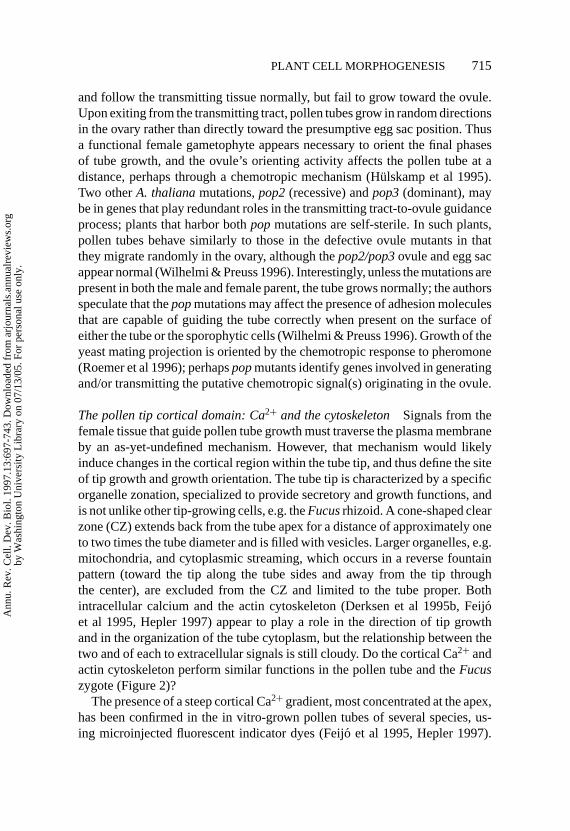
and follow the transmitting tissue normally, but fail to grow toward the ovule.Upon exiting from the transmitting tract, pollen tubes grow in random directionsin the ovary rather than directly toward the presumptive egg sac position. Thusa functional female gametophyte appears necessary to orient the final phasesof tube growth, and the ovule’s orienting activity affects the pollen tube at adistance, perhaps through a chemotropic mechanism (H¨ulskamp et al 1995).Two otherA. thalianamutations,pop2(recessive) andpop3(dominant), maybe in genes that play redundant roles in the transmitting tract-to-ovule guidanceprocess; plants that harbor bothpopmutations are self-sterile. In such plants,pollen tubes behave similarly to those in the defective ovule mutants in thatthey migrate randomly in the ovary, although thepop2/pop3ovule and egg sacappear normal (Wilhelmi & Preuss 1996). Interestingly, unless the mutations arepresent in both the male and female parent, the tube grows normally; the authorsspeculate that thepopmutations may affect the presence of adhesion moleculesthat are capable of guiding the tube correctly when present on the surface ofeither the tube or the sporophytic cells (Wilhelmi & Preuss 1996). Growth of theyeast mating projection is oriented by the chemotropic response to pheromone(Roemer et al 1996); perhapspopmutants identify genes involved in generatingand/or transmitting the putative chemotropic signal(s) originating in the ovule.
The pollen tip cortical domain: Ca2+ and the cytoskeletonSignals from thefemale tissue that guide pollen tube growth must traverse the plasma membraneby an as-yet-undefined mechanism. However, that mechanism would likelyinduce changes in the cortical region within the tube tip, and thus define the siteof tip growth and growth orientation. The tube tip is characterized by a specificorganelle zonation, specialized to provide secretory and growth functions, andis not unlike other tip-growing cells, e.g. theFucusrhizoid. A cone-shaped clearzone (CZ) extends back from the tube apex for a distance of approximately oneto two times the tube diameter and is filled with vesicles. Larger organelles, e.g.mitochondria, and cytoplasmic streaming, which occurs in a reverse fountainpattern (toward the tip along the tube sides and away from the tip throughthe center), are excluded from the CZ and limited to the tube proper. Bothintracellular calcium and the actin cytoskeleton (Derksen et al 1995b, Feij´oet al 1995, Hepler 1997) appear to play a role in the direction of tip growthand in the organization of the tube cytoplasm, but the relationship between thetwo and of each to extracellular signals is still cloudy. Do the cortical Ca2+ andactin cytoskeleton perform similar functions in the pollen tube and theFucuszygote (Figure 2)?
The presence of a steep cortical Ca2+ gradient, most concentrated at the apex,has been confirmed in the in vitro-grown pollen tubes of several species, us-ing microinjected fluorescent indicator dyes (Feij´o et al 1995, Hepler 1997).
Ann
u. R
ev. C
ell.
Dev
. Bio
l. 19
97.1
3:69
7-74
3. D
ownl
oade
d fr
om a
rjou
rnal
s.an
nual
revi
ews.
org
by W
ashi
ngto
n U
nive
rsity
Lib
rary
on
07/1
3/05
. For
per
sona
l use
onl
y.

P1: ARK/dat P2: ARK
September 3, 1997 15:25 Annual Reviews AR041-20
716 FOWLER & QUATRANO
Furthermore, dissipation of the gradient either by injection of BAPTA-typecalcium buffers (Pierson et al 1994) or by treatment with the Ca2+ channelblocker La3+ (Malho et al 1995) causes growth of the pollen tube to cease. Thenormal fluctuations that occur in the growth rate of cultured tubes correlatequite closely with fluctuations in [Ca2+] at the tip: higher [Ca2+] appears toprecede an increased growth rate by a small margin (Pierson et al 1996). Thushigh Ca2+ levels at the tube tip are necessary for continued growth and mayinfluence growth rate. Moreover, several lines of evidence indicate that the siteof highest [Ca2+] defines the site of tip growth and thus the growth orientation(similar to the rhizoid growth site inFucus; Figure 2b). When tube growth isdisrupted by caffeine or by a mild temperature shock, the Ca2+ gradient is alsodissipated; the tube recovers by undergoing an initial symmetric expansion,followed by normal, directed tip growth at a site predicted by a re-establishedCa2+ gradient (Pierson et al 1996). So-called undulating pollen tubes, whichappear occasionally in culture, show highly repetitive switching between left-oriented growth and right-oriented growth; in such tubes, the tip-high gradientalso changes position from side to side in a manner that precedes the directionalchange in growth (Malh´o & Trewavas 1996). Finally, experimental manipula-tion of the gradient, by localized photoactivation of either caged Ca2+ or a cagedCa2+ chelator, can control the future direction of tube growth, thus providingconvincing evidence that a localized high [Ca2+] at the CZ cortex is sufficientto control growth orientation (Malh´o & Trewavas 1996). Importantly, localizedphotoactivation of caged Ca2+ in the tube proper (away from the CZ) does notreorient growth to that site (Malh´o & Trewavas 1996); this suggests that theCZ and/or its cortex, in contrast to other regions of the tube, are specialized torespond to the Ca2+ gradient.
Localization of Ca2+ influx to the pollen tube tip led to the original hypothesisof an intracellular tip-associated gradient (Jaffe et al 1975) and suggests thatCa2+ channel activity is also localized. High-resolution ratiometric dye imagingindicates that the region of high [Ca2+], and thus the area of Ca2+ entry intothe tip, is very limited; the patch of plasma membrane at the apex throughwhich Ca2+ enters is estimated at 2.5µm in diameter in the≈15µm diameterlily pollen tube (Pierson et al 1996). The existence of calcium channels in thetip is supported by a close correlation between the presence and location ofan extracellular tip-directed influx of Ca2+ and the intracellular Ca2+ gradient(Pierson et al 1994) and also by the ability of extracellular Mn2+ (which can passthrough most calcium channels) to enter preferentially at the tip (Malh´o et al1995). The tip-localized activity of the channels correlates with the presenceor absence of the Ca2+ gradient, suggesting that channel activity is a factor inregulating the gradient (Malh´o et al 1995). Localized calcium channel activityis a conserved feature of tip-growing cells, e.g. in root hairs (Hermann & Felle
Ann
u. R
ev. C
ell.
Dev
. Bio
l. 19
97.1
3:69
7-74
3. D
ownl
oade
d fr
om a
rjou
rnal
s.an
nual
revi
ews.
org
by W
ashi
ngto
n U
nive
rsity
Lib
rary
on
07/1
3/05
. For
per
sona
l use
onl
y.

P1: ARK/dat P2: ARK
September 3, 1997 15:25 Annual Reviews AR041-20
PLANT CELL MORPHOGENESIS 717
1995), in fungal hyphae (Garill et al 1992), and inFucusrhizoids (Taylor et al1996). Several models could account for localized calcium channel activity,including (a) localized stretch-activation of channels at sites of cell expansion(i.e. the region of vesicle secretion at the tube tip) (Pierson et al 1996); (b)preferential inactivation of channels displaced from the tip via endocytosis(Derksen et al 1995a, Derksen 1996); or (c) anchoring of active channels inthe tip region, or exclusion of active channels from non-tip regions, possibly bymeans of a cytoskeletal network (Cai et al 1997). These models are not mutuallyexclusive, and some bear similarity to the putative ASC inFucusrhizoid tips(Figure 2). However, any explanation must take into account the ability of thepollen tube to homeostatically regulate its Ca2+ gradient (Malh´o & Trewavas1996), rapidly change its direction of growth, and respond to external, orientingsignals. Imposition of a local external Ca2+ gradient near the tip influences theinternal Ca2+ gradient and growth orientation, suggesting that such a gradientin the pistil ECM could serve to guide tube growth in vivo (Malh´o & Trewavas1996). Although high concentrations of Ca2+ have been detected near ovules(Chaubal & Reger 1990), further work is needed to address this possibility.
The existence of an organized cytoskeletal framework in the extreme tip ofthe tube is currently a matter of some debate (Derksen 1996, Cai et al 1997).Until recently, most reports indicated the presence of an actin MF network atthe tube apex (Pierson & Cresti 1992, Derksen et al 1995b). However, newdata, using more advanced techniques, call these results into question (Lancelle& Hepler 1992, Miller et al 1996). Furthermore, fixation techniques used inearlier work do not preserve the CZ zonation, supporting the argument forexclusion of MFs from the CZ and tube tip (Doris & Steer 1996). In any case,cytoskeletal organization at the apex is clearly different from that at the cortex ofthe tube proper, which contains characteristic polarized arrays of MTs and MFs,hypothesized to aid in cytoplasmic streaming and other tip-directed movementprocesses (Lancelle & Hepler 1992, Derksen et al 1995b, Cai et al 1997). Thecortical cytoskeletal arrays in the tube proper terminate as they approach theCZ, and MTs and MFs that have been observed in the CZ apparently are notorganized at the cortex in a specific array (Lancelle & Hepler 1992, Cai et al1993, Miller et al 1996). Cytochalasins have long been known to disrupt tubegrowth, cytoplasmic streaming, and organelle movement—almost certainly bytheir disruption of an actomyosin network in the tube proper (Cai et al 1997).Some in vivo evidence also points to a necessary function for MTs in tube growth(Joos et al 1995), perhaps for maintaining the cytoplasm and the generative cellsnear the tube tip, and the vacuole near the tube base (Joos et al 1994).
Several proteins putatively associated with the cortical cytoskeleton in pollentubes have been identified. Homologues of motor proteins in the myosin,kinesin, and dynein families, which may function in organelle movement,
Ann
u. R
ev. C
ell.
Dev
. Bio
l. 19
97.1
3:69
7-74
3. D
ownl
oade
d fr
om a
rjou
rnal
s.an
nual
revi
ews.
org
by W
ashi
ngto
n U
nive
rsity
Lib
rary
on
07/1
3/05
. For
per
sona
l use
onl
y.

P1: ARK/dat P2: ARK
September 3, 1997 15:25 Annual Reviews AR041-20
718 FOWLER & QUATRANO
have been described (Cai et al 1996b, Asada & Collings 1997). An animalcentrosome-related antigen, possibly associated with cortical MTs and MTOCactivity, has been localized to the cortex and is associated with plasma mem-branes (Cai et al 1996a). Perhaps most interesting is the localization of Ropprotein antigens to the plasma membrane at and near the pea tube apex (Linet al 1996); Rop genes make up a plant-specific multigene branch of therac/rho/Cdc42p family of GTPases (Yang & Watson 1993, Delmer et al 1995).GTPases of the rac family regulate the organization of the actin cytoskeletonin yeasts, mammalian tissue culture cells, andD. melangaster(Tapon & Hall1997); thus the localized Rop protein(s) could play a crucial role in determin-ing the distribution of MFs at and near the pollen tip (Lin et al 1996). Futureexperiments are needed to determine the roles of these putative cytoskeleton-associated proteins and whether they influence or respond to the position of thepollen tip growth site.
Function of the tip growth site The most conspicuous function of the growthsite at the tube apex is to serve as a region for fusion of secretory vesicles tothe plasma membrane, thus providing material for the deposition of new cellwall and plasma membrane. However, the growth site is also likely to conveyinformation, providing a focal point around which the rest of the cell organizesits characteristic zonation and cytoskeletal distribution.
The CZ cytoplasm adjacent to the tube apex is packed with vesicles thatare transported to the CZ via cytoplasmic streaming (Lancelle & Hepler 1992,Battey & Blackbourne 1993, Derksen et al 1995a,b). In contrast to the vesicles atthe cortex of the tube proper, those in the CZ move randomly (Pierson et al 1990),perhaps because they lack an associated cytoskeletal network. The mechanismby which the vesicles fuse with the plasma membrane, and by which fusion isrestricted to the apex, is unknown. Speculation has focused on a possible role forthe Ca2+ gradient itself in regulating the docking and/or fusion of the vesicles tothe plasma membrane through Ca2+-binding proteins (Battey & Blackbourne1993, Derksen et al 1995b, Malh´o & Trewavas 1996, Pierson et al 1996). Forexample, annexins, which have been localized to pollen tube tips, associate withthe plasma membrane in the presence of Ca2+, and might assist vesicle fusion(Blackbourn et al 1992, Moss 1997).
Other functions of the apex site are even less clear. The localized secretionmay target specific molecules to the cell wall, for example, extensins (Rubinsteinet al 1995), or the molecule(s) responsible for TTS deglycosylation. AGPssecreted at the tip, or associated with the plasma membrane, may reinforce thesignals that orient the Ca2+gradient. An intriguing report indicates that exposureof the pollen tube to the Yariv reagent not only inhibits growth but also causesa rise in [Ca2+] throughout a broad region of the tube tip (Roy et al 1996). It
Ann
u. R
ev. C
ell.
Dev
. Bio
l. 19
97.1
3:69
7-74
3. D
ownl
oade
d fr
om a
rjou
rnal
s.an
nual
revi
ews.
org
by W
ashi
ngto
n U
nive
rsity
Lib
rary
on
07/1
3/05
. For
per
sona
l use
onl
y.

P1: ARK/dat P2: ARK
September 3, 1997 15:25 Annual Reviews AR041-20
PLANT CELL MORPHOGENESIS 719
is unknown how the boundary between the CZ and the tube proper, and theirdistinct cytoskeletal arrangements, are established, stabilized, and continuallymodified as growth occurs. The calcium gradient appears important for correcttip zonation, as injection of BAPTA-type buffers to dissipate the gradient alsodisturbs the integrity of the CZ and alters the pattern of cytoplasmic streaming atthe tip (Pierson et al 1994). However, the mechanisms that connect the [Ca2+]-defined cortical site to cytoplasmic organization include many possibilities:spatially regulated endocytosis (Derksen et al 1995a), direct [Ca2+] regulationof the cytoskeleton, or cytoskeleton-plasma membrane-cell wall connections.
Although integrins in animal cells can function as a transmembrane link be-tween the cytoskeleton and the extracellular matrix (see focal adhesions above),no protein with similar sequence (that might function at the tube tip) has beenfound in plants. However, anA. thalianagene, wall-associated kinase1 (WAK1),encodes a transmembrane protein that has a cytoplasmic kinase motif and an ex-tracellular, cell wall–associated domain with similarities to the cell wall proteinextensin (He et al 1996; BD Kohorn, personal communication); thus,WAK1might provide an integrin-like function in plants. Also of potential relevanceis the putative transmembrane complex associated with cellulose deposition.MFs, MTs, and annexins have been linked to the deposition and alignmentof cellulose microfibrils, possibly through a cellulose synthase (CS) complex(Andrawis et al 1993, Delmer & Amor 1995, Calvert et al 1996). In addition,a plasma membrane-bound sucrose synthase may be associated with the CScomplex that could channel carbon directly to the complex from sucrose viaUDP-glucose (Amor et al 1995). Enhanced expression of a Rac13 gene at thetime of actin reorganization and the initiation of cellulose synthesis in cottonfibers (Delmer et al 1995) may indicate a role for this small GTPase in activat-ing CS (similar to the Rho1 activation of glucan synthase in the yeast bud; seeabove), and/or in regulating F-actin organization. With the isolation of a highlyconserved candidate cellulose synthase gene from cotton (Pear et al 1996), theorganization and role of the components of the CS complex in wall expansionand tip growth in plants can be explored with respect to transmembrane com-plexes linking the cytoskeleton to the extracellular matrix (see D Cosgrove, thisvolume).
Thus despite important advances toward understanding the physiology ofthe tube apex, some connections between the identified players remain to beestablished. Does compromising the cytoskeleton affect the Ca2+ gradient?Caffeine treatment causes the tube’s cortical MF to redistribute into the tubecenter and closer to the apex and the CZ to shorten (Miller et al 1996); doesdissipation of the Ca2+gradient cause similar effects? How does an extracellulargradient of TTS protein affect the location of the highest [Ca2+] within thetip? Such questions can be addressed using the knowledge and tools currently
Ann
u. R
ev. C
ell.
Dev
. Bio
l. 19
97.1
3:69
7-74
3. D
ownl
oade
d fr
om a
rjou
rnal
s.an
nual
revi
ews.
org
by W
ashi
ngto
n U
nive
rsity
Lib
rary
on
07/1
3/05
. For
per
sona
l use
onl
y.

P1: ARK/dat P2: ARK
September 3, 1997 15:25 Annual Reviews AR041-20
720 FOWLER & QUATRANO
available. For example, the report describing an effect of the Yariv reagent on tip[Ca2+] implies a link between extracellular AGPs and the regulation of the Ca2+
gradient (Roy et al 1996). Does this treatment also affect the cytoskeleton? Arethere localized DHP receptors at the growth site, as observed at theFucusrhizoidtip? Establishing such connections with the methods at hand, and consideringthe analogous models from other systems (e.g.Fucus, yeast), should provide amore complete framework around which to design future experiments.
ROOT HAIRS Certain epidermal root cells initiate tip growth at a specific siteon their outer cell wall, producing a tube-like extension, the root hair. A geneticapproach to understanding root hair development inA. thaliana(Schiefelbein& Somerville 1990) is beginning to bear fruit, and it may also provide infor-mation relevant to pollen tubes and other growing cells. For example, therhd3mutation, originally isolated through a defect in root hair morphology, disruptscell expansion throughout the plant and is found in a novel conserved proteinwith GTP-binding motifs (Wang et al 1997). AlthoughA. thalianaroot hairscharacteristically initiate from the apical end (i.e. nearest the root meristem)of epidermal cells, plants carrying therhd6 mutation possess fewer root hairsthan wild-type, and the extant hairs initiate from a more basal position than inthe wild-type (Masucci & Schiefelbein 1996). Similar defects were observed inthe auxin-, ethylene-, and ABA-resistant mutantaxr2and the ethylene-resistantmutantetr1and could be phenocopied by the application of an ethylene biosyn-thesis inhibitor. Further, therhd6 mutant phenotype could be alleviated by in-cluding auxin or ACC (an ethylene precursor) in the growth medium. Thisimplies that therhd6gene acts in the signaling process that identifies the site ofroot hair initiation (analogous to choosing the rhizoid site inFucus; Figure 2)and also that the phytohormones ethylene and auxin are involved in this signal-ing (Masucci & Schiefelbein 1996). As in pollen tubes and theFucusrhizoid,Ca2+ appears to have important roles in root hair growth; Ca2+ influx has beendetected at the tip of theA. thalianaroot hair (Schiefelbein et al 1992). Do MFsor Ca2+ play a role not only in growth, but also in determining the root hairinitiation site?
A potential link between secretion and root hair growth is provided by oneof the several root hair mutants identified in maize (Wen & Schnable 1994).A preliminary report indicates that the maizerth1 gene has homology to theS. cerevisiae SEC3gene (T-J Wen & PS Schnable, personal communication),a subunit of the Exocyst complex that is required for secretion (TerBush et al1996). Is the RTH1 protein localized to a cortical site at the root hair tip,analogous to Exocyst localization at the growing yeast bud tip? A final linkbetween pollen tubes and root hairs is revealed by theA. thaliana tip1mutant,which exhibits defects in both root hair and pollen tube growth (Schiefelbein
Ann
u. R
ev. C
ell.
Dev
. Bio
l. 19
97.1
3:69
7-74
3. D
ownl
oade
d fr
om a
rjou
rnal
s.an
nual
revi
ews.
org
by W
ashi
ngto
n U
nive
rsity
Lib
rary
on
07/1
3/05
. For
per
sona
l use
onl
y.

P1: ARK/dat P2: ARK
September 3, 1997 15:25 Annual Reviews AR041-20
PLANT CELL MORPHOGENESIS 721
et al 1993). Are there pollen tube mutants with altered morphology, similar tothe already identified mutations that affect the root hair?
Spatial Regulation of Cell DivisionSpatial regulation of division plane placement allows plants to control theirmorphology by altering cell division patterns (Lloyd 1995, Smith 1996)(Figure 1e-h). The central role of the cortical cytoskeleton in this process haslong been recognized; in many plant cells the cortical microtubule (MT) pre-prophase band (PPB) predicts the future division site, i.e. where the new cellwall between daughters will fuse with the existing walls (Wick 1991a,b). A bandof F-actin is also present and associated with the MT PPB, but both the MT andMF bands disappear before metaphase. Thus in contrast to an animal cell, thedivision plane of a plant cell is not dependent on spindle orientation but rather isdetermined and marked cortically prior to anaphase. After anaphase, the phrag-moplast, a complex array of MTs, MFs, vesicles, and membrane structures,appears between the daughter nuclei and builds the new cell wall centrifugally,such that it eventually fuses with the previously defined cortical division site(Staehelin & Hepler 1996). The mechanisms that position the PPB and its as-sociated cues at the cortex and those that transform the PPB-associated cuesinto a correctly positioned new cell wall are still unknown.
THE CORTICAL CYTOSKELETON AND THE DIVISION SITE The general frame-work for cell division in plants recounted above has been established through thework of many investigators (Mineyuki & Gunning 1990, Wick 1991a, Clearyet al 1992, Samuels et al 1995, Staehelin & Hepler 1996). The cortical MT andMF PPBs appear prior to the onset of mitosis but disappear before metaphase,leaving only an actin-depleted zone that remains notably free of cortical actin(relatively abundant elsewhere) in certain cells (Cleary et al 1992, Liu & Palevitz1992, Cleary 1995). Large, vacuolated cells contain a network of cytoplasmicactin filaments that persists through division and connects the spindle, andthen the phragmoplast, to the cell cortex and division site (Traas et al 1987,Lloyd & Traas 1988). These actin filaments are called phragmosomal filamentsbecause they are associated with the disk of cytoplasm (the phragmosome)that encompasses the nucleus and the future division plane during division invacuolated cells (Staiger & Lloyd 1991). The presence of an actin-depleted zonein several types of dividing cells and phragmosomal filaments in vacuolated cellsindicates that the cortex at the division site retains a persistent specializationafter loss of the PPBs.
TheB. subtilisdivision site is also cortically determined but is restricted toa choice between three pre-set alternate positions (one medial, two polar). Arethere a limited number of sites for PPB formation, or can it form at any site on the
Ann
u. R
ev. C
ell.
Dev
. Bio
l. 19
97.1
3:69
7-74
3. D
ownl
oade
d fr
om a
rjou
rnal
s.an
nual
revi
ews.
org
by W
ashi
ngto
n U
nive
rsity
Lib
rary
on
07/1
3/05
. For
per
sona
l use
onl
y.

P1: ARK/dat P2: ARK
September 3, 1997 15:25 Annual Reviews AR041-20
722 FOWLER & QUATRANO
plant cell cortex? The signals that position PPB formation (and the associateddivision site) are unknown. Nuclear migration to the center of the cell, whichoccurs prior to division, has been implicated as a possible determinant of PPBposition in fern protonemata (Murata & Wada 1991) but not in higher plantcells (Mineyuki & Palevitz 1990). Actin PPB formation precedes formationof the MT PPB in cells induced to divide by wounding (Goodbody & Lloyd1990), but treatment with cytochalasin does not block formation of a MT PPB;rather, it prevents narrowing of the MT PPB to a precise site (Mineyuki &Palevitz 1990, Eleftheriou & Palevitz 1992). Thus although actin may not benecessary in the initial phases of MT PPB formation, it has been suggestedthat actin plays a crucial role in the exact determination of the division site byrestricting the MT PPB to a narrow ring (Mineyuki & Palevitz 1990, Eleftheriou& Palevitz 1992). This model provides one explanation for the observation thatcytochalasin treatment leads to the misplacement of new cell walls (reviewed inWick 1991b). However, because actin appears to be involved in several differentprocesses during cell division, it is unclear which MF cytoskeletal array(s) isdisrupted in these experiments, and thus which one(s) is crucial for positioningof the cell wall. One hypothesis to account for the positioning of the PPB toproduce equal daughters in large, vacuolated cells is that a default orientationis chosen, perhaps by physical tensions between the nucleus and cell cortexexerted through connecting MTs and/or MFs, corresponding to the shortestpossible path across the cell (Flanders et al 1990, Lloyd 1991); however, manyplant cells do not divide in this default orientation, so other mechanisms mustexist to position the PPB at non-default sites. Thus current interpretations favorthe notion that the division site is not restricted to one of a limited number ofpredetermined sites, but that any cortical site can be chosen based on interactionsamong the MF and MT cytoskeleton and cortical components.
Several recently isolated mutations may lie in genes that are important forcortical cytoskeleton organization, including formation of the PPBs at the di-vision site (Torres-Ruiz & J¨urgens 1994, Traas et al 1995). Mutations in theTON1andTON2genes ofA. thaliana(one of which is likely allelic toFASS)lead to cells of highly irregular shape. Mutant embryonic and root cells appearto divide in random orientations, rather than in the regular patterns seen inwild-type plants and are shorter than wild-type, suggesting an additional defectin cell elongation (Torres-Ruiz & J¨urgens 1994, Scheres et al 1995, Traas et al1995). Plant cells mutant for eitherTON gene have disorganized interphasearrays of cortical MTs and never form PPBs, although MTs are associated withthe cortex, and the spindles and phragmoplasts appear normal (Traas et al 1995).Cells in plants mutant for thetangled-1(tan-1) gene of maize also divide inabnormal orientations but, in contrast to theton/fassmutants, appear to elongatenormally (Smith et al 1996). Analysis of the cytoskeleton intan-1cells shows
Ann
u. R
ev. C
ell.
Dev
. Bio
l. 19
97.1
3:69
7-74
3. D
ownl
oade
d fr
om a
rjou
rnal
s.an
nual
revi
ews.
org
by W
ashi
ngto
n U
nive
rsity
Lib
rary
on
07/1
3/05
. For
per
sona
l use
onl
y.

P1: ARK/dat P2: ARK
September 3, 1997 15:25 Annual Reviews AR041-20
PLANT CELL MORPHOGENESIS 723
that both PPB positioning and phragmoplast guidance are affected by the mu-tation (LG Smith & AL Cleary, personal communication). Thus the productsof these genes are likely to contribute to the normal regulation of the corticalMT cytoskeleton, and the signals that serve to position cortical MTs (and theMT PPB) may act through them.
Placement of the division site in asymmetric divisionsStomatal complex for-mation involves stereotyped patterns of cell division and is a well-studied modelfor asymmetric divisions (Sylvester et al 1996). The pattern begins with anasymmetric epidermal cell division, producing a smaller daughter cell thatwill eventually generate the stomatal guard cells. In several dicot species (e.g.A. thaliana), this initial asymmetric division is followed by one or more similardivisions (Yang & Sack 1995). Each further asymmetric division occurs onlyin the smaller daughter of the previous division; eventually, a smaller daughtercell in this series, the guard mother cell (GMC), divides equally to form thetwo guard cells. In monocots, the smaller daughter of the initial asymmetricdivision is the GMC and directly produces the two guard cells. In many mono-cot species, small subsidiary cells (SCs) are formed by asymmetric division ofsubsidiary mother cells (SMCs), located laterally adjacent to the GMCs. TheSMCs divide only after formation of the GMC, such that the smaller daughter(the subsidiary cell) is adjacent to the GMC. Thus the division asymmetry ofthe SMC appears to be induced by the presence of the GMC. These observa-tions suggest that a conserved mechanism involving an asymmetric division,perhaps to segregate cell fate determinants (Figure 1f, h), is operating in theinitial steps of stomatal complex development. How is the process that leadsto placement of the division site altered in these asymmetric, compared withdefault-symmetric, divisions?
In vivo studies inTradescantiaSMC cells have found several differencesbetween asymmetric and symmetric division that may be important (Cleary1995, Cleary & Mathesius 1995, Kennard & Cleary 1997). The first sign ofasymmetry in the vacuolated SMC is the accumulation of cytoplasm at a cor-tical site adjacent to the GMC, followed by migration of the nucleus near thesame region (Kennard & Cleary 1997). After nuclear migration, MFs accumu-late at the same SMC cortical site between the SMC nucleus and the GMC,and concomitantly in the adjacent cortex of the GMC (Cleary 1995, Cleary &Mathesius 1995). Actin filaments also connect the nucleus to the newly accu-mulated cortical actin, and treatment with cytochalasin B (CB), but not oryzalin,prevents the migration of centrifuged SMC nuclei toward the GMC, suggestingthat MFs, but not MTs, are necessary for normal nuclear migration (Kennard& Cleary 1997). In contrast, that migration is not necessary to induce corticalMF localization, because in a small subset of SMC cells, the actin accumulates
Ann
u. R
ev. C
ell.
Dev
. Bio
l. 19
97.1
3:69
7-74
3. D
ownl
oade
d fr
om a
rjou
rnal
s.an
nual
revi
ews.
org
by W
ashi
ngto
n U
nive
rsity
Lib
rary
on
07/1
3/05
. For
per
sona
l use
onl
y.

P1: ARK/dat P2: ARK
September 3, 1997 15:25 Annual Reviews AR041-20
724 FOWLER & QUATRANO
without nuclear migration (Cleary & Mathesius 1995). The polar accumulationof actin persists throughout mitosis. The remainder of SMC mitosis appearsto occur by the general division mechanism already discussed: asymmetricallyshifted MT (Cho & Wick 1989) and MF (Cleary 1995) PPBs appear at an SMCcortex site that predicts the future division plane, and on their disappearance,they leave an actin-depleted zone; no phragmosomal MFs have been observedin SMCs (Cho & Wick 1990, Cleary 1995). A similar series of events occurs inthe asymmetric division that produces the GMC, suggesting that these eventsmay be general features of plant asymmetric division. These studies supportand extend earlier observations in stomatal complex cells of winter rye (Cho &Wick 1990, 1991).
The signals that initiate these asymmetries, though clearly associated withthe GMC, remain elusive. One clue to the nature of the signals is that localapplication of pressure to SMC cell walls leads to cytoplasmic and nuclearmigration to the pressure point (Kennard & Cleary 1997); the authors suggestthat stretch-activated ion channels and ion fluxes are intermediates in this sig-naling process. A simple model is that an extracellular cue originating from theGMC is interpreted by the SMC to orient both nuclear migration and the localaccumulation of cortical actin. Either of these asymmetric events could serve toreinforce the GMC-originating spatial cue, thus establishing a more permanentpolarity, which might in turn influence, extracellularly, the actin cytoskeletonof the adjacent GMC and, intracellularly, the SMC cytokinetic apparatus(Figure 4a, b). The localized actin/actin-capping protein/dynactin complex intheC. elegansP1 cell and the rhizoid tip site in theFucuszygote may functionas anchors to help rotate a centrosome and establish their asymmetric divisionplanes (see Figure 2c). Does the polarly localized actin in the SMC have ananalogous function, anchoring the asymmetric position of the nucleus? Does italso shift the division site (Figure 4a, b)?
Division site function The division site and associated PPBs have two ascribedmain functions (reviewed in Wick 1991a,b): The first is to assist in defining thespindle apparatus location and orientation. Although the division site deter-mines where the phragmoplast will eventually fuse, centrifugation experimentsindicate that the phragmoplast must be correctly situated (within an allowablemargin of error) as it finishes forming the cell plate in order to produce a cor-rectly placed wall. Thus the nucleus is generally positioned at the center ofthe PPB disk, and the spindle poles are often oriented perpendicular to thisdisk during karyokinesis—although in many plant cells spindle orientation isoblique to the disk (Palevitz 1993). MTs and MFs that connect the PPB to thenucleus during prophase have been observed (Flanders et al 1990, Katsuta et al1990), and in vacuolated cells, the phragmosomal MFs persist through division
Ann
u. R
ev. C
ell.
Dev
. Bio
l. 19
97.1
3:69
7-74
3. D
ownl
oade
d fr
om a
rjou
rnal
s.an
nual
revi
ews.
org
by W
ashi
ngto
n U
nive
rsity
Lib
rary
on
07/1
3/05
. For
per
sona
l use
onl
y.

P1: ARK/dat P2: ARK
September 3, 1997 15:25 Annual Reviews AR041-20
PLANT CELL MORPHOGENESIS 725
a b
c d
Figure 4 A model illustrating the possible role of cortical domains in asymmetric cell divisionand cell fate determination in plants. The mechanistic details in this model are largely unknown.The model incorporates a postulated role for targeted secretion to the cell wall/membrane as acomponent for defining and/or stabilizing the cortical sites, as well as influencing the fate ofdaughter cells. The model is based on observations inFucusand other plants, as well as in otherorganisms (e.g. yeast,C. elegans). (a) An initial directional cue (extracellular or intracellular) leadsto formation of an asymmetry in the cortex (rectangle). The cortical asymmetry directs targetedsecretion to the cell wall, perhaps through interaction with the cytoskeleton, and secretion of specificcomponents into the plasma membrane/cell wall (dark line) stabilizes the asymmetries on one faceof the cell (see Figure 2b). (b) The stabilized cortical domain directs (dotted arrows) the asymmetricplacement of the division site (solid circle) around the perimeter of the cell (see Figure 3a), as wellas the movement of the nucleus (hatched oval) toward the division site. Assembly of a localizedcortical domain at the division site could also be stabilized by cell wall/membrane interactions. (c)During cytokinesis, the division sites, through interaction (dashed arrows) with the phragmoplastand/or cell plate (diagonal shading), control the position of plate fusion to the parental walls. Theinitial cortical asymmetry may be transient (dotted rectangle). However, the persisting, positionalcues secreted into the cell wall/plasma membrane remain. (d ) The localized cortical differences(dark line) stabilized in (a) are now partitioned into a single daughter cell. The specialized cellwall/membrane may affect the developmental fate of that daughter cell by signaling to cytoplasmiccomponents and/or to the nucleus (see Figure 3b).
Ann
u. R
ev. C
ell.
Dev
. Bio
l. 19
97.1
3:69
7-74
3. D
ownl
oade
d fr
om a
rjou
rnal
s.an
nual
revi
ews.
org
by W
ashi
ngto
n U
nive
rsity
Lib
rary
on
07/1
3/05
. For
per
sona
l use
onl
y.

P1: ARK/dat P2: ARK
September 3, 1997 15:25 Annual Reviews AR041-20
726 FOWLER & QUATRANO
(Lloyd & Traas 1988), suggesting a mechanism that accounts for the centralposition of the nucleus and spindle. Drug studies indicate that nuclear migra-tion to the future division plane and therein its maintenance can be perturbedby destabilizing MTs in certain cell types, and MFs in other types (reviewedin Wick 1991b). Thus one function of the cortical division/PPB site may be toanchor cortical ends of nucleus/spindle-positioning cytoskeletal arrays.
The second function of the division site is to ensure that the growing phrag-moplast fuses with the existing cell walls at the correct position, i.e. the locationselected by the plant prior to mitosis based on developmental and environmen-tal cues (Figure 4c). This function is often called the memory of the PPB, butin another view this memory function is the raison d’ˇetre for the PPB. A fewmarkers are now known (e.g. the actin-depleted zone) indicating that the divi-sion site does indeed possess persisting differences after the disappearance ofthe MT and MF PPBs. Another indication of a localized, persisting function isthat wounding of the division site by micro-needle punctures during anaphaseleads to a localized inhibition of phragmoplast growth near the wound site intelophase (Gunning & Wick 1985). The mechanism responsible for this persis-tence is unknown, although some possibilities have been suggested. The p34cdc2
kinase has been localized to the PPB in both symmetrically dividing root tipcells (Mineyuki et al 1991) and in asymmetrically dividing stomatal complexcells (Colasanti et al 1993) and has been postulated to help mark the cortex viaphosphorylation of cortex proteins. The persistence of the division site couldalso be due to specialization of the cell wall at the former PPB site (clearlydiscussed in Mineyuki & Gunning 1990). Observations in several cell types,including A. thalianaGMCs (L Zhao, M Geisler & FD Sack, personal com-munication), indicate that the cell wall adjacent to the PPB shows localizedthickening and, in some cases, that vesicles associate with the PPB MT (Galatiset al 1984). Thus, the PPB cytoskeletal structures could direct the secretion ofa specific vesicle population at the division site (analogous to that occurring attheFucusrhizoid tip; Figure 2b, c), leading to a persisting, localized corticalspecialization (perhaps through ECM/plasma membrane linkages) that enablesphragmoplast fusion in the final stage of mitosis (Mineyuki & Palevitz 1990).
The mechanism that causes the nascent cell wall to fuse with the defined corti-cal site is under intense scrutiny (Figure 4c). One proposal is that phragmosomalMFs connecting the division site to the growing phragmoplast in highly vac-uolated cells guide it to the correct cortical site (Traas et al 1987, Lloyd &Traas 1988, Staiger & Lloyd 1991); hence, the division site is a cortical site thatanchors these MFs. However, such division site-to-phragmoplast MF are notvisible in smaller, less vacuolated cells (Cho & Wick 1990, Cleary et al 1992,Liu & Palevitz 1992, Cleary 1995). In smaller cells, e.g.Tradescantiastamenhairs, growing phragmoplasts displaced by centrifugation or micromanipulation
Ann
u. R
ev. C
ell.
Dev
. Bio
l. 19
97.1
3:69
7-74
3. D
ownl
oade
d fr
om a
rjou
rnal
s.an
nual
revi
ews.
org
by W
ashi
ngto
n U
nive
rsity
Lib
rary
on
07/1
3/05
. For
per
sona
l use
onl
y.

P1: ARK/dat P2: ARK
September 3, 1997 15:25 Annual Reviews AR041-20
PLANT CELL MORPHOGENESIS 727
migrate back to their normal position and fuse at the division site (Gunning& Wick 1985, Mineyuki & Palevitz 1990), supporting the notion that activeguidance of the phragmoplast to the division site occurs. As noted previously,cytochalasin treatment causes abnormal placement of new cell walls (Wick1991b); whether this is from a direct loss of actin function in phragmoplastguidance, or the indirect result of abnormal nuclear or spindle positioning, lossof actin PPB function, or compromising the MF in the phragmoplast itself, orother CB effects, remains to be clarified.
Another proposal, which does not exclude the previous actin-based model,is that the division site contains localized factors necessary for the final matura-tion of the phragmoplast (Mineyuki & Gunning 1990). This model is supportedby real-time observation of normal and experimentally manipulated cytokine-sis inTradescantiastamen hairs. During normal cytokinesis, the forming cellplate within the phragmoplast is initially wrinkled and wavy and retains thismorphology until shortly after attachment to the parental cell walls, at whichtime the existing cell plate flattens (Mineyuki & Palevitz 1990). This flatteningeffect appears to spread gradually from the division site onto cell plates thatare attached to the division site on only one side. However, cell plates that areforced to fuse to the parental cell walls at positions other than the division siteremain wavy, suggesting that they lack the maturation factors necessary fornormal structure (Mineyuki & Palevitz 1990). Thus the division site appears tobe the sole repository of cortical maturation factors, perhaps further insuringthat the new cell wall is positioned only at the pre-mitotically determined site.
THE PHRAGMOPLAST AND CYTOKINESIS Our discussion has so far centeredon two important cortical sites established during cell division—the divisionsite and the polar actin accumulation site in developing stomatal complexes (e.g.SMCs). However, there is another important cortical site; the new cortex form-ing at the nascent cell plate. Cytokinesis in higher plants is accomplished byproducing a new cell wall between the two daughters, and the cellular apparatusresponsible for the new wall is the phragmoplast (Figure 4c) (Staehelin & Hepler1996, Verma & Gu 1996). The phragmoplast is a complex array of MTs and MFsthat directs Golgi-derived vesicles to the region between the two daughter nu-clei; these vesicles fuse to form a membranous plate that expands from the centerof the cell toward the periphery. The plate goes through specific stages of matu-ration as it accumulates vesicle-derived material and other cell wall components,forming a cell wall that separates the newly generated plasma membrane/cellcortex of the daughter cells. Thus the cell plate can be considered a specializedcortical region that serves as a target for vesicle fusion and as a site of local-ized wall synthesis activity, e.g. callose synthase (Kakimoto & Shibaoka 1992).However, the cell plate initiates in the cytoplasm, as a collection of vesicles that
Ann
u. R
ev. C
ell.
Dev
. Bio
l. 19
97.1
3:69
7-74
3. D
ownl
oade
d fr
om a
rjou
rnal
s.an
nual
revi
ews.
org
by W
ashi
ngto
n U
nive
rsity
Lib
rary
on
07/1
3/05
. For
per
sona
l use
onl
y.

P1: ARK/dat P2: ARK
September 3, 1997 15:25 Annual Reviews AR041-20
728 FOWLER & QUATRANO
then fuse (Samuels et al 1995, Staehelin & Hepler 1996, Verma & Gu 1996);the mechanisms that govern this unique maturation process are unknown.
The initial phragmoplast structure is a cylindrically shaped MT aggregate,consisting of two disks of oppositely oriented MTs that occupy the space roughlycorresponding to the recent spindle interzone. The exact signals that positionthe phragmoplast are not known, but results support the idea that this originalMT array is formed in late anaphase by MTs from the spindle (Zhang et al1993). One hypothesis to explain its position is that the phragmoplast MTs areinitially organized by MTOC activity at the reforming nuclear membranes ofthe two daughter nuclei, as the faster-growing plus ends of the MTs interdigitateat the center of the phragmoplast, with their minus ends toward the daughternuclei (Lambert 1993). At roughly the same stage, MFs form parallel and ad-jacent to the phragmoplast MTs, but do not meet at the phragmoplast center,leaving a space devoid of F-actin that corresponds to the site of the future cellplate (Zhang et al 1993). Vesicles associated with the phragmoplast cytoskeletalelements are deposited at the phragmoplast equator, where the cylindrical arrayof MTs interdigitate. As the cell plate forms from these vesicles, the cylindricalMT and MF arrays expand radially toward the cell walls, correlating with anexpanded region in which vesicles are deposited into the phragmoplast cen-tral region (Schopfer & Hepler 1991, Zhang et al 1993). Disruption of the MTarrays in the phragmoplast after the daughter nuclei separate prevents cytokine-sis and produces binucleate cells, presumably by blocking vesicle transport tothe cell plate. Further, a protein with MT plus-end-directed motor activity hasbeen identified and isolated from tobacco phragmoplasts (Asada & Shibaoka1994), and a kinesin-like protein likely to have minus-end-directed motor ac-tivity has been detected in the phragmoplast by immunofluorescence (Liu et al1996). Plus-end-directed motors are candidates to provide the force necessaryto transport vesicles on phragmoplast MTs to the forming cell plate. The func-tion of MFs in the phragmoplast is less clear, but available evidence supportsthe hypothesis that the phragmoplast cytoskeletal arrays function to transportvesicles to a target site for assembly into the cell plate.
A detailed characterization of the vesicle fusion/cell plate formation pro-cess by transmission electron microscopy (TEM) indicates that several distinctstages are recognizable (Samuels et al 1995). Vesicles arrive at the equato-rial region and rapidly fuse through connections established by thin (20 nm)tubules, which can extend relatively long (up to 500 nm) distances betweentwo vesicles. This rudimentary network of vesicles and tubules is transformedinto a continuous, mesh-like tubulo-vesicular network (TVN). The TVN is sur-rounded by a complex recognized in TEM as a fuzzy coat and is associated withnumerous MTs and with the initial synthesis of callose, which is deposited intothe lumen of the network. The TVN matures into a network of larger, smoother
Ann
u. R
ev. C
ell.
Dev
. Bio
l. 19
97.1
3:69
7-74
3. D
ownl
oade
d fr
om a
rjou
rnal
s.an
nual
revi
ews.
org
by W
ashi
ngto
n U
nive
rsity
Lib
rary
on
07/1
3/05
. For
per
sona
l use
onl
y.

P1: ARK/dat P2: ARK
September 3, 1997 15:25 Annual Reviews AR041-20
PLANT CELL MORPHOGENESIS 729
membrane tubules, accompanied by the spreading of callose deposits alongthe lumenal face of the membrane and a loss of associated MTs. Coincidentwith a near-simultaneous fusion of the plate membrane network with the parentplasma membrane at hundreds of sites, the plate begins to flatten and graduallyclose the last cytoplasmic gaps. The plate maturation process proceeds spatiallyin concert with the centrifugal expansion of the phragmoplast; i.e. the plate’scenter, which is formed initially, is more mature than the plate’s edges, whichare formed later, as the phragmoplast MT array shifts from the center. Hencethe formation of the cell plate can be viewed as a three-part process: targetedvesicle transport, specialized membrane/vesicle fusion, and maturation of themembrane structure through continued vesicle fusion and synthesis of cell wallwithin the membrane compartment. In one view, cell plate formation is thecreation of an extracellular compartment inside the cell (Gu & Verma 1997).
What molecules and mechanisms govern the formation of the cell plate? Thetransition from the early vesicle/thin tubule network to the TVN is blocked bythe addition of caffeine (Samuels & Staehelin 1996); however, the centrifugalexpansion of the vesicle network continues. After addition of caffeine, the TVNfuzzy coat never appears, callose is deposited in much smaller quantities, andeventually the vesicle network disintegrates, perhaps lacking some essentialstabilizing component (Samuels & Staehelin 1996). Application of caffeinemay disrupt Ca2+ gradients at the plate, and other evidence from Ca2+ bufferinjections (J¨urgens et al 1994) supports the idea that localized Ca2+ gradientsare a necessary element in plate formation. Ca2+ is a potential regulator ofvesicle fusion at theFucuscortical growth site (Figure 2b) and in tip-growingpollen tubes (see above), and it may regulate vesicle fusion at the cell plate(Jurgens et al 1994). However, the initial vesicle/tubule fusion events occur inthe presence of caffeine (Samuels & Staehelin 1996), indicating that any [Ca2+]changes caused by caffeine must perturb later vesicle fusion. A Ca2+-activatedcallose synthase has been detected in isolated phragmoplasts (Kakimoto &Shibaoka 1992), raising the possibility that altering endogenous [Ca2+] reducesthis activity, thereby removing a stabilizing function of callose in the growingcell plate.
Phragmoplastin, a member of the dynamin protein family that all possessGTPase activity and appear to be involved in vesicle budding from membranes,is potentially involved in vesicle fusion at the cell plate, where it localizes(Dombrowski & Raikhel 1995, Gu & Verma 1996). Expression of a greenfluorescent protein (GFP)-phragmoplastin fusion has allowed visualization ofits dynamics, further linking it to cell plate formation: GFP-phragmoplastinappears first at the center of the cell plate in tobacco BY-2 cells, and redistributesoutward as the cell plate grows. Moreover, this process is likely to involveMTs because treatment with taxol prevents the centrifugal redistribution of
Ann
u. R
ev. C
ell.
Dev
. Bio
l. 19
97.1
3:69
7-74
3. D
ownl
oade
d fr
om a
rjou
rnal
s.an
nual
revi
ews.
org
by W
ashi
ngto
n U
nive
rsity
Lib
rary
on
07/1
3/05
. For
per
sona
l use
onl
y.

P1: ARK/dat P2: ARK
September 3, 1997 15:25 Annual Reviews AR041-20
730 FOWLER & QUATRANO
GFP-phragmoplastin (Gu & Verma 1997). In some cell lines over-expressingGFP-phragmoplastin, cell plate formation is slowed significantly, and obliquecell plates are formed, providing a functional link between phragmoplastinand cytokinesis (Gu & Verma 1997). In the presence of the nonhydrolyzableGTP-γ -S nucleotide analogue, animal dynamin causes formation of and coatsthe surface of elongated membrane tubules (Takei et al 1995). This raises thepossibility that phragmoplastin is a component of the cell plate vesicle fusionmachinery, catalyzing the formation of the thin tubules connecting vesicles inthe initial stage of plate formation (Gu & Verma 1997).
More direct functional evidence has been obtained for involvement of theA. thaliana KNOLLE (KN) protein in cell plate formation:knolle mutantsexhibit incomplete cell walls and multinucleate cells, indicating a defect in cy-tokinesis (Lukowitz et al 1996). KN is in the syntaxin family, the members ofwhich in both yeast and mammalian cells serve as t-SNAREs; that is, as recep-tors on the target membrane for specific vesicle fusion (Rothman & Wieland1996). Thus KN may serve as a cell plate-localized receptor for the vesiclesthat are transported by the phragmoplast (Lukowitz et al 1996). Several othercytokinesis-defective mutants have been identified: in pea,cyd(Liu et al 1995);and inA. thaliana, keule(Assaad et al 1996),cyd1(Yang et al 1997), andtso1(Liu et al 1997). Thetso1phenotype is confined to floral organs, suggestingthat cytokinesis, despite being a ubiquitous cellular function, involves differentgenes in different plant organs.
Important goals for the near future are not only to clone these genes in or-der to pursue their molecular characterization and intracellular localization, butalso to evaluate the mutant phenotypes with respect to the framework alreadyestablished for cytokinesis, e.g. which, if any, of the known cytoskeletal ar-rays are disrupted in the mutants? Which stage(s) of cell plate formation areabnormal? An early indication is that vesicle fusion may be the central pro-cess that is controlled in cell plate formation. A general outline can be drawnthat reflects some of the examples recounted inFucusand other organisms: asite is established and vesicles are transported to that site via the cytoskeleton,the fusion of those vesicles occurs via mechanisms involving phragmoplastin,KNOLLE, and Ca2+ (which could be components of a localized complex), andthe (newly formed) membrane site is stabilized by callose deposition or othercaffeine-sensitive processes.
CONSEQUENCES OF ASYMMETRIC DIVISION Asymmetric divisions, whichproduce daughter cells of unequal sizes, often produce daughters of differentcell types. For example, after the first zygotic division of higher plant embryos,cells derived from the smaller of the daughters form most of the embryo proper,whereas the larger of the daughters contributes only to the embryonic root and
Ann
u. R
ev. C
ell.
Dev
. Bio
l. 19
97.1
3:69
7-74
3. D
ownl
oade
d fr
om a
rjou
rnal
s.an
nual
revi
ews.
org
by W
ashi
ngto
n U
nive
rsity
Lib
rary
on
07/1
3/05
. For
per
sona
l use
onl
y.

P1: ARK/dat P2: ARK
September 3, 1997 15:25 Annual Reviews AR041-20
PLANT CELL MORPHOGENESIS 731
suspensor (Goldberg et al 1994). InD. melangaster, the neuroblast asymmetricdivision partitions the asymmetrically localized Numb and Prospero proteins toone daughter, and thus determines the fate of both daughters, as the presence orabsence of Numb and/or Prospero establishes alternative cell fates (Doe 1996).A similar mechanism appears to be at work in theFucuszygote, wherein the de-terminants appear to be asymmetrically distributed in the cell wall (Figure 3b).Are similarly localized molecules partitioned to only one daughter to specifycell fate in higher plants? Are such determinants present in the cytoplasm, an-chored to a cortical site, as with Numb and Prospero, or are they secreted tolocalized regions of the cell wall, as in theFucuszygote?
The vegetative and generative cells in pollenThe first mitotic division of thehaploid microspore, also known as pollen mitosis I (PMI), produces cells un-equal both in size and in developmental fate. The larger vegetative cell (VC) pro-vides the metabolic activity necessary for pollen germination and tube growth,whereas the smaller generative cell (GC) divides again to form the two sperm nu-clei, which fertilize the egg sac. The two cells have distinct morphological char-acteristics and gene expression patterns. In tobacco, thelat52promoter is suffi-cient to direct VC-specific transcription of a nuclear-targetedβ-glucuronidase(GUS) fusion, providing a useful marker for VC cell identity (Twell 1992). Fur-thermore, isolated microspores can be cultured to produce mature binucleatepollen with normal VC and GC characteristics (Eady et al 1995). Cultured cellstreated with high levels of the MT-disrupting drug colchicine do not go throughPMI, and instead produce a single-celled pollen grain with VC characteristics.Treatment with low levels of colchicine allows PMI to proceed normally inmost microspores, but a small percentage of resulting pollen grains containstwo cells of equal size that both display VC characteristics, including expres-sion of lat52-GUS (Eady et al 1995). Thus the colchicine treatment preventsone of the PMI daughters from adopting a GC fate, and concurrently producesan apparently symmetric division. The authors postulate that the asymmetricPMI is necessary to establish the GC fate and that a MT structure is involvedin anchoring asymmetric factors influencing the division and GC fate. In twoorchid species, specialized MT structures have been observed in the generativepole cytoplasm extending toward the cortical region that the GC will occupy(Brown & Lemmon 1991, 1994); however, similar arrays have thus far not beenobserved in tobacco microspores. A more detailed look at mitosis and the cy-toskeleton in the colchicine-treated tobacco microspores would serve to definebetter the mechanism that interrupts the establishment of the GC.
Asymmetric division in the root meristemIn the A. thalianaroot meristem,asymmetric divisions occur in the ring of cortex/endodermis initial (CEI) cellsto give rise to a smaller daughter that forms an interior ring of cells (the
Ann
u. R
ev. C
ell.
Dev
. Bio
l. 19
97.1
3:69
7-74
3. D
ownl
oade
d fr
om a
rjou
rnal
s.an
nual
revi
ews.
org
by W
ashi
ngto
n U
nive
rsity
Lib
rary
on
07/1
3/05
. For
per
sona
l use
onl
y.

P1: ARK/dat P2: ARK
September 3, 1997 15:25 Annual Reviews AR041-20
732 FOWLER & QUATRANO
endodermis) and a larger daughter that forms the more exterior ring (the cortex).Histological and clonal analyses indicate that all endodermis cells derive fromthe smaller daughters and all cortex cells derive from the larger daughters (Dolanet al 1993, Scheres et al 1994). Plants mutant for theSCARECROWgene haveonly one cell layer in place of the normal two (cortex and endodermis) presentin wild-type; interestingly, the single mutant cell layer exhibits cell-type mark-ers that suggest it expresses both cortex and endodermis fates (Di Laurenzioet al 1996). The loss of a cell layer apparently is from lack of the layer-formingCEI asymmetric divisions in the root meristem (Benfey et al 1993, Scheres et al1995). SCR encodes what is likely to be a transcription factor, and the authorssuggest that its function is to positively regulate the asymmetric division per se,and not the specification of either cortex or endodermis cell fate (Di Laurenzioet al 1996). They base their arguments on the observation that both cell fates areexpressed in the mutant layer, implying that specification of either cell fate doesnot require SCR. Thus its primary function must be in generating the two celllayers via the CEI division, and this division is required for correct cortex andendodermis development. TheA. thaliana short-root(shr) mutation also abol-ishes the CEI division, but produces a single layer expressing only the cortexcell fate (Benfey et al 1993). Several further questions come to mind regardingthese experiments: Inscr plants is an asymmetry never established in the CEIto direct the division, or is an asymmetry present but simply not acted upon?Would a symmetric division in the periclinal orientation in thescrCEI producetwo layers expressing distinct cell fates?
GNOM/EMB30 and cell polarity Thegnommutations were initially isolatedbased on the apical-basal pattern defects they cause in theA. thalianaembryo(Mayer et al 1991); consequently, they were shown to be allelic toemb30mu-tations that were isolated previously based on their embryo lethal phenotype(Meinke 1985, Mayer et al 1993).gnomandemb30mutants exhibit a rangeof developmental abnormalities, with the most reproducible phenotype beingirregularities in cell shape, most likely from defects in both division plane ori-entation and cell expansion throughout development, including in the zygoticdivision (Mayer et al 1993, Shevell et al 1994). In these mutants, the zygoticdivision often appears to be nearly symmetric, rather than the wild-type asym-metric (Mayer et al 1993), and several other typically asymmetric divisions alsolack asymmetry (Shevell et al 1994). [Stomatal complexes are formed ingnomplants, suggesting that the asymmetric division associated with stomatal devel-opment still occurs (Mayer et al 1993); however, an analysis ofgnomstomataldevelopment has not been published.] Further analysis ofgnomalleles, usinganAtLTP1marker (which is expressed at the apical end of the wild-type em-bryo) indicates thatgnomembryos can have normal, reversed, or no apical-basal
Ann
u. R
ev. C
ell.
Dev
. Bio
l. 19
97.1
3:69
7-74
3. D
ownl
oade
d fr
om a
rjou
rnal
s.an
nual
revi
ews.
org
by W
ashi
ngto
n U
nive
rsity
Lib
rary
on
07/1
3/05
. For
per
sona
l use
onl
y.

P1: ARK/dat P2: ARK
September 3, 1997 15:25 Annual Reviews AR041-20
PLANT CELL MORPHOGENESIS 733
polarity (Vroemen et al 1996), suggesting that although not absolutely requiredfor establishing the apical-basal pattern, theEMB30/GNOMgene provides animportant function for this process. One interpretation of theemb30/gnomphe-notype is that the wild-type gene serves to promote asymmetric divisions (Mayeret al 1993). However, the variety of abnormalities associated withemb30/gnommutants suggests that the wild-type gene function contributes to many differentcellular processes (Shevell et al 1994).
The EMB30/GNOMgene encodes a protein containing a motif, the Sec7domain, which is conserved in plants, yeast, and animals (Shevell et al 1994).Recent data provide more insight into the function of the Sec7 domain and,possibly, GNOM function; e.g. the Sec7 domain of the mammalian cytohesin-1 gene has been shown to interact withβ2 integrin and to regulate adhesion(Kolanus et al 1996). An exciting parallel also exists between theemb30/gnomphenotype and theFucusBFA experiments. TwoS. cerevisiaegenes,GEA1andGEA2;one human gene,ARNO; and a purified bovine brain protein, GEP, haverecently been shown to code for guanine nucleotide exchange factors (GEFs)for small GTPases in the ARF family (Chardin et al 1996, Morinaga et al1996, Peyroche et al 1996). Furthermore, each contains a Sec7 domain, andthe ARNO Sec7 domain alone is sufficient to provide GEF activity (Chardinet al 1996). These proteins should serve as positive regulators of ARF factors,which are necessary for budding of vesicles from ER and Golgi membranes forintracellular transport and secretion (Rothman & Wieland 1996). These resultssuggest that the EMB30/GNOM protein is also likely to be an ARF exchangefactor and that it is important for vesicle transport in plant cells. The potentiallink to the BFA secretion inhibition inFucusis suggested by the observationthat when purified in native form, both Gea1p and bovine GEP activity aresensitive to BFA (Morinaga et al 1996, Peyroche et al 1996). Thus the BFAtreatment ofFucuszygotes and the mutation ofEMB30/GNOMin A. thalianamay produce similar effects in vivo (e.g. misplaced division planes, altered cellpolarity, incorrect cell fate specification) by eliminating ARF GEF activity, andhence vesicle transport. Taken together, these observations further strengthenthe idea that secretion plays a crucial role in plant development, perhaps throughthe deposition of specific molecules into defined regions of the cell wall thatact as developmental cues, either specifying specific cell fates or conferringpositional information by defining cell polarity.
CONCLUSIONS
Cortical domains, asymmetrically localized in response to directional cues frominside or outside of the cell, appear to be of central importance in establishingcell polarity, orienting cell division, and determining daughter cell fates in a
Ann
u. R
ev. C
ell.
Dev
. Bio
l. 19
97.1
3:69
7-74
3. D
ownl
oade
d fr
om a
rjou
rnal
s.an
nual
revi
ews.
org
by W
ashi
ngto
n U
nive
rsity
Lib
rary
on
07/1
3/05
. For
per
sona
l use
onl
y.

P1: ARK/dat P2: ARK
September 3, 1997 15:25 Annual Reviews AR041-20
734 FOWLER & QUATRANO
wide variety of organisms. Mechanisms governing cell morphogenesis in plantsrequire that the plane of cell division and the site of cell expansion be closelyregulated to generate morphological and developmental diversity at the cellu-lar level (Figure 1). A basic framework (Figure 2) for the role of cytoskeletaland cell wall interactions with the plasma membrane in localized cell expan-sion and placement of the division plane has been developed in the brown algaFucus, where one can experimentally manipulate both the site of localized ex-pansion (Figure 1d ) and the placement of the asymmetric plane of division(Figure 1f ). Asymmetries in the plasma membrane (e.g. ion channel activity)and cortex (Ca2+ and F-actin) of zygotes are established in response to a gradi-ent of light. Stabilization of this polarity requires MF-mediated local secretionof vesicles derived from the Golgi, resulting in the assembly of a persisting lo-calized cortical site on the shaded side of the light gradient. TheFucuszygotethen undergoes localized expansion by continuing to direct its secretory appa-ratus toward that domain, resulting in the deposition of unique plasma mem-brane/cell wall components at the expansion site (Figure 2). Recent evidenceindicates that this cortical domain at the tip of the elongating rhizoid conveyspositional information that orients the first asymmetric plane of division andinfluences the developmental fate of the daughter cells (Figure 3). ForFucus,the interaction between the cytoskeleton and plasma membrane is essential forthis morphogenetic sequence to occur. However, results inFucusalso highlightthe role of targeted secretion and links to the extracellular matrix in providingpositional information for the critical morphogenetic events that follow. Howmight these principles be applied to understanding cell morphogenesis in higherplants?
Examples of cell morphogenesis, including localized expansion (e.g. pollentubes and root hairs) in higher plants, clearly emphasize the importance ofF-actin, ion channels, and gradients of Ca2+ in assembling the cortical domainactively involved in expansion. Furthermore, these examples of localized cellexpansion indicate that signals (some transmitted from external sources) directthe orientation of the secretory apparatus. However, there is no direct evidence ina tip-growing system for a physical or signaling link between the cytoskeletalnetwork and cell wall through a transmembrane complex. Both WAK1 andSRK are molecules that might be examples of such a link, i.e. transmembraneproteins with known cell wall motifs linked through a transmembrane region toa kinase domain. Studies addressing the nature and function of molecules thatare integral components of the cell wall and might interact with the tip corticaldomain (e.g. AGPs, TTS), as well as analysis of mutations (e.g.pop, rhd6,rth1) that affect localized expansion, should clarify the mechanisms involvedin directional signaling. The localization of rac family GTPases and annexinsto the pollen tube tip is suggestive of models for cortical complexes at theyeast bud site and focal adhesions in mammalian cells. The putative plant
Ann
u. R
ev. C
ell.
Dev
. Bio
l. 19
97.1
3:69
7-74
3. D
ownl
oade
d fr
om a
rjou
rnal
s.an
nual
revi
ews.
org
by W
ashi
ngto
n U
nive
rsity
Lib
rary
on
07/1
3/05
. For
per
sona
l use
onl
y.

P1: ARK/dat P2: ARK
September 3, 1997 15:25 Annual Reviews AR041-20
PLANT CELL MORPHOGENESIS 735
transmembrane cellulose/sucrose synthase complex (Delmer & Amor 1995),which might include a rac-like GTPase, also has obvious similarities to thesame yeast cortical complex (Bussey 1996).
Except for the clear role of the MF and MT cytoskeleton, the nature of the sig-nals and cortical complexes that orient cell divisions, in stomatal developmentfor example, is unknown. Unanswered questions remain about the mechanismsthat define and maintain the identity of the various cortical domains (e.g. the di-vision site) and the mechanisms that direct cytoplasmic function in response tothose domains (e.g. growth of the cell plate to the division site; Figure 4b, c). It isclear that the location of the MT and MF PPBs, and of wall thickenings in somecells, predicts the site of division and that actin is required for correct placementof the cell wall. Furthermore, in the asymmetrically dividing SMCs, circum-stantial evidence for cell-cell interactions that orient and reorganize the actincytoskeleton is strong. However, these observations only suggest an interactionbetween the cell wall and cytoskeleton—no direct evidence for a physical linkexists. Whether specific secretory events, similar to those important for deter-mining the division plane inFucus, establish such links will become more clearthrough the analysis of mutants with abnormal divisions and the genes identifiedby such mutations (e.g.tangled, fass, tso1, cyd, keule). However, both directedsecretion and the cytoskeleton have established roles in cell plate formation; thefunctions of several proteins (e.g. MT motors, phragmoplastin, KNOLLE) arebeing investigated with respect to their role in controlling vesicle targeting andfusion at the developing plate. Analysis of other mutants (e.g.scarecrow) andwork with in vitro systems such as cultured tobacco microspores will contributeto our understanding of the mechanism(s) by which cell fate determinants arepartitioned to daughter cells, and whether such mechanisms also involve secre-tion and the cytoskeleton. The two cell division mutations currently analyzedat the molecular level (emb30/gnomand knolle) are in genes with putativesecretory functions.
By incorporating the ideas concerning the establishment of a stable corticalasymmetry inFucus(Figure 2) and relevant principles from research in otherorganisms with current knowledge about plant cell division, a general modelfor an asymmetric division resulting in daughters with different fates in higherplants can be postulated (Figure 4). The model does not specify mechanisms,but highlights the role of cortical domains in defining the division site and inpartitioning developmental determinants, and will help provide a framework forsubsequent experimentation. Although the extent to which global growth con-trols that may be operative during organ morphogenesis (Kaplan & Hagemann1991) affect the mechanisms in the proposed model is unclear, the model couldaccount for some of the local interactions (Meyerowitz 1996, 1997) that orientcell expansion and cell division within meristems and during the developmentof specific cellular features of plant organs. The model also illustrates that
Ann
u. R
ev. C
ell.
Dev
. Bio
l. 19
97.1
3:69
7-74
3. D
ownl
oade
d fr
om a
rjou
rnal
s.an
nual
revi
ews.
org
by W
ashi
ngto
n U
nive
rsity
Lib
rary
on
07/1
3/05
. For
per
sona
l use
onl
y.
P1: ARK/dat P2: ARK
September 3, 1997 15:25 Annual Reviews AR041-20
736 FOWLER & QUATRANO
localized cell wall domains and associated cortical sites can be instrumentalin a number of cellular processes. This general mechanism could be used byplant cells in many different contexts, merely by altering the components ofthe cortical site and the location of directed secretion, based on existing envi-ronmental and developmental signals. Within several years, genetic approacheswill identify many more genes that play a role in oriented cell division and cellexpansion in higher plants. Analysis of mutants, coupled with cytological (e.g.microinjection, immunolocalization) and molecular tools (cell/tissue specificpromoters, GFP-fusion expression), can be used to test the local function ofthese gene products for a much more complete analysis of the mechanismsinvolved.
ACKNOWLEDGMENTS
We would especially like to thank Sid Shaw for stimulating conversations andsalient comments, usually made over coffee. We also thank Ken Belanger, LeighBrian, David Cove, Margit Foss, Mark Peifer, and Laurie Smith for criticalreview of the manuscript; and Susan Whitfield for assistance with the figures.The authors were supported by grants from the National Science Foundation:#BIR-9404025 to JEF and #IBN-96-04672 to RSQ.
Visit the Annual Reviews home pageathttp://www.annurev.org.
Literature Cited
Amor Y, Haigler CH, Johnson S, Wainscott M,Delmer DP. 1995. A membrane-associatedform of sucrose synthase and its potential rolein synthesis of cellulose and callose in plants.Proc. Natl. Acad. Sci. USA92:9353–57
Andrawis A, Solomon M, Delmer DP. 1993.Cotton fiber annexins: a potential role inthe regulation of callose synthase.Plant J.3:763–72
Arigoni F, Pogliano K, Webb CD, Stragier P,Losick R. 1995. Localization of protein im-plicated in establishment of cell type to sitesof asymmetric division.Science270:637–40
Asada T, Collings D. 1997. Molecular motorsin higher plants.Trends Plant Sci.2:29–37
Asada T, Shibaoka H. 1994. Isolation ofpolypeptides with microtubule-translocatingactivity from phragmoplasts of tobacco BY-2cells.J. Cell Sci.107:2249–57
Assaad FF, Mayer U, Wanner G, J¨urgens G.1996. TheKEULE gene is involved in cy-tokinesis inArabidopsis. Mol. Gen. Genet.253:267–77
Battey NH, Blackbourne HD. 1993. The con-trol of exocytosis in plant cells.New Phytol.89:307–38
Benfey P, Linstead P, Roberts K, Schiefelbein J,Hauser M-T, Aeschbacher R. 1993. Root de-velopment inArabidopsis: four mutants withdramatically altered root morphogenesis.De-velopment119:57–70
Berger F, Brownlee C. 1993. Ratio confocalimaging of free cytoplasmic calcium gradi-ents in polarising and polarisedFucus zy-gotes.Zygote1:9–15
Berger F, Brownlee C. 1994. Photopolarizationof Fucussp. zygote by blue light involves aplasma membrane redox chain.Plant Phys-iol. 105:519–27
Berger F, Taylor A, Brownlee C. 1994. Cell fatedetermination by the cell wall in earlyFucusdevelopment.Science263:1421–23
Blackbourn HD, Barker PJ, Huskisson NS, Bat-tey NH. 1992. Properties and partial proteinsequence of plant annexins.Plant Physiol.99:864–71
Ann
u. R
ev. C
ell.
Dev
. Bio
l. 19
97.1
3:69
7-74
3. D
ownl
oade
d fr
om a
rjou
rnal
s.an
nual
revi
ews.
org
by W
ashi
ngto
n U
nive
rsity
Lib
rary
on
07/1
3/05
. For
per
sona
l use
onl
y.

P1: ARK/dat P2: ARK
September 3, 1997 15:25 Annual Reviews AR041-20
PLANT CELL MORPHOGENESIS 737
Bouget F-Y, Gerttula S, Quatrano RS. 1995.Spatial redistribution of poly(A)+ RNA dur-ing polarization of theFucus zygote isdependent upon microfilaments.Dev. Biol.171:258–61
Bouget F-Y, Gerttula S, Shaw SL, Quatrano RS.1996. Localization of actin mRNA during theestablishment of cell polarity and early celldivision inFucusembryos.Plant Cell8:189–201
Brawley SH, Robinson KR. 1985. Cytochalasintreatment disrupts the endogenous currentsassociated with cell polarization in fucoid zy-gotes: studies of the role of F-actin in embryo-genesis.J. Cell Biol.100:1173–84
Brown RC, Lemmon BE. 1991. Pollen develop-ment in orchids. 3. A novel generative polemicrotubule system predicts unequal pollenmitosis.J. Cell Sci.99:273–81
Brown RC, Lemmon BE. 1994. Pollen mitosisin the slipper orchidCypripedium fascicula-tum. Sex. Plant Reprod.7:87–94
Brownlee C, Berger F. 1995. Extracellular ma-trix and pattern in plant embryos: on the look-out for developmental information.TrendsGenet.11:344–48
Burridge K, Chrzanowska-Wodnicka M. 1996.Focal adhesions, contractility, and signaling.Annu. Rev. Cell Dev. Biol.12:463–519
Bussey H. 1996. Cell shape determination: apivotal role for Rho.Science272:224–25
Cai G, Bartalesi A, Del Casino C, MoscatelliA, Tiezzi A, Cresti M. 1993. The kinesin-immunoreactive homologue fromNicotianatabacum pollen tubes: biochemical prop-erties and subcellular localization.Planta191:496–506
Cai G, Moscatelli A, Cresti M. 1997. Cytoskele-tal organization and pollen tube growth.Trends Plant Sci.2:86–91
Cai G, Moscatelli A, Del Casino C, Chevrier V,Mazzi M, et al. 1996a. The anti-centrosomemonoclonal antibody 6C6 reacts with aplasma membrane-associated polypeptide of77 kDa fromNicotiana tabacumpollen tubes.Protoplasma190:68–78
Cai G, Moscatelli A, Del Casino C, Cresti M.1996b. Cytoplasmic motors and pollen tubegrowth.Sex. Plant Reprod.9:59–64
Calvert CM, Gant SJ, Bowles DJ. 1996. Tomatoannexins p34 and p35 bind to F-actin anddisplay nucleotide phosphodiesterase activ-ity inhibited by phospholipid binding.PlantCell 8:333–42
Chant J. 1996. Septin scaffolds and cleavageplanes in Saccharomyces.Cell 84:187–90
Chant J, Stowers L. 1995. GTPase cascadeschoreographing cellular behavior: move-ment, morphogenesis, and more.Cell 81:1–4
Chardin P, Paris S, Antonny B, RobineauS, Berard-Dufour S et al. 1996. A human
exchange factor for ARF contains Sec7-and pleckstrin-homology domains.Nature384:481–84
Chaubal R, Reger B. 1990. Relatively high cal-cium is localized in synergid cells of wheatovaries.Sex. Plant Reprod.3:98–102
Cheung AY. 1996. Pollen-pistil interactions dur-ing pollen-tube growth.Trends Plant Sci.1:45–51
Cheung AY, Wang H, Wu HM. 1995. A flo-ral transmitting tissue-specific glycoproteinattracts pollen tubes and stimulates theirgrowth.Cell 82:383–93
Cho S-O, Wick SM. 1989. Microtubule ori-entation during stomatal differentiation ingrasses.J. Cell Sci.92:581–94
Cho S-O, Wick SM. 1990. Distribution andfunction of actin in the developing stomatalcomplex of winter rye (Secale cerealecv.Puma).Protoplasma157:154–64
Cho S-O, Wick SM. 1991. Actin in the develop-ing stomatal complex of winter rye: a compar-ison of actin antibodies and rh-phalloidin la-beling of control and CB-treated tissues.CellMotil. Cytoskel.19:25–36
Cleary AL. 1995. F-actin redistributions at thedivision site in living Tradescantiastom-atal complexes as revealed by microinjec-tion of rhodamine-phalloidin.Protoplasma185:152–65
Cleary AL, Gunning BES, Wasteneys GO, Hep-ler PK. 1992. Microtubule and F-actin dy-namics at the division site in livingTrades-cantiastamen hair cells.J. Cell Sci.103:977–88
Cleary AL, Mathesius U. 1995. Rearrangementsof F-actin during stomatogenesis visualizedby confocal microscopy in fixed and per-meabilisedTradescantialeaf epidermis.Bot.Acta109:15–24
Colasanti J, Cho S-O, Wick S, Sundaresan V.1993. Localization of the functional p34cdc2homolog of maize in root tip and stomatalcomplex cells: association with predicted di-vision sites.Plant Cell5:1101–11
Cove DJ, Knight CD, Lamparter T. 1997.Mosses as model systems.Trends Plant Sci.2:99–105
Cove DJ, Quatrano RS, Hartmann E. 1996. Thealignment of the axis of asymmetry in regen-erating protoplasts of the moss,Ceratodonpurpureus,is determined independently ofaxis polarity.Development122:371–79
Cyr RJ. 1994. Microtubules in plant morpho-genesis: role of the cortical array.Annu. Rev.Cell Biol. 10:153–80
Davis RW, Smith JD, Cobb BG. 1990. A lightand electron microscope investigation of thetransfer cell region of maize caryopses.Can.J. Bot.68:471–79
Delmer DP, Amor Y. 1995. Cellulose biosyn-
Ann
u. R
ev. C
ell.
Dev
. Bio
l. 19
97.1
3:69
7-74
3. D
ownl
oade
d fr
om a
rjou
rnal
s.an
nual
revi
ews.
org
by W
ashi
ngto
n U
nive
rsity
Lib
rary
on
07/1
3/05
. For
per
sona
l use
onl
y.

P1: ARK/dat P2: ARK
September 3, 1997 15:25 Annual Reviews AR041-20
738 FOWLER & QUATRANO
thesis.Plant Cell7:987–1000Delmer DP, Pear JR, Andrawis A, Stalker DM.
1995. Genes encoding small GTP-bindingproteins analogous to mammalian rac arepreferentially expressed in developing cottonfibers.Mol. Gen. Genet.248:43–51
Derksen J. 1996. Pollen tubes: a model systemfor plant cell growth.Bot. Acta109:341–45
Derksen J, Rutten T, Lichtscheidel IK, de WinAHN, Pierson ES, Rongen G. 1995a. Quan-titative analysis of the distribution of or-ganelles in tobacco pollen tubes: implicationsfor exocytosis and endocytosis.Protoplasma188:267–76
Derksen J, Ruttan T, van Amstel T, de Win A,Doris F, Steer M. 1995b. Regulation of pollentube growth.Acta Bot. Neerl.44:93–119
Dickinson HG. 1996. Plant signalling comes ofage: identification of self-pollen inBrassicainvolves a transmembrane receptor kinase.Trends Plant Sci.1:136–38
Di Laurenzio L, Wysocka-Diller J, Malamy JE,Pysh L, Helariutta Y, et al. 1996. TheSCARE-CROWgene regulates an asymmetric divisionthat is essential for generating the radial orga-nization of the Arabidopsis root.Cell86:423–33
Doe CQ. 1996. Asymmetric cell division andneurogenesis.Curr. Opin. Genet. Dev.6:562–66
Dolan L, Janmaat K, Willemsen V, Linstead P,Poethig S, et al. 1993. Cellular organizationof the Arabidopsis thalianaroot. Develop-ment119:71–84
Dombrowski JE, Raikhel NV. 1995. Isolation ofa cDNA encoding a novel GTP-binding pro-tein ofArabidopsis thaliana. Plant Mol. Biol.28:1121–26
Domian IJ, Quon KC, Shapiro L. 1996. The con-trol of temporal and spatial organization dur-ing theCaulobactercell cycle.Curr. Opin.Genet. Dev.6:538–44
Doris FP, Steer MW. 1996. Effects of fixativesand permeabilisation buffers on pollen tubes:implications for localisation of actin micro-filaments using phalloidin staining.Proto-plasma195:25–36
Drgonova J, Drgon T, Tanaka K, Koll´ar R, ChenG-C, et al. 1996. Rho1p, a yeast protein at theinterface between cell polarization and mor-phogenesis.Science272:277–79
Drubin DG, Nelson WJ. 1996. Origins of cellpolarity.Cell 84:335–44
Duncan L, Alper S, Arigoni F, Losick R,Stragier P. 1995. Activation of cell-specifictranscription by a serine phosphatase atthe site of asymmetric division.Science270:641–44
Eady C, Lindsey K, Twell D. 1995. The signif-icance of microspore division and divisionsymmetry for vegetative cell-specific tran-
scription and generative cell differentiation.Plant Cell7:65–74
Eleftheriou EP, Palevitz BA. 1992. The effect ofcytochalasin D on preprophase band organi-zation in root tip cells of Allium.J. Cell Sci.103:989–98
Erickson HP, Taylor DW, Taylor KA, BramhillD. 1996. Bacterial cell division proteinFtsZ assembles into protofilament sheetsand minirings, structural homologs of tubu-lin polymers. Proc. Natl. Acad. Sci. USA93:519–23
Errington J. 1993.Bacillus subtilissporulation:regulation of gene expression and control ofmorphogenesis.Microbiol. Rev.57:1–33
Faure J-E, Aldon D, Rougier M, Dumas C.1996. Emerging data on pollen tube growthand fertilization in flowering plants, 1990–1995.Protoplasma193:132–43
Feijo JA, Malho R, Obermeyer G. 1995. Iondynamics and its possible role during in vitropollen germination and tube growth.Proto-plasma187:155–67
Flanders DJ, Rawlins DJ, Shaw PJ, Lloyd CW.1990. Nucleus-associated microtubules helpdetermine the division plane of plant epider-mal cells: avoidance of four-way junctionsand the role of cell geometry.J. Cell Biol.110:1111–22
Fowler JE, Quatrano RS. 1995. Cell polarity,asymmetric division, and cell fate determi-nation in brown algal zygotes.Semin. Dev.Biol. 6:347–58
Galatis B, Apostolakos P, Katsaros C. 1984. Ex-perimental studies on the function of the cor-tical cytoplasmic zone of the preprophase mi-crotubule band.Protoplasma122:11–26
Garill A, Lew RR, Heath IB. 1992. Stretch-activated Ca2+ and Ca2+-activated K+cha-nnels in the hyphal tip plasma membrane ofthe oomyceteSaprolegnia ferax. J. Cell Sci.101:721–30
Gens JS, Reuzeau C, Doolittle KW, Mc-Nally JG, Pickard BG. 1996. Covisualiza-tion by computational optical-sectioning mi-croscopy of integrin and associated proteinsat the cell membrane of living onion proto-plasts.Protoplasma194:215–30
Gober JW, Marques MV. 1995. Regulation ofcellular differentiation inCaulobacter cres-centus. Microbiol. Rev.59:31–47
Goldberg RB, de Paiva G, Yadegari R. 1994.Plant embryogenesis: zygote to seed.Science266:605–14
Gonczy P, Hyman AA. 1996. Cortical domainsand the mechanisms of asymmetric cell divi-sion.Trends Cell Biol.6:382–87
Goodbody KC, Lloyd CW. 1990. Actin fila-ments line up acrossTradescantiaepider-mal cells, anticipating wound-induced divi-sion planes.Protoplasma157:92–101
Ann
u. R
ev. C
ell.
Dev
. Bio
l. 19
97.1
3:69
7-74
3. D
ownl
oade
d fr
om a
rjou
rnal
s.an
nual
revi
ews.
org
by W
ashi
ngto
n U
nive
rsity
Lib
rary
on
07/1
3/05
. For
per
sona
l use
onl
y.

P1: ARK/dat P2: ARK
September 3, 1997 15:25 Annual Reviews AR041-20
PLANT CELL MORPHOGENESIS 739
Goodner B, Quatrano RS. 1993.Fucusembryo-genesis: a model to study the establishmentof polarity.Plant Cell5:1471–81
Gu X, Verma DPS. 1996. Phragmoplastin, adynamin-like protein associated with cellplate formation in plants.EMBO J.15:695–704
Gu X, Verma DPS. 1997. Dynamics of phrag-moplastin in living cells during cell plateformation and uncoupling of cell elongationfrom the plane of cell division.Plant Cell9:157–69
Gunning BE, Wick SM. 1985. Preprophasebands, phragmoplasts, and spatial control ofcytokinesis.J. Cell Sci. Suppl.2:157–79
Guo S, Kemphues KJ. 1996. A non-musclemyosin required for embryonic polarity inCaenorhabditis elegans. Nature382:455–58
Guo SG, Kemphues KJ. 1995.par-1, a generequired for establishing polarity in C. ele-gans embryos, encodes a putative Ser/Thr ki-nase that is asymmetrically distributed.Cell81:611–20
He Z-H, Fujiki M, Kohorn BD. 1996. A cellwall associated, receptor-like protein kinase.J. Biol. Chem.271:19789–93
Henry CA, Jordan JR, Kropf DL. 1996. Lo-calized membrane-wall adhesions inPelvetiazygotes.Protoplasma190:39–52
Hepler PK. 1997. Tip growth in pollen tubes:calcium leads the way.Trends Plant Sci.2:79–80
Hermann A, Felle H. 1995. Tip growth in roothair cells ofSinapsis albaL.: significance ofinternal and external Ca2+ and pH.New Phy-tol. 128:523–33
Horvitz HR, Herskowitz I. 1992. Mechanismsof asymmetric cell division: two Bs or nottwo Bs, that is the question.Cell 68:237–55
Huber O, Sumper M. 1994. Algal-CAMs: iso-forms of a cell adhesion molecule in em-bryos of the algaVolvox with homology toDrosophilafasciclin I.EMBO J.13:4212–22
Hueros G, Varotto S, Salamini F, Thomp-son RD. 1995. Molecular characterization ofBET1, a gene expressed in the endospermtransfer cells of maize.Plant Cell7:747–57
Hulskamp M, Misera S, J¨urgens G. 1994. Ge-netic dissection of trichome cell developmentin Arabidopsis.Cell 76:555–66
Hulskamp M, Schneitz K, Pruitt RE. 1995. Ge-netic evidence for a long-range activity thatdirects pollen tube guidance inArabidopsis.Plant Cell7:57–64
Jaffe LA, Weisenseel MH, Jaffe LF. 1975. Cal-cium accumulations within the growing tipsof pollen tubes.J. Cell Biol.67:488–92
Jaffe LF. 1958. Tropistic responses of zygotesof the Fucaceae to polarized light.Exp. CellRes.15:282–99
Jansen R-P, Dowzer C, Michaelis C, Galova M,
Nasmyth K. 1996. Mother cell-specific HOexpression in budding yeast depends on theunconventional myosin Myo4p and other cy-toplasmic proteins.Cell 84:687–97
Jauh GY, Lord EM. 1996. Localization ofpectins and arabinogalactan-proteins in lily(Lilium longiflorumL.) pollen tube and style,and their possible roles in pollination.Planta199:251–61
Jenal U, Shapiro L. 1996. Cell cycle controlledproteolysis of a flagellar motor protein that isasymmetrically distributed in theCaulobac-ter predivisional cell.EMBO J.15:101–14
John M, Rohrig H, Schmidt J, Walden R, SchellJ. 1997. Cell signalling by oligosaccharides.Trends Plant Sci.2:111–15
Joos U, van Aken J, Kristen U. 1994. Micro-tubules are involved in maintaining the cel-lular polarity in pollen tubes ofNicotianasylvestris. Protoplasma179:5–15
Joos U, van Aken J, Kristen U. 1995. The anti-microtubule drug carbetamide stopsNico-tiana sylvestrispollen tube growth in thestyle.Protoplasma187:182–91
Jurgens M, Hepler LH, Rivers BA, Hepler PK.1994. BAPTA-calcium buffers modulate cellplate formation in stamen hairs ofTrades-cantia: evidence for calcium gradients.Pro-toplasma183:86–99
Kakimoto T, Shibaoka H. 1992. Synthesis ofpolysaccharides in phragomplasts isolatedfrom tobacco BY2 cells.Plant Cell Physiol.33:353–61
Kammerer H, Cove DJ. 1996. Genetic anal-ysis of the effects of re-transformation oftransgenic lines of the mossPhyscomitrellapatens. Mol. Gen. Genet.250:380–82
Kaplan DR, Hagemann W. 1991. The relation-ship of cell and organism in vascular plants—are cells the building blocks of plant form?Bioscience41:693–703
Katsuta J, Hashiguchi U, Shibaoka H. 1990. Therole of the cytoskeleton in positioning of thenucleus in premitotic tobacco BY-2 cells.J.Cell Sci.95:413–22
Kennard JL, Cleary AL. 1997. Pre-mitotic nu-clear migration in subsidiary mother cells ofTradescantiaoccurs in G1 of the cell cycleand requires F-actin.Cell Motil. Cytoskelet.36:55–67
Kirk DL. 1995. Asymmetric division, cellsize and germ-soma specification inVolvox.Semin. Dev. Biol.6:369–79
Knapp E. 1931. Entwicklungsphysiologischeuntersuchungen an Fucaceen-Eiren. I. ZurKenntnis der Polaritat der eier von Cystosirabarbata.Planta14:731–51
Knaus HG, Moshammer T, Friedrich K, KangHC, Haugland RP, Glossman H. 1992. Invivo labeling of L-type Ca2+ channels byfluorescent dihydropyridines: evidence for a
Ann
u. R
ev. C
ell.
Dev
. Bio
l. 19
97.1
3:69
7-74
3. D
ownl
oade
d fr
om a
rjou
rnal
s.an
nual
revi
ews.
org
by W
ashi
ngto
n U
nive
rsity
Lib
rary
on
07/1
3/05
. For
per
sona
l use
onl
y.

P1: ARK/dat P2: ARK
September 3, 1997 15:25 Annual Reviews AR041-20
740 FOWLER & QUATRANO
functional, extracellular heparin-binding site.Proc. Natl. Acad. Sci. USA89:3586–90
Knoblich JA, Jan LY, Jan YN. 1995. Asymmet-ric segregation of Numb and Prospero duringcell division.Nature377:624–27
Kolanus W, Nagel W, Schiller B, Zeitlmann L,Godar S, et al. 1996.αLβ2 integrin/LFA-1binding to ICAM-1 induced by cytohesin-1, a cytoplasmic regulatory molecule.Cell86:233–42
Kraut R, Campos-Ortega JA. 1996.inscuteable,a neural precursor gene ofDrosophila,en-codes a candidate for a cytoskeleton adaptorprotein.Dev. Biol.174:65–81
Kropf DL. 1992. Establishment and expressionof cellular polarity in fucoid zygotes.Micro-biol. Rev.56:316–39
Kropf DL. 1994. Cytoskeletal control of cell po-larity in a plant zygote.Dev. Biol.165:361–71
Kropf DL, Berge SK, Quatrano RS. 1989.Actin localization duringFucusembryoge-nesis.Plant Cell1:191–200
Kropf DL, Kloareg G, Quatrano RS. 1988. Cellwall is required for fixation of the embryonicaxis inFucuszygotes.Science239:187–90
Lambert A-M. 1993. Microtubule-organizingcenters in higher plants.Curr. Opin. Cell Biol.5:116–22
Lancelle SA, Hepler PK. 1992. Ultrastructureof freeze-substituted pollen tubes ofLiliumlongiflorum. Protoplasma167:215–30
Levin PA, Losick R. 1996. Transcription fac-tor Spo0A switches the localization of thecell division protein FtsZ from a medial toa bipolar pattern inBacillus subtilis. GenesDev.10:478–88
Lin H, Schagat T. 1997. Neuroblasts: a modelfor the asymmetric division of stem cells.Trends Genet.13:33–39
Lin Y, Wang Y, Zhu J-K, Yang Z. 1996. Local-ization of a Rho GTPase implies a role in tipgrowth and movement of the generative cellin pollen tubes.Plant Cell8:293–303
Liu B, Cyr RJ, Palevitz BA. 1996. A kinesin-likeprotein, KatAp, in the cells ofArabidopsisand other plants.Plant Cell8:119–32
Liu B, Palevitz BA. 1992. Organization of corti-cal microfilaments in dividing root cells.CellMotil. Cytoskelet.23:252–64
Liu C-M, Johnson S, Wang TL. 1995.cyd, amutant of pea that alters embryo morphol-ogy is defective in cytokinesis.Dev. Genet.16:321–31
Liu Z, Running MP, Meyerowitz EM. 1997.TSO1functions in cell division duringAra-bidopsisflower development.Development124:665–72
Lloyd C. 1995. Life on a different plane.Curr.Biol. 5:1085–87
Lloyd CW. 1991. How does the cytoskeletonread the laws of geometry in aligning the di-
vision plane of plant cells?Dev. Suppl.1:55–65
Lloyd CW, Traas JA. 1988. The role of F-actinin determining the division plane of carrotsuspension cells. Drug studies.Development102:211–21
Lord EM, Sanders LC. 1992. Roles for the ex-tracellular matrix in plant development andpollination: a special case of cell movementin plants.Dev. Biol.153:16–28
Lukowitz W, Mayer U, Jurgens G. 1996. Cy-tokinesis in the Arabidopsis embryo involvesthe syntaxin-related KNOLLE.Cell 84:61–71
Machesky LM, Hall A. 1996. Rho: a connectionbetween membrane receptor signalling andthe cytoskeleton.Trends Cell Biol.6:304–10
Malho R, Read ND, Trewavas AJ, Pais MS.1995. Calcium channel activity during pollentube growth and reorientation.Plant Cell7:1173–84
Malho R, Trewavas AJ. 1996. Localized api-cal increases of cytosolic free calcium controlpollen tube orientation.Plant Cell8:1935–49
Masucci JD, Schiefelbein JW. 1996. Therhd6mutation ofArabidopsis thalianaalters root-hair initiation through an auxin- and ethylene-associated process.Plant Physiol.106:1335–46
Mayer U, Buttner G, Jurgens G. 1993. Apical-basal pattern formation in theArabidopsisembryo: studies on the role of thegnomgene.Development117:149–62
Mayer U, Ruiz RAT, Berleth T, Mis´era S,Jurgens G. 1991. Mutations affecting bodyorganization in theArabidopsisembryo.Na-ture353:402–7
Meinke DW. 1985. Embryo-lethal mutants ofArabidopsis thaliana: analysis of mutantswith a wide range of lethal phases.Theor.Appl. Genet.69:543–52
Meyerowitz EM. 1996. Plant development: lo-cal control, global patterning.Curr. Opin.Genet. Dev.6:475–79
Meyerowitz EM. 1997. Genetic control of celldivision patterns in developing plants.Cell88:299–308
Miller DD, Lancelle SA, Hepler PK. 1996.Actin microfilaments do not form a densemeshwork inLilium longiflorumpollen tubetips.Protoplasma195:123–32
Miller SM, Schmitt R, Kirk DL. 1993.Jordan,an activeVolvox transposable element sim-ilar to higher plant transposons.Plant Cell5:1125–38
Mineyuki Y, Gunning BES. 1990. A role forpreprophase bands of microtubules in matu-ration of new cell walls, and a general pro-posal on the function of preprophase bandsites in cell division in higher plants.J. CellSci.97:527–37
Ann
u. R
ev. C
ell.
Dev
. Bio
l. 19
97.1
3:69
7-74
3. D
ownl
oade
d fr
om a
rjou
rnal
s.an
nual
revi
ews.
org
by W
ashi
ngto
n U
nive
rsity
Lib
rary
on
07/1
3/05
. For
per
sona
l use
onl
y.

P1: ARK/dat P2: ARK
September 3, 1997 15:25 Annual Reviews AR041-20
PLANT CELL MORPHOGENESIS 741
Mineyuki Y, Palevitz B. 1990. Relationshipbetween preprophase band organization,F-actin and the division site inAllium. J. CellSci.97:283–95
Mineyuki Y, Yamashita M, Nagahama Y. 1991.p34CDC2 kinase homolog in the preprophaseband.Protoplasma162:182–86
Morinaga N, Tsai S-C, Moss J, Vaughan M.1996. Isolation of a brefeldin A-inhibitedguanine nucleotide-exchange protein forADP ribosylation factor (ARF) 1 and ARF3that contains a Sec7-like domain.Proc. Natl.Acad. Sci. USA93:12856–60
Moss SE. 1997. Annexins.Trends Cell Biol.7:87–89
Murata T, Wada M. 1991. Effects of centrifuga-tion on preprophase-band formation inAdi-antumprotonemata.Planta183:391–98
Nuccitelli R. 1978. Ooplasmic segregation andsecretion inPelvetiaegg is accompanied bythe membrane-generated electrical current.Dev. Biol.62:13–33
Oppenheimer D, Marks MD. 1995. Cloning ofthe STALKLESSlocus of Arabidopsisandits role in trichome morphogenesis.J. Cell.Biochem.21A:447 (Suppl.)
Osteryoung KW, Vierling E. 1995. Conservedcell and organelle division.Nature376:473–74
Palevitz BA. 1993. Morphological plasticity ofthe mitotic apparatus in plants and its devel-opmental consequences.Plant Cell5:1001–9
Pear JR, Kawagoe Y, Schreckengost WE,Delmer DP, Stalker DM. 1996. Higher plantscontain homologs of the bacterialcelAgenesencoding the catalytic subunit of cellu-lose synthase.Proc. Natl. Acad. Sci. USA93:12637–42
Peyroche A, Paris S, Jackson CL. 1996. Nu-cleotide exchange on ARF mediated by yeastGea1 protein.Nature384:479–81
Pierson ES, Cresti M. 1992. Cytoskeleton andcytoplasmic organization of pollen and pollentubes.Int. Rev. Cytol.140:73–125
Pierson ES, Lichtscheidl IK, Derksen J. 1990.Structure and behavior of organelles in livingpollen tubes ofLilium longiflorum. J. Exp.Bot.41:1461–68
Pierson ES, Miller DD, Callaham DA, Ship-ley AM, Rivers BA, et al. 1994. Pollen tubegrowth is coupled to the extracellular calciumion flux and the intracellular calcium gradi-ent: effect of BAPTA-type buffers and hyper-tonic media.Plant Cell6:1815–28
Pierson ES, Miller DD, Callaham DA, van AkenJ, Hackett G, Hepler PK. 1996. Tip-localizedcalcium entry fluctuates during pollen tubegrowth.Dev. Biol.174:160–73
Qadota H, Python CP, Inoue SB, Arisawa M,Anraku Y, et al. 1996. Identification of yeast
Rho1p GTPase as a regulatory subunit of 1,3-β-glucan synthase.Science272:279–81
Quatrano RS. 1973. Separation of processes as-sociated with the differentiation of two-celledFucusembryos.Dev. Biol.30:209–13
Quatrano RS. 1982. Cell-wall formation inFu-cuszygotes: a model system to study the as-sembly and localization of wall polymers.In Cellulose and Other Natural PolymerSystems,ed. RM Brown, pp. 45–59. Lon-don/New York: Plenum. 519 pp.
Quatrano RS, Shaw SL. 1997. Role of the cellwall in the determination of cell polarity andthe plane of cell division inFucusembryos.Trends Plant Sci.2:15–21
Ridley AJ, Hall A. 1992. The small GTP-binding protein rho regulates the assemblyof focal adhesions and actin stress fibers inresponse to growth factors.Cell 70:389–99
Robinson KR. 1996a. Calcium and the pho-topolarization of Pelvetia zygotes.Planta198:378–84
Robinson KR. 1996b. Fucoid zygotes germinatefrom their darkest regions, not their brightestones.Plant Physiol.112:1401
Robinson KR, Jaffe LF. 1975. Polarizing fucoideggs drive a calcium current through them-selves.Science187:70–72
Robinson KR, McCaig CD. 1980. Electricalfields, calcium gradients, and cell growth.Ann. NY Acad. Sci.339:132–38
Roemer T, Vallier LG, Snyder M. 1996. Selec-tion of polarized growth sites in yeast.TrendsCell Biol. 6:434–41
Rothfield LI, Justice SS. 1997. Bacterial cell di-vision: the cycle of the ring.Cell 88:581–84
Rothman JE, Wieland FT. 1996. Protein sortingby transport vesicles.Science272:227–34
Roy S, Jauh GY, Hepler PK, Lord EM. 1996.Disrupting the balance between cell wall as-sembly and Ca2+dependent exocytosis in lilypollen tube using AGP binding Yariv phenyl-glycoside.Plant Physiol.111:171 (Suppl.)
Rubinstein AL, Marquez J, Su´arez-Cervera M,Bedinger PA. 1995. Extensin-like glycopro-teins in the maize pollen tube wall.Plant Cell7:2211–25
Samuels AL, Giddings TH, Staehelin LA. 1995.Cytokinesis in tobacco BY-2 and root tipcells: a new model of cell plate formation inhigher plants.J. Cell Biol.130:1345–57
Samuels AL, Staehelin LA. 1996. Caffeineinhibits cell plate formation by disruptingmembrane reorganization just after the vesi-cle fusion step.Protoplasma195:144–55
Schaefer DG, Zyrd J-P. 1997. Efficient genetargeting in the mossPhyscomitrella patens.Plant J.11:1195–1206
Scheres B, Di Laurenzo L, Willemsen V,Hauser M-T, Janmaat K, et al. 1995. Mu-tations affecting the radial organization of
Ann
u. R
ev. C
ell.
Dev
. Bio
l. 19
97.1
3:69
7-74
3. D
ownl
oade
d fr
om a
rjou
rnal
s.an
nual
revi
ews.
org
by W
ashi
ngto
n U
nive
rsity
Lib
rary
on
07/1
3/05
. For
per
sona
l use
onl
y.

P1: ARK/dat P2: ARK
September 3, 1997 15:25 Annual Reviews AR041-20
742 FOWLER & QUATRANO
theArabidopsisroot display specific defectsthroughout the embryonic axis.Development121:53–62
Scheres B, Wolkenfelt H, Willemsen V, TerlouwM, Lawson E, et al. 1994. Embryonic ori-gin of theArabidopsisprimary root and rootmeristem initials.Development120:2475–87
Schiefelbein J, Galway M, Masucci J, Ford S.1993. Pollen tube and root-hair tip growthis disrupted in a mutant ofArabidopsisthaliana. Plant Physiol.103:979–85
Schiefelbein JW, Shipley A, Rowse P. 1992.Calcium influx at the tip of growing roothair cells of Arabidopsis thaliana. Planta187:455–59
Schiefelbein JW, Somerville C. 1990. Geneticcontrol of root hair development inArabidop-sis thaliana. Plant Cell2:235–43
Schopfer CR, Hepler PK. 1991. Distributionof membranes and the cytoskeleton duringcell plate formation in pollen mother cells ofTradescantia. J. Cell Sci.100:717–28
Shapiro L. 1993. Protein localization and asym-metry in the bacterial cell.Cell 73:841
Shaw SL, Quatrano RS. 1996a. Polar localiza-tion of a dihydropyridine receptor on livingFucuszygotes.J. Cell Sci.109:335–42
Shaw SL, Quatrano RS. 1996b. The role of tar-geted secretion in the establishment of cellpolarity and orientation of the division planein Fucuszygotes.Development122:2623–30
Shevell DE, Leu W-M, Gillmor CS, Xia G, Feld-mann KA, Chua N-H. 1994.EMB30 is es-sential for normal cell division, cell expan-sion, and cell adhesion in Arabidopsis andencodes a protein that has similarity to Sec7.Cell 77:1051–62
Smith LG. 1996. What is the role of cell divisionin leaf development?Semin. Cell Dev. Biol.7:839–48
Smith LG, Hake S, Sylvester AW. 1996. Thetangled-1mutation alters cell division orien-tations throughout maize leaf developmentwithout altering leaf shape.Development122:481–89
Spana E, Doe CQ. 1995. The prospero tran-scription factor is asymmetrically localizedto the cell cortex during neuroblast mitosis inDrosophila. Development121:3187–95
Spana EP, Kopczynski C, Goodman CS, DoeCQ. 1995. Asymmetric localization ofnumbautonomously determines sibling neuronidentity in theDrosophilaCNS.Development121:3489–94
Staehelin LA, Hepler PK. 1996. Cytokinesis inhigher plants.Cell 84:821–24
Staiger CJ, Lloyd CW. 1991. The plant cy-toskeleton.Curr. Opin. Cell Biol.3:33–42
Stein JC, Dixit R, Nasrallah ME, NasrallahJB. 1996. SRK, the stigma-specific S locusreceptor kinase ofBrassica, is targeted to
the plasma membrane in transgenic tobacco.Plant Cell8:429–45
Sylvester AW, Smith L, Freeling M. 1996. Ac-quisition of identity in the developing leaf.Annu. Rev. Cell Dev. Biol.12:257–304
Takei K, McPherson PS, Schmid S, CamilliPD. 1995. Tubular membrane invagina-tions coated by dynamin rings and inducedby GTP-γ -S in nerve terminals.Nature374:186–90
Tapon N, Hall A. 1997. Rho, Rac and Cdc42 GT-Pases regulate the organization of the actincytoskeleton.Curr. Opin. Cell Biol.9:86–92
Taylor AR, Manison NFH, Fernandez C, WoodJ, Brownlee C. 1996. Spatial organizationof calcium signaling involved in cell vol-ume control in theFucusrhizoid.Plant Cell8:2015–31
TerBush DR, Maurice T, Roth D, Novick P.1996. The Exocyst is a multiprotein complexrequired for exocytosis inSaccharomycescerevisiae. EMBO J.15:6483–94
Torres-Ruiz RA, J¨urgens G. 1994. Mutationsin theFASSgene uncouple pattern formationand morphogenesis inArabidopsisdevelop-ment.Development120:2967–78
Traas J, Bellini C, Nacry P, Kronenberger J,Bouchez D, Caboche M. 1995. Normal dif-ferentiation patterns in plants lacking micro-tubular preprophase bands.Nature375:676–77
Traas JA, Doonan JD, Rawlins DJ, Shaw PJ,Watts J, Lloyd CW. 1987. An actin networkis present in the cytoplasm throughout the cellcycle of carrot cells and associates with thedividing nucleus.J. Cell Biol.105:387–95
Twell D. 1992. Use of a nuclear-targetedβ-glucuronidase fusion protein to demonstratevegetative cell-specific gene expression in de-veloping pollen.Plant J.2:887–92
Verma DPS, Gu X. 1996. Vesicle dynamicsduring cell plate formation in plants.TrendsPlant Sci.1:145–49
Vroemen CW, Langeveld S, Mayer U, RipperG, Jurgens G, et al. 1996. Pattern formation intheArabidopsisembryo revealed by position-specific lipid transfer protein gene expres-sion.Plant Cell8:783–91
Waddle JA, Cooper JA, Waterston RH. 1994.Transient localized accumulation of actinin Caenorhabditis elegansblastomeres withoriented asymmetric divisions.Development120:2317–28
Wagner TA, Cove DJ, Sack FD. 1997. A pos-itively gravitropic mutant mirrors the wild-type protonemal response in the mossCer-atodon purpureus. Planta202:149–54
Wang H, Lockwood SK, Hoeltzel MF, Schiefel-bein JW. 1997. TheROOT HAIR DEFEC-TIVE3 gene encodes an evolutionarily con-served protein with GTP-binding motifs and
Ann
u. R
ev. C
ell.
Dev
. Bio
l. 19
97.1
3:69
7-74
3. D
ownl
oade
d fr
om a
rjou
rnal
s.an
nual
revi
ews.
org
by W
ashi
ngto
n U
nive
rsity
Lib
rary
on
07/1
3/05
. For
per
sona
l use
onl
y.

P1: ARK/dat P2: ARK
September 3, 1997 15:25 Annual Reviews AR041-20
PLANT CELL MORPHOGENESIS 743
is required for regulated cell enlargement inArabidopsis. Genes Dev.11:799–811
Wang H, Wu H-M, Cheung AY. 1993. Devel-opment and pollination regulated accumula-tion and glycosylation of a stylar transmit-ting tissue-specific proline-rich protein.PlantCell 5:1639–50
Wen T-J, Schnable PS. 1994. Analyses of mu-tants of three genes that influence root hairdevelopment inZea mays(Gramineae) sug-gest that root hairs are dispensable.Am. J.Bot.81:833–42
White J, Strome S. 1996. Cleavage plane spec-ification in C. elegans: how to divide thespoils.Cell 84:195–98
Wick SM. 1991a. The preprophase band. InTheCytoskeletal Basis of Plant Growth and Form,ed. CW Lloyd, pp. 231–44. London/NewYork: Academic. 330 pp.
Wick SM. 1991b. Spatial aspects of cytokinesisin plant cells.Curr. Opin. Cell Biol.3:253–60
Wilhelmi LK, Preuss D. 1996. Self-sterilityin Arabidopsisdue to defective pollen tubeguidance.Science274:1535–37
Wilhelmi LK, Preuss D. 1997. Blazing newtrails.Plant Physiol.113:307–12
Wu HM, Wang H, Cheung AY. 1995. A pollentube growth stimulatory glycoprotein is de-glycosylated by pollen tubes and displaysa glycosylation gradient in the flower.Cell82:395–403
Yang M, Nadaeu JA, Sack FD. 1996. Character-ization of acytokinesis defective(cyd1) mu-tant of Arabidopsis. Mol. Biol. Cell Suppl.7:329a
Yang M, Sack FD. 1995. Thetoo many mouthsand four lips mutations affect stomatal pro-duction inArabidopsis. Plant Cell7:2227–39
Yang Z, Watson JC. 1993. Molecular cloningand characterization of rho, a ras-relatedsmall GTP-binding protein from the gardenpea.Proc. Natl. Acad. Sci. USA90:8732–36
Yasuhara H, Sonobe S, Shibaoka H. 1995. Ef-fects of brefeldin A on the formation of thecell plate in tobacco BY-2 cells.Eur. J. CellBiol. 66:274–81
Zhang D, Wadsworth P, Hepler PK. 1993. Dy-namics of microfilaments are similar, but dis-tinct from microtubules during cytokinesis inliving, dividing plant cells.Cell Motil. Cy-toskel.24:151–55
Ann
u. R
ev. C
ell.
Dev
. Bio
l. 19
97.1
3:69
7-74
3. D
ownl
oade
d fr
om a
rjou
rnal
s.an
nual
revi
ews.
org
by W
ashi
ngto
n U
nive
rsity
Lib
rary
on
07/1
3/05
. For
per
sona
l use
onl
y.

Annual Review of Cell and Developmental Biology Volume 13, 1997
CONTENTSGenetics of Transcriptional Regulation in Yeast: Connections to the RNA Polymerase II CTD, Marian Carlson 1
Mitochondrial Preprotein Translocase, Nikolaus Pfanner, Elizabeth A. Craig, Angelika Hönlinger 25
Left-Right Asymmetry in Animal Development, William B. Wood 53
Microtubule Polymerization Dynamics, Arshad Desai, Timothy J. Mitchison 83
Molecular and Functional Analysis of Cadherin-Based Adherens Junctions, Alpha S. Yap, William M. Brieher, Barry M. Gumbiner 119
Genetic Analysis of the Actin Cytoskeleton in the Drosophila Ovary, Douglas N. Robinson, Lynn Cooley 147
Assembly and Enlargement of the Primary Cell Wall in Plants, Daniel J. Cosgrove 171
Light Control of Plant Development, Christian Fankhauser, Joanne Chory 203
Adipocyte Differentiation and Leptin Expression, Cheng-Shine Hwang, Thomas M. Loftus, Susanne Mandrup, M. Daniel Lane 231
Cyclin-Dependent Kinases: Engines, Clocks, and Microprocessors, David O. Morgan 261
Initiation of DNA Replication in Eukaryotic Cells, Anindya Dutta, Stephen P. Bell 293
The LIN-12/Notch Signaling Pathway and Its Regulation, Judith Kimble, Pat Simpson 333
Implications of Atomic-Resolution Structures for Cell Adhesion, Daniel J. Leahy 363
Bacterial Cell Division, David Bramhill 395
Neural Cell Adhesion Molecules of the Immunoglobulin Superfamily: Role in Axon Growth and Guidance, Frank S. Walsh, Patrick Doherty 425
The Two-Component Signaling Pathway of Bacterial Chemotaxis: A Molecular View of Signal Transduction by Receptors, Kinases, Joseph J. Falke, Randal B. Bass, Scott L. Butler, Stephen A. Chervitz, Mark A. Danielson
457
Cellular Functions Regulated by SRC Family Kinases, Sheila M. Thomas, Joan S. Brugge 513
Formation and Function of Spemann's Organizer, Richard Harland, John Gerhart 611
Nuclear Assembly, Tracey Michele Gant, Katherine L. Wilson 669
Ann
u. R
ev. C
ell.
Dev
. Bio
l. 19
97.1
3:69
7-74
3. D
ownl
oade
d fr
om a
rjou
rnal
s.an
nual
revi
ews.
org
by W
ashi
ngto
n U
nive
rsity
Lib
rary
on
07/1
3/05
. For
per
sona
l use
onl
y.

Plant Cell Morphogenesis: Plasma Membrane Interactions with the Cytoskeleton and Cell Wall, John E. Fowler, Ralph S. Quatrano 697
The Design Plan of Kinesin Motors, Ronald D. Vale, Robert J. Fletterick 745
Structure, Function, and Regulation of the Vacuolar (H+)-ATPase, Tom H. Stevens, Michael Forgac 779
Ann
u. R
ev. C
ell.
Dev
. Bio
l. 19
97.1
3:69
7-74
3. D
ownl
oade
d fr
om a
rjou
rnal
s.an
nual
revi
ews.
org
by W
ashi
ngto
n U
nive
rsity
Lib
rary
on
07/1
3/05
. For
per
sona
l use
onl
y.


![Plasma Membrane [7.2] Goals: Understand the concept of homeostasis in relation to the plasma membrane Demonstrate and understand how the plasma membrane.](https://static.fdocuments.net/doc/165x107/5697c01d1a28abf838cd0a9a/plasma-membrane-72-goals-understand-the-concept-of-homeostasis-in-relation.jpg)