
Physiology of the liver - ijrpb.org · In this paper the functions of the liver was summarized...
12
International Journal of Research in Pharmacy and Biosciences Volume 4, Issue 8, 2017, PP 13-24 ISSN 2394-5885 (Print) & ISSN 2394-5893 (Online) International Journal of Research in Pharmacy and Biosciences V4 ● I8 ● 2017 13 Physiology of the liver Ozougwu, Jevas C. Ph.D Physiology and Biomedical Research unit, Department of Biological Sciences, College of Basic and Applied Sciences, Rhema University, Nigeria. *Corresponding Author: Ozougwu, Jevas C, Physiology and Biomedical Research unit, Department of Biological Sciences, College of Basic and Applied Sciences, Rhema University, Nigeria. INTRODUCTION The liver is the largest solid organ, the largest gland and one of the most vital organs that functions as a centre for metabolism of nutrients and excretion of waste metabolites [1]. Its primary function is to control the flow and safety of substances absorbed from the digestive system before distribution of these substances to the systemic circulatory system [2]. A total loss of liver function could leads to death within minutes, demonstrating the liver‘s great importance [3], in view of this, this study was undertaken to review the physiology of the liver with a view to keep it functioning at its optimum and maintaining good health so as to avoid liver damages such as fatty liver, liver fibrosis and cirrhosis. Origin of the Liver The cells that will eventually make up the adult liver originated during embryogenesis from the ventral foregut definitive endoderm [4] . The different developmental stages of the liver involves establishment of competence for liver formation, after which liver specification, hepatic bud formation, growth and finally differentiation will occur [5]. During the development of the liver, as well as certain duration after partus, the metabolic profile of the young liver is far from that of the adult ABSTRACT In this paper the functions of the liver was summarized which includes firstly, Secretion of bile, the liver assists intestinal digestion by secreting 700 to 1200 ml of bile per day. Bile is an alkaline, bitter-tasting, yellowish green fluid that contains bile salts (conjugated bile acids), cholesterol, bilirubin (a pigment), electrolytes and water. It is formed by hepatocytes and secreted into the canaliculi. Bile salts, which are conjugated bile acids, are required for the intestinal emulsification and absorption of fats. Secondly, Metabolism of bilirubin which is a byproduct of destruction of aged red blood cells and gives bile a greenish black color and produces the yellow tinge of jaundice. Also Vascular and hematologic functions,because of its extensive vascular network, the liver can store a large volume of blood. The amount stored at anyone duration depends on pressure relationships in the arteries and veins. Moreover, the liver has hemostatic functions, It synthesizes prothrombin, fibrinogen, and clotting factors. Vitamin K, a fat-soluble vitamin, is essential for the synthesis of other clotting factors. Because bile salts are needed for reabsorption of fats, vitamin K absorption depends on adequate bile production in the liver. Furthermore Metabolism of nutrients, the liver plays essential roles in the metabolism of fat, protein and carbohydrates. Also, Metabolic detoxification, the liver alters exogenous and endogenous chemicals (e.g. drugs), foreign molecules, and hormones to make them less toxic or less biologically active. This process, called metabolic detoxification, diminishes intestinal or renal tubular reabsorption of potentially toxic substances and facilitates their intestinal and renal excretion. In this way alcohol, barbiturates, amphetamines, steroids and hormones (including estrogens, aldosterone, antidiuretic hormone, and testosterone) are metabolized or detoxified, preventing excessive accumulation and adverse effects. Also, Storage of minerals and vitamins, the liver stores certain vitamins and minerals, including iron and copper, in periods of excessive intake and releases them in periods of need. The liver can store vitamins B 12 and D for several months and vitamin A for several years. The liver also stores vitamins E and K. Iron is stored in the liver as ferritin, an iron-protein complex and is released when needed for red blood cell production. Finally, the liver has immunologic functions as the liver contain cells involved in adaptive and innate immunity. Keywords: Liver, Physiology, Functions, Lobule, Hepatocytes.
Transcript of Physiology of the liver - ijrpb.org · In this paper the functions of the liver was summarized...

International Journal of Research in Pharmacy and Biosciences
Volume 4, Issue 8, 2017, PP 13-24
ISSN 2394-5885 (Print) & ISSN 2394-5893 (Online)
International Journal of Research in Pharmacy and Biosciences V4 ● I8 ● 2017 13
Physiology of the liver
Ozougwu, Jevas C. Ph.D
Physiology and Biomedical Research unit, Department of Biological Sciences, College of Basic and Applied Sciences, Rhema University, Nigeria.
*Corresponding Author: Ozougwu, Jevas C, Physiology and Biomedical Research unit, Department
of Biological Sciences, College of Basic and Applied Sciences, Rhema University, Nigeria.
INTRODUCTION
The liver is the largest solid organ, the largest
gland and one of the most vital organs that
functions as a centre for metabolism of nutrients
and excretion of waste metabolites [1]. Its
primary function is to control the flow and
safety of substances absorbed from the digestive
system before distribution of these substances to
the systemic circulatory system [2]. A total loss
of liver function could leads to death within
minutes, demonstrating the liver‘s great
importance [3], in view of this, this study was
undertaken to review the physiology of the liver
with a view to keep it functioning at its optimum
and maintaining good health so as to avoid liver
damages such as fatty liver, liver fibrosis and
cirrhosis.
Origin of the Liver
The cells that will eventually make up the adult
liver originated during embryogenesis from the ventral foregut definitive endoderm [4] . The
different developmental stages of the liver
involves establishment of competence for liver formation, after which liver specification,
hepatic bud formation, growth and finally
differentiation will occur [5]. During the development of the liver, as well as certain
duration after partus, the metabolic profile of the
young liver is far from that of the adult
ABSTRACT
In this paper the functions of the liver was summarized which includes firstly, Secretion of bile, the liver
assists intestinal digestion by secreting 700 to 1200 ml of bile per day. Bile is an alkaline, bitter-tasting,
yellowish green fluid that contains bile salts (conjugated bile acids), cholesterol, bilirubin (a pigment),
electrolytes and water. It is formed by hepatocytes and secreted into the canaliculi. Bile salts, which are
conjugated bile acids, are required for the intestinal emulsification and absorption of fats. Secondly,
Metabolism of bilirubin which is a byproduct of destruction of aged red blood cells and gives bile a
greenish black color and produces the yellow tinge of jaundice. Also Vascular and hematologic
functions,because of its extensive vascular network, the liver can store a large volume of blood. The
amount stored at anyone duration depends on pressure relationships in the arteries and veins. Moreover,
the liver has hemostatic functions, It synthesizes prothrombin, fibrinogen, and clotting factors. Vitamin K, a
fat-soluble vitamin, is essential for the synthesis of other clotting factors. Because bile salts are needed for
reabsorption of fats, vitamin K absorption depends on adequate bile production in the liver. Furthermore
Metabolism of nutrients, the liver plays essential roles in the metabolism of fat, protein and carbohydrates.
Also, Metabolic detoxification, the liver alters exogenous and endogenous chemicals (e.g. drugs), foreign
molecules, and hormones to make them less toxic or less biologically active. This process, called metabolic
detoxification, diminishes intestinal or renal tubular reabsorption of potentially toxic substances and
facilitates their intestinal and renal excretion. In this way alcohol, barbiturates, amphetamines, steroids
and hormones (including estrogens, aldosterone, antidiuretic hormone, and testosterone) are metabolized
or detoxified, preventing excessive accumulation and adverse effects. Also, Storage of minerals and
vitamins, the liver stores certain vitamins and minerals, including iron and copper, in periods of excessive
intake and releases them in periods of need. The liver can store vitamins B12 and D for several months and
vitamin A for several years. The liver also stores vitamins E and K. Iron is stored in the liver as ferritin, an
iron-protein complex and is released when needed for red blood cell production. Finally, the liver has
immunologic functions as the liver contain cells involved in adaptive and innate immunity.
Keywords: Liver, Physiology, Functions, Lobule, Hepatocytes.
Physiology of the liver
14 International Journal of Research in Pharmacy and Biosciences V4 ● I8 ● 2017
phenotype. Prior to birth, and shortly thereafter,
many metabolic changes occur in the liver [6]. These allow the organism to adapt to uptake of
nutrients from food, but also change its ability to
metabolize xenobiotics. As the organism matures, with duration an adult pattern of
metabolic enzymes develops. During the
development of the hepatocellular carcinoma,
frequently the gene expression pattern of the hepatocytes reverts to a more fetal-like stage
[7]. In certain cases this leads to the expression
of metabolic enzymes otherwise found only during embryogenesis. This partly fetal-like
expression pattern is also noticeable in many
human hepatoma cell lines that are often used for in vitro toxicology studies
General Description of the Liver
The liver weighs approximately 1500g and
accounts for approximately 2.5% of adult body weight [8]. The surface of the liver is smooth
and dome shaped, where it is related to the
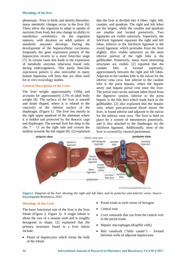
concavity of the inferior surface of the diaphragm. (Figure 1) The liver lies mainly in
the right upper quadrant of the abdomen where
it is hidden and protected by the thoracic cage
and diaphragm. The normal liver lies deep to the ribs 7 – 11 on the right side and crosses the
midline towards the left nipple [8]. [2] explained
that the liver is divided into 4 lobes: right, left,
caudate, and quadrate. The right and left lobes are the largest, while the caudate and quadrate
are smaller and located posteriorly. Two
ligaments are visible anteriorly. Superiorly, the falciform ligament separates the right and left
lobes. Inferior to the falciform ligament is the
round ligament, which protrudes from the liver
slightly. Also visible anteriorly on the most inferior portion of the right lobe is the
gallbladder. Posteriorly, many more interesting
structures are visible. [2] reported that the caudate lobe is located superiorly,
approximately between the right and left lobes.
Adjacent to the caudate lobe is the sulcus for the inferior vena cava. Just inferior to the caudate
lobe is the porta hepatis, where the hepatic
artery and hepatic portal vein enter the liver.
The portal vein carries nutrient laden blood from the digestive system. Inferior to the porta
hepatis is the bile duct which leads back to the
gallbladder. [2] also explained that the hepatic vein, where post-processed blood leaves the
liver, is found inferior and adjacent to the sulcus
for the inferior vena cava. The liver is held on
place by a system of mesenteries posteriorly, and is also attached to the diaphragm via the
falciform ligament. Additionally, most of the
liver is covered by visceral peritoneum.
Figure1. Diagram of the liver showing the right and left lobes and its posterior and anterior veiws. Source :
Encyclopedia Britannica, 2010
Histology of the Liver
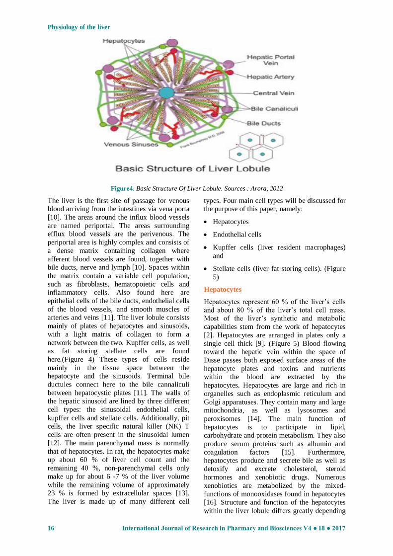
The basic functional unit of the liver is the liver
lobule (Figure 2, Figure 3). A single lobule is
about the size of a sesame seed and is roughly
hexagonal in shape. [2] explained that the
primary structures found in a liver lobule
include:
Plates of hepatocytes which forms the bulk
of the lobule
Portal triads at each corner of hexagon
Central vein
Liver sinusoids that run from the central vein
to the portal triads
Hepatic macrophages (Kupffer cells)
Bile canaliculi (―little canals‖) – formed
between walls of adjacent hepatocytes

Physiology of the liver
International Journal of Research in Pharmacy and Biosciences V4 ● I8 ● 2017 15
Space of Disse – a small space between the
sinusoids and the hepatocytes
The portal triads consist of three vessels: a hepatic portal arteriole, a hepatic portal venule,
and a bile duct. The blood from the arteriole and
the venule both flow in the same direction – through the sinusoids toward the central vein,
which eventually leads to the hepatic vein and
the inferior vena cava. Secreted bile flows in the
opposite direction – through the bile canaliculi away from the central vein, toward the portal
triad, and exiting via the bile duct. As blood
flows through the sinusoids and the space of disse toward the central vein, nutrients are
processed and stored by the hepatocytes, and
worn out blood cells and bacteria are engulfed
by the Kupffer cells [2].
Figure2. Cellular architecture of the liver. (A) The schematic shows an adult liver (red), with the gall bladder
and extra hepatic ducts (green), in relation to the stomach and intestine (yellow). The extra hepatic duct system
consists of the hepatic ducts (hd), which drain bile from the liver into the common hepatic duct (chd) to the gall
bladder via the cystic duct (cd) and into the duodenum through the common bile duct (cbd). (B) A schematic of
the cellular architecture of the liver showing the hepatocytes (pink) arranged in hepatic plates separated by
sinusoid spaces radiating around a central vein. Bile canaliculi on the surface of adjoining hepatocytes drain
bile into the bile ducts (green), which run parallel to portal veins (blue) and hepatic arteries (red) to form the
“portal triad”.
Source: Zorn, 2013.
Figure3. Diagram Showing Liver Lobules.
Source: [9]
Physiology of the liver
16 International Journal of Research in Pharmacy and Biosciences V4 ● I8 ● 2017
Figure4. Basic Structure Of Liver Lobule. Sources : Arora, 2012
The liver is the first site of passage for venous blood arriving from the intestines via vena porta
[10]. The areas around the influx blood vessels
are named periportal. The areas surrounding efflux blood vessels are the perivenous. The
periportal area is highly complex and consists of
a dense matrix containing collagen where
afferent blood vessels are found, together with bile ducts, nerve and lymph [10]. Spaces within
the matrix contain a variable cell population,
such as fibroblasts, hematopoietic cells and inflammatory cells. Also found here are
epithelial cells of the bile ducts, endothelial cells
of the blood vessels, and smooth muscles of arteries and veins [11]. The liver lobule consists
mainly of plates of hepatocytes and sinusoids,
with a light matrix of collagen to form a
network between the two. Kupffer cells, as well as fat storing stellate cells are found
here.(Figure 4) These types of cells reside
mainly in the tissue space between the hepatocyte and the sinusoids. Terminal bile
ductules connect here to the bile cannaliculi
between hepatocystic plates [11]. The walls of the hepatic sinusoid are lined by three different
cell types: the sinusoidal endothelial cells,
kupffer cells and stellate cells. Additionally, pit
cells, the liver specific natural killer (NK) T cells are often present in the sinusoidal lumen
[12]. The main parenchymal mass is normally
that of hepatocytes. In rat, the hepatocytes make up about 60 % of liver cell count and the
remaining 40 %, non-parenchymal cells only
make up for about 6 -7 % of the liver volume
while the remaining volume of approximately 23 % is formed by extracellular spaces [13].
The liver is made up of many different cell
types. Four main cell types will be discussed for the purpose of this paper, namely:
Hepatocytes
Endothelial cells
Kupffer cells (liver resident macrophages)
and
Stellate cells (liver fat storing cells). (Figure
5)
Hepatocytes
Hepatocytes represent 60 % of the liver‘s cells and about 80 % of the liver‘s total cell mass. Most of the liver‘s synthetic and metabolic
capabilities stem from the work of hepatocytes
[2]. Hepatocytes are arranged in plates only a
single cell thick [9]. (Figure 5) Blood flowing toward the hepatic vein within the space of
Disse passes both exposed surface areas of the
hepatocyte plates and toxins and nutrients within the blood are extracted by the
hepatocytes. Hepatocytes are large and rich in
organelles such as endoplasmic reticulum and
Golgi apparatuses. They contain many and large mitochondria, as well as lysosomes and
peroxisomes [14]. The main function of
hepatocytes is to participate in lipid, carbohydrate and protein metabolism. They also
produce serum proteins such as albumin and
coagulation factors [15]. Furthermore, hepatocytes produce and secrete bile as well as
detoxify and excrete cholesterol, steroid
hormones and xenobiotic drugs. Numerous
xenobiotics are metabolized by the mixed-functions of monooxidases found in hepatocytes
[16]. Structure and function of the hepatocytes
within the liver lobule differs greatly depending

Physiology of the liver
International Journal of Research in Pharmacy and Biosciences V4 ● I8 ● 2017 17
on proximity to periportal or perivenous areas.
Periportal type of hepatocytes are often smaller, but have larger mitochondria, and a larger Golgi
apparatus as compared to the perivenous type.
Perivenous hepatocytes on the other hand have larger endoplasmic reticulum. Functionally,
periportal hepatocytes are more involved in
gluconeogenesis, while perivenous are involved
in glycolysis [10]. Additionally, perivenous hepatocytes are dorminant with respect to P
450- dependent hydroxylation reactions [17]
and glutamine synthetase [18].
Endothelial Cells
The sinusoidal endothelial cells line the walls of the hepatic sinusoid and perform a function of filtration due to the presence of fenestrae [10].
(Figure 5) These cells also demonstrate large
endocytic capacity for extracellular matrix
components and immune complexes. In general they engulf smaller size particles and may play a
role in clearance of viruses, but do not posses
phagocytic function [19]. They may also function as antigen presenting cells and secrete
certain cytokines and eicosanoids [12].
Kupffer Cells
The liver harbors large amounts of kupffer cells, which represent the largest tissue resident
macrophage population of the body [20].
(Figure 5). They are located within the sinusoid and are in constant contact with gut-derived
particles that lead to low but constant amount of
activation of these monocyte derived cells. Upon activation they are able to secrete a vast
range of inflammatory mediators such as
cytokines, reactive oxygen species, eicosanoids
and nitric oxide [12]. Kupffer cells have
receptors that enable them to bind cells covered
with immunoglobulins or bind to complement receptors and subsequently phagocytose cell
[21]. Kupffer cells are even actively phagocytic
in vitro and contain high levels of peroxidase, acid phosphatase and glucose 6-phosphate
dehydrogenase [22].
Stellate Cells
The liver plays a central role in uptake and storage of vitamins A (Retinol) and stores about
95 % of retinoids found in the body. The fat
storing perisinusoidal cells of the liver, stellate cells are the main vitamin A storing cells
(Figure 5). They harbor large amounts of retinol
and retinyl palmitate in lipid droplets within their cell cytoplasm [23]. They are located in the
space of Disse (between hepatocytes and
sinusoid) and generally protrude to come into
contact with several sinusoids [23]. Additionally, they function to control the
turnover of extracellular matrix and regulate
sinusoid contractility. The stellate cells may become activated under stressful conditions and
transformed into myofibroblast – like cells
which play a key role in inflammatory fibrotic
response [12]. When activated, stellate cells not only proliferate, but also produce increased
amounts of extracellular matrix per cell.
Transforming growth factor beta (TGF β) is one of the most important signals to activate stellate
cells, which leads to a higher transcriptional rate
of mRNAs coding for extracellular matrix components such as collagen I, fibronectin and
proteoglycans [10]. Lipid peroxidation products
are also an important stimulus, whose effect
may be augmented in oxidative stress conditions [24].
Figure5. Diagram showing main cell types of Liver - hepatocytes, endothelial cells, Kupffer cells and Stellate
cells. Source: Tissupath specialist pathway services, Retrieved 2017. www. Tissupath.com.au

Physiology of the liver
18 International Journal of Research in Pharmacy and Biosciences V4 ● I8 ● 2017
FUNCTIONS OF THE LIVER
The liver has numerous functions best grouped into secretion of bile, metabolism of bilirubin,
vascular and hematologic functions, metabolism of nutrients, metabolic detoxification and storage of
minerals and vitamins. (Table 1)
Table1. Summary Of Major Functions Of The Liver
Secretion of Bile
Metabolism of Bilirubin
Vascular and Hematologic Functions Important blood reservoir
Metabolism of Nutrients
Fat - fatty acid oxidation, synthesis of cholesterol/lipoproteins and production of ketoacids
Protein – Amino acid production, turnover of proteins
Carbohydrate – converts galactose/fructose to glucose, gluconeogenesis and contains 100g of glycogen
for release
Metabolic Detoxification
Toxins
Hormones
Drugs
Storage of Minerals and Vitamins
Iron
Copper
Vitamins ADEKB12 Glycogen
Endocrine functions
Activation of vitamin D
Conversion of thyroxine (T4) to T3
secretes angiotensinogen
metabolises hormones
Immunological/ Protective Functions
Reticuloendothelial Component
Filters the portal blood from bacteria
Important in antigen presentation
Phagocytosis via kupffer cells
Removes haemolysis products
Inactivation Of Toxins and Drugs Phase I (oxidation, reduction and hydrolysis)
Phase II (conjugation/ cytochrome P450 system)
[25] and [8] explained the functions of the liver as follows:
Secretion of Bile
The liver assists intestinal digestion by secreting 700 to 1200 ml of bile per day. Bile is an
alkaline, bitter-tasting, yellowish green fluid that
contains bile salts (conjugated bile acids),
cholesterol, bilirubin (a pigment), electrolytes and water. It is formed by hepatocytes and
secreted into the canaliculi. Bile salts, which are
conjugated bile acids, are required for the intestinal emulsification and absorption of fats.
Having facilitated fat emulsification and
absorption, most bile salts are actively absorbed
in the terminal ileum and returned to the liver via the portal circulation for resecretion as
follows:
Bile has two fractional components: the
acid-dependent fraction and the acid-
independent fraction. Hepatocytes secrete
the bile acid-dependent fraction of the
bile. This fraction consists of bile acids,
cholesterol, lecithin (a phospholipid), and
bilirubin (a bile pigment). The bile acid-
independent fraction of the bile, which is
secreted by the hepatocytes and epithelial
cells of the bile canaliculi, is a
bicarbonate-rich aqueous fluid that gives
bile its alkaline pH.
Bile salts are conjugated in the liver from
primary and secondary bile acids. The
primary bile acids are cholic acid and
chenodeoxycholic (chenic) acid. These
acids are synthesized from cholesterol by
the hepatocytes. The secondary bile acids
are deoxycholic acid and lithocholic acid.
These acids are formed in the small

Physiology of the liver
International Journal of Research in Pharmacy and Biosciences V4 ● I8 ● 2017 19
intestine by the action of intestinal
bacteria, after which they are absorbed
and flow to the liver.
Both forms of bile acids are conjugated
with amino acids in the liver to form bile
salts.
Conjugation makes the bile acids more
water soluble, thus restricting their
diffusion from the duodenum and ileum.
Metabolism of Bilirubin
Bilirubin is a byproduct of destruction of aged red blood cells. It gives bile a greenish black
color and produces the yellow tinge of jaundice.
Aged red blood cells are taken up and
destroyed by macrophages of the
mononuclear phagocyte system,
primarily in the spleen and liver (in the
liver these macrophages are Kupffer
cells). Within these cells hemoglobin is
separated into its component parts—
heme and globin. The globin component
is further degraded into its constituent
amino acids, which are recycled to form
new protein. The heme moiety is
converted to biliverdin by the enzymatic
cleavage of iron. The iron attaches to
transferrin in the plasma and can be
stored in the liver or used by the bone
marrow to make new red blood cells. The
biliverdin is enzymatically converted to
bilirubin in the macrophage of the
mononuclear phagocytic system and then
is released into the plasma. In the plasma,
bilirubin binds to albumin and is known
as unconjugated bilirubin or free
bilirubin, which is lipid soluble.
In the liver, unconjugated bilirubin
moves from plasma in the sinusoids into
the hepatocyte. Within hepatocytes, it
joins with glucuronic acid to form
conjugated bilirubin, which is water
soluble. Conjugation transforms bilirubin
from a lipid-soluble substance that can
cross biologic membranes to water-
soluble substance that can be excreted in
the bile. When conjugated bi1irubin
reaches the distal ileum and colon, it is
deconjugated by bacteria and converted
to urobilinogen. Most of the urobilinogen
is then excreted in the urine, and a small
amount is eliminated in feaces.
Vascular and Hematologic Functions
Because of its extensive vascular network, the liver can store a large volume of blood. The
amount stored at any one duration depends on
pressure relationships in the arteries and veins.
The liver can also release blood to maintain
systemic circulatory volume in the event of
hemorrhage.
Kupffer cells in the sinusoids of the liver
remove bacteria and foreign particles from
the portal blood. Because the liver receives all of the venous blood from the gut and
pancreas, the Kupffer cells play an important
role in destroying intestinal bacteria and preventing infections.
The liver also has hemostatic functions. It
synthesizes prothrombin, fibrinogen, and
clotting factors. Vitamin K, a fat-soluble vitamin, is essential for the synthesis of other
clotting factors. Because bile salts are needed
for reabsorption of fats, vitamin K absorption
depends on adequate bile production in the liver.
Metabolism of Nutrients
Fats: Fat is synthesized from carbohydrate
and protein, primarily in the liver. Fat absorbed by lacteals in the intestinal villi
enters the liver through the lymphatics,
primarily as triglycerides. In the liver the triglycerides can be hydrolyzed to glycerol
and free fatty acids and used to produce
metabolic energy adenosine triphosphate
(ATP), or they can be released into the bloodstream as lipoprotein. The lipoproteins
are carried by the blood to adipose cells for
storage. The liver also synthesizes phospholipids and cholesterol, which are
needed for the hepatic production of bile
salts, steroid hormones, components of
plasma membranes and other special molecules.
Proteins - The plasma proteins, including
albumins and globulins (excluding gamma-
globulin), are synthesized by the liver. The liver also synthesizes several non essential
amino acids and serum enzymes including
aspartate aminotransferase, alanine aminotransferase, lactate dehydrogenase and
alkaline phosphatase.

Physiology of the liver
20 International Journal of Research in Pharmacy and Biosciences V4 ● I8 ● 2017
Carbohydrates: The liver contributes to the
stability of blood glucose levels by releasing
glucose during states of hypoglycemia (low blood sugar) and taking up glucose during
states of hyperglycemia (high blood sugar)
and storing it as glycogen (glyconeogenesis) or converting it to fat. When all glycogen
stores have been used, the liver can convert
amino acids and glycerol to glucose.
Metabolic Detoxification
The liver alters exogenous and endogenous
chemicals (e.g. drugs), foreign molecules, and
hormones to make them less toxic or less biologically active. This process, called
metabolic detoxification, diminishes intestinal
or renal tubular reabsorption of potentially toxic substances and facilitates their intestinal and
renal excretion. In this way alcohol,
barbiturates, amphetamines, steroids and
hormones (including estrogens, aldosterone, antidiuretic hormone, and testosterone) are
metabolized or detoxified, preventing excessive
accumulation and adverse effects. Although metabolic detoxification is usually protective, in
some durations the products of metabolic
detoxification become toxins. Those of alcohol
metabolism, for example, are acetaldehyde and hydrogen. Excessive intake of alcohol over a
prolonged period causes these end products to
damage the hepatocytes. Acetaldehyde damages cellular mitochondria, and the excess hydrogen
promotes fat accumulation. This is how alcohol
impairs the liver‘s ability to function.
The adult liver is the main organ responsible for
detoxifying and metabolizing a variety of
exogenous as well as endogenous compounds,
rendering them more hydrophilic, which often affects their potency and level [10]. The
enzymes responsible for the actions are
primarily produced in hepatocytes and mainly divided into two groups phase I and phase II.
The phase I enzymes are predominantly from
the P- 450 family of genes, whose general function is to add polar groups, such as hydroxyl
groups, to lipophilic molecules thus rendering
them more hydrophilic [26]. The main function
of the phase II enzymes is to covalently attach a water soluble moiety to the polar group added
by the phase I enzymes. Usually such molecules
are sugars or peptides, such as glucuronic acid or glutathione. This usually renders the
compound less reactive. Examples of phase II
enzymes are glutathione-S-transferase and
UDP– glucuronosyl transferase. If the phase II reaction is impaired for some reasons or the
phase I reaction is induced, this may leave the
organism with an excess of reactive molecules from the phase I reaction, which can be
detrimental. This can occur in the case of drug
induced hepatotoxicity, when reactive metabolites of the parent compound are formed,
which subsequently negatively affects cellular
functions [27].
Storage of Minerals and Vitamins
The liver stores certain vitamins and minerals,
including iron and copper, in durations of
excessive intake and releases them in durations of need. The liver can store vitamins B12 and D
for several months and vitamin A for several
years. The liver also stores vitamins E and K. Iron is stored in the liver as ferritin, an iron-
protein complex and is released as needed for
red blood cell production.
The Immunologic Function of the Liver
The liver is the main hematopoietic organ
during certain stages in fetal development and
continues to be a hematopoietic organ even after birth. It can produce all leukocytes lineages
from resident hematopoietic stem cells [28, 29].
The portal tract of the liver contains many
different cells of hematopoietic origin, as well as hematopoietic stem cells [11]. The liver
contains cells involved in adaptive and innate
immunity.
Innate Immunity of the Liver
In comparison to other organs, the liver is
particularly rich in cells of the innate immune system. The main cell types here are kupffer
cells and NK T cells. NK T cells are not strictly
part of innate immunity but functionally
somewhere in between adaptive and innate. Of hepatic lymphocytes, approximately 30 % are
NK cells, indicating the great contribution of
NK cells to liver immunity. This may be compared to the approximately 15% that the two
cell types combined contribute to in peripheral
blood lymphocytes [30]. NK cells are one of the main producers of liver INFγ in response to
lipopolysaccharide (LPS), which partly depends
on the activation of NK cells by IL-I2 derived
from activated Kupffer cells. They show high cytotoxic level towards tumor cells, with the
help of the trail-ligand, which is up regulated by
IL-2. Kupffer cells are derived from circulating monocytes and play a particularly important role
in initiating inflammation in the liver. Kupffer
cells differ in properties depending if they are
periportal or perivenous. Periportal cells are larger and more active in phagocytosis,

Physiology of the liver
International Journal of Research in Pharmacy and Biosciences V4 ● I8 ● 2017 21
mirroring their function as the first line of
defense of the body towards gut derived bacteria entering the blood stream and reaching the liver
via the vena porta [31] . Perivenous kupffer cells
on the other hand, are smaller and produce larger amounts of nitric oxide, as well as
prostaglandins [32]. Ex vivo culture of kupffer
cells have shown that the perivenous type
secretes more than double the amount of TNF α upon LPS stimulation. Kupffer cells are one of
the main cell types to secrete cytokines, which
then regulate the function not only of the kupffer cells themselves but also that of other
cell types such as the NK cells. Stimulation of
kupffer cells by bacteria and bacterial LPS leads to production of IL-12 [33] , as well as TNFα.
Other cytokines known to be produced by
kupffer cells upon LPS stimulation include IL-6,
TGF β, IL-1β and IL-8 [34]. Cytokines derived from kupffer cells have in turn been proven to
stimulate hepatocytes to further increase
chemotactic response by secretion of IL-8 [35]. Thus kupffer cells and NK cells hence mainly
secrete Th1 type cytokines that activate the
immune system. Immunosuppressive cytokines
such as IL-10 may instead be secreted by stellate cells and regulatory T cells (but also by
kupffer cells and NK cells) [36].
Adaptive Immunity
Adaptive immunity can be classified into
humoral immunity and cell-mediated immunity,
mediated principally by B and T lymphocytes, respectively. T cells promote differentiation of
B cells to antibody secreting plasma cells. T
cells kill infected cells and secrete cytokines
such as TNF α, IFN γ and IL -6. TNF α derived from kupffer cells play an important role in
stimulating activation of T cells which then
elicit a cytotoxic response [37]. Intrahepatic accumulation of highly activated CD8+ T cells
is part of the pathogenesis process in hepatitis,
including alcoholic hepatitis [38]. NK T cells constitute a distinct subpopulation of T cells that
is particularly abundant in the liver, as
previously mentioned. Infact, they are not
strictly a part of adaptive immune response, but can be seen as having a function in between
adaptive and innate immunity. These cells
produce large amounts of the Th 2 cytokine IL-4 [39] but also the Th1 cytokine IFN γ [40]. This
ability of secreting both Th 1 and Th 2 type
cytokines is particularly feature of NK cells. NK
T cells are often present in the lumen of the sinusoid. They exhibit MHC- unrestricted
killing of a variety of tumor cells, an level
which is enhanced by IFN γ [12]. NK T cells
have been shown to ‗crawl‘ within the hepatic
sinusoid, and stop upon T cell antigen receptor activation [41]. Naïve CD8+ T cells are also
known to accumulate in the liver, where they
may be activated, but at durations to a lesser degree than in lymph nodes. Thus, low-grade
activation of T cells in liver rather leads to
tolerance [42]. There is also evidence for
regulatory T cells expressing IL-10 [36]. B cells have not been well studied in adult liver,
however, there is a substantial B cell population
showing similarities to splenic B cells. It has been shown that B cells play a role in liver
fibrosis, as B cell deficient mice show
significantly less fibrotic lesions after carbon tetrachloride induced liver injury. This effect is
independent of antibody production. But also B
cell antibody-dependent responses play a part in
liver injury, as antibody production has been shown to be of importance in alcohol induced
liver damage [43].
Interrelationships of the Liver with Other
Organs
The liver interacts with many other organs.
Following the flow of blood, the liver receives
its arterial blood supply from the hepatic arteries [3]. The hepatic arteries are distal to the celiac
trunk, which is distal to the abdominal aorta.
Thus the liver receives its oxygenated blood supply from the heart. Nutrient laden blood
from the digestive system and blood leaving the
spleen enters the liver through the hepatic portal vein . Processed blood leaving the liver through
the hepatic veins drains into the inferior vena
cava, completing the connection to the heart.
The liver affects digestion through its formation of bile, which is secreted into the small intestine
[2]. The gallbladder is essentially an overflow
area for the liver‘s bile duct. The liver is full of lymph glands, which provide fluid drainage and
immune system support. The liver synthesizes
many blood proteins, showing its relation to that ―organs‖. The liver also has a supply of nerves,
showing its relationship with the nervous
system. Finally, liver disease often causes
problems in the renal system, demonstrating a relationship with the kidneys [2]. The liver has
many important functions in maintaining the
physiologic balance of the human body.
CONCLUSIONS
In conclusion this paper summarized the
functions of the liver which includes firstly, Secretion of bile, the liver assists intestinal
digestion by secreting 700 to 1200 ml of bile per
day. Bile salts, which are conjugated bile acids,

Physiology of the liver
22 International Journal of Research in Pharmacy and Biosciences V4 ● I8 ● 2017
are required for the intestinal emulsification and
absorption of fats. Secondly, Metabolism of bilirubin which is a byproduct of destruction of
aged red blood cells and gives bile a greenish
black color and produces the yellow tinge of jaundice. Also Vascular and hematologic
functions, because of its extensive vascular
network, the liver can store a large volume of
blood. The amount stored at any one duration depends on pressure relationships in the arteries
and veins. Moreover, the liver has hemostatic
functions, It synthesizes prothrombin, fibrinogen, and clotting factors. Vitamin K, a
fat-soluble vitamin, is essential for the synthesis
of other clotting factors. Because bile salts are needed for reabsorption of fats, vitamin K
absorption depends on adequate bile production
in the liver. Furthermore Metabolism of
nutrients, the liver plays essential roles in the metabolism of fat, protein and carbohydrates.
Also, Metabolic detoxification, the liver alters
exogenous and endogenous chemicals (e.g. drugs), foreign molecules, and hormones to
make them less toxic or less biologically active.
This process, called metabolic detoxification,
diminishes intestinal or renal tubular reabsorption of potentially toxic substances and
facilitates their intestinal and renal excretion. In
this way alcohol, barbiturates, amphetamines, steroids and hormones (including estrogens,
aldosterone, antidiuretic hormone, and
testosterone) are metabolized or detoxified, preventing excessive accumulation and adverse
effects. Also, Storage of minerals and vitamins,
the liver stores certain vitamins and minerals,
including iron and copper, in periods of excessive intake and releases them in periods of
need. The liver can store vitamins B12 and D for
several months and vitamin A for several years. The liver also stores vitamins E and K. Iron is
stored in the liver as ferritin, an iron-protein
complex and is released as needed for red blood cell production. Finally, the liver has
immunologic functions as the liver contain cells
involved in adaptive and innate immunity.
REFERENCES
[1] Ozougwu JC, Eyo JE. Hepatoprotective effects
of Allium cepa extracts on paracetamol-induced
liver damage in rat. African Journal of
Biotechnology 2014, 13(26): 2679 -2688.
[2] Allen SE.The liver: Anatomy, Physiology,
Disease and Treatment. 2002 North Eastern
University Press, USA.
[3] Ozougwu JC. Comparative hepatoprotective
and antioxidant effects of Allium cepa, Allium
sativum and Zingiber officinale methanolic
extracts against paracetamol-induced liver
damage in Rattus novergicus. 2014 Ph.D
Research Thesis, Department Of Zoology and
Environmental Biology, University of Nigeria,
Nsukka. 222pp
[4] Watt AJ, Zhao R, Li J, Duncan SA.
Development of the mammalian liver and
ventral pancreas is dependent on GATA4. BMC
Developmental Biology 2007, 7: 37 - 45.
[5] Burke ZD, Thowfeequ S, Tosh D. Liver
specification: a new role for rats in liver
development. Current Biology 2006, 16(17):
688 - 690.
[6] Zaret KS. Molecular genetics of early liver
development. Annual Review of Physiology
1996, 58: 231 - 251.
[7] Nardone G, Romano M, Calabro A, Pedone
PV, De Sio I, Persico M.. Activation of fetal
promoters of insulin like growth factors II gene
in hepatitis C virus-related chronic hepatitis,
cirrhosis and hepatocellular carcinoma.
Hepatology 1996, 23(6): 1304 - 1312.
[8] Moore KL, Dalley AF. Clinically Oriented
Anatomy. 2006 5th Edition Lippincott Williams
and Wilkins. 1209 pp.
[9] Singh I. Textbook of Human Histology with
colour Atlas. 2007, 5th Edition Jay Pee Brothers
Medical Publishers Ltd. 365 pp.
[10] Butura A. Drug and Alcohol Induced
Hepatotoxicity. 2008 Ph. D Thesis Department
of Physiology and Pharmacology Karolinska
Institutet, Stockholom, Sweden. 55 pp.
[11] Grisham JW. Cell type in rat liver cultures:
their identification and isolation. Molecular and
Cellular Biochemistry 1983, 53(2): 23 – 33.
[12] Kmiec Z. Co-operation of liver cells in health
and diseases. Advances in Anatomy,
Embryology and Cell Biology 2001, 161: 3 –
12.
[13] Hendriks HF, Brouwer A, Knook DL. Isolation,
purification and characterization of liver cell
types. Methods in Enzymology 1990, 190: 49 -
58.
[14] Wanson JC, Bernaert D, May C. Morphology
and functional properties of isolated and
cultured hepatocytes. Progress in Liver
Diseases 1979, 6: 1 - 22.
[15] Jeejeebhoy K, Phillips MJ. Isolated mammalian
hepatocytes in culture. Gastroenterology 1976,
71(6): 1086 - 1096.
[16] Sirica AE, Pitot AC. Drug metabolism and
effects of carcinogens in cultural hepatic cells.
Pharmacological Review 1979, 31(3): 205 -
228.
[17] Smith MT, Wills ED. Effects of dietary lipid
and phenobarbitone on the distribution and

Physiology of the liver
International Journal of Research in Pharmacy and Biosciences V4 ● I8 ● 2017 23
concentration of cytochrome P-450 in the liver
studied by quantitative cytochemistry.
Federation of European Biochemical Society
Letters 1981, 127(1): 33 - 36.
[18] Gebhardt R, Mecke D. Heterogenous
distribution of glutamine synthease among rat
liver parenchymal cells in situ and in primary
culture. EMBO Journal 1983, 2(4): 567 - 570.
[19] Breiner KM, Urban S, Schaller H. Endothelial
cell-mediated uptake of a hepatitis B virus: a
new concept of liver targeting of hepatotropic
microorganisms. Hepatology 2001, 34(4): 803 -
808.
[20] Knook DL, Blansjaar N, Sleyster EC. Isolation
and characterization of kupffer and endothelial
cells from the rat liver. Experimental Cell
Research 1977, 109(2): 317 - 329.
[21] Smedsrod B, Pertoft H, Eggertsen G,
Sundstrom C. Functional and morphological
characterization of cultures of kupffer cells and
liver endothelial cells prepared by means of
density separation in per cells, and selective
substrate adherence. Cell and Tissue Research
1985, 24 1(3): 639 - 649.
[22] Munthe-Kaas AC, Berg T, Seglen PO, Seljelid
R. Distribution of lysosomal enzyme in
different types of rat liver cells. Experimental
cell Research 1976, 99(1): 146 - 54.
[23] Knook DL, Seffelaar AM, De-Leeuw AM. Fat-
storing cells of the rat liver. Their isolation and
purification. Experimental Cell Research 1982,
139(2): 468 - 471.
[24] Friedman SL. Hepatic satellite cells: Protein,
multifunctional and enigmatic cells of the liver.
Physiological Review 2008, 88(1): 125 - 172.
[25] Guyton AC, Hall JE.Textbook of Medical
Physiology. 2006, 11th Edition, Saunder
Philadelphia, Pennsylvania. 1116 pp.
[26] Park BK, Pirmohamed M, Kitteringham NR.
The role of cytochrome P450 enzymes in
hepatic and extrahepatic human drug toxicity.
Pharmacology and Therapeutics 1995, 68(3):
385 – 424.
[27] Liu ZX, Govindarajan S, Kaplowitz N. Innate
immune system plays a critical role in
determining the progression and severity of
acetaminophen hepatotoxicity.
Gastroenterology 2004, 127: 1760 – 1774.
[28] Abo T, Watanabe H, Iiai T, Kimura M,
Ohtsuka K, Sato K, Ogawa M, Hirahara H,
Hashimoto S, Sekikawa H. Extrathymic
pathways of T-cell differentiation in the liver
and other organs. International Reviews of
Immunology 1994, 11(1): 61 - 102.
[29] Taniguchi H, Toyoshima T, Fukao K, Nakauchi
H. Presence of hematopoietic stem cells in the
adult liver. Nature Medicine 1996, 2(2): 198 –
203.
[30] Mechal WZ, Azzaroli F, Crispe IN.
Immunology of the healthy liver.
Gastroenterology 2001, 120: 250 - 260.
[31] Itoh T, Okanoue M, Morimoto Y, Nagao T,
Mori N, Hori K, Kashima K. Functional
heterogeneity of rat liver microphages:
interleukin-1 secretion and 1A antigen
expression in contrast with phagocytic activity.
Liver 1992, 12 (1): 26 -33
[32] Mustafa SB, Olson MS. Expression of nitric-
oxide synthase in rat kupffer cells is regulated
by cAMP. Journal of Biological Chemistry
1998, 273(9): 5073 – 5080.
[33] Takahashi M, Ogasawara K, Takeda K,
Hashimoto W, Sakihara H. LPS induces NK I+
alpha beta T cells with potent cytotoxicity in
the liver of mice via production of 1L- 12 from
kupffer cells. Journal of Immunology 1996,
156(7): 2436 – 2442.
[34] Kamimura S, Tsukamato, H. (1995). Cytokine
gene expression by kupffer cells. Hepatology
1995, 22(4): 1304 -1309.
[35] Thornton AJ, Ham J, Kunkel SL. Kupffer cell-
derived cytokines induce the synthesis of a
leukocyte chemotactic peptide, interleukin–8,
in human hepatoma and primary hepatocyte.
Hepatology 1991, 14(6): 1112 - 1122.
[36] Erhardt A, Biburger M, Papadopoulos T, Tiegs
G. IL-10, regulatory T cells, and kupffer cells
mediate tolerance in concanavalin A- induced
liver injury in mice. Hepatology 2007, 45(2):
475 - 485.
[37] Schumann J, Wolf D, Pahl A, Brune K,
Papadopoulos T, Van Rooijen N, Tiegs, G.
Importance of kupffer cells for T-cells
dependent liver injury in mice. American
Journal of Pathology 2000. 157(5): 1671 –
1683.
[38] Bowen DG, Warren A, Davis T. Cytokine-
dependent hepatitis due to intrahepatic murine
CD8 Tcell activation by bone marrow derived
cells. Gastroentology 2002, 123(4): 1252 -
1264.
[39] Yoshimoto T, Paul, WE. CD4 POS promptly
produce interleukin-4 in response to in vivo
challenge with anti-CD3. Journal of
Experimental Medicine 1994, 179(4): 1285 -
1295.
[40] Burdin N, Brossay L, Koezuka Y, Smiley ST,
Grusby MJ, Gui M. Selective ability of mouse
CD1 to present glycolipids: alpha–
galactosylceramides specifically stimulates S V
alpha 14+ NKT lymphocytes. Journal of
Immunology 1998, 161(7): 3271 - 3281.

Physiology of the liver
24 International Journal of Research in Pharmacy and Biosciences V4 ● I8 ● 2017
[41] Geissmann F, Cameron TO, Sidobre S,
Manlongat N, Kronenberg M. Intravascular
immune surveillance by cells patrolling liver
sinusoids. Plos Biology 2005, 3(4): 113 - 120.
[42] Bowen DG, McCaughan GW, Bertolino P.
Intrahepatic immunity: a tale of two sites.
Trends in Immunology 2005, 26(10): 512 – 517
[43] Albano E, French SW, Ingleman-Sundberg M.
Hydroxyethyl radicals in ethanol hepatotoxicity.
Frontiers in Bioscience 1999, 4: 533 – 540.
Citation: J. Ozougwu, "Physiology of the liver", International Journal of Research in Pharmacy and
Biosciences, vol. 4, no. 8, pp. 13-24, 2017.
Copyright: © 2017 J. Ozougwu. This is an open-access article distributed under the terms of the Creative Commons Attribution License, which permits unrestricted use, distribution, and reproduction in any
medium, provided the original author and source are credited.