
Photoluminescence of Photocatalytic Degradaded...
7
Photoluminescence of Photocatalytic Degradaded Malachite Green Dye by using Mn-doped ZnS Jyoti Tolia 1,2 , Mousumi Chakraborty 2+ and Z.V. P Murthy 2 1 Department of Chemical Engineering,V.V.P. Engineering College, Rajkot , Gujarat, India. 2 Department of Chemical Engineering, SVNIT, Surat 395007, Gujarat, India. Abstract. Mechanochemical method was used to synthesize Mn doped ZnS and undoped ZnS. The particles were characterized using XRD measurements and TEM analysis. The particle size for different doping concentration was found to be 4.19nm, 3.8nm, 2.68nm. Also the particle size of pure ZnS was found to be 4.79nm. The FTIR results showed that Mn 2+ ions were incorporated in host ZnS. The photoluminescence study revealed that PL intensity increases with increase in doping concentration. The as prepared nanoparticles were further used for degradation of malachite green dye where the dye adsorption/ interaction on photocatalyst were studied using PL spectra. The PL spectra of dye absorbed photocatalyst showed that PL intensity increases manifolds due to dye adsorption. The maximum dye degradation (97%) was found for Zn 1-x Mn x S (x=0.01) concentration with maximum rate constant (0.026 min -1 ). Keywords: Mechanochemical method, Photocatalytic degradation, ZnS, Mn doped ZnS, Malachite green, Dye interaction 1. Introduction The ZnS nanoparticles belong to group II-VI semiconductor with wide bandgap of 3.6eV. The important property of a semiconductor material is that it can be doped with impurities that can alter its electronic properties in a controllable way. These particles can be modified from bulk and can be improved in their properties through doping as they find wide application as optoelectronic devices such as electroluminescent, photoluminscent, photocatalyst, photoconductor, photovoltaic cells, DNA markers, bio sensors, light emitting diodes and lasers [1-3]. To understand the physics and applications of ZnS as nanophosphors through doping as quantum dots, huge amount of research effort is being done in recent years. To utilize the phenomena of quantum confinement further, device fabrication with nano dimensions is also being done world over. The recent discovery of doped nanocrystaliine (DNC) luminescent materials in the size range of 3-5 nm provided alternative in low voltage phosphor applications. The additional benefit of using the doped nanocrystalline phosphor is that non-radiative contributions decrease with decrease in particle size. The original result of doped quantum-dot material or doped nanocrystals with high luminescent efficiency and ultrafast decay time was reported by Bhargava et al. [4] Thus, ZnS quantum dots are particularly suitable for use as luminescent host materials for a very large variety of dopants like Cu 2+ , Ni 2+ ,Pb 2+ , Mn 2+ and rare- earth elements. The doping ions act as recombination centers for the excited electron–hole pairs and result in strong and characteristic luminescence [5-7]. Synthesis of the extremely small size of particles results in quantum confinement of the photogenerated electron–hole pair, leading to a blue shift in the absorption spectrum. It is hence, highly important to synthesize such low-dimensional nanosized semiconductor particles with a narrow size distribution. Typical QD sizes range between 2–20 nm, but according to some literature their diameter should be strictly below 10 nm [8]. ZnS-Mn quantum dots exhibiting wide application as luminescent and nanophosphor material, literature reports various synthesis methods like, chemical precipitation, micro-emulsion, precipitation in a solid polymer matrix, solgel, laser ablation, electrochemical fabrication and solvothermal [9-10]. Although extensive work on Mn doped ZnS has been found in literature, large number of research is still in progress to explore nanophosphor property of Mn-ZnS + Corresponding authors. Tel.: +91 261 2201641; Fax: +91 261 2227334 E-mail: [email protected], [email protected] 489 2011 International Conference on Biology, Environment and Chemistry IPCBEE vol.24 (2011) © (2011)IACSIT Press, Singapoore
Transcript of Photoluminescence of Photocatalytic Degradaded...
Photoluminescence of Photocatalytic Degradaded Malachite Green Dye by using Mn-doped ZnS
Jyoti Tolia 1,2, Mousumi Chakraborty 2+ and Z.V. P Murthy 2 1 Department of Chemical Engineering,V.V.P. Engineering College, Rajkot , Gujarat, India.
2 Department of Chemical Engineering, SVNIT, Surat 395007, Gujarat, India.
Abstract. Mechanochemical method was used to synthesize Mn doped ZnS and undoped ZnS. The particles were characterized using XRD measurements and TEM analysis. The particle size for different doping concentration was found to be 4.19nm, 3.8nm, 2.68nm. Also the particle size of pure ZnS was found to be 4.79nm. The FTIR results showed that Mn2+ ions were incorporated in host ZnS. The photoluminescence study revealed that PL intensity increases with increase in doping concentration. The as prepared nanoparticles were further used for degradation of malachite green dye where the dye adsorption/ interaction on photocatalyst were studied using PL spectra. The PL spectra of dye absorbed photocatalyst showed that PL intensity increases manifolds due to dye adsorption. The maximum dye degradation (97%) was found for Zn1-xMnxS (x=0.01) concentration with maximum rate constant (0.026 min-1).
Keywords: Mechanochemical method, Photocatalytic degradation, ZnS, Mn doped ZnS, Malachite green, Dye interaction
1. Introduction The ZnS nanoparticles belong to group II-VI semiconductor with wide bandgap of 3.6eV. The important
property of a semiconductor material is that it can be doped with impurities that can alter its electronic properties in a controllable way. These particles can be modified from bulk and can be improved in their properties through doping as they find wide application as optoelectronic devices such as electroluminescent, photoluminscent, photocatalyst, photoconductor, photovoltaic cells, DNA markers, bio sensors, light emitting diodes and lasers [1-3]. To understand the physics and applications of ZnS as nanophosphors through doping as quantum dots, huge amount of research effort is being done in recent years. To utilize the phenomena of quantum confinement further, device fabrication with nano dimensions is also being done world over. The recent discovery of doped nanocrystaliine (DNC) luminescent materials in the size range of 3-5 nm provided alternative in low voltage phosphor applications. The additional benefit of using the doped nanocrystalline phosphor is that non-radiative contributions decrease with decrease in particle size. The original result of doped quantum-dot material or doped nanocrystals with high luminescent efficiency and ultrafast decay time was reported by Bhargava et al. [4] Thus, ZnS quantum dots are particularly suitable for use as luminescent host materials for a very large variety of dopants like Cu2+, Ni2+,Pb2+, Mn2+ and rare- earth elements. The doping ions act as recombination centers for the excited electron–hole pairs and result in strong and characteristic luminescence [5-7]. Synthesis of the extremely small size of particles results in quantum confinement of the photogenerated electron–hole pair, leading to a blue shift in the absorption spectrum. It is hence, highly important to synthesize such low-dimensional nanosized semiconductor particles with a narrow size distribution. Typical QD sizes range between 2–20 nm, but according to some literature their diameter should be strictly below 10 nm [8]. ZnS-Mn quantum dots exhibiting wide application as luminescent and nanophosphor material, literature reports various synthesis methods like, chemical precipitation, micro-emulsion, precipitation in a solid polymer matrix, solgel, laser ablation, electrochemical fabrication and solvothermal [9-10]. Although extensive work on Mn doped ZnS has been found in literature, large number of research is still in progress to explore nanophosphor property of Mn-ZnS +Corresponding authors. Tel.: +91 261 2201641; Fax: +91 261 2227334 E-mail: [email protected], [email protected]
489
2011 International Conference on Biology, Environment and ChemistryIPCBEE vol.24 (2011) © (2011)IACSIT Press, Singapoore
by controlling the size, shape and crystallinity and various parameters affecting the size and shape of these materials. The original result of Mn doped ZnS quantum-dot materials or doped nanocrystal possessing high luminescent efficiency was reported by Bhargava et al. [4]. As ZnS can be doped with Mn very easily, where few Mn atoms substitute for Zn atoms; here in present work synthesis of ZnS and Mn doped ZnS was carried out using mechanochemical method. The as- prepared particles were characterized using XRD, TEM, FTIR and were further used to study the effect of Mn concentration on luminosity. The particles were used as photocatalyst for degradation of malachite green dye and were studied to observe the change in PL dye absorbed photocatalyst.
2. Experimental
2.1. Material and method Metal acetate (AR grade, sd fine chemicals) and sodium sulfide (LR grade, Loba chemicals) were used
as starting material for synthesis of ZnS and Mn doped ZnS. Metal acetate and the flakes of sodium sulfide (1:1 molar ratio) were sealed under air atmosphere inside tungsten-carbide vial along with 1:10 powder mixture to ball ratio. The mixture was milled for 6h in Planetary ball mill (Fritsch Pulverisette 6) apparatus rotating at 350 rpm. Similar synthesis method was used for preparation of ZnS 1-xMnxS where x=0.01, 0.22 and 0.3.
The as prepared nanoparticles were used for photocatalytic degradation of malachite green dye in a cylindrical quartz glass batch reactor of 250ml. 125W UV lamp with water cooling system was used for irradiation. The dye polluntant along with catalyst was placed inside the cylindrical quartz glass such that the reaction mixture was between the reactor walls and UV lamp system. 50mg/L dye solution was prepared in distilled water maintaing pH=4. The necessary quantity of catalyst = 2g/L was added to dye solution. The initial sample was taken at start time when UV irradiation was started. The samples were collected at every 15minutes time interval till 90minutes. The samples collected were analyzed for concentration change in dye using spectrophotometer.
2.2. Characterization The mechanochemically synthesized samples of ZnS and Mn-ZnS nanoparticles were characterized
before using them as photo-catalysts. XRD measurement was carried out using a diffractometer (Philips, X’ Pert-MPD) employing Ni-filtered Cu Kα radiation with 40 kV and 30 mA at scanning rate of 5o/min and XRD were compared with the data obtained from TEM (Technai-20, 200KV, Phillips,Holland). IR spectra (Thermonicolet, model no 6700, USA) was used to confirm Mn doping in ZnS. The absorption spectra were analyzed at different time interval using a UV-Vis spectrophotometer (HACH, Germany). For photoluminescence spectral studies, the samples were excited by 300-400nm ultraviolet light from (Horiba Jobin Yvon, Fluoromax-4, Japan) spectroflurometer was excited by 200-400 nm ultraviolet light The luminescence intensity was measured over the wavelength range 250-800 nm.
3. Results and discussion
3.1. Characterization The undoped and Mn-doped ZnS photocatalyst prepared by mechanochemical method was characterized
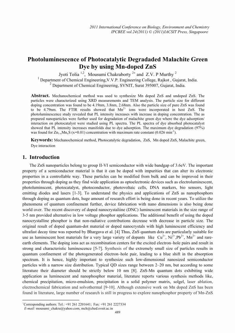
using XRD. XRD analysis in fig: 1(a) reveals hexagonal wurzite α-ZnS (JCPDS 72-0163) together with cubic spherelite β-ZnS (JCPDS 5- 0566) as the only products of mechanochemical synthesis. Coexistence of both the phases has also been reported in literature [12-13]. This can be due to phase transformation as the consequence of the motion of dislocations in activated solid state. The spectra was matched for various diffraction peaks at 2θ values of 29.03o, 48.49o and 56.87o corresponding to the diffraction planes (103), (1011) and (203) respectively for wurzite ZnS while the peaks were also found to match at 28.792o, 48.121o and 56.874o corresponding to the diffraction planes (111), (220) and (311) respectively for cubic ZnS..The value of 4.79nm was calculated using Scherrer formula [14]. The particle size was also found to match TEM analysis (not shown here). The XRD patterns shown in fig: 1 (b, c and d) of the ZnS:Mn nanocrystals show very broad diffraction peaks with increasing concentration of manganese which is the characteristics of nanosized materials. For all the samples the three main diffraction peak positions corresponds cubic zinc
490
blend ZnS structure(JCPDS No: 05-0566). Thus, it is due Mn doping the crystal structure has completely changed to cubic Mn doped ZnS. The diffraction peaks from manganese impurities were detected at 2θ position value 35.734°, 36.375° and 35.953° for Zn1-x MnxS where x= 0.01, 0.22 and 0.3 respectively. This indicates that manganese ions are dispersed into the ZnS matrix. The cystalline size were calculated to be 4.19nm, 3.86nm and 2.68nm for Zn1-x MnxS where x= 0.01, 0.22 and 0.3 respectively. The particle size is found to decrease with increase doping concentration.
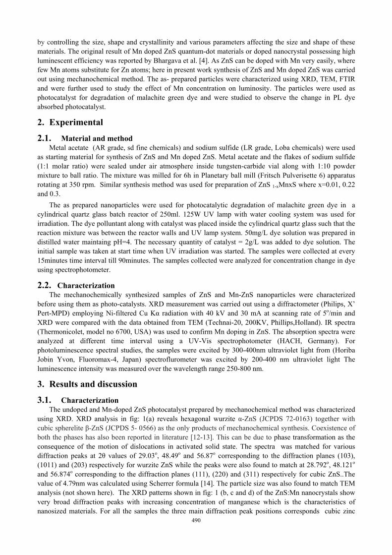
Fig: 1 XRD of ZnS and Zn1-x MnxS, Fig: 2 IR spectra of ZnS and Zn1-x MnxS, where x= 0.01(b), 0.22(c) and 0.3(d) where x= 0.01(b), 0.22(c) and 0.3(d)
The IR spectra in fig: 2(a,b,c,d), the peaks are assigned to the samples at room temperature. The bands around 3000-3600cm-1 are due to the hydrogen stretching frequency (O-H stretching). This bands corresponds to valence vibrations of the occluded water. Such bands are found in all the samples in figures. The weak bands at 2373.7cm-1 in fig: 3a corresponds to C=O stretching vibrations. This absorption peak has completely disappeared on adding Mn for doping to ZnS. The bands around 1625.9cm-1, 1625.4cm-1, 1629.4cm-1, 1637.1cm-1 and 1618.3cm-1 correspond to O-H bending. The bands around 1200 and 1100cm-1 are for ZnS-Mn2+ ions due to symmetric stretching and the characteristic frequency of these inorganic ions. The bands at 1090.6cm-1, 1008.9cm-1, 1009.5cm-1, 1015.4cm-1, 1012.6cm-1, 1012.8cm-1 are weak shoulders with asymmetric stretching. The peaks at 637cm-1, 621cm-1, 669cm-1, 664cm-1 and 650cm-1 are assigned to the ZnS band and symmetric bending which arises from Zn-S and Mn-S vibrations.
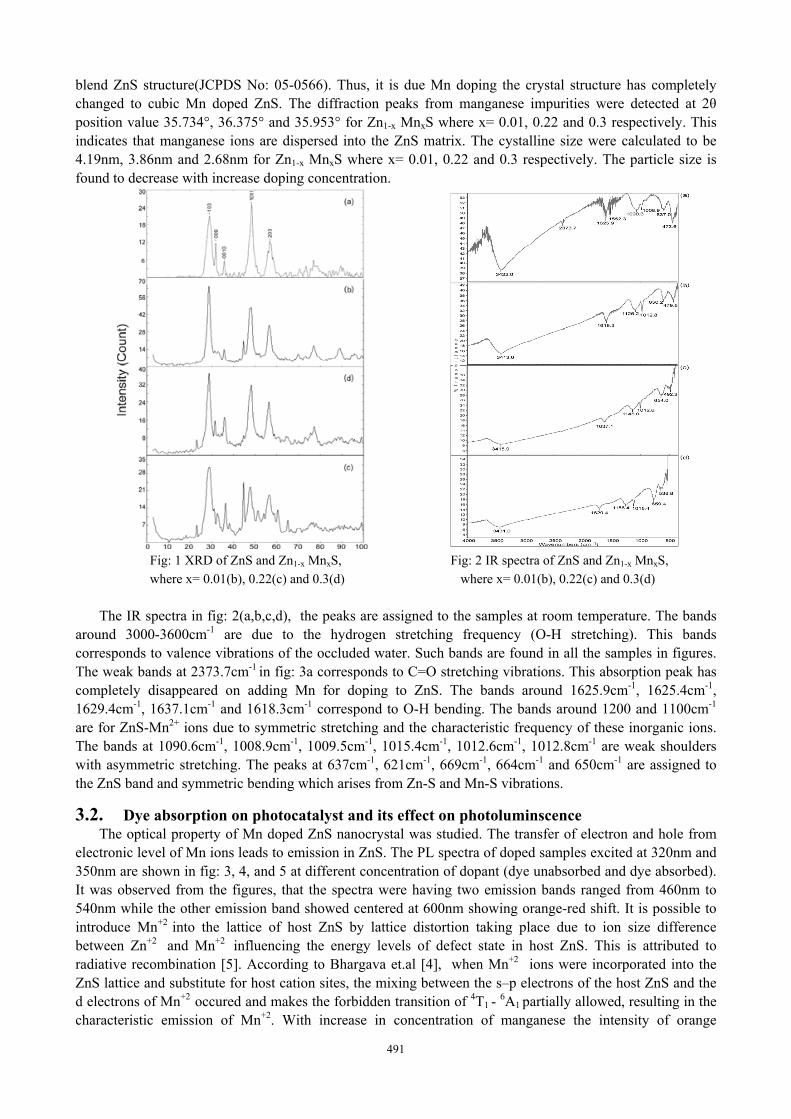
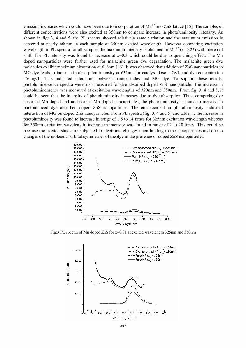
3.2. Dye absorption on photocatalyst and its effect on photoluminscence The optical property of Mn doped ZnS nanocrystal was studied. The transfer of electron and hole from
electronic level of Mn ions leads to emission in ZnS. The PL spectra of doped samples excited at 320nm and 350nm are shown in fig: 3, 4, and 5 at different concentration of dopant (dye unabsorbed and dye absorbed). It was observed from the figures, that the spectra were having two emission bands ranged from 460nm to 540nm while the other emission band showed centered at 600nm showing orange-red shift. It is possible to introduce Mn+2
into the lattice of host ZnS by lattice distortion taking place due to ion size difference between Zn+2
and Mn+2 influencing the energy levels of defect state in host ZnS. This is attributed to
radiative recombination [5]. According to Bhargava et.al [4], when Mn+2 ions were incorporated into the
ZnS lattice and substitute for host cation sites, the mixing between the s–p electrons of the host ZnS and the d electrons of Mn+2
occured and makes the forbidden transition of 4T1 - 6A1 partially allowed, resulting in the characteristic emission of Mn+2. With increase in concentration of manganese the intensity of orange
491
492
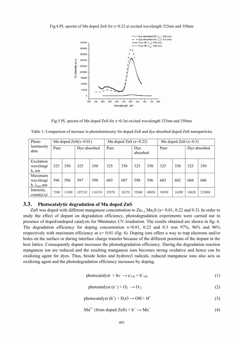
emission increases which could have been due to incorporation of Mn+2 into ZnS lattice [15]. The samples of different concentrations were also excited at 350nm to compare increase in photoluminosity intensity. As shown in fig: 3, 4 and 5, the PL spectra showed relatively same variation and the maximum emission is centered at nearly 600nm in each sample at 350nm excited wavelength. However comparing excitation wavelength in PL spectra for all samples the maximum intensity is obtained in Mn+2 (x=0.22) with mere red shift. The PL intensity was found to decrease at x=0.3 which could be due to quenching effect. The Mn doped nanoparticles were further used for malachite green dye degradation. The malachite green dye molecules exhibit maximum absorption at 618nm [16]. It was observed that addition of ZnS nanoparticles to MG dye leads to increase in absorption intensity at 631nm for catalyst dose = 2g/L and dye concentration =50mg/L. This indicated interaction between nanoparticles and MG dye. To support these results, photoluminescence spectra were also measured for dye absorbed doped ZnS nanoparticle. The increase in photoluminensence was measured at excitation wavelengths of 320nm and 350nm. From fig: 3, 4 and 5, it could be seen that the intensity of photoluminosity increases due to dye absorption. Thus, comparing dye absorbed Mn doped and unabsorbed Mn doped nanoaprticles, the photoluminosity is found to increase in photoinduced dye absorbed doped ZnS nanoparticles. The enhancement in photoluminosity indicated interaction of MG on doped ZnS nanoparticles. From PL spectra (fig: 3, 4 and 5) and table: 1, the increase in photoluminosity was found to increase in range of 1.5 to 14 times for 325nm excitation wavelength whereas for 350nm excitation wavelength, increase in intensity was found in range of 2 to 20 times. This could be because the excited states are subjected to electronic changes upon binding to the nanoparticles and due to changes of the molecular orbital symmetries of the dye in the presence of doped ZnS nanoparticles.
Fig:3 PL spectra of Mn doped ZnS for x=0.01 at excited wavelength 325nm and 350nm
493
Fig:4 PL spectra of Mn doped ZnS for x=0.22 at excited wavelength 325nm and 350nm
Fig:5 PL spectra of Mn doped ZnS for x=0.3at excited wavelength 325nm and 350nm
Table 1: Comparison of increase in photoluminosity for doped ZnS and dye absorbed doped ZnS nanoparticles
Photo luminosity data
Mn doped ZnS(x=0.01) Mn doped ZnS (x=0.22) Mn doped ZnS (x=0.3) Pure Dye absorbed Pure Dye
absorbed Pure Dye absorbed
Excitation wavelength, nm
325 350 325 350 325 350 325 350 325 350 325 350
Maximum wavelength, λmax nm
596 596 597 598 603 607 598 596 603 602 604 606
Intensity, count(a.u) 7280 13100 107210 116310 35970 24170 52840 48050 10930 16200 18620 323880
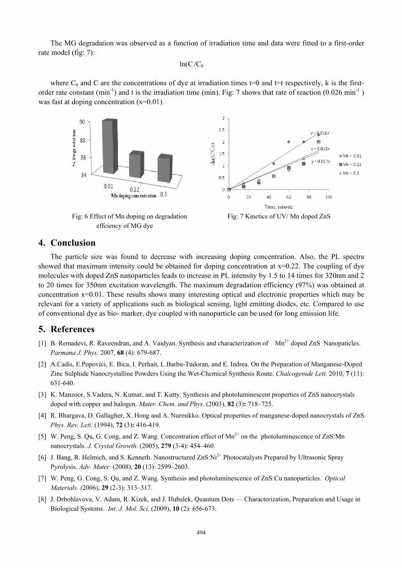
3.3. Photocatalytic degradation of Mn doped ZnS ZnS was doped with different manganese concentration to Zn1-x MnxS (x= 0.01, 0.22 and 0.3). In order to
study the effect of dopant on degradation efficiency, photodegradation experiments were carried out in presence of doped/undoped catalysts for 90minutes UV irradiation. The results obtained are shown in fig: 6. The degradation efficiency for doping concentration x=0.01, 0.22 and 0.3 was 97%, 96% and 96% respectively with maximum efficiency at x= 0.01 (fig: 6). Doping ions offers a way to trap electrons and/or holes on the surface or during interface charge transfer because of the different positions of the dopant in the host lattice. Consequently dopant increases the photodegradation efficiency. During the degradation reaction manganese ion are reduced and the resulting manganese ions becomes strong oxidative and hence can be oxidizing agent for dyes. Thus, beside holes and hydroxyl radicals, reduced manganese ions also acts as oxidizing agent and the photodegradation efficiency increases by doping.
photocatalyst + hν → e-CB + h+
VB (1)
photoatalyst (e- ) + O2 → O·2 (2)
photocatalyst (h+) + H2O → OH·+ H+ (3)
Mn2+ (from doped ZnS) + h+ → Mn+ (4)
494
The MG degradation was observed as a function of irradiation time and data were fitted to a first-order rate model (fig: 7):
ln(C /C0
where C0 and C are the concentrations of dye at irradiation times t=0 and t=t respectively, k is the first-order rate constant (min-1) and t is the irradiation time (min). Fig: 7 shows that rate of reaction (0.026 min-1 ) was fast at doping concentration (x=0.01).
Fig: 6 Effect of Mn doping on degradation Fig: 7 Kinetics of UV/ Mn doped ZnS
effciency of MG dye
4. Conclusion The particle size was found to decrease with increasing doping concentration. Also, the PL spectra
showed that maximum intensity could be obtained for doping concentration at x=0.22. The coupling of dye molecules with doped ZnS nanoparticles leads to increase in PL intensity by 1.5 to 14 times for 320nm and 2 to 20 times for 350nm excitation wavelength. The maximum degradation efficiency (97%) was obtained at concentration x=0.01. These results shows many interesting optical and electronic properties which may be relevant for a variety of applications such as biological sensing, light emitting diodes, etc. Compared to use of conventional dye as bio- marker, dye coupled with nanoparticle can be used for long emission life.
5. References [1] B. Remadevi, R. Raveendran, and A. Vaidyan. Synthesis and characterization of Mn2+ doped ZnS Nanopaticles.
Parmana J. Phys. 2007, 68 (4): 679-687.
[2] A.Cadis, E.Popovici, E. Bica, I. Perhait, L.Barbu-Tudoran, and E. Indrea. On the Preparation of Manganese-Doped Zinc Sulphide Nanocrystalline Powders Using the Wet-Chemical Synthesis Route. Chalcogenide Lett. 2010, 7 (11): 631-640.
[3] K. Manzoor, S.Vadera, N. Kumar, and T. Kutty. Synthesis and photoluminescent properties of ZnS nanocrystals doped with copper and halogen. Mater. Chem. and Phys. (2003), 82 (3): 718–725.
[4] R. Bhargava, D. Gallagher, X. Hong and A. Nurmikko. Optical properties of manganese-doped nanocrystals of ZnS. Phys. Rev. Lett. (1994), 72 (3): 416-419.
[5] W. Peng, S. Qu, G. Cong, and Z. Wang. Concentration effect of Mn2+ on the photoluminescence of ZnS:Mn nanocrystals. J. Crystal Growth. (2005), 279 (3-4): 454–460.
[6] J. Bang, R. Helmich, and S. Kenneth. Nanostructured ZnS:Ni2+ Photocatalysts Prepared by Ultrasonic Spray Pyrolysis. Adv. Mater. (2008), 20 (13): 2599–2603.
[7] W. Peng, G. Cong, S. Qu, and Z. Wang. Synthesis and photoluminescence of ZnS:Cu nanoparticles. Optical Materials. (2006), 29 (2-3): 313–317.
[8] J. Drbohlavova, V. Adam, R. Kizek, and J. Hubalek. Quantum Dots — Characterization, Preparation and Usage in Biological Systems. Int. J. Mol. Sci. (2009), 10 (2): 656-673.

495
[9] S. Wei Lu, B. Lee, Z. Lin Wang, W. Tong, B. Wagner, W. Park, and C. Summers. Synthesis and photoluminescence enhancement of Mn2+-doped ZnS nanocrystals. J. Luminescence. (2001), 92 (1-2): 73-78.
[10] D. Denzler, M. Olschewski, and K. Sattlera. Luminescence studies of localized gap states in colloidal ZnS nanocrystals. J. Appl. Phys. (1998), 84 (51): 2841-2845.
[11] E. Mohagheghpour, M. Rabiee, F. Moztarzadeh, M. Tahriri, M. Jafarbeglou, D. Bizari, and H. Eslami. Controllablesynthesis, characterization and optical properties of ZnS:Mn nanoparticles as a novel biosensor. Mater. Sci. Eng. C,. (2009), 29 (6): 1842-1848.
[12] B. Geng, X, Liu, X. Wei, and L. Zhang. Structure and optical properties of periodically twinned ZnS nanowires. Appl. Phys. Lett., (2006), 88 (16): 163104-163104-3.
[13] S. Sambasivam, S. Joseph, D.Reddy, B. Reddy, and C. Jayasankar. Synthesis and characterization of thiophenol passivated Fe doped ZnS nanoparticles. Mater. Sci. Eng., B. (2008), 150 (2): 125-129.
[14] J. Cao, J. Yang, Y. Zhang, L. Yang, Y. Wang, M. Wei, Y. Liu, M. Gao, X. Liuc, and Z. Xied. Optimized doping concentration of manganese in zinc sulfide nanoparticles for yellow-orange light emission. J. Alloys Compd. (2009), 486 (1-2): 890–894.
[15] G. Parshetti, S. Kalme, G. Saratale, and S. Govindwar. Biodegradation of Malachite Green by Kocuria rosea MTCC 1532. Acta Chimica Slovenica. (2006), 53 (1-2): 492–498.


