
Photo-acoustic phase conjugation for biomedical imaging
19
1 5/19/2011, Reid McCargar Photo-acoustic phase conjugation for biomedical imaging Reid McCargar, Research Assistant
Transcript of Photo-acoustic phase conjugation for biomedical imaging

15/19/2011, Reid McCargar
Photo-acoustic phase conjugation for biomedical imaging
Reid McCargar, Research Assistant

25/19/2011, Reid McCargar
Outline
• Brief overview – current diagnostic imaging technologies and the motivation for new technologies
• Wavefront aberration by scattering
• Dynamic holography and phase conjugation
• The acousto-optic effect
• Synthetic guide star acousto-optic tomography
35/19/2011, Reid McCargar
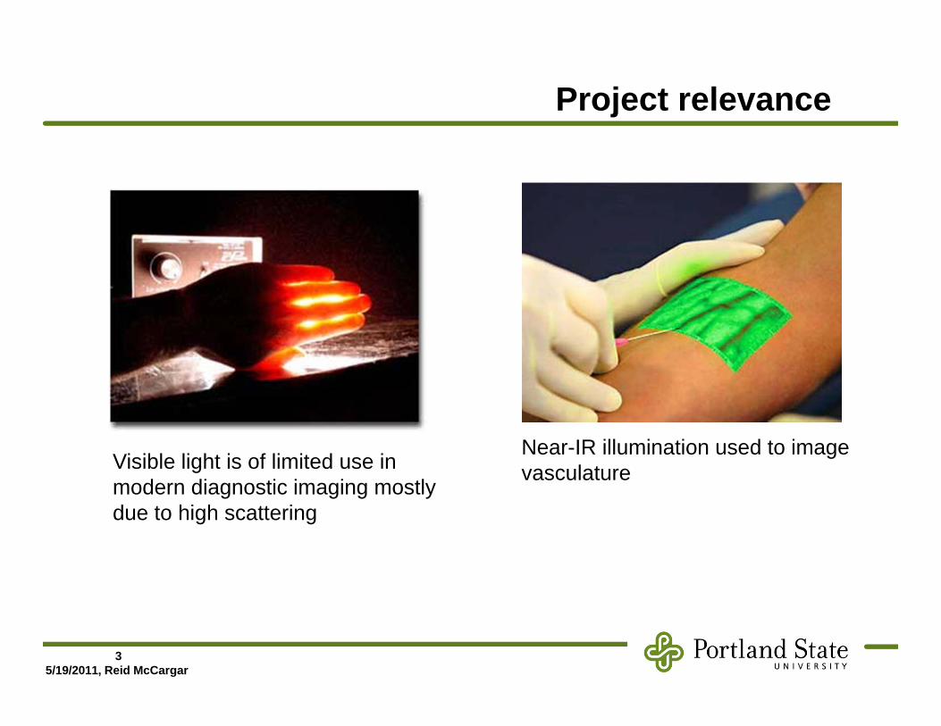
Project relevance
Near-IR illumination used to image vasculature Visible light is of limited use in
modern diagnostic imaging mostly due to high scattering

45/19/2011, Reid McCargar
Current imaging technologies
55/19/2011, Reid McCargar
Why change anything?
• Not all features of medical interest have adequate contrast when viewed with currently-available technologies.
• Many of the imaging technologies involve ionizing radiation, require chemical contrast agents, or have prohibitive cost.

65/19/2011, Reid McCargar
Why not use optical wavelengths?
With 2-way propagation, imaging may be possible down to a centimeter or so.
75/19/2011, Reid McCargar
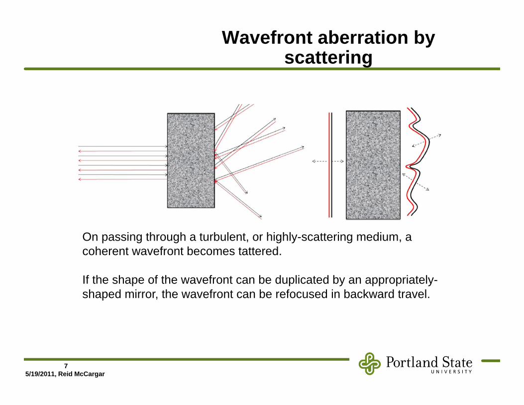
Wavefront aberration by scattering
On passing through a turbulent, or highly-scattering medium, a coherent wavefront becomes tattered.
If the shape of the wavefront can be duplicated by an appropriately-shaped mirror, the wavefront can be refocused in backward travel.

85/19/2011, Reid McCargar
Wavefront sensing
• Problem with recreating the shape of a tattered wave: optical detectors are phase-insensitive.
Locally regarded as plane-waves, a wavefront shape can be detected by focal position shifts

95/19/2011, Reid McCargar
Implementation
GS
CCDWS
CPU
Historically used for sharpening astronomical images, but can you imagine the implementation in diagnostic imaging?

105/19/2011, Reid McCargar
Not good – lasers inside people
GS
CC
DW
S
CPU

115/19/2011, Reid McCargar
Dynamic holography
E1
E2 = k E1 *E4 = k E3 *
E3
Material with transmittance changes due to interference of E1 and E3

125/19/2011, Reid McCargar
Acousto-optic frequency shifting
Ultrasound transducer
Transmitted zero-order-beam
1st -order diffracted beam, up-shifted in frequency by facoustic
Acoustic wave propagation

135/19/2011, Reid McCargar
Synthetic guide star
Ultrasound transducer
LaserPhotodetector identifies frequency-shifted photons as having passed through ultrasound focus

145/19/2011, Reid McCargar
Putting the pieces together – Xu, et. al

155/19/2011, Reid McCargar
A less bewildering look
AO AO
US
CCD
R (f0) R* (f0 )
fs=f0-F

165/19/2011, Reid McCargar
Results
Tissue analog imaged to 5 mm with better resolution than ultrasound focus

175/19/2011, Reid McCargar
Conclusions
• It is possible to image tissue with visible light to perhaps several centimeters.
• Applications of this new technology are mostly untested.

185/19/2011, Reid McCargar
References

195/19/2011, Reid McCargar
Questions?
Thank you