Phosphorylation of the proto-oncogene product eukaryotic initiation ...
4
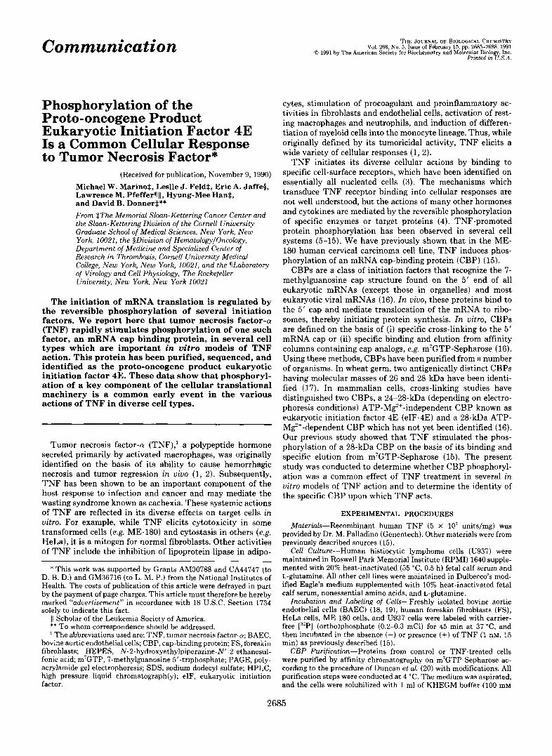
Communication Vol. 266, No. 5, Issue of February 15, pp. 2685~2689,1991 THE JOURNAL OF BIOLOGICAL CHEMISTRY d 1991 by The American Society for Biochernlstry and Molecular Blology, Inc. Printed in U.S.A. Phosphorylation of the Proto-oncogene Product Eukaryotic Initiation Factor 4E Is a Common Cellular Response to Tumor Necrosis Factor* (Received for publication, November 9, 1990) Michael W. MarinoS, Leslie J. FeldS, Eric A. Jaffep, Lawrence M. Pfefferv 11, Hyung-Mee HanS, and David B. Dormer$** From $The Memorial Sloan-Kettering Cancer Center and the Sloan-Kettering Division of the Cornell University Graduate School of Medical Sciences, New York, New York, 10021, the §Division of HematologylOncology, Department of Medicine and Specialized Center of Research in Thrombosis, Cornell University Medical College,New York, New York, 10021, and the TLaboratory of Virology and Cell Physiology, The Rockefeller University, New York, New York 10021 The initiation of mRNA translation is regulated by the reversible phosphorylation of several initiation factors. We report here that tumor necrosis factor-a (TNF) rapidly stimulates phosphorylation of one such factor, an mRNA cap binding protein, in several cell types which are important in vitro models of TNF action. This protein has been purified, sequenced, and identified as the proto-oncogene product eukaryotic initiation factor 4E. These data show that phosphoryl- ation of a key component of the cellular translational machinery is a common earlyevent in the various actions of TNF in diverse cell types. Tumor necrosis factor-cu (TNF),' a polypeptide hormone secreted primarily by activated macrophages, was originally identified on the basis of its ability to cause hemorrhagic necrosis and tumor regression in vivo (1, 2). Subsequently, TNF has been shown to be an important component of the host response to infection and cancer and may mediate the wasting syndrome known as cachexia. These systemic actions of TNF are reflected in its diverse effects on target cells in uitro. For example, while T N F elicitscytotoxicity in some transformed cells (e.g. ME-180) and cytostasis in others (e.g. HeLa), it is a mitogen for normal fibroblasts. Other activities of TNF include the inhibition of lipoprotein lipase in adipo- * This work was supported by Grants AM30788 and CA44747 (to D. B. D.) and GM36716 (to L. M. P.) from the National Institutes of Health. The costs of publication of this article were defrayed in part by the payment of page charges. This article must therefore be hereby marked "advertisement" in accordance with 18 U.S.C. Section 1734 solely to indicate this fact. 1) Scholar of the Leukemia Society of America. ** To whom correspondence should be addressed, The abbreviations used are: TNF, tumor necrosis factor-a;BAEC, bovine aortic endothelial cells; CBP, cap-binding protein; FS, foreskin fibroblasts; HEPES, N-2-hydroxyethylpiperazine-N"2-ethanesul- fonic acid m7GTP, 7-methylguanosine 5"triphosphate; PAGE, poly- acrylamide gel electrophoresis; SDS, sodium dodecyl sulfate; HPLC, high pressure liquid chromatograph(y); eIF, eukaryotic initiation factor. cytes, stimulation of procoagulant and proinflammatory ac- tivities in fibroblasts and endothelial cells, activation of rest- ing macrophages and neutrophils, and induction of differen- tiation of myeloid cells into the monocyte lineage. Thus, while originally defined by its tumoricidal activity, TNF elicits a wide variety of cellular responses (1, 2). TNF initiatesits diverse cellular actions by bindingto specific cell-surface receptors, which have been identified on essentiallyallnucleated cells (3). The mechanisms which transduce TNF receptor binding into cellular responses are not well understood, but the actions of many other hormones and cytokines are mediated by the reversible phosphorylation of specific enzymes or target proteins (4). TNF-promoted proteinphosphorylationhas been observed in several cell systems (5-15). We have previously shown that in the ME- 180 human cervical carcinoma cell line, TNF induces phos- phorylation of an mRNA cap-binding protein (CBP) (15). CBPs are a class of initiation factors that recognize the 7- methylguanosinecapstructure found on the 5' end of all eukaryotic mRNAs (except those in organelles) and most eukaryotic viral mRNAs (16). In uiuo, these proteins bind to the 5' cap and mediate translocation of the mRNA to ribo- somes, thereby initiating protein synthesis. In uitro, CBPs are defined on the basisof (i) specific cross-linking to the 5' mRNA cap or (ii) specific binding and elution from affinity columns containing cap analogs, e.g. m7GTP-Sepharose (16). Using these methods, CBPs have been purified from a number of organisms. In wheat germ, two antigenically distinct CBPs having molecular masses of 26 and 28 kDa have been identi- fied (17). In mammalian cells, cross-linking studies have distinguished two CBPs, a 24-28-kDa (depending onelectro- phoresis conditions) ATP-Mg'+-independent CBP known as eukaryotic initiation factor 4E (eIF-4E) and a 28-kDa ATP- M$+-dependent CBP which has not yet been identified (16). Our previous study showed that TNF stimulated the phos- phorylation of a 28-kDa CBP on the basisof its binding and specific elutionfromm7GTP-Sepharose (15). The present study was conducted to determine whether CBP phosphoryl- ation was a common effect of TNF treatment in several in uitro models of TNF action and to determine the identity of the specific CBP upon which TNF acts. EXPERIMENTAL PROCEDURES Materials-Recombinant human TNF (5 X lo7 units/mg) was provided by Dr. M. Palladino (Genentech). Other materials were from previously described sources (15). Cell Culture-Human histiocytic lymphoma cells (U937) were maintained in Roswell Park Memorial Institute (RPMI) 1640 supple- mented with 20% heat-inactivated (55 "C, 0.5 h) fetal calf serum and L-glutamine. All other cell lines were maintained in Dulbecco's mod- ified Eagle's medium supplemented with 10% heat-inactivated fetal calf serum, nonessential amino acids, and L-glutamine. Incubation and LabelingofCells-Freshlyisolated bovine aortic endothelial cells (BAEC) (18, 19), human foreskin fibroblasts (FS), HeLa cells, ME-180 cells, and U937 cells were labeled with carrier- free E3'P] (0rtho)phosphate (0.2-0.3 mCi) for 45 min at 37 "C, and then incubated in the absence (-) or presence (+) of TNF (1 nM, 15 min) as previously described (15). CBPPurification-Proteins from control or TNF-treated cells were purified by affinity chromatography on m7GTP-Sepharose ac- cording to the procedure of Duncan et al. (20) with modifications. All purification steps were conducted at 4 "C. The medium was aspirated, and the cells were solubilized with 1 ml of KHEGM buffer (100 mM 2685
Transcript of Phosphorylation of the proto-oncogene product eukaryotic initiation ...
Communication Vol. 266, No. 5, Issue of February 15, pp. 2685~2689,1991 THE JOURNAL OF BIOLOGICAL CHEMISTRY
d 1991 by The American Society for Biochernlstry and Molecular Blology, Inc. Printed in U.S.A.
Phosphorylation of the Proto-oncogene Product Eukaryotic Initiation Factor 4E Is a Common Cellular Response to Tumor Necrosis Factor*
(Received for publication, November 9, 1990)
Michael W. MarinoS, Leslie J. FeldS, Eric A. Jaffep, Lawrence M. Pfefferv 11, Hyung-Mee HanS, and David B. Dormer$** From $The Memorial Sloan-Kettering Cancer Center and the Sloan-Kettering Division of the Cornell University Graduate School of Medical Sciences, New York, New York, 10021, the §Division of HematologylOncology, Department of Medicine and Specialized Center of Research in Thrombosis, Cornell University Medical College, New York, New York, 10021, and the TLaboratory of Virology and Cell Physiology, The Rockefeller University, New York, New York 10021
The initiation of mRNA translation is regulated by the reversible phosphorylation of several initiation factors. We report here that tumor necrosis factor-a (TNF) rapidly stimulates phosphorylation of one such factor, an mRNA cap binding protein, in several cell types which are important in vitro models of TNF action. This protein has been purified, sequenced, and identified as the proto-oncogene product eukaryotic initiation factor 4E. These data show that phosphoryl- ation of a key component of the cellular translational machinery is a common early event in the various actions of TNF in diverse cell types.
Tumor necrosis factor-cu (TNF),' a polypeptide hormone secreted primarily by activated macrophages, was originally identified on the basis of its ability to cause hemorrhagic necrosis and tumor regression in v ivo (1, 2). Subsequently, TNF has been shown to be an important component of the host response to infection and cancer and may mediate the wasting syndrome known as cachexia. These systemic actions of TNF are reflected in its diverse effects on target cells i n uitro. For example, while T N F elicits cytotoxicity in some transformed cells (e.g. ME-180) and cytostasis in others (e.g. HeLa), it is a mitogen for normal fibroblasts. Other activities of TNF include the inhibition of lipoprotein lipase in adipo-
* This work was supported by Grants AM30788 and CA44747 (to D. B. D.) and GM36716 (to L. M. P.) from the National Institutes of Health. The costs of publication of this article were defrayed in part by the payment of page charges. This article must therefore be hereby marked "advertisement" in accordance with 18 U.S.C. Section 1734 solely to indicate this fact.
1) Scholar of the Leukemia Society of America. ** To whom correspondence should be addressed, The abbreviations used are: TNF, tumor necrosis factor-a; BAEC,
bovine aortic endothelial cells; CBP, cap-binding protein; FS, foreskin fibroblasts; HEPES, N-2-hydroxyethylpiperazine-N"2-ethanesul- fonic ac id m7GTP, 7-methylguanosine 5"triphosphate; PAGE, poly- acrylamide gel electrophoresis; SDS, sodium dodecyl sulfate; HPLC, high pressure liquid chromatograph(y); eIF, eukaryotic initiation factor.
cytes, stimulation of procoagulant and proinflammatory ac- tivities in fibroblasts and endothelial cells, activation of rest- ing macrophages and neutrophils, and induction of differen- tiation of myeloid cells into the monocyte lineage. Thus, while originally defined by its tumoricidal activity, TNF elicits a wide variety of cellular responses (1, 2).
TNF initiates its diverse cellular actions by binding to specific cell-surface receptors, which have been identified on essentially all nucleated cells (3). The mechanisms which transduce T N F receptor binding into cellular responses are not well understood, but the actions of many other hormones and cytokines are mediated by the reversible phosphorylation of specific enzymes or target proteins (4). TNF-promoted protein phosphorylation has been observed in several cell systems (5-15). We have previously shown that in the ME- 180 human cervical carcinoma cell line, T N F induces phos- phorylation of an mRNA cap-binding protein (CBP) (15).
CBPs are a class of initiation factors that recognize the 7- methylguanosine cap structure found on the 5' end of all eukaryotic mRNAs (except those in organelles) and most eukaryotic viral mRNAs (16). In uiuo, these proteins bind to the 5' cap and mediate translocation of the mRNA to ribo- somes, thereby initiating protein synthesis. In uitro, CBPs are defined on the basis of (i) specific cross-linking to the 5' mRNA cap or (ii) specific binding and elution from affinity columns containing cap analogs, e.g. m7GTP-Sepharose (16). Using these methods, CBPs have been purified from a number of organisms. In wheat germ, two antigenically distinct CBPs having molecular masses of 26 and 28 kDa have been identi- fied (17). In mammalian cells, cross-linking studies have distinguished two CBPs, a 24-28-kDa (depending on electro- phoresis conditions) ATP-Mg'+-independent CBP known as eukaryotic initiation factor 4E (eIF-4E) and a 28-kDa ATP- M$+-dependent CBP which has not yet been identified (16). Our previous study showed that TNF stimulated the phos- phorylation of a 28-kDa CBP on the basis of its binding and specific elution from m7GTP-Sepharose (15). The present study was conducted to determine whether CBP phosphoryl- ation was a common effect of TNF treatment in several in uitro models of T N F action and to determine the identity of the specific CBP upon which TNF acts.
EXPERIMENTAL PROCEDURES
Materials-Recombinant human TNF (5 X lo7 units/mg) was provided by Dr. M. Palladino (Genentech). Other materials were from previously described sources (15).
Cell Culture-Human histiocytic lymphoma cells (U937) were maintained in Roswell Park Memorial Institute (RPMI) 1640 supple- mented with 20% heat-inactivated (55 "C, 0.5 h) fetal calf serum and L-glutamine. All other cell lines were maintained in Dulbecco's mod- ified Eagle's medium supplemented with 10% heat-inactivated fetal calf serum, nonessential amino acids, and L-glutamine.
Incubation and Labeling of Cells-Freshly isolated bovine aortic endothelial cells (BAEC) (18, 19), human foreskin fibroblasts (FS), HeLa cells, ME-180 cells, and U937 cells were labeled with carrier- free E3'P] (0rtho)phosphate (0.2-0.3 mCi) for 45 min at 37 "C, and then incubated in the absence (-) or presence (+) of TNF (1 nM, 15 min) as previously described (15).
CBP Purification-Proteins from control or TNF-treated cells were purified by affinity chromatography on m7GTP-Sepharose ac- cording to the procedure of Duncan et al. (20) with modifications. All purification steps were conducted at 4 "C. The medium was aspirated, and the cells were solubilized with 1 ml of KHEGM buffer (100 mM
2685


Phosphorylation of the Proto-oncogene Product eIF-4E 2687
c
Retention time, min
FIG. 3. HPLC fractionation and amino acid sequence analy- sis of peptides from tryptic digestion of CBP. CBP was purified from ME-180 cells (80 X IO8 cells) by affinity chromatography on m7GTP-Sepharose (3 ml). CBP fractionated by SDS-PAGE was transferred to nitrocellulose and then digested with trypsin. CBP fragments were separated by HPLC and sequenced.
M A T V E P E T T P T P N P p T T E E E K . T E S N Q E V A N P E H Y
I K . H P L Q N R . W A L W F F K . N D K m S K . T W Q A N L R * L I r -
S K . F D T V E D F W A L Y N H I Q L S S N L M P G C D Y S L F K - D
G I E P M W E D E K . N N R . G G R A W L I T L N K A Q Q R * R . S D
L D R . F W L E T L L C L 1 G E S P D D Y S D D V C G A V V N V R . A
1 I
I I
I I
K . G D K . I A I W T T E C E N R . E A V T H I G R * V Y K * E R - L
G F P P K . I V I G Y Q S H A D T A T K . S G S T T K . N R . F V V I I z
FIG. 4. Primary structure of human eIF-4E from Ref. 23 showing the amino acid sequences derived from purified CBP (boxed). Trypsin cleavage sites within the eIF-4E sequence are indicated ( - ) . The one-letter code is used to specify the amino acids.
addition, the peak eluting a t 29.8 min yielded a second amino acid sequence (Thr-Trp-Gln-Ala-Asn-Leu-Arg) which also corresponded to an eIF-4E tryptic peptide. Thus, purification and sequence analysis demonstrate that it is the 28-kDa mRNA cap-binding protein eIF-4E which is phosphorylated in response to TNF treatment.
DISCUSSION
The effects of TNF are diverse and highly cell type-specific. We have studied the role of protein phosphorylation in T N F signal transduction using in uitro models in which T N F induces cytotoxicity and cytostasis (ME-180 and HeLa cells, respectively), differentiation (U937 cells), mitogenesis (fibro- blasts), and also inflammatory responses (FS, U937, and endothelial cells). In each of these cell lines T N F promotes the rapid phosphorylation of the 28-kDa mRNA cap-binding protein eIF-4E, which has recently been implicated in the translational control of gene expression.
eIF-4E is thought to be the first initiation factor to interact with the mRNA (24) and may have a discriminatory role in selecting specific messages for translation initiation (25, 26). In uitro studies suggest that phosphorylation “activates” eIF- 4E and correlates with recruitment of translationally con- trolled mRNAs to active ribosomes (27, 28). Central to the TNF-induced proinflammatory response of the macrophage, endothelial cell, and fibroblast is the induction of several important inflammatory mediators which include interleukin- 1, TNF, and several colony-stimulating factors (2). Recent evidence suggests that these mediators are encoded by
mRNAs which are regulated at the translational level (29). It is therefore of particular interest that TNF promotes the phosphorylation of eIF-4E, a factor implicated in the regula- tion of translation in these cells.
eIF-4E phosphorylation also appears to play an important role in the regulation of cell growth. In yeast, eIF-4E is involved in controlling the transition of cells from Gl- into S - phase of the cell cycle (30). Mutation of the yeast eIF-4E gene leads to growth arrest in G1 but can be complemented by a mutation which leads to constitutive activation of the cata- lytic subunit of cyclic AMP-dependent protein kinase (30). In sea urchin oocytes, eIF-4E phosphorylation increases after fertilization and decreases during mitosis (31). Similarly, in fibroblasts, the phosphorylation state of eIF-4E increases during mitogen stimulation of quiescent cells (27) and de- creases during mitosis (32). Recently, transfection and over- expression of eIF-4E were shown to lead to malignant trans- formation of NIH 3T3 cells (33), establishing that eIF-4E is the product of a proto-oncogene. Transformation did not occur when cells were transfected with an eIF-4E mutant (Ser53 + Ala53) lacking the major eIF-4E phosphorylation site (33). Thus, the phosphorylation of eIF-4E promoted by TNF may prove relevant to the growth effects induced by TNF in ME-180 cells (cytotoxicity), HeLa cells (cytostasis), fibro- blasts (mitogenesis), and U937 cells (differentiation). In con- clusion, our study suggests that TNF-promoted phosphoryl- ation of eIF-4E may mediate or modulate some of TNF’s effects on cell growth and metabolism.
Acknowledgments-We are grateful to George Lam for preparation of the endothelial cells, Drs. Lloyd Old and Nahum Sonenberg for helpful discussions, Genentech for providing recombinant human TNF-a, and especially William Lane for his technical assistance.
1.
2.
3.
4.
5.
6. 7.
8.
9. 10.
11.
12.
13. 14.
15.
16.
17.
18.
19.
REFERENCES Rosenblum, M. G., and Donato, N. J. (1989) Crit. Reu. Immunol.
Kunkel, S. L., Remick, D. G., Strieter, R. M., and Larrick, J. W .
Kull, F. C., Jr . (1988) Nat. Immun. Cell Growth Regul. 7, 254-
Rosen, 0. M., and Krebs, E. G. (1981) Cold Spring Harbor Conf.
Hepburn, A,, Demolle, D., Boeynaems, J.-M., Fiers, W., and
Kaur, P., and Saklatvala, J. (1988) FEES Lett. 241, 6-10 Berkow, R. L., and Dodson, M. R. (1988) J. Leukocyte Biol. 44,
Schutze, S., Scheurich, P., Pfizenmaier, K., and Kronke, M.
Bird, T. A., and Saklatvala, J. (1989) J. Immunol. 142, 126-133 Donato, N. J., Gallick, G. E., Steck, P. A., and Rosenblum, M. G.
(1989) J. Biol. Chem. 264, 20474-20481 Robaye, B., Hepburn, A., Lecoq, R., Fiers, W., Boeynaems, J. M.,
and Dumont, J. E. (1989) Biochem. Biophys. Res. Commun.
Donato, N. J., Ince, C., Rosenblum, M. G., and Gallick, G. E.
Arrigo A.-P. (1990) Mol. Cell. Biol. 10, 1276-1280 Kohno, M., Nishizawa, N., Tsujimoto, M., and Nomoto, H. (1990)
Marino, M. W., Pfeffer, L. M., Guidon, P. T., Jr., and Donner,
Sonenberg, N. (1988) Prog. Nucleic Acid Res. Mol. Biol. 35, 173-
Browning, K. S., Lax, S. R., and Ravel, J. M. (1987) J. Biol.
Jaffe, E. A,, Grulich, J., Weksler, B. B., Hampel, G., and Watan-
Jaffe, E. A., Nachman, R. L., Becker, C. G., and Minick, C. R.
9, 21-44
(1989) Crit. Reu. Immunol. 9, 93-117
265
Cell Proliferation 8, 715-808
Dumont, J. E. (1988) FEES Lett. 227, 175-178
345-352
(1989) J. Biol. Chem. 264,3562-3567
163,301-308
(1989) J . Cell. Biochem. 41, 139-157
Biochem. J . 267, 91-98
D. B. (1989) Proc. Natl. Acad. Sci. U. S. A. 86, 8417-8421
207
Chem. 262, 11228-11232
abe, K. (1987) J. Biol. Chem. 262,8557-8565
(1973) J. Clin. Inuest. 52, 2745-2756

2688 Phosphorylation of the Proto-oncogene Product eIF-4E
20. Duncan, R., Milburn, S. C., and Hershey, J. W. B. (1987) J. Biol. Chem. 262,380-388
21. Aebersold, R. H., Leavitt, J., Saavedra, R. A., Hood, L. E., and Kent, S. B. H. (1987) Proc. Natl. Acad. Sci. U. S. A. 84,6970- 6974
22. Stone. K. L. (1989) in Techntiues in Protein Chemistrv (Hueli. T., ed) p. 377, Academic Press, New York
“~ “,
23. Rvchlik. W.. Domier. L. L.. Gardner. P. R.. Hellman. G. M.. and ‘Rhoads, R. E. (1987) P&x. Natl. &ad. &i. U. S. k. 84, b45- 949
24. Rhoads. R. E. (1988) Trends Biochem. Sci. 13.52-56 25. Ray, B: K., Brendier, T. G., Adya, S., Daniels-McQueen, S.,
Miller, J. K., Hershev, J. W. B., Grifo, J. A.. Merrick, W. C.. and T’hach, R. E. (1983) Proc. fiatl. &ad. sci. U. S.’ A. SO; 663-667
26. Lawson. T. G., Cladaras. M. H.. Ray. B. K.. Lee. K. A., Abramson. R. D.,‘Mer&k, W. C.; and Thach, R. El (1988) J. Biol. Chem: 263,7266-7276
27. Kaspar, R. L., Rychlik, W., White, M. W., Rhoads, R. E., and Morris, D. R. (1990) J. Biol. Chem. 265,3619-3622
28. Joshi-Barve, S., Rychlik, W., and Rhoads, R. E. (1990) J. Biol. Chem. 265,2979-2983
29. Han, J., Brown, T., and Beutler, B. (1990) J. Exp. Med. 171, 465-475
30. Brenner, C., Nakayama, N., Goebl, M., Tanaka, K., Toh-e, A., and Matsumoto, K. (1988) Mol. Cell. Biol. 8, 3556-3559
31. Waltz, K., and Lopo, A. C. (1987) in Translational Control (Math- ews, M., Hershey, J. W. B., and Safer, B., eds) p. 157, Cold Spring Harbor Laboratory, Cold Spring Harbor, NY
32. Bonneau, A.-M., and Sonenberg, N. (1987) J. Biol. Chem. 262, 11134-11139
33. Lazaris-Karatzas, A., Montine, K. S., and Sonenberg, N. (1990) Nature 345,544-547
34. Morrissey, J. H. (1991) Anal. Biochem. 117,307-310