Persuasion is a common social exchange in which one person or group attempts to convince another of...
31
For Review Only Jounal of Cognitive Neuroscience
Transcript of Persuasion is a common social exchange in which one person or group attempts to convince another of...

For Review Only
Jounal of Cognitive Neuroscience

For Review Only
1
RUNNING HEAD: NEURAL BASES OF PERSUASION
The Neural Correlates of Persuasion: A common network across cultures and media
Emily B. Falk1, Lian Rameson1, Elliot T. Berkman1, Betty Liao1, Yoona Kang2, Tristen K. Inagaki1,
Matthew D. Lieberman1
1University of California, Los Angeles, 2Yale University
Abstract Word Count: 227 Main Text Word Count: 4815 Correspondence should be addressed to: Matthew Lieberman Department of Psychology 1285 Franz Hall, UCLA Los Angeles, CA 90095-1563 email: [email protected]
Page 1 of 30 Jounal of Cognitive Neuroscience
123456789101112131415161718192021222324252627282930313233343536373839404142434445464748495051525354555657585960

For Review Only
NEURAL BASES OF PERSUASION 2
Abstract
Persuasion is at the root of countless social exchanges in which one person or group is motivated to
have another share its beliefs, desires, or behavioral intentions. Here, we report the first three
functional magnetic resonance imaging (fMRI) studies to investigate the neurocognitive networks
associated with feeling persuaded by an argument. In the first two studies, American and Korean
participants, respectively, were exposed to a number of text-based persuasive messages. In both
Study One and Study Two, feeling persuaded was associated with increased activity in posterior
superior temporal sulcus (pSTS) bilaterally, temporal pole (TP) bilaterally, and dorsomedial
prefrontal cortex (DMPFC). The findings suggest a discrete set of underlying mechanisms in the
moment that the persuasion process occurs, and are strengthened by the fact that the results
replicated across two diverse linguistic and cultural groups. Additionally, in a third study, region of
interest (ROI) analyses demonstrated that neural activity in this network was also associated with
persuasion when a sample of American participants viewed video-based messages. In sum, across
three studies, including two different cultural groups and two types of media, persuasion was
associated with a consistent network of regions in the brain. Activity in this network has been
associated with social cognition and mentalizing and is consistent with models of persuasion that
emphasize the importance of social cognitive processing in determining the efficacy of persuasive
communication.
Page 2 of 30Jounal of Cognitive Neuroscience
123456789101112131415161718192021222324252627282930313233343536373839404142434445464748495051525354555657585960

For Review Only
NEURAL BASES OF PERSUASION 3
Persuasion is a common social exchange in which one person or group attempts to convince
another of its beliefs, desires, or behavioral intentions. Aristotle devoted an entire volume to the
mechanisms of persuasion, attesting to the enduring significance of this type of human interaction
(Aristotle, 1926). He suggested that an individual might be persuaded as a result of the logic of an
argument (logos), the emotional appeal of an argument (pathos), or factors related to the source of
the persuasive message (ethos). Reasoning, emotion, and characteristics of the message source
have continued to be central factors examined in modern models of persuasion and attitude change,
although the terminology used to describe these factors has changed to include ideas such as
cognitive elaboration, affective appeal, and perceived similarity to the message source (Albarracin,
Johnson, & Zanna, 2005; Chaiken, Liberman, & Eagly, 1989; Crano & Prislin, 2008; Eagly &
Chaiken, 1993; Johnson, Maio, & Smith-McLallen, 2005; Petty & Cacioppo, 1986; Stayman &
Batra, 1991; Zajonc & Markus, 1982).
Because behavioral methods can only assess one measure at a time, it has not been possible
to assess the simultaneous cognitive, affective, and social processes that may occur in concert
during persuasion attempts or determine the relative priority with which each contributes to
effective persuasion. Limitations of introspective self-reports are well documented (Nisbett &
Wilson, 1977; Wilson & Schooler, 1991); even implicit measures, which circumvent self-report
difficulties, are incapable of assessing persuasion processes at the moment they are occurring
without simultaneously imposing a concurrent cognitive task. Using behavioral methods, attempts
to measure persuasion while it is actually occurring would almost certainly alter the persuasion
process itself.
Although having limitations of its own, fMRI has some important advantages in the study of
persuasion and therefore is an important complement to existing methodologies. Critically, fMRI
allows the neurocognitive processes associated with persuasion to be assessed as they unfold and
Page 3 of 30 Jounal of Cognitive Neuroscience
123456789101112131415161718192021222324252627282930313233343536373839404142434445464748495051525354555657585960

For Review Only
NEURAL BASES OF PERSUASION 4
thus the processes operative at the moment of persuasion can be identified without interruption.
Additionally, fMRI is not constrained to examine a single process at a time. Because there are well-
established neural networks associated with cognitive, affective, and social processes (Cabeza &
Nyberg, 2000; Lieberman, 2007), the presence or absence of each of these processes can be
examined simultaneously. Based on previous persuasion research, a number of candidate
neurocognitive networks that might contribute to the persuasion processes were identified. If
argument logic, emotional appeal, and message source characteristics are factors that impact
persuasion under different circumstances, as both Aristotle and modern research suggests, then
deliberative reasoning (associated with activity in the lateral prefrontal and parietal cortices),
emotional processing (associated with activity in the limbic system), and social cognition
(associated with activity in dorsomedial prefrontal cortex, posterior superior temporal sulcus, and
temporal poles), respectively, are psychological processes that should relate to experiencing an
argument as persuasive (Albarracin, et al., 2005; Cabeza & Nyberg, 2000; Campbell & Babrow,
2004; Chaiken, et al., 1989; Crano & Prislin, 2008; Eagly & Chaiken, 1993; Johnson, et al., 2005;
Lieberman, 2007; Petty & Cacioppo, 1986; Stayman & Batra, 1991; Zajonc & Markus, 1982). In
addition, memory encoding (Chaiken, et al., 1989; Stayman & Batra, 1991) and self-referential
processing (Meyers-Levy & Peracchio, 1995), the former of which has been associated with activity
medial temporal lobe and left ventrolateral prefrontal cortex (VLPFC), and the latter of which has
been associated with activity in medial prefrontal cortex and precuneus/posterior cingulate, may
contribute to persuasion effects under some circumstances.
In this paper, we report three functional magnetic resonance imaging (fMRI) studies that
begin to elucidate the neurocognitive networks associated with feeling persuaded across two
different cultural/linguistic groups (Americans and Koreans), and across two different categories of
media conveying persuasive messages (text-based arguments and video-based commercials). We
Page 4 of 30Jounal of Cognitive Neuroscience
123456789101112131415161718192021222324252627282930313233343536373839404142434445464748495051525354555657585960

For Review Only
NEURAL BASES OF PERSUASION 5
used a within subjects design allowing us to correlate the individual experience of persuasion with
neural activity in order to explore which of the above networks and regions are reliably associated
with persuasion across individuals. We also conducted between groups analyses to examine these
effects across two cultural groups to identify points of convergence and divergence as a function of
culture.
Materials and Methods (Studies One and Two)
In a first study, fifteen American participants simultaneously read and heard arguments related
to a number of different objects and activities (e.g. flossing, blood donation) during an fMRI
scanning session. Participants were reminded of each argument and asked to rate its persuasiveness
shortly after exiting the scanner. In order to identify the neural mechanisms associated with finding
an argument persuasive, we compared blood-oxygenation-level-dependent (BOLD) response as
participants were exposed to trials that they subsequently rated as persuasive relative to BOLD
response during trials that they subsequently rated as unpersuasive.
Numerous social science phenomena studied exclusively within Western countries (i.e. North
America, Western Europe) were once thought to be universal until examination of those phenomena
in East Asian populations revealed strong cross-cultural differences (Markus & Kitayama, 1991;
Nisbett, 2003). Likewise, persuasive effects have been shown to differ along cultural dimensions
such as individualism/collectivism (Aaker & Williams, 1998; Khaled, Ronald, Noble, & Biddle,
2008; Kreuter & Mcclure, 2004; Uskul, Sherman, & Fitzgibbon, 2009). We therefore conducted a
second study within a cultural neuroscience framework (Chiao & Ambady, 2007) using the same
methodology but with a culturally different sample to replicate the findings and examine whether
they would generalize across cultural boundaries. Topics and wording were also reviewed by
individuals from America and Korea to confirm similar relevance of the topics and presentation in
each culture.
Page 5 of 30 Jounal of Cognitive Neuroscience
123456789101112131415161718192021222324252627282930313233343536373839404142434445464748495051525354555657585960

For Review Only
NEURAL BASES OF PERSUASION 6
Participants (Study One). Fifteen participants (7 female, mean age = 20.75 , sd = 3.21) were
recruited from the UCLA subject pool and through mass emails and posted fliers, and received
either course credit or financial compensation for their participation. All participants were right-
handed, European American, born and raised in the United States, and spoke English as their first
language. Participants also met the following criteria related to fMRI safety: 1) were not
claustrophobic; 2) had no metal in their bodies (other than tooth fillings); 3) were not pregnant/
breast-feeding. Potential participants were excluded if they were currently taking any psychoactive
medication.
Participants (Study Two). Fourteen participants (11 female, mean age = 22.06, sd = 3.96)
were recruited from the UCLA subject pool and from mass emails and posted fliers, and received
either course credit or financial compensation for their participation. All participants were right-
handed, Asian, were born and raised for more than half of their lifetime in Korea, and spoke Korean
as their first language. Participants met identical safety criteria to Study One.
Materials (Studies One & Two). Materials for studies one and two included text-based
persuasive arguments about 20 different objects and activities. Each set of arguments about a given
object or activity consisted of five phrases (one main argument and four supporting phrases),
resulting in 100 total phrases across the 20 blocks. Phrases were developed by a team of American
and Korean researchers to minimize cultural biases. The phrases were selected to be highly
comprehensible, range in level of persuasiveness, and pertain to objects and activities about which
people were likely to have weak initial attitudes. In Study One, all phrases and instructions were
presented in English. In Study Two, phrases and instructions were presented in Korean. Individual
difference measures relevant to culture including individualism/collectivism (Singelis, Triandis,
Bhawuk, & Gelfand, 1995; Triandis, 1995) and independence/interdependence (Singelis, 1994)
were collected from each participant.
Page 6 of 30Jounal of Cognitive Neuroscience
123456789101112131415161718192021222324252627282930313233343536373839404142434445464748495051525354555657585960

For Review Only
NEURAL BASES OF PERSUASION 7
Translation (Study Two). Instructions and stimuli were all translated by a native Korean
speaking, professional translator with prior experience working in and translating for the
psychological sciences. After discussion of the aims of the research, the primary translator
provided a first draft translation, which was reviewed by a bilingual member of the research team,
and corrections were made in line with the scientific goals of the study. After approval of all
changes by the primary translator, a second, native English speaking, translator was hired to provide
a back-translation to correct any errors. All mismatches were addressed and the final translation
was approved by the primary translator, the secondary translator and a bilingual reviewer on the
research team.
Procedure (Studies One & Two). While in an fMRI scanner, each participant viewed all 20
blocks (100 phrases) arranged into four runs, with order of the runs counterbalanced across subjects.
Each run contained five randomly ordered blocks, with each block pertaining to a different object or
activity. Each block began with one argument phrase followed by four supporting phrases, for a
total of five phrases about any given object or activity. Blocks ranged from 33-61 seconds in
English, and 33-57 seconds in Korean, and were separated by a 15 second fixation-cross baseline
period. Participants were instructed to read each phrase, to consider each phrase carefully, and were
told that they would later be asked some questions about what they had read (persuasion was not
mentioned at any point prior to the post-scan questionnaires). The instructions were repeated before
each run. In order to control for reading speed, each phrase displayed on the screen was also
presented aurally via pre-recorded cues. Following the scanner session, participants were asked to
rate whether each group of phrases as a whole was persuasive on a four point scale (This
paragraph, as a whole, is PERSUASIVE: 1 = Disagree Strongly 2 = Disagree Somewhat 3 = Agree
Somewhat 4 = Agree Strongly). Participants also rated the extent to which they believed that the
arguments were based on information and based on feelings, using the same four-point scale. Aside
Page 7 of 30 Jounal of Cognitive Neuroscience
123456789101112131415161718192021222324252627282930313233343536373839404142434445464748495051525354555657585960

For Review Only
NEURAL BASES OF PERSUASION 8
from language, Korean and American participants completed an identical task.
Data Acquisition and Analysis. Imaging data were acquired using a Siemens Allegra 3-Tesla
head-only MRI scanner at the UCLA Ahmanson-Lovelace Brainmapping Center. Head motion was
minimized using foam padding and surgical tape; goggles were also fixed in place using surgical
tape connecting to the head coil and scanner bed. A set of high-resolution structural T2-weighted
echo-planar images were acquired coplanar with the functional scans (spin-echo; TR=5000 ms;
TE=33ms; matrix size = 128 x 128; 36 sagittal slides; FOV=20 cm; 3 mm thick; voxel size = 1.6 x
1.6 x 3.0mm). Four functional runs were recorded (echo-planar T2-weighted gradient-echo,
TR=2000 ms, TE=25 ms, flip angle = 90, matrix size = 64 x 64, 36 axial slices, FOV=20 cm, 3mm
thick; voxel size = 3.1 x 3.1 x 3.0 mm) lasting 328, 312, 310, 298 seconds respectively for Study
One, and 321, 302, 307, 295 seconds respectively for Study Two.
The data were analyzed using Statistical Parametric Mapping (SPM5, Wellcome Department
of Cognitive Neurology, Institute of Neurology, London, UK). Images were realigned to correct for
motion, slice timed, normalized into standard stereotactic space (Montreal Neurological Institute,
MNI), and smoothed with an 8mm Gaussian kernel, full width at half maximum. The task was
modeled for each participant using a weighted linear contrast, comparing neural responses during
arguments rated persuasive [rating of 3 or 4] or unpersuasiveness [rating of 1 or 2]; the subjects’
primary ratings were used to sort the blocks (persuasive or not) for each individual and then a 1, -1
dummy variable was used for persuasive or not. All analyses were run at a threshold of p<.001,
uncorrected, with a 5 voxel extent threshold. All coordinates are reported in MNI space.
Results (Studies One and Two)
Study One: Persuasiveness of Text-Based Messages (American Participants). In examining the
neural response to persuasive compared to unpersuasive arguments in American participants
viewing text-based messages, dorsomedial prefrontal cortex (DMPFC), bilateral posterior superior
Page 8 of 30Jounal of Cognitive Neuroscience
123456789101112131415161718192021222324252627282930313233343536373839404142434445464748495051525354555657585960

For Review Only
NEURAL BASES OF PERSUASION 9
temporal sulcus (pSTS), and bilateral temporal pole (TP), were each more active during the
presentation of an argument that was subsequently rated as persuasive compared to arguments that
were rated as unpersuasive (Table 2a, Figure 1). These three regions have been repeatedly observed
to be co-active in ‘theory of mind’ and mentalizing studies (Frith & Frith, 2003) and do not
typically appear together during other kinds of processing (Cabeza & Nyberg, 2000). Mentalizing
refers to the ability to infer the mental states (desires, intentions and beliefs) of other people, and
has been extensively studied in the brain (Frith & Frith, 2003).
Bilateral medial temporal lobe and left ventrolateral prefrontal cortex (VLPFC), regions often
implicated in memory processes (Badre & Wagner, 2007; Wagner, Schacter, Rotte, Koutstaal,
Maril, Dale, Rosen, & Buckner, 1998), were also more active to persuasive, relative to
unpersuasive, arguments. Visual cortex was the only other brain region where activity was greater
during persuasive than unpersuasive passages.
Study Two: Persuasiveness of Text-Based Messages (Korean Participants). The results of
Study Two were remarkably consistent with Study One (Figure 1; Table 2a). In fact, there was no
brain region significantly activated to persuasive, relative to unpersuasive messages, in one sample
that was not significantly activated in the other sample. A conjunction analysis also confirmed that
there was overlap in all key regions at p< .005, uncorrected (Table 4).
Cross Cultural Differences. Examining individual differences that commonly differ by
cultural group, we found that the American sample was higher in independence (mean_amerincan =
5.15; mean_korean = 4.48, t(27) = 2.88, p<.01), and horizontal individualism (mean_american =
6.73, mean_korean = 6.13, t(27) =2.28 , p<.05), while the Korean group was higher in vertical
collectivism (mean_american = 5.02, mean_korean = 6.09, t(27) = 2.85, p<.01). Group means for
measures of interdependence (mean_american = 4.76, mean_korean = 5.13) and vertical
individualism (mean_american = 5.61, mean_korean = 5.30) were in the expected direction, but
Page 9 of 30 Jounal of Cognitive Neuroscience
123456789101112131415161718192021222324252627282930313233343536373839404142434445464748495051525354555657585960

For Review Only
NEURAL BASES OF PERSUASION 10
were not statistically significant at p<.05.
Examining behavioral responses to the persuasive messages, the correlation across average
block persuasiveness ratings followed a similar pattern between groups (r = .83), as did the average
information ratings (r = .85). Furthermore, none of the average persuasion ratings for a block
differed across groups at p<.05 (See Table 1a). A paired samples t-test (pairing across items) also
suggested that there were no significant differences in average persuasion (t(19) = 1.41, p=n.s.) or
information ratings (t(19)=1.72, p=n.s.) across samples. While the average block emotion scores
were also highly correlated between samples (r=.75), on average Korean participants rated the
arguments as more emotional than did the American participants (t(19)=2.81, p = .01).
Comparing neural activation in the two samples, although the same set of brain regions were
active in the American and Korean samples, there were statistical differences in activity when the
samples were directly compared to one another. A variety of areas were more active in American
participants (compared to Korean participants) when viewing arguments that were later rated as
persuasive (compared to those that were rated as unpersuasive). These included areas that are
typically implicated in emotion processing (amygdala, ventral striatum), social cognition (pSTS,
posterior cingulate cortex), and memory encoding (medial temporal lobe; see Table 3, Figure 2).
In examining areas that were more active in Korean participants (compared to American
participants) for persuasive (compared to unpersuasive arguments), the only regions showing
increased activity were in areas of inferior occipital cortex associated with visual processing.
Materials and Methods (Study Three)
In addition to replicating across culturally diverse groups, we explored whether the results
would replicate across stimulus modality (i.e. beyond text-based persuasive messages). Therefore,
in a third study, we measured BOLD signal as participants viewed a series of video-based
commercials. The design and the analysis of this study differed from the first two in the following
Page 10 of 30Jounal of Cognitive Neuroscience
123456789101112131415161718192021222324252627282930313233343536373839404142434445464748495051525354555657585960

For Review Only
NEURAL BASES OF PERSUASION 11
ways: in terms of design, participants viewed professionally-developed video-based commercials
as persuasive stimuli instead of text-based messages, and participants rated how persuasive they
found each video immediately after seeing the clip instead of waiting to exit the scanner as they had
in studies One and Two; in terms of analysis, we interrogated specific regions based on the
activations reported above in addition to whole-brain analyses. This analysis was motivated by the
strong similarity in the activations observed in Study One and Two, and tested whether the same
discrete network of brain regions were associated with persuasion across stimulus modality and
diverse participant samples. To begin to test this, in Study Three, we created a set of regions of
interest (ROIs) based on functional responses during Study One and examined the relationship of
activity in those regions to persuasion in Study Three.
Participants (Study Three). Twenty-seven European-American participants (15 female, mean
age = 20.11 , sd = 2.66) were recruited from the UCLA subject pool and through mass emails and
posted fliers, and received either course credit or financial compensation for their participation.
Participants met identical exclusion and safety criteria as in Study One.
Materials (Study Three). Widely viewed commercials were piloted to develop a final set of
test videos. All videos were selected to be highly comprehensible, to range in level of
persuasiveness, and pertain to objects and activities about which people were likely to have weak
initial attitudes.
Procedure (Study Three). While in an fMRI scanner, each participant viewed all commercials
arranged into two runs, with order of the runs counterbalanced across subjects. Commercials
ranged from 30 sec to 75 seconds, and were separated by a 15 second fixation-cross period.
Participants were instructed to watch each video, and were told that they would later be asked some
questions about what they had seen. Directly following each video clip, participants were asked to
rate whether the clip was persuasive on a four-point scale (PERSUASIVE: 1 = Not at all, 4 =
Page 11 of 30 Jounal of Cognitive Neuroscience
123456789101112131415161718192021222324252627282930313233343536373839404142434445464748495051525354555657585960

For Review Only
NEURAL BASES OF PERSUASION 12
Definitely). Equivalent ratings were also made for INFORMATIVE and EMOTIONAL.
Data Acquisition and Analysis. Imaging data were acquired using the same physical setup
and imaging parameters as described in Studies One and Two. Two functional runs were recorded
lasting 481 seconds and 422 seconds, respectively. The data were analyzed using Statistical
Parametric Mapping (SPM5, Wellcome Department of Cognitive Neurology, Institute of
Neurology, London, UK). Images were realigned to correct for motion, normalized into standard
stereotactic space (Montreal Neurological Institute, MNI), and smoothed with an 8mm Gaussian
kernel, full width at half maximum.
The task was modeled at the first level in two ways: first using an ANOVA model to compare
activity during the task to activity during rest, and then as a regression relating neural activity to
online persuasiveness ratings for each video. Based on the results from Studies One and Two, and
the prior literature linking posterior superior temporal sulcus, temporal poles, and dorsomedial
prefrontal cortex to social cognition, we hypothesized that activity in this network would be
associated with persuasion during Study Three. To directly test this hypothesis, we extracted
regions of interest (ROI) based on functional activations from Study One (thresholded at p = .005,
uncorrected) that were within the dorsomedial prefrontal cortex, temporal poles and posterior
superior temporal sulcus as defined by the Automated Anatomical Labeling atlas (AAL; (Tzourio-
Mazoyer, Landeau, Papathanassiou, Crivello, Etard, Delcroix, Mazoyer, & Joliot, 2002). Thus, we
created functionally defined ROIs based on Study One effects that were anatomically constrained
by a priori hypotheses. For each subject, we created six ROIs (right pSTS, left pSTS, right TP, left
TP, and two regions in DMPFC) that each represented the average across all voxels within the
circumscribed region using Marsbar (Brett, Anton, Valabregue, & Poline, 2002).
Lastly, in order to explore whether regions outside of the putative social cognition network
were also activated in response to persuasive, compared to unpersuasive videos, we conducted a
Page 12 of 30Jounal of Cognitive Neuroscience
123456789101112131415161718192021222324252627282930313233343536373839404142434445464748495051525354555657585960
For Review Only
NEURAL BASES OF PERSUASION 13
further exploratory whole-brain analysis, using a threshold of p < .001, uncorrected, with a 5 voxel
extent threshold. All coordinates are reported in MNI space.
Results (Study Three)
Comparing the two American groups behaviorally, the video-based messages in study three
were rated as less persuasive than the text-based messages in study one (mean_american_text =
2.98, mean_american_video = 2.39; t(30) = 2.78, p<.01), with the video-based messages being rated
as less informative (mean_american_text = 3.07, mean_american_video = 2.12; t(30) = 4.64, p<.01)
and more emotional (mean_american_text = 2.46, mean_american_video = 2.91; t(26) = 1.84,
p=.03) than the text-based messages (Table 1b). Examining the neural data, however, results from
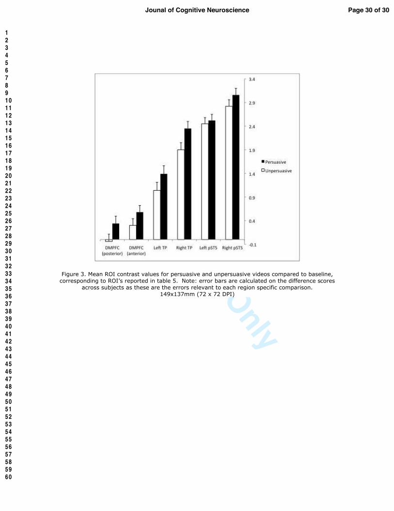
our ROI analysis revealed that all regions of the social cognition network were associated with
persuasion, with the exception of the ROI in left pSTS (Table 5; Figure 3). Results from our whole-
brain search demonstrated that as in the Studies One and Two, finding arguments persuasive was
associated with increased activity in DMPFC, bilateral pSTS, bilateral TP, and left VLPFC (Figure
1; Table 2a). Aside from these regions, the only other region that was significantly activated in
response to persuasive compared to unpersuasive videos was ventromedial prefrontal cortex
(VMPFC), a region that has typically been associated with affective processing and implicit
evaluation (Knutson, Wood, Spampinato, & Grafman, 2006; Koenigs & Tranel, 2008; Mcclure, Li,
Tomlin, Cypert, Montague, & Montague, 2004).
Discussion
Taken together, these results suggest that across linguistically and culturally diverse groups,
as well as across different media, a distinct set of neural regions typically invoked by mentalizing
tasks are associated with the experience of persuasion. Moreover, using an ROI approach, nearly
all mentalizing regions that were sensitive to the experience of persuasion in a text-based message
task were also sensitive to the experience of persuasion in a video-based message task.
Page 13 of 30 Jounal of Cognitive Neuroscience
123456789101112131415161718192021222324252627282930313233343536373839404142434445464748495051525354555657585960
For Review Only
NEURAL BASES OF PERSUASION 14
In sum, across all three studies, increased activity in DMPFC, pSTS, TP, and left VLPFC
while viewing persuasive messages was associated with feeling persuaded afterwards. Consistent
with work documenting the neural underpinnings of expert effects (Klucharev, Smidts, &
Fernandez, 2008), persuasion was associated with increased activity in the medial temporal lobes
and visual cortex in the first two studies, in which participants viewed text based messages and
made ratings following the scanner session, but not in the third study when participants viewed
video based messages and made ratings directly following each message. Persuasion was also
associated with increased activity in the VMPFC in the third study.
The DMPFC, pSTS, and TP have well-documented roles in social cognitive and mentalizing
tasks (Frith & Frith, 2003). The present work extends the role of this network to include the
experience of persuasion. The notion that persuasion relies on a social cognition network is
consistent with Emerson’s proposal that the goal of persuasion “is to bring another out of his bad
sense into your good sense” (Emerson, 1880). To the extent that coordinated activity in this
mentalizing network reflects consideration of another person’s mental state and perspective, our
results suggest that Emerson may have been pretty close to the mark. Our results are also in line
with prior behavioral research that has suggested a relationship between social cognition and
persuasion (Campbell & Babrow, 2004). However, most behavioral studies of persuasion have not
focused directly on perspective taking as a mechanism of persuasion, and thus these results suggest
an important new direction for persuasion research.
The overlap between the brain regions associated with persuasion effects and mentalizing in
Study Three is potentially revealing about how persuasion operates. In Studies One and Two, there
was a single voice conveying all of the arguments; however, in Study Three, there was no obvious
person serving as the message source in the video advertisements. Thus, in Study Three, there was
no individual to mentalize about or whose perspective to take. One intriguing prospect is that
Page 14 of 30Jounal of Cognitive Neuroscience
123456789101112131415161718192021222324252627282930313233343536373839404142434445464748495051525354555657585960

For Review Only
NEURAL BASES OF PERSUASION 15
mentalizing about a particular person’s beliefs, desires, and intentions is just a special case of
thinking about beliefs, desires, and intentions more generally, regardless of whether they are tied to
a particular individual’s mind or presented as part of a more general argument. In other words,
these regions may be involved in considering a point-of-view with or without a particular source.
Humans are surrounded by signs and other artifacts that suggest particular beliefs (e.g. smoking is
bad) without these signs referring back to a particular person who is promoting this belief.
Although we typically associate perspectives and points-of-view with individuals, content often has
a perspective long after its association with the content creator is lost.
Left VLPFC was the only other region that was more active in response to persuasive
compared to unpersuasive messages in all three studies. Given that mid-VLPFC (pars triangularis)
was the specific region of VLPFC activated in each study, it is plausible that this region plays a role
in selecting among competing beliefs and memory representations regarding the persuasion topic.
This sub-region of VLPFC has been regularly observed in studies of memory selection (selecting
among multiple activated memory representations) and emotional reappraisal (in which a new
interpretation for an event is selected over a prior interpretation) (Badre & Wagner, 2007; Ochsner
& Gross, 2005). As persuasion involves adopting a new interpretation over an existing one, VLPFC
may play a role in this selection process. Still, it is not yet clear what role VLPFC is playing in
persuasion, from the current findings alone.
Our results also speak to the modulation of neural responses by message medium. Although
the majority of regions observed in any one study were replicated across all three, and five out of
six regions in the main mentalizing network of interest were significantly active when using ROIs
from Study One to predict activity in Study Three, there were some differences between the
responses to persuasive text-based versus video-based arguments. For example, medial temporal
lobe was observed in response to persuasive compared to unpersuasive text based messages, while
Page 15 of 30 Jounal of Cognitive Neuroscience
123456789101112131415161718192021222324252627282930313233343536373839404142434445464748495051525354555657585960

For Review Only
NEURAL BASES OF PERSUASION 16
VMPFC was observed in response to persuasive compared to unpersuasive commercials. It is
possible that this difference is related to the informational versus emotional content of the material.
VMPFC has been associated with emotional processing and medial temporal lobe has been
associated with cognitive processing. Thus, each region may have been sensitive to types of
appeals that were differentially emphasized through the two media. Manipulation checks
concerning the behavioral data support this distinction; the text-based messages in Studies One and
Two were rated as more information-based than the commercials in Study Three, whereas the
commercials were rated as more feelings-based than the text appeals.
The differential activations in medial temporal lobe and VMPFC may also reflect the temporal
distance between the persuasive messaging and self-reports of persuasion. In the first two studies,
persuasion was reported after leaving the scanner and thus encoded associations about the
persuasive messages, supported by medial temporal lobe, may have played a role in discriminating
which messages would subsequently be remembered as persuasive. In contrast, in the third study,
self-reports of persuasion were obtained after each message rendering memory processes less
relevant and immediate affective responses more relevant. VMPFC has been observed in multiple
studies of automatic affect (Kawasaki, Kaufman, Damasio, Damasio, Granner, Bakken, Hori,
Howard, & Adolphs, 2001; Knutson, et al., 2006) and non-reflective evaluations (Koenigs &
Tranel, 2008). Indeed, the VMPFC and medial temporal lobe trade-off is reminiscent of similar
results from studies of evaluation in the “Pepsi Challenge” (Koenigs & Tranel, 2008; Mcclure, et
al., 2004). In one fMRI study (Mcclure, et al., 2004), soda preferences based solely on immediate
experience of taste were associated with VMPFC activity, whereas soda preferences after seeing
brand names, which would presumably activate previously encoded associations, were linked to
medial temporal lobe activity.
Despite these differences, the results were remarkably consistent across American (Study
Page 16 of 30Jounal of Cognitive Neuroscience
123456789101112131415161718192021222324252627282930313233343536373839404142434445464748495051525354555657585960

For Review Only
NEURAL BASES OF PERSUASION 17
One) and Korean (Study Two) subjects when the same medium was used. When analyzed
separately, each group activated the same set of regions as the other. This provides initial support
for the generalizability of the results in the context of this type of communication. Nevertheless,
when pitted against one another, some differences did emerge cross-culturally. Specifically,
Americans appeared to engage brain regions involved in socioemotional processing to a greater
degree than did Koreans when reading persuasive, relative to unpersuasive, messages (Table 3;
Figure 2). Interestingly, while Korean participants explicitly rated the arguments as more emotional
than did the American participants, American participants showed comparatively more activity in
regions associated with affective processing (amygdala, ventral striatum). Given that there has been
relatively little research on cross-cultural differences in persuasion and the fact that cultural
neuroscience (Chiao & Ambady, 2007; Han & Northoff, 2008) is a relatively new field, the
implication of these differences is unclear. Future work that specifically targets known cultural
differences should help to make sense of the activation differences observed. For example, it will
be of interest to explore whether the neural response to differently framed messages (e.g.
individually framed versus collectively framed messages; gain/approach framed versus
loss/avoidance framed messages) elicit differing neural responses, in parallel with behavioral
studies suggesting differences along these dimensions (Aaker & Williams, 1998; Khaled, et al.,
2008; Uskul, et al., 2009). This will also complement interdisciplinary applications of cultural
psychology to fields such as public health and health communication (Kreuter & Mcclure, 2004).
In summary, these studies identify for the first time the neurocognitive processes occurring at
the moment that persuasion occurs. Neural activations associated with feeling persuaded were
almost exclusively, and repeatedly, associated with a neural network involved in mentalizing and
perspective taking. Furthermore, the specific regions identified within this network that were active
in response to persuasion following text-based messages also generalized to a task in which
Page 17 of 30 Jounal of Cognitive Neuroscience
123456789101112131415161718192021222324252627282930313233343536373839404142434445464748495051525354555657585960

For Review Only
NEURAL BASES OF PERSUASION 18
participants were persuaded by video-based commercials. Building on the baseline provided here,
future work can use neuroimaging to further advance our understanding of how people are
persuaded and by what means.
Acknowledgements: Funding for this work was made possible by a National Science Foundation
Graduate Research Fellowship (E.F). The authors also wish to acknowledge feedback and
assistance from Scott Gerwehr, Shelley Taylor, Chris Frith, Traci Mann, Brett Hemenway, Shalin
Pei, Mihn-Chau Do and Chu Kim. For generous support the authors also wish to thank the Brain
Mapping Medical Research Organization, Brain Mapping Support Foundation, Pierson-Lovelace
Foundation, The Ahmanson Foundation, William M. and Linda R. Dietel Philanthropic Fund at the
Northern Piedmont Community Foundation, Tamkin Foundation, Jennifer Jones-Simon Foundation,
Capital Group Companies Charitable Foundation, Robson Family and Northstar Fund. This work is
dedicated to the memory of Scott Gerwehr.
Page 18 of 30Jounal of Cognitive Neuroscience
123456789101112131415161718192021222324252627282930313233343536373839404142434445464748495051525354555657585960

For Review Only
NEURAL BASES OF PERSUASION 19
References Aaker, J. L., & Williams, P. (1998). Empathy versus pride: The influence of emotional appeals
across cultures. Journal of Consumer Research, 25(3), 241-261. Albarracin, D., Johnson, B. T., & Zanna, M. P. (Eds.). (2005). The Handbook of Attitudes. Mahwah,
NJ: Lawrence Erlbaum. Aristotle (1926). The art of rhetoric / Aristotle ; with an English translation by J.H. Freese.
London: Loeb Classical Library/Harvard University Press. Badre, D., & Wagner, A. D. (2007). Left ventrolateral prefrontal cortex and the cognitive control of
memory. Neuropsychologia, 45(13), 2883-2901. Brett, M., Anton, J., Valabregue, R., & Poline, J. (2002). Region of interest analysis using an SPM
toolbox. Paper presented at the The 8th International Conference on Functional Mapping of the Human Brain.
Cabeza, R., & Nyberg, L. (2000). Imaging cognition II: An empirical review of 275 PET and fMRI studies. J Cogn Neurosci, 12(1), 1-47.
Campbell, R. G., & Babrow, A. S. (2004). The role of empathy in responses to persuasive risk communication: overcoming resistance to HIV prevention messages. Health Commun, 16(2), 159-182.
Chaiken, S., Liberman, A., & Eagly, A. H. (1989). Heuristic and systematic information processing within and beyond the persuasion context. In J. A. B. J. S. Uleman (Ed.), Unintended thought (pp. 212-252). New York, NY: Guilford Press.
Chiao, J., & Ambady, N. (2007). Cultural neuroscience: Parsing universality and diversity across levels of analysis. In S. Kitayama & D. Cohen (Eds.), Handbook of Cultural Psychology (pp. 237-254). NY: Guilford Press.
Crano, W., & Prislin, R. (2008). Attitudes and Attitude Change. New York: Psychology Press. Eagly, A. H., & Chaiken, S. (1993). The psychology of attitudes. Orlando, FL: Harcourt Brace
Jovanovich College Publishers. Emerson, R. W. (1880). Letters and Social Aims (Vol. IV). Cambridge MA: Riverside Press. Frith, U., & Frith, C. D. (2003). Development and neurophysiology of mentalizing. Philos Trans R
Soc Lond B Biol Sci, 358(1431), 459-473. Han, S., & Northoff, G. (2008). Culture-sensitive neural substrates of human cognition: a
transcultural neuroimaging approach. Nat Rev Neurosci, 9(8), 646-654. Johnson, B. T., Maio, G. R., & Smith-McLallen, A. (Eds.). (2005). Communication and Attitude
Change: Causes, Processes, and Effects. Mahwah, NJ Lawrence Erlbaum Associates Publishers.
Kawasaki, H., Kaufman, O., Damasio, H., Damasio, A. R., Granner, M., Bakken, H., et al. (2001). Single-neuron responses to emotional visual stimuli recorded in human ventral prefrontal cortex. Nat Neurosci, 4(1), 15-16.
Khaled, R., Ronald, F., Noble, J., & Biddle, R. (2008). A Qualitative Study of Culture and Persuasion in a Smoking Cessation Game. Paper presented at the Persuasive Technology: Third International Conference, Oulu, Finland.
Klucharev, V., Smidts, A., & Fernandez, G. (2008). Brain mechanisms of persuasion: how 'expert power' modulates memory and attitudes. Soc Cogn Affect Neurosci, nsn022.
Knutson, K., Wood, J., Spampinato, M., & Grafman, J. (2006). Politics on the brain: An fMRI investigation. PSNS, 1(1), 25-40.
Koenigs, M., & Tranel, D. (2008). Prefrontal cortex damage abolishes brand-cued changes in cola preference. Soc Cogn Affect Neurosci, 3(1), 1-6.
Page 19 of 30 Jounal of Cognitive Neuroscience
123456789101112131415161718192021222324252627282930313233343536373839404142434445464748495051525354555657585960

For Review Only
NEURAL BASES OF PERSUASION 20
Kreuter, M., & Mcclure, S. (2004). The role of culture in health communication. Annual Review of Public Health, 25, 439-455.
Lieberman, M. D. (2007). Social cognitive neuroscience: A review of core processes. Annu. Rev. Psychol., 58, 259-289.
Markus, H. R., & Kitayama, S. (1991). Culture and the self: Implications for cognition, emotion, and motivation. Psychol Rev, 98(2), 224-253.
Mcclure, S., Li, J., Tomlin, D., Cypert, K., Montague, L., & Montague, P. (2004). Neural Correlates of Behavioral Preference for Culturally Familiar Drinks. Neuron, 44(2), 379-387.
Meyers-Levy, J., & Peracchio, L. A. (1995). Moderators of the impact of self-reference on persuasion. Journal of Consumer Research, 22(4), 408-423.
Nisbett, R., & Wilson, T. (1977). Telling more than we can know: Verbal reports on mental processes. Psychol Rev, 84(3), 231-259.
Nisbett, R. E. (2003). The geography of thought: How Asians and Westerners think differently ... and why. New York, NY, US: Free Press, 263.
Ochsner, K. N., & Gross, J. J. (2005). The cognitive control of emotion. Trends Cogn Sci, 9(5), 242-249.
Petty, R. E., & Cacioppo, J. T. (1986). Communication and persuasion: Central and peripheral routes to attitude change. NewYork: Springer-Verlag.
Singelis, T. (1994). The Measurement of Independent and Interdependent Self-Construals. Personality and Social Psychology Bulletin, 20(5), 580-591.
Singelis, T., Triandis, H., Bhawuk, D., & Gelfand, M. (1995). Horizontal and Vertical Dimensions of Individualism and Collectivism: A Theoretical and Measurement Refinement. Cross-Cultural Research, 29(3), 240-275.
Stayman, D. M., & Batra, R. (1991). Encoding and retrieval of ad affect in memory. Journal of Marketing Research, 28(2), 232-239.
Triandis, H. (1995). Individualism and Collectivism. Boulder, CO: Westview. Tzourio-Mazoyer, N., Landeau, B., Papathanassiou, D., Crivello, F., Etard, O., Delcroix, N., et al.
(2002). Automated anatomical labeling of activations in SPM using a macroscopic anatomical parcellation of the MNI MRI single-subject brain. Neuroimage, 15(1), 273-289.
Uskul, A., Sherman, D., & Fitzgibbon, J. (2009). The cultural congruency effect: Culture, regulatory focus, and the effectiveness of gain- vs. loss-framed health messages Journal of Experimental Social Psychology, 45(3), 535-541.
Wagner, A. D., Schacter, D. L., Rotte, M., Koutstaal, W., Maril, A., Dale, A. M., et al. (1998). Building memories: remembering and forgetting of verbal experiences as predicted by brain activity. Science, 281(5380), 1188-1191.
Wilson, T., & Schooler, J. (1991). Thinking Too Much: Introspection can reduce the quality of preferences and decisions. Journal of Personality and Social Psychology, 60(2), 181-192.
Zajonc, R. B., & Markus, H. (1982). Affective and cognitive factors in preferences. Journal of Consumer Research, 9(2), 123-131.
Page 20 of 30Jounal of Cognitive Neuroscience
123456789101112131415161718192021222324252627282930313233343536373839404142434445464748495051525354555657585960

For Review Only
NEURAL BASES OF PERSUASION 21 Table 1a. Behavioral responses, Text-based messages.
Table 1b.
Behavioral responses, Video-based messages.
Page 21 of 30 Jounal of Cognitive Neuroscience
123456789101112131415161718192021222324252627282930313233343536373839404142434445464748495051525354555657585960

For Review Only
NEURAL BASES OF PERSUASION 22
Table 2a.
Brain regions showing increased activity for persuasive relative to non-persuasive passages (thresholded at
p<.001, uncorrected, 5 voxel extent).
Page 22 of 30Jounal of Cognitive Neuroscience
123456789101112131415161718192021222324252627282930313233343536373839404142434445464748495051525354555657585960

For Review Only
NEURAL BASES OF PERSUASION 23 Table 2b.
Brain regions showing increased activity for non-persuasive relative to persuasive passages (thresholded at
p<.001, uncorrected, 5 voxel extent).
Page 23 of 30 Jounal of Cognitive Neuroscience
123456789101112131415161718192021222324252627282930313233343536373839404142434445464748495051525354555657585960

For Review Only
NEURAL BASES OF PERSUASION 24
Note: DMPFC = dorsomedial prefrontal cortex; pSTS = posterior superior temporal sulcus; TP = temporal pole;
VLPFC = Ventrolateral prefrontal Cortex; HCMP = hippocampus; VMPFC = Ventromedial Prefrontal Cortex;
OFC = Orbitofrontal Cortex; Amer = American participants; Kor = Korean participants; vox = number of voxels
in cluster.
Page 24 of 30Jounal of Cognitive Neuroscience
123456789101112131415161718192021222324252627282930313233343536373839404142434445464748495051525354555657585960

For Review Only
NEURAL BASES OF PERSUASION 25 Table 3.
Regional differences between the American sample and the Korean sample for persuasive
relative to unpersuasive arguments. It should be noted that these are relative activations
across groups and thus may reflect the difference between two within group deactivations
(thresholded at p<.001, uncorrected, 5 voxel extent).
Note: pSTS = posterior superior temporal sulcus; SubgenACC = subgenual anterior cingulate
cortex.
Page 25 of 30 Jounal of Cognitive Neuroscience
123456789101112131415161718192021222324252627282930313233343536373839404142434445464748495051525354555657585960

For Review Only
NEURAL BASES OF PERSUASION 26 Table 4.
Results of conjunction analysis of activations in studies one and two, run at p<.005,
uncorrected for each analysis.
Table 5.
Results of ROI Analyses in Study Three. ROIs were developed using functional activations in
Study One that fell within the anatomically defined posterior superior temporal sulcus,
temporal pole, and dorsomedial prefrontal cortex. T-statistics were computed by averaging
over all voxels in the ROI using Marsbar.
Page 26 of 30Jounal of Cognitive Neuroscience
123456789101112131415161718192021222324252627282930313233343536373839404142434445464748495051525354555657585960

For Review Only
NEURAL BASES OF PERSUASION 27 Figure Captions
Figure 1. Neural regions that were more active during persuasive than non-persuasive
passages in Study One (Americans, text based messages), Study Two (Koreans, text based
messages), and Study Three (Americans, video based messages). For display purposes, all
activity in this figure uses a threshold of p = .005, uncorrected. Note: Korean activations
were statistically equivalent in many of the displayed regions but appear weaker because the
color scales are different (see scales on left). Also, only a small portion of the actual VLPFC
cluster appears in axial slice selected for the Korean sample. As shown in the Table 2a, the
spatial extent of these activations is comparable. DMPFC=dorsomedial prefrontal cortex;
pSTS = posterior superior temporal sulcus; TP = temporal pole; HCMP = hippocampus;
VLPFC = ventrolateral prefrontal cortex.
Figure 2. Neural regions that were more active in American participants than Korean
participants for persuasive, compared to unpersuasive arguments. For display purposes, all
activity in this figure uses a threshold of p = .005, uncorrected. pSTS = posterior superior
temporal sulcus; Post. Cingulate = Posterior Cingulate.
Figure 3. Mean ROI contrast values for persuasive and unpersuasive videos compared to
baseline, corresponding to ROI’s reported in table 5. Note: error bars are calculated on the
difference scores across subjects as these are the errors relevant to each region specific
comparison.
Page 27 of 30 Jounal of Cognitive Neuroscience
123456789101112131415161718192021222324252627282930313233343536373839404142434445464748495051525354555657585960

For Review Only
Page 28 of 30Jounal of Cognitive Neuroscience
123456789101112131415161718192021222324252627282930313233343536373839404142434445464748495051525354555657585960

For Review Only
Page 29 of 30 Jounal of Cognitive Neuroscience
123456789101112131415161718192021222324252627282930313233343536373839404142434445464748495051525354555657585960
For Review Only
Page 30 of 30Jounal of Cognitive Neuroscience
123456789101112131415161718192021222324252627282930313233343536373839404142434445464748495051525354555657585960