Permeability and Metabolic Properties of a Trophoblast ... · Permeability and Metabolic Properties...
9
EXPERIMENTAL CELL RESEARCH 234, 147–155 (1997) ARTICLE NO. EX973603 Permeability and Metabolic Properties of a Trophoblast Cell Line (HRP-1) Derived from Normal Rat Placenta Fenglin Shi, Michael J. Soares,* Michael Avery, Fei Liu, Xiaoman Zhang, and Kenneth L. Audus 1 Department of Pharmaceutical Chemistry, The University of Kansas, Lawrence, Kansas 66047; and *Department of Physiology, The University of Kansas Medical Center, Kansas City, Kansas 66160 hemomonochorial [2]. The three trophoblast layers of The HRP-1 cell line is derived from normal rat pla- the labyrinth zone of the rat chorioallantoic placenta centa and appears morphologically similar to and re- appear to be the rate-limiting permeability barrier to tains characteristic expression of cellular markers of the exchanges of substances between maternal and fe- labyrinthine trophoblast cells. In this study, mono- tal compartments [3]. The labyrinth zone, in order from layers of HRP-1 cells grown on permeable supports the maternal blood sinus inward to the fetal capillary, were evaluated as a potential in vitro system to study consists of an initial trophoblast layer (layer I) com- trophoblast transport and metabolism. The cell line posed of mononucleated cells in direct contact with the was shown to express and retain functional activity of maternal blood. The second and third trophoblast lay- the predominant placental cytochrome P450 isozyme, ers (layers II and III, respectively) are both syncytial. CYP1A1. Additionally, the HRP-1 cells retain func- Only layer I and the fetal capillary endothelium are tional activity of angiotensin I converting enzyme and permeable to lanthanum and horseradish peroxidase carboxypeptidase N-like enzyme, peptidases charac- [4 – 6]. Therefore, within the rat placenta, layers II and teristic of the trophoblast. The permeation of several III are considered to be the rate-limiting permeability hydrophilic, inert markers across the HRP-1 mono- barriers [3]. layers was observed to be dependent on effective mo- A single syncytiotrophoblast forms the rate-limiting lecular size and to be passive in nature. Functional permeability barrier in the human placenta. The en- asymmetry of the HRP-1 cells was illustrated by the zyme alkaline phosphatase is predominantly localized predominant permeation of linoleic acid in the apical- to the maternal-facing syncytial plasma membrane of to-basolateral direction across the monolayers. Trans- this rate-limiting permeability barrier. This is notewor- ferrin passage across HRP-1 monolayers was concen- thy in that, in the rat placenta, alkaline phosphatase tration-dependent, was bidirectional, and could be in- hibited by unlabeled transferrin, features typical of is also localized to the maternal-facing plasma mem- the trophoblast transport system for transferrin. Col- brane of layer II trophoblasts [7, 8]. Similarly, in the lectively, these properties suggest that the HRP-1 cell mouse, alkaline phosphatase is present in the tropho- line may provide a useful tool for evaluating some of blast layer constituting the rate-limiting permeability the permeability and metabolic properties of the tro- barrier [9]. These observations indicate that alkaline phoblast. q 1997 Academic Press phosphatase seems to be co-localized with the tropho- blast layer forming the rate-limiting permeability bar- rier of the placenta of rat, human, and mouse [3, 7–9] INTRODUCTION and could be a marker for trophoblasts responsible for determining some of the permeability properties of the The placenta serves as a mediator between the ma- placenta. ternal and fetal circulations, providing for nutrient and Cell lines such as HRP-1 have been derived from the waste transport, gas exchange, and hormone produc- labyrinth zone of the normal rat placenta. The HRP- tion [1]. The mammalian chorioallantoic placenta dif- 1 cells appear morphologically similar to labyrinthine fers among species and is classified based on the tissue trophoblast cells [10, 11] and retain expression of alka- layers intervening between the maternal and the fetal line phosphatase [12, 13], cytokeratins [13], and trans- circulations. The laboratory rat placenta has three tro- ferrin receptors [13, 14], additional markers for laby- phoblast layers and is classified as hemotrichorial. The rinth zone trophoblasts. The HRP-1 cell line is a tro- human placenta, having only one layer, is classified as phoendodermal stem cell population that arises after midgestation with the capability of differentiating into cells resembling trophoblast and yolk sac phenotypes. 1 To whom reprint requests should be addressed. Fax: (913) 864- 5736. E-mail: [email protected]. Harvested from the midgestation chorioallantoic pla- 147 0014-4827/97 $25.00 Copyright q 1997 by Academic Press All rights of reproduction in any form reserved.
Transcript of Permeability and Metabolic Properties of a Trophoblast ... · Permeability and Metabolic Properties...

EXPERIMENTAL CELL RESEARCH 234, 147–155 (1997)ARTICLE NO. EX973603
Permeability and Metabolic Properties of a Trophoblast Cell Line(HRP-1) Derived from Normal Rat Placenta
Fenglin Shi, Michael J. Soares,* Michael Avery, Fei Liu, Xiaoman Zhang, and Kenneth L. Audus1
Department of Pharmaceutical Chemistry, The University of Kansas, Lawrence, Kansas 66047; and*Department of Physiology, The University of Kansas Medical Center, Kansas City, Kansas 66160
hemomonochorial [2]. The three trophoblast layers ofThe HRP-1 cell line is derived from normal rat pla- the labyrinth zone of the rat chorioallantoic placenta
centa and appears morphologically similar to and re- appear to be the rate-limiting permeability barrier totains characteristic expression of cellular markers of the exchanges of substances between maternal and fe-labyrinthine trophoblast cells. In this study, mono- tal compartments [3]. The labyrinth zone, in order fromlayers of HRP-1 cells grown on permeable supports the maternal blood sinus inward to the fetal capillary,were evaluated as a potential in vitro system to study consists of an initial trophoblast layer (layer I) com-trophoblast transport and metabolism. The cell line posed of mononucleated cells in direct contact with thewas shown to express and retain functional activity of maternal blood. The second and third trophoblast lay-the predominant placental cytochrome P450 isozyme, ers (layers II and III, respectively) are both syncytial.CYP1A1. Additionally, the HRP-1 cells retain func- Only layer I and the fetal capillary endothelium aretional activity of angiotensin I converting enzyme and permeable to lanthanum and horseradish peroxidasecarboxypeptidase N-like enzyme, peptidases charac- [4–6]. Therefore, within the rat placenta, layers II andteristic of the trophoblast. The permeation of several
III are considered to be the rate-limiting permeabilityhydrophilic, inert markers across the HRP-1 mono-barriers [3].layers was observed to be dependent on effective mo-
A single syncytiotrophoblast forms the rate-limitinglecular size and to be passive in nature. Functionalpermeability barrier in the human placenta. The en-asymmetry of the HRP-1 cells was illustrated by thezyme alkaline phosphatase is predominantly localizedpredominant permeation of linoleic acid in the apical-to the maternal-facing syncytial plasma membrane ofto-basolateral direction across the monolayers. Trans-this rate-limiting permeability barrier. This is notewor-ferrin passage across HRP-1 monolayers was concen-thy in that, in the rat placenta, alkaline phosphatasetration-dependent, was bidirectional, and could be in-
hibited by unlabeled transferrin, features typical of is also localized to the maternal-facing plasma mem-the trophoblast transport system for transferrin. Col- brane of layer II trophoblasts [7, 8]. Similarly, in thelectively, these properties suggest that the HRP-1 cell mouse, alkaline phosphatase is present in the tropho-line may provide a useful tool for evaluating some of blast layer constituting the rate-limiting permeabilitythe permeability and metabolic properties of the tro- barrier [9]. These observations indicate that alkalinephoblast. q 1997 Academic Press phosphatase seems to be co-localized with the tropho-
blast layer forming the rate-limiting permeability bar-rier of the placenta of rat, human, and mouse [3, 7–9]
INTRODUCTION and could be a marker for trophoblasts responsible fordetermining some of the permeability properties of the
The placenta serves as a mediator between the ma- placenta.ternal and fetal circulations, providing for nutrient and Cell lines such as HRP-1 have been derived from thewaste transport, gas exchange, and hormone produc- labyrinth zone of the normal rat placenta. The HRP-tion [1]. The mammalian chorioallantoic placenta dif- 1 cells appear morphologically similar to labyrinthinefers among species and is classified based on the tissue trophoblast cells [10, 11] and retain expression of alka-layers intervening between the maternal and the fetal line phosphatase [12, 13], cytokeratins [13], and trans-circulations. The laboratory rat placenta has three tro- ferrin receptors [13, 14], additional markers for laby-phoblast layers and is classified as hemotrichorial. The rinth zone trophoblasts. The HRP-1 cell line is a tro-human placenta, having only one layer, is classified as phoendodermal stem cell population that arises after
midgestation with the capability of differentiating intocells resembling trophoblast and yolk sac phenotypes.1 To whom reprint requests should be addressed. Fax: (913) 864-
5736. E-mail: [email protected]. Harvested from the midgestation chorioallantoic pla-
147 0014-4827/97 $25.00Copyright q 1997 by Academic Press
All rights of reproduction in any form reserved.
AID ECR 3603 / 6i23$$$261 06-09-97 13:48:57 eca

148 SHI ET AL.
5 min. Molecular weight markers (4 to 250 kDa) and purified bovinecenta, HRP-1 cells are readily adaptable to propagationserum albumin (0.5 mg) served as controls. Electrophoresis of micro-both in vitro and in vivo [12, 14]. The purpose of thissomes with SDS–PAGE was monitored at 95 V for 2.5 h. Followingstudy was to evaluate the suitability of subcultures of electrophoresis, protein was either transferred electrophoretically to
HRP-1 cells for investigation of trophoblast transport PVDF or polyacrylamide gels and then exposed to Coomassie bluestain (0.1% Coomassie blue, 45% methanol, 2% acetic acid) for 1 h.and metabolism in vitro. Unlike most trophoblast cul-Destaining polyacrylamide not used in electrophoretic transfer oftures, the HRP-1 cells grow to form confluent mono-microsomal protein required destaining overnight to visualize thelayers which can facilitate trans-trophoblast transportprotein bands. No proteins greater than 69 kDa were detected.
experiments. Furthermore, the appearance of alkaline Following SDS–PAGE, microsomal proteins were transferred elec-phosphatase in the HRP-1 cells, an enzyme coincident trophoretically to PVDF membrane (45 V, 2 h). After electrophoresis
transfer, PVDF membranes were immersed in Ponceau S stainingin trophoblasts that provide the rate-limiting perme-solution for 5 min to verify the presence of microsomal protein. Theability properties of the placenta in vivo, prompted con-Ponceau S stain was removed in an aqueous solution containing 10%sideration of the cell line’s potential utility in studying acetic acid (v/v) for approximately 4–5 min. To block nonspecific
trophoblast transport and metabolism. binding with the monoclonal antibody, membranes were incubatedfor 2 h with PBS, pH 7.4, containing 10% dry milk (w/v), 2% bovineserum albumin (w/v), and 0.1% Tween 20. The membranes wereMATERIALS AND METHODSthen incubated with monoclonal antibody CYP1A1 (10 mg/ml) or apreimmune mouse antibody (nonspecific IgG, 10 mg/ml) in 10 mMTransferrin (human) fluorescein conjugate, 7-ethoxyresorufin, andTris–HCl, pH 7.4, containing 0.1% Tween 20. All microsomal proteinresorufin were purchased from Molecular Probes (Junction City, OR).bound to PVDF membranes was incubated overnight at room temper-Tissue culture medium, antibiotics, b-glucuronidase/arysulfatase,ature with the monoclonal antibody or the nonspecific IgG. A second-DMSO, fluorescein, fluorescein isothiocyanate-conjugated dextrans,ary antibody, IgG peroxidase conjugate specific for the CYP1A1anti-mouse IgG (Fab) peroxidase conjugate, and angiotensin I con-monoclonal antibody, was diluted 1:80,000 in 10 mM Tris–HCl con-verting enzyme assay kit were purchased from Sigma Chemical Co.taining 0.05% Tween 20 and incubated with the PVDF membrane(St. Louis, MO). Monoclonal antibody, CYP1A1, and induced liverfor 1 h at 257C. CYP1A1 was detected by enhanced chemilumines-samples were purchased from XenoTech (Kansas City, KS). Hyper-cence by incubating PVDF immunoblots at 257C for 45 s and thenfilm-ECL was purchased from Amersham (Arlington Heights, IL).exposing them to Hyperfilm-ECl.ScintiVerse BD was purchased from Fisher Scientific (Fair Lawn,
Activity assays for cytochrome P450. Microsomal cytochromeNJ). b-Mercaptoethanol was purchased from Bio-Rad LaboratoriesP450 1A (CYP 1A1) activity was measured fluorometrically by the(Hercules, CA). Heat-inactivated fetal bovine serum was purchasedO-dealkylation of 7-ethoxyresorufin producing resorufin according tofrom JRH Biosciences (Lenexa, KS). [14C]Sucrose and [14C]urea wereDutton and Parkinson [16] with minor modifications according topurchased from ICN Biomedicals, Inc. (Irvine, CA). [3H]MannitolShiverick et al. [17]. Reaction mixtures of 1 ml final volume wereand [3H]methotrexate were purchased from DuPont NEN Researchincubated at 377C. The reaction mixture contained potassium phos-Products (Boston, MA). [14C]Linoleic acid was purchased from Amer-phate buffer (100 mM, pH 7.4), MgCl2 (3 mM), EDTA (1 mM), NADPsham Life Sciences Co. (Little, CA). Protein assay kits (BCA Protein(1 mM), glucose 6-phosphate (5 mM), glucose-6-phosphate dehydro-Assay with bovine serum standards) were purchased from Piercegenase (1 unit/ml), and 7-ethoxyresorufin (10 mM). Blanks did notLife Science and Analytical Research Products (Rockford, IL). Allcontain NADP. Reactions were started by the addition of theother reagents were of the highest grade commercially available.NADPH-generating system and stopped after 8 min with 2 ml of ice-Culture of HRP-1 cells. HRP-1 cells [11, 12] were maintained incold acetone. Protein was removed by centrifugation at 1000g for 10culture medium containing RPMI 1640, NaHCO3, pH 7.4, 1 mMmin and the supernatant was transferred to clean quartz cuvettes.sodium pyruvate, 50 mM b-mercaptoethanol, 100 units/ml penicillin,Fluorescence was measured at an emission of 585 nm and an excita-100 mg/ml streptomycin, and 5–10% heat-inactivated fetal bovinetion wavelength of 535 nm in an SLM Aminco 4800 spectrofluoro-serum. The cells were seeded in 75- or 175-cm2 flask and incubatedmeter.at 377C with 5% CO2 [12]. The cells were fed every other day. The
The method of Donato et al. [18] was used to assay CYP1A1 inHRP-1 cells were harvested for passaging after 3 to 4 days by tryp-intact cell cultures. HRP-1 cells were grown in 6-well culture dishessin–EDTA (0.25% trypsin and 0.02% EDTA in Hanks’ balanced saltand 7-ethoxyresorufin and dicumarol were added to the cells. Follow-solution) treatment. Cells from passages 12–16 were used for all ofing addition of b-glucuronidase/arysulfatase in 0.1 M sodium acetate,the studies described herein.the cell supernatants were further incubated for 2 h at 377C. TheMicrosome preparation. HRP-1 cells were grown in 175-cm2
amount of resorufin was measured with excitation of 530 nm andflasks to confluent monolayers. The HRP-1 cells were washed withemission of 590 mm.ice-cold phosphate-buffered saline (PBS), pH 7.4. The cells were then
bathed in a buffer containing 50 mM Tris–HCl, pH 7.4, 150 mM Angiotensin converting enzyme and carboxypeptidase N-like activ-KCl, and 2 mM EDTA. The cells were scraped from the flask and ity. To assay both angiotensin converting enzyme and carboxypepti-centrifuged at 1000g for 10 min. The cell pellet was then resuspended dase N-like enzyme activities, HRP-1 cells were grown to confluentin buffer containing 0.25 M sucrose in 50 mM Tris–HCl, pH 7.4. monolayers in 175-cm2 flasks.Sonication of the suspension for 1–2 min produced a suspension of For angiotensin converting enzyme, homogenates were preparedcytosolic and nuclear proteins. To remove mitochondrial proteins according to Shi and Audus [19]. Briefly, the tissues were homoge-from suspended membrane fragments, cell fractions were centrifuged nized in 0.25 M sucrose and frozen on dry ice and thawed, six times.at 10,000g for 15 min. The supernatant was centrifuged at 100,000g The homogenates were centrifuged at 15,000g for 5 min and thefor 1 h at 107C. The microsomal pellet was resuspended in 0.25 M supernatant was saved at 0707C until assayed. Angiotensin con-sucrose and the protein concentration determined by BCA. All micro- verting enzyme activity in the supernatants was assayed with asomal preparations were stored at 0707C. Sigma Chemical Co. 305-UV kit. Absorbance of the samples was
measured at 340 nm with a Shimadzu UV–visible spectrophotome-Immunoblots. Microsome preparations (0.5 to 10 mg HRP-1 pro-ter. Blanks contained boiled supernatant.tein and 0.05 mg induced rat liver protein) were subjected to SDS–
Carboxypeptidase N-like enzyme activity in the HRP-1 cells wasPAGE, 12% acrylamide Tris–glycine [15]. All microsomal sampleswere heated to 1007C for 10 min and then centrifuged at 10,000g for assayed according to the method of Skidgel and Erdos [20]. Briefly,
AID ECR 3603 / 6i23$$$262 06-09-97 13:48:57 eca
149HRP-1 PERMEABILITY AND METABOLISM
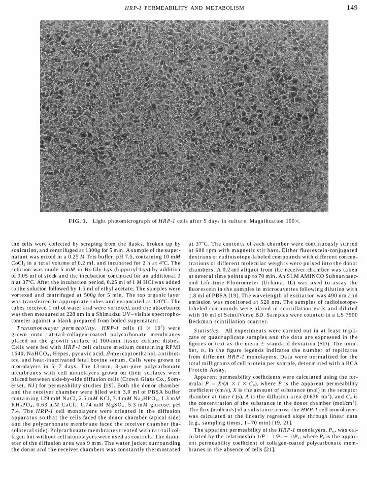
FIG. 1. Light photomicrograph of HRP-1 cells after 5 days in culture. Magnification 1001.
the cells were collected by scraping from the flasks, broken up by at 377C. The contents of each chamber were continuously stirredsonication, and centrifuged at 1300g for 5 min. A sample of the super- at 600 rpm with magnetic stir bars. Either fluorescein-conjugatednatant was mixed in a 0.25 M Tris buffer, pH 7.5, containing 10 mM dextrans or radioisotope-labeled compounds with different concen-CoCl2 in a total volume of 0.2 ml, and incubated for 2 h at 47C. The trations or different molecular weights were pulsed into the donorsolution was made 5 mM in Bz-Gly-Lys (hippuryl-Lys) by addition chambers. A 0.2-ml aliquot from the receiver chamber was takenof 0.05 ml of stock and the incubation continued for an additional 3 at several time points up to 70 min. An SLM AMINCO Subnanosec-h at 377C. After the incubation period, 0.25 ml of 1 M HCl was added ond Life-time Fluorometer (Urbana, IL) was used to assay theto the solution followed by 1.5 ml of ethyl acetate. The samples were fluorescein in the samples in microcuvettes following dilution withvortexed and centrifuged at 500g for 5 min. The top organic layer 1.8 ml of PBSA [19]. The wavelength of excitation was 490 nm andwas transferred to appropriate tubes and evaporated at 1207C. The emission was monitored at 520 nm. The samples of radioisotope-tubes received 1 ml of water and were vortexed, and the absorbance labeled compounds were placed in scintillation vials and dilutedwas then measured at 228 nm in a Shimadzu UV–visible spectropho- with 10 ml of ScintiVerse BD. Samples were counted in a LS 7500tometer against a blank prepared from boiled supernatant. Beckman scintillation counter.
Transmonolayer permeability. HRP-1 cells (1 1 107) were Statistics. All experiments were carried out in at least tripli-grown onto rat-tail-collagen-coated polycarbonate membranes cate or quadruplicate samples and the data are expressed in theplaced on the growth surface of 100-mm tissue culture dishes. figures or text as the mean { standard deviation (SD). The num-Cells were fed with HRP-1 cell culture medium containing RPMI ber, n, in the figure legends indicates the number of replicates1640, NaHCO3, Hepes, pyruvic acid, b-mercaptoethanol, antibiot- from different HRP-1 monolayers. Data were normalized for theics, and heat-inactivated fetal bovine serum. Cells were grown to
total milligrams of cell protein per sample, determined with a BCAmonolayers in 5–7 days. The 13-mm, 3-mm-pore polycarbonateProtein Assay.membranes with cell monolayers grown on their surfaces were
Apparent permeability coefficients were calculated using the for-placed between side-by-side diffusion cells (Crown Glass Co., Som-mula: P Å X/(A 1 t 1 Cd), where P is the apparent permeabilityerset, NJ) for permeability studies [19]. Both the donor chambercoefficient (cm/s), X is the amount of substance (mol) in the receptorand the receiver chamber were filled with 3.0 ml of PBSA bufferchamber at time t (s), A is the diffusion area (0.636 cm2), and Cd iscontaining 129 mM NaCl, 2.5 mM KCl, 7.4 mM Na2HPO4, 1.3 mMthe concentration of the substance in the donor chamber (mol/cm3).KH2PO4, 0.63 mM CaCl2 , 0.74 mM MgSO4, 5.3 mM glucose, pHThe flux (mol/cm/s) of a substance across the HRP-1 cell monolayers7.4. The HRP-1 cell monolayers were oriented in the diffusionwas calculated at the linearly regressed slope through linear dataapparatus so that the cells faced the donor chamber (apical side)(e.g., sampling times, 1–70 min) [19, 21].and the polycarbonate membrane faced the receiver chamber (ba-
The apparent permeability of the HRP-1 monolayers, Pe , was cal-solateral side). Polycarbonate membranes treated with rat-tail col-culated by the relationship 1/P Å 1/Pe / 1/Pc , where Pc is the appar-lagen but without cell monolayers were used as controls. The diam-ent permeability coefficient of collagen-coated polycarbonate mem-eter of the diffusion area was 9 mm. The water jacket surrounding
the donor and the receiver chambers was constantly thermostated branes in the absence of cells [21].
AID ECR 3603 / 6i23$$$262 06-09-97 13:48:57 eca

150 SHI ET AL.
FIG. 2. Light photomicrograph of a cross section of HRP-1 cells grown on polycarbonate membranes. Cells were stained with hematoxylinand eosin and observed at a magnification of 4001.
RESULTS blots using the nonspecific mouse IgG revealed no im-munoreactivity (not shown).
The level of 7-ethoxyresorufin O-deethylase (EROD)HRP-1 cells grow rapidly and form monolayers ofactivity, a functional marker for CYP1A1, was low butflattened polygonal-shaped cells on rat-tail-collagen-consistent in both HRP-1 microsomes and intact HRP-coated surfaces, including polycarbonate membranes1 cells as summarized in Fig. 4. The EROD activityor plastic culture dishes. A typical 5-day-old subculturewas essentially constant over a period of 2 to 12 daysof HRP-1 cell monolayers grown on a plastic culturein microsomes prepared from cultures and in the intactdish is shown in Fig. 1. Cross sections of HRP-1 cellscell cultures. Two additional enzymes were chosen forgrown on polycarbonate membranes were used to verifyassay in the HRP-1 cell line as part of the functionalthe presence of a single layer of cells. Figure 2 shows
a representative example of HRP-1 cells grown on thepolycarbonate support after 5 days in culture.
A Western blot of microsomal protein from the HRP-1, shown in Fig. 3, indicated the expression of smallamounts of the cytochrome P450 isozyme CYP1A1 inthe cells. The cell proteins were run against a positivecontrol, the induced rat liver sample, and a negativecontrol, bovine serum albumin (not shown). The mono-clonal antibody specifically recognizes the CYP1A1 pro-tein (upper band) and also cross-reacts with the in-duced form of the isozyme, CYP1A2 (lower band), inthe HRP-1 and liver samples but not in the negativecontrol, bovine serum albumin. The cross-reactivity
FIG. 3. Western blot of microsomal protein from confluent HRP-was particularly obvious in the induced rat liver sam- 1 cell monolayers and microsomal protein from induced rat liver.ple. Minor immunoreactivity for CYP1A2 also ap- The monoclonal antibody recognizes cytochrome P450 isozyme
CYP1A1 and reacts with CYP1A2.peared in the HRP-1 cells as shown in Fig. 3. Similar
AID ECR 3603 / 6i23$$$263 06-09-97 13:48:57 eca
151HRP-1 PERMEABILITY AND METABOLISM
tems both transcellularly and paracellularly and hada permeability much greater than any of the purelyparacellular markers. Methotrexate, on the otherhand, was example of an agent that undergoes exten-sive binding and uptake by trophoblasts and, conse-quently, had a much lower permeability than might bepredicted by the molecular weight.
The difference between the apical-to-basolateral andthe basolateral-to-apical permeation of linoleic acidacross HRP-1 monolayers was used to assess the func-tional asymmetry of the cells. Linoleic acid was mixedwith bovine serum albumin in a ratio of 6:1 (fattyacid:albumin), and the permeation of the fatty acid wasmonitored in both directions across collagen-coatedpolycarbonate membranes alone and in the presenceof HRP-1 monolayers. Figure 6 summarizes data thatindicated a predominant apical-to-basolateral perme-ation of linoleic acid across HRP-1 monolayers. By con-trast, passage of linoleic acid across collagen-coatedpolycarbonate membranes in the absence of cells wasnot asymmetric and substantially greater. The passage
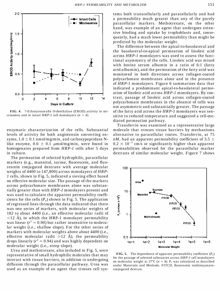
FIG. 4. 7-Ethoxyresorufin O-deethylase (EROD) activity in mi- of the fatty acid across the HRP-1 monolayers was sen-crosomes and in intact HRP-1 cell monolayers (n Å 4). sitive to reduced temperature and suggested a cell-me-
diated permeation pathway.Transferrin was examined as a representative large
molecule that crosses tissue barriers by mechanismsenzymatic characterization of the cells. Substantiallevels of activity for both angiotensin converting en- alternative to paracellular routes. Transferrin, at 75
nM, had an apparent permeability coefficient of 3.5 {zyme, 1.0{ 0.1 nmol/mg/min, and carboxypeptidase N-like enzyme, 0.6 { 0.1 mmol/mg/min, were found in 0.2 1 1005 cm/s or significantly higher than apparent
permeabilities observed for the paracellular markerhomogenates prepared from HRP-1 cells after 5 daysin culture. dextrans of similar molecular weight. Figure 7 shows
The permeation of selected hydrophilic, paracellularmarkers (e.g., mannitol, surose, fluorescein, and fluo-rescein conjugated dextrans with average molecularweights of 4400 to 147,800) across monolayers of HRP-1 cells, shown in Fig. 5, indicated a sieving effect basedon effective molecular size. The passage of the markersacross polycarbonate membranes alone was substan-tially greater than with HRP-1 monolayers present andwas used to calculate the apparent permeability coeffi-cients for the cells (Pe) shown in Fig. 5. The applicationof regressed lines through the data indicated that therewas one series of markers, with molecular weights of182 to about 4400 (i.e., an effective molecular radii ofõ12 A), in which the HRP-1 monolayer permeabilitywas linear (r2 Å 0.90) but rather insensitive to molecu-lar weight (i.e., shallow slope). For the other series ofmarkers with molecular weights above about 4400 (i.e.,effective molecular radii ú12 A), the permeabilitydrops linearly (r2 Å 0.94) and was highly dependent onmolecular weight (i.e., steep slope).
Urea and methotrexate, also included in Fig. 5, wereFIG. 5. The dependence of apparent permeability coefficient (Pe)representative of small hydrophilic molecules that may
for the passage of selected substances across HRP-1 cell monolayersinteract with tissue barriers, in addition to undergoing on molecular weight at 377C (n Å 4). Pe was calculated as describeddiffusion through the paracellular pathway. Urea was under Materials and Methods. FITCD, fluorescein isothiocyanate-
conjugated dextran.used as an example of an agent that crosses cell sys-
AID ECR 3603 / 6i23$$$263 06-09-97 13:48:57 eca

152 SHI ET AL.
FIG. 6. Bidirectional permeation of [14C]linoleic acid across HRP-1 monolayers and collagen-coated polycarbonate membranes (n Å 4).Except for the indicated bar all data were collected from experimentsperformed at 377C. FIG. 8. Apparent permeability coefficients for the bidirectional
passage of 75 nM transferrin across HRP-1 monolayers in the ab-sence of any treatment (Control), or the presence of a 10-fold excessof unlabeled transferrin (/750 nM Transferrin), or at 47C (Low Tem-
that the passage of transferrin across HRP-1 cell mono- perature) (n Å 4).layers was concentration-dependent and was saturableat higher concentrations. The half-maximal concentra-tion of the saturable transport was about 60 nM (esti-
HRP-1 monolayers was inhibited by an excess amountmated with Sigma Plot, version 2.0). In addition, theof unlabeled transferrin and by low temperatures, aspermeation rate of transferrin across the monolayersillustrated in Fig. 8, an additional indication of thewas bidirectional with the trend of apical-to-basolat-involvement of a transcytotic mechanism.eral permeation being only slightly greater than the
permeation rate of the protein in the basolateral-to-DISCUSSIONapical direction. The passage of transferrin across the
HRP-1 is a well-studied normal rat cell line that hasproven useful for identifying factors controlling tropho-blast growth and differentiation. Particularly notableis the rapid and convenient growth of HRP-1 cells andtheir retention of morphological and biochemical fea-tures consistent with normal trophoblasts of the laby-rinth zone [11, 12], including the presence of a typicalnutrient transporter [22]. Perhaps more importantly,the HRP-1 cell line has the ability to form monolayers,a property that permits evaluation of transcellulartransport and access to apical and basolateral surfacesof the monolayer. By contrast, primary cultures of hu-man cytotrophoblasts form a syncytium that does notcompletely cover a growth surface [23] and does notpermit trans-trophoblast transport studies. This nor-mal cell line could also potentially provide a convenientand more easily manipulated corollary to rat studiesin vivo. The purpose of our study was to initiate anevaluation of the applicability of the HRP-1 monolayersfor mechanistic studies of trans-trophoblast transportand metabolism of therapeutic agents and drugs ofFIG. 7. Apparent permeability coefficients for the bidirectional
passage of transferrin across HRP-1 monolayers at 377C (n Å 4). abuse in vitro.
AID ECR 3603 / 6i23$$$263 06-09-97 13:48:57 eca

153HRP-1 PERMEABILITY AND METABOLISM
Several drugs of abuse and other xenobiotics undergo urea, small-molecular-radius (õ10-A) markers (e.g.,mannitol, sucrose, and fluorescein-conjugated dextran,oxidative metabolism in the human placenta. The pla-
centa has at least one constitutive and one induced MW 4000) had a placental clearance in rabbit that wasdependent on and linearly related to the effective mo-form of the cytochrome P450 isozyme, CYP1A1, which
is localized in the trophoblast [24]. Expression and re- lecular size of the marker. Although Faber’s modelwould predict a steeper drop in the placental perme-tention of CYP1A1 activity then will be a required
marker for any cell culture system proposed for study- ability for larger molecules [27], in the HRP-1 cells theobserved decrease in permeability for larger moleculesing placental drug metabolism. The HRP-1 cell line
retains low levels of CYP1A1 relative to the induced was substantial. The considerable decrease in Pe of thelarger molecules may represent restricted diffusion asrat liver and as documented by Western blots and mea-
sured by EROD activity. The low EROD activity in the observed in the rat placenta in vivo for high-molecular-weight markers [30]. Qualitatively, the general profileHRP-1 monolayers was in good agreement with the
literature on rat [17] and human placenta [24, 25], both of permeability versus effective molecular size for ourin vitro system was in very good agreement with rela-of which retain very low consitutive levels of CYP1A1
relative to the liver. As examples, in the nonsmoking tionships developed for the permeation of hydrophilicsubstances through aqueous pores across trophoblastshuman placental microsome, the EROD activity was
reported to be in the range of 0.20 to 0.35 pmol/mg/min constituting the rat placenta [30].The effective molecular size of a substance is but one[25] and in rat labyrinth zone microsomes, õ1 pmol/
mg/min [17]. Our immunoblots also confirmed indica- factor in determining the penetration of a tissue barrierby a given molecule. In this study we examined urea,tions of the induced CYP1A1 form, CYP1A2, in the
HRP-1 cells. In the rat and human, induction of methotrexate, linoleic acid, and transferrin permeationacross the HRP-1 monolayers to further establish theCYP1A2 in the placenta occurs through exposure to
polycyclic hydrocarbons [17, 24]. potential utility of the in vitro system in characterizingsome trans-trophoblast permeation processes. Urea isThe plasma membrane of the syncytiotrophoblast
facing the maternal blood has been found to be rich in believed to cross tissue barriers by both transcellularand paracellular pathways [29]. Relative to mannitolcertain peptidases that may regulate the passage of
peptides across the placental barrier. In particular, mi- for instance, urea has a 10-fold and a 4-fold higherpermeability across the syncytiotrophoblast maternalcrovillar membranes from human syncytiotrophoblasts
were observed to be enriched in angiotensin I con- plasma membrane and basal membrane, respectively[31]. The passage of urea across HRP-1 monolayers wasverting enzyme, angiotensinase A, carboxypeptidase
N-like enzyme, and neutral endopeptidase (‘‘Enkepha- greater than would be predicted solely on the basis ofeffective molecular size and at least three times fasterlinase’’) [26]. On that basis, we elected to determine
whether two of the peptidases, angiotensin I converting than mannitol. Therefore, the enhanced permeabilityof urea in vitro might also be accounted for by bothenzyme and carboxypeptidase N-like enzyme, were re-
tained in the HRP-1 system. The presence of significant paracellular and transcellular permeation across thecells. By contrast, methotrexate has significant bind-activity of both enzymes was observed in the HRP-1
cells. Relative to one of the few available reports on ing, uptake, and accumulation within trophoblasts[32]. The passage of methotrexate across HRP-1 mono-peptidase activity in the placenta [26], our observations
suggest the cell line may be useful in examining tropho- layers was substantially lower than expected based onmolecular weight and might be accounted for in partblast metabolism of some peptides. In ongoing studies,
this in vitro system is being used to examine the meta- by trophoblast binding and uptake which limits theavailability of the drug for diffusion across the mono-bolic fate of both drugs of abuse and opioid peptides in
order to validate the utility of the system in evaluating layers.A critical feature of an in vitro system proposed astrophoblast metabolism.
Passive diffusion is the primary mechanism by which a representative model of a polarized tissue barrier isthat the in vitro system also retains a functional asym-xenobiotics cross the placental barrier [3, 27–30]. An
appropriate in vitro system might be expected to pro- metry. Lafond et al. [33] established that linoleic acidtransport by human trophoblasts was saturable, polar-vide at least a qualitative representation of the sieving
of molecules observed in the placenta. The passive per- ized, and dependent on the presence of extracellularalbumin. Based on the appearance of asymmetricmeation of selected hydrophilic markers across the
HRP-1 monolayers was found to be linearly related to transport of linoleic acid in that study [33], the fattyacid was selected as a marker to test for functionalmolecular size. The Pe was also proportional to the wa-
ter diffusion coefficients for the markers (not shown), asymmetry of the HRP-1 cells. HRP-1 cells were shownto transport linoleic acid in predominantly the apical-consistent with the trans-trophoblast passage of small
hydrophilic solutes in the placenta [27–30]. Faber [27], to-basolateral direction consistent with the observa-tions of Lafond et al. [33]. Our observation affirms that,for example, previously demonstrated that, excluding
AID ECR 3603 / 6i23$$$263 06-09-97 13:48:57 eca

154 SHI ET AL.
at least with respect to linoleic acid, HRP-1 monolayers the process of trophoblast differentiation. Generally,the development of tissue culture systems presents thegrown on permeable supports have functional asymme-
try. In related studies we have also gone on to establish opportunity to evaluate cellular-, biochemical-, and mo-lecular-level mechanisms in the transport and metabo-saturable, albumin-dependent linoleic acid transport
across the HRP-1 cell line that was inhibited by excess lism of therapeutic agents and drugs of abuse at inac-cessible tissue barriers. The advantages of such in vitrounlabeled fatty acid (X. Zhang, F. Liu, M. J. Soares,
and K. L. Audus, unpublished results). systems include the capacity for screening large num-bers of newly synthesized agents, the requirement forTransferrin has been shown to cross human tropho-
blasts by an apparent transcytotic mechanism in a cho- minimal amounts of material, and a reduction in thenumbers of costly and possibly controversial animalriocarcinoma-derived cell line [34, 35] and by endocyto-
sis on both apical and basolateral surfaces of normal studies [42]. Therefore, the availability of appropriatein vitro trophoblast systems that might also be used totrophoblast cultures [36]. In the rat, transferrin uptake
has been shown to occur through the typical transferrin test proposed strategies in which, for example, newlysynthesized therapeutic agents might be effectively in-endocytic process [37]. Since, previous works had dem-
onstrated the presence of functional transferrin recep- activated to prevent crossing of the placental barrier[43] is particularly important in the development andtors on the HRP-1 cells [13, 14], we investigated the
possibility that the HRP-1 cells retain a similar appar- design of agents directed toward maternal-only ther-apy and limiting unnecessary fetal exposure.ent transferrin uptake system. We were able to demon-
strate that transferrin passage across the HRP-1 mono-layer system was greater than that predicted on the This research was supported by NIDA N01DA-4-7405. The authors
also gratefully acknowledge the support of Corning Costar Corp. forbasis of its molecular size. In addition, the passage ofthe Cellular and Molecular Biopharmaceutics Handling Laboratory.transferrin across the monolayers was concentration-
dependent, saturable, bidirectional, and inhibited byREFERENCESunlabeled transferrin. These properties were consis-
tent with the concentration dependence of functional1. Ala-Kokko, T. I., Vahakangas, K., and Pelkonen, O. (1993) Actatransferrin receptors on the HRP-1 cells [13, 14] and
Anaesthesiol. Scand. 37, 47–49.the observations for apparent transferrin transcytosis2. Enders, A. G. (1965) Am. J. Anat. 116, 29–68.in human trophoblast cultures [34–36] and endocytosis3. Sibley, C. P. (1994) Placenta 15, 675–691.in rat trophoblast cultures [37]. Although results here4. Aoki, A., Metz, J., and Forssmann, W. G. (1978) Cell Tissue Res.and work with the BeWo cell system have suggested
192, 409–422.transcytosis of transferrin [34, 35], in vivo observations
5. Metz, J., Heinrich, D., and Forssmann, W. G. (1976) Anat. Ana-have generally presupposed that transplacental trans- tom. Embryol. 149, 123–148.fer does not occur [38]. However, in the rat, evidence 6. Metz, J., Aoki, A., and Forssmann, W. G. (1978) Cell Tissue Res.suggests that in early pregnancy maternal transferrin 192, 391–407.may be the primary source of fetal transferrin [39] and 7. Jones, C. J. P., and Fox, H. (1976) J. Pathol. 118, 143–151.that later in gestation, cells within the placenta itself 8. Glazier, J. D., Jones, C. J. P., and Sibley, C. P. (1990) Placentamay serve as an extrahepatic source of transferrin [40]. 11, 451–463.While in vitro studies [34, 35] including our own sug- 9. Matsubara, S., Tamada, T., Sayama, M., and Saito, T. (1993)
Asia Ocean. J. Obstetr. Gynaecol. 19, 441–447.gest transtrophoblast passage of transferrin, the pre-10. Hunt, J. S., Suzuki, Y., Wood, G. W., and Soares, M. J. (1988)cise processing of transferrin by trophoblasts requires
Placenta 9, 147–158.further study.11. Hunt, J. S., Deb, S., Faria, T. N., Wheaton, D., and Soares, M. J.Our results suggest that the HRP-1 cell line should
(1989) Placenta 10, 161–177.have potential applications in the study of certain tro-12. Soares, M. J., Schaberg, K. D., Pinal, C. S., De, S. K., Bhatia,phoblast transport and metabolism processes. Clearly,
P., and Andrews, G. K. (1987) Dev. Biol. 124, 134–144.the cell system will have to be validated for intended
13. Hunt, J. S., and Soares, M. J. (1988) Placenta 9, 159–171.applications in the study of a specific trophoblast trans-14. De, M., Hunt, J. S., and Soares, M. J. (1988) Biol. Reprod. 38,port system. Exploiting HRP-1 and other monolayer- 1123–1128.
forming cell lines (e.g., BeWo) in the study of the trans- 15. Laemmli, U. D. (1970) Nature 227, 680–685.port and metabolism of therapeutic agents and drugs 16. Dutton, D. R., and Parkinson, A. R. (1989) Arch. Biochem. Bio-of abuse could contribute new and more detailed in- phys. 268, 617–629.sights into trophoblast processes regulating the distri- 17. Shiverick, K. T., Swanson, C., Salhab, A. S., and James, M. O.bution of substances between maternal and fetal com- (1986) J. Pharmacol. Exp. Ther. 238, 1108–1113.partments. Since the HRP-1 cells do undergo differenti- 18. Donato, M. T., Castell, J. V., and Gomez-Lechon, M. J. (1995)
Drug Metab. Disp. 23, 553–558.ation in the presence of an appropriate stimulus [41],19. Shi, F., and Audus, K. L. (1994) Neurochem. Res. 19, 427–433.this system could provide an in vitro system with which
to study placental transport and metabolism during 20. Skidgel, R. A., and Erdos, E. G. (1984) in Methods of Enzymatic
AID ECR 3603 / 6i23$$$263 06-09-97 13:48:57 eca

155HRP-1 PERMEABILITY AND METABOLISM
Analysis (Bergmeyer, H. U., Ed.), Vol. 5, pp. 60–72, Verlag 32. Sweiry, J. H., and Yudilevich, D. L. (1985) Biochim. Biophys.Acta 821, 497–501.Chemie, Weinheim.
33. Lafond, J., Simoneau, L., Savard, R., and Gagnon, M.-C. (1994)21. Adson, T., Burton, P. S., Raub, T. J., Barsuhn, C. L., Audus,Eur. J. Biochem. 226, 707–713.K. L., and Ho, N. F. H. (1995) J. Pharm. Sci. 84, 1197–1204.
34. Cerneus, D. P., and van der Ende, A. (1991) J. Cell Biol. 114,22. Das, U., Sadiq, H. F., Soares, M. J., Hays, W. W., and Devaskar,1149–1158.S. U. (1996) submitted for publication.
35. Cerneus, D. P., Strous, G. J., and van der Ende, A. (1993) J.23. Ringler, G. E., and Strauss, J. F. (1990) Endocr. Rev. 11, 105–Cell Biol. 122, 1223–1230.123.
36. Verrijt, C. E. H., Cleton-Soeteman, M. I., Kroos, M. J., and van24. Pasanen, M., and Pelkonen, O. (1994) Crit. Rev. Toxicol. 24,Dijk, J. P. (1996) Placenta 17, A.34.211–229.
37. McArdle, H. J., Douglas, A. J., and Morgan, E. H. (1985) J. Cell25. Pasanen, M., and Pelkonen, O. (1990) Placenta 11, 75–85.Physiol. 122, 405–409.
26. Johnson, A. R., Skidgel, R. A., Gafford, J. T., and Erdos, E. G. 38. Contractor, S. F., and Eaton, B. M. (1986) Cell Biochem. Funct.(1984) Peptides 5, 789–796. 4, 69–74.27. Faber, J. J. (1993) Am. J. Physiol. 265, H1804–H1808. 39. Huxham, I. M., and Beck, F. (1985) Dev. Biol. 110, 75–83.28. Faber, J. J., and Thornburg, K. L. (1983) Placental Physiology, 40. Boockfor, F. R., Harris, S. E., Barto, J. M., and Bonner, J. M.
Raven Press, New York. (1994) Placenta 15, 501–509.29. Stulc, J. (1989) Placenta 10, 113–119. 41. Soares, M. J., De, M., Pinal, C. S., and Hunt, J. S. (1989) Biol.
Reprod. 40, 435–447.30. Robinson, N. R., Atkinson, D. E., Jones, C. J. P., and Sibley,C. P. (1988) Placenta 9, 361–372. 42. Audus, K. L., Bartel, R. L., Hidalgo, I. J., and Borchardt, R. T.
(1990) Pharm. Res. 7, 435–451.31. Jansson, T., Powell, T. L., and Illsley, N. P. (1993) Physiology468, 261–274. 43. Rapaka, R. S., and Porreca, F. (1991) Pharm. Res. 8, 1–8.
Received January 6, 1997Revised version received March 24, 1997
AID ECR 3603 / 6i23$$$264 06-09-97 13:48:57 eca