Percutaneous Needle Nephrostomy - BMJ
4
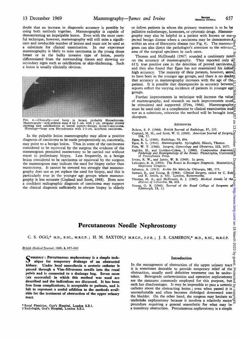
13 December 1969 Mammography-7ames and Irvine BRnT 657 doubt that an increase in diagnostic accuracy is possible by using both methods together. Mammography is capable of demonstrating an impalpable lesion. Even with the most care- ful technique, however, mammography will still miss a signifi- cant and irreducible number of lesions and must not be used as a substitute for clinical examination. In our experience mammography is likely to miss carcinoma in the young dense breast or in the bulky invasive type of lesion, poorly differentiated from the surrounding tissues and showing no secondary signs such as calcification or skin-thickening. Such a lesion is usually clinically obvious. FIG. 4.-Clinically-oval lm in brat poal fibroadenoma. Mammogram-we,_ -defined mas 6 by 3 cm .It 11 cm.iirreg.lar.density Histology-large~~~~ are .fibroadenosis with...1... 5 cm....... siroscrioa ma .point to a benign lesion Thus in some of the crin considered~~~~~~~~~~~~~~~~~~~~~~~~~~~~~~~~~~~~~~~~~~ to be equiocal byte sugo th evdec ofth +. .. .. :. .. .. .. .. .. .. .. .. .. .. .. .. .. .._.. ._ . .... .. ...... FIG. 4. Clinically oval lump in breast, probably fibroadenoma. Mammogram- well-defined mass 6 by 3 cm. with 1 cm. irregular density showing tiny calcifications at lateral aspect benign lesion + carcinoma Histology- large area fibroadenosis with l 5-cm. scirrhous carcinoma. In the palpable lesion mammography may allow a positive diagnosis of carcinoma to be made preoperatively or, conversely, may point to a benign lesion. Thus in some of the carcinomas considered to be equivocal by the surgeon the evidence of the mammogram permitted mastectomy to be carried out without resort to preliminary biopsy. Less frequently, in a benign lesion considered to be carcinoma or equivocal by the surgeon the mammogram may indicate the need for biopsy rather than mastectomy. It cannot be stressed too strongly that mammo- graphy does not as yet replace the need for biopsy, and this is particularly true in the younger age groups where mammo- graphy is less accurate (Copland and Scott, 1968). Even now a confident radiographic diagnosis of carcinoma may support the clinical diagnosis sufficiently to obviate biopsy in elderly or infirm patients in whom the primary treatment is to be by palliative radiotherapy, hormones, or cytotoxic drugs. Mammo- graphy may also be helpful in a patient with known or sus- pected benign disease where a carcinoma may be demonstrated within an area of fibrocystic disease (see Fig. 4). The mammo- gram can also direct the pathologist's attention to the relevant area of the surgical specimen in such cases. Thomas and McDonald (1967) sounded a cautionary note on the accuracy of mammography. They reported only a 61% true positive rate in the detection of proved carcinoma, and they also found that Egan has not maintained his initial high accuracy. The majority of their patients, however, seem to have been in the younger age groups, and there is no doubt that accuracy in mammography increases with the age of the patient. It is possible that discrepancies in accuracy between reports reflect the varying incidence of patients in younger age groups. Further improvements in technique will increase the value of mammography, and research on such improvements must be stimulated and supported (Fitts, 1966). Mammography must be used only as a complement to clinical examination and not as a substitute, otherwise the method will be brought into disrepute. REFERENCES Bohrer, S. P. (1964). British Journal of Radiology, 37, 237. Copland, M. M., and Scott, W. G. (1968). American 7ournal of Surgery, 116, 57. Egan, R. L. (1960). Radiology, 75, 894. Egan, R. L. (1964). Mammography. Springfield, Illinois, Thomas. Fitts, W. T. (1966). Surgery, Gynecology and Obstetrics, 122, 1077. Ingleby, H., and Gershon-Cohen, J. (1960). Comparative Anatomy, Pathology and Roentgenology of the Breast. Philadelphia, University of Pennsylvania Press. Irvine, R. W., and James, W. B. (1969). In press. Leborgne, R. A. (1953). The Breast in Roentgen Diagnosis. Montevideo, Impresora Uruguya. Salomon, A. (1913). Archiv fur klinische Chirurgie, 101, 573. Samuel, E., and Young, B. (1964). Clinical Surgery, edited by C. Rob and R. Smith, p. 431. London, Butterworths. Thomas, M. A., and McDonald, E. J. (1967). Medical Annals "of the District of Columbia, 36, 468. Young, G. B. (1968). Journal of the Royal College of Surgeons of Edinburgh, 13, 12. Percutaneous Needle Nephrostomy C. S. OGG,* M.D., B.SC., M.R.C.P.; H. M. SAXTON,t M.R.C.P., F.F.R.; J. S. CAMERON,* M.D., B.SC., M.R.C.P. British Medical Journal, 1969, 4, 657-660 Summary: Percutaneous nephrostomy is a simple tech- nique for temporary drainage of an obstructed kidney. Under local anaesthesia a ureteric catheter is passed through a Vim-Silverman needle into the renal pelvis and is connected to a drainage bag. Seven cases (six successful) in which this method was used are described and the indications are discussed. It has been free from complications, is acceptable to patients, and is felt to represent a useful addition to the methods avail- able for the treatment of obstruction of the upper urinary tract. * Renal Physician, Guy's Hospital, London S.E.1. t Radiologist, Guy's Hospital, London S.E.1. Introduction In the management of obstruction of the upper urinary tract it is sometimes desirable to provide temporary relief of the obstruction, usually until definitive treatment can be under- taken. Retrograde catheterization and operative nephrostomy are the measures commonly employed for this purpose, but each has disadvantages. It may be impossible to pass a ureteric catheter above the obstructing lesion; even when passed it is uncomfortable and often becomes dislodged downward into the bladder. On the other hand, the surgeon may hesitate to undertake nephrostomy because it involves a relatively major procedure requiring a general anaesthetic for what may be a transitory obstruction. Percutaneous nephrostomy is a simple on 18 March 2022 by guest. Protected by copyright. http://www.bmj.com/ Br Med J: first published as 10.1136/bmj.4.5684.657 on 13 December 1969. Downloaded from
Transcript of Percutaneous Needle Nephrostomy - BMJ
13 December 1969 Mammography-7ames and Irvine BRnT 657
doubt that an increase in diagnostic accuracy is possible byusing both methods together. Mammography is capable ofdemonstrating an impalpable lesion. Even with the most care-ful technique, however, mammography will still miss a signifi-cant and irreducible number of lesions and must not be used asa substitute for clinical examination. In our experiencemammography is likely to miss carcinoma in the young densebreast or in the bulky invasive type of lesion, poorlydifferentiated from the surrounding tissues and showing nosecondary signs such as calcification or skin-thickening. Sucha lesion is usually clinically obvious.
FIG. 4.-Clinically-oval lm in brat poal fibroadenoma.Mammogram-we,_ -defined mas 6 by 3 cm.It11 cm.iirreg.lar.density
Histology-large~~~~are .fibroadenosiswith...1...5 cm.......siroscrioa
ma .point to a benign lesion Thus in some of the crin
considered~~~~~~~~~~~~~~~~~~~~~~~~~~~~~~~~~~~~~~~~~~tobeequiocalbytesugo th evdec ofth
+. .. .. :. .. .. .. .. .. .. .. .. .. .. .. .. ..
.._.. ._ . .... .. ......
FIG. 4. Clinically oval lump in breast, probably fibroadenoma.Mammogram- well-defined mass 6 by 3 cm. with 1 cm. irregular densityshowing tiny calcifications at lateral aspect benign lesion+ carcinomaHistology- large area fibroadenosis with l 5-cm. scirrhous carcinoma.
In the palpable lesion mammography may allow a positivediagnosis of carcinoma to be made preoperatively or, conversely,may point to a benign lesion. Thus in some of the carcinomasconsidered to be equivocal by the surgeon the evidence of themammogram permitted mastectomy to be carried out withoutresort to preliminary biopsy. Less frequently, in a benignlesion considered to be carcinoma or equivocal by the surgeonthe mammogram may indicate the need for biopsy rather thanmastectomy. It cannot be stressed too strongly that mammo-graphy does not as yet replace the need for biopsy, and this isparticularly true in the younger age groups where mammo-graphy is less accurate (Copland and Scott, 1968). Even nowa confident radiographic diagnosis of carcinoma may supportthe clinical diagnosis sufficiently to obviate biopsy in elderly
or infirm patients in whom the primary treatment is to be bypalliative radiotherapy, hormones, or cytotoxic drugs. Mammo-graphy may also be helpful in a patient with known or sus-pected benign disease where a carcinoma may be demonstratedwithin an area of fibrocystic disease (see Fig. 4). The mammo-gram can also direct the pathologist's attention to the relevantarea of the surgical specimen in such cases.Thomas and McDonald (1967) sounded a cautionary note
on the accuracy of mammography. They reported only a61% true positive rate in the detection of proved carcinoma,and they also found that Egan has not maintained his initialhigh accuracy. The majority of their patients, however, seemto have been in the younger age groups, and there is no doubtthat accuracy in mammography increases with the age of thepatient. It is possible that discrepancies in accuracy betweenreports reflect the varying incidence of patients in younger agegroups.
Further improvements in technique will increase the valueof mammography, and research on such improvements mustbe stimulated and supported (Fitts, 1966). Mammographymust be used only as a complement to clinical examination andnot as a substitute, otherwise the method will be brought intodisrepute.
REFERENCES
Bohrer, S. P. (1964). British Journal of Radiology, 37, 237.Copland, M. M., and Scott, W. G. (1968). American 7ournal of Surgery,
116, 57.Egan, R. L. (1960). Radiology, 75, 894.Egan, R. L. (1964). Mammography. Springfield, Illinois, Thomas.Fitts, W. T. (1966). Surgery, Gynecology and Obstetrics, 122, 1077.Ingleby, H., and Gershon-Cohen, J. (1960). Comparative Anatomy,
Pathology and Roentgenology of the Breast. Philadelphia, Universityof Pennsylvania Press.
Irvine, R. W., and James, W. B. (1969). In press.Leborgne, R. A. (1953). The Breast in Roentgen Diagnosis. Montevideo,
Impresora Uruguya.Salomon, A. (1913). Archiv fur klinische Chirurgie, 101, 573.Samuel, E., and Young, B. (1964). Clinical Surgery, edited by C. Rob
and R. Smith, p. 431. London, Butterworths.Thomas, M. A., and McDonald, E. J. (1967). Medical Annals "of the
District of Columbia, 36, 468.Young, G. B. (1968). Journal of the Royal College of Surgeons of
Edinburgh, 13, 12.
Percutaneous Needle Nephrostomy
C. S. OGG,* M.D., B.SC., M.R.C.P.; H. M. SAXTON,t M.R.C.P., F.F.R.; J. S. CAMERON,* M.D., B.SC., M.R.C.P.
British Medical Journal, 1969, 4, 657-660
Summary: Percutaneous nephrostomy is a simple tech-nique for temporary drainage of an obstructed
kidney. Under local anaesthesia a ureteric catheter ispassed through a Vim-Silverman needle into the renalpelvis and is connected to a drainage bag. Seven cases(six successful) in which this method was used aredescribed and the indications are discussed. It has beenfree from complications, is acceptable to patients, and isfelt to represent a useful addition to the methods avail-able for the treatment of obstruction of the upper urinarytract.
* Renal Physician, Guy's Hospital, London S.E.1.t Radiologist, Guy's Hospital, London S.E.1.
IntroductionIn the management of obstruction of the upper urinary tractit is sometimes desirable to provide temporary relief of theobstruction, usually until definitive treatment can be under-taken. Retrograde catheterization and operative nephrostomyare the measures commonly employed for this purpose, buteach has disadvantages. It may be impossible to pass a uretericcatheter above the obstructing lesion; even when passed it isuncomfortable and often becomes dislodged downward intothe bladder. On the other hand, the surgeon may hesitate toundertake nephrostomy because it involves a relatively majorprocedure requiring a general anaesthetic for what may bea transitory obstruction. Percutaneous nephrostomy is a simple
on 18 March 2022 by guest. P
rotected by copyright.http://w
ww
.bmj.com
/B
r Med J: first published as 10.1136/bm
j.4.5684.657 on 13 Decem
ber 1969. Dow
nloaded from

and effective alternative without these drawbacks, and in thispaper we describe our experience with the method.
Technique
The procedure was first described by Goodwin, Casey, andWoolf (1955) and has recently been modified by Cobb (1967).We have based our technique on the latter author's descriptionbut have used fluoroscopy with image intensification for theinitial localization of the kidney. The procedure may bedescribed as having two stages-percutaneous pyelography,followed by percutaneous nephrostomy. The kidney is firstmade visible for fluoroscopy, commonly by high-dose uro-
graphy. In most cases the faint outlines of the calices andpelvis have become visible, but occasionally we have been guidedby the nephrogram alone. When the degree of opacity seems
adequate the patient is turned prone and settled as comfortablyas possible. A fine exploring needle-for example, Harrislumbar puncture No. 5-is introduced under local anaesthesiainto the pelvi-caliceal system. With acute obstruction the urineis under pressure and wells up when the needle enters the pelvis,but in one of our cases where the obstruction was of longstanding it was necessary to aspirate to confirm the correctposition of the needle.
Contrast medium-for example, 10-15 ml. of Conray 280-is injected via a flexible connexion to produce better visualiza-tion of the upper urinary tract and to confirm the presenceand site of obstruction. The needle is then withdrawn, notingthe depth to which it has been introduced. Next a Vim-Silverman renal biopsy needle is passed, under fluoroscopiccontrol, to a corresponding depth and so into the renal pelvis.The stylet is removed, and, provided urine emerges, a No. 5(French) whistle-tipped ureteric catheter is threaded throughthe needle and advanced well into the pelvis. Usually thecatheter passes easily into the pelvis, but occasionally rotationof the bevel or suitable angulation of the needle is required.The needle is withdrawn over the catheter, which is then fixedto the skin and connected to a drainage bag. Drainage con-
tinues until the obstruction has been relieved, and the catheteris then removed.
Case 1
This patient was a 52-year-old woman who in 1966 was found tohave lymphosarcoma. She had been treated with cyclophosphamideand remained reasonably well until February 1968. She thencomplained of haematuria and left renal colic, and shortly after-wards became anuric. Bilateral retrograde pyelography showed thatboth ureters were severely narrowed in their mid-portions. At rightnephrostomy extensive retroperitoneal deposits of lymphosarcomawere found. The nephrostomy did not drain adequately and theblood urea rose to 280 mg./100 ml. On 1 March 1968 a per-cutaneous pyelogram (Fig. 1) showed complete obstruction of theleft ureter. Left percutaneous nephrostomy was performed and gooddrainage resulted, Three days later the blood urea had fallen to38 mg./100 ml. Radiotherapy to the abdomen was begun on
8 March, and the course was completed on 3 April. Spontaneouspassage of urine began 10 days after the beginning of treatment,and injection of contrast medium through the percutaneous cathetershowed that the ureter was no longer obstructed (Fig. 2). On23 March the nephrostomy catheter was removed. At first thepatient appeared to be maintaining her improvement, but furtherlymphosarcomatous masses developed and six weeks later she died.
Case 2
A 61-year-old man underwent abdomino-perineal resection forcarcinoma of the rectum in 1967. In October 1968 he was admittedto hospital in a semicomatose state with a history of nine days'anuria and with a blood urea of 310 mg./100 ml. Peritonealdialysis produced a gradual improvement, and retrograde uretero-grams showed bilateral obstruction at the pelvic brim. Percutaneousnephrostomy on 15 October produced adequate drainage of the left
BurrwsMEDICAL JOURNAl
kidney, and his blood urea stabilized at 200 mg./100 ml. Ten dayslater ureterostomy was performed and the ureteric catheter was
removed. Nine months later he was well, with a blood urea ofabout 120 mg./100 ml.
FIG. 1.-Case 1. Left percutaneous pyelogram(1 March 1968) showing ureteric obstruction at
L3-4 level.
FIG. 2.-Case 1. Injection of contrast mediumthrough the percutaneous catheter shows that after
radiotherapy the ureter is patent throughout itslength (22 March 1968).
Case 3
This 47-year-old man with epilepsy had been treated with pheno-barbitone and phenytoin for several years. Three weeks before
admission he was put on acetazolamide, and 17 days later developedbilateral loin pain and became anuric. On admission his blood
urea was 118 mg./100 ml., but later rose to 212 mg./100 ml.
At cystoscopy debris was seen extruding from the left ureteric orifice
658 13 December 1969 Percutaneous Nephrostomy-Ogg et al.
on 18 March 2022 by guest. P
rotected by copyright.http://w
ww
.bmj.com
/B
r Med J: first published as 10.1136/bm
j.4.5684.657 on 13 Decem
ber 1969. Dow
nloaded from

Percutaneous Nephrostomy---Ogg et al.
and it was impossible to catheterize either ureter. Urography con-firmed the presence of bilateral obstruction, and on 27 February1969 percutaneous nephrostomy was performed on the left side.This drained well, and soon afterwards the right side cleared spon-
taneously, possibly owing to the osmotic diuresis produced by theurographic contrast. The blood urea rapidly returned to normallevels and an antegrade pyelogram showed that the left ureter was
patent. After seven days the catheter was removed. An intravenousurogram was normal and he was discharged from hospital. It was
presumed that the acetazolamide had produced ureteric " sludge"with bilateral obstruction.
Case 4
A 21-year-old man with a long history of enuresis was admittedto hospital because of sudden onset of left loin pain. He had a
urinary infection and a blood urea of 188 mg./100 ml. Aturography both kidneys were found to be large, with minimalfunction on the left side and no excretion on the right. Voidingcystography showed a grossly trabeculated bladder with no outletobstruction and no reflux, the appearances suggesting a neurogenicbladder. Left retrograde pyelography showed a large hydro-nephrosis with pelvi-ureteric narrowing and angulation. Becauseof the risk of infection following retrograde pyelography a per-
cutaneous nephrostomy was performed on 5 May 1969. When thepelvis was punctured it was found that the urine was not underpressure, and aspiration was needed to confirm the correct positionof the needle. The low pressure was attributed to the chronicityof the obstruction. Drainage was continued until pyeloplasty threedays later. His right kidney was later removed. At the time ofwriting his blood urea was steady at about 150 mg./100 ml.
Case 5
This 68-year-old man underwent abdomino-perineal resection of a
rectal carcinoma in February 1969. In April he noticed diminishedurine output and he became anuric on 1 May. Six days later hewas admitted to Guy's Hospital with a blood urea of 280 mg./100ml. High-dose urography confirmed the presence of obstruction on
both sides, and bilateral percutaneous nephrostomy was performed.In spite of some blood-staining both catheters drained well, andwithin two days his blood urea had fallen to 32 mg./100 ml.This fall was accompanied by a massive diuresis, reaching a peakof 10 litres in one day. Subsequent laparotomy showed extensiveperitoneal deposits; his condition deteriorated and he died on
2 June.Case 6
This 68-year-old man was known to have a solitary left kidneywith moderate pelvi-ureteric narrowing and hydronephrosis. InMay 1969 he became oliguric, urography showing a considerableincrease in the hydronephrosis. His blood urea was 250 mg./100 ml.In spite of clear visualization of the kidney it proved impossibleto puncture the pelvis even when a relatively long needle was used.Subsequently a nephrostomy was performed and later a pyeloplasty;no indication of trauma due to the needling was seen.
Case 7
This 40-year-old man had a left pyelolithotomy in 1967. Aright-sided stone present at that time increased in size, and inMarch 1969 a right pyelolithotomy was carried out. This was a
difficult operation and was followed by severe urinary infection.Two months later urography showed no excretion by the rightkidney, and retrograde pyelography revealed obstruction at thepelvi-ureteric junction. It was decided to undertake percutaneousnephrostomy to determine the extent of any residual function.This was performed on 23 May and a good flow of urine wasobtained. A week later the creatinine clearance of the right kidneywas 11l3 ml. per minute, while that of the left side was 42 ml.per minute. It therefore appeared that the right kidney provideda significant proportion of the total renal function, and a rightureterocalicotomy was successfully carried out on 10 June.
Discussion
The practical value of a technique which affords reliabletemporary relief of obstruction will be readily apparent. Time
BRyHMEDICAL JOURNAL 659
is gained during which the patient's condition can improve,infection be controlled or prevented, and the diagnosis be con-firmed or treatment planned. Though the indications are
admittedly restricted, it is difficult to see why the procedurehas not been more widely used. Renal puncture is generallyaccepted as safe, whether for renal biopsy or for the diagnostictapping of cysts. Percutaneous pyelography, an essential pro-liminary to this method, has been advocated in a number ofpapers over the past 15 years (Weens and Florence, 1954;Wickbom, 1954; Casey and Goodwin, 1955; Floyd and Guy,1956; Lundin and Wadstrdm, 1965; Maldonado, 1956;Bosniak, Scheff, and Kaufman, 1968; Lalli, 1968). Yet onethe renal pelvis has been located and visualized in this waythe additional manoeuvre required for drainage is so simplethat we can see little object in performing antegrade pyelo-graphy for obstruction without at the same time attemptingpercutaneous nephrostomy.Our experience of the method has been that in six of the
seven cases in which it was attempted it was straightforward,and in all cases it was without complications. In one patient(Case 6), though the pelvis was considerably enlarged andclearly visible, it was not possible to puncture it. At the time
it was thought that this was due to inspissation of the pelviccontents, but at subsequent nephrostomy that did not proveto be the case, and it was not possible to see why puncturehad failed. In retrospect it is thought that it might have beenhelpful to establish the relative depths of the needle and kidneyby taking a single x-ray film in the transverse plane.So few accounts have been given of this technique that it
would be unrealistic to discuss the incidence of complications.In the two patients (Cases 4 and 7) who have undergoneexploration of the kidney shortly after removal of the catheterthere was no indication of any harmful effect on the kidneyor pelvis. As Goodwin et al. (1955) and Cobb (1967) pointout, there are no vital structures in the path of the needleexcept the renal vessels. The probability of accidental punctureof a vessel during exploratory needling under screen control isno greater than during preliminary needling for renal biopsy;such accidental puncture is most unlikely to cause significantbleeding. Once the pelvi-caliceal system has been visualizedby direct injection of contrast medium it becomes possibleto direct the larger (Vim-Silverman) needle to the lateral sideof the pelvis, so avoiding the larger renal vessels.
Obstruction of the catheter may occur, and Cobb (1967)successfully inserted a second catheter when this happened.A possible cause of such obstruction is kinking due to excessivecoiling in the renal pelvis, but though several of our cases
have shown marked coiling this has not affected drainage,possibly because we have usually employed " woven " cathetersas Cobb recommended. The surprising feature of the techniqueis the good drainage produced by the relatively narrow catheter-so much so that in Case 3, where the output through twocatheters reached as high as 10 litres in a day, there was some
difficulty in maintaining adequate fluid balance. The periodof drainage in this series ranges from 3 to 29 days, but Case 3of Goodwin et al. drained satisfactorily for five months andthe tube showed no sign of encrustation. Though we havenot used the method in the presence of infection, the experienceof Goodwin et al. (1955) and of Cobb (1967) shows that thisis not a bar to the technique. In their cases the infectionresponded rapidly to adequate drainage.
Cases in which percutaneous nephrostomy is suitable foruse do not often occur. The indications for the proceduremay widen as it becomes more generally adopted, but at presentthey may be summarized as follows:
(1) Short-term relief of obstruction in the anuric patient in whomretrograde catheterization is impossible. This may be because oftechnical difficulty with catheterization, because of the severity ofthe ureteric narrowing, because the condition of the patient pre-cludes anaesthesia, or because the ureter is not available forcatheterization-for example, after implantation into an ileal loop.
13 December 1969
on 18 March 2022 by guest. P
rotected by copyright.http://w
ww
.bmj.com
/B
r Med J: first published as 10.1136/bm
j.4.5684.657 on 13 Decem
ber 1969. Dow
nloaded from
660 13December 1969 Percutaneous Nephrostomy-Ugg et al. BRL"Iu660 13 D~ecemb~er 1969J ~C4 MEDICAL JOURNAL
(2) Short-term relief of obstruction in a patient who is awaitingpyeloplasty, when there is a risk that pyonephrosis may develop-for example, after retrograde catheterization. We would also suggestthat it might be of value to produce preliminary decompression ofthe renal pelvis and allow contraction of the stretched tissues, makingsubsequent surgery easier.
(3) Assessment of potential recovery of an obstructed kidney orin a non-functioning moiety of an obstructed duplex kidney.
For the most part these indications require little comment.Cases with suspected malignant disease involving both uretersraise difficult problems of management, since the benefit result-ing from relief of obstruction is likely to be temporary and itmay at times be more humane not to allow the patient torecover sufficiently from renal failure to experience the directeffects of the growth. But in other cases of anuria due toobstruction there is every advantage in improving the generalcondition of the patient and diminishing the risk of infectionbefore curative surgery. In unilateral obstructive disease thereis an interesting further possibility, since a period of satis-factory drainage makes it possible to assess the likely returnof function with adequate relief of obstruction (see our Case 7and Goodwin et al. (1955) Case 2). This, in our view, offersa most promising area for further extension of the method.The importance of conserving renal tissue is undoubted, yetafter a period of chronic obstruction it may be impossible topredict whether adequate function can be restored to a kidneyeven if the obstruction is satisfactorily relieved. Percutaneousnephrostomy offers a practical way of resolving such uncer-tainties.
The particular technique recommended is not definitive.For the purpose of simple drainage the No. 5 ureteric catheteris adequate, but narrower catheters could be preferable forother purposes-for example, long-term manometric studiesand the instillation of antibiotics, chemotherapeutic agents, orstone-dissolving fluids. At times a wider catheter might beneeded. Whatever the future extensions of the method, theexisting technique is, in our view, a significant advance in themanagement of obstructive disease of the upper urinary tract.
We would like to thank the physicians and surgeons who referredthese patients to us, and Mr. F. R. Kilpatrick and Mr. C. H. Kinderfor their advice and help.
REFERENCES
Bosniak, M. A., Scheff, S., and Kaufman, S. (1968). 7ournal of Urology,99, 241.
Casey, W. C., and Goodwin, W. E. (1955). 7ournal of Urology, 74, 164.Cobb, B. (1967). 7ournal of Urology, 98, 309.Floyd, E., and Guy, J. C. (1956). 7ournal of the Medical Association of
Georgia, 45, 13.Goodwin, W. E., Casey, W. C., and Woolf, W. (1955). 7ournal of the
American Medical Association, 157, 891.Lalli, A. F. (1968). Radiology, 90, 331.Lundin, E., and Wadstr6m, L. B. (1965). Acta Chirurgica Scandinavica.
130, 267.Maldonado, J. (1966). 7ournal of Urology, 96, 651.Weens, H. S., and Florence, T. J. (1954). 7ournal of Urology, 72, 589.Wickbom, I. (1954). Acta Radiologica, 41, 505.
Latent Cytomegalovirus Infection in Blood Donors
PETER DIOSI,* M.D.; EVA MOLDOVAN,* M.D.; NICHOLAS TOMESCUt M.D.
British Medical Journal, 1969, 4, 660-662
ummary: Twenty-one out of 32 apparently healthyblood donors aged 21 to 65 years yielded positive
complement fixation tests with a cytomegalovirus antigen,at titres ranging from 1:8 to 1:64. Virus was present inleucocyte cultures of fresh peripheral blood of two sero-positive subjects from a total of 35 donors examined.Plasma and 48-hour stored blood specimens failed to dis-close virus in culture. Viruria could not be demonstrated,and there was no evidence of recent illness in the studygroup. These findings suggest that subclinical viraemiais not uncommon in blood donors.
Introduction
Wyatt et al. (1951) were the first to assume that cytomegalo-virus infection could be the result of the introduction of virusinto a non-carrier by transfusion with blood from a carrier.Since then the occurrence of cytomegaloviraemia has beenshown by the recovery of cytomegalovirus strains from thebuffy-coat layer of a blood sample received from a patient withgiant-cell type hepatitis (Stulberg et al., 1966), as well as fromwashed leucocytes obtained from the peripheral blood in casesof congenital cytomegalic inclusion disease (Demidova et al.,1968), post-transfusion mononucleosis (Foster and Jack, 1968),
post-perfusion syndrome (Lang et al., 1968), lymphatic leuk-aemia (Harnden et al., 1967; Diosi and Roth, 1969), andmyeloid leukaemia (Jack et al., 1968).
Since generalized cytomegalic inclusion disease and cyto-megalovirus mononucleosis have been shown to develop afterthe transfusion of fresh blood (Kairidinen et al., 1966a;Henson, 1967), in connexion with renal haemodialysis (Cutforthet al., 1968), or surgery with extracorporeal circulation (Ander-son and Larsson, 1963 ; Embil et at., 1968 ; Marton et at.,1968; Foster and Jack, 1969), attempts were made in the pre-sent study to recover cytomegalovirus from donated freshblood and from blood specimens stored for 48 hours beforetheir administration.
* Institute of Hygiene, Timisoara, Rumania.t Centre of Blood Transfusions, Timisoara, Rumania.
Materials and Methods
The study group, which came from the Blood TransfusionCentre in Timiaoara, consisted of 35 blood donors aged 21 to65 years, belonging at random to various blood groups. Routinehaemograms and urine analyses yielded normal values in eachcase, while serology for syphilis and hepatocellular functiontests were negative. None of the individuals surveyed showedany sign of disease.
Virological Techniques
Details of the laboratory methods employed have for themost part been reported elsewhere (Diosi and Roth, 1969).
on 18 March 2022 by guest. P
rotected by copyright.http://w
ww
.bmj.com
/B
r Med J: first published as 10.1136/bm
j.4.5684.657 on 13 Decem
ber 1969. Dow
nloaded from