Peptidoglycan synthesis in Mycobacterium tuberculosis … · Peptidoglycan synthesis in...
6
Peptidoglycan synthesis in Mycobacterium tuberculosis is organized into networks with varying drug susceptibility Karen J. Kieser a , Catherine Baranowski a , Michael C. Chao b , Jarukit E. Long c , Christopher M. Sassetti c,d , Matthew K. Waldor b,d,e , James C. Sacchettini f , Thomas R. Ioerger g , and Eric J. Rubin a,e,1 a Department of Immunology and Infectious Disease, Harvard T. H. Chan School of Public Health, Boston, MA 02115; b Division of Infectious Diseases, Brigham & Women’s Hospital, Boston, MA 02115; c Department of Microbiology and Physiological Systems, University of Massachusetts Medical School, Worcester MA 01655; d Howard Hughes Medical Institute, Chevy Chase, MD 20815; e Department of Microbiology and Immunobiology, Harvard Medical School, Boston, MA 02115; f Department of Biochemistry and Biophysics and Department of Chemistry, Texas A&M University, College Station, TX 77843; and g Department of Computer Science, Texas A&M University, College Station, TX 77843 Edited by Carolyn R. Bertozzi, University of California, Berkeley, CA, and approved September 8, 2015 (received for review July 17, 2015) Peptidoglycan (PG), a complex polymer composed of saccharide chains cross-linked by short peptides, is a critical component of the bacterial cell wall. PG synthesis has been extensively studied in model organisms but remains poorly understood in mycobacteria, a genus that includes the important human pathogen Mycobacte- rium tuberculosis (Mtb). The principle PG synthetic enzymes have similar and, at times, overlapping functions. To determine how these are functionally organized, we carried out whole-genome transposon mutagenesis screens in Mtb strains deleted for ponA1, ponA2, and ldtB, major PG synthetic enzymes. We identified distinct factors required to sustain bacterial growth in the absence of each of these enzymes. We find that even the homologs PonA1 and PonA2 have unique sets of genetic interactions, suggesting there are distinct PG synthesis pathways in Mtb. Either PonA1 or PonA2 is required for growth of Mtb, but both genetically interact with LdtB, which has its own distinct genetic network. We further provide evidence that each interaction network is differentially susceptible to antibiotics. Thus, Mtb uses alternative pathways to produce PG, each with its own biochemical characteristics and vulnerabilities. Mycobacterium tuberculosis | transposon sequencing | genetic interaction | peptidoglycan | cell wall O ne of the leading causes of infectious disease deaths world- wide is tuberculosis (TB), caused by Mycobacterium tubercu- losis (Mtb). One-third of the human population is thought to harbor Mtb and ∼1.5 million individuals died of TB last year (1). Mtb’s success as a pathogen is due in part to its unusual cell wall, which is notorious for its complexity and is implicated in Mtb’s innate resistance to many commonly used antibiotics (2). A critical component of the bacterial cell wall (including Mtb’s) is peptidoglycan (PG), a complex polymer that provides structural support and counteracts turgor pressure (3). PG is essential for cell survival, and its synthesis is targeted by many potent anti- biotics (2). PG consists of long glycan chains composed of two different sugars (Fig. 1A) that are cross-linked via short peptide side chains that extend from the glycan chains. Notably, generation of mature PG occurs outside of the cell membrane and is mediated by enzymes that incorporate new PG subunits, which are formed in the cytoplasm, into the PG polymer. PonA1 and PonA2 are the two enzymes in Mtb that can both polymerize glycan strands and cross-link peptides [known as bifunctional penicillin binding proteins (PBPs), Fig. 1A]. The predominant peptide cross-links in mycobacteria join the third amino acids (3–3 link) of adjacent stem peptides (4, 5), which are synthesized by L,D-transpeptidases (Ldts) such as LdtB, one of the major Ldts in Mtb (Fig. 1A). The peptides can also be joined by cross-linking the fourth and third amino acids (4–3 link) (Fig. 1A) through the action of bifunctional or monofunctional (capable of only peptide cross-linking) PBPs. The activity of these distinct factors must be coordinated to ensure proper cell-wall synthesis. One method of coordination is the use of large protein complexes, the elongation complex and divisome, which mediate cell-wall biogenesis during cell elongation or di- vision, respectively (2). The essential activity of these enzymes makes them prime drug targets; indeed, PBPs and Ldts are inhibited by carbapenems and penicillin (6, 7), which remains one of the most clinically important drugs in use. Although the biosynthesis and structure of PG have been in- vestigated for decades, predominantly in organisms such as Escherichia coli or Bacillus subtilis, the mechanisms that coordinate the biochemical activities required to polymerize and modify the cell wall remain incompletely understood. Moreover, much less is known about PG synthesis in many pathogenic organisms, including Mtb (2). However, previous studies in Mtb suggest that PG synthesis in this pathogen does not strictly conform to the E. coli paradigm. For example, E. coli has three bifunctional PBPs [PBP1A, PBP1B, and PBP1C (3)], whereas Mtb has just two [PonA1 and PonA2 (8)]. Additionally, PBP2 (known as PBPA in mycobacteria) is a monofunctional PBP and is required for cell elongation in E. coli, but instead seems to function in cell septation in mycobacteria (3, 9). The structure of PG is also different in Mtb than in E. coli: Mycobacterial PG has an unusual prevalence of 3–3 peptide linkages. The abundance of 3–3 cross-links in mycobacterial PG throughout different growth stages (5) suggests that Ldts are Significance The rise of drug-resistant Mycobacterium tuberculosis (Mtb) underscores the critical need for a better understanding of es- sential physiological processes. Among these is cell-wall syn- thesis, the target of many antibiotics. To understand how Mtb orchestrates synthesis of its cell wall, we performed whole- genome interaction studies in cells with different peptidoglycan synthesis mutations. We found that different enzymes become required for bacterial growth in ΔponA1, ΔponA2, or ΔldtB cells, suggesting that discrete cell envelope biogenesis networks exist in Mtb. Furthermore, we show that these networks’ enzymes are differentially susceptible to cell-wall–active drugs. Our data provide insight into the essential processes of cell-wall synthesis in Mtb and highlight the role of different synthesis networks in antibiotic tolerance. Author contributions: K.J.K., C.B., and E.J.R. designed research; K.J.K., C.B., and J.E.L. performed research; K.J.K., C.B., M.C.C., J.E.L., C.M.S., M.K.W., J.C.S., and T.R.I. contributed new reagents/analytic tools; K.J.K., C.B., M.C.C., T.R.I., and E.J.R. analyzed data; and K.J.K. and E.J.R. wrote the paper. The authors declare no conflict of interest. This article is a PNAS Direct Submission. 1 To whom correspondence should be addressed. Email: [email protected]. This article contains supporting information online at www.pnas.org/lookup/suppl/doi:10. 1073/pnas.1514135112/-/DCSupplemental. www.pnas.org/cgi/doi/10.1073/pnas.1514135112 PNAS | October 20, 2015 | vol. 112 | no. 42 | 13087–13092 MICROBIOLOGY
Transcript of Peptidoglycan synthesis in Mycobacterium tuberculosis … · Peptidoglycan synthesis in...

Peptidoglycan synthesis in Mycobacterium tuberculosisis organized into networks with varyingdrug susceptibilityKaren J. Kiesera, Catherine Baranowskia, Michael C. Chaob, Jarukit E. Longc, Christopher M. Sassettic,d,Matthew K. Waldorb,d,e, James C. Sacchettinif, Thomas R. Ioergerg, and Eric J. Rubina,e,1
aDepartment of Immunology and Infectious Disease, Harvard T. H. Chan School of Public Health, Boston, MA 02115; bDivision of Infectious Diseases,Brigham & Women’s Hospital, Boston, MA 02115; cDepartment of Microbiology and Physiological Systems, University of Massachusetts Medical School,Worcester MA 01655; dHoward Hughes Medical Institute, Chevy Chase, MD 20815; eDepartment of Microbiology and Immunobiology, Harvard MedicalSchool, Boston, MA 02115; fDepartment of Biochemistry and Biophysics and Department of Chemistry, Texas A&M University, College Station, TX 77843;and gDepartment of Computer Science, Texas A&M University, College Station, TX 77843
Edited by Carolyn R. Bertozzi, University of California, Berkeley, CA, and approved September 8, 2015 (received for review July 17, 2015)
Peptidoglycan (PG), a complex polymer composed of saccharidechains cross-linked by short peptides, is a critical component of thebacterial cell wall. PG synthesis has been extensively studied inmodel organisms but remains poorly understood in mycobacteria,a genus that includes the important human pathogen Mycobacte-rium tuberculosis (Mtb). The principle PG synthetic enzymes havesimilar and, at times, overlapping functions. To determine how theseare functionally organized, we carried out whole-genome transposonmutagenesis screens in Mtb strains deleted for ponA1, ponA2, andldtB, major PG synthetic enzymes. We identified distinct factorsrequired to sustain bacterial growth in the absence of each of theseenzymes. We find that even the homologs PonA1 and PonA2 haveunique sets of genetic interactions, suggesting there are distinct PGsynthesis pathways in Mtb. Either PonA1 or PonA2 is required forgrowth of Mtb, but both genetically interact with LdtB, which hasits own distinct genetic network. We further provide evidence thateach interaction network is differentially susceptible to antibiotics.Thus, Mtb uses alternative pathways to produce PG, each with itsown biochemical characteristics and vulnerabilities.
Mycobacterium tuberculosis | transposon sequencing |genetic interaction | peptidoglycan | cell wall
One of the leading causes of infectious disease deaths world-wide is tuberculosis (TB), caused by Mycobacterium tubercu-
losis (Mtb). One-third of the human population is thought toharbor Mtb and ∼1.5 million individuals died of TB last year (1).Mtb’s success as a pathogen is due in part to its unusual cell wall,which is notorious for its complexity and is implicated in Mtb’sinnate resistance to many commonly used antibiotics (2). Acritical component of the bacterial cell wall (including Mtb’s) ispeptidoglycan (PG), a complex polymer that provides structuralsupport and counteracts turgor pressure (3). PG is essential forcell survival, and its synthesis is targeted by many potent anti-biotics (2).PG consists of long glycan chains composed of two different
sugars (Fig. 1A) that are cross-linked via short peptide sidechains that extend from the glycan chains. Notably, generation ofmature PG occurs outside of the cell membrane and is mediatedby enzymes that incorporate new PG subunits, which are formedin the cytoplasm, into the PG polymer. PonA1 and PonA2 arethe two enzymes in Mtb that can both polymerize glycan strandsand cross-link peptides [known as bifunctional penicillin bindingproteins (PBPs), Fig. 1A]. The predominant peptide cross-linksin mycobacteria join the third amino acids (3–3 link) of adjacentstem peptides (4, 5), which are synthesized by L,D-transpeptidases(Ldts) such as LdtB, one of the major Ldts in Mtb (Fig. 1A). Thepeptides can also be joined by cross-linking the fourth and thirdamino acids (4–3 link) (Fig. 1A) through the action of bifunctionalor monofunctional (capable of only peptide cross-linking) PBPs.The activity of these distinct factors must be coordinated to ensure
proper cell-wall synthesis. One method of coordination is the useof large protein complexes, the elongation complex and divisome,which mediate cell-wall biogenesis during cell elongation or di-vision, respectively (2). The essential activity of these enzymesmakes them prime drug targets; indeed, PBPs and Ldts are inhibitedby carbapenems and penicillin (6, 7), which remains one of the mostclinically important drugs in use.Although the biosynthesis and structure of PG have been in-
vestigated for decades, predominantly in organisms such asEscherichia coli or Bacillus subtilis, the mechanisms that coordinatethe biochemical activities required to polymerize and modify thecell wall remain incompletely understood. Moreover, much lessis known about PG synthesis in many pathogenic organisms,including Mtb (2). However, previous studies in Mtb suggest thatPG synthesis in this pathogen does not strictly conform to theE. coli paradigm. For example, E. coli has three bifunctionalPBPs [PBP1A, PBP1B, and PBP1C (3)], whereas Mtb has justtwo [PonA1 and PonA2 (8)]. Additionally, PBP2 (known asPBPA in mycobacteria) is a monofunctional PBP and is requiredfor cell elongation in E. coli, but instead seems to function in cellseptation in mycobacteria (3, 9).The structure of PG is also different in Mtb than in E. coli:
Mycobacterial PG has an unusual prevalence of 3–3 peptidelinkages. The abundance of 3–3 cross-links in mycobacterial PGthroughout different growth stages (5) suggests that Ldts are
Significance
The rise of drug-resistant Mycobacterium tuberculosis (Mtb)underscores the critical need for a better understanding of es-sential physiological processes. Among these is cell-wall syn-thesis, the target of many antibiotics. To understand how Mtborchestrates synthesis of its cell wall, we performed whole-genome interaction studies in cells with different peptidoglycansynthesis mutations. We found that different enzymes becomerequired for bacterial growth in ΔponA1, ΔponA2, or ΔldtB cells,suggesting that discrete cell envelope biogenesis networks existin Mtb. Furthermore, we show that these networks’ enzymesare differentially susceptible to cell-wall–active drugs. Our dataprovide insight into the essential processes of cell-wall synthesisin Mtb and highlight the role of different synthesis networks inantibiotic tolerance.
Author contributions: K.J.K., C.B., and E.J.R. designed research; K.J.K., C.B., and J.E.L.performed research; K.J.K., C.B., M.C.C., J.E.L., C.M.S., M.K.W., J.C.S., and T.R.I. contributednew reagents/analytic tools; K.J.K., C.B., M.C.C., T.R.I., and E.J.R. analyzed data; and K.J.K.and E.J.R. wrote the paper.
The authors declare no conflict of interest.
This article is a PNAS Direct Submission.1To whom correspondence should be addressed. Email: [email protected].
This article contains supporting information online at www.pnas.org/lookup/suppl/doi:10.1073/pnas.1514135112/-/DCSupplemental.
www.pnas.org/cgi/doi/10.1073/pnas.1514135112 PNAS | October 20, 2015 | vol. 112 | no. 42 | 13087–13092
MICRO
BIOLO
GY
active during normal growth; however, their cellular roles or reg-ulation during growth and PG biogenesis remain largely unknown.Whereas penicillins and cephalosporins target only enzymes thatproduce 4–3 cross-links, Ldts can be targeted by carbapenems(7). Recent work suggests that these agents might be far moreefficacious against both dividing and nondividing bacteria (10).Although little is known aboutMtb’s five encoded Ldts (11), one,LdtB, is implicated in antibiotic tolerance (11–13), is required fornormal virulence in a mouse model of TB (12), and is importantfor normal cell shape (13).Previous studies have also revealed that PG biosynthesis dif-
fers between Mtb and the related saprophytic Mycobacteriumsmegmatis (Msm). As opposed toMtb,Msm has three bifunctionalPBPs: PonA1, PonA2, and PonA3 (14). PonA1 is required forMsm but not Mtb growth in culture (15, 16); however, PonA1 isrequired for robust growth of Mtb during infection (16). In con-trast, Mtb and Msm ponA2 mutants do not have growth defects inculture (14, 17). However, Mtb strains with inactivated ponA1 orponA2 exhibit similar survival defects during growth in a host (16,18), suggesting that these two similar bifunctional enzymes havenonredundant and important contributions to PG synthesis duringinfection. Collectively, the differences in PG synthase functionalitymay imply that different PG synthetic pathways exist across spe-cies, which may have consequences for a pathogen’s virulenceduring infection.
Here, we interrogated PG synthesis in Mtb by investigating thegenetic interactions of ponA1, ponA2, and ldtB, which encodethree PG synthases critical for Mtb’s growth during infection. Toidentify these interactions, we performed genome-wide transposonmutagenesis screens inMtbmutant strains that lacked one of theseenzymes. Advances in high-throughput sequencing technologycoupled with the power of whole-genome studies provide uniqueinsights into key bacterial processes, such as cell-wall biosynthesis.Such studies have been performed to a limited extent in bacteria,and further work would substantially expand our understanding ofthe organization of prokaryotic metabolic processes. In this study,we identified diverse genetic interaction networks for ponA1,ponA2, and ldtB, suggesting that these synthases are embeddedwithin distinct cellular networks for assembling Mtb’s PG. Wefound that either ponA1 or ponA2 is required for cell growth, andthat ldtB interacts with both ponA1 and ponA2. Moreover, mu-tants that lack these enzymes have differential susceptibility toagents that interfere with cell-wall biogenesis. Thus, the Mtb cellwall is synthesized using multiple interacting networks that areboth overlapping and unique.
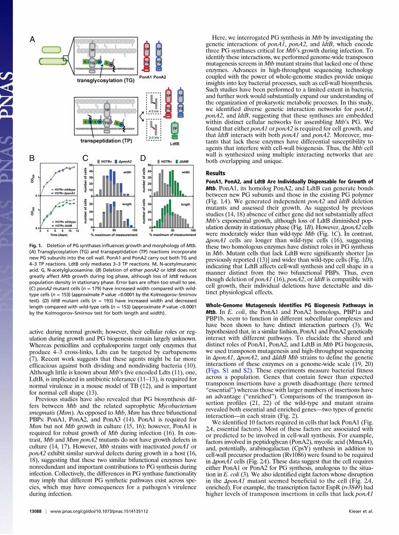
ResultsPonA1, PonA2, and LdtB Are Individually Dispensable for Growth ofMtb. PonA1, its homolog PonA2, and LdtB can generate bondsbetween new PG subunits and those in the existing PG polymer(Fig. 1A). We generated independent ponA2 and ldtB deletionmutants and assessed their growth. As suggested by previousstudies (14, 18) absence of either gene did not substantially affectMtb’s exponential growth, although loss of LdtB diminished pop-ulation density in stationary phase (Fig. 1B). However, ΔponA2 cellswere moderately wider than wild-type Mtb (Fig. 1C). In contrast,ΔponA1 cells are longer than wild-type cells (16), suggestingthese two homologous enzymes have distinct roles in PG synthesisin Mtb. Mutant cells that lack LdtB were significantly shorter [aspreviously reported (13)] and wider than wild-type cells (Fig. 1D),indicating that LdtB affects cell-wall synthesis and cell shape in amanner distinct from the two bifunctional PBPs. Thus, eventhough deletion of ponA1 (16), ponA2, or ldtB is compatible withcell growth, their individual deletions have detectable and dis-tinct physiological effects.
Whole-Genome Mutagenesis Identifies PG Biogenesis Pathways inMtb. In E. coli, the PonA1 and PonA2 homologs, PBP1a andPBP1b, seem to function in different subcellular complexes andhave been shown to have distinct interaction partners (3). Wehypothesized that, in a similar fashion, PonA1 and PonA2 geneticallyinteract with different pathways. To elucidate the shared anddistinct roles of PonA1, PonA2, and LdtB in Mtb PG biogenesis,we used transposon mutagenesis and high-throughput sequencingin ΔponA1, ΔponA2, and ΔldtB Mtb strains to define the geneticinteractions of these enzymes on a genome-wide scale (19, 20)(Figs. S1 and S2). These experiments measure bacterial fitnessacross a population. Genes that contain fewer than expectedtransposon insertions have a growth disadvantage (here termed“essential”) whereas those with larger numbers of insertions havean advantage (“enriched”). Comparisons of the transposon in-sertion profiles (21, 22) of the wild-type and mutant strainsrevealed both essential and enriched genes—two types of geneticinteraction—in each strain (Fig. 2).We identified 10 factors required in cells that lack PonA1 (Fig.
2A, essential factors). Most of these factors are associated withor predicted to be involved in cell-wall synthesis. For example,factors involved in peptidoglycan (PonA2), mycolic acid (MmaA4),and, potentially, arabinogalactan (CpsY) synthesis in addition tocell-wall precursor production (Rv1086) were found to be requiredin ΔponA1 cells (Fig. 2A). These data suggest that the cell requireseither PonA1 or PonA2 for PG synthesis, analogous to the situa-tion in E. coli (3). We also identified eight factors whose disruptionin the ΔponA1 mutant seemed beneficial to the cell (Fig. 2A,enriched). For example, the transcription factor EspR (rv3849) hadhigher levels of transposon insertions in cells that lack ponA1
Fig. 1. Deletion of PG synthases influences growth and morphology ofMtb.(A) Transglycosylation (TG) and transpeptidation (TP) reactions incorporatenew PG subunits into the cell wall. PonA1 and PonA2 carry out both TG and4–3 TP reactions. LdtB only mediates 3–3 TP reactions. M, N-acetylmuramicacid. G, N-acetylglucosamine. (B) Deletion of either ponA2 or ldtB does notgreatly affect Mtb growth during log phase, although loss of ldtB reducespopulation density in stationary phase. Error bars are often too small to see.(C) ponA2 mutant cells (n = 179) have increased width compared with wild-type cells (n = 153) (approximate P value <0.0001 by the Kolmogorov–Smirnovtest). (D) ldtB mutant cells (n = 193) have increased width and decreasedlength compared with wild-type cells (n = 153) (approximate P value <0.0001by the Kolmogorov–Smirnov test for both length and width).
13088 | www.pnas.org/cgi/doi/10.1073/pnas.1514135112 Kieser et al.

compared with wild-type Mtb, suggesting that cells that lack EspRmay grow more rapidly than wild-type cells under these condi-tions. This could indicate that EspR regulates ponA1 transcription.Indeed, we found multiple canonical EspR binding sites (23) inthe ponA1 promoter region (Fig. S3).Our screen identified widely different genetic connections for
ponA2 compared with ponA1. As predicted from the screen per-formed in ΔponA1 cells, ponA1 was required in ΔponA2 cells (Fig.2B). Relatively few factors were shared between ΔponA1 andΔponA2 cells. For example, rv3490 and rv1248c were required inboth backgrounds. However, there were many differences, such ascpsY, rv1086, pra, and fadB, and the differences were in both theessential and enriched classes (Fig. 2 A and B). Thus, althougheither PonA1 or PonA2 is required for Mtb growth, both enzymeshave predominantly different genetic interactions.ldtB also interacts with a number of loci including some, such
as ponA2, that overlap with ponA1 interactions (Fig. 2C). In theabsence of either of the bifunctional enzymes that form 4–3cross-links, 3–3 cross-linking may become more important formaintaining cell integrity. In addition, ldtB interacts with genesinvolved in cell-wall precursor synthesis (treS) and other stepsin peptidoglycan metabolism, including the NlpP60 hydrolaseRv2190c (24). Collectively, these data show that the PG synthasesPonA1, PonA2, and LdtB participate in largely distinct geneticnetworks (Fig. 2D).
PG Synthases Are Variably Required in ponA1, ponA2, or ldtB MutantCells. We took advantage of the depth and saturation of themutant libraries to establish the relative contributions of PBPs
and Ldts to bacterial fitness in the different mutant strain back-grounds. We analyzed the frequency of transposon insertions inthe four PBP and five Ldt loci as well as a putative penicillinbinding protein (Rv2864c) in each mutant strain. We found thatparticular PG synthases had differential insertion profiles in cellsthat lack ponA1, ponA2, or ldtB (Fig. 3). For example, transposondisruption of pbpA generated a relative growth disadvantage inponA1 mutant cells compared with wild type whereas trans-poson disrupted pbpA exhibited growth advantages in ponA2and ldtB mutant cells. These data suggest that PBPA may bemore important for PG synthesis in cells without PonA1 than incells without PonA2 or LdtB. Our data also support a role forrv2864c in PG synthesis. In ponA1 mutant cells, rv2864c was dis-rupted at just 15% of the wild-type frequency but was disrupted at82% or 51% of the wild-type frequency in ponA2 or ldtB mutantcells, respectively (Fig. 3).
PonA2 Is Required for PG Synthesis in the Absence of PonA1. Wechose select genes for individual validation of essentiality usinga previously described allelic exchange method (16, 25) (Fig. 4Aand Fig. S4). For example, we generated a ponA2 deletion in astrain whose only copy of ponA1 is at the L5 phage integrationsite (ΔponA1 L5::ponA1wt). We also generated a ponA2 deletionin wild-type H37Rv Mtb as a control. We assessed the impact ofponA2 loss in the mutant background and found that ΔponA1L5::ponA1wt ΔponA2 cells grew at rates similar to and exhibitedrod-shaped morphology similar to wild-type Mtb (Fig. S5 A and B).However, although we could integrate a wild-type ponA1 copy intothe L5 site in ΔponA2 or ΔponA1 L5::ponA1wt ΔponA2 backgrounds
Fig. 2. ponA1, ponA2, and ldtB have largely distinct genetic interactions. Loci whose sequence reads were significantly different between wild-type andmutant cells [(A) ΔponA1, red circles; (B) ΔponA2, blue circles; (C) ΔldtB, green circles] with a P value <0.001 (represented by the dotted line) by the Mann–Whitney u test are plotted according to their P value and fold change in sequence reads from wild type (calculated from the geometric mean). Gray circles,nonsignificant loci. Gray circles above the dotted line are loci that are <90% significant in the simulations (SI Materials and Methods). (D) Venn diagramrepresentation of the distinct interaction networks of ponA1, ponA2, and ldtB.
Kieser et al. PNAS | October 20, 2015 | vol. 112 | no. 42 | 13089
MICRO
BIOLO
GY

(Fig. 4B and Fig. S6A), when we transformed an L5 empty vectorwe only obtained a significant number of colonies in the ΔponA2cells that still encode ponA1 at the original locus (Fig. 4B andFig. S6A). This confirms that PonA2 is required in the absence ofPonA1 in Mtb and that even though these enzymes participate inlargely distinct genetic networks they have at least partially com-plementary roles in PG biogenesis in Mtb.
Rv1086 Is Required for Cell-Wall Synthesis in Cells That Lack PonA1.Rv1086, a Z-isoprenyl diphosphate synthase, carries out the firstcommitted step in the synthesis of the lipid carrier for cell-wallprecursors (26). As with ponA2, we used allelic exchange to testwhether rv1086 was required in cells that lack ponA1 (Fig. 4C).Indeed, we obtained robust growth only when we transformedΔponA1 L5::ponA1wt Δrv1086 cells [which grew similarly to andexhibited cell shape similar to wild-type Mtb (Fig. S5 C–E)] withanother ponA1 copy (Fig. 4D and Fig. S6B). These data dem-onstrate that rv1086 is required in cells that lack ponA1 and mayindicate that PonA2 requires a dedicated pool of precursors.
LdtB Is Critical for Normal Bacterial Fitness in Cells Without PonA1.An important prediction of the findings from our ΔponA1 andΔponA2 screens is that both strains require ldtB for optimalgrowth (Fig. 4E and Fig. S7). Whereas bifunctional PBPs are crit-ical in many bacterial species (3), mycobacterial PG exhibits verydifferent architecture with its prevalence of 3–3 cross-links (4, 5).Using allelic exchange, we found that only tiny ΔponA1 ΔldtBcolonies could be seen after 21 d of growth, when ΔldtB colonieswere already large (Fig. 4F and Fig. S6C); these colonies required35 d to reach a similar size (Fig. S6D). Thus, ldtB is required forrobust growth in the absence of ponA1.
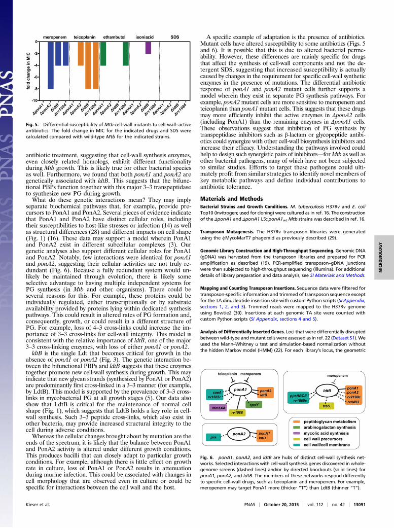
Distinct Cell-Wall Synthesis Networks Exhibit Differential Tolerance toCell-Wall–Active Drugs. Our data suggest that PonA1, PonA2, andLdtB participate in distinct genetic networks with partially over-lapping but largely unique genetic interactions for each enzyme.We hypothesized that these networks would have different cel-lular activity and could therefore be differentially susceptible todrugs that target cell-wall synthesis. To test this hypothesis, wetreated cells that lack ponA1, ponA2, ldtB, or other members oftheir interaction networks with drugs that inhibit the variouscomponents ofMtb’s cell wall, including meropenem, which blocksPG transpeptidases; teicoplanin, which binds directly to PG toprevent further cross-linking (10, 27); ethambutol, which targetsarabinogalactan synthesis; and isoniazid, which targets mycolicacid synthesis. We found that ΔponA1 cells had the same mini-mum inhibitory concentration (MIC) for meropenem and teico-planin as wild-type Mtb (Fig. 5); however, ΔponA2 and ΔldtB cellswere four- to eightfold more susceptible to both meropenem andteicoplanin. Furthermore, mutants that lack ponA1 exhibit afourfold increased susceptibility to ethambutol. However, theΔponA2 or ΔldtB mutants do not exhibit heightened sensitivity
to this antibiotic. ΔponA1, ΔponA2, or ΔldtB cells exhibited nochange in MIC when treated with SDS, and only ΔldtB cells wereslightly more sensitive to isoniazid. The Δrv1086 mutant waseightfold more sensitive to teicoplanin and fourfold more sen-sitive to meropenem and ethambutol compared with wild-typeMtb (Fig. 5). Together, these data suggest that the enzymes thatcomprise distinct PG synthetic networks have different roles inantibiotic tolerance in Mtb.
DiscussionCell-wall synthesis requires the collaboration of multiple en-zymes. In most bacterial species, individual PBPs are dispensable.This is generally interpreted as evidence of functional redundancy,suggesting that enzymes have overlapping functions. Clearly, this isnot entirely true in both Msm and Mtb. In Msm, a single bi-functional PBP, PonA1, is essential for normal growth, but ponA1and ponA2 are both required for robust growth of Mtb duringinfection. Our network analysis suggests that PonA1 and PonA2are not identical—each is genetically associated with overlappingand, importantly, unique factors. This may suggest that althoughPonA1 and PonA2 mediate similar reactions, the pathways bywhich each synthesizes PG are different. This likely has functionalconsequences for bacterial growth in specific conditions. Indeed,we found that mutant cells exhibit differential survival under
Fig. 3. Importance of PG synthases in ponA1, ponA2, or ldtB mutant cells.Sequence reads corresponding to transposon insertions at the indicated lociin the ΔponA1, ΔponA2, and ΔldtB mutant cells. Sequence reads at eachlocus are normalized to the respective locus in wild-type H37Rv cells.
Fig. 4. Diverse cell-wall synthesis factors become required in ΔponA1 cells.(A) Transposon insertions (vertical bars), determined from high-throughputsequencing, are visualized at the ponA2 locus in either wild-type (gray) orΔponA1 (red) cells. (B) Colony-forming units (CFUs) were counted from allelicexchanges with L5-integrating vectors that do or do not encode ponA1 inthe experimental ΔponA1 L5::ponA1wt ΔponA2 strain or the control ΔponA2strain (both control strain transformations had lawn growth, and CFUs werearbitrarily set to 6,000). (C) Transposon insertions at the rv1086 locus in wild-type (gray) and ΔponA1 (red) cells. (D) CFU counts from L5 allelic exchangesin the experimental ΔponA1 L5::ponA1wt Δrv1086 strain or the controlΔrv1086 strain. (E) Transposon insertions at the ldtB locus in wild-type (gray)or ΔponA1 (red) cells. (F) CFU counts from L5 allelic exchanges in the ex-perimental ΔponA1 L5::ponA1wt ΔldtB strain.
13090 | www.pnas.org/cgi/doi/10.1073/pnas.1514135112 Kieser et al.
antibiotic treatment, suggesting that cell-wall synthesis enzymes,even closely related homologs, exhibit different functionalityduring Mtb growth. This is likely true for other bacterial speciesas well. Furthermore, we found that both ponA1 and ponA2 aregenetically associated with ldtB. This suggests that the bifunc-tional PBPs function together with this major 3–3 transpeptidaseto synthesize new PG during growth.What do these genetic interactions mean? They may imply
separate biochemical pathways that, for example, provide pre-cursors to PonA1 and PonA2. Several pieces of evidence indicatethat PonA1 and PonA2 have distinct cellular roles, includingtheir susceptibilities to host-like stresses or infection (14) as wellas structural differences (28) and different impacts on cell shape(Fig. 1) (16). These data may support a model wherein PonA1and PonA2 exist in different subcellular complexes (3). Ourgenetic analyses also support different cellular roles for PonA1and PonA2. Notably, few interactions were identical for ponA1and ponA2, suggesting their cellular activities are not truly re-dundant (Fig. 6). Because a fully redundant system would un-likely be maintained through evolution, there is likely someselective advantage to having multiple independent systems forPG synthesis (in Mtb and other organisms). There could beseveral reasons for this. For example, these proteins could beindividually regulated, either transcriptionally or by substrateavailability provided by proteins lying within dedicated synthesispathways. This could result in altered rates of PG formation and,consequently, growth, or could result in a different structure ofPG. For example, loss of 4–3 cross-links could increase the im-portance of 3–3 cross-links for cell-wall integrity. This model isconsistent with the relative importance of ldtB, one of the major3–3 cross-linking enzymes, with loss of either ponA1 or ponA2.ldtB is the single Ldt that becomes critical for growth in the
absence of ponA1 or ponA2 (Fig. 3). The genetic interaction be-tween the bifunctional PBPs and ldtB suggests that these enzymestogether promote new cell-wall synthesis during growth. This mayindicate that new glycan strands (synthesized by PonA1 or PonA2)are predominantly first cross-linked in a 3–3 manner (for example,by LdtB). This model is supported by the prevalence of 3–3 cross-links in mycobacterial PG at all growth stages (5). Our data alsoshow that LdtB is critical for the maintenance of normal cellshape (Fig. 1), which suggests that LdtB holds a key role in cell-wall synthesis. Such 3–3 peptide cross-links, which also exist inother bacteria, may provide increased structural integrity to thecell during adverse conditions.Whereas the cellular changes brought about by mutation are the
ends of the spectrum, it is likely that the balance between PonA1and PonA2 activity is altered under different growth conditions.This produces bacilli that can closely adapt to particular growthconditions. For example, although there is little effect on growthrate in culture, loss of PonA1 or PonA2 results in attenuationduring murine infection. This could be associated with changes incell morphology that are observed even in culture or could bespecific for interactions between the cell wall and the host.
A specific example of adaptation is the presence of antibiotics.Mutant cells have altered susceptibility to some antibiotics (Figs. 5and 6). It is possible that this is due to altered bacterial perme-ability. However, these differences are mainly specific for drugsthat affect the synthesis of cell-wall components and not the de-tergent SDS, suggesting that increased susceptibility is actuallycaused by changes in the requirement for specific cell-wall syntheticenzymes in the presence of mutations. The differential antibioticresponse of ponA1 and ponA2 mutant cells further supports amodel wherein they exist in separate PG synthesis pathways. Forexample, ponA2mutant cells are more sensitive to meropenem andteicoplanin than ponA1mutant cells. This suggests that these drugsmay more efficiently inhibit the active enzymes in ΔponA2 cells(including PonA1) than the remaining enzymes in ΔponA1 cells.These observations suggest that inhibition of PG synthesis bytranspeptidase inhibitors such as β-lactam or glycopeptide antibi-otics could synergize with other cell-wall biosynthesis inhibitors andincrease their efficacy. Understanding the pathways involved couldhelp to design such synergistic pairs of inhibitors—forMtb as well asother bacterial pathogens, many of which have not been subjectedto similar studies. Efforts to target these pathogens could ulti-mately profit from similar strategies to identify novel members ofkey metabolic pathways and define individual contributions toantibiotic tolerance.
Materials and MethodsBacterial Strains and Growth Conditions. M. tuberculosis H37Rv and E. coliTop10 (Invitrogen; used for cloning) were cultured as in ref. 16. The constructionof the ΔponA1 and ΔponA1 L5::ponA1wt Mtb strains was described in ref. 16.
Transposon Mutagenesis. The H37Rv transposon libraries were generatedusing the ϕMycoMarT7 phagemid as previously described (29).
Genomic Library Construction and High-Throughput Sequencing. Genomic DNA(gDNA) was harvested from the transposon libraries and prepared for PCRamplification as described (19). PCR-amplified transposon–gDNA junctionswere then subjected to high-throughput sequencing (Illumina). For additionaldetails of library preparation and data analysis, see SI Materials and Methods.
Mapping and Counting Transposon Insertions. Sequence data were filtered fortransposon-specific information and trimmed of transposon sequence exceptfor the TA dinucleotide insertion site with custom Python scripts (SI Appendix,sections 1, 2, and 3). Trimmed reads were mapped to the H37Rv genomeusing Bowtie2 (30). Insertions at each genomic TA site were counted withcustom Python scripts (SI Appendix, sections 4 and 5).
Analysis of Differentially Inserted Genes. Loci that were differentially disruptedbetweenwild-type andmutant cells were assessed as in ref. 22 (Dataset S1).Weused the Mann–Whitney u test and simulation-based normalization withoutthe hidden Markov model (HMM) (22). For each library’s locus, the geometric
Fig. 5. Differential susceptibility ofMtb cell-wall mutants to cell-wall–activeantibiotics. The fold change in MIC for the indicated drugs and SDS werecalculated compared with wild-type Mtb for the indicated strains.
Fig. 6. ponA1, ponA2, and ldtB are hubs of distinct cell-wall synthesis net-works. Selected interactions with cell-wall synthesis genes discovered in whole-genome screens (dashed lines) and/or by directed knockouts (solid lines) forponA1, ponA2, and ldtB. The members of these networks respond differentlyto specific cell-wall drugs, such as teicoplanin and meropenem. For example,meropenem may target PonA1 more (thicker “T”) than LdtB (thinner “T”).
Kieser et al. PNAS | October 20, 2015 | vol. 112 | no. 42 | 13091
MICRO
BIOLO
GY

mean of the sequence reads was calculated (Dataset S2) with a customMATLAB script using the encoded geomean function (SI Appendix, section 10).
Recombinant DNA Constructs and Gene Knockouts. ponA1 was subcloned intothe L5 pMC1s vector as in ref. 16. The ponA2 knockout cassette was ampli-fied by PCR from a custom phage (31). The rv1086 and ldtB knockout cas-settes consisted of 500 nucleotides 5′ or 3′ of rv1086 or ldtB flanking ahygromycin cassette with PmeI sites at each end (Gen9). Digestion with PmeI(New England Biolabs) generated linear recombineering products. The ponA2,ldtB, and rv1086 deletions were generated in the ΔponA1 L5::ponA1wt strain(16) via recombineering (for more details, see SI Materials and Methods).
Allelic Exchange in Mtb. Allelic exchange occurs as described (16, 23). TheΔponA1 L5::ponA1wt loxed strain wherein ponA2, rv1086, or ldtB was de-leted was used for allelic exchange. Simultaneous control transformationswith the same L5 vectors were performed in ΔponA2, Δrv1086, and ΔldtBcells. Colony-forming units were counted at 21 d, except for the ΔponA1 L5::tetRΔldtB plate, which was counted at 35 d.
Optical Density Measurements. Population growth curves forMtb strains wereperformed as in ref. 16.
Light Microscopy and Image Analysis. Cells were fixed overnight in 1% for-malin at 4 °C in the Biosafety Level 3 facility. Cells were imaged and mor-phology analyzed as in ref. 16. Final images were prepared in Fiji (32).
Antibiotic MIC Assays. The antibacterial effects of meropenem, teicoplanin,ethambutol, isoniazid, and SDS (Sigma Aldrich) were determined as in ref. 16.
Data Representation and Statistical Analysis. Prism 6.0 (GraphPad Software)was used tographandanalyzenumerical data. Statistical tests in Prism calculatedsignificance of measurements as reported in figure legends. The Venn diagramwas generated with BioVenn (33). Transposon insertions were visualized inDNAplotter (34) or Artemis (35) by converting the insertion counts to appro-priate data structures with custom Python scripts (SI Appendix, sections 8 and 9).
ACKNOWLEDGMENTS. We thank the E.J.R. and Fortune laboratories forthoughtful discussions and technical expertise and M. Larsen for the phageencoding the ponA2 knockout cassette. This work was supported by Na-tional Institutes of Health Grants U19 AI107774 (to E.J.R., T.R.I., C.M.S.,and J.C.S.), U01 GM094568 (to E.J.R.), F32 GM108355-02 (to M.C.C.), andF32 A1093049 (to J.E.L.), the Howard Hughes Medical Institute (C.M.S. andM.K.W.), and a National Science Foundation Graduate Research Fellowship(Grants DGE1144152 and DGE0946799 to K.J.K.).
1. World Health Organization (2014) Global Tuberculosis Report (World Health Orga-
nization, Geneva).2. Kieser KJ, Rubin EJ (2014) How sisters grow apart: Mycobacterial growth and division.
Nat Rev Microbiol 12(8):550–562.3. Typas A, Banzhaf M, Gross CA, Vollmer W (2012) From the regulation of peptidoglycan
synthesis to bacterial growth and morphology. Nat Rev Microbiol 10(2):123–136.4. Lavollay M, et al. (2008) The peptidoglycan of stationary-phase Mycobacterium tu-
berculosis predominantly contains cross-links generated by L,D-transpeptidation.
J Bacteriol 190(12):4360–4366.5. Kumar P, et al. (2012) Meropenem inhibits D,D-carboxypeptidase activity in Myco-
bacterium tuberculosis. Mol Microbiol 86(2):367–381.6. Dubée V, et al. (2012) Inactivation of Mycobacterium tuberculosis l,d-transpeptidase
LdtMt1 by carbapenems and cephalosporins. Antimicrob Agents Chemother 56(8):
4189–4195.7. Wivagg CN, Bhattacharyya RP, Hung DT (2014) Mechanisms of β-lactam killing and re-
sistance in the context of Mycobacterium tuberculosis. J Antibiot (Tokyo) 67(9):645–654.8. Cole ST, et al. (1998) Deciphering the biology of Mycobacterium tuberculosis from the
complete genome sequence. Nature 393(6685):537–544.9. Dasgupta A, Datta P, Kundu M, Basu J (2006) The serine/threonine kinase PknB of
Mycobacterium tuberculosis phosphorylates PBPA, a penicillin-binding protein re-
quired for cell division. Microbiology 152(Pt 2):493–504.10. Hugonnet J-E, Tremblay LW, Boshoff HI, Barry CE, 3rd, Blanchard JS (2009) Meropenem-
clavulanate is effective against extensively drug-resistant Mycobacterium tuberculosis.
Science 323(5918):1215–1218.11. Sanders AN, Wright LF, Pavelka MS, Jr (2014) Genetic characterization of mycobac-
terial L,D-transpeptidases. Microbiology 160(Pt 8):1795–1806.12. Gupta R, et al. (2010) The Mycobacterium tuberculosis protein LdtMt2 is a nonclassical
transpeptidase required for virulence and resistance to amoxicillin.NatMed 16(4):466–469.13. Schoonmaker MK, Bishai WR, Lamichhane G (2014) Nonclassical transpeptidases of
Mycobacterium tuberculosis alter cell size, morphology, the cytosolic matrix, protein
localization, virulence, and resistance to β-lactams. J Bacteriol 196(7):1394–1402.14. Patru MM, Pavelka MS, Jr (2010) A role for the class A penicillin-binding protein
PonA2 in the survival of Mycobacterium smegmatis under conditions of nonreplication.
J Bacteriol 192(12):3043–3054.15. Hett EC, Chao MC, Rubin EJ (2010) Interaction and modulation of two antagonistic
cell wall enzymes of mycobacteria. PLoS Pathog 6(7):e1001020.16. Kieser KJ, et al. (2015) Phosphorylation of the peptidoglycan synthase PonA1 governs
the rate of polar elongation in mycobacteria. PLoS Pathog 11(6):e1005010.17. Vandal OH, Pierini LM, Schnappinger D, Nathan CF, Ehrt S (2008) A membrane protein
preserves intrabacterial pH in intraphagosomal Mycobacterium tuberculosis. Nat Med
14(8):849–854.
18. Vandal OH, et al. (2009) Acid-susceptible mutants of Mycobacterium tuberculosisshare hypersusceptibility to cell wall and oxidative stress and to the host environment.J Bacteriol 191(2):625–631.
19. Long JE, et al. (2015) Identifying essential genes in Mycobacterium tuberculosis byglobal phenotypic profiling. Methods Mol Biol 1279:79–95.
20. Joshi SM, et al. (2006) Characterization of mycobacterial virulence genes throughgenetic interaction mapping. Proc Natl Acad Sci USA 103(31):11760–11765.
21. Chao MC, et al. (2013) High-resolution definition of the Vibrio cholerae essential geneset with hidden Markov model-based analyses of transposon-insertion sequencingdata. Nucleic Acids Res 41(19):9033–9048.
22. Pritchard JR, et al. (2014) ARTIST: High-resolution genome-wide assessment of fitnessusing transposon-insertion sequencing. PLoS Genet 10(11):e1004782.
23. Blasco B, et al. (2012) Virulence regulator EspR of Mycobacterium tuberculosis is anucleoid-associated protein. PLoS Pathog 8(3):e1002621.
24. Parthasarathy G, et al. (2012) Rv2190c, an NlpC/P60 family protein, is required for fullvirulence of Mycobacterium tuberculosis. PLoS One 7(8):e43429.
25. Pashley CA, Parish T (2003) Efficient switching of mycobacteriophage L5-basedintegrating plasmids in Mycobacterium tuberculosis. FEMS Microbiol Lett 229(2):211–215.
26. Schulbach MC, et al. (2001) Purification, enzymatic characterization, and inhibition ofthe Z-farnesyl diphosphate synthase from Mycobacterium tuberculosis. J Biol Chem276(15):11624–11630.
27. Dong SD, et al. (2002) The structural basis for induction of VanB resistance. J AmChem Soc 124(31):9064–9065.
28. Calvanese L, et al. (2014) Structural and binding properties of the PASTA domain ofPonA2, a key penicillin binding protein from Mycobacterium tuberculosis. Biopolymers101(7):712–719.
29. Sassetti CM, Boyd DH, Rubin EJ (2001) Comprehensive identification of conditionallyessential genes in mycobacteria. Proc Natl Acad Sci USA 98(22):12712–12717.
30. Langmead B, Salzberg SL (2012) Fast gapped-read alignment with Bowtie 2. NatMethods 9(4):357–359.
31. Jain P, et al. (2014) Specialized transduction designed for precise high-throughputunmarked deletions in Mycobacterium tuberculosis. MBio 5(3):e01245–e14.
32. Schindelin J, et al. (2012) Fiji: An open-source platform for biological-image analysis.Nat Methods 9(7):676–682.
33. Hulsen T, de Vlieg J, AlkemaW (2008) BioVenn - a web application for the comparisonand visualization of biological lists using area-proportional Venn diagrams. BMCGenomics 9:488.
34. Carver T, Thomson N, Bleasby A, Berriman M, Parkhill J (2009) DNAPlotter: Circularand linear interactive genome visualization. Bioinformatics 25(1):119–120.
35. Carver T, Harris SR, BerrimanM, Parkhill J, McQuillan JA (2012) Artemis: An integratedplatform for visualization and analysis of high-throughput sequence-based experimentaldata. Bioinformatics 28(4):464–469.
13092 | www.pnas.org/cgi/doi/10.1073/pnas.1514135112 Kieser et al.