Peptides for Autoimmune diseases
42
Peptides for Autoimmune diseases Dr. Luis Martinez Regenera Global XanoGene Clinic
Transcript of Peptides for Autoimmune diseases

Peptides for Autoimmune diseases
Dr. Luis Martinez
Regenera Global
XanoGene Clinic

Objectives
DESCRIBE IMMUNE DYSREGULATION IN AUTOIMMUNE DISEASES
DISCUSS MECHANISMS OF ACTION FOR IMMUNE MODULATION AS EXERTED BY
MULTIPLE PEPTIDES
DISCUSS CASES AND PROTOCOLS FOR INCORPORATING PEPTIDES IN THE
MANAGEMENT OF IMMUNE DISEASES.

Autoimmune disease statistics
50 million Americans with ADs
80-100 different autoimmune diseases identified with evidence suggesting 40 more
ADs share close genetic relationships
Common immunosuppressants lead to marked side effects
New treatment options are required to address ADs

Autoimmune Disorders-affected systems
• Thyroid: Graves disease, Hashimoto’s
• Bones: Rheumatoid arthritis, ankylosing spondylitis
• Brain/Nervous system: MS, ALS, GBS
• Gastrointestinal system: Crohn’s disease, Ulcerative colitis
• Skin: Vitiligo, psoriasis

Peptides and ADs
CAN MODULATE IMMUNE SYSTEM
INCREASED TREGS TH1-TH2 SHIFTS
TH17
CYTOKINE MODULATION

Peptide based immunotherapy
Can serve as adjunct to current approaches to managing autoimmune
diseases
Perceived challenges
Indications Availability Continuity of care


Key Concepts in Autoimmunity

http://drugline.org/img/term/autoimmunity-1507_1.gif
Self vs Non Self
Antigens Identification

TE vs TR
It is all in the balance


http://sanjosefuncmed.com/wp-content/uploads/2013/05/pathogenesis-leaky-gut-autoimmune-connection.jpg
Leaky gut and autoimmunity
Is it all in the gut?

Central Tolerance
The regulatory environment


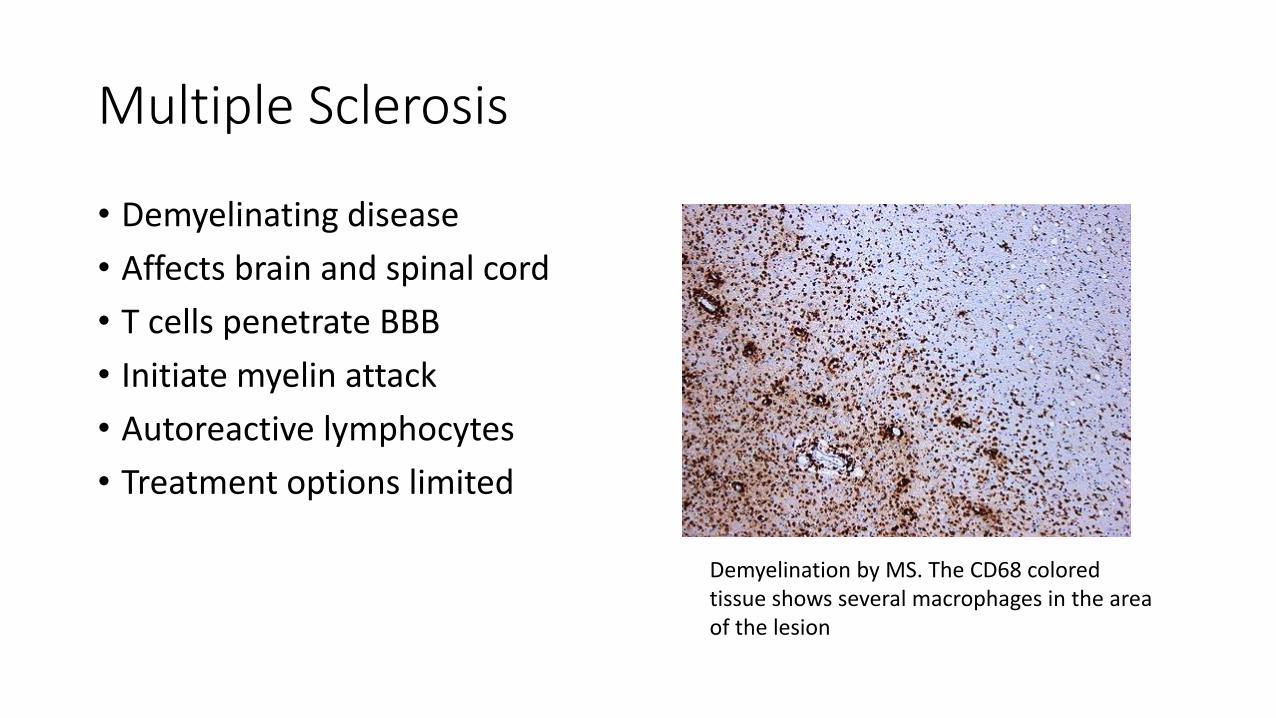
Multiple Sclerosis
• Demyelinating disease
• Affects brain and spinal cord
• T cells penetrate BBB
• Initiate myelin attack
• Autoreactive lymphocytes
• Treatment options limited
Demyelination by MS. The CD68 colored tissue shows several macrophages in the area of the lesion

The Peptides

Opioid Growth Factor (OGF)
Opioid Growth Factor (OGF) known as
Metkephalin (Met5)
Endogenous pentapepide
OGF activates a specific receptor called Opioid Growth Factor receptor (OGFr or ζ-
opioid receptor).
OGF and OGFr axis regulates cell growth
in normal and abnormal cells

OGF and Immunity
OGF blocks release of proinflammatory cytokines including Interleukins IL6 and IL12, TNFα, NF-ĸB (nuclear factor kappa light chain enhancer of activated B cells)
Can alter T and B lymphocyte production
Modulates immune response TH2/TH1/T17
Biphasic response
Increased regulatory T cells

Brain Res. 2012 Sep 7;1472:138-48. doi: 10.1016/j.brainres.2012.07.006. Epub 2012 Jul 20.Opioid growth factor arrests the progression of clinical disease and spinal cord pathology in established experimental autoimmune encephalomyelitis.Campbell AM1, Zagon IS, McLaughlin PJ.Author informationAbstractAn endogenous neuropeptide, opioid growth factor (OGF), chemically termed [Met(5)]-enkephalin, arrested the progression of established disease in a mouse model of multiple sclerosis (MS) called experimental autoimmune encephalomyelitis (EAE). This study treated mice who demonstrated 2 consecutive days of behavioral decline following injections of myelin oligodendrocyte glycoprotein (MOG) with daily injections of OGF (10mg/kg) or saline (0.1ml) for 40 days. Within 6 days of OGF treatment, mice initially demonstrating clinical signs of EAE had significant reductions (45% reduction) in their behavioral scores relative to EAE mice receiving saline. Behavior was attenuated for the entire 40-day period with mice receiving OGF showing only limp tails and wobbly gait in comparison to saline-treated EAE mice who displayed paralysis of one or more limbs. Neuropathological studies revealed that OGF treatment initiated after the appearance of disease reduced the number of activated astrocytes and damaged neurons, decreased demyelination, and inhibited T cell proliferation. These results demonstrate that OGF can halt the progression of established EAE, return aberrant pain sensitivity to normal levels, inhibit proliferation of T cells and astrocytes, and prevent further spinal cord pathology. The data extend our observations that OGF given at the time of disease induction prevented disease onset, reduced the severity of clinical signs of disease, and reversed neurological deficits in a non-toxic manner. Our data substantiate the role of the OGF-OGFr axis in EAE and support the use of OGF as a biotherapy for MS.

Exp Biol Med (Maywood). 2017 Sep;242(15):1524-1533. doi: 10.1177/1535370217724791. Epub 2017 Aug 2.Featured Article: Serum [Met5]-enkephalin levels are reduced in multiple sclerosis and restored by low-dose naltrexone.Ludwig MD1, Zagon IS1, McLaughlin PJ1.Author informationAbstract
Low-dose naltrexone is a widely used off-label therapeutic prescribed for a variety of immune-related disorders. The mechanism underlying low-dose naltrexone's efficacy for fatigue, Crohn's disease, fibromyalgia, and multiple sclerosis is, in part, intermittent blockade of opioid receptors followed by upregulation of endogenous opioids. Short, intermittent blockade by naltrexone specifically blocks the opioid growth factor receptor resulting in biofeedback events that increase production of the endogenous opioid growth factor (OGF) (chemically termed [Met5]-enkephalin) facilitating interactions between opioid growth factor and opioid growth factor receptor that ultimately, result in inhibited cell proliferation. Preclinical studies have reported that enkephalin levels are deficient in animal models of experimental autoimmune encephalomyelitis, a mouse model of multiple sclerosis. Our hypothesis is that serum enkephalin levels are diminished in humans with multiple sclerosis and experimental autoimmune encephalomyelitis mice, and that change in serum opioid growth factor levels may serve as a reasonable candidate biomarker for the onset of experimental autoimmune encephalomyelitis and response to therapy. To address this, we designed a two-part study to measure endogenous opioids in multiple sclerosis patients, and to investigate the temporal pattern of decline in serum enkephalin concentrations in mice with chronic progressive experimental autoimmune encephalomyelitis and treated with low-dose naltrexone. For comparison, we investigated whether low-dose naltrexone exposure in normal mice also resulted in altered enkephalin levels. In both animal models, we monitored tactile and heat sensitivity, as well as differential white blood cell counts as indicators of inflammation. Serum [Met5]-enkephalin levels were lower in humans with multiple sclerosis relative to non-multiple sclerosis patients, and low-dose naltrexone restored their levels. In experimental autoimmune encephalomyelitis mice, [Met5]-enkephalin levels were depressed prior to the appearance of clinical disease, and were restored with low-dose naltrexone treatment. Low-dose naltrexone therapy had no effect on serum [Met5]-enkephalin or β-endorphin in normal mice. Thus, [Met5]-enkephalin (i.e. opioid growth factor) may be a reasonable candidate biomarker for multiple sclerosis, and may signal new pathways for treatment of autoimmune disorders. Impact statement This report presents human and animal data identifying a novel biomarker for the onset and
progression of multiple sclerosis (MS). Humans diagnosed with MS have reduced serum levels of OGF (i.e. [Met5]-enkephalin) relative to non-MS neurologic patients, and low-dose naltrexone (LDN) therapy restored their enkephalin levels. Serum OGF levels were reduced in mice immunized with MOG35-55 prior to any clinical behavioral sign of experimental autoimmune encephalomyelitis, and LDN therapy restored their serum OGF levels. β-endorphin concentrations were not altered by LDN in humans or mice. Thus, blood levels of OGF may serve as a new, selective biomarker for the progression of MS, as well as response to therapy.

Thymosin Alpha 1 (TA-1)
• Endogenous human peptide
• Derived from prothymosin
• 28 aa fragment
• Helps restore immune function (remember thymic involution and aging)
• Enhances cell mediated immunity
• Increases efficiency of antigen presenting cells

Mult Scler. 2018 Feb;24(2):127-139. doi: 10.1177/1352458517695892. Epub 2017 Feb 1.Thymosin-α1 expands deficient IL-10-producing regulatory B cell subsets in relapsing-remitting multiple sclerosis patients.Giacomini E1, Rizzo F1, Etna MP1, Cruciani M1, Mechelli R2, Buscarinu MC2, Pica F3, D'Agostini C4, Salvetti M2, Coccia EM1, Severa M1.Author informationAbstractBACKGROUND: B cells are key pathogenic effectors in multiple sclerosis (MS) and several therapies have been designed to restrain B cell abnormalities by directly targeting this lymphocyte population.OBJECTIVES: Moving from our data showing a Toll-like receptor (TLR)7-driven dysregulation of B cell response in relapsing-remitting multiple sclerosis (RRMS) and having found a low serum level of Thymosin-α1 (Tα1) in patients, we investigated whether the addition of this molecule to peripheral blood mononuclear cells (PBMCs) would influence the expansion of regulatory B cell subsets, known to dampen autoimmune inflammation.METHODS: Serum Tα1 level was measured by enzyme immunoassay. Cytokine expression was evaluated by Cytometric Bead Array (CBA), enzyme-linked immunosorbent assay (ELISA), and real-time reverse transcription polymerase chain reaction (RT-PCR). B cell subsets were analyzed by flow cytometry.RESULTS: Tα1 pre-treatment induces an anti-inflammatory status in TLR7-stimulated RRMS PBMC cultures, reducing the secretion of pro-inflammatory interleukin (IL)-6, IL-8, and IL-1β while significantly increasing the regulatory IL-10 and IL-35. Indeed, Tα1 treatment enhanced expansion of CD19+CD24+CD38hi transitional-immature and CD24low/negCD38hi plasmablast-like regulatory B cell subsets, which likely inhibit both interferon (IFN)-γ and IL-17 production.CONCLUSION: Our study reveals a deficient ability of B cells from MS patients to differentiate into regulatory subsets and unveils a novel anti-inflammatory and repurposing potential for Tα1 in MS targeting B cell response

Clin Exp Immunol. 2016 Oct;186(1):39-45. doi: 10.1111/cei.12833. Epub 2016 Aug 1.Serum thymosin α 1 levels in patients with chronic inflammatory autoimmune diseases.Pica F1, Chimenti MS2, Gaziano R1, Buè C1, Casalinuovo IA1, Triggianese P2, Conigliaro P2, Di Carlo D1, Cordero V3, Adorno G3, Volpi A4, Perricone R2, Garaci E1,5.Author informationAbstractThymosin alpha 1 (Tα1) is a powerful modulator of immunity and inflammation. Despite years of studies, there are a few reports evaluating serum Tα1 in health and disease. We studied a cohort of healthy individuals in comparison with patients affected by chronic inflammatory autoimmune diseases. Sera from 120 blood donors (healthy controls, HC), 120 patients with psoriatic arthritis (PsA), 40 with rheumatoid arthritis (RA) and 40 with systemic lupus erythematosus (SLE), attending the Transfusion Medicine or the Rheumatology Clinic at the Policlinico Tor Vergata, Rome, Italy, were tested for Tα1 content by means of a commercial enzyme-linked immunosorbent assay (ELISA) kit. Data were analysedin relation to demographic and clinical characteristics of patients and controls. A gender difference was found in the HC group, where females had lower serum Tα1 levels than males (P < 0·0001). Patients had lower serum Tα1 levels than HC (P < 0·0001), the lowest were observed in PsA group (P < 0·0001 versus all the other groups). Among all patients, those who at the time of blood collection were taking disease-modifying anti-rheumatic drugs (DMARD) plus steroids had significantly higher Tα1 levels than those taking DMARD alone (P = 0·044) or no treatment (P < 0·0001), but not of those taking steroids alone (P = 0·280). However, whichever type of treatment was taken by the patients, serum Tα1 was still significantly lower than in HC and there was no treatment-related difference in PsA group. Further prospective studies are necessary to confirm and deepen these observations. They might improve our understanding on the regulatory role of Tα1 in health and disease and increase our knowledge of the pathogenesis of chronic inflammatory autoimmune diseases.


BPC 157
• Composed of 15 amino acids
• Partial sequence of body protection peptide
• Can heal digestive system
• Leaky gut = antigen release into blood stream
• Reduce leaky gut = control source of immune dysregulation
• Additionally beneficial in GI autoimmune diseases (UC/Crohn’s)
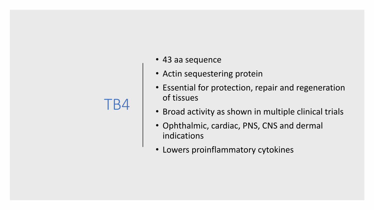
TB4
• 43 aa sequence
• Actin sequestering protein
• Essential for protection, repair and regeneration of tissues
• Broad activity as shown in multiple clinical trials
• Ophthalmic, cardiac, PNS, CNS and dermal indications
• Lowers proinflammatory cytokines

Neurobiol Dis. 2016 Apr;88:85-95. doi: 10.1016/j.nbd.2016.01.010. Epub 2016 Jan 12.Thymosin beta4 promotes oligodendrogenesis in the demyelinating central nervous system.Zhang J1, Zhang ZG2, Li Y2, Lu M3, Zhang Y2, Elias SB2, Chopp M4.Author informationAbstractMultiple sclerosis (MS) is a demyelinating disease of the central nervous system (CNS). No effective remyelination therapies are in use. We hypothesized that thymosin beta4 (Tβ4) is an effective remyelination treatment by promoting differentiation of oligodendrocyte progenitor cells (OPCs), and that the epidermal growth factor receptor (EGFR) signaling pathway contributes to this process. Two demyelination animal models were employed in this study: 1) experimental autoimmune encephalomyelitis (EAE), an animal model of MS. EAE mice were treated daily for 30days, with Tβ4 or saline treatment initiated on the day of EAE onset; and 2) cuprizone diet model, a non-inflammatory demyelination model. The mice were treated daily for 4weeks with Tβ4 or saline after fed a cuprizone diet for 5weeks. Immunofluorescent staining and Western blot were performed to measure the differentiation of OPCs, myelin and axons, respectively. To obtain insight into mechanisms of action, the expression and activation of the EGFR pathway was measured. AG1478, an EGFR inhibitor, was employed in a loss-of-function study. Data revealed that animals in both demyelination models exhibited significant reduction of myelin basic protein (MBP(+)) levels and CNPase(+) oligodendrocytes. Treatment of EAE mice with Tβ4 significantly improved neurological outcome. Double immunofluorescent staining showed that Tβ4 significantly increased the number of newly generated oligodendrocytes identified by BrdU(+)/CNPase(+) cells and MBP(+) mature oligodendrocytes, and reduced axonal damage in the EAE mice compared with the saline treatment. The newly generated mature oligodendrocytes remyelinated axons, and the increased mature oligodendrocytes significantly correlated with functional improvement (r=0.73, p<0.05). Western blot analysis revealed that Tβ4 treatment increased expression and activation of the EGFR pathway. In the cuprizone demyelination model, Tβ4 treatment was confirmed that significantly increased OPC differentiation and remyelination, and increased the expression of EGFR and activated the EGFR pathway in the demyelinating corpus callosum. In cultured OPCs, blockage of the activation of the EGFR pathway with AG1478 abolished the Tβ4-increased OPC differentiation. Collectively, these findings indicate that: 1) Tβ4 increases proliferation of OPCs and the maturation of OPCs to myelinating oligodendrocytes which in concert, likely contribute to the beneficial effect of Tβ4 on EAE, 2)
EGFR upregulated and activated by Tβ4 may mediate the process of OPC differentiation, and 3) Tβ4 could potentially be developed as a therapy for MS patients, and for other demyelinating neurological

Vasoactive Intestinal Peptide (VIP)
28 aa peptideBelongs to
glucagon/secretin superfamily
Produced in many tissues and organs
Multiple functions (vasodilation,
glycogenolysis, increases heart
contractility)
Short half lifeHas shown promise in
regulating autoimmunity

VIP and innate immune cell function
Mostly excitatory effects on mast cells
Inhibitory effects on macrophages and monocytes (VPAC 1 receptor)
Stimulation of IL10
Down regulation of Toll like receptors
Increased Tregs
Decreased TH1 cytokine expression


Putting it all together: Integrative Approach
Immuneregulation:
TA1
OGF/Met Enk
VIP
Neuro repair TB4
GI/Leaky gut BPC 157
Systemic repair: HGH analogues/secretagogues

Dosing
Peptide Dosing Comments
TA1 450 mcg SQ Evaluate clinical response and markers; titrate adjustingly
TB 4 750mcg SQ Alternate dosing after 1st month
VIP 50mcg QID Alternating nostrils; watch out for adverse effects; taper down post 1 month; check lipase
OGF 250mcg/kg IV Consider LDN as an option due to current limits with OFG/Met Enk
BPC 157 200 mcg SQ daily; Alt 500 mcg caps PO

Some pearls
• Always correct the gut first before starting VIP tx; will allow for a faster, more effective clinical response
• May start TA1 alongside gut repair/BPC
• Addressing the environment may be challenging, reason why it is more about fortifying and regulating bodies barriers and immune response
• Chelation therapy can help greatly if metals an issue (lymphocyte dysregulation with metal toxicity)

Case example
• 25 y/o female patient with 2 year hx of SLE
• ANA +, Anti-dsDNA +, elevated ESR (increasing)
• Malar rash and synovitis of MCP,PIP joints
• Had been on/off biologics, currently on steroid tx
• Referring flare ups not adequately controlled by current pharmacotherapy

Continued
• Refers history of asthma and IBS
• Allergic to cat hair, pollen
• Lactose intolerant
• Started on gluten free diet
• BPC SQ QD + TA1 SQ QD
• Two weeks after, with notable improvement in GI issues, VIP was started IN QID

Continued
• 6 weeks later, ESR near normal
• Arthralgia gone
• Steroid weaning started (now at 5mg prednisone QD)
• Rash diminished
• Patient feeling “better than ever”
• VIP reduced to 2x day
• BPC and TA 1 d/c

Conclusions
• Autoimmune diseases share some common altered immunological pathways
• TH subset balance, Tregs predominance, chronic inflammation and leaky gut tend to be presentations/causes
• Multiple peptides have shown immunomodulatory as well as anti inflammatory and tissue repairing effects
• Can be considered as part of an integrative approach for the management of autoimmune diseases
• Additional studies will shed new light on future applications and peptides