Pediatric Cervical Spine OMT Module · OMT Treatment Guidelines For unstable, symptomatic cervical...
44
Pediatric Cervical Spine OMT Module AOBP with thanks to: Dawn C. Dillinger, DO Robert Hostoffer, DO, FACOP, FAAP Eric Hegybeli, DO
Transcript of Pediatric Cervical Spine OMT Module · OMT Treatment Guidelines For unstable, symptomatic cervical...
Pediatric Cervical Spine OMT Module
AOBP with thanks to:
Dawn C. Dillinger, DO
Robert Hostoffer, DO,
FACOP, FAAP
Eric Hegybeli, DO

Cervical Vertebrae

Cervical Spine
Provides pathway for neural, vascular and
musculoskeletal communication between
head and thorax
Consists of 7 vertebrae: C1 and C2 are
atypical and C3-7 are typical
Posterior spinal muscles are continuous from
the cervical spine to the sacrum

Muscles of Neck

Deeper Muscles of Neck

Posterior Neck Muscles

Atypical Cervical Vertebrae

Neural and Vascular Communications of the Neck

Pediatric Cervical Spine Anomalies
May be seen isolated or in association with
other disorders
Most remain asymptomatic and undiagnosed
Patients with known associations should
have an evaluation of the cervical spine
Neck stability depends on integrity of
surrounding ligaments and joint capsules


Causes of Pediatric Cervical Instability
Congenital – Vertebral or bony anomalies
– Ligamentous or combined ligamentous and bony
– Syndromic disorders (ex: Down Syndrome)
Acquired – Trauma
– Infection
– Inflammatory (JRA)
– Metabolic (Rickets)
– Tumor (including neurofibromatosis)

Osteopathic Exam of the Cervical Spine
Inspection of skin and asymmetry in position of the
neck
Active motion testing by the patient through all
ranges of motion
Palpation of the cervical spine in first the seated and
then supine positions
Passive motion testing of both regional and
segmental motion to determine somatic dysfunction

C1 Structure (Atlas)
1. Posterior Arch
2. Transverse Foramin
3. Transverse Process
4. Inferior Articular Facet
5. Anterior Arch
The primary motions are flexion and extension
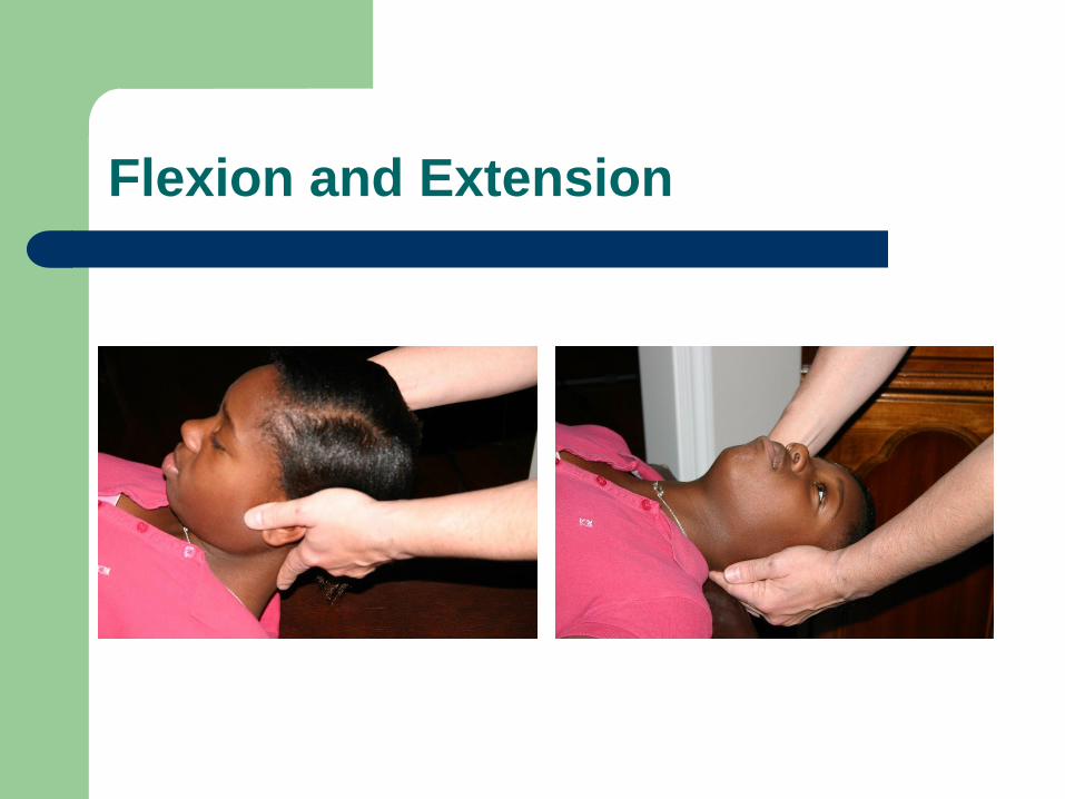
Flexion and Extension

C2 structure (Axis)
1. Posterior arch
2.Transverse Foramin
3.Transverse Process
4. Inferior Articular facet
5. Anterior Arch
The primary motion is
rotation

Rotation

Cervical Spine (C3 through C7)
1. Spinous process
2. Superior articular facets
3. Vertebral foramen
4. Transverse foramina
5. Transverse processes
6. Body
The primary motion of these
vertebrae is sidebending
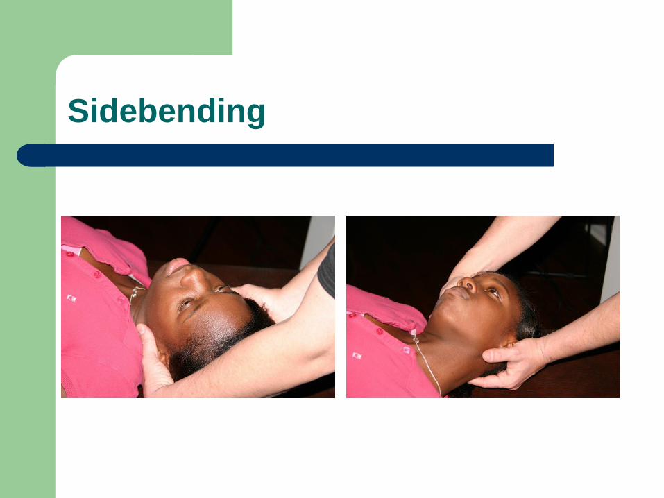
Sidebending

Neck Injuries
Most common injuries to the neck in children are soft
tissue
– Contusions, muscle strains, ligament sprains
Often overlap between all 3 types of injuries
Cervical disk injuries in sports usually result from
uncontrolled lateral bending
Cervical injuries are less common than lumbar
injuries and are uncommon in pediatrics

Neck Injuries
Red flags in pediatric athletes with neck pain – Midline cervical spine pain
– Neck pain on range of motion
– Focal neurologic defects
– Loss of consciousness
If red flag present, neck fracture needs to be ruled-out with C-spine X-rays (A/P, lateral, oblique, open-mouth)
If unable to actively flex or extend neck: CT scan should be performed
Neck Injuries
Radiographic signs of instability
– Interspinous widening
– Vertebral subluxation
– Vertebral compression fracture
– Loss of cervical lordosis
MRI sensitive in diagnosing ligamentous and
spinal cord injuries

Treatment of Neck Injuries
If negative radiographic exam and normal neurologic
exam, neck can be immobilized in a soft collar for
comfort
Rest and NSAIDS benefit minor injuries
Range of motion exercises when collar is removed
Cervical disk problems may also require cervical
traction
Strength training should be initiated after symptoms
improve to help prevent recurrence

OMT Treatment Guidelines
For unstable, symptomatic cervical spine problems:
– Avoid HVLA of cervical spine
– Decrease muscle tension: may need to treat upper thoracic
and rib dysfunction to accomplish
– Counterstrain, cranial, and indirect techniques are thought
to be least traumatic to the neck
– Muscle energy may be appropriate if it does not cause pain
– Traction may be appropriate with the proper direction of
force

OMT for neck pain
Manipulation of the cervical spine for neck
pain and headache is the second most
common use of spinal manipulative therapy

OMT for neck pain
True incidence of complications from OMT is
unknown since reporting has only been in form of
case reports and surveys
Attempts have been made to relate vertebral basilar
accidents to chiropractic manipulation of cervical
spine, but the literature does not support this relation
Of the 1500 patients reported in clinical trials of
manipulation of multiple kinds, no complications
have been reported

OMT: Cradling and Traction
The physician will place the fingers close to
the cervical spine, but lateral to the spinous
processes bilaterally, then apply an anteriorly
directed force bilaterally with slight traction
through the arms of the physician.


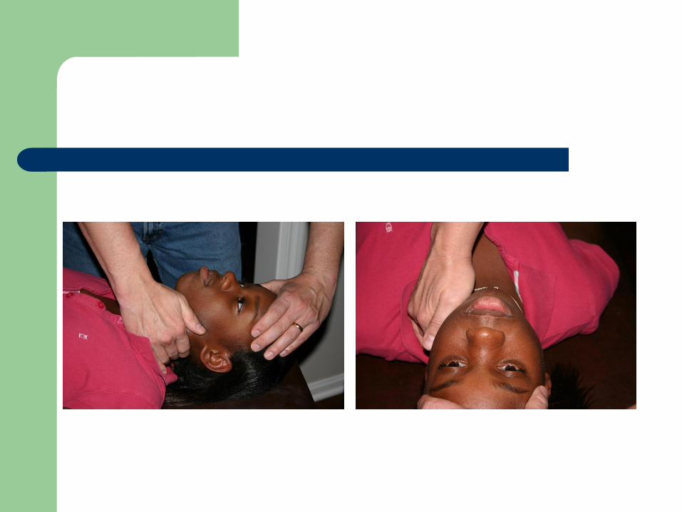
OMT: Counter-Lateral Traction
Place one hand on the frontal bone, the other
hand on the lateral aspect of the cervical
spine along the articular facets. While
applying pressure on the frontal bone away
from you, the other hand stretches the
muscles of the neck toward you.

Occipital-Atlantal Cervical High Velocity Low Amplitude
The patient is supine. Place your hand on the
ramus of the mandible with fingers extending
downward toward the chin. Apply a sudden
increase in the rotation of the neck by
pressing downward toward the table on the
ramus of the mandible, without extending the
cervical spine


3rd to 7th Cervical High Velocity Low Amplitude
The patient is supine. Rotate and sidebend the neck to point of maximum resistance at somatic dysfunction. The index finger of your hand is posterolateral to the articular process.The patient’s head may be flexed or extended depending on the dysfunction and then the corrective thrust is made with the index finger in an arc conforming to the plane of the facets.

HVLA Cervical Spine

Evidence-based Medicine: neck pain
JAOA study 2005 comparing efficacy of single dose ketorolac to OMT for the management of acute neck pain in the ED
Convenience sample of 58 patients 18-50y – 29 patients received ketorolac
– 29 patients received OMT
Subjective measures of pain on an 11-point scale were gathered immediately before intervention and one hour post-intervention
Subjects perceived pain was also recorded at one hour post-intervention on a 5 point pain relief scale

Evidence-based Medicine: neck pain
3 enrolling osteopathic physicians who specialize in
emergency medicine and routinely use OMT in the
ED
Patients randomly assigned to intervention group but
unable to be blinded due to study design
All patients received an initial structural exam
OMT performed included any or a combination of
HVLA thrust, muscle energy and soft tissue
All OMT interventions lasted less than 5 minutes

Evidence-based Medicine: neck pain
OMT group showed a statistically significant
decrease in self-reported pain INTENSITY
No significant difference in perceived pain RELIEF at
one hour between the groups
Some patients had self treated with NSAIDS prior to
ED arrival
OMT was more effective than ketorolac for
decreasing pain levels among the 40 patients who
had not taken NSAIDS within 24 hours of ED arrival

Evidence-based Medicine: neck pain
Although a placebo arm was not used, most
patients in study were unfamiliar with OMT
8 patients reported an adverse event with
ketorolac including: arm soreness, dizziness,
drowsiness, dyspepsia, nausea, vomiting
1 patient reported an adverse event with
OMT which was transient “arm felt funny”

Evidence-based Medicine: neck pain
Authors conclusions: – OMT is significantly better than IM ketorolac at
decreasing pain intensity
– OMT is as efficacious as ketorolac in providing pain relief for acute neck pain in he ED
– Less side effects with OMT
Previous studies suggest patients do better with a combination of medication and manipulation

Question 1
The motion through the first cervical vertebrae is
best described as:
A. Flexion
B. Opposition
C. Rotation
D. Sidebending
E. Translation

Question 2
According to a study of OMT utilized for neck pain in ER patients, which of the following is a possible expected outcome after OMT?
A. Better pain relief than the use of muscle relaxant medication
B. Better pain relief than the use of NSAIDS
C. Decreased pain intensity compared to NSAIDS
D. Increased neck pain
E. No improvement in neck pain

Question 3
The motion through C3-7 is best described as which
of the following?
A. Extension
B. Flexion
C. Opposition
D. Rotation
E. Sidebending

Question 4
A 16 year old football player presents to the ER with neck pain after being tackled. He denies LOC, paresthesias or weakness, but on exam he is unable to actively flex or extend his neck. Which of the following is the imaging of choice?
A. Bone scan
B. CT scan
C. MRI
D. Ultrasound
E. X-ray

Question 5
According to OMT treatment guidelines, how should
the athlete in question 4 be initially treated with
OMT?
A. Counterstrain
B. Facilitated Positional Release
C. HVLA
D. Muscle energy
E. Rib raising

References
McReynold T, Sheridan B. Intramuscular ketorolac versus osteopathic manipulative treatemtn in the management of acute neck pain in the emergency department: a randomized clinical trial. JAOA. 2005; 105(2): 57-68.
Ward R. Foundations for osteopathic medicine. Philadelphia, PA: Lippincott Williams and Wilkins; 2003.
Kliegman R, Jenson H, Behrman R, Stanton B. Nelson textbook of pediatrics, 18th ed. Philadelphia, PA: Saunders Elsevier; 2007.