A new role of PET/CT for target delineation for ... brief review addresses the potential role of PET...
5
www.nuclmed.gr 139 www.nuclmed.gr Hellenic Journal of Nuclear Medicine May - August 2012 Prototype Brief Review Abstract Fluorine-18-fluorodeoxyglucose- positron emission tomography ( 18 F-FDG PET) in head and neck cancer patients is useful for staging, identification of macroscopic disease, detection of invaded lymph nodes and distant metastases, delineation of radiotherapy target volume and assessment of treatment response. This brief review addresses the potential role of PET in radiotherapy plan- ning as compared to MRI and CT scan. Positron emission tomography is considered by radiation oncologists a useful test for the identification of the specific target volume for treatment. In addi- tion, a number of hypoxia-related PET radiopharmaceuticals such as the fluorine-18-fluoromisoni- dazole ( 18 F-FMISO) and the fluorine-18-fluoroazomycin arabinoside ( 18 F-FAZA) are now available in order to identify hypoxic tumor subvolumes helping to implement new radiotherapy techniques. Magnetic resonance imaging (MRI) has the advantage to discriminate the soft tissue contrast from the tumor, against computerized tomography (CT), but PET/CT scans have the additional advan- tage to incorporate the metabolic imaging for improving the delineation of variable and hypoxic tumor tissue in the head and neck region. Regardless of the method used for determining the gross tumor volume, clinical examination remains irreplaceable. In conclusion, PET/CT offers com- plementary information for the delineation of the primary tumor and the corresponding lymph nodes compared to the use of MRI and CT and can support the use of modern radiotherapy tech- niques, having fewer toxicities. Introduction F luorine-18-fluorodeoxyglucose-positron emission tomography ( 18 F-FDG-PET) has been used in head and neck squamous cell carcinoma (HNSCC) patients for stag- ing, identification of macroscopic cancer disease, for the detection of metastatic lymph nodes, distant metastases and for the assessment of treatment response [1, 2]. Avoiding unnecessary radiation of normal tissues may allow dose escalation in inten- sity modulated radiation therapy (IMRT). Due to the high radiation dose administered in IMRT, the localization of the primary tumor and of the cervical lymph nodes metastases is quite important [3, 4]. Currently, computed tomography (CT) assisted volume definition remains the gold standard for radiotherapy planning. However, CT scans are limited by soft tissue resolu- tion, dental artifacts and may fail to clearly identify critical normal structures like in the spinal cord, the optic chiasm and optic nerves [5]. On the other hand, magnetic resonance imaging (MRI) helps in the delineation of vari- ous organs and offers excellent soft tissue discrimination [6, 7]. However, MRI and PET do not provide electron density data required for dosimetric calculation. This brief review addresses the promising role of 18 F-FDG-PET scan in radiotherapy plan- ning as related with MRI and the CT scan. Details of the search Our literature review was based on database search in PubMed/MEDLINE from 2000- May 2012. Terms used were: head and neck cancer, MRI, PET, delineation, contour, radiotherapy and synonyms combined with one or more of the following: radiation, three dimentional conformal radiotherapy (3D-CRT), intensity modulated radiotherapy (IMRT), PET-CT, diffusion MRI (DW-MRI). Furthermore, these terms were combined with the respective keywords for each paragraph. Only papers published in English were included. A new role of PET/CT for target delineation for radiotherapy treatment planning for head and neck carcinomas Anna Zygogianni 1,2 MD, PhD George Kyrgias 2 MD, PhD John Kouvaris 1 MD, PhD Kyriaki Pistevou-Gompaki 3 MD, PhD Vassilis Kouloulias 4 MSc, MD, PhD 1. 1 st Radiology Department, Radiotherapy Unit, Aretaieion University Hospital, Athens, Greece 2. Radiotherapy Department, Larisa University Hospital, University of Thessaly, Greece 3. Department of Radiotherapy, AHEPA University Hospital, Thessaloniki, Greece 4. 2 nd Radiology Department, Radiotherapy Unit, Attikon University Hospital, Athens, Greece *** Keywords: PET/CT - Head and neck cancer - Target delineation - Radiotherapy Correspondence address: Dr. A. Zygogianni, Lecturer of Radiation Oncology University of Thessaly, Medical School, Biopolis, Larissa 41110, Greece. Email: [email protected] Tel: +302413502054 Mobile: +306977748628 Fax: +302413501013 Received: 24 June 2012 Accepted revised: 20 July 2012 Hell J Nucl Med 2012; 15(2): 139-143 Published on line 27 July 2012:
-
Upload
nguyendang -
Category
Documents
-
view
216 -
download
1
Transcript of A new role of PET/CT for target delineation for ... brief review addresses the potential role of PET...
www.nuclmed.gr 1 39www.nuclmed.gr Hellenic Journal of Nuclear Medicine May - August 2012
Prototype Brief Review
AbstractFluorine-18-fluorodeoxyglucose- positron emission tomography (18F-FDG PET) in head and neck cancer patients is useful for staging, identification of macroscopic disease, detection of invaded lymph nodes and distant metastases, delineation of radiotherapy target volume and assessment of treatment response. This brief review addresses the potential role of PET in radiotherapy plan-ning as compared to MRI and CT scan. Positron emission tomography is considered by radiation oncologists a useful test for the identification of the specific target volume for treatment. In addi-tion, a number of hypoxia-related PET radiopharmaceuticals such as the fluorine-18-fluoromisoni-dazole (18F-FMISO) and the fluorine-18-fluoroazomycin arabinoside (18F-FAZA) are now available in order to identify hypoxic tumor subvolumes helping to implement new radiotherapy techniques. Magnetic resonance imaging (MRI) has the advantage to discriminate the soft tissue contrast from the tumor, against computerized tomography (CT), but PET/CT scans have the additional advan-tage to incorporate the metabolic imaging for improving the delineation of variable and hypoxic tumor tissue in the head and neck region. Regardless of the method used for determining the gross tumor volume, clinical examination remains irreplaceable. In conclusion, PET/CT offers com-plementary information for the delineation of the primary tumor and the corresponding lymph nodes compared to the use of MRI and CT and can support the use of modern radiotherapy tech-niques, having fewer toxicities.
Introduction
F luorine-18-fluorodeoxyglucose-positron emission tomography (18F-FDG-PET) has been used in head and neck squamous cell carcinoma (HNSCC) patients for stag-ing, identification of macroscopic cancer disease, for the detection of metastatic
lymph nodes, distant metastases and for the assessment of treatment response [1, 2]. Avoiding unnecessary radiation of normal tissues may allow dose escalation in inten-sity modulated radiation therapy (IMRT). Due to the high radiation dose administered in IMRT, the localization of the primary tumor and of the cervical lymph nodes metastases is quite important [3, 4].
Currently, computed tomography (CT) assisted volume definition remains the gold standard for radiotherapy planning. However, CT scans are limited by soft tissue resolu-tion, dental artifacts and may fail to clearly identify critical normal structures like in the spinal cord, the optic chiasm and optic nerves [5].
On the other hand, magnetic resonance imaging (MRI) helps in the delineation of vari-ous organs and offers excellent soft tissue discrimination [6, 7]. However, MRI and PET do not provide electron density data required for dosimetric calculation.
This brief review addresses the promising role of 18F-FDG-PET scan in radiotherapy plan-ning as related with MRI and the CT scan.
Details of the search
Our literature review was based on database search in PubMed/MEDLINE from 2000-May 2012. Terms used were: head and neck cancer, MRI, PET, delineation, contour, radiotherapy and synonyms combined with one or more of the following: radiation, three dimentional conformal radiotherapy (3D-CRT), intensity modulated radiotherapy (IMRT), PET-CT, diffusion MRI (DW-MRI). Furthermore, these terms were combined with the respective keywords for each paragraph. Only papers published in English were included.
A new role of PET/CT for target delineation for radiotherapy treatment planning for head and neck carcinomas
Anna Zygogianni1,2 MD, PhDGeorge Kyrgias2 MD, PhD John Kouvaris1 MD, PhDKyriaki Pistevou-Gompaki3 MD, PhDVassilis Kouloulias4 MSc, MD, PhD
1. 1st Radiology Department, Radiotherapy Unit, Aretaieion University Hospital, Athens, Greece2. Radiotherapy Department, Larisa University Hospital, University of Thessaly, Greece 3. Department of Radiotherapy, AHEPA University Hospital, Thessaloniki, Greece4. 2nd Radiology Department, Radiotherapy Unit, Attikon University Hospital, Athens, Greece
***Keywords: PET/CT - Head and neck cancer- Target delineation- Radiotherapy
Correspondence address:Dr. A. Zygogianni, Lecturer of Radiation OncologyUniversity of Thessaly,Medical School, Biopolis, Larissa 41110, Greece.Email: [email protected]: +302413502054 Mobile: +306977748628Fax: +302413501013
Received: 24 June 2012Accepted revised: 20 July 2012
Hell J Nucl Med 2012; 15(2): 139-143 Published on line 27 July 2012:
Hellenic Journal of Nuclear Medicine www.nuclmed.gr1 40 May - August 2012
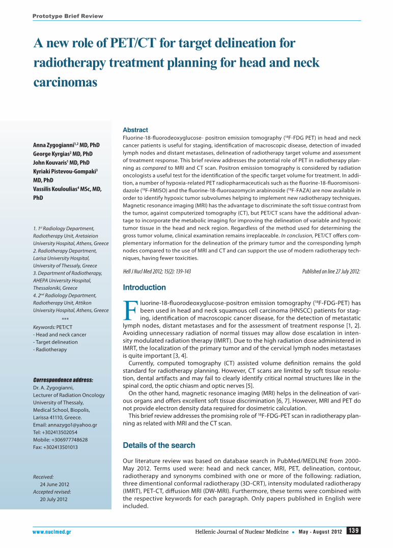
Before the application of PET, the radiological diagnosis of a positive lymph node was only based on its size [20]. We now know that malignant involvement of smaller sized lymph nodes is not uncommon (Fig. 2).
Figure 2. Primary tumor in the head seen on A) CT, B) MRI and C) 18F-FDG PET.
Others, in a meta-analysis of 32 studies with 1236 patients showed that 18F-FDG-PET in staging neck cancer had a sensi-tivity of 79% and specificity of 86%. In a subanalysis of stud-ies in which 18F-FDG-PET and MRI and CT were used, the sen-sitivity and specificity of PET was higher than 80% and 86% versus 75% and 79% for MRI and CT, respectively. Therefore, 18F-FDG-PET offers a modest improvement in the diagnosis of head and neck cancer and of its staging [21].
Integrated PET/CT has been shown to have higher ac-curacy than 18F-FDG-PET alone [22]. Others in 233 patients applied 18F-FDG-PET in addition to conventional imaging staging and concluded that PET imaging alters the stage of the disease based on nodal involvement in 43% of the patients [1].
Tumor and lymph nodes delineation with PET/CT The increased diagnostic accuracy with PET/CT reduced in-terobserver variability in target delineation and modified the estimated extension of gross tumor volume (GTV), the clinical target volume (CTV) and the planning target volume (PTV) for both the primary tumor and the regional lymph nodes [23-28]. The aim of a radiotherapist is to increase the radiotherapy dose for target volume and simultaneously de-crease the dose in normal tissues.
Others have studied 12 patients by PET/CT and found that the GTV had decreased in four patients and increased in two patients. However, the comparison between the changes of GTV in primary tumor and in lymph nodes was not de-scribed [23].
Others, by PET/CT found in 17/21 patients that the GTV de-lineation was corrected. They performed a separate analysis of the tumor GTV and of the lymph nodes. Increase in the tumor GTV was noticed in 3 and a reduction in 14 patients while nodal delineation was increased in 7 and decreased in 3 of the 21 patients [24].
Others, enrolled 40 patients in order to compare the GTV identified on CT to that obtained from PET. The GTV was changed in 37 of the 40 patients. Thirty of them showed re-duction and 7 increase. No comparison was made between the primary tumor and the lymph nodes [25].
Others, in 38 patients compared PET/CT delineation with CT target definition. They showed that the mean to-tal PET/CT based GTV was significantly smaller than that on CT. However this difference was not statistically significant when separate analyses of the CT based and PET/CT based GTV of the primary tumor versus the GTV of nodal disease were performed [26].
Applications of 18F-FDG-PETResearch into the biochemistry of PET radioisotopes indi-
cated preferential uptake of particular markers as a function of tumor metabolism with particular metabolic characteris-tics such as hypoxia and high proliferation ability. Also par-ticular radiopharmaceuticals should penetrate the lipophilic cell membrane of cancer cells easily with fast metabolic clearance but not normal cell membrane [8]. Also particular radiopharmaceuticals should easily penetrate the lipophilic cell membrane of cancer cells with fast metabolic clearance but abnormal cell membrane [8].
The use of 18F-FDG in PET studies is focused on the detec-tion of high proliferative tumors characterized by hypoxic areas [9]. To make use of this latter parameter, other PET radiopharmaceuticals such as the fluorine-18-fluoromisoni-dazole (18F-FMISO) and the fluorine-18-fluoroazomycin arab-inoside (18F-FAZA) are now applied. Furthermore, nitroimida-zoles suitable for quantifying hypoxia status in tumors have been used as radiopharmaceuticals for PET, too [10, 11].
The PET/CT may therefore facilitate not only the deline-ation of the primary tumor and the corresponding lymph nodes metastases in HNSCC patients (Fig. 1) but has specific tumor hypoxia detection abilities.
Figure 1. A patient with a tumor of the oropharynx (SUV=7.2) with a positive lymph node (SUV=6.1) at the left submandibular region.
Comparing MRI, CT and PET/CT for the identification of the primary or the unknown tumorClinical examination is important to locate the primary can-cer tumor followed by diagnostic imaging consisting of CT, MRI and PET. Conventional imaging with CT and/or MRI in detecting the primary tumor have high sensitivities, of 86% and 88% respectively [12] (Fig.2).
When these methods show insufficient detectability, PET may play an important role in tumor delineation. Its sensitiv-ity has been reported to be 93%-100% [13-15].
The potential role of PET in patients with unknown prima-ry tumors is also essential, especially if the tumor is too small or lies in a position difficult to access. Several studies con-cluded that PET was capable of detecting the unknown pri-mary tumor in about 25% of the cases [16, 17], as confirmed by biopsy [18].
MRI, CT and PET/CT for the detection of lymph-node metastasesIn IMRT planning, the detection of the status of regional lymph nodes is very important, because positive lymph nodes are included in high dose target volume. The risk of regional recurrence is high if these lymph nodes are left untreated. However, the elective nodal irradiation in high-er doses may not spare normal organs such as the parotid glands, larynx and pharynx [19].
Α Β C
Α Β C
Prototype Brief Review

Hellenic Journal of Nuclear Medicine May - August 2012www.nuclmed.gr 1 4 1
optical estimation in order to set the threshold of the 18F-FDG uptake to precisely depict the actual limit between the tumor and normal tissue. More studies on this issue are war-ranted [31].
Clinical examination and findings There is often significant variability between Radiation Oncologists regarding the target delineation by MRI and PET/CT. If mucosal disease and the tumor bed are too small their size may be underestimated [16, 17]. The extension of the tumor can be precisely assessed by clinical examina-tion. Regardless of the method used for determining the GTV, clinical examination remains irreplaceable [28]. For example, a clinical examination at the base of the tongue can diagnose cancer extending in the mucosa by palpa-tion, while this diagnosis could be missed by CT and/or 18F-FDG PET/CT [19]. The same has been reported by others with superficial tumor extension to the contralateral side of larynx or the subglottis, which not detected by CT, MRI or PET [30, 28].
Hypoxic tumors identificationThe application of 18F-FDG PET in hypoxic tumors was in-tended to implement an integrated boost in these regions for better tumor control, even though it has been ques-tioned. Comparing the 18F-FDG PET before and during ra-diotherapy, hypoxia imaging showed significant spatial variability, representing reoxygenation and changes in the locations of tumoral hypoxia [19, 31]. These findings reduce the possibility for using hypoxia imaged subvolumes as targets. Nevertheless, hypoxia-related treatment options are emerging; thus, a number of hypoxia-related PET trac-ers such as 18F-FMISO and 18F-FAZA are available now in an effort to identify hypoxic tumor subvolumes [8]. Studying
A recently published study by PET/CT and CT included 25 patients with HNSCC of whom 20/25 had the primary GTV tumor smaller than the GTV contoured on CT. The difference was statistically significant (P=0.0001). In addition, 60 lymph nodes were identified on PET while only 55 on CT [27].
Other researchers have studied 41 patients and compared the GTV contoured on CT, MRI, PET and on physical exami-nation (PE). For primary tumors, mean GTV was significantly larger when studied by PET/CT than by MRI/CT fusion but without statistical difference (P=0.39, Cl=0.62). Qualitative analyses to explain the low CI revealed that the superficial extent of primary disease was often underestimated when GTV was contoured without knowledge of physical exami-nation findings. Similar trends were observed for nodal GTV. Finally, it was concluded that the target volume delineation was not complete without consideration of physical exami-nation [28] (Table 1).
Discussion
The 18F-FDG-PET sensitivity for tumor tissue provides excel-lent contrast between malignant and the surrounding nor-mal tissues and renders easier the identification of the target volume by the radiation oncologists. Numerous recommen-dations have been made to obtain consistency of target de-lineation by collaboration between radiologists and nuclear medicine physicians [18]. Accurate delineation of the GTV is critically important and often achieved by using 18F-FDG-PET, while the boundary between the tumor and the normal tissue can be difficult to define on CT [29].
The application of 18F-FDG-PET in radiotherapy depart-ments has been increased [29, 30], though, the modality has its limitations, mainly there is no objective manner besides
Table 1. Studies comparing CT and/or MRI versus PET/CT for the target delineation concerning head and neck tumors
References N Imaging modalities
CT/MRI/PET-based primary volume P CT/MRI/PET-based LN
volume P
[22] 12 CT, PET/CT
2/12 pz with ≥25% increased with PET/CT, 4/12 pz with ≥25% decreased with PET/CT
N/A Without differentiation from primary tumor N/A
[23] 21 CT, PET/CT CT 64.7cm3
PET /CT 42.8cm3 0.002 CT 29.9cm3
PET/CT 37.2cm3 NS
[24] 40 CT, PET/CT CT 37.0cm3
PET/CT 20.3cm3 N/A Without differentiation from primary tumor N/A
[25] 38 CT, PET/CT CT 34.54cm3
PET/CT 29.38cm3 0.046 No data NS
[26] 29 CT, PET/CT CT 24ccPET/CT 18cc 0.001
CT 55 suspicious lymph nodes, PET/CT 60 suspicious lymph nodes
NS
[27] 41 CT,MRI,PET CT/PET 33.9mLCT/MRI 34.9mL 0.39 CT/PET 34.9mL
CT/MRI 34.4mL NS
Ν: number of patients; N/A, non applied; NS, non significant; P: significance value against PET; pz: patients
Prototype Brief Review
Hellenic Journal of Nuclear Medicine www.nuclmed.gr1 42 May - August 2012
tomography/computed tomography imaging in the preoper-ative staging of head and neck squamous cell carcinoma. Oral Oncol 2007; 43(9): 887-93.
13. Gordin A, Golz A, Daitzchman M et al. Fluorine-18 fluorode-oxyglucose positron emission tomography/computed tomog-raphy imaging in patients with carcinoma of the nasopharynx: diagnostic accuracy and impact on clinical management. Int J Radiat Oncol Biol Phys 2007; 68: 370-6.
14. King AD, Ma BB, Yau YY et al. The impact of 18F-FDG PET/CT on assessment of nasopharyngeal carcinoma at diagnosis. Br J Radiol 2008; 81: 291-8.
15. Ng SH, Yen TC, Liao CT et al. 18F-FDG PET and CT/MRI in oral cavity squamous cell carcinoma: A prospective study of 124 patients with histologic correlation. J Nucl Med 2005; 46: 1136-43.
16. Johansen J, Buus S, Loft A et al. Prospective study of 18FDG PET in the detection and management of patients with lymph node metastases to the neck from an unknown primary tumor. Results from the DAHANCA-13 study. Head Neck 2008; 30(4): 471-8.
17. Rusthoven KE, Rusthoven KE, Koshy M, Paulino AC. The role of fluorodeoxyglucose positron emission tomography in cervi-cal lymph node metastases from an unknown primary tumor. Cancer 2004; 101(11): 2641-9.
18. Troost E, Schinagl D,Bussink J et al. Clinical evidence on PET/CT for radiation therapy planning in head and neck tumours. Ra-diother Oncol 2010; 96: 328-34.
19. Eisbruch A, Gregoire V. Balancing Risk and Reward in Target Delineation for Highly Conformal Radiotherapy in Head and Neck Cancer. Semin Radiat Oncol 2009; 19: 43-52.
20. Van den Brekel MW, Stel HV, Castelijns JA et al. Cervical lymph node metastasis: assessment of radiologic criteria. Radiology 1990; 177(2): 379-84.
21. Kyzas PA, Evangelou E, Denaxa-Kyza D, Ioannidis JP. 18F-fluoro-deoxyglucose positron emission tomography to evaluate cer-vical node metastases in patients with head and neck squa-mous cell carcinoma: a meta-analysis. J Natl Cancer Inst 2008; 100(10): 712-20.
22. Syed R, Bomanji JB, Nagabhushan N et al. Impact of combined 18F-FDG PET/CT in head and neck tumours. Br J Cancer 2005; 92(6): 1046-50.
23. Ciernik IF, Dizendorf E, Baumert BG et al. Radiation treatment planning with an integrated positron emission and computer tomography (PET/CT): A feasibility study. Int J Radiat Oncol Biol Phys 2003; 57: 853-63.
24. Heron DE, Andrade RS, Flickinger J et al. Hybrid PET-CT simula-tion for radiation treatment planning in head-and neck can-cers: a brief technical report. Int J Radiat Oncol Biol Phys 2004; 60(5): 1419-24.
25. Paulino Paulino AC, Koshy M, Howell R et al. Comparison of CT- and FDGPET-defined gross tumor volume in intensity-modu-lated radiotherapy for head-and-neck cancer. Int J Radiat On-col Biol Phys 2005; 61: 1385-92.
26. Guido A, Fuccio L, Rombi B et al. Combined 18F-FDG-PET/CTi-maging in radiotherapy target delineation for head-and-neck cancer. Int J Radiat Oncol Biol Phys 2009; 73: 759-63.
27. Delouya G, Igidbashian L, Houle A et al. 18F-FDG-PET imaging in radiotherapy tumor volume delineation in treatment of head and neck cancer. Radiotherapy and Oncology 2011; 101: 362-8.
28. Thiagarajan A, Caria N, Schoeder H et al. Target Volume Deline-ation in Oropharyngeal Cancer:Impact of PET, MRI, and Physical Examination. Int J Radiation Oncol Biol Phys 2012; 83(1): 220-7.
the application of these tracers, it is necessary to image pa-tients at least after 2.5h, because of the slow improvement of the contrast between hypoxic and normal tissues. For 18F-labeled hypoxia tracers, count statistics are of concern because of the necessity to have late-time-point images [32]. Compared to 18F-FMISO, the clearance of 18F-FAZA from blood and from non target tissues is faster and therefore has a higher tumor to background ratio [31]. Before these tracers can be clinically implemented for patient selection, dose escalation or adaptive radiotherapy, additional stud-ies are required.
In conclusion, besides clinical diagnosis, GTV and lymph node metastases can be better diagnosed by PET/CT than by MRI and CT modalities. Studies on hypoxic GTV are also suggesting the importance of using PET/CT with specific tracers.
The authors declare that they have no conflicts of interest.
Bibliography
1. Lonneux M, Hamoir M, Reychler H et al. Positron emission tomography with [18F] fluorodeoxyglucose improves staging and patient management in patients with head and neck sq-uamous cell carcinoma: A multicenter prospective study. J Clin Oncol 2010; 28: 1190-5.
2. Prestwich RJD, Sykes J, Carey B et al. Improving Target Defi-nition for Head and Neck Radiotherapy: A Place for Magnetic Resonance Imaging and 18-Fluoride Fluorodeoxyglucose Pos-itron Emission Tomography? Clin Oncol 2012: 1-13.
3. Chen AM, Farwell DG, Luu Q et al. Marginal misses after post-operative intensity modulated radiotherapy for head and neck cancer. Int J Radiat Oncol Biol Phys 2011; 80(5): 1423-9.
4. David MB, Eisbruch A. Delineating neck targets for intensity modulated radiation therapy of head and neck cancer. What we learned from marginal recurrences? Front Radiat Ther Oncol 2007; 40: 193-207.
5. Cooper JS, Mukherji SK, Toledano AY et al. An evaluation of the variability of tumor-shape definition derived by experienced observers from CT images of supraglottic carcinomas (ACRIN protocol6658). Int J Radiat Oncol Biol Phys 2007; 67(4): 972-5.
6. Gallagher FA. An introduction to functional and molecular im-aging with MRI. Clin Radiol 2010; 65(7): 557-66.
7. Vandecaveye V, De Keyzer F, Dirix P et al. Applications of diffu-sion-weighted magnetic resonance imaging in head and neck squamous cell carcinoma. Neuroradiology 2010; 52(9): 773-84.
8. Marcu L, Bezak E, Filip S. The role of PET imaging in overcoming radiobiological challenges in the treatment of advanced head and neck cancer. Cancer Treatment Reviews 2012; 38: 185-93.
9. Busk M, Horsman MR, Jakobsen S et al. Cellular uptake of PET tracers of glucose metabolism and hypoxia and their linkage. Eur J Nucl Med Mol Imag 2008; 35: 2294-303.
10. Gagel B, Gagel B, Piroth M et al. pO polarography, contrast enhanced color duplex sonography (CDS), [18F] fluoromisoni-dazole, [18F] fluorodeoxyglucose positron emission tomogra-phy: validated methods for the evaluation of therapy-relevant tumor oxygenation or only bricks in the puzzle of tumor hy-poxia? BMC Cancer 2007; 7: 113.
11. Souvatzoglou M, Grosu AL, Röper B et al. Tumour hypoxia im-aging with [18F]FAZA PET in head and neck cancer patients: a pilot study. Eur J Nucl Med Mol Imaging 2007; 34: 1566-75.
12. Roh JL, Yeo NK, Kim JS et al. Utility of 2-[18F] fluoro-2-deoxy-D-glucose positron emission tomography and positron emission
Prototype Brief Review
Hellenic Journal of Nuclear Medicine May - August 2012www.nuclmed.gr 1 4 3
31. Steenbakkers RJHM. Implementing PET/CT imaging for head and neck cancer radiotherapy. Tijdschr Nucl Geneesk 2012; 34(2): 883-8.
32. Wang W, Lee N, Georgy JC et al. Pharmacokinetic Analysis of Hypoxia 18F-Fluoromisonidazole Dynamic PET in Head and Neck cancer. J Nucl Med 2010; 51(1): 37-45.
29. Dimitrakopoulou- Strauss A, Pan L, Strauss L. Parametric imaging: a promising approach for the evaluation of dynamic PET-18F-FDG studies the DKFZ experience. Hell J Nucl Med 2010; 13(1): 18-22.
30. Daisne JF, Duprez T, Weynand B et al. Tumor volume in pharyn-golaryngeal squamous cell carcinoma: Comparison at CT, MR imaging, and FDG PET and validation with surgical specimen. Radiology 2004; 233: 93-100.
Prototype Brief Review
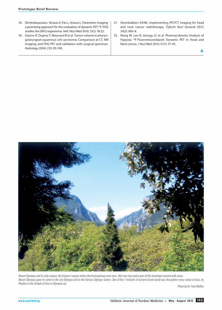
Mount Olympus and its only canyon, the Enipea’s canyon where the homonymous river runs. Afar one may notice part of the mountain covered with snow.Mount Olympus gave its name to the city Olympia and to the famous Olympic Games. One of the 7 miracles of ancient Greek world was the golden-ivory statue of Zeus, by Phedias in the Temple of Zeus in Olympia city.
Photo by Dr. Fani Melfou