
Medical Assistant Role Delineation Chart
28
ADMINISTRATIVE Administrative Procedures ■ Perform basic administra- tive medical assisting functions ■ Schedule, coordinate and monitor appointments ■ Schedule inpatient/outpa- tient admissions and procedures ■ Understand and apply third-party guidelines ■ Obtain reimbursement through accurate claims submission ■ Monitor third-party reim- bursement ■ Understand and adhere to managed care policies and procedures ■ Negotiate managed care contracts Practice Finances ■ Perform procedural and diagnostic coding ■ Apply bookkeeping principles CLINICAL ■ Manage accounts receivable ■ Manage accounts payable ■ Process payroll ■ Document and maintain accounting and banking records ■ Develop and maintain fee schedules ■ Manage renewals of busi- ness and professional insurance policies ■ Manage personnel benefits and maintain records ■ Perform marketing, financial, and strategic planning Fundamental Principles ■ Apply principles of aseptic technique and infection control ■ Comply with quality assurance practices ■ Screen and follow up patient test results Diagnostic Orders ■ Collect and process specimens ■ Perform diagnostic tests Patient Care ■ Adhere to established pa- tient screening procedures ■ Obtain patient history and vital signs ■ Prepare and maintain ex- amination and treatment areas ■ Prepare patient for exami- nations, procedures and treatments ■ Assist with examinations, procedures and treatments ■ Prepare and administer medications and immu- nizations ■ Maintain medication and immunization records ■ Recognize and respond to emergencies ■ Coordinate patient care information with other health care providers ■ Initiate IV and administer IV medications with ap- propriate training and as permitted by state law GENERAL Professionalism ■ Display a professional manner and image ■ Demonstrate initiative and responsi- bility ■ Work as a member of the health care team ■ Prioritize and perform multiple tasks ■ Adapt to change ■ Promote the CMA credential ■ Enhance skills through continuing education ■ Treat all patients with compassion and empathy ■ Promote the practice through positive public relations Communication Skills ■ Recognize and respect cultural diversity ■ Adapt communications to individual’s ability to understand ■ Use professional telephone technique ■ Recognize and respond effectively to verbal, nonverbal, and written com- munications ■ Use medical terminology appropriately ■ Utilize electronic technology to receive, organize, prioritize and transmit infor- mation ■ Serve as liaison Legal Concepts ■ Perform within legal and ethical boundaries ■ Prepare and maintain medical records ■ Document accurately ■ Follow employer’s established policies dealing with the health care contract ■ Implement and maintain federal and state health care legislation and regulations ■ Comply with established risk manage- ment and safety procedures ■ Recognize professional credentialing criteria ■ Develop and maintain personnel, policy and procedure manuals Instruction ■ Instruct individuals according to their needs ■ Explain office policies and procedures ■ Teach methods of health promotion and disease prevention ■ Locate community resources and dis- seminate information ■ Develop educational materials ■ Conduct continuing education activities Operational Functions ■ Perform inventory of supplies and equipment ■ Perform routine maintenance of ad- ministrative and clinical equipment ■ Apply computer techniques to support office operations ■ Perform personnel management functions ■ Negotiate leases and prices for equip- ment and supply contracts ■ Denotes advanced skills. SOURCE: Reprinted by permission of the American Association of Medical Assistants from the AAMA Role Delineation Study: Occupational Analysis of the Medical Assisting Profession. Medical Assistant Role Delineation Chart HIGHLIGHT indicates material covered in this chapter.
Transcript of Medical Assistant Role Delineation Chart

ADMINISTRATIVE
Administrative Proceduresn Perform basic administra-
tive medical assistingfunctions
n Schedule, coordinate andmonitor appointments
n Schedule inpatient/outpa-tient admissions andprocedures
n Understand and applythird-party guidelines
n Obtain reimbursementthrough accurate claimssubmission
n Monitor third-party reim-bursement
n Understand and adhere tomanaged care policies andprocedures
n Negotiate managed carecontracts
Practice Financesn Perform procedural and
diagnostic codingn Apply bookkeeping
principles
CLINICAL
n Manage accounts receivable
n Manage accounts payablen Process payrolln Document and maintain
accounting and bankingrecords
n Develop and maintain feeschedules
n Manage renewals of busi-ness and professionalinsurance policies
n Manage personnel benefitsand maintain records
n Perform marketing,financial, and strategicplanning
Fundamental Principlesn Apply principles of aseptic
technique and infectioncontrol
n Comply with qualityassurance practices
n Screen and follow uppatient test results
Diagnostic Ordersn Collect and process
specimensn Perform diagnostic tests
Patient Caren Adhere to established pa-
tient screening proceduresn Obtain patient history and
vital signsn Prepare and maintain ex-
amination and treatmentareas
n Prepare patient for exami-nations, procedures andtreatments
n Assist with examinations,procedures and treatments
n Prepare and administermedications and immu-nizations
n Maintain medication andimmunization records
n Recognize and respond toemergencies
n Coordinate patient careinformation with otherhealth care providers
n Initiate IV and administerIV medications with ap-propriate training and aspermitted by state law
GENERAL
Professionalismn Display a professional manner and
imagen Demonstrate initiative and responsi-
bilityn Work as a member of the health care
teamn Prioritize and perform multiple
tasksn Adapt to changen Promote the CMA credentialn Enhance skills through continuing
educationn Treat all patients with compassion and
empathyn Promote the practice through positive
public relations
Communication Skillsn Recognize and respect cultural
diversityn Adapt communications to individual’s
ability to understandn Use professional telephone technique
n Recognize and respond effectively toverbal, nonverbal, and written com-munications
n Use medical terminology appropriatelyn Utilize electronic technology to receive,
organize, prioritize and transmit infor-mation
n Serve as liaison
Legal Conceptsn Perform within legal and ethical
boundariesn Prepare and maintain medical recordsn Document accuratelyn Follow employer’s established policies
dealing with the health care contractn Implement and maintain federal
and state health care legislation andregulations
n Comply with established risk manage-ment and safety procedures
n Recognize professional credentialingcriteria
n Develop and maintain personnel,policy and procedure manuals
Instructionn Instruct individuals according to their
needsn Explain office policies and proceduresn Teach methods of health promotion
and disease preventionn Locate community resources and dis-
seminate informationn Develop educational materialsn Conduct continuing education
activities
Operational Functionsn Perform inventory of supplies and
equipmentn Perform routine maintenance of ad-
ministrative and clinical equipmentn Apply computer techniques to support
office operationsn Perform personnel management
functionsn Negotiate leases and prices for equip-
ment and supply contracts
n Denotes advanced skills.
SOURCE: Reprinted by permission of the American Association of Medical Assistants from the AAMA Role Delineation Study: Occupational Analysis of the Medical Assisting Profession.
Medical Assistant Role Delineation ChartHIGHLIGHT indicates material covered in this chapter.
MEDIMC43_0131715771.QXD 3/7/07 2:55 PM Page 900

chapter
43HematologyOUTLINE
The Medical Assistant’s Role 902
Blood Formation and Components 902
Function of Blood 903
Blood Specimen Collection 904
Routine Blood Tests 909
Erythrocyte Sedimentation Rate 921
Phenylketonuria 921
Mono Testing 921
n Define and spell the terms to learnfor this chapter.
n List the components of blood, in-cluding the liquid and cellular por-tions and functions of each.
n Describe how to prepare a patientfor collection of a blood specimen
via venipuncture and capillary punc-ture methods.
n Discuss how to process a bloodspecimen for routine testing in aphysician’s office.
n State the normal values for each ofthe blood tests discussed.
Learning ObjectivesAfter completing this chapter, you should be able to:
Terms to Learn
antecubital space
capillaries
electrolytes
erythrocyte sedimentationrate (ESR)
hematology
hematopoiesis
hemoglobin
heparin
plasma
platelets
Case Study
MARY O’SHEA, CMA HAS JUST BEEN HIRED AS A MEDICAL ASSISTANT at
a physician’s office. The first task that Mary has been asked to do is to obtain blood
samples from a male patient. The order in the patient’s chart indicates that blood
samples are to be drawn for the following tests: CBC, serum chemistry tests, and a
prothrombin time. What method(s) should Mary use to collect the samples?
MEDIMC43_0131715771.QXD 3/7/07 2:55 PM Page 901

ematology is the study of blood andthe tissues that produce it. Blood andits components are studied to detectpathological conditions and to deter-mine the appropriate course of treat-ment. Blood analysis is one of the mostcommon diagnostic tests performed in
the doctor’s office. As a result, the medical assistant musthave a thorough understanding of how to collect, han-dle, package, and analyze a blood specimen correctly.
The Medical Assistant’s RoleWhen the physician orders a blood test, the role of themedical assistant is to collect the specimen. The actualtesting of the blood is not commonly done in the med-ical office but rather is done in an outside laboratorythat is contracted with the patient’s medical insurance.When a patient’s blood is drawn, the medical assistantmust ensure that proper specimen labeling has oc-curred and that the blood is stored correctly until thelaboratory courier has arrived for pickup.
Blood Formation and ComponentsThe formation of blood cells is called hematopoiesis.Hematopoiesis begins at the stem cell level during fetaldevelopment. Blood is formed with both cellular andliquid components.
Cellular Formation and ComponentsAll blood cells originate from the hematopoietic stemcell, but mature into one of seven individual cells:
1. Red blood cells (erythrocytes)
2. White blood cells (leukocytes)—five types
A: Granular leukocytes (have granules in theircytoplasm):
• Neutrophil
• Eosinophil
• Basophil
B: Nongranular leukocytes (do not have gran-ules in their cytoplasm):
• Lymphocyte
• Monocyte
3. Platelets (thrombocytes)
Hematopoiesis occurs primarily in the bone marrow ofthe adult. Lymphocytes, one of the types of white bloodcells (WBCs), are produced in the lymph nodes.
Red Blood CellsRed blood cells (RBCs) are formed in the bone mar-row. They are important for the human body because
they contain hemoglobin. Hemoglobin has two func-tions. The first is to carry oxygen from the lungs to thecells of the body. The second function is to carry car-bon dioxide (a waste product) from the body back tothe lungs, where it can be expelled with exhalation.When the hemoglobin is carrying oxygen, it is calledoxyhemoglobin. When it is carrying carbon dioxide, itis called carboxyhemoglobin. Arterial blood has ahigher concentration of oxygen, explaining its brightred color. Venous blood is darker in color because ofthe carboxyhemoglobin.
The formation of red blood cells is controlled some-what by erythropoietin, which is secreted by the kid-neys in an adult, and by the liver in a fetus. When theamount of erythropoietin is decreased, red blood cellswill not be formed in proper amounts, which may re-sult in certain types of anemia. For instance, patientswho are being treated with chemotherapy may developanemia that then can be treated with an artificialerythropoietin called Procrit that assists in the repro-duction of red blood cells. Red blood cells last forabout 4 months and are continually being reproducedin the body. The normal RBC range for a male adult is4.5 to 6 million/mm3. The normal female RBC range is4 to 5.5 million/mm3.
White Blood CellsWhite blood cells are also known as leukocytes, whichare produced in the bone marrow and are divided intoseveral different types. They originate in the bone mar-row from stem cells. White blood cells are larger thanred blood cells and their principal function is to defendagainst infection. The five types of white blood cellsare neutrophils, lymphocytes, monocytes, eosinophils,and basophils. The range of WBCs in an adult is 4.5 to11 thousand/mm3
NEUTROPHILS Neutrophils are divided into two cat-egories: segmented neutrophils and nonsegmented neu-trophils. Segmented neutrophils have a nucleus that isdivided into multiple segments connected by smallthin threads. Nonsegmented neutrophils are also calledstabs (or bands) and are more immature neutrophilsthan the segmented ones. The presence of a large num-ber of stabs may indicate the existence of a bacterialinfection. Neutrophils are named this because the gran-ules are neutral in color on the laboratory-stained slide.The body reproduces neutrophils on an ongoing basis,and they only survive for a few days. Reproduction isincreased when bacterial infection is occurring. Neu-trophils combat infection by phagocytosis. Phagocyto-sis is the process in which the neutrophil surrounds,swallows, and digests the bacteria.
EOSINOPHILS Eosinophils are also assumed to beproduced by the bone marrow. Detection of a largenumber of eosinophils can indicate a parasitic condi-
902 Chapter 43 Hematology
H
MEDIMC43_0131715771.QXD 3/7/07 2:55 PM Page 902

tion or the presence of certain allergic conditions.Eosinophils have granules that produce a red color onthe laboratory-stained slide. Eosinophils are called thisdue to the stain eosin, which is used in the staining ofblood smears.
BASOPHILS Like the other white cells, basophils arethought to be produced by the bone marrow, and theyproduce heparin. Heparin is a substance that preventsclotting. When an individual has a condition that iscreating inflammation, heparin may be used to assist indiminishing or preventing the occurrence of clotting.Increased amounts of basophils may be found in pa-tients who have had their spleen removed. Patientswho have had excessive exposure to radiation mayalso have increased basophils.
LYMPHOCYTES Lymphocytes are produced in thebone marrow and in the lymphoid tissue such asthe spleen and lymph nodes. The function of lympho-cytes is primarily to produce antibodies against foreignsubstances such as bacteria, viruses, and pollens. Lym-phocytes are small and large and can proliferate intoB and T cells. B cells may convert into plasma cells,which produce antibodies. T cells can produce helpercells, cytotoxic cells, and suppressor cells. To diagnosean individual with HIV, testing is performed to evalu-ate the type and amount of T cells present. Lympho-cytes do not have granules and are nonsegmented.
MONOCYTES Monocytes are formed in the bonemarrow from stem cells. Monocytes assist in phagocy-tosis. They ingest foreign particles or bacteria that theneutrophils are unable to digest and assist in cleaningup cellular debris that may have been left from the in-fection. An increase in monocytes is seen in patientswho have certain diseases such as tuberculosis, ty-phoid, and Rocky Mountain spotted fever.
PlateletsPlatelets (also called thrombocytes) are the smallestcells found in the blood and are formed in the bonemarrow. The main function of platelets is to assist inthe clotting of blood. Platelets increase around an areathat is bleeding to assist in the formation of clots. Theplatelets and the injured tissue release thromboplastin.The thromboplastin combines with other elements inthe blood to produce thrombin. The thrombin acts ona protein in the blood called fibrinogen, resulting inthe formation of fibrin. Fibrin is tiny threads that cre-ate a mesh that catches the red cells and other cells toform a clot. There are typically between 150,000 and400,000 platelets/mm3.
Understanding the process of normal clotting and ab-sence of normal clotting is important because laboratorytests are designed to determine why clotting is not occur-ring properly, particularly in patients who are receivinganticlotting drugs such as heparin and Coumadin.
Liquid Blood Formationand ComponentsFor the medical assistant to understand how blood isformed requires a thorough comprehension of the cel-lular and liquid components of blood. The liquid com-ponent of blood is called plasma. Plasma is about 55%of the composition of blood and carries cellular ele-ments and other substances. Plasma transports sub-stances in the blood to the different parts of the body.Plasma does contain fibrinogen, which converts to fib-rin during the clotting process. Plasma without thefibrinogen is called serum.
Ninety percent of plasma is water, while the other10% is solid substances, called solutes. These solutes mayinclude the plasma proteins (albumin, globulin, fibrino-gen, and prothrombin), electrolytes (sodium, potassium,and chloride), glucose, amino acids, lipids and carbohy-drates, metabolic waste products (urea, lactic acid, uricacid), creatinine, respiratory gases (oxygen and carbondioxide), and miscellaneous substances (hormones, anti-bodies, enzymes, vitamins, and mineral salts).
Function of BloodThe function of blood is transportation and protection.It carries oxygen and nutrients to the body and removesthe waste product carbon dioxide. The blood carriesthe waste products to the liver, kidneys, and skin forelimination.
UNIT 4 Clinical Medical Assisting 903
Drawing blood from an older individual cansometimes be challenging due to the conditionof their veins. Patients do not want to have anymore needle sticks than necessary. To ensurethat a successful needle stick occurs requiresboth experience and patience. If patients will bereturning to the office for blood work at a latertime, inform the patient to drink a lot of fluidsprior to arrival at the office. Being well hydratedis helpful for finding veins. Use of items such asa small ball placed in the patient’s hand tosqueeze in order to pump up the veins is alsohelpful. If the hand must be used for the drawsite, place a warm cloth over the area to allow forthe vein to rise up. All of these techniques canhelp in making the first try a success.
LifespanConsiderations
MEDIMC43_0131715771.QXD 3/7/07 2:55 PM Page 903

The heart pumps blood through the body by way ofthe arteries, veins, and capillaries. The capillaries con-nect the arteries and veins that pump the blood to andfrom the heart. When blood flows away from the heart
it flows in arteries, and when it returns back to the heartit flows through veins. Arteries have thick walls that al-low them to withstand the pressure sustained when theheart is pumping. The blood carried in the arteries con-tains oxygen. This blood with its high level of oxygensustains tissue function. As oxygen is being releasedfrom the blood, carbon dioxide is being transported tothe lungs to be expelled as a waste product.
The blood regulates body temperature. When thebody becomes warm, the capillaries dilate and releaseheat, which in turn cools the body. When the body iscold, the capillaries constrict allowing for less bloodflow, which increases the body temperature.
Blood Specimen CollectionLaboratory testing of blood and the collection of allblood and body fluids is strictly regulated by OSHAregulations, and the CDC’s Universal Precautions mustbe followed at all times. CLIA (Clinical Laboratory
904 Chapter 43 Hematology
Quality Control for Collecting a Blood Specimen
PROCEDURE43-1
OBJECTIVE: Perform quality control procedure while collecting a blood specimenwithout errors.
Equipment and Supplies
antiseptic cleaner; biohazard waste container; nec-essary sterile equipment; specimen collection con-tainer; disposable alcohol wipe; disposable gloves;appropriate requisition or paperwork required ofcollection; pen or pencil; patient’s chart
Method
1. Review request and verify test ordered.2. Prepare necessary equipment and work area.3. Perform hand hygiene and don gloves.4. Identify the patient and explain the procedure,
and make sure he or she understands theprocedure.
5. Confirm that patient has followed any pretestpreparation requirements.
6. Collect the specimen properly, using the appro-priate equipment and technique.
7. Use the appropriate collection container andthe right preservatives.
8. Immediately label the specimen with thepatient’s name, date and time of collection,test’s name, and the name of the personcollecting the specimen.
9. Follow correct procedures for disposing of haz-ardous specimen waste and decontaminatingwork area and equipment according to OSHAguidelines.
10. Remove gloves and dispose in appropriate con-tainer. Perform hand hygiene. Dispose of all usedneedles, etc., in biohazard waste container.
11. Thank the patient and observe for any signs orsymptoms of inappropriate response to theprocedure.
12. Document the procedure in patient’s chart.13. If the specimen is to be transported to an out-
side laboratory, prepare it for transport in theproper container, with all the appropriate infor-mation according to OSHA guidelines.
It is legal in most states for a medical assistantto perform a venipuncture. Check with the localAmerican Association of Medical Assistants (AAMA)chapter for specifics in your state. When perform-ing basic in-office lab tests, the medical assistantmust keep all results confidential. Confidentialityis a moral and ethical obligation for all health careteam members.
Legaland Ethical Issues
MEDIMC43_0131715771.QXD 3/7/07 2:55 PM Page 904

Improvement Amendments) sets the standards that alllaboratories must adhere to, including training of per-sonnel and testing and transport of specimens (seeChapter 42). It is important when performing speci-men collection that the medical assistant follow the reg-ulation guidelines established by these organizations(see Procedure 43-1).
The type and amount of specimen to be acquired isdetermined by the test to be done. If a very smallamount is needed, then the specimen may be obtainedby capillary puncture. Larger volumes are collectedthrough venipuncture.
VenipunctureThree methods of venipuncture are used: the vacuumtube method, the syringe and needle method, and thebutterfly method (see also Legal and Ethical Issues).
MethodsThe most common method of venipuncture is the vac-uum container method because multiple samples canbe obtained at the same time, requiring fewer “sticks”for the patient and faster collection for the medical as-sistant. In using the vacuum container method, it is im-portant to use a large vein, because the vacuum cancollapse smaller veins. If the patient has no accessiblelarger veins, then it is appropriate to use a small needlewith a syringe to obtain the specimen. (See Figures 43-1and 43-2 and Procedure 43-2 for obtaining venousblood with a sterile syringe and needle.)
The butterfly method uses a needle that is attachedto 6-to12-inch tubing. The end of the tubing can at-
tach to the syringe or the vacuum container tubeholder. The butterfly method is used for small veinsthat are difficult to draw with the standard vacuumcontainer method or syringe and needle method. It iscalled the butterfly method because the needle on theend has a winged portion that keeps the needle fromturning and anchors the needle into the small vein.The needle used for the butterfly method is a small 21-,23-, or 25-gauge needle. The drawback to performingthe butterfly method is the cost. The needle is moreexpensive than a standard needle. The butterflymethod is not used for the majority of blood drawsdue to its expense.
EquipmentFigure 43-3 shows the equipment that a medical assis-tant will need to perform a venipuncture using the
UNIT 4 Clinical Medical Assisting 905
FIGURE 43-1 Vacutainer evacuated specimen tubeswith Hemogard closure blood collection tubes.
FIGURE 43-2 Vacutainer brand safety lock needle holder.
FIGURE 43-3 Venipuncture equipment.
MEDIMC43_0131715771.QXD 3/7/07 2:55 PM Page 905
vacuum container method. All equipment should be as-sembled and the expiration dates checked prior to at-tempting to use. Expired tubes should not be used,because they may not have a vacuum. There are varioustypes of tubes, marked by their color. Each tube has adifferent chemical additive (anticoagulant) to keep theblood from clotting for different types of tests.
• Red top tubes: Contain no anticoagulant andhave a sterile interior. A red top tube is typicallyused for blood bank and serological testing.
• Serum separator tubes (SST tube): these tubes canhave a gold top or a red/gray top (sometimes calledtiger top or marbled top). The SST tubes contain agel that separates the serum from the clot after thetube has been centrifuged. These tubes also containa clot activator that allows the blood to clot faster.SST tubes are routinely used for chemistry testing.
• Lavender/purple top tubes: These tubes containethylenediaminetetraacetic acid (EDTA) and areused for testing whole blood or plasma. This
tube is used when testing a CBC or glycosylatedhemoglobin.
• Green top tubes: These tubes contain heparin andare used for testing whole blood or plasma.
• Light blue top tubes: These tubes contain sodiumcitrate and are used when testing a PT/INR orPTT (clotting tests).
• Gray top tubes: These tubes contain potassiumoxylate and are used when performing a glucosetolerance test.
• Yellow top tubes: These tubes contain SodiumPolyanethol Sulfonate (SPS) and are used whendrawing blood for blood cultures.
Some laboratories may use additional tubes.Pink, tan, black, and royal blue topped tubes aresometimes used for specific laboratory tests.
Vacuum blood tubes come in 5-, 7-, 10-, and 15-mLsizes. The amount of blood needed for each test differs,so the tube sizes differ accordingly.
906 Chapter 43 Hematology
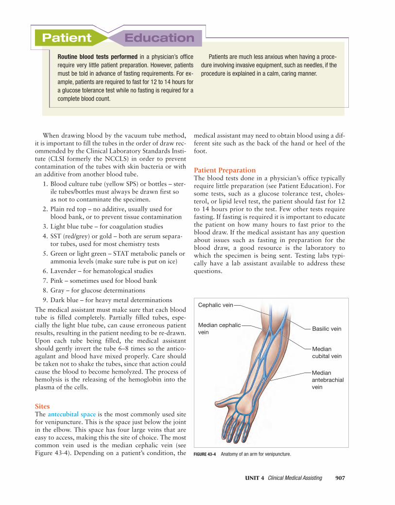
Obtaining Venous Blood with a Sterile Syringeand Needle
PROCEDURE43-2
OBJECTIVE: Perform a venipuncture using the syringe and needle method.
Equipment and Supplies
sterile 22-gauge needle and 10- to 20-mL syringe;appropriate vacuum specimen tubes for testsordered; tourniquet; examination gloves; alcoholsponge; cotton balls or dry gauze square; adhesivebandage; patient’s record; pen; lab coat; biohazardsharps containerNote: Always identify the patient by his or her arm-band and by asking the patient his or her name.
Method
1. Perform hand hygiene and assemble necessaryequipment and supplies.
2. Securely attach the sterile needle to thesyringe.
3. Put on examination gloves.4. Apply a tourniquet 3 to 4 inches above the
venipuncture site.5. Palpate vein and clean the venipuncture site
with an alcohol sponge and then dry with aclean gauze.
6. Have the patient make a fist and hold it shutuntil told to release it.
7. Make sure there is no air in the syringe.8. Remove the needle guard and insert the needle
into the vein.9. Slowly pull back the syringe plunger until the
proper amount of blood has been obtained.10. Instruct the patient to open his or her fist.11. Release the tourniquet and withdraw the needle
quickly.12. Fill the appropriate vacuum tubes to the proper
level.13. Apply gentle pressure to the puncture site with
a piece of cotton or gauze with the patient’sarm slightly raised for a few minutes. This mayprevent hematomas from occurring.
14. Apply a bandage to the puncture site.15. Discard the used needle and syringe into a
biohazard sharps container.16. Remove gloves and discard into appropriate
container.17. Perform hand hygiene.18. Record procedure in patient’s record.19. Label tubes and send to lab.
MEDIMC43_0131715771.QXD 3/7/07 2:55 PM Page 906
When drawing blood by the vacuum tube method,it is important to fill the tubes in the order of draw rec-ommended by the Clinical Laboratory Standards Insti-tute (CLSI formerly the NCCLS) in order to preventcontamination of the tubes with skin bacteria or withan additive from another blood tube.
1. Blood culture tube (yellow SPS) or bottles – ster-ile tubes/bottles must always be drawn first soas not to contaminate the specimen.
2. Plain red top – no additive, usually used forblood bank, or to prevent tissue contamination
3. Light blue tube – for coagulation studies
4. SST (red/grey) or gold – both are serum separa-tor tubes, used for most chemistry tests
5. Green or light green – STAT metabolic panels orammonia levels (make sure tube is put on ice)
6. Lavender – for hematological studies
7. Pink – sometimes used for blood bank
8. Gray – for glucose determinations
9. Dark blue – for heavy metal determinations
The medical assistant must make sure that each bloodtube is filled completely. Partially filled tubes, espe-cially the light blue tube, can cause erroneous patientresults, resulting in the patient needing to be re-drawn.Upon each tube being filled, the medical assistantshould gently invert the tube 6–8 times so the antico-agulant and blood have mixed properly. Care shouldbe taken not to shake the tubes, since that action couldcause the blood to become hemolyzed. The process ofhemolysis is the releasing of the hemoglobin into theplasma of the cells.
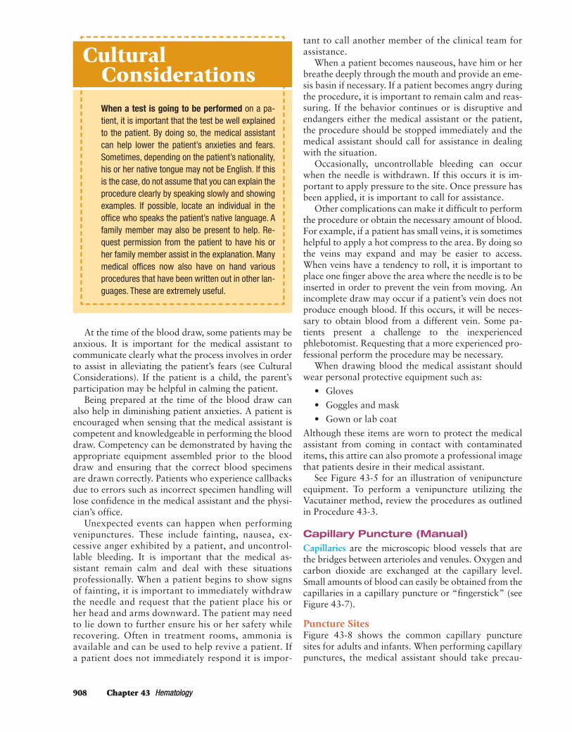
SitesThe antecubital space is the most commonly used sitefor venipuncture. This is the space just below the jointin the elbow. This space has four large veins that areeasy to access, making this the site of choice. The mostcommon vein used is the median cephalic vein (seeFigure 43-4). Depending on a patient’s condition, the
medical assistant may need to obtain blood using a dif-ferent site such as the back of the hand or heel of thefoot.
Patient PreparationThe blood tests done in a physician’s office typicallyrequire little preparation (see Patient Education). Forsome tests, such as a glucose tolerance test, choles-terol, or lipid level test, the patient should fast for 12to 14 hours prior to the test. Few other tests requirefasting. If fasting is required it is important to educatethe patient on how many hours to fast prior to theblood draw. If the medical assistant has any questionabout issues such as fasting in preparation for theblood draw, a good resource is the laboratory towhich the specimen is being sent. Testing labs typi-cally have a lab assistant available to address thesequestions.
UNIT 4 Clinical Medical Assisting 907
Patient EducationRoutine blood tests performed in a physician’s officerequire very little patient preparation. However, patientsmust be told in advance of fasting requirements. For ex-ample, patients are required to fast for 12 to 14 hours fora glucose tolerance test while no fasting is required for acomplete blood count.
Patients are much less anxious when having a proce-dure involving invasive equipment, such as needles, if theprocedure is explained in a calm, caring manner.
Cephalic vein
Median cephalicvein
Basilic vein
Mediancubital vein
Medianantebrachialvein
FIGURE 43-4 Anatomy of an arm for venipuncture.
MEDIMC43_0131715771.QXD 3/7/07 2:55 PM Page 907
908 Chapter 43 Hematology908 Chapter 43 Hematology
At the time of the blood draw, some patients may beanxious. It is important for the medical assistant tocommunicate clearly what the process involves in orderto assist in alleviating the patient’s fears (see CulturalConsiderations). If the patient is a child, the parent’sparticipation may be helpful in calming the patient.
Being prepared at the time of the blood draw canalso help in diminishing patient anxieties. A patient isencouraged when sensing that the medical assistant iscompetent and knowledgeable in performing the blooddraw. Competency can be demonstrated by having theappropriate equipment assembled prior to the blooddraw and ensuring that the correct blood specimensare drawn correctly. Patients who experience callbacksdue to errors such as incorrect specimen handling willlose confidence in the medical assistant and the physi-cian’s office.
Unexpected events can happen when performingvenipunctures. These include fainting, nausea, ex-cessive anger exhibited by a patient, and uncontrol-lable bleeding. It is important that the medical as-sistant remain calm and deal with these situationsprofessionally. When a patient begins to show signsof fainting, it is important to immediately withdrawthe needle and request that the patient place his orher head and arms downward. The patient may needto lie down to further ensure his or her safety whilerecovering. Often in treatment rooms, ammonia isavailable and can be used to help revive a patient. Ifa patient does not immediately respond it is impor-
tant to call another member of the clinical team forassistance.
When a patient becomes nauseous, have him or herbreathe deeply through the mouth and provide an eme-sis basin if necessary. If a patient becomes angry duringthe procedure, it is important to remain calm and reas-suring. If the behavior continues or is disruptive andendangers either the medical assistant or the patient,the procedure should be stopped immediately and themedical assistant should call for assistance in dealingwith the situation.
Occasionally, uncontrollable bleeding can occurwhen the needle is withdrawn. If this occurs it is im-portant to apply pressure to the site. Once pressure hasbeen applied, it is important to call for assistance.
Other complications can make it difficult to performthe procedure or obtain the necessary amount of blood.For example, if a patient has small veins, it is sometimeshelpful to apply a hot compress to the area. By doing sothe veins may expand and may be easier to access.When veins have a tendency to roll, it is important toplace one finger above the area where the needle is to beinserted in order to prevent the vein from moving. Anincomplete draw may occur if a patient’s vein does notproduce enough blood. If this occurs, it will be neces-sary to obtain blood from a different vein. Some pa-tients present a challenge to the inexperiencedphlebotomist. Requesting that a more experienced pro-fessional perform the procedure may be necessary.
When drawing blood the medical assistant shouldwear personal protective equipment such as:
• Gloves
• Goggles and mask
• Gown or lab coat
Although these items are worn to protect the medicalassistant from coming in contact with contaminateditems, this attire can also promote a professional imagethat patients desire in their medical assistant.
See Figure 43-5 for an illustration of venipunctureequipment. To perform a venipuncture utilizing theVacutainer method, review the procedures as outlinedin Procedure 43-3.
Capillary Puncture (Manual)Capillaries are the microscopic blood vessels that arethe bridges between arterioles and venules. Oxygen andcarbon dioxide are exchanged at the capillary level.Small amounts of blood can easily be obtained from thecapillaries in a capillary puncture or “fingerstick” (seeFigure 43-7).
Puncture SitesFigure 43-8 shows the common capillary puncturesites for adults and infants. When performing capillarypunctures, the medical assistant should take precau-
When a test is going to be performed on a pa-tient, it is important that the test be well explainedto the patient. By doing so, the medical assistantcan help lower the patient’s anxieties and fears.Sometimes, depending on the patient’s nationality,his or her native tongue may not be English. If thisis the case, do not assume that you can explain theprocedure clearly by speaking slowly and showingexamples. If possible, locate an individual in theoffice who speaks the patient’s native language. Afamily member may also be present to help. Re-quest permission from the patient to have his orher family member assist in the explanation. Manymedical offices now also have on hand variousprocedures that have been written out in other lan-guages. These are extremely useful.
CulturalConsiderations
MEDIMC43_0131715771.QXD 3/7/07 2:55 PM Page 908

UNIT 4 Clinical Medical Assisting 909
tions to avoid using the thumb and index fingers on thehand of adults, because the skin is thicker there thanon other fingers. The tissues on the lateral sides of thefingers are less sensitive than that in the middle, buta larger specimen can be obtained from the medialportions of the fingers. Earlobes are also a commonsite for adults. When doing a capillary puncture on in-fants, their fingers are not large enough to yield a suf-ficient sample, so the heels may be a better option.
Equipment and SuppliesFigure 43-9 shows the equipment needed for a capil-lary puncture. Lancets may be either manual or auto-matic (see Figure 43-10). The primary advantage of theautomatic lancet is that the depth of the puncture iscontrolled by a spring-loaded mechanism, causing lesspain to the patient. After a lancet is used, it should im-mediately be placed in a sharps container to preventneedle sticks. Procedure 43-4 outlines the process for amanual capillary puncture.
Routine Blood TestsBlood analysis is one vital and routine tool of medi-cine. The medical assistant performs routine bloodtests and procedures in the physician’s office. Morecomplex tests are performed by specially trained indi-
UNIT 4 Clinical Medical Assisting 909
FIGURE 43-5 Demonstrating venipuncture equipment.
A B
C D
viduals, either a medical lab technician (MLT) or med-ical technologist (MT). Figures 43-12 and 43-13 showdifferent hematology analyzing systems.
Physicians frequently order a complete blood count(CBC). A CBC typically consists of a microhematocrit,hemoglobin, WBC count, RBC count, platelet count,and differential white blood cell count (diff).
The medical assistant may be asked to perform acoagulation test. This test is done to determine how wella patient’s blood is clotting. It is done on a routine basiswith patients who are on blood-thinning medicationssuch as Coumadin. This test is called a prothrombintime, more commonly referred to as a protime. Pro-thrombin is a protein in the blood plasma that is thenconverted to thrombin as part of the clotting process.
Often blood tests ordered for patients are ordered inpanels. Common panels include the lipid panel and theliver panel. Included in the lipid panel are such testsas cholesterol, triglycerides, and high-density lipid. Aliver panel includes tests such as SGOT and SGPT thatcan be used in the diagnosis of hepatitis. When per-forming blood tests, it is always essential that the med-ical assistant utilize the office lab manual to determinehow much blood and the specific blood tube requiredfor each test.
Diabetics monitor their blood sugar by the use ofa portable machine such as the One Touch (Figure43-14) or the Accu-chek. A patient who is diagnosed
text continued on page 914
MEDIMC43_0131715771.QXD 3/7/07 2:55 PM Page 909

910 Chapter 43 Hematology
Performing a Venipuncture Using the Vacutainer Method
PROCEDURE43-3
OBJECTIVE: Perform venipuncture by correctly assembling, locating, and enteringvein and withdrawing blood sample.
Equipment and Supplies
biohazard sharps container; Vacutainer tubes; multi-sample needle; 2 or 3 2-inch gauze squares;alcohol pads; examination gloves; Vacutainersleeve; tourniquet; bandage; cotton balls; adhesive;ink pen; lab coat; patient record; ammonia ampulesNote: Follow Standard Precautions and safetyguidelines when working with blood samples. Usecare to avoid splashing or spilling blood. Wipe up allspills using guidelines established by OSHA.
Method
1. Perform hand hygiene.2. Assemble equipment (refer to Figure 43-6A).3. Identify the patient and explain the procedure.
Ensure the patient is either sitting or lyingdown.Rationale: Explaining the procedure helps de-crease anxiety. A sitting or lying down positionis safer if the patient becomes faint (to preventfalls).
A B
FIGURE 43-6 Venipuncture procedure.
C D
MEDIMC43_0131715771.QXD 3/7/07 2:55 PM Page 910
UNIT 4 Clinical Medical Assisting 911
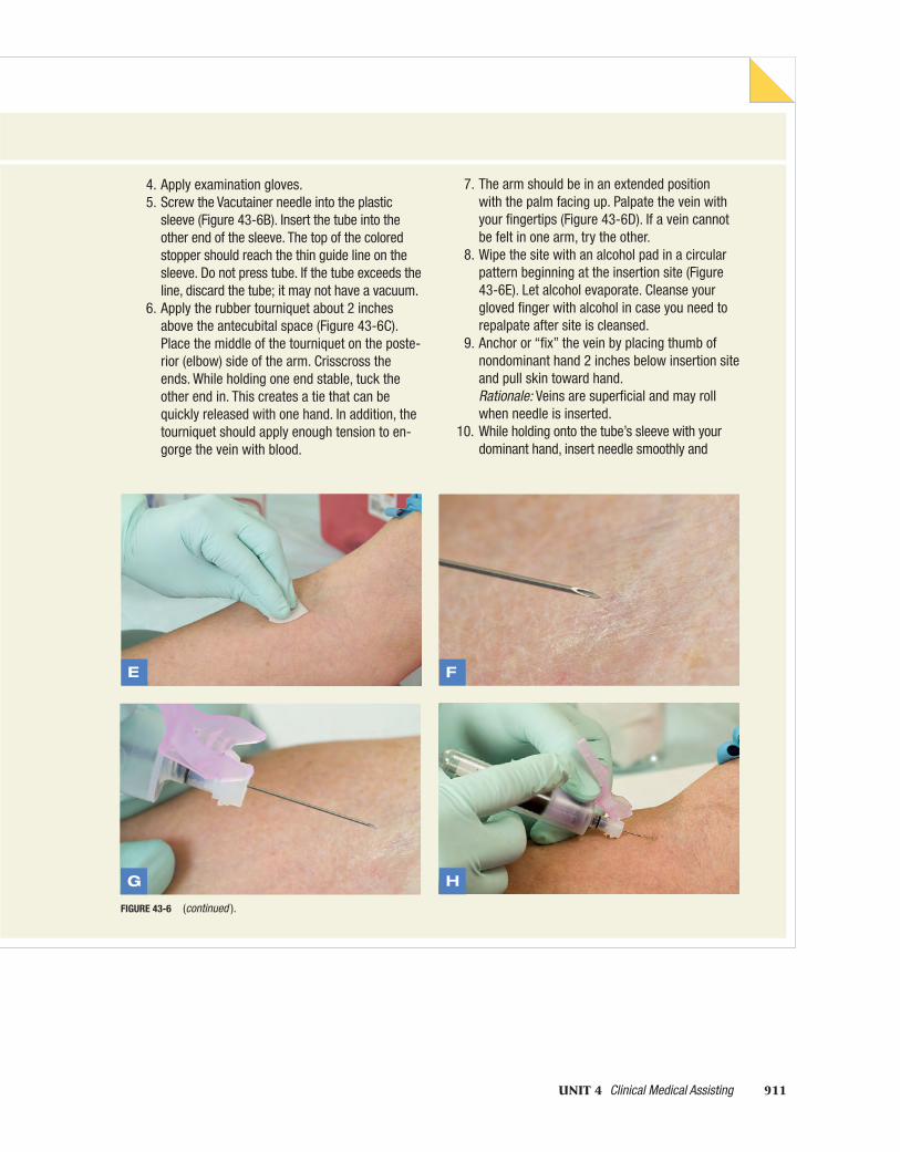
4. Apply examination gloves.5. Screw the Vacutainer needle into the plastic
sleeve (Figure 43-6B). Insert the tube into theother end of the sleeve. The top of the coloredstopper should reach the thin guide line on thesleeve. Do not press tube. If the tube exceeds theline, discard the tube; it may not have a vacuum.
6. Apply the rubber tourniquet about 2 inchesabove the antecubital space (Figure 43-6C).Place the middle of the tourniquet on the poste-rior (elbow) side of the arm. Crisscross theends. While holding one end stable, tuck theother end in. This creates a tie that can bequickly released with one hand. In addition, thetourniquet should apply enough tension to en-gorge the vein with blood.
7. The arm should be in an extended positionwith the palm facing up. Palpate the vein withyour fingertips (Figure 43-6D). If a vein cannotbe felt in one arm, try the other.
8. Wipe the site with an alcohol pad in a circularpattern beginning at the insertion site (Figure43-6E). Let alcohol evaporate. Cleanse yourgloved finger with alcohol in case you need torepalpate after site is cleansed.
9. Anchor or “fix” the vein by placing thumb ofnondominant hand 2 inches below insertion siteand pull skin toward hand.Rationale: Veins are superficial and may rollwhen needle is inserted.
10. While holding onto the tube’s sleeve with yourdominant hand, insert needle smoothly and
G H
FIGURE 43-6 (continued ).
E F
MEDIMC43_0131715771.QXD 3/7/07 2:55 PM Page 911

912 Chapter 43 Hematology
Performing a Venipuncture Using the Vacutainer Method
PROCEDURE (continued)43-3
rapidly at a 15–20-degree angle with the bevelup (see Figure 43-6F). The needle only needs tobe inserted just past the bevel. If inserted too far,it will puncture both vein walls. Also keep needlein line with the vein.
11. While stabilizing the sleeve, push the tube intothe sleeve. Use your thumb to push the tubeand hold sleeve with index and middle fingeron the flange (Figure 43-6H).Rationale: The sleeve must be perfectly stableor needle will damage the fragile vein and willresult in a hematoma (bruise).
12. Allow the tube to fill. The vacuum will automati-cally fill the tube three-quarters full.
13. Remove the tube very carefully without movingthe needle and apply a second tube if needed(Figure 43-6I). Gently roll the tube five to sixtimes after removing it from the sleeve to allowthe blood to mix with the additive. If both a redand purple tube are needed, collect blood in thered tube that contains no additive first.
I J
FIGURE 43-6 (continued ).
14. Release tourniquet once the last tube has been in-serted into the adaptor. Fill last tube remove it, andswiftly remove the needle and cover site with a cleangauze pad. Be careful not to push onto the needlewhen covering the puncture site, since that maycause the needle to scratch the patient’s arm. Gentlyinvert collection tube.Rationale: the tube must be removed from the adaptorbefore the needle has been removed or additionalbleeding may occur at the puncture site.
15. Immediately have patient apply firm, continuous pressure using a cotton ball or gauze square (Figure 43-6J).Rationale: Firm, continuous pressure will decreasechance of hematoma (bruise) formation.
16. Properly dispose of needle in biohazard container(Figures 43-6K, 43-6L and 43-6M).
17. Gently invert all tubes collected 8 to 10 times in afigure-eight pattern.Rationale: RBCs are fragile and may hemolyze if han-dled improperly.
MEDIMC43_0131715771.QXD 3/7/07 2:55 PM Page 912

UNIT 4 Clinical Medical Assisting 913
18. Assess the patient. Check the venipuncture sitefor bleeding, then apply some cotton and a stripof adhesive or a bandage (Figure 43-6N). Ask ifthe patient is dizzy or lightheaded.Rationale: If patient is dizzy or lightheaded,he or she may faint when attempting to stand.
19. Label tubes with patient’s name, date, time, IDnumber, specimen type, tests to be done, andphlebotomist’s initials. Fill out laboratory requi-sition sheet (Figure 43-6O).
20. Remove gloves. Perform hand hygiene.21. Record procedure on patient’s medical
record.
Charting Example
2/28/XX 1.00 P.M.
Withdrew 10 mL of blood from L arm, no complica-tions. Sent blood to in-office lab for CBC.
M. King, RMA
K
L M
N O
FIGURE 43-6 (continued )
MEDIMC43_0131715771.QXD 3/7/07 2:55 PM Page 913

914 Chapter 43 Hematology
FIGURE 43-7 A fingerstick is useful for obtaining small amounts of blood.
FIGURE 43-10 Spring-activated lancet.
with diabetes may be anxious about performing thistest. Proper education of the patient will be helpful inaddressing their concerns. A recheck visit or follow-upcall to the patient may be necessary to ensure that thepatient’s comfort level is achieved. This daily test is verycritical to address the patient’s diabetic condition.
The physician will often order a glycosylated hemo-globin (HbgA1C) to test the long-term control of dia-betes. For this test it is critical that the patient beinstructed to fast. If the patient fails to fast, the test re-sults will be inaccurate. Concerns expressed by the pa-tient regarding his or her medications should be directedto the physician. The physician will then determine ifchanges in medication must occur. To screen for bloodglucose levels in diabetics, a glucometer may be utilized(see Procedure 43-5).
Other tests that a physician may order on a dia-betic patient could include the glucose tolerance test.Although this test was traditionally done in a physi-cian’s office, it is now done more often in the outpa-
tient department at a hospital. The patient shouldbe given clear instructions as to the location of theoutpatient hospital department. Often hospitals pro-vide maps that are useful to provide to the patient.The patient should be informed that this test requiresseveral hours. This is due in part to the preparation,which involves drinking a solution and several blooddraws.
Laboratory results are reported from the lab to thephysician’s office in a variety of ways including phonecalls from the laboratory or by copies of the report be-ing faxed or sent by courier. If the medical assistantreceives a call from the laboratory with results, it is im-portant to write down the correct results. Often physi-cians’ offices will have standard forms that are used torecord laboratory results. Lab slips for common testssuch as urinalysis and common blood panels are oftenprovided. This makes the process of recording thephone information easier. If a lab report does come inby phone it must be followed up with the hard-copycontaining the results from the lab. The contents ofmost lab slips include the patient’s name, the writtenresults of the test, and the range values. Table 43-1provides a list of some commonly performed labora-tory tests and the range values.
FIGURE 43-8 Capillary puncture sites.
EarlobeRing/great finger Infant ’s heel
FIGURE 43-9 Capillary puncture equipment.
MEDIMC43_0131715771.QXD 3/7/07 2:55 PM Page 914

Performing a Capillary Puncture (Manual)
PROCEDURE43-4
OBJECTIVE: Perform a capillary stick using a lancet or spring-loaded lancet following correctaseptic technique and obtaining an adequate sample.
Equipment and Suppliesbiohazard sharps container; examination gloves; alcoholpad; 2 3 2 gauze square; lancet or spring-loaded lancet;capillary tubes; sealing clay; ammonia ampules; bandage;lab coat
Note: Follow Standard Precautions and safety guidelines.Use care to avoid splashing or spilling blood. Wipe up allspills using guidelines established by OSHA.
Method1. Perform hand hygiene.
Rationale: Hand washing helps prevent the spread ofinfection.
2. Assemble equipment.3. Identify the patient and explain the procedure. Have
the patient either sitting or lying.4. Apply examination gloves.5. Select either the ring or great finger on the nondomi-
nant hand. Wipe the site with an alcohol pad. Letalcohol evaporate.
6. Remove plastic protective tip to expose the lancet.7. Grasp patient’s hand and gently squeeze the finger
1 inch below the chosen puncture site.8. Puncture the site using a quick, jabbing motion
across the fingerprints to obtain a full round drop ofblood (Figure 43-11A). Do not puncture the directcenter of the finger pad since the skin is generallytougher there. Immediately discard lancet in sharpscontainer. (A spring-loaded lancet may also be used.)
9. Wipe away the first drop of blood with a gauze square.Rationale: The first drop contains alcohol and tissuefluid and will not provide accurate test results (Fig-ure 43-11B).
10. Obtain the sample using a microhematocrit capillarytube (Figure 43-11C). The finger may be gently mas-saged to increase blood flow. Seal one end of thecapillary tube in clay sealer (Figure 43-11D).Rationale: The reason for gently massaging ratherthan squeezing the finger is to avoid mixing the bloodwith other tissue fluids that may in turn hemolyze thered blood cells.
11. Apply clean gauze over the site and ask patient toapply firm, continuous pressure until the bleedingstops.Rationale: Firm, continuous pressure will decreasehematoma formation.
12. Assess patient and the site. Apply a bandage, ifneeded. Ask patient if he or she is dizzy orlightheaded.
13. Remove gloves and perform hand hygiene.14. Record procedure on patient’s medical record.
Charting Example
2/28/XX 1:30 P.M.
Performed capillary puncture on L ring finger, nocomplications.
M. King, RMA
A B
C D
FIGURE 43-11 Capillary puncture procedure.
MEDIMC43_0131715771.QXD 3/7/07 2:55 PM Page 915

916 Chapter 43 Hematology
Microhematocrit ProcedureThe microhematocrit (crit) provides the physician withinformation about the patient’s red blood cell volume. Alow hematocrit indicates anemia (decrease in the quan-tity of circulating RBCs) or hemorrhage. An elevatedhematocrit indicates dehydration or polycythemia. Anormal hematocrit is 40% to 50% in males and 35% to45% in females.
To perform a microhematocrit, the patient’s bloodmust be drawn using capillary tubes. The patient’sfinger is cleansed with an alcoholic sponge and thendried with sterile gauze. The patient’s finger is thenpunctured utilizing an automatic lancet or a manuallancet. The first drop of blood is wiped away with drysterile gauze. The second and subsequent drops aredrawn up using a capillary tube that is either tiltedhorizontally or slightly downward. When the tip ofthe tube touches the blood, the tube will automatically
draw the blood up by capillary action. The tubeshould be filled two-thirds to three-quarters of theway full and then sealed on each end. These tubesare then placed in a microhematocrit centrifuge that
FIGURE 43-12 Whole-blood analyzer.
FIGURE 43-13 The COULTER STKS hematology analyzer system.
FIGURE 43-14 ONE TOUCH Profile diabetes blood sugar monitoring system.
TABLE 43-1 Common Laboratory Tests and Their Normal Values
Total cholesterol 130–200 mg/dL
Glucose 70–120 mg/dL
Triglycerides 40–150 mg/dL
Creatinine 0.7–1.4 mg/dL
Uric acid 3.5–7.5 mg/dL
BUN 8–20 mg/dL
Sodium 132–142 mEq/L
Potassium 5–5 mEq/L
Chloride 98–106 mEq/L
CO2 25–32 mEq/L
White blood cell count 5,000–10,000/mm3
Red blood cell count 3.5–5.5 × 10/mm3
Hemoglobin 12–16 g/dL
Hematocrit 35.5–49%
Sedimentation rate 0–10 mm/hr
Platelet count 15,000–35,000/mm3
MEDIMC43_0131715771.QXD 3/7/07 2:55 PM Page 916

UNIT 4 Clinical Medical Assisting 917
For some medical assistants blood draws canbe both challenging and rewarding. If you find thatperforming laboratory tests are not your strength,be sure to work on this area while you are still inschool. Ask your instructor to provide extra time inclass or outside of class to test your skills.When se-lecting an externship it will be important to selectone that will continue to provide you encourage-ment and assistance in gaining skills in this area.Communication regarding areas that continue to bechallenging will be important so that the right typeof help can be provided to you so that your skillscan be developed. With some assistance and prac-tice you will become proficient.
Preparing for
Externship
Screening for Glucose (Blood Sugar) Level
PROCEDURE43-5
OBJECTIVE: Determine blood glucose level using a glucometer.
Equipment and Supplies
sterile lancet; reagent strips; glucometer; examina-tion gloves; cotton balls; alcohol sponges; gauzesquares; watch or clock; pen; lab coat; patient’srecord
Method
1. Identify the patient and make sure the patient isfasting if required.
2. Assemble the equipment and supplies.3. Make sure the glucometer has been turned on
the required amount of time and has been cali-brated for accuracy according to the manufac-turer’s instructions.
4. Wash hands and put on examination gloves.5. Perform a capillary blood puncture utilizing a
sterile lancet.6. Remove a plastic reagent strip from the bottle
without touching the chemically treated portion.7. Apply a large drop of blood from the capillary
puncture to the chemically treated area so thatit is completely covered.
8. Immediately begin timing for the duration specified by the manufacturer.
9. When the duration specified is complete, placethe reagent strip into the glucometer and closethe door.
10. At this time provide the patient with a drygauze square to hold over the puncture siteafter wiping the site with an alcohol sponge.
11. Record the number of mg (milligrams) of glu-cose per 100 mL (milliliters) as displayed by theinstrument.
12. Discard all used equipment and supplies.Remove gloves and wash hands.
Note: Several different types of testing instrumentsmay be used to perform this test. It is critical to followthe manufacturer’s instructions for the testing equip-ment provided.
performs cellular separation. For the complete proce-dure, review Procedure 43-6 and Figure 43-15.
Hemoglobin DeterminationHemoglobin (Hgb) provides the physician with infor-mation regarding the amount of hemoglobin presentin the sample. A low Hgb may indicate iron-deficiencyanemias, while elevated readings are present in patientswith polycythemia and in extreme situations, suchas burns. Normal values for adult females are 12 to16 g/dL, and for males 14–18 g/dL.
Hemoglobin can either be measured by an auto-mated blood analyzer or manually by using a hemo-globinometer (see Figures 43-16, 43-17, and 43-18).Typically, the manual method is less accurate and notas reliable as the automated blood analyzer.
Hemoglobin values can be determined utilizing twomethods: the specific gravity method or the cyanmethe-moglobin method. The specific gravity method is a screen-ing method for blood donors. The cyanmethemoglobin
MEDIMC43_0131715771.QXD 3/7/07 2:55 PM Page 917

Performing a Microhematocrit
PROCEDURE43-6
OBJECTIVE: Perform a microhematocrit on a capillary blood sample using proper aseptic technique.
Equipment and Supplies
biohazard sharps container; examination gloves;capillary tubes; sealing clay; microhematocritcentrifuge; whole blood; hematocrit card or otherreaderNote: Follow Standard Precautions and safetyguidelines when working with blood samples. Usecare to avoid splashing or spilling blood. Wipe up allspills using guidelines established by OSHA.
Method
1. Perform hand hygiene and apply examinationgloves.
2. Assemble equipment as shown in Figure 43-15A.3. Fill two capillary tubes three-quarters full. The
blood specimen can be obtained from a vacuumtube of anticoagulated blood using a plain capil-lary tube or directly from a fingerstick siteusing a heparinized capillary tube. Seal oneend in the sealing clay.
4. Place capillary tubes in centrifuge with thesealed ends against the rubber gasket (Figure43-15B). If more than one patient’s blood isbeing tested, mark down the number of the slotthe patient’s tube is in. Spin for 3 to 5 minutes
at 10,000 rpms. (Always check manufacturer’srecommendations for proper time and speed.)After centrifuging, the sample will be separatedinto three layers:n Top layer is the plasma.n Middle layer, or the buffy coat, is made up of
WBCs and platelets.n Bottom layer is packed RBCs.
5. Remove tubes immediately after centrifuge stops.Rationale: If tubes are not removed immedi-ately, blood may begin to mix together.
6. Determine results. Use the Hct card by placingthe sealing clay just below the zero line on bothtubes. Then on both tubes match the top of theplasma with the 100 line. Read results on bothtubes directly below the buffy coat. Then addthose results together and divide by 2.
7. Discard tubes into sharps container.8. Remove gloves and perform hand hygiene.9. Record the value as a percentage on the
patient’s medical record.
Charting Example
2/28/XX 1:45 P.M. Hct 47%.
M. King, CMA
A
B
FIGURE 43-15 (A) Centrifuge and supplies; (B) loading a centrifuge.
MEDIMC43_0131715771.QXD 3/7/07 2:55 PM Page 918

UNIT 4 Clinical Medical Assisting 919
6.0 million/mm3. An increase in the number of circu-lating RBCs may indicate polycythemia, whereas adecrease may indicate anemia.
A manual RBC count is similar to a manual WBCcount. Both require small samples of the specimen tobe diluted in a special solution. Then the sample isplaced on a hemocytometer, which is placed on a micro-scope used to count the red and white blood cells.
Automated testing for RBCs is more common thanmanual testing. Always follow the machine’s proce-dure manual exactly. The manual method of countingred blood cells is done with the hemacytometer.
Differential White Blood Cell CountA differential white blood cell count (diff) determinesthe percentages of each type of WBC, RBC morphology,and platelet estimation. Performing this test manually isa skill that requires practice to achieve proficiency. Thetesting is done using a microscope with a bright light
is a more specific and accurate method to give exact he-moglobin levels using hemoglobin analyzers. (See Proce-dure 43-7 for a hemoglobin determination using thehemoglobinometer.)
White Blood Cell CountNormal WBC counts range from 4.5 to 11 thousand/mm3 in adults. An elevated level usually indicates in-fection (leukocytosis) or, if grossly elevated, may beleukemia. A low level usually indicates a viral infectionor auto immunodeficiency.
A WBC count can be performed either manuallyby the medical assistant using a microscope or byan automated blood analyzer. The manual method ofobtaining white blood counts is through the use of ahemacytometer. A hemacytometer is a special slide count-ing chamber that allows for counting of cells under themicroscope. If testing is preformed manually, the med-ical assistant may use an automated tabulator to as-sist in counting the various types of cells (see Figure43-19). Always follow the instructions on the automatedanalyzer exactly to ensure the validity of the results.
Red Blood Cell CountNormal RBC volume in an adult female is 4.5 to 5.0million/mm3. In the adult male, it is about 5.0 to
FIGURE 43-18 VITROS DT60II system.
FIGURE 43-16 Hemoglobinometer.
FIGURE 43-17 Nova 16 analyzer.
MEDIMC43_0131715771.QXD 3/7/07 2:55 PM Page 919

920 Chapter 43 Hematology
and 1003 magnification with an oil immersion slide.Focus near the edge of the stained slide where the cellsare feathered, and where the cells are one layer thick.This test can also be performed by the automated ana-lyzer (see Procedure 43-8 for slide preparation and alsoFigures 43-20 and 43-21.)
Red Blood CellsRBCs are the most numerous, are salmon colored, andappear oval with a slightly pale center. RBCs have nonucleus or granules. RBCs with a nucleus are calledreticulocytse (immature RBCs). Normal looking RBCsare recorded as “normal RBC morphology.”
PlateletsPlatelets are the smallest of the formed blood elements.They are about half the size of a RBC. They stain purpleFIGURE 43-19 Electronic tabulator.
Determining Hemoglobin Using the Hemoglobinometer
PROCEDURE43-7
OBJECTIVE: Perform a blood test to determine hemoglobin levels using thehemoglobinometer.
Equipment and Supplies
hemoglobinometer; glass slide chamber; hemolysisapplicator (plastic or wooden); sterile lancet; cottonballs; dry gauze square; alcohol sponges; examina-tion gloves; patient’s record; lab coat; biohazardsharps containerNote: Follow Standard Precautions and safetyguidelines when working with blood samples. Usecare to avoid splashing or spilling blood. Wipe up allspills using guidelines established by OSHA.
Method
1. Perform hand hygiene and put on examinationgloves.
2. Gather the necessary equipment and supplies.3. Using a manual lancet or auto lancet, obtain
capillary blood.4. Pull the glass chamber out of the hemoglobi-
nometer and position the lower part of the slideso that it is slightly offset.
5. Place a large drop of capillary blood onto theslide.
6. Wipe the patient’s puncture site with a cottonball and provide the patient with a dry gauzesquare to apply mild pressure to the puncture.This should stop further bleeding.
7. Mix blood with hemolysis applicator until theblood becomes clear.
8. Push the glass chamber into the clip and placeinto the slot on the left side of the hemoglobi-nometer.
9. Hold the hemoglobinometer in your left hand ateye level while using your left thumb to turn onthe light by depressing the bottom button. Lookinto the instrument to see a split green field.
10. Slide the button on the right side of the meterwith your right thumb and index finger whilelooking into the meter until a matching greenfield occurs. Leave the sliding scale on thecalibrated line where the solid green fieldappeared.
11. Read the hemoglobin value at the top scale.The results are read as grams of hemoglobinper 100 mL of blood (g/dL).
12. Wash chamber and reusable hemolysis appli-cator with a detergent solution, rinse, dry, andreturn to the instrument for the next test.
13. Remove gloves and perform hand hygiene.Discard gloves and nonreusable supplies in appropriate containers.
14. Record the results in the patient’s record.
MEDIMC43_0131715771.QXD 3/7/07 2:55 PM Page 920

UNIT 4 Clinical Medical Assisting 921
and tend to appear in a clump, although they may alsoexist singly. They appear if they have a rough outeredge and contain small granules. There are between200,000 and 300,000 platelets per cubic millimeterof the blood. There are typically between 5 and 20platelets in one field of view. If this number is counted,then record the platelet count as “adequate plateletestimation.”
White Blood CellsThere are five types of white blood cells, each of whichhas a distinct identifying characteristic when using theWright’s staining method. Count 100 WBCs, then ex-press each of the cell types as a percentage. Normalvalues for adults are:
• Neutrophils 50–70%
• Eosinophils 1–4%
• Basophils 0–1%
• Lymphocytes 20–35%
• Monocytes 3–8%
Values may differ between manual and automatedanalyses.
GRANULATED WHITE BLOOD CELLS The neu-trophil, or seg, is the most numerous WBC. The neu-trophil has small cytoplasmic granules that stain pinkor lilac with a multilobed nucleus with small strandsconnecting each of the lobes, which stain purple. Aband neutrophil is an immature neutrophil. It appearssimilar to the neutrophil except that the nucleus isnonsegmented and curved with a band-like structure.Cytoplasmic granules stain blue to pink.
Eosinophils have segmented nuclei and large, red-dish staining granules that are found in the cytoplasm.Basophils are rarely seen in a diff count. This WBChas an S-shaped nucleus and large, irregularly shapedpurplish-blue granules that almost entirely cover thenucleus.
ANGRANULOCYTIC WHITE BLOOD CELLS Lym-phocytes have a single round or lightly indented nu-cleus, which almost completely fills the cell. Thecytoplasm is clear and stains a pale blue. Lymphocytesare the smallest WBC.
Monocytes are the largest WBC and have a distinctkidney-bean-shaped nucleus. The cytoplasm is abun-dant and clear, and it stains a grayish blue.
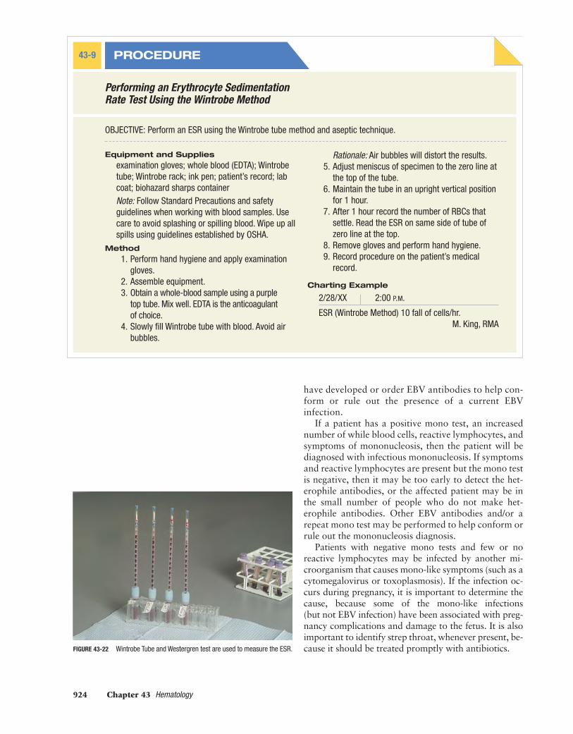
Erythrocyte Sedimentation RateThe erythrocyte sedimentation rate (ESR) (also called thesed rate) measures the rate at which RBCs settle at thebottom of a tube. Drawing a patient’s ESR can be doneutilizing either the Wintrobe or Westergren method.
When performing the Wintrobe method, the Wintrobetube is calibrated in mm/hr. The rate is the height of theRBCs in the bottom of the tube (see Procedure 43-9 andFigure 43-22).
Depending on the type of method used, the normalvalues may vary. Utilizing the Wintrobe method thenormal ESR in an adult female is 0 to 20 mm/hr and inan adult male 0 to 9 mm/hr. Increased values maymean inflammation. An individual’s ESR may also beelevated due to a variety of reasons including menstru-ation, pregnancy, and malignant tumors.
An ESR itself is not diagnostic, but it is used in con-junction with other tests for diagnosis. As an example,when testing for rheumatoid arthritis or fibromyalgia,an ESR may be done in conjunction with the antinu-clear antibody test (ANA).
The sed rate is related to the condition of the redblood cells and the amount of fibrinogen in the plasma.When an individual presents with a disease, the sur-faces of the membranes of the cells are affected. Whena sed rate test is conducted on a patient, the rate atwhich the RBCs fall indicates the existence of possibleconditions.
PhenylketonuriaPhenylketonuria (PKU) is a congenital disease causedby a defect in the metabolism of the amino acidphenylalanine. The unmetabolized protein accumu-lates in the bloodstream and, if undetected and un-treated, will result in mental retardation. The PKU testis always performed on newborns to determine thepresence of the unmetabolized protein phenylalanine.The test is typically performed in the hospital but maybe performed in the office if not done in the hospital.To perform the PKU test see Procedure 43-10.
Mono TestingThe mono test, which is also known as the mononu-cleosis spot test, is used to help determine whethera patient has inefectious mononucleosis (see Proce-dure 43-11). It is frequently ordered along with aCBC (complete blood count). A step test may be or-dered with the mono test to determine whether a per-son’s sore throat is due to a streptococcal infectioninstead of or in addition to mononucleosis.
A mono test is primarily ordered when an adoles-cent patient has symptoms such as fever, headache,swollen glands, and fatigue that the doctor suspectsare due to infectious mononucleosis. The test may berepeated when it is initially negative but suspicion ofmono remains high. The physician may order a re-peat test in a week or so to see if heterophile antibodies
MEDIMC43_0131715771.QXD 3/7/07 2:55 PM Page 921

922 Chapter 43 Hematology
Preparing Slides
PROCEDURE43-8
OBJECTIVE: Prepare a slide for a differential WBC count using correct asepticprocedure.
Equipment and Supplies
clean, glass slides; whole blood (EDTA); examina-tion gloves; biohazard container; eye dropper;Wright’s stain; lab coat; ink pen; patient recordNote: Follow Standard Precautions and safetyguidelines when working with blood samples.Use care to avoid splashing or spilling blood. Wipeup all spills using guidelines established by OSHA.
Method
1. Perform hand hygiene and apply examinationgloves.
2. Assemble equipment.
3. Obtain a whole-blood sample using EDTA asthe anticoagulant of choice. Blood must bemixed thoroughly before use.
4. Using a dropper, place one drop of room tem-perature blood on the end of a clean, glass slide(Figure 43-20A).
5. Using the short side of another clean, glass slide,back the slide to the drop of blood. Allow theblood to spread across the short side of the slide(Figure 43-20B). Holding the spreader slide at a30-degree angle, spread the blood across thelength of the slide (Figure 43-20C). Use gentle,
A B
C D
FIGURE 43-20 Blood smear.
MEDIMC43_0131715771.QXD 3/7/07 2:55 PM Page 922

UNIT 4 Clinical Medical Assisting 923
FIGURE 43-21 Wright’s staining process.
C D
continuous pressure, and a smooth glidingmotion to create a smear as pictured in Figure43-20D. Notice that the smear has a thick sidethat gradually changes to a thin side. The thinside has a feathered edge, and the blood coversone-half to three-quarters the length of the slide.
6. Allow the slide to air dry on a rack (Figure 43-21A).Rationale: Air drying will not distort cells.
7. Label on the frosted edge of the slide with patient’s name and the date.
8. Stain slide using Wright’s staining method.Flood slide with stain for exactly 45 seconds oramount of time indicated by manufacturer(Figure 43-21B).
9. Rinse with distilled water as in Figure 43-21C.Rinse until water is clear (Figure 43-21D).
10. Allow slide to air dry before examining underthe microscope.
11. Abnormal findings may need to be referred toa laboratory technician for analysis. Recordresults on the patient’s medical record.
Charting Example
2/28/XX 2:15 P.M.
Differential cell count: neutrophils 62%, lymphocyte28%, monocytes 5%, eosinophils 5%, basophils0%, RBC morphology—normal, adequate plateletestimation.
M. King, RMA
A B
MEDIMC43_0131715771.QXD 3/7/07 2:55 PM Page 923
924 Chapter 43 Hematology
Performing an Erythrocyte SedimentationRate Test Using the Wintrobe Method
PROCEDURE43-9
OBJECTIVE: Perform an ESR using the Wintrobe tube method and aseptic technique.
Equipment and Supplies
examination gloves; whole blood (EDTA); Wintrobetube; Wintrobe rack; ink pen; patient’s record; labcoat; biohazard sharps containerNote: Follow Standard Precautions and safetyguidelines when working with blood samples. Usecare to avoid splashing or spilling blood. Wipe up allspills using guidelines established by OSHA.
Method
1. Perform hand hygiene and apply examinationgloves.
2. Assemble equipment.3. Obtain a whole-blood sample using a purple
top tube. Mix well. EDTA is the anticoagulantof choice.
4. Slowly fill Wintrobe tube with blood. Avoid airbubbles.
Rationale: Air bubbles will distort the results.5. Adjust meniscus of specimen to the zero line at
the top of the tube.6. Maintain the tube in an upright vertical position
for 1 hour.7. After 1 hour record the number of RBCs that
settle. Read the ESR on same side of tube ofzero line at the top.
8. Remove gloves and perform hand hygiene.9. Record procedure on the patient’s medical
record.
Charting Example
2/28/XX 2:00 P.M.
ESR (Wintrobe Method) 10 fall of cells/hr.M. King, RMA
FIGURE 43-22 Wintrobe Tube and Westergren test are used to measure the ESR.
have developed or order EBV antibodies to help con-form or rule out the presence of a current EBVinfection.
If a patient has a positive mono test, an increasednumber of while blood cells, reactive lymphocytes, andsymptoms of mononucleosis, then the patient will bediagnosed with infectious mononucleosis. If symptomsand reactive lymphocytes are present but the mono testis negative, then it may be too early to detect the het-erophile antibodies, or the affected patient may be inthe small number of people who do not make het-erophile antibodies. Other EBV antibodies and/or arepeat mono test may be performed to help conform orrule out the mononucleosis diagnosis.
Patients with negative mono tests and few or noreactive lymphocytes may be infected by another mi-croorganism that causes mono-like symptoms (such as acytomegalovirus or toxoplasmosis). If the infection oc-curs during pregnancy, it is important to determine thecause, because some of the mono-like infections (but not EBV infection) have been associated with preg-nancy complications and damage to the fetus. It is alsoimportant to identify strep throat, whenever present, be-cause it should be treated promptly with antibiotics.
MEDIMC43_0131715771.QXD 3/7/07 2:55 PM Page 924

UNIT 4 Clinical Medical Assisting 925
Performing a PKU Test
PROCEDURE43-10
OBJECTIVE: Collect blood specimen for PKU testing.
Equipment and Supplies
sterile lancet; alcohol sponge; examination gloves;sterile dry gauze; special filter paper card, typicallysupplied by the State Health Department
Method
1. Perform hand hygiene and don examinationgloves.
2. Cleanse the infant’s heel with an alcohol sponge.3. Puncture the lateral portion of the infant’s heel
with a sterile disposable lancet.4. Wipe puncture site with a dry sterile gauze.5. Allow large blood droplet to form.
6. Touch the blood droplet to the center of thecircle on one side of the special filter papercard.
7. Ensure the blood has completely soaked throughthe paper card by looking at the reverse side.
8. Fill all required five circles on the paper card.9. Do not squeeze heel excessively to avoid
collecting tissue fluid mixed with blood.10. Set card in an appropriate area to air dry for
2 hours at room temperature.11. When completely dry place in the state-
provided envelope and mail within 48 hours.
Performing a Mono Test
PROCEDURE43-11
OBJECTIVE: Student will perform a mono test.
Equipment and Supplies
antiseptic cleaner; biohazard waste container; dis-posable lancet; disposable gloves; capillary tube;test tube; mono test diluent; mono test stick(s);blood specimen
Method
1. Perform hand hygiene.2. Don gloves.3. Assemble equipment and supplies.4. Perform a fingertip stick on patient.5. Fill a capillary tube end to end, dispensing all of
the blood into the test tube.6. Slowly add 1 drop of diluent to the bottom of
the test tube.7. Mix.8. Remove the test stick(s) from the container.
Recap the container immediately.9. Place the absorbent end of the test stick into
the treated sample. Leave the test stick in thetest tube.
10. Read result at 5 minutes. Positive results maybe read as soon as the red control line appears.
11. Discard used test tubes, lancet, and test sticksin the biohazard waste container.
12. Remove gloves and dispose of them correctly.Perform hand hygiene.
13. Document findings in patient record:Positive: A blue test line and a red control line isa positive result.Negative: A red control line but no blue testline, is a negative result.
Note: While the medical assistant is responsible forreporting abnormal (or positive results in the case of amono test) to the physician, “interpreting” such testsas being positive or negative is NOT within the scopeof practice for the medical assistant as such, shouldnever be done. The medical assistant is only allowedto report the findings of “positive” or “negative.”14. Clean work area and equipment according to
OSHA guidelines.
MEDIMC43_0131715771.QXD 3/7/07 2:55 PM Page 925

926 Chapter 43 Hematology
Chapter Review
COMPETENCY REVIEW1. Define and spell the terms to learn for this chapter.2. List, in order, the steps for performing a venipuncture.3. Prepare a list of the different types of venipuncture collection tubes. Explain what makes
each unique and the order in which they should be used.4. Make a list of the types of white blood cells and the values of each that are expected in a
normal differential.5. Create a normal list (for male and female where applicable) for the WBC, RBC, Hct, Hgb
and ESR.6. List the equipment needed to perform an erythrocyte sedimentation rate test.
SUMMARYThe proper collection, handling, and processing ofblood specimens are vital to reliable test results. Inaddition, it is critical that a medical assistant has athorough understanding of patient preparation andthe theory of blood formation, including the cellular
and liquid components of blood. Lastly, knowledgeof and competency in, the performance of routineblood tests—CBCs, differentials, hemoglobins, mi-crohematocrits, ESRs—are essential to proper patientcare.
PREPARING FOR THE CERTIFICATION EXAM
1. A collection of blood below the surface of theskin is called:A. hematomaB. hemolysisC. hematemesisD. hematuriaE. hematology
2. Which type of blood collection tube containsa clot activator and a gel that permanently separates the serum from the clot aftercentrifugation?A. red topB. purple topC. green topD. marbled topE. gray top
3. The best vein to use in order to obtain a venoussample of blood is the:A. median cephalicB. medial cubitalC. basilic veinD. accessory cephalicE. small vein in the hand
4. The most commonly used anticoagulant forblood tests isA. heparinB. potassium oxylateC. sodium citrateD. potassium citrateE. EDTA
5. When drawing blood for a PT/INR and/or aPTT, which type of blood collection tubeshould be used?A. red topB. purple topC. green topD. light blue topE. gray top
6. If an angle of more than 15 degrees is usedwhen inserting a needle into a patient’s arm:A. the needle may not puncture the veinB. the needle may breakC. the needle may go through both vein wallsD. the blood sample will hemolyzedE. a significant degree of pain will be caused
continued on next page
MEDIMC43_0131715771.QXD 3/7/07 2:55 PM Page 926
UNIT 4 Clinical Medical Assisting 927
7. If the blood collection tube is pushed past thethin guideline on the plastic sleeve:A. blood will hemolyzeB. the needle will dullC. the vacuum may be compromisedD. blood will enter the tube too quicklyE. the blood sample will be contaminated
8. Another name for a capillary puncture is a:A. fingerstickB. arterial blood drawC. phlebotomyD. arterioles drawE. venipuncture
9. The purpose behind applying pressure and agauze or bandage after a capillary puncture is to:A. prevent an infection by keeping the site sterileB. decrease the likelihood of hematomaC. cause a sterile clot to form.D. prevent the spread of infectionE. reassure the patient that the procedure has
ended
10. When performing an ESR, how long should thesample sit?A. 20 minutesB. 40 minutesC. 60 minutesD. 80 minutesE. 90 minutes
CRITICAL THINKING1. What method(s) should Mary use to collect the samples?
2. What type of Vacutainer tubes should she use to collect the samples?
3. What individual tests comprise a CBC?
ON THE JOBIt is Wednesday and Drs. Joseph and Burg are not seeing patients. The office though is openfor blood draws. Angie, a medical assistant, is in the office alone.
Matt, a 17-year-old upcoming college freshman, has just arrived to have his labs drawn sothat Dr. Joseph can complete Matt’s college physical. He is extremely nervous, although he istrying very hard to exhibit a relaxed manner. Angie tries to reassure Matt, but the more shetries, the more nervous he gets. At Matt’s urging to “just get it over with,” Angie decides to goahead and draw his blood. Unfortunately, as soon as the first collection tube begins to fill withblood, Matt notices it and slumps forward. He has fainted. What should Angie do?
What is your response?
1. What is the first thing that Angie should do?2. Was Angie at all negligent in drawing Matt’s blood given that he was extremely nervous?3. Does the fact that Matt is a minor influence how Angie should handle this situation? If
so, how?4. Does Matt have some level of complicity in this incident? If so, describe.5. Is this considered a medical emergency.6. Would the situation have been different or handled differently if Angie hadn’t been alone?7. Does an incident report of some sort need to be filed? If so, what should be included and
who should receive a copy?
INTERNET ACTIVITYGo online and research materials to further understand OSHA regulations, the Centers forDisease Control and Prevention (CDC) Standard Precautions, and the laboratory regulationsestablished by CLIA 1988.
MediaLink More on hematology, including interactive resources, can be found on theStudent CD-ROM accompanying this textbook.
MEDIMC43_0131715771.QXD 3/7/07 2:55 PM Page 927