Patient-derived organoids and high grade serous ovarian ...
14
REVIEW Open Access Patient-derived organoids and high grade serous ovarian cancer: from disease modeling to personalized medicine Camilla Nero 1,2 , Giuseppe Vizzielli 1 , Domenica Lorusso 1,2 , Eleonora Cesari 1,3 , Gennaro Daniele 1 , Matteo Loverro 1 , Giovanni Scambia 1,2* and Claudio Sette 3* Abstract Background: High grade serous ovarian cancer (HGSOC) is among the deadliest human cancers and its prognosis remains extremely poor. Tumor heterogeneity and rapid acquisition of resistance to conventional chemotherapeutic approaches strongly contribute to poor outcome of patients. The clinical landscape of HGSOC has been radically transformed since the advent of targeted therapies in the last decade. Nevertheless, the lack of predictive biomarkers informing on the differential clinical benefit in select subgroups, and allowing patient-centric approaches, currently limits the efficacy of these novel therapies. Thus, rational selection of the best possible treatment for each patient represents a clinical priority in order to improve outcome, while limiting undesirable effects. Main body: In this review, we describe the state of the art and the unmet needs in HGSOC management, illustrate the treatment options that are available and the biomarkers that are currently employed to orient clinical decisions. We also describe the ongoing clinical trials that are testing new therapeutic approaches for HGSOC. Next, we introduce the organoid technology as a promising, expanding strategy to study cancer and to develop personalized therapeutic approaches. In particular, we discuss recent studies that have characterized the translational potential of Patient’s Derived Organoids (PDOs) to inform on drug sensitivity of HGSOC patients. Conclusions: PDOs can predict the response of patients to treatments and may therefore guide therapeutic decisions. Although preliminary results appear encouraging, organoids still need to be generated and expanded efficiently to enable drug screening in a clinically meaningful time window. A new generation of clinical trials based on the organoid technology should guarantee tailored approaches to ovarian cancer management, as it is now clear that the one-size-fits-all approach cannot lead to efficient and meaningful therapeutic advancements. Keywords: Ovarian cancer, Organoids, Target therapy, 3D cultures, Drug screening © The Author(s). 2021 Open Access This article is licensed under a Creative Commons Attribution 4.0 International License, which permits use, sharing, adaptation, distribution and reproduction in any medium or format, as long as you give appropriate credit to the original author(s) and the source, provide a link to the Creative Commons licence, and indicate if changes were made. The images or other third party material in this article are included in the article's Creative Commons licence, unless indicated otherwise in a credit line to the material. If material is not included in the article's Creative Commons licence and your intended use is not permitted by statutory regulation or exceeds the permitted use, you will need to obtain permission directly from the copyright holder. To view a copy of this licence, visit http://creativecommons.org/licenses/by/4.0/. The Creative Commons Public Domain Dedication waiver (http://creativecommons.org/publicdomain/zero/1.0/) applies to the data made available in this article, unless otherwise stated in a credit line to the data. * Correspondence: [email protected]; [email protected] 1 Fondazione Policlinico Universitario A. Gemelli IRCCS, L.go Agostino gemelli, 8, 00168 Roma, Italy 3 Dipartimento di Neuroscienze, Sezione di Anatomia Umana, Università Cattolica del Sacro Cuore, Roma, Italy Full list of author information is available at the end of the article Nero et al. Journal of Experimental & Clinical Cancer Research (2021) 40:116 https://doi.org/10.1186/s13046-021-01917-7
Transcript of Patient-derived organoids and high grade serous ovarian ...

REVIEW Open Access
Patient-derived organoids and high gradeserous ovarian cancer: from diseasemodeling to personalized medicineCamilla Nero1,2, Giuseppe Vizzielli1, Domenica Lorusso1,2, Eleonora Cesari1,3, Gennaro Daniele1, Matteo Loverro1,Giovanni Scambia1,2* and Claudio Sette3*
Abstract
Background: High grade serous ovarian cancer (HGSOC) is among the deadliest human cancers and its prognosisremains extremely poor. Tumor heterogeneity and rapid acquisition of resistance to conventional chemotherapeuticapproaches strongly contribute to poor outcome of patients. The clinical landscape of HGSOC has been radicallytransformed since the advent of targeted therapies in the last decade. Nevertheless, the lack of predictivebiomarkers informing on the differential clinical benefit in select subgroups, and allowing patient-centricapproaches, currently limits the efficacy of these novel therapies. Thus, rational selection of the best possibletreatment for each patient represents a clinical priority in order to improve outcome, while limiting undesirableeffects.
Main body: In this review, we describe the state of the art and the unmet needs in HGSOC management, illustratethe treatment options that are available and the biomarkers that are currently employed to orient clinical decisions.We also describe the ongoing clinical trials that are testing new therapeutic approaches for HGSOC. Next, weintroduce the organoid technology as a promising, expanding strategy to study cancer and to developpersonalized therapeutic approaches. In particular, we discuss recent studies that have characterized thetranslational potential of Patient’s Derived Organoids (PDOs) to inform on drug sensitivity of HGSOC patients.
Conclusions: PDOs can predict the response of patients to treatments and may therefore guide therapeuticdecisions. Although preliminary results appear encouraging, organoids still need to be generated and expandedefficiently to enable drug screening in a clinically meaningful time window. A new generation of clinical trials basedon the organoid technology should guarantee tailored approaches to ovarian cancer management, as it is nowclear that the one-size-fits-all approach cannot lead to efficient and meaningful therapeutic advancements.
Keywords: Ovarian cancer, Organoids, Target therapy, 3D cultures, Drug screening
© The Author(s). 2021 Open Access This article is licensed under a Creative Commons Attribution 4.0 International License,which permits use, sharing, adaptation, distribution and reproduction in any medium or format, as long as you giveappropriate credit to the original author(s) and the source, provide a link to the Creative Commons licence, and indicate ifchanges were made. The images or other third party material in this article are included in the article's Creative Commonslicence, unless indicated otherwise in a credit line to the material. If material is not included in the article's Creative Commonslicence and your intended use is not permitted by statutory regulation or exceeds the permitted use, you will need to obtainpermission directly from the copyright holder. To view a copy of this licence, visit http://creativecommons.org/licenses/by/4.0/.The Creative Commons Public Domain Dedication waiver (http://creativecommons.org/publicdomain/zero/1.0/) applies to thedata made available in this article, unless otherwise stated in a credit line to the data.
* Correspondence: [email protected];[email protected] Policlinico Universitario A. Gemelli IRCCS, L.go Agostino gemelli,8, 00168 Roma, Italy3Dipartimento di Neuroscienze, Sezione di Anatomia Umana, UniversitàCattolica del Sacro Cuore, Roma, ItalyFull list of author information is available at the end of the article
Nero et al. Journal of Experimental & Clinical Cancer Research (2021) 40:116 https://doi.org/10.1186/s13046-021-01917-7

BackgroundThe overall survival of patients with ovarian cancer (OC)had not substantially changed for several decades, mainlydue to the lack of early diagnosis coupled with frequentacquisition of resistance to therapeutic treatments [1, 2].Nevertheless, recent progress in traditional and noveltherapeutic strategies have led to a significant improve-ment in patient’s outcome especially for the high gradeserous histotype (HGSOC), which is the most commonand most malignant among the OC subtypes [3–5]. Inparticular, several treatment options have become avail-able in the past 5 years, both for frontline and recurrentdisease settings.Decisions regarding the best treatment option for
each individual patient do not always have an obvious“optimal choice”, due to a number of variables thatare not always easy to decipher. First, both the typeand the timing of treatment(s) have to be considered.Moreover, while algorithms based on patient’s charac-teristics and biological factors are necessary, they arenot diagnostically exhaustive. Lastly, the identificationof successful therapies is also hampered by the highlevel of complexity and genetic heterogeneity existingeven within single tumor types. With more thera-peutic options being available, the identification ofbiological markers and/or experimental models thatare able to predict treatment response has progres-sively become a clinical priority.
In this scenario, an emerging technology that holdspromise of significantly impacting the clinical manage-ment of patients in the near future is represented bypatient-derived Organoids (PDOs). Indeed, elegant stud-ies carried out in the past 2–3 years have clearly indi-cated that HGSOC PDOs have the potential to faithfullyreproduce many of the challenging characteristics of thetumor from which they derive in a reasonable timeframe and at sustainable costs [6–10]. This technologymay offer the possibility of attaining truly personalizeddrug-based therapy. On this basis, it is conceivable thatPDOs could be introduced as a clinical test to guide theselection of therapeutic treatments and to improve man-agement of HGSOC patients in the near future.
State of the art and unmet needs in ovariancancer clinical managementTreatment options and predictive biomarkersThe current treatment options for advanced stage andrecurrent HGSOC according to ESMO and NCCN rec-ommendations [11, 12] are summarized in Fig. 1.Treating HGSOC has become an increasingly complex
chess game in which clinicians should be fully aware ofthe consequences that each move implies. Identifying tu-mors that will respond to the available therapies in thatprecise moment of the disease is an emerging unmetneed that will progressively overcome every other trad-itional debate. In this regard, the efficacy of current
Fig. 1 HGSOC treatment options according to ESMO and NCCN recommendations in front line a and recurrent b clinical settings
Nero et al. Journal of Experimental & Clinical Cancer Research (2021) 40:116 Page 2 of 14

available treatments measured by progression or relapserate is represented in Fig. 2.The standard of care for newly diagnosed HGSOC pa-
tients consists of primary debulking surgery (PDS) andplatinum-based chemotherapy. The amount of residualdisease after surgery remains a key prognostic variablesupporting the role of PDS with maximal debulking oftumour [13, 14]. Although desirable, optimal or subopti-mal (less than 1 cm in maximum diameter of residualtumour) surgeries are only achieved in 25–40% of casesundergoing PDS worldwide [15]. Thus, neoadjuvantchemotherapy (NACT) followed by interval debulkingsurgery (IDS) has been tested as an alternative option.At least four randomized controlled trials were con-ducted to compare NACT versus PDS in the past years.Although debates on this topic continue, NACT has be-come an established practice of care in patients withhigh tumor load or severe comorbidities [16–19].The combination of carboplatin and paclitaxel is the
‘backbone’ of the chemotherapeutic treatment for ad-vanced HGSOC [20–22]. While most HGSOC patientsshow a good response to the conventional platinum-based chemotherapy, ~ 15% of women experience
primary resistance to these treatments [20–24]. To date,there are no validated predictive markers of primaryplatinum refractory or resistant disease. Although limitedand biologically unfounded, the time since the last platinumchemotherapy currently defines resistance (≤ 6months) orsensitivity (> 6months) to the treatment in HGSOC pa-tients that experience recurrent disease [25, 26].Subsequent randomized trials have been designed
adding a third drug, either in combination or in main-tenance. These trials lead to the approval of Bevacizu-mab and two types of poly-ADP-ribose polymerase(PARP) inhibitors (Olaparib and Niraparib) [26–29].Bevacizumab is a humanized monoclonal antibodyagainst vascular endothelial growth factor A (VEGF-A).Unfortunately, no molecular biomarkers can faithfullypredict its benefit in patients. Indeed, neither angiogenicmarkers (i.e. CD31, microvessel density and tumorVEGF-A levels) nor predictive signatures based on com-binations of biomarkers (i.e. mesothelin, FLT4, alpha-1acid glycoprotein and CA125 or Ang1 and Tie2) wereprospectively validated in large clinical trials and, there-fore, were not introduced into clinical practice [30, 31].Currently, only clinical biomarkers, including stage,
Fig. 2 Efficacy of the currently available treatments in HGSOC. Data are expressed as the median rate of freedom from disease progression andfrom death at a certain interval time
Nero et al. Journal of Experimental & Clinical Cancer Research (2021) 40:116 Page 3 of 14

debulking status, residual tumor and presence of ascites,are used to select patients for first line treatment withBevacizumab. Olaparib and Niraparib are inhibitors ofthe PARP-1 and -2 enzymes and are able to selectivelykill cancer cells that are defective for the BRCA 1 and 2tumor suppressors. PARP1/2, like BRCA1/2, are in-volved in the DNA damage response pathway that pro-motes homologous recombination (HR) and repair ofthe DNA lesions. Thus, concomitant inhibition of thispathway by BRCA1/2 mutations and PARP inhibitors(PARPi) results in synthetic lethality of cancer cells [32,33]. However, although mutations in BRCA1/2 genesrepresent the strongest hallmark of sensitivity to PARPi,up to 40% of HGSOC patients bearing these mutationsfail to respond to treatment [32]. On the other hand, tu-mors harboring other mutations that impair the HRpathway exhibited remarkable responses to PARPi [32–35]. Thus, direct evaluation of HR proficiency may helpstratify more accurately the HGSOC patients who wouldbenefit of treatment with PARPi. However, current as-says of HR proficiency are believed to be unsuitable toexclude patients from PARPi therapy, at least in secondline platinum sensitive relapse, in which platinum sensi-tivity is still considered the best predictor of response toPARPi [11]. The evaluation of genome-wide loss of het-erozygosity (LOH) or genomics scars (a score based ontelomeric imbalance, LOH and large-scale rearrange-ments) could indirectly assess HR deficiency status,including also patients whose tumors harbor HR-impairing mutations different from those in the BRCA1/2 genes. However, attempts to use these tests as predict-ive markers in the maintenance setting were not com-pletely successful and the tests were not conclusive inup to 20% of patients due to technical issues [36, 37].Moreover, reversion of HR deficiency, which may occurupon development of resistance to platinum and toPARPi and likely contributes to clinical drug resistance,is a major limitation of current HR assays [38, 39].Overall, targeted therapies produced a paradigm shift
in the treatment of HGSOC, transforming it into achronic condition. Up to 80% of HGSOC patients re-lapses within 24months and treatment options at thattime are also conditioned by first line treatments [40].The best treatment sequence or combination challengesclinicians as much as the optimal selection of patientsfor each treatment. Differently from Bevacizumab whoseefficacy beyond progression has been clearly reported, itis at present unclear whether PARPi maintain efficacy ina second treatment and if they can be used also inpatients who have previously received PARPi [41, 42].
Drugs under investigationMost ongoing clinical trials for HGSOC are focused onimmune check point inhibitors which are mainly based
on inhibiting the PD-1/PD-L1 pathway. The efficacy ofthese agents depends on many factors, including PD-L1expression, abundance of tumor infiltrating lymphocytes(TILs), neoantigen load and tumor mutational burden[43, 44]. Initial over-optimism about these agents inHGSOC treatment has been tempered by the disap-pointing results emerged in clinical trials [45–50].In particular, in recurrent patients, immune check
point inhibitors failed to demonstrate any relevant bene-fit both as single agent (KEYNOTE-100, single arm trialon Pembrolizumab) and in combination with chemo-therapy (JAVELIN 200, randomized trial pegylated lipo-somal doxorubicin and Avelumab vs pegylated liposomaldoxorubicin single agent vs Avelumab single agent; MK-3475, single arm trial on Pembrolizumab in combinationwith weekly paclitaxel) or with antiangiogenetic agents(NCT02873962, single arm on the combination of anti-PD1 Nivolumab with Bevacizumab) [46, 47, 49]. Clinicalresults were disappointing also in newly diagnosed pa-tients. Indeed, the JAVELIN 100 phase III randomizedtrial, which tested Avelumab combined with platinum-based chemotherapy and as maintenance vs chemother-apy alone, was prematurely terminated for futility at thepre-planned interim analysis [48]. Likewise, the combin-ation of Atezolizumab/Bevacizumab failed to demon-strate an improvement in progression free survival withrespect to Bevacizumab alone in newly diagnosedHGSOC patients in the recently presented IMAGYN050trial [50].The combination of immune check point inhibitors
with PARPi is supported by a strong rationale. In-deed, HR deficient (HRD) tumors are characterizedby elevated PD-L1 expression and persistence of non-lethal DNA defects, which continuously stimulate in-nate immune cells to release pro-inflammatory sub-stances. This tumor microenvironment probablyinduces the switch from a Th1-mediated immunity tochronic inflammation and immunosuppression [51].PARPi, by triggering catastrophic DNA damage, espe-cially in HRD cells, could restore a productive Th1immune response and reset the tumor microenviron-ment [52]. In line with this hypothesis, it was re-ported that in mouse models bearing mutations inthe Brca1/2 genes, PARPi increased the mutationaltumor load, promoted the recruitment of TILs andactivated the interferon-mediated pathway by syner-gizing with immune check point inhibitors [52].Lastly, the redundant nature of immune control and
the cross talk between signaling pathways strongly sup-port the blockade of multiple immune checkpoints as astrategy to improve the efficacy of anti-PD1-PDL1 ther-apy and to overcome resistance [53]. On this basis, manyclinical trials are currently verifying both hypotheses infirst line and recurrent clinical settings [54–62].
Nero et al. Journal of Experimental & Clinical Cancer Research (2021) 40:116 Page 4 of 14

Patient-derived organoidsHuman organoids are stem cell-derived three-dimensional (3D) culture systems exhibiting interestingperspectives in both basic and translational research [6,63, 64]. Organoid cultures have been generated from al-most all endoderm-derived tissues and were demon-strated to faithfully recapitulate the features of the tissueof origin [63]. Furthermore, these 3D cultures can alsobe used to propagate tumor tissues, thus providing anin vitro model for the study of human cancers [6, 64].Tumor organoid lines have been obtained from manytypes of epithelial cancers, such as lung, esophageal,bladder, endometrial, ovarian, renal, colorectal, gastro-intestinal, pancreatic, prostate, breast and liver cancer [6,63, 64], and were also recently cultured from glioblast-oma tissues [65, 66].An important feature of PDOs is that they recapitulate
the cellular heterogeneity that characterizes the tumorfrom which they are derived, thus allowing high qualitymodeling of human carcinogenesis [6, 63, 64]. PDOs canbe expanded for long-term, cryopreserved in biobanksand efficiently recovered after thawing [6, 63–66]. Fur-thermore, the relatively simple culture conditions andlimited costs required to maintain them, make PDOs ex-cellent models also for in vitro drug screening [6]. In-deed, several studies in the past three years have clearlyshown that drug responses in PDOs summarize patient’sresponses in the clinic [6, 63–66].The success rate of initiation, time of establishment,
ease of maintenance and growth rates of PDOs vary con-siderably, depending on the type of cancer and on thepercentage of proliferating cells in the specific biopsy.Nevertheless, once established the PDOs display severaladvantages with respect to other cancer model systems.Compared to the classical 2D cancer cell lines, organoidsare more difficult to operate and more expensive tomaintain. However, they represent more reliably thepathological features of the tumor, as PDOs maintaingenetic stability and tumor heterogeneity [6, 8–10, 67–74], which instead are lost during the long-termselection required to establish 2D tumor cell lines. Incomparison with patient-derived xenografts (PDXs) orgenetically engineered animal models, PDOs are lesstime consuming, less expensive and more appropriatefor high-throughput drug screening. Furthermore, PDOsare usually obtained with higher success rate than PDXsand can also be formed from non-transformed cell cul-tures or preinvasive cancer models [6, 8–10, 67–74].Lastly, with respect to spheroids, which are clusters ofproliferating cells that assemble in 3D sphere-like struc-tures floating in the culture medium [75], PDOs offer abetter representation of the architecture of the tumor, asthey assemble onto a reconstituted matrix that resem-bles the basal lamina of epithelia and recapitulate the
cellular heterogeneity of the tumor mass. Nevertheless,one limitation of PDOs is currently represented by thelack of reliable protocols capable to faithfully reproducethe tumor microenvironment, which comprises thestroma, immune cells and blood vessels [6, 8–10, 67–74]. However, the recent development of organoidscocultured with tumor microenvironment componentsholds promise for the possibility to evaluate complexfeatures of tumors in the near future [76, 77]. Short termPDO cultures were recently shown to maintain tumorinfiltrating immune cells and have been successfullyemployed for comparative analyses of immune check-point therapies responses [77]. Moreover, in cancerscharacterized by a high tumor mutational burden, co-culture of PDOs with peripheral blood lymphocytes gen-erated CD8+ T cell clones specifically reactive againstneoplastic cells, thus potentially valuable for adoptivecell transplantation [78]. Another current limitation toapplication of PDO platforms to clinical trials is the rela-tively limited information regarding their predictivevalue in terms of response to treatments. Nevertheless,eighteen clinical trials including various cancer types arecurrently evaluating this issue, by testing the consistencyand accuracy of PDOs to predict the clinical efficacy ofanti-cancer drugs (NCT03979170, NCT04279509,NCT03577808, NCT04261192, NCT03925233,NCT03544255, NCT03453307, NCT03952793,NCT03655015, NCT03990675, NCT04371198,NCT04342286, NCT04777604, NCT04736043,NCT03500068, NCT04278326, NCT03890614). Theseissues are crucial because, in spite of the great improve-ment that PDOs have brought to preclinical cancer re-search, several challenges still need to be addressedbefore they can exert a concrete impact in clinical ad-vance. For instance, validation of short-term organoidsas tools that faithfully mimic the tumor microenviron-ment elements (i.e. fibroblasts, immune cells, vascularpopulations) is necessary in order to use PDOs as accur-ate platform for high-throughput drug and immunother-apy screens on single patients’ cancer sample.Furthermore, reliability of the drug response of PDOs asprognostic factor needs to be evaluated on large cohortsbefore it can enter in clinical routine at single patientlevel. It is conceivable that once these limitations areovercome, PDOs will be routinely used for a new gener-ation of personalized clinical trials.
Ovarian cancer PDOsTo investigate the state of the art in the researchemploying PDOs from OC tissues, we carried out a sys-tematic review and meta-analysis (PRISMA) of the lit-erature indexed in PubMed, MEDLINE and EMBASEelectronic databases using the following terms: ‘orga-noids’ AND ‘ovarian cancer’. All types of articles were
Nero et al. Journal of Experimental & Clinical Cancer Research (2021) 40:116 Page 5 of 14

included, with the exception of case reports and com-mentaries or studies that did not fully report clinical andtechnical data on organoids features, establishment andmaintenance (see the consort diagram represented inFig. 3).Several protocols to obtain PDO cultures from OC
have been reported in the past three years [6, 8–10, 67–74]. The main experimental conditions and key findingsof these studies are summarized in Table 1. Overall, thepublished data indicate the efficient derivation (successrate, ranging from 60 to > 90%) of a wide variety of OCsubtypes (high grade and low grade serous carcinoma,carcinosarcoma, clear cell carcinoma, endometrioid car-cinoma, mucinous and serous borderline tumor, malig-nant Brenner tumor) [69]. The timing required for theestablishment of OC PDOs in culture varied significantlybetween cases, ranging from one to four weeks [6, 8–10,67–74]. Importantly, it was possible to obtain PDOsfrom both the primary tumor and the metastatic lesions,therefore deriving multiple organoid lines from individ-ual patients, a strategy that allows to also address theheterogeneity of the tumor.A key feature of OC PDOs is to maintain the main
hallmarks of the original tumor, including histologicalcharacteristics, biomarker expression, genomic profileand tumor heterogeneity [6, 8–10, 67–74]. In particular,
OC PDOs were shown to faithfully recapitulate the gen-omic landscape of the original tumor and to be exploit-able for functional profiling of DNA repair efficiencyand response to therapeutic drugs. Indeed, when it wascompared to the response of the patient, PDOs generallydemonstrated similar features [6, 8–10, 67–74]. For in-stance, the HRD mutational signature of the PDO couldpredict sensitivity of both the organoid and the primarytumor to treatment with PARPi [8, 10]. Moreover, OCPDOs were successfully used to directly test HR profi-ciency by biological assays and this parameter was alsoin line with the sensitivity of the organoid to PARPi [9].This observation suggests that OC PDOs could be usedto assess the HRD status independently of the muta-tional signature, thus potentially uncovering also defectsin genes and pathway not yet associated with HR.Recent development in the organoid field have yielded
culture conditions that allow long-term expansion ofOC PDOs through slight modifications of the mediumand growth factors utilized [70]. Together with the dem-onstration that OC PDOs can be cryopreserved [9, 70,74], these results suggest that it will be possible to estab-lish stable biobanks of PDOs with highly detailed fea-tures, to match the wide heterogeneity in tumor featuresthat clinicians currently face. Such biobanks could bepotentially employed for parallel screening of new drugs
Fig. 3 Consort Diagram
Nero et al. Journal of Experimental & Clinical Cancer Research (2021) 40:116 Page 6 of 14
Table
1Keyde
tails
andfinding
sof
stud
ieson
OCPD
Os
Pauli2
017*
[67]
Hill
2018
[8]
Phan
2019
[68]
Kop
per
2019
[9]
Maru20
19[69]
Hoffm
an20
20[70]
Sun
2020
[71]
Nan
ki20
20[72]
Che
n20
20[73]
Mae
nhou
dt
2020
[74]
deWitte
2020
[10]
Patients
materials(n
ofpatients)
Fresh
Tissue
-Freshtissue
-Pleuraleffu
sion
Freshtissue
Freshtissue
-Pleuraleffu
sion
-Ascites
Freshtissue
Freshtissue
Freshtissue
Freshtissue
-Ascites
-Pleural
effusion
Freshtissue
-Freshtissue
-Ascites
Orig
inof
biolog
ical
material(nof
samples)
Ovary
-Omen
tum
(15)
-Ovary
(12)
-Pleuraleffu
sion
(1)
-Mesen
tery
(5)
-Diaph
ragm
(1)
-Ovary
(1)
-Periton
eum
(1)
-Ovary
(28)
-Periton
eum
(4)
-Omen
tum
(9)
-Pleuraleffu
sion
(2)
-Lymph
node
(1)
-Diaph
ragm
(3)
-Bo
wel(1)
-Uterus(1)
-Abd
ominalwall
(3)
-Ascites(4)
NA
-Periton
eum
(10)
-Omen
tum
(5)
NA
Ovary
Ascites(8)
-Pleural
effusion
(1)
-Omen
tum
(3)
-Ovary
(3)
-Rectum
(1)
-NA(24)
-Omen
tum
(7)
-Ovary/adn
exa
(19)
-Periton
eum
(2)
-Lymph
node
(1)
-Ascites(3)
-Uterus(2)
-Abd
ominalwall
(2)
Clinicalsetting
(nof
samples
perpatient)
NA
-Untreated
(10)
-NACT(12)
-Recurren
t(2)
-Untreated
(2)
-NACT(2)
-Untreated
(17)
-NACT(9)
-Recurren
t(7)
NA
-Untreated
(12)
-NACT(1)
-Untreated
(4)
-Treated(6)
-Untreated
(6)***
-NACT(1)***
-Untreated
(3)***
-Treated
(11)***
-Untreated
(9)
-NACT(20)
-Recurren
t(2)
-Untreated
(21)
-NACT(9)
-Recurren
t(6)
Histological
type
s(n)
S(1)
HGS(22)
LGS(1)
HGM
(1)
HGS(1)
HGP(1)
CS(1)
HGS(2)
E(2)
HG(2)
LGS(14)
CC(1)
SBT(4)
M(5)
MBT
(7)
E(4)
BBT(1)
HGS(4)
MBT
(1)
MCB(2)
SBT(2)
M(1)
HGS(45)
S(10)
HGS(10)
CC(10)
E(5)
MBT
(3)
Others**
(7)
HGS(5)
HGP(1)
HGS(22)
LGS(2)
MMMT(1)
M(1)
CC(1)
HGS(11)
E(1)
HG(2)
LGS(4)
CC(1)
S/MBT
(2)
M(3)
Num
berof
patients/
numbe
rof
organo
ids
1/1
23/33
4/4
32/56
15/9
13/15
10/10
35/28
6/14
27/12
23/36
Overallsuccess
rate
(%)
100
80–90%
100%
65%
60%
30%
n.s.
80%
n.s.
44%
n.s.
Onset
oforgano
idform
ation
(days)
n.s
7–14
days
n.s.
3–14
days
n.s.
n.s.
n.s.
7–21
days
3–4days
n.s.
20days
Expansion
5passages
2passages
n.s.
3–31
passages
n.s.
6–26
passages
n.s.
≥4passages
n.s.
1–2passages
n.s.
Extracellular
matrix
Matrig
elMatrig
elMatrig
elor
Cultrex
BME
Matrig
elMatrig
elMatrig
elMatrig
elMatrig
elCultrex
BME
Matrig
elMatrig
el
Culturin
gMed
ium
-AdvancedDMEM
-Glutamax
1X-B27
−100U/m
lPen
icillin
−100μ
g/ml
Streptom
ycin
-Prim
ocin
100μ
g/ml
-N-Acetylcysteine
1.25
mM
-Mou
seRecombinant
-AdvancedDMEM
/F12,−1%
penicillin
streptom
ycin-
Glutamax
1X-1%
HEPES
−100
ng/m
LR-spon
din1
-100
ng/m
LNog
gin
−50
ng/m
LEG
F−10
ng/m
LFG
F-10
−10
ng/m
LFG
F2
PrEG
Mmed
ium
orMam
mocult
-ADF
±25%
cond
ition
edhu
man
Wnt3A
med
ium
−25%
cond
ition
edhu
man
RSPO
1med
ium
−12
mM
HEPES
−1%
GlutaMAX
−2%
B27
-advanced
DMEM
/F12
-50
ng/m
lhu
man
EGF
−250ng
/mlR
-spon
din1
−100ng
/ml
Nog
gin
-10
μMY27632
-Advanced
DMEM
/F12
1X-pe
nicillin
streptom
ycin
100U·ml−
1/
100mg·ml−
1
-10
mM
HEPES
-GlutaMax
100x
1X-Nicotinam
ide
n.s.
-Advanced
DMEM
/F12
−2mM
HEPES
-1×GlutaMAX-I
-1XB27supp
lemen
t−10
nMLeu15-Gastrin
I -1mMN-acetylcystein
−100ng
/mL
recombinant
human
-DMEM
/F12
−10%
R-spon
din1
-2%
B27
supp
lemen
t-10
mM
HEPE
S−1%
Glutamax
−1.25
mMN-
-Y27632
10μM
-DMEM
/F12
-L-glutamine
1X -Pen/Strep1X
-A83–01
0.5μM
-Nicotinam
ide
1or
5mM
-ADF
±25%
cond
ition
edhu
man
Wnt3A
med
ium
−25%
cond
ition
edhu
man
RSPO
1med
ium
−12
mM
HEPES
Nero et al. Journal of Experimental & Clinical Cancer Research (2021) 40:116 Page 7 of 14

Table
1Keyde
tails
andfinding
sof
stud
ieson
OCPD
Os(Con
tinued)
Pauli2
017*
[67]
Hill
2018
[8]
Phan
2019
[68]
Kop
per
2019
[9]
Maru20
19[69]
Hoffm
an20
20[70]
Sun
2020
[71]
Nan
ki20
20[72]
Che
n20
20[73]
Mae
nhou
dt
2020
[74]
deWitte
2020
[10]
EGF50
ng/m
L-Hum
anRecombinant
FGF-
1020
ng/m
L-Recombinant
Hum
anFG
F-basic1
ng/m
L-Y-2763210uM
-A-83-01500nM
-SB202190
10uM
-Nicotinam
ide10
mM
-PG
E21uM
-Nog
gin50
mL
-R-Spon
din25
mL
−1×
B27
−10
mM
Nicotinam
ide−
1.25
mMN-
acetylcysteine
-1uM
Prostaglandin
E2 -10uM
SB202190
-500nm
A83–01
−1%
N2
−10
ngml−
1
human
EGF−100
ngml−
1hu
man
nogg
in−100ng
ml−
1
human
FGF10–1
mM
nicotin
amide
−9μM
ROCK
inhibitor
−0.5μM
TGF-βR
Kinase
InhibitorIV
-hydrocortison
-forskolin
-hereg
ulinβ-1
−1μM
Jagg
ed-1
-L-glutam
ine
solutio
n-pen
icillin/
Streptom
ycin
and
amph
otericin
Bsuspen
sion
1mM
-N2 supp
lemen
t1X
-B27
supp
lemen
t1X
-SB431542
0.5μM
- -R-Spon
din1,
mou
se25%
-EG
F10
ng·ml−
1
-Y-276329μM
IGF-1
−50
ng/m
Lrecombinant
human
FGF-2
-20%
Afamin/W
nt3a
CM
-1μg
/mLhu
manR-spon
din
-100ng
/mLNog
gin
−500nM
A-83-01
−200U/m
Lpe
nicillin/
streptom
ycin
−10
μMY-27632
acetylcysteine
−100μg
/mL
Prim
ocin
-1%
Antibiotic-
Antim
ycotic
-1mM
nicotin
amide
−0.5μM
A83–01
−5nM
Neuregu
lin1
-5ng
/mL
FGF-7
-20
ng/m
LFG
F-10
−100ng
/mL
Nog
gin
-5ng
/mLEG
F-0.5μM
SB202190
-5μM
Y-27632
-N21X
B27minus
vitamin
AN-
acetylcysteine
1X -17-β
Estradiol
1.25
mM
-p3
8i1or
10μM
-EG
F50
ng/m
l±bFGF2ng
/ml
±FG
F1010
ng/m
l-Nog
gin(re
cor
CM)10%
or100ng
/mL
-RSPO
1(re
cor
CM)25%
or50
ng/m
L±IGF1
20ng
/mL
±HGF10
ng/
mL
±NRG
150
ng/
mL
−1%
GlutaMAX
−2%
B27
−1%
N2
−10
ngml−
1
human
EGF−
100ng
ml−
1
human
nogg
in−100ng
ml−
1
human
FGF10
−1mM
nicotin
amide
−9μM
ROCK
inhibitor
−0.5μM
TGF-βR
Kinase
Inhibitor
IV -hydrocortison
-forskolin
-hereg
ulinβ-1
Gen
omic
characterization
WES
(atpassage5)
WES
Not
perfo
rmed
WGS
409-ge
nepane
l(on
3ou
tof
9PD
Os)
121-ge
nepane
lRN
A-Seq
analysis
1053-gen
epane
l(at
med
ianpassage4)
RNA-Seq
analysis
WGS
WGS
Con
cordance
rate
86%
Allele-spe
cific
copy
numbe
r,high
concordancein
ploidy
andge
nomic
burden
>98%
insomatic
mutation,
allelic
imbalanceand
copy
numbe
rvariantions
n.s
Highin
insomatic
mutation,
allelic
imbalanceand
copy
numbe
rvariatio
ns
Highin
somatic
mutations
Highin
somatic
mutations
Not
perfo
rmed
Med
ian59.1%
ofge
nevariantswereshared
;high
concordancein
copy
numbe
rvariatio
ns
n.s
Highin
insomatic
mutationand
copy
numbe
rvariatio
ns
67%
ofsing
lenu
cleo
tide
variants,
comparable
copy-num
ber
states
Drugscreen
ing
Not
perfo
rmed
Carbo
platin
Olaparib
Prexasertib
VE-822
Seliciclib
Milciclib
PHA-793887
PHA-767491
BS-181
HCl
BMS-265246
Flavop
irido
lHCl
BMS-387032
AT7519
Dinaciclib
Deg
rasyn
R547
Alvocidib
AZD
5438
JNJ-7706621
THZ1
Palbociclib
Paclitaxel
Carbo
platin
Alpelisib
Pictilisib
MK2206
AZD
8055
Niraparib
Adavosertib
Gem
citabine
Paclitaxel
Cisplatin
Carbo
platin
Cisplatin
Cisplatin
Carbo
platin
Paclitaxel
Docetaxel
Vino
relbine
Eribulin
Topo
tecan
SN-38
Etop
oside
Doxorub
icin
Gem
citabine
Tamoxifen
Trabectedin
Olaparib
Vorin
ostat
Belinostat
Ced
iranib
Pazopanib
Carbo
platin
Taxol
Mocetinostat
Tram
etinib
LY294002
AZD
5363
BBI503
MK-
1775
Sorafe-
nibAPR-246
CB5083
Napabucasin
Paclitaxel
Cisplatin
Doxorub
icin
Gem
citabine
Carbo
platin
Paclitaxel
Gem
citabine
Olaparib
Niraparib
Rucaparib
Afatin
ibVemurafen
ibFlavop
irido
lAdavosertib
Alpelisib
Adavosertib
Afatin
ibAZD
8055
Pictilisib
Cob
imetinib
Nero et al. Journal of Experimental & Clinical Cancer Research (2021) 40:116 Page 8 of 14

Table
1Keyde
tails
andfinding
sof
stud
ieson
OCPD
Os(Con
tinued)
Pauli2
017*
[67]
Hill
2018
[8]
Phan
2019
[68]
Kop
per
2019
[9]
Maru20
19[69]
Hoffm
an20
20[70]
Sun
2020
[71]
Nan
ki20
20[72]
Che
n20
20[73]
Mae
nhou
dt
2020
[74]
deWitte
2020
[10]
SNS-032
WZ3
146
WZ8
040
IMD0354
PD184352
AZD
8330
Omipalisib
BGT226
Quizartinib
BGT226
Deg
rasyn
Lapatin
ibDito
sylate
Sorafenib
Tosylate
WZ8
040
Lapatin
ibCHIR-124
CUDC-907
CUDC-101
NVP-AEW
541
PHA-665752
GSK690693
Sunitin
ibEverolim
usTram
etinib
Gefitinib
Lapatin
ib
SSerous
HGShigh
grad
eserous
LGSLo
wgrad
eserous
HGM
Highgrad
emixed
type
HGPHighgrad
epe
riton
eal
CScarcinosarcoma
Een
dometrio
idCC
Clear
cell
SBTSerous
borderlin
etumor
Mmucinou
sMBT
Mucinou
sbo
rderlin
etumor
BBTBo
rderlin
eBren
nertumor
MBT
Maligna
ntbren
nertumor
MCB
Mucinou
sCystoad
enom
abo
rderlin
eMMMTMaligna
ntmixed
meson
ephrictumor
NACT
neoa
djuv
antchem
othe
rapy
n.s.no
tspecified
*thestud
ycontains
data
from
othe
rcancer
type
s**
dysgerminom
a,thecom
a,serous
cystad
enofibroma,carcinosarcoma,an
dfib
roma
***on
lyreferred
tosuccessfullo
rgan
oids
Nero et al. Journal of Experimental & Clinical Cancer Research (2021) 40:116 Page 9 of 14
or drug combinations in a multicenter setting, thus fa-voring the employment of OC PDOs in clinical trials.Three clinical trials are currently ongoing to evaluate
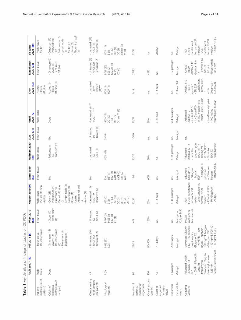
the role of PDOs in predicting the clinical efficacy of anti-cancer drugs in OC (NCT04279509, NCT04768270 andNCT04555473). The NCT04279509 trial is a single-centrestudy aimed at prospectively determine if high-throughputdrug screen assays using PDOs can accurately select che-motherapeutic agents that result in objective response inpatients with refractory solid tumours (head and necksquamous cell carcinoma, colorectal, breast and epithelialOC). NCT04768270 is a single-centre study aimed at ver-ify whether PDOs can help guide precision treatments forOC patients. NCT04555473 is a longitudinal observationalphase II study of the reliability of HGSOC PDOs as modelfor the patients’ response to treatments and it is con-ducted by our group. In this latter trial, PDOs are beingestablished from both PDS and IDS cases preceded byNACT. Since organoids represent a model system com-parable to PDXs, we tested the null hypothesis that thepossibility of correctly identifying the drug-sensitivitycould improve from 80% (as assessed by xenografts) to atleast 95%. The first step was planned to include 7 patients;if 5 or more PDOs fail in the correct identification ofdrug-sensitivity compared to patients’ response, the trialwill be terminated. If the trial proceeds to the secondstage, a total of 43 patients will be studied. Considering apatient dropout of approximately 10%, the study wasplanned to enroll a minimum of 48 patients (see Fig. 4).
ConclusionsHGSOC remains a devastating cancer for which newtherapeutic strategies are urgently needed. In this sce-nario, PDOs represent reliable experimental modelsthat can address several clinical challenges. However,current major bottlenecks regarding their use to sup-port clinical decisions are related to efficacy, time,costs and accuracy in mimicking the overall cancercomplexity.In order to increase the efficacy, it will be crucial
to standardize the procedures of tissue manipulation,the media and the growth factors required for eachtype of PDO. Such standardize guidelines would re-duce the need of specialist skills and might promotethe widespread usage of this technology in clinicalsettings. Major limits to significantly decrease thetime for PDOs outgrowth are related to the quality ofthe biopsy. In particular, the possibility of havingtissue fragments of sufficient size and enriched inproliferating cancer cells from laparoscopic biopsiesor ultrasound guided biopsy would allow higher effi-ciency of PDO formation also from these surgical in-terventions. Costs will likely drop as technologiesmature and protocols become standardized. However,median costs for PDO generation and maintenanceare relatively high at the moment, thus representing apotential limitation for their application to routinemanagement of patients. Lastly, the most limiting as-pect of currently available PDOs is represented by the
Fig. 4 NCT04555473 trial design
Nero et al. Journal of Experimental & Clinical Cancer Research (2021) 40:116 Page 10 of 14

lack of tumor microenvironment. Large efforts areongoing to include stromal, immune and vascularcells in these cultures, thus to represent more faith-fully the disease. This is particularly relevant forHGSOC, where several targeted therapies involve im-mune checkpoints and angiogenic inhibitors. Never-theless, mimicking the whole microenvironmentcharacterizing the tumor is challenging, especiallywith respect to vascularization of the PDO. Improve-ment in these procedures will likely require substan-tial time and costs before the best and mostreproducible conditions are set up.PDOs will certainly serve as a complement to other
traditional models to study cancer, such as primaryhuman tissues and animal models, which are cur-rently the gold standard in biomedical research. How-ever, once PDOs are improved and optimized, theymay have unique characteristics to be introduced infuture clinical trials as empirical predictor of patients’response to therapies (see Fig. 5).
AbbreviationsPDOs: Patients derived organoids; OC: Ovarian cancer; HGSOC: High gradeserous ovarian cancer; PDS: Primary debulking surgery; IDS: Intervaldebulking surgery; NACT: Neoadjuvant chemotherapy; PDX: Patient-derivedxenograft; LOH: Loss of heterozygosis; PARPi: Poly ADP ribose polymeraseinhibitors; HRD: Homologous recombination deficiency; HR: Homologousrecombination; PD-1: Programmed cell death protein 1; PD-L1: Programmedcell death protein 1 ligand; TILs: Tumor infiltrating lymphocytes; VEGF-A: Vascular endothelial growth factor A; 3D: Three-dimensional
AcknowledgementsThis work is partially supported by grants from Associazione Italiana Ricercasul Cancro (AIRC IG23416) and Italian Ministry of Health (GR-2019-12371435).
Authors’ contributionsCN, GV, GD, EC, ML and DL performed the literature search. CN, GV and CSanalyzed selected papers. EC, ML and CN realized Figures and Tables. CN, GVand DL were dedicated to Writing and Original Draft Preparation. GS and CSsupervised the whole process. All authors read and approved the finalmanuscript.
FundingThis work is partially supported by grants from Associazione Italiana Ricercasul Cancro (AIRC IG23416) and Italian Ministry of Health (GR-2019-12371435).
Availability of data and materialsNot applicable.
Declarations
Ethical approval and consent to participateNot applicable.
Consent for publicationNot applicable.
Competing interests- C. Nero, G. Vizzielli, C. Sette, E. Cesari, M. Loverro have no conflict of interestto declare.- G. Daniele has served on advisory board of Beigene and received supportfor travel and accomodation from Roche.- G Scambia has served on advisory boards for TESARO Bio Italy S.r.l,Johnson & Johnson, Clovis Oncology Italy S.r.l. He received support.for travel or accommodation from MSD Italy S.r.l and Clovis.Oncology Italy S.r.l, and institutional research funding from MSD Italy S.r.l.- D. Lorusso has served on advisory boards for Clovis Oncology,AstraZeneca, Genmab/Seattle Genetics, MSD, ImmunoGen,
Fig. 5 PDOs based future clinical trials
Nero et al. Journal of Experimental & Clinical Cancer Research (2021) 40:116 Page 11 of 14

PharmaMar, Roche, and Tesaro/GSK, received support for travel or.accommodation from AstraZeneca, GSK and Roche and institutional.research funding from Merck, GSK, Clovis, Pharmamar.
Author details1Fondazione Policlinico Universitario A. Gemelli IRCCS, L.go Agostino gemelli,8, 00168 Roma, Italy. 2Dipartimento di Scienze della vita e sanità pubblica,Università Cattolica del Sacro Cuore, Roma, Italy. 3Dipartimento diNeuroscienze, Sezione di Anatomia Umana, Università Cattolica del SacroCuore, Roma, Italy.
Received: 3 February 2021 Accepted: 17 March 2021
References1. Bray F, Ferlay J, Soerjomataram I, Siegel RL, Torre LA, Jemal A. Global cancer
statistics 2018: GLOBOCAN estimates of incidence and mortality worldwidefor 36 cancers in 185 countries. CA Cancer J Clin. 2018;68(6):394–424.https://doi.org/10.3322/caac.21492.
2. Matulonis UA, Sood AK, Fallowfield L, Howitt BE, Sehouli J, Karlan BY.Ovarian cancer. Nat Rev Dis Primers. 2016;2(1):16061. https://doi.org/10.1038/nrdp.2016.61.
3. Oza AM, Cook AD, Pfisterer J, Embleton A, Ledermann JA, Pujade-Lauraine E,et al. Standard chemotherapy with or without bevacizumab for womenwith newly di- agnosed ovarian cancer (ICON7): overall survival results of aphase 3 randomised trial. Lancet Oncol. 2015;16:928–36 https://doi.org/10.1016/S1470-2045(15)00086-8.
4. Kaufman B, Shapira-Frommer R, Schmutzler RK, Audeh MW, Friedlander M,Balmaña J, et al. Olaparib monotherapy in patients with advanced cancerand a germline BRCA1/2 mutation. J Clin Oncol. 2015;33(3):244–50. https://doi.org/10.1200/JCO.2014.56.2728.
5. van Driel WJ, Koole SN, Sikorska K, Schagen van Leeuwen JH, SchreuderHWR, Hermans RHM, de Hingh IHJT, van der Velden J, Arts HJ, MassugerLFAG, Aalbers AGJ, Verwaal VJ, Kieffer JM, Van de Vijver KK, van Tinteren H,Aaronson NK, Sonke GS. Hyperthermic Intraperitoneal Chemotherapy inOvarian Cancer. N Engl J Med. 2018; 378(3): 230–240. doi: https://doi.org/10.1056/NEJMoa1708618. PMID: 29342393.
6. Tuveson D, Clevers H., Cancer modeling meets human organoidtechnology. Science. 2019; 364(6444): 952–955. doi: https://doi.org/10.1126/science.aaw6985. PMID: 31171691.
7. Lõhmussaar K, Boretto M, Clevers H. Human-derived model Systems inGynecological Cancer Research. Trends Cancer 2020;6(12):1031–1043.doi: https://doi.org/10.1016/j.trecan.2020.07.007. Epub 2020 Aug 24.PMID: 32855097
8. Hill SJ, Decker B, Roberts EA, Horowitz NS, Muto MG, Worley MJ, et al.Prediction of DNA repair inhibitor response in short-term patient-derivedovarian Cancer Organoids. Cancer Discov. 2018;8:1404–21.
9. Kopper O, DeWitte CJ, Lõhmussaar K, ValleInclan JE, Hami N, Kester L, et al.An organoid platform for ovarian cancer captures intra- and interpatientheterogeneity. Nat Med. 2019;25:838–49.
10. de Witte CJ, Espejo Valle-Inclan J, Hami N, Lõhmussaar K, Kopper O, VreulsCPH, et al. Patient-derived ovarian Cancer Organoids mimic clinicalresponse and exhibit heterogeneous inter- and Intrapatient drug responses.Cell Rep. 2020;31(11):107762. https://doi.org/10.1016/j.celrep.2020.107762.
11. Colombo N, Sessa C, du Bois A, Ledermann J, McCluggage WG, McNeish I,Morice P, Pignata S, Ray-Coquard I, Vergote I, Baert T, Belaroussi I, DashoraA, Olbrecht S, Planchamp F, Querleu D; ESMO-ESGO ovarian Cancerconsensus conference working group. ESMO-ESGO consensus conferencerecommendations on ovarian cancer: pathology and molecular biology,early and advanced stages, borderline tumours and recurrent disease†. AnnOncol 2019;30(5):672–705. doi: https://doi.org/10.1093/annonc/mdz062.PMID: 31046081.
12. NCCN Clinical Practice Guidelines in Oncology (NCCN Guidelines®) OvarianCancer Including Fallopian Tube Cancer and Primary Peritoneal CancerVersion 1. 2020 — March 11, 2020.
13. Polterauer S, Vergote I, Concin N, et al. Prognostic Value of Residual TumorSize in Patients With Epithelial Ovarian Cancer FIGO Stages IIa-IV: analysis ofthe OVCaD Data. Int J Gynecol Cancer. 2012;22:380–5. https://doi.org/10.1097/IGC.0b013e31823de6ae.
14. Bristow RE, Tomacruz RS, Armstrong DK, et al. Survival effect of maximalcytoreductive surgery for advanced ovarian carcinoma during the platinum
era: a meta-analysis. J Clin Oncol. 2002;20:1248–59. https://doi.org/10.1200/JCO.20.5.1248.
15. Dauplat J, Le Bouedec G, Pomel C, Scherer C. Cytoreductive surgery foradvanced stages of ovarian cancer. Semin Surg Oncol. 2000;19:42e8.
16. Vergote I, Trope' CG, Amant F, Kristensen GB, Ehlen T, Johnson N, et al.Neoadjuvant chemotherapy or primary surgery in stage IIIC or IV ovariancancer. N Engl J Med. 2010;363:943e53.
17. Kehoe S, Hook J, Nankivell M, Jayson GC, Kitchener H, Lopes T, et al. Primarychemotherapy versus primary surgery for newly diagnosed advancedovarian cancer (CHORUS): an openlabel, randomised, controlled, non-inferiority trial. Lancet. 2015;386:249e57.
18. Onda T, Matsumoto K, Shibata T, Sato A, Fukuda H, Konishi I, et al. Phase IIItrial of upfront debulking surgery versus neoadjuvant chemotherapy forstage III/IV ovarian, tubal and peritoneal cancers: Japan clinical oncologygroup study JCOG0602. Jpn J Clin Oncol. 2008;38:74e7.
19. Fagotti A, Ferrandina G, Vizzielli G, Fanfani F, Gallotta V, Chiantera V,Costantini B, Margariti PA, Gueli Alletti S, Cosentino F, Tortorella L, ScambiaG. Phase III randomised clinical trial comparing primary surgery versusneoadjuvant chemotherapy in advanced epithelial ovarian cancer with hightumour load (SCORPION trial): final analysis of peri-operative outcome. Eur JCancer 2016;59:22–33. doi: https://doi.org/10.1016/j.ejca.2016.01.017. Epub2016 Mar 19. PMID: 26998845.
20. Ozols RF, Bundy BN, Greer BE, Fowler JM, Clarke-Pearson D, Burger RA,Mannel RS, DeGeest K, Hartenbach EM, Baergen R; Gynecologic oncologygroup. Phase III trial of carboplatin and paclitaxel compared with cisplatinand paclitaxel in patients with optimally resected stage III ovarian cancer: agynecologic oncology group study. J Clin Oncol 2003;21(17):3194–3200. doi:https://doi.org/10.1200/JCO.2003.02.153. Epub 2003 Jul 14. PMID: 12860964.
21. du Bois A, Lück HJ, Meier W, Adams HP, Möbus V, Costa S, Bauknecht T,Richter B, Warm M, Schröder W, Olbricht S, Nitz U, Jackisch C, Emons G,Wagner U, Kuhn W, Pfisterer J; Arbeitsgemeinschaft GynäkologischeOnkologie ovarian Cancer study group. A randomized clinical trial ofcisplatin/paclitaxel versus carboplatin/paclitaxel as first-line treatment ofovarian cancer. J Natl Cancer Inst 2003;95(17):1320–1329. doi: https://doi.org/10.1093/jnci/djg036. PMID: 12953086.
22. International Collaborative Ovarian Neoplasm Group. Paclitaxel pluscarboplatin versus standard chemotherapy with either single-agentcarboplatin or cyclophosphamide, doxorubicin, and cisplatin in womenwith ovarian cancer: the ICON3 randomised trial. Lancet. 2002; 360(9332):505–515. doi: https://doi.org/10.1016/S0140-6736(02)09738-6. Erratum in:Lancet. 2003 Feb 22;361(9358):706. PMID: 12241653.
23. Katsumata N, Yasuda M, Isonishi S, Takahashi F, Michimae H, Kimura E, AokiD, Jobo T, Kodama S, Terauchi F, Sugiyama T, Ochiai K; JapaneseGynecologic Oncology Group. Long-term results of dose-dense paclitaxeland carboplatin versus conventional paclitaxel and carboplatin fortreatment of advanced epithelial ovarian, fallopian tube, or primaryperitoneal cancer (JGOG 3016): a randomised, controlled, open-label trial.Lancet Oncol. 2013; 14(10):1020–1026. doi: https://doi.org/10.1016/S1470-2045(13)70363-2. Epub 2013 Aug 13. PMID: 23948349.
24. Parmar MK, Ledermann JA, Colombo N, du Bois A, Delaloye JF, KristensenGB, Wheeler S, Swart AM, Qian W, Torri V, Floriani I, Jayson G, Lamont A,Tropé C; ICON and AGO collaborators. Paclitaxel plus platinum-basedchemotherapy versus conventional platinum-based chemotherapy inwomen with relapsed ovarian cancer: the ICON4/AGO-OVAR-2.2 trial.Lancet. 2003;361(9375):2099–2106. doi: https://doi.org/10.1016/s0140-6736(03)13718-x. PMID: 12826431.
25. Friedlander M, Trimble E, Tinker A, Alberts D, Avall-Lundqvist E, Brady M,et al. Clinical trials in recurrent ovarian cancer. Int J Gynecol Cancer. 2011;21(4):771–5. https://doi.org/10.1097/IGC.0b013e31821bb8aa.
26. Stuart G., Kitchener H., Bacon M., Dubois A., Friedlander M., Ledermann J.,et al. . (2011) 2010. Gynecologic Cancer intergroup (GCIG) consensusstatement on clinical trials in ovarian cancer: report from the fourth ovarianCancer consensus conference. Int J Gynecol Cancer 21: 750–755.
27. Oza AM, Cook AD, Pfisterer J, Embleton A, Ledermann JA, Pujade-Lauraine E,Kristensen G, Carey MS, Beale P, Cervantes A, Park-Simon TW, Rustin G, JolyF, Mirza MR, Plante M, Quinn M, Poveda A, Jayson GC, Stark D, Swart AM,Farrelly L, Kaplan R, Parmar MK, Perren TJ; ICON7 trial investigatorsStandardchemotherapy with or without bevacizumab for women with newlydiagnosed ovarian cancer (ICON7): overall survival results of a phase 3randomised trial. Lancet Oncol. 2015; 16(8): 928–936. doi: https://doi.org/10.1016/S1470-2045(15)00086-8. Epub 2015 Jun 23.PMID: 26115797.
Nero et al. Journal of Experimental & Clinical Cancer Research (2021) 40:116 Page 12 of 14

28. González-Martín A, Pothuri B, Vergote I, DePont Christensen R, GraybillW, Mirza MR, McCormick C, Lorusso D, Hoskins P, Freyer G, Baumann K,Jardon K, Redondo A, Moore RG, Vulsteke C, O'Cearbhaill RE, Lund B,Backes F, Barretina-Ginesta P, Haggerty AF, Rubio-Pérez MJ, Shahin MS,Mangili G, Bradley WH, Bruchim I, Sun K, Malinowska IA, Li Y, Gupta D,Monk BJ; PRIMA/ENGOTOV26/ GOG-3012 Investigators. Niraparib inPatients with Newly Diagnosed Advanced Ovarian Cancer. N Engl JMed. 2019; 381(25): 2391–2402. doi: https://doi.org/10.1056/NEJMoa1910962. Epub 2019 Sep 28. PMID: 31562799.
29. Moore K, Colombo N, Scambia G, Kim BG, Oaknin A, Friedlander M,Lisyanskaya A, Floquet A, Leary A, Sonke GS, Gourley C, Banerjee S, Oza A,González-Martín A, Aghajanian C, Bradley W, Mathews C, Liu J, Lowe ES,Bloomfield R, DiSilvestro P. Maintenance Olaparib in patients with newlydiagnosed advanced ovarian Cancer. N Engl J Med 2018 Dec 27;379(26):2495–2505. doi: https://doi.org/10.1056/NEJMoa1810858. Epub 2018 Oct 21.PMID: 30345884.
30. Collinson F, Hutchinson M, Craven RA, et al. Predicting response tobevacizumab in ovarian cancer: a panel of potential biomarkers informingtreatment selection. Clin Cancer Res. 2013;19(18):5227–39. https://doi.org/10.1158/1078-0432.CCR-13-0489.
31. Backen A, Renehan AG, Clamp AR, et al. The combination of circulatingAng1 and Tie2 levels predicts progression-free survival advantage inbevacizumab-treated patients with ovarian cancer. Clin Cancer Res. 2014;20(17):4549–58. https://doi.org/10.1158/1078-0432.CCR-13-3248.
32. Audeh MW, Carmichael J, Penson RT, et al. Oral poly (ADP-ribose)polymerase inhibitor olaparib in patients with BRCA1 or BRCA2 mutationsand recurrent ovarian cancer: a proof-of-concept trial. Lancet. 2010;376(9737):245–51. https://doi.org/10.1016/S0140-6736(10)60893-8.
33. Gelmon KA, Tischkowitz M, Mackay H, et al. Olaparib in patients withrecurrent high-grade serous or poorly differentiated ovarian carcinoma ortriple-negative breast cancer: a phase 2, multicentre, open-label, non-randomised study. Lancet Oncol. 2011;12(9):852–61. https://doi.org/10.1016/S1470-2045(11)70214-5.
34. Fong PC, Yap TA, Boss DS, et al. Poly (ADP)-ribose polymerase inhibition:frequent durable responses in BRCA carrier ovarian cancer correlating withplatinum-free interval. J Clin Oncol. 2010;28(15):2512–9. https://doi.org/10.1200/JCO.2009.26.9589.
35. McCabe N, Turner NC, Lord CJ, et al. Deficiency in the repair of DNAdamage by homologous recombination and sensitivity to poly (ADP-ribose)polymerase inhibition. Cancer Res. 2006;66(16):8109–15. https://doi.org/10.1158/0008-5472.CAN-06-0140.
36. Coleman RL, Swisher EM, Oza AM, Scott CL, Giordano H, Lin KK, et al.McNeishRefinement of prespecified cutoff for genomic loss ofheterozygosity (LOH) in ARIEL2 part 1: A phase II study of rucaparib inpatients (pts) with high grade ovarian carcinoma (HGOC). J Clin Oncol.2016;34(15_suppl):5540.
37. Konstantinopoulos PA, Ceccaldi R, Shapiro GI, D’Andrea AD. Homologousrecombination deficiency: exploiting the fundamental vulnerability ofovarian cancer. Cancer Discov. 2015;5(11):1137–54. https://doi.org/10.1158/2159-8290.CD-15-0714.
38. Norquist B, Wurz KA, Pennil CC, Garcia R, Gross J, Sakai W, et al. Secondarysomatic mutations restoring BRCA1/2 predict chemotherapy resistance inhereditary ovarian carcinomas. J Clin Oncol. 2011;29(22):3008–15. https://doi.org/10.1200/JCO.2010.34.2980.
39. Quigley D, Alumkal JJ, Wyatt AW, Kothari V, Foye A, Lloyd P, et al. Analysisof Circulating Cell-Free DNA Identifies Multiclonal Heterogeneity of BRCA2Reversion Mutations Associated with Resistance to PARP Inhibitors. CancerDiscov. 2017;7(9):999–1005. https://doi.org/10.1158/2159-8290.CD-17-0146.
40. Luvero D, Milani A, Ledermann JA. Treatment options in recurrent ovariancancer: latest evidence and clinical potential. Ther Adv Med Oncol. 2014;6(5): 229–239. doi: https://doi.org/10.1177/1758834014544121. PMID:25342990; PMCID: PMC4206613.
41. Robert L. Coleman, Mark F. Brady, Thomas J Herzog, Deborah Kay Armstrong,Paul Sabbatini, Joan L. Walker, Byoung Kim, Keiichi Fujiwara, Krishnansu SujataTewari, David M. O'Malley. Bevacizumab after bevacizumab in platinum-sensitive recurrent ovarian cancer: A subgroup analysis of GOG0213. J ClinOncol. DOI: https://doi.org/10.1200/JCO.2016.34.15_suppl.5523 Journal ofClinical Oncology 34, no. 15_suppl (2016) 5523.
42. https://clinicaltrials.gov/ct2/show/NCT0310698743. Wu Y, Chen W, Xu ZP, Gu W. PD-L1 Distribution and Perspective for Cancer
Immunotherapy-Blockade, Knockdown, or Inhibition. Front Immunol. 2019;
10: 2022. doi: https://doi.org/10.3389/fimmu.2019.02022. PMID: 31507611;PMCID: PMC6718566.
44. Spranger S, Gajewski TF. Impact of oncogenic pathways on evasionof antitumour immune responses. Nat Rev Cancer. 2018;18(3):139–47.https://doi.org/10.1038/nrc.2017.117. Epub 2018 Jan 12.
45. Matulonis UA, Shapira-Frommer R, Santin AD, Lisyanskaya AS, Pignata S,Vergote I, et al. Antitumor activity and safety of pembrolizumab in patientswith advanced recurrent ovarian cancer: results from the phase II KEYNOTE-100 study. Ann Oncol. 2019;30(7):1080–7. 31046082. https://doi.org/10.1093/annonc/mdz135.
46. SGO 2019, Available on line: https://www.cancernetwork.com/view/sgo-2019-despite-missed-endpoint-javelin-trial-pd-l1-subgroup-analysis-redeems
47. Dose Dense Paclitaxel With Pembrolizumab (MK-3475) in Platinum ResistantOvarian Cancer - Full Text View - https://clinicaltrials.gov/ct2/show/NCT02440425.
48. Ledermann JA, Colombo N, Oza M, et al. Avelumab in combination withand/or following chemotherapy vs chemotherapy alone in patients withpreviously untreated epithelial ovarian cancer: Results from the phase 3javelin ovarian 100 trial. 2020. Society of Gynecologic Oncology AnnualMeeting on Women’s Cancer. LBA 25, Scientific Plenary.
49. Liu JF, Herold C, Gray KP, Penson RT, Horowitz N, Konstantinopoulos PA,Castro CM, Hill SJ, Curtis J, Luo W, Matulonis UA, Cannistra SA, Dizon DS.Assessment of Combined Nivolumab and Bevacizumab in Relapsed OvarianCancer: A Phase 2 Clinical Trial. JAMA Oncol. 2019; 5(12): 1731–1738. doi:https://doi.org/10.1001/jamaoncol.2019.3343. Epub ahead of print. PMID:31600397; PMCID:PMC6802049.
50. Moore KN, Pignata S. Trials in progress: IMagyn050/GOG 3015/ENGOT-OV39.A phase III, multicenter, randomized study of atezolizumab versus placeboadministered in combination with paclitaxel, carboplatin, and bevacizumabto patients with newly diagnosed stage III or stage IV o. Int. J GynecolCancer. 2019;29(2):430–3.
51. Higuchi T, Flies DB, Marjon NA, et al. CTLA-4 blockade synergizestherapeutically with PARP inhibition in BRCA1-deficient ovarian Cancer.Cancer Immunol Res. 2015;3(11):1257–68. https://doi.org/10.1158/2326-6066.CIR-15-0044.
52. Stewart RA, Pilie PG, Yap TA. Development of PARP and immune-checkpoint inhibitor combinations. Cancer Res. 2018;78(24):6717–25. https://doi.org/10.1158/0008-5472.CAN-18-2652.
53. Nguyen LT, Ohashi PS. Clinical blockade of PD1 and LAG3--potentialmechanisms of action. Nat Rev Immunol 2015;15(1):45–56. doi: https://doi.org/10.1038/nri3790. PMID: 25534622.
54. Available on line: https://clinicaltrials.gov/ct2/show/NCT0373764355. Available on line: https://clinicaltrials.gov/ct2/show/NCT03740165?cond=
ENGOT+OV+43&draw=2&r ank=1.56. Available on line: https://clinicaltrials.gov/ct2/show/NCT0360285957. Available on line: https://clinicaltrials.gov/ct2/show/NCT0352224658. Available on line: https://clinicaltrials.gov/ct2/show/NCT0359827059. Available on line: https://clinicaltrials.gov/ct2/show/NCT0248440460. Available on line: https://clinicaltrials.gov/ct2/show/NCT0257172561. Available on line: https://www.mito-group.it/studi/studio-mito-33-nitche/62. Addition of a CTLA-4–Targeted Therapy to a Checkpoint Inhibitor in Ovarian
Cancer - The ASCO Post. [https://www.ascopost.com/News/59329].63. Jihoon Kim, Bon-Kyoung Koo, Juergen A. Knoblich Human organoids:
model systems for human biology and medicine Nat Rev Mol Cell Biol.2020: 1–14. doi: https://doi.org/10.1038/s41580-020-0259-3 [Epub ahead ofprint] PMCID: PMC7339799.
64. Drost J, Clevers H. Organoids in cancer research. Nat Rev Cancer 2018;18(7):407–418. doi: https://doi.org/10.1038/s41568-018-0007-6. PMID: 29692415.
65. Jacob F, Salinas RD, Zhang DY, Nguyen PTT, Schnoll JG, Wong SZH, ThokalaR, Sheikh S, Saxena D, Prokop S, Liu DA, Qian X, Petrov D, Lucas T, Chen HI,Dorsey JF, Christian KM, Binder ZA, Nasrallah M, Brem S, O'Rourke DM, MingGL, Song H. A Patient-Derived Glioblastoma Organoid Model and BiobankRecapitulates Inter- and Intra-tumoral Heterogeneity. Cell. 2020; 180(1): 188–204.e22. doi: https://doi.org/10.1016/j.cell.2019.11.036. Epub 2019 Dec 26.PMID: 31883794.
66. Zhu Z, Mesci P, Bernatchez JA, Gimple RC, Wang X, Schafer ST,Wettersten HI, Beck S, Clark AE, Wu Q, Prager BC, Kim LJY, Dhanwani R,Sharma S, Garancher A, Weis SM, Mack SC, Negraes PD, Trujillo CA,Penalva LO, Feng J, Lan Z, Zhang R, Wessel AW, Dhawan S, DiamondMS, Chen CC, Wechsler-Reya RJ, Gage FH, Hu H, Siqueira-Neto JL,Muotri AR, Cheresh DA, Rich JN. Zika Virus Targets Glioblastoma Stem
Nero et al. Journal of Experimental & Clinical Cancer Research (2021) 40:116 Page 13 of 14

Cells through a SOX2-Integrin alpha(v)beta(5) Axis. Cell Stem Cell. 2020;26(2): 187–204.e10. doi: https://doi.org/10.1016/j.stem.2019.11.016. Epub2020 Jan 16.PMID: 31956038.
67. Pauli C, Hopkins BD, Prandi D, Shaw R, Fedrizzi T, Sboner A, et al.Personalized in vitro and in vivo Cancer models to guide precisionmedicine. Cancer Discov. 2017;7(5):462–77. https://doi.org/10.1158/2159-8290.CD-16-1154.
68. Phan N, Hong JJ, Tofig B, Mapua M, Elashoff D, Moatamed NA, Huang J,Memarzadeh S, Damoiseaux R, Soragni A. A simple high-throughputapproach identifies actionable drug sensitivities in patient-derived tumororganoids. Commun Biol. 2019; 2: 78. doi: https://doi.org/10.1038/s42003-019-0305-x. PMID: 30820473; PMCID: PMC6389967.
69. Maru Y, Tanaka N, Itami M, Hippo Y. Efficient use of patient-derivedorganoids as a preclinical model for gynecologic tumors. Gynecol Oncol.2019;154(1):189–98. https://doi.org/10.1016/j.ygyno.2019.05.005. Epub 2019May 14.
70. Hoffmann K, Berger H, Kulbe H, Thillainadarasan S, Mollenkopf HJ, ZemojtelT, Taube E, Darb-Esfahani S, Mangler M, Sehouli J, Chekerov R, Braicu EI,Meyer TF, Kessler M. Stable expansion of high-grade serous ovarian cancerorganoids requires a low-Wnt environment. EMBO J. 2020; 39(6): e104013.doi: https://doi.org/10.15252/embj.2019104013. Epub 2020 Feb 3. PMID:32009247; PMCID: PMC7073464.
71. Sun H, Wang H, Wang X, Aoki Y, Wang X, Yang Y, Cheng X, Wang Z, WangX. Aurora-A/SOX8/FOXK1 signaling axis promotes chemoresistance viasuppression of cell senescence and induction of glucose metabolism inovarian cancer organoids and cells. Theranostics. 2020; 10(15): 6928–6945.doi: https://doi.org/10.7150/thno.43811. PMID: 32550913; PMCID:PMC7295065.
72. Nanki Y, Chiyoda T, Hirasawa A, Ookubo A, Itoh M, Ueno M, Akahane T,Kameyama K, Yamagami W, Kataoka F, Aoki D. Patient-derived ovariancancer organoids capture the genomic profiles of primary tumoursapplicable for drug sensitivity and resistance testing. Sci Rep. 2020; 10(1):12581. doi: https://doi.org/10.1038/s41598-020-69488-9. PMID: 32724113;PMCID: PMC7387538.
73. Chen H, Gotimer K, De Souza C, Tepper CG, Karnezis AN, Leiserowitz GS,Chien J, Smith LH. Short-term organoid culture for drug sensitivity testing ofhigh-grade serous carcinoma. Gynecol Oncol 2020;157(3):783–792. doi:https://doi.org/10.1016/j.ygyno.2020.03.026. Epub 2020 Apr 4. PMID:32253045.
74. Maenhoudt N, Defraye C, Boretto M, Jan Z, Heremans R, Boeckx B, HermansF, Arijs I, Cox B, Van Nieuwenhuysen E, Vergote I, Van Rompuy AS,Lambrechts D, Timmerman D, Vankelecom H. Developing Organoids fromOvarian Cancer as Experimental and Preclinical Models. Stem Cell Rep. 2020;14(4): 717–729. doi: https://doi.org/10.1016/j.stemcr.2020.03.004. Epub 2020Apr 2. PMID: 32243841; PMCID: PMC7160357.
75. Park H, Hwang S, Jeong JY, Jung SG, Choi MC, Joo WD, Song SH, Lee C, AnHJ. Integrative analysis of transcription factors and microRNAs in ovariancancer cell spheroids. J Ovarian Res. 2020 ; 13(1): 16. doi: https://doi.org/10.1186/s13048-020-00618-7. PMID: 32046751; PMCID: PMC7014770.
76. Neal JT, Li X, Zhu J, Giangarra V, Grzeskowiak CL, Ju J, Liu IH, Chiou SH,Salahudeen AA, Smith AR, Deutsch BC, Liao L, Zemek AJ, Zhao F, Karlsson K,Schultz LM, Metzner TJ, Nadauld LD, Tseng YY, Alkhairy S, Oh C, Keskula P,Mendoza-Villanueva D, De La Vega FM, Kunz PL, Liao JC, Leppert JT,Sunwoo JB, Sabatti C, Boehm JS, Hahn WC, Zheng GXY, Davis MM, Kuo CJ.Organoid Modeling of the Tumor Immune Microenvironment. Cell. 2018;175(7): 1972–1988.e16. doi: https://doi.org/10.1016/j.cell.2018.11.021. PMID:30550791; PMCID: PMC6656687.
77. Wan C, Keany MP, Dong H, Al-Alem LF, Pandya UM, Lazo S, Boehnke K,Lynch KN, Xu R, Zarrella DT, Gu S, Cejas P, Lim K, Long HW, Elias KM,Horowitz NS, Feltmate CM, Muto MG, Worley MJ, Berkowitz RS, MatulonisUA, Nucci MR, Crum CP, Rueda BR, Brown M, Liu XS, Hill SJ. Enhancedefficacy of simultaneous PD-1 and PD-L1 immune checkpoint blockade inhigh grade serous ovarian cancer. Cancer Res. 2020:canres.1674.2020. doi:https://doi.org/10.1158/0008-5472.CAN-20-1674. Epub ahead of print. PMID:33158814.
78. Dijkstra KK, Cattaneo CM, Weeber F, Chalabi M, van de Haar J, Fanchi LF,Slagter M, van der Velden DL, Kaing S, Kelderman S, van Rooij N, vanLeerdam ME, Depla A, Smit EF, Hartemink KJ, de Groot R, Wolkers MC, SachsN, Snaebjornsson P, Monkhorst K, Haanen J, Clevers H, Schumacher TN,Voest EE. Generation of Tumor-Reactive T Cells by Co-culture of PeripheralBlood Lymphocytes and Tumor Organoids. Cell. 2018; 174(6): 1586–1598.
e12. doi: https://doi.org/10.1016/j.cell.2018.07.009. Epub 2018 Aug 9. PMID:30100188; PMCID: PMC6558289.
Publisher’s NoteSpringer Nature remains neutral with regard to jurisdictional claims inpublished maps and institutional affiliations.
Nero et al. Journal of Experimental & Clinical Cancer Research (2021) 40:116 Page 14 of 14