Pathophysiology, Special Considerations, and...
77
A Case of Scoliosis Repair: Pathophysiology, Special Considerations, and Intraoperative Management A Case of Scoliosis Repair: Pathophysiology, Special Considerations, and Intraoperative Management Karim Rafaat, M.D. Karim Rafaat, M.D.
Transcript of Pathophysiology, Special Considerations, and...
A Case of Scoliosis Repair: Pathophysiology, Special Considerations,
and Intraoperative Management
A Case of Scoliosis Repair: Pathophysiology, Special Considerations,
and Intraoperative Management
Karim Rafaat, M.D.Karim Rafaat, M.D.
OutlineOutline
Case presentation Overview of diagnostic/treatment
considerations Intraoperative management
Case presentation Overview of diagnostic/treatment
considerations Intraoperative management
Case PresentationCase Presentation
Karim Rafaat, M.D.
Fellow in Pediatric AnesthesiaLucile Packard Children’s Hospital
Stanford University
Karim Rafaat, M.D.
Fellow in Pediatric AnesthesiaLucile Packard Children’s Hospital
Stanford University
Case Report:C.C.Case Report:C.C.
IdentificationIdentification
12 yo male with complex medical history
Scheduled for posterior spinal instrumentation and fusion with multiple vertebrectomies
12 yo male with complex medical history
Scheduled for posterior spinal instrumentation and fusion with multiple vertebrectomies
Past Medical HistoryPast Medical History
Midthoracic Myelomeningocele -repaired as an infant in Mexico
paralysis to T6Chiari II malformationHydrocephalus, s/p VP Shunt placementDevelopmental DelayScoliosis
Midthoracic Myelomeningocele -repaired as an infant in Mexico
paralysis to T6Chiari II malformationHydrocephalus, s/p VP Shunt placementDevelopmental DelayScoliosis
Past Medical HistoryPast Medical History
Failure to ThriveSeizuresFailure to ThriveSeizures
Past Surgical HistoryPast Surgical History
Per mother’s report, has had over 30 surgeries: Repair of midthoracic myelomeningoceleVP Shunt, s/p multiple revisionsUrostomyVentral hernia repairBilat ear tubesG-tube placement
Per mother’s report, has had over 30 surgeries: Repair of midthoracic myelomeningoceleVP Shunt, s/p multiple revisionsUrostomyVentral hernia repairBilat ear tubesG-tube placement
Birth/Developmental HistoryBirth/Developmental History
Born at 32 wksWheelchair dependent
Cannot standUrostomy - incontinent of urineIncontinent of stoolSensory level approximately at umbilicus
Born at 32 wksWheelchair dependent
Cannot standUrostomy - incontinent of urineIncontinent of stoolSensory level approximately at umbilicus
Family HistoryFamily History
Negative for any bleeding problemsNegative for any problems with anesthesia
Negative for any bleeding problemsNegative for any problems with anesthesia
MedicationsMedications
Phenobarbital 15mg per G-tube bidNitrofurantoin 3mg per G-tube bidPhenobarbital 15mg per G-tube bidNitrofurantoin 3mg per G-tube bid
Physical ExamPhysical Exam
Vital Signs:T - 36.8 CHR - 90BP - 122/85RR - 20SpO2 - 99% on RA
Vital Signs:T - 36.8 CHR - 90BP - 122/85RR - 20SpO2 - 99% on RA
Physical ExamPhysical Exam
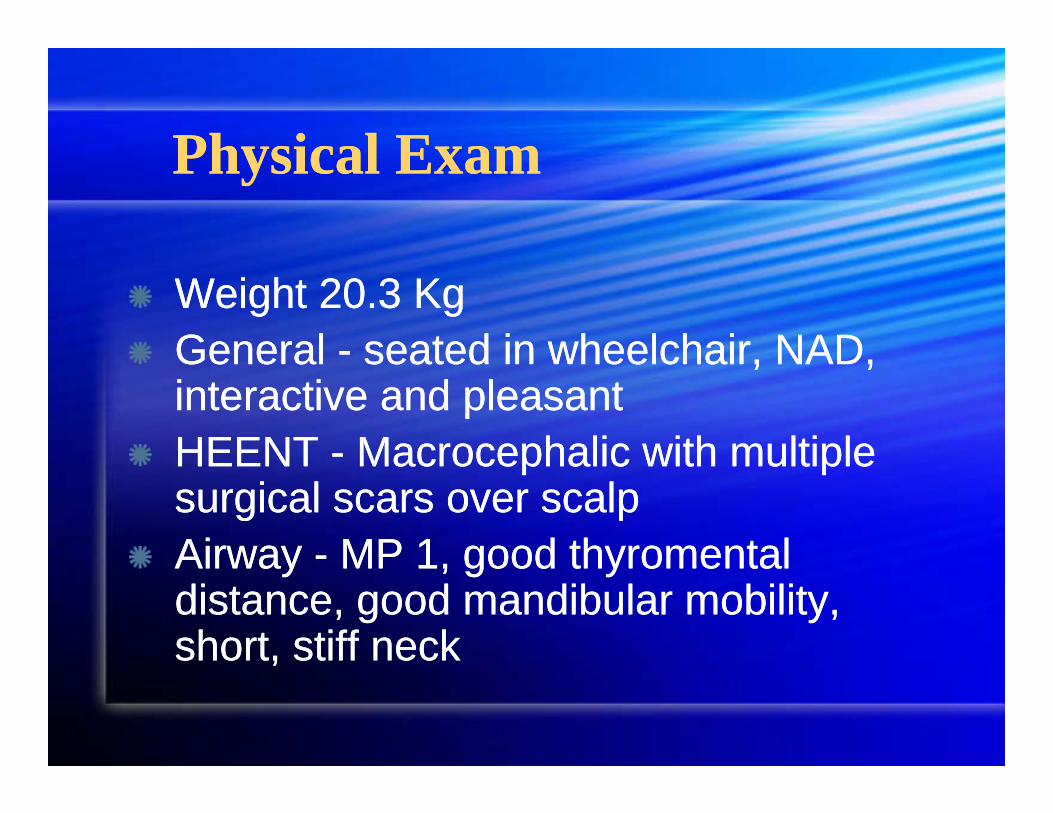
Weight 20.3 KgGeneral - seated in wheelchair, NAD, interactive and pleasantHEENT - Macrocephalic with multiple surgical scars over scalpAirway - MP 1, good thyromental distance, good mandibular mobility, short, stiff neck
Weight 20.3 KgGeneral - seated in wheelchair, NAD, interactive and pleasantHEENT - Macrocephalic with multiple surgical scars over scalpAirway - MP 1, good thyromental distance, good mandibular mobility, short, stiff neck
Physical ExamPhysical Exam
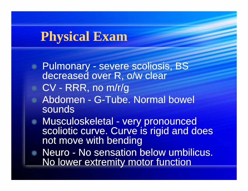
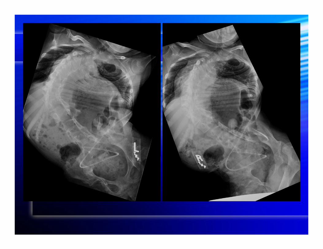
Pulmonary - severe scoliosis, BS decreased over R, o/w clearCV - RRR, no m/r/gAbdomen - G-Tube. Normal bowel soundsMusculoskeletal - very pronounced scoliotic curve. Curve is rigid and does not move with bendingNeuro - No sensation below umbilicus. No lower extremity motor function
Pulmonary - severe scoliosis, BS decreased over R, o/w clearCV - RRR, no m/r/gAbdomen - G-Tube. Normal bowel soundsMusculoskeletal - very pronounced scoliotic curve. Curve is rigid and does not move with bendingNeuro - No sensation below umbilicus. No lower extremity motor function
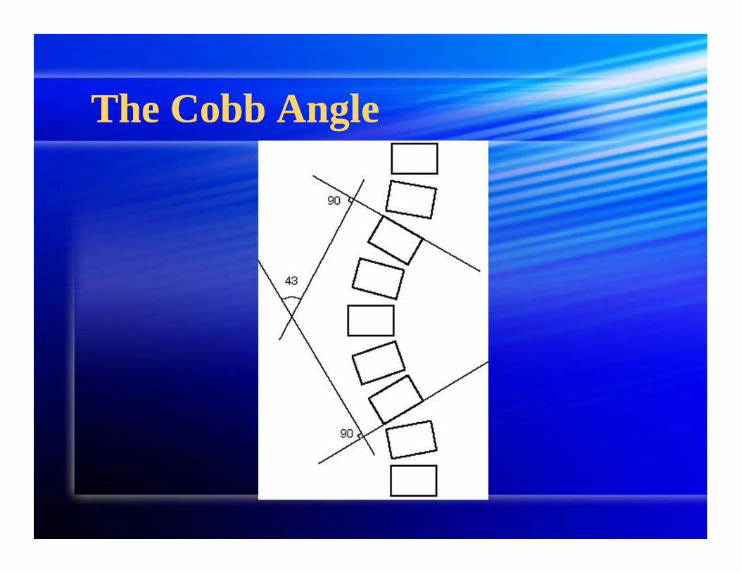
The Cobb AngleThe Cobb Angle

LabsLabs
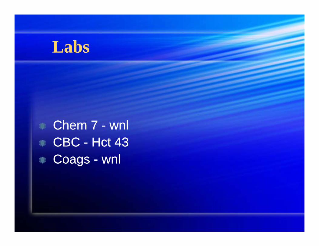
Chem 7 - wnlCBC - Hct 43Coags - wnl
Chem 7 - wnlCBC - Hct 43Coags - wnl
OperationOperation
Consisted of:Vertebrectomy T12-L1Spinal cord excision below T12Posterior spinal instrumentation and fusion T1-S1
Consisted of:Vertebrectomy T12-L1Spinal cord excision below T12Posterior spinal instrumentation and fusion T1-S1
Intraoperative CourseIntraoperative Course
Intubated easily with 5.0 cuffed ETT and MAC blade3 PIV’s total: 1x22G, 2x20GRadial A-lineRIJ 5Fr double lumen central catheter
Intubated easily with 5.0 cuffed ETT and MAC blade3 PIV’s total: 1x22G, 2x20GRadial A-lineRIJ 5Fr double lumen central catheter
Intraoperative CourseIntraoperative Course
Amicar bolus 75mg/kg, drip at 75 mg/kg/hrLungs with reasonable compliance (!) and no difficulties with gas exchangeStable hemodynamics (when surgical losses properly attended to)Stable UOPPersistent and significant bleeding from bony surgical sites
Amicar bolus 75mg/kg, drip at 75 mg/kg/hrLungs with reasonable compliance (!) and no difficulties with gas exchangeStable hemodynamics (when surgical losses properly attended to)Stable UOPPersistent and significant bleeding from bony surgical sites

Intraoperative CourseIntraoperative Course
Operative course stable (with some effort), with no unexpected complications until......
Operative course stable (with some effort), with no unexpected complications until......

Guess What’s Missing..........Guess What’s Missing..........
The spinal cord was incised and excised below T12The spinal cord was incised and excised below T12
Intraoperative CourseIntraoperative Course
Acute spinal shock manifested as sudden hypotension, unresponsive to fluidsNeosynephrine bolused frequently, while drip prepared
Dose ranged from 0.1 - 2 mcg/kg/min
Vasopressin drip startedDose ranged from 0.2 - 1.2 units/hrUsual shock dose 10-50 mU/kg/hr
Acute spinal shock manifested as sudden hypotension, unresponsive to fluidsNeosynephrine bolused frequently, while drip prepared
Dose ranged from 0.1 - 2 mcg/kg/min
Vasopressin drip startedDose ranged from 0.2 - 1.2 units/hrUsual shock dose 10-50 mU/kg/hr
Intraoperative CourseIntraoperative Course
Volatiles reduced, and midazolam bolused to ensure amnesiaDespite mild, persistent hypotension (MAPs in 50s, with visits to the 40s), sufficient end-organ oxygen delivery maintained as evidenced by continued UOP > 0.5 cc/kg/hr and lack of a base deficit on ABG
Volatiles reduced, and midazolam bolused to ensure amnesiaDespite mild, persistent hypotension (MAPs in 50s, with visits to the 40s), sufficient end-organ oxygen delivery maintained as evidenced by continued UOP > 0.5 cc/kg/hr and lack of a base deficit on ABG
Intraoperative CourseIntraoperative Course
Case ended smoothly, secondary to consistent leg work
1200 cc crystalloids250cc colloids6 units PRBC, 2 units FFP, 1 unit PlatletsAforementioned pressors, plus boluses
Case ended smoothly, secondary to consistent leg work
1200 cc crystalloids250cc colloids6 units PRBC, 2 units FFP, 1 unit PlatletsAforementioned pressors, plus boluses
Brief Post-Op CourseBrief Post-Op Course
Pt admitted to PICU on 0.5 mcg/kg/min Neosynephrine and 1 unit/hr VasopressinHct 30 with Coags wnlPressors off by next morning Extubated that same day to NCDischarged home after 5 days
Pt admitted to PICU on 0.5 mcg/kg/min Neosynephrine and 1 unit/hr VasopressinHct 30 with Coags wnlPressors off by next morning Extubated that same day to NCDischarged home after 5 days
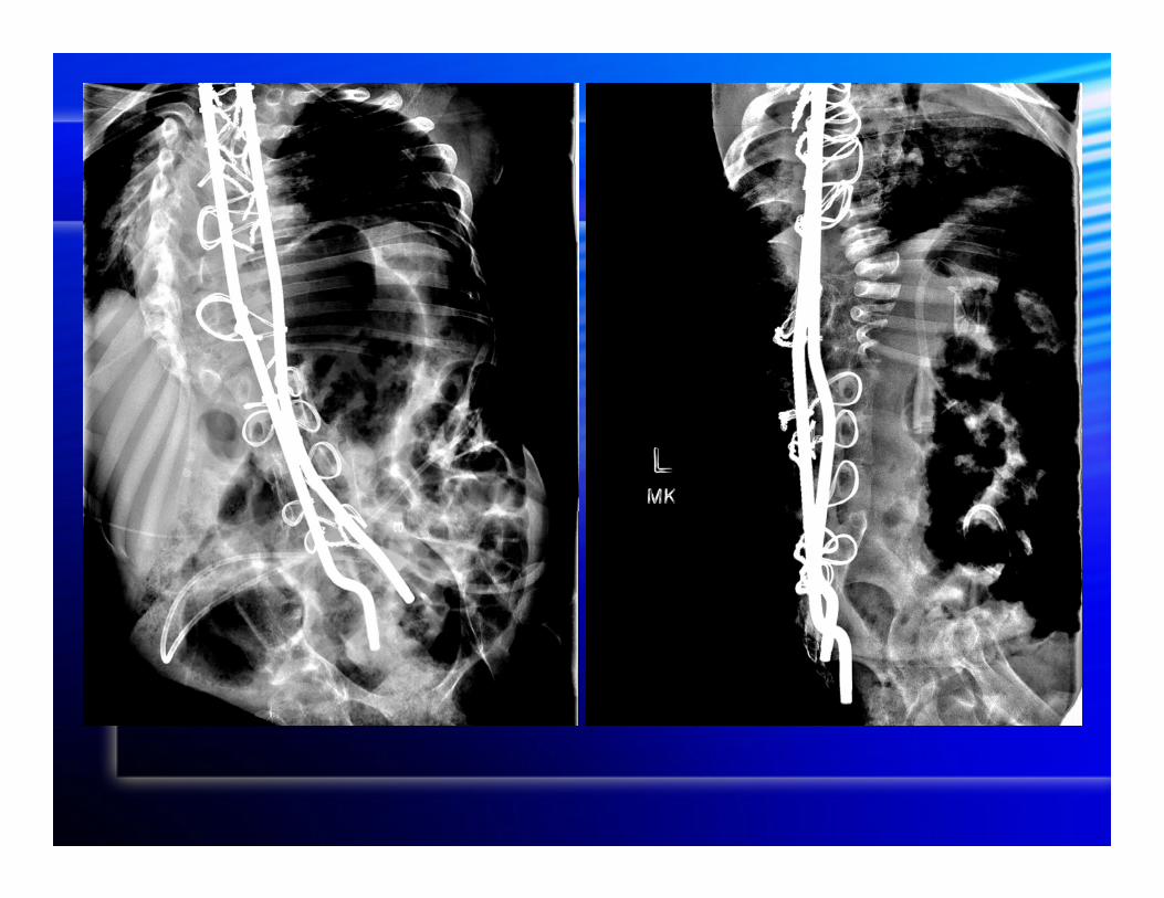

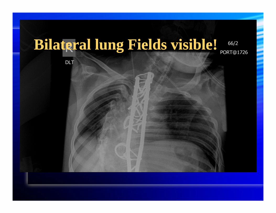
Bilateral lung Fields visible!Bilateral lung Fields visible!
Scoliosis: An OverviewScoliosis: An Overview
OutlineOutline
Definition Etiology Associated co-morbidities Conservative treatment Indications for surgery Predictors of complications
Definition Etiology Associated co-morbidities Conservative treatment Indications for surgery Predictors of complications

DefinitionDefinition
A lateral spinal curvature of >10º ~2% of children affected at some stage
of life ~10% of affected patients will require
corrective surgery
A lateral spinal curvature of >10º ~2% of children affected at some stage
of life ~10% of affected patients will require
corrective surgery
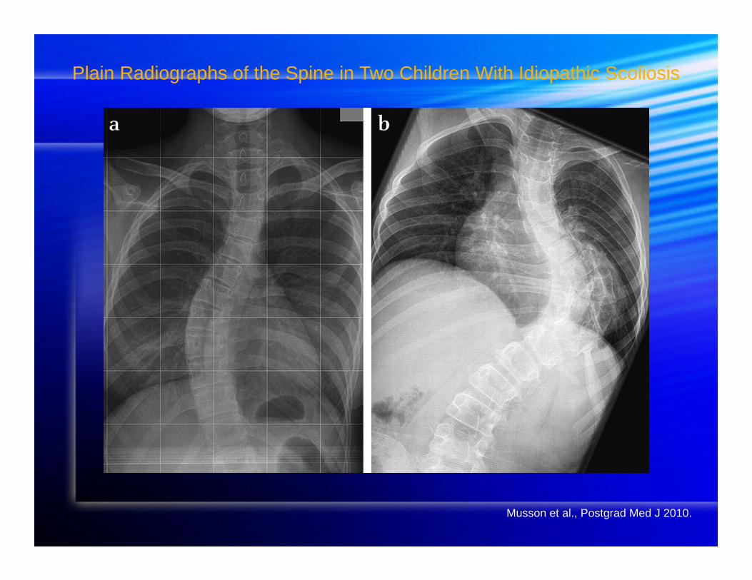
Plain Radiographs of the Spine in Two Children With Idiopathic Scoliosis
Musson et al., Postgrad Med J 2010.
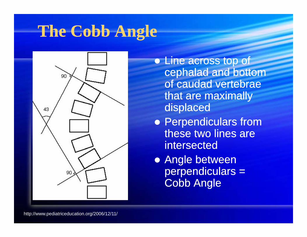
The Cobb AngleThe Cobb Angle Line across top of
cephalad and bottom of caudad vertebrae that are maximally displaced
Perpendiculars from these two lines are intersected
Angle between perpendiculars = Cobb Angle
Line across top of cephalad and bottom of caudad vertebrae that are maximally displaced
Perpendiculars from these two lines are intersected
Angle between perpendiculars = Cobb Angle
http://www.pediatriceducation.org/2006/12/11/
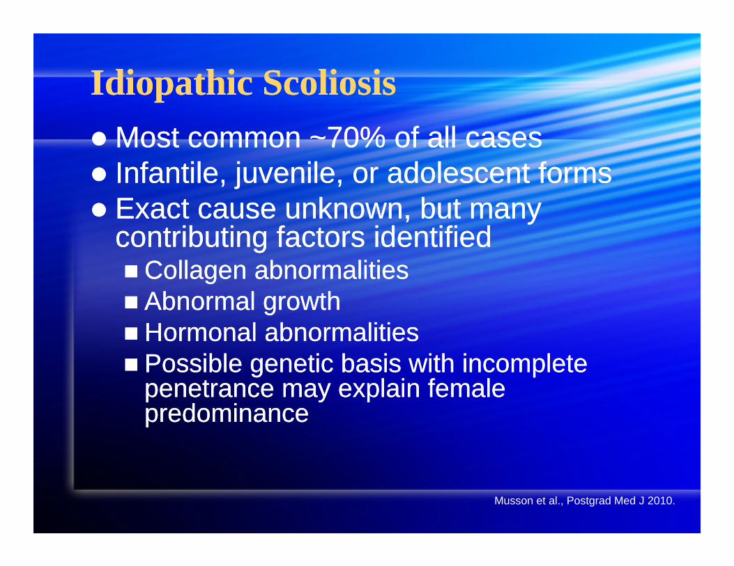
Idiopathic ScoliosisIdiopathic ScoliosisMost common ~70% of all cases Infantile, juvenile, or adolescent forms Exact cause unknown, but many
contributing factors identifiedCollagen abnormalities Abnormal growthHormonal abnormalities Possible genetic basis with incomplete
penetrance may explain female predominance
Most common ~70% of all cases Infantile, juvenile, or adolescent forms Exact cause unknown, but many
contributing factors identifiedCollagen abnormalities Abnormal growthHormonal abnormalities Possible genetic basis with incomplete
penetrance may explain female predominance
Musson et al., Postgrad Med J 2010.
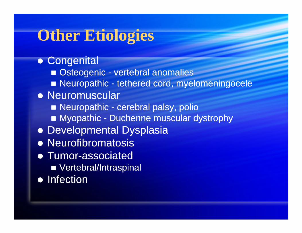
Other EtiologiesOther Etiologies Congenital
Osteogenic - vertebral anomalies Neuropathic - tethered cord, myelomeningocele
Neuromuscular Neuropathic - cerebral palsy, polio Myopathic - Duchenne muscular dystrophy
Developmental Dysplasia Neurofibromatosis Tumor-associated
Vertebral/Intraspinal Infection
Congenital Osteogenic - vertebral anomalies Neuropathic - tethered cord, myelomeningocele
Neuromuscular Neuropathic - cerebral palsy, polio Myopathic - Duchenne muscular dystrophy
Developmental Dysplasia Neurofibromatosis Tumor-associated
Vertebral/Intraspinal Infection
Pulmonary ComorbiditiesPulmonary Comorbidities
Restrictive lung pattern Decrease in lung volumes Vital capacity most significant FRC, TLC, IC, ERV also
Impaired respiratory muscle functionChest wall deformity = inspiratory muscles
working at mechanical disadvantage Arterial hypoxemia from V/Q mismatch
Restrictive lung pattern Decrease in lung volumes Vital capacity most significant FRC, TLC, IC, ERV also
Impaired respiratory muscle functionChest wall deformity = inspiratory muscles
working at mechanical disadvantage Arterial hypoxemia from V/Q mismatch
Pulmonary ComorbiditiesPulmonary Comorbidities
Slope of ventilatory response to CO2may be decreased
Higher respiratory rates and lower tidal volumes minimize work of breathing
Pulmonary compromise increases with curve progression
Slope of ventilatory response to CO2may be decreased
Higher respiratory rates and lower tidal volumes minimize work of breathing
Pulmonary compromise increases with curve progression
Cardiac ComorbiditiesCardiac Comorbidities
Chronic hypoxemia HPV Pulmonary Hypertension
RVH RV failureMVP common among scoliosis patients Scoliosis associated with congenital
heart disease (no specific lesion)
Chronic hypoxemia HPV Pulmonary Hypertension
RVH RV failureMVP common among scoliosis patients Scoliosis associated with congenital
heart disease (no specific lesion)
Natural HistoryNatural History
Significant curve progression may eventually lead to intolerable cardiopulmonary compromise
Treatment is either conservative (aimed at slowing/stopping curve progression) or surgical
Significant curve progression may eventually lead to intolerable cardiopulmonary compromise
Treatment is either conservative (aimed at slowing/stopping curve progression) or surgical
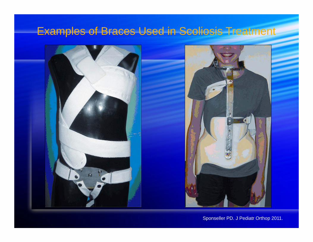
Examples of Braces Used in Scoliosis Treatment
Sponseller PD. J Pediatr Orthop 2011.
Conservative ManagementConservative Management
Bracing is the mainstay of treatment Goal = slow or prevent curve
progression via external forces guiding growth of spine
Curve correction with bracing is not commonly observed
Bracing is the mainstay of treatment Goal = slow or prevent curve
progression via external forces guiding growth of spine
Curve correction with bracing is not commonly observed
Indications for BracingIndications for Bracing
Curve of 25-45º in patient going through a rapid growth period (Risser 0-1 years)
Some patients with smaller curves showing recent progression
Curve of 25-45º in patient going through a rapid growth period (Risser 0-1 years)
Some patients with smaller curves showing recent progression
Sponseller PD. J Pediatr Orthop 2011.
Characteristics Predicting Failure of Bracing TreatmentCharacteristics Predicting Failure of Bracing Treatment
Overweight High thoracic curve (above T8) Lordotic thoracic spineWithin a year of skeletal maturity 1-year post-menarche Treatment non-compliance
Overweight High thoracic curve (above T8) Lordotic thoracic spineWithin a year of skeletal maturity 1-year post-menarche Treatment non-compliance
Sponseller PD. J Pediatr Orthop 2011.
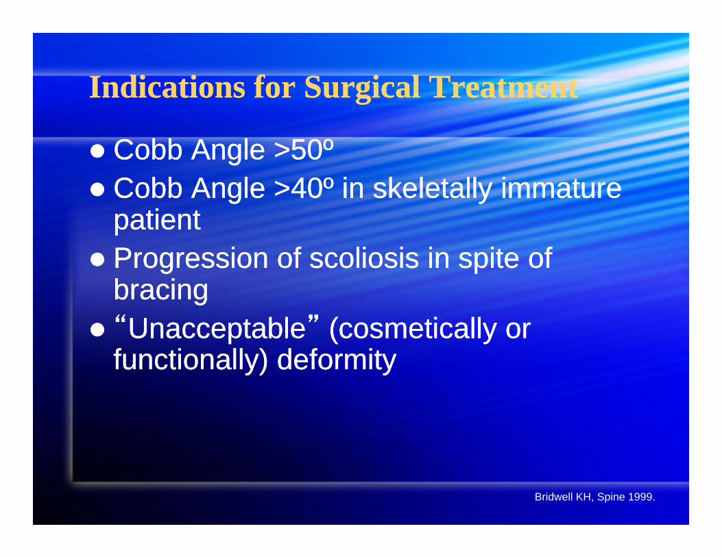
Indications for Surgical TreatmentIndications for Surgical Treatment
Cobb Angle >50º Cobb Angle >40º in skeletally immature
patient Progression of scoliosis in spite of
bracing “Unacceptable” (cosmetically or
functionally) deformity
Cobb Angle >50º Cobb Angle >40º in skeletally immature
patient Progression of scoliosis in spite of
bracing “Unacceptable” (cosmetically or
functionally) deformity
Bridwell KH, Spine 1999.
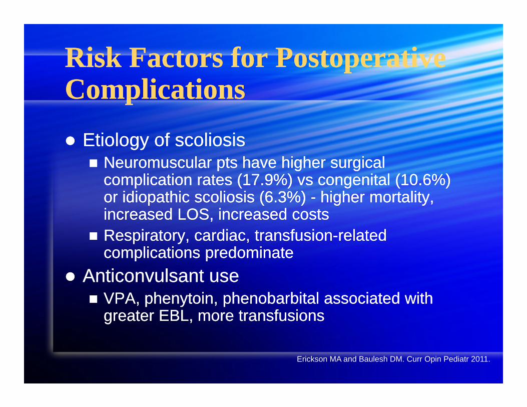
Risk Factors for Postoperative Complications Risk Factors for Postoperative Complications Etiology of scoliosis
Neuromuscular pts have higher surgical complication rates (17.9%) vs congenital (10.6%) or idiopathic scoliosis (6.3%) - higher mortality, increased LOS, increased costs
Respiratory, cardiac, transfusion-related complications predominate
Anticonvulsant use VPA, phenytoin, phenobarbital associated with
greater EBL, more transfusions
Etiology of scoliosis Neuromuscular pts have higher surgical
complication rates (17.9%) vs congenital (10.6%) or idiopathic scoliosis (6.3%) - higher mortality, increased LOS, increased costs
Respiratory, cardiac, transfusion-related complications predominate
Anticonvulsant use VPA, phenytoin, phenobarbital associated with
greater EBL, more transfusions
Erickson MA and Baulesh DM. Curr Opin Pediatr 2011.
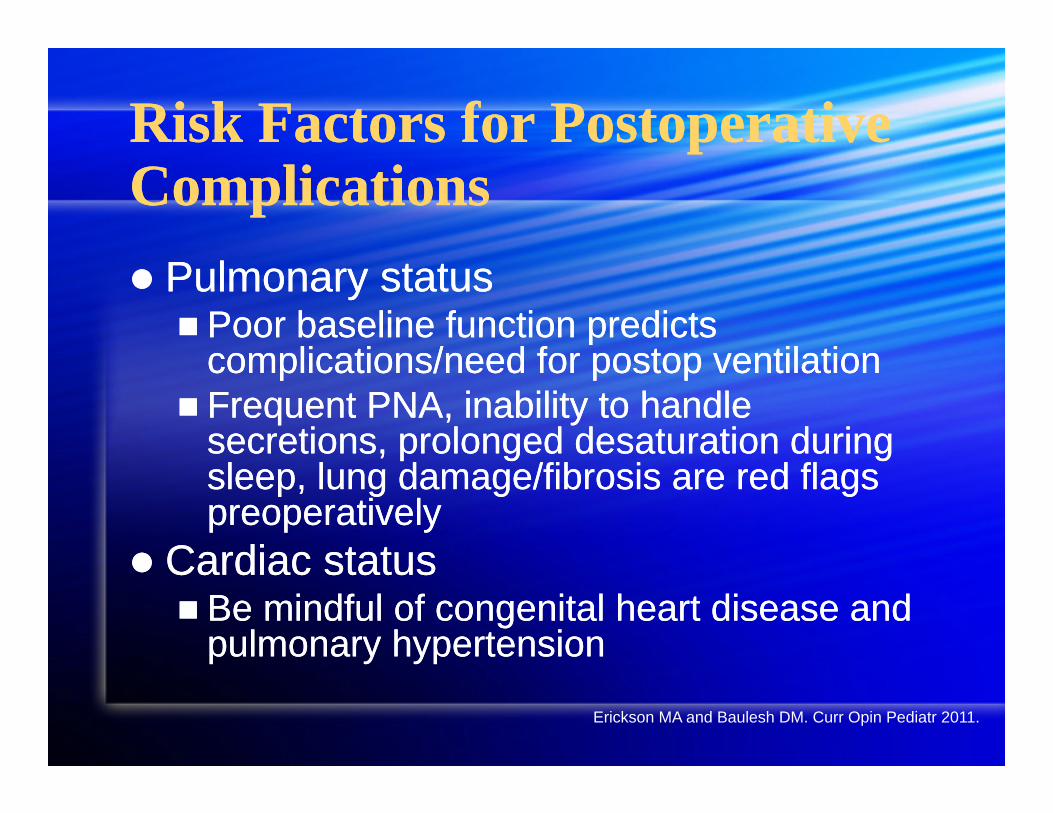
Risk Factors for Postoperative ComplicationsRisk Factors for Postoperative Complications Pulmonary status Poor baseline function predicts
complications/need for postop ventilation Frequent PNA, inability to handle
secretions, prolonged desaturation during sleep, lung damage/fibrosis are red flags preoperatively
Cardiac status Be mindful of congenital heart disease and
pulmonary hypertension
Pulmonary status Poor baseline function predicts
complications/need for postop ventilation Frequent PNA, inability to handle
secretions, prolonged desaturation during sleep, lung damage/fibrosis are red flags preoperatively
Cardiac status Be mindful of congenital heart disease and
pulmonary hypertension
Erickson MA and Baulesh DM. Curr Opin Pediatr 2011.
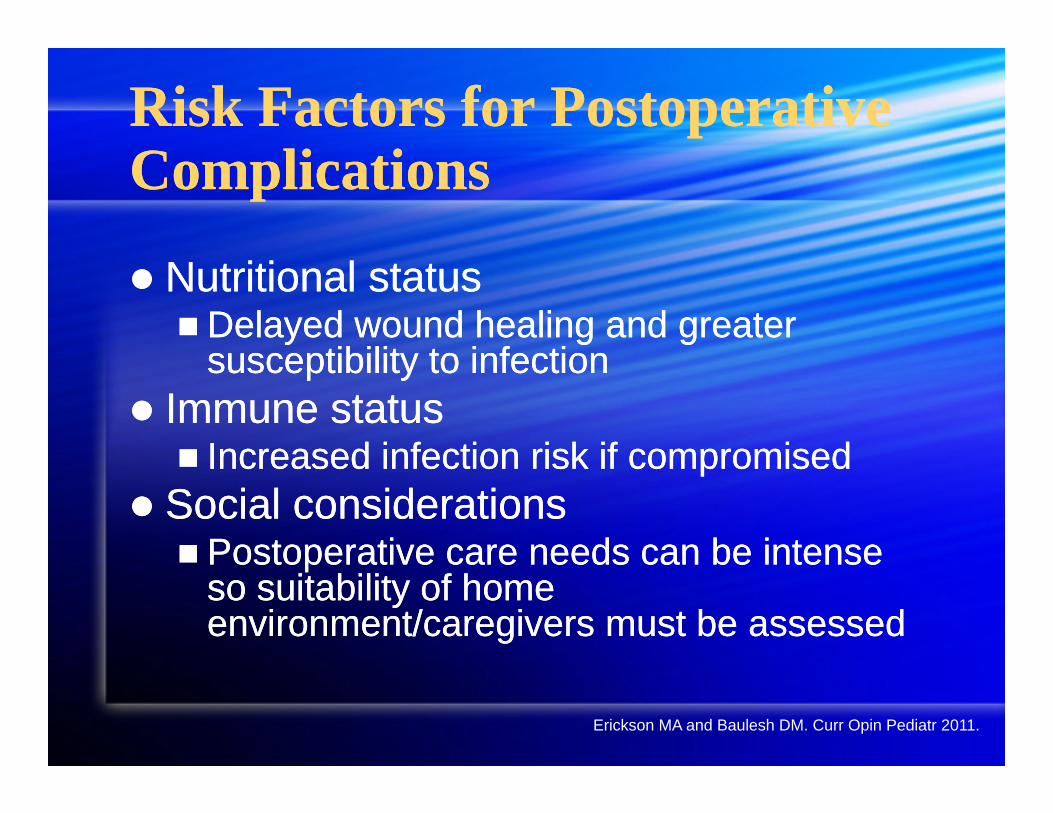
Risk Factors for Postoperative ComplicationsRisk Factors for Postoperative Complications
Nutritional statusDelayed wound healing and greater
susceptibility to infection Immune status Increased infection risk if compromised
Social considerations Postoperative care needs can be intense
so suitability of home environment/caregivers must be assessed
Nutritional statusDelayed wound healing and greater
susceptibility to infection Immune status Increased infection risk if compromised
Social considerations Postoperative care needs can be intense
so suitability of home environment/caregivers must be assessed
Erickson MA and Baulesh DM. Curr Opin Pediatr 2011.
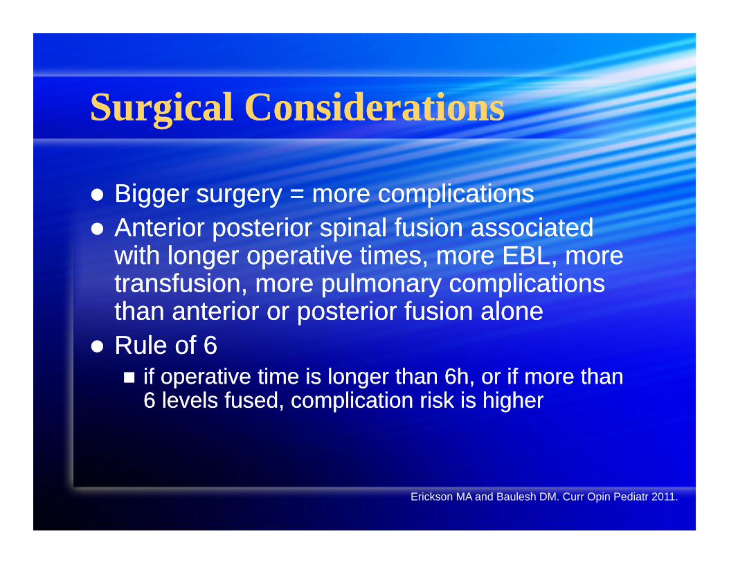
Surgical ConsiderationsSurgical Considerations
Bigger surgery = more complications Anterior posterior spinal fusion associated
with longer operative times, more EBL, more transfusion, more pulmonary complications than anterior or posterior fusion alone
Rule of 6 if operative time is longer than 6h, or if more than
6 levels fused, complication risk is higher
Erickson MA and Baulesh DM. Curr Opin Pediatr 2011.
SummarySummary
Definition and Etiology Cobb Angle Comorbidities Bracing Risk factors for complications
Definition and Etiology Cobb Angle Comorbidities Bracing Risk factors for complications
ReferencesReferences
Bridwell KH. Surgical treatment of idiopathic adolescent scoliosis. Spine 1999; 24: 2607-16.
Erickson MA, Baulesh DM. Pathways that distinguish simple from complex scoliosis repair and their outcomes. Curr Opin Pediatr 2011; 23: 339-45.
Musson RE et al. Imaging in childhood scoliosis: a pictorial review. Postgrad Med J 2010; 86: 419-27.
Sponseller PD. Journal of Pediatric Orthopaedics 2011; 31: S53-S60.
Zayas VM. “Scoliosis” in Anethesiology: Problem-Oriented Patient Management. Yao et al. Eds. 2008; Lippincott Williams & Wilkins.
http://www.pediatriceducation.org/2006/12/11/
Intraoperative ManagementIntraoperative Management
OutlineOutline
Airway management Access and monitors Prone positioning Anesthesia and neurophysiologic
monitoring Transfusion management Postoperative pain managementManagement of spinal shock
Airway management Access and monitors Prone positioning Anesthesia and neurophysiologic
monitoring Transfusion management Postoperative pain managementManagement of spinal shock
Airway evaluationAirway evaluation
Assessment of cervical spine stability (Chiari II malformation) flexion of the neck may cause compression of the
medulla1
Assessment of any coexisting craniofacial abnormalities
Implications for mask ventilation and intubation: is the patient is a difficult airway? what is the primary plan for airway management? what are the backup plans in case the primary
plan fails?
Positioning during inductionPositioning during induction
Patient positioning during induction and airway management: can the patient be laid supine? (severe
kyphosis/meningomyelocele) lateral or semi-lateral induction and airway
management may be necessary
Patient positioning during induction and airway management: can the patient be laid supine? (severe
kyphosis/meningomyelocele) lateral or semi-lateral induction and airway
management may be necessary
Access and monitorsAccess and monitors
Access in addition to large bore peripheral access,
consider central access for patients with anticipated increased bleeding risk
Monitoring In addition to standard ASA monitors, CVP as a
monitor for trending volume status Arterial line as a close monitor of hemodynamic
changes with the ability to sample blood gases for Hct, assessment of acid base status, etc.
Intraoperative prone positioning1,2Intraoperative prone positioning1,2
Sources of morbidity in the prone position Facial compression, ocular injury -> loss of vision/blindness Neck/cord injury from excessive extension or flexion Inadequate intraabominal excursion leading to impaired
ventilation and increased venous pressure (more bleeding) Brachial plexus injury from excessive extension (greater than
90 degrees) Femoral nerve injury from compression by bolsters
Tape ETT securely Frequent checks of eyes, face, airway, and neck
positioning
Intraoperative managementIntraoperative management
If the airway is unexpectedly lost in the prone position, what is the plan for reacquiring airway control? Plan for supporting oxygenation and
ventilation in the prone position Expeditious turning of patient to supine
position (proximity of OR stretcher) Plan for reintubation
If the airway is unexpectedly lost in the prone position, what is the plan for reacquiring airway control? Plan for supporting oxygenation and
ventilation in the prone position Expeditious turning of patient to supine
position (proximity of OR stretcher) Plan for reintubation
Intraoperative anesthetic managementIntraoperative anesthetic management Stable, balanced anesthetic consisting of
volatiles and intravenous infusions to provide satisfactory and consistent conditions for neurophysiologic monitoring
Avoid large boluses or sudden changes in anesthetic
Communication with neurophysiologist Backup anesthetic plans in the event of
hemodynamic instability (conversion to more cardiostable medications i.e. ketamine)
Stable, balanced anesthetic consisting of volatiles and intravenous infusions to provide satisfactory and consistent conditions for neurophysiologic monitoring
Avoid large boluses or sudden changes in anesthetic
Communication with neurophysiologist Backup anesthetic plans in the event of
hemodynamic instability (conversion to more cardiostable medications i.e. ketamine)
Anesthetic effects on SSEPs and MEPs2,3Anesthetic effects on SSEPs and MEPs2,3
All anesthetics affect spinal monitoring to varying degrees
Nitrous oxide decreases SSEP amplitude without an increase in latency
Volatiles anesthetics cause dose dependent decrease in amplitude and increase in latency
Hypoxia, hypotension, hypothermia, and hematocrit below 15% also affect both SSEPs and MEPs
Abnormal SSEPs and MEPsAbnormal SSEPs and MEPs
What if SSEPs and MEPs become abnormal during surgery? Ensure adequate oxygenation, ventilation,
and hemodynamics (adequate spinal cord perfusion)
Communication with surgeon as to possible surgical causes (instrumentation?)
What if SSEPs and MEPs become abnormal during surgery? Ensure adequate oxygenation, ventilation,
and hemodynamics (adequate spinal cord perfusion)
Communication with surgeon as to possible surgical causes (instrumentation?)
Risk factors associated with increasedperioperative and postoperative complications4
Risk factors associated with increasedperioperative and postoperative complications4
Neuromuscular disease Genetic syndromes Traumatic nerve/muscle
injuries Seizure disorders Decreased cognitive
ability Poor pulmonary status Restrictive lung disease
Neuromuscular disease Genetic syndromes Traumatic nerve/muscle
injuries Seizure disorders Decreased cognitive
ability Poor pulmonary status Restrictive lung disease
Frequent pneumonias Sleep apnea Malnutrition Cardiac disease Immune compromised Social status Ambulatory status Increasing complexity of
surgical procedure
Frequent pneumonias Sleep apnea Malnutrition Cardiac disease Immune compromised Social status Ambulatory status Increasing complexity of
surgical procedure
Risk factors associated with increasedperioperative and postoperative complications4
Risk factors associated with increasedperioperative and postoperative complications4
Pediatric patients with secondary scoliosis tend to have greater blood loss than those with idiopathic scoliosis
The exact reasons are unknown and are still under investigation but platelet dysfunction, poor vascular response, increased bleeding time, and fibrinolysis are some of the proposed reasons
Transfusion managementTransfusion management
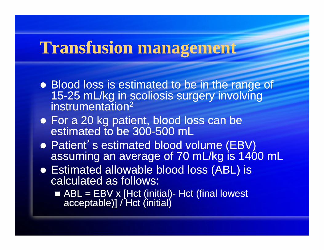
Blood loss is estimated to be in the range of 15-25 mL/kg in scoliosis surgery involving instrumentation2
For a 20 kg patient, blood loss can be estimated to be 300-500 mL
Patient’s estimated blood volume (EBV) assuming an average of 70 mL/kg is 1400 mL
Estimated allowable blood loss (ABL) is calculated as follows: ABL = EBV x [Hct (initial)- Hct (final lowest
acceptable)] / Hct (initial)
Given a starting Hct of 43 and assuming a lowest acceptable Hct of 20, the allowable blood loss would be: ABL = 1400 mL (43-20) / 43 = 750 mL
Given a starting Hct of 43 and assuming a lowest acceptable Hct of 20, the allowable blood loss would be: ABL = 1400 mL (43-20) / 43 = 750 mL
Given the anticipated large bleeding from a long complicated multilevel repair as well as patient risk factors, what evidence based interventions can minimize blood loss?
Given the anticipated large bleeding from a long complicated multilevel repair as well as patient risk factors, what evidence based interventions can minimize blood loss?
Minimize intraabdominal pressure to prevent further engorgement of the vertebral venous plexus and venous bleeding
Isovolemic hemodilution Intraoperative blood salvage Deliberate hypotension Antifibrinolytic drugs
Minimize intraabdominal pressure to prevent further engorgement of the vertebral venous plexus and venous bleeding
Isovolemic hemodilution Intraoperative blood salvage Deliberate hypotension Antifibrinolytic drugs
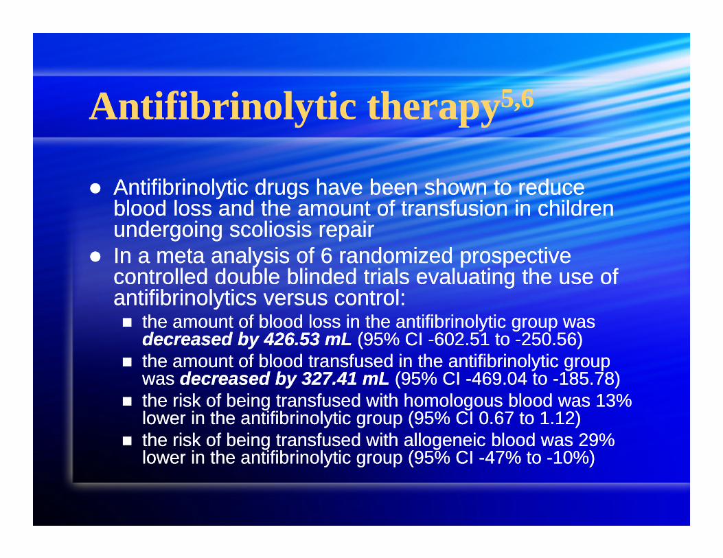
Antifibrinolytic therapy5,6Antifibrinolytic therapy5,6
Antifibrinolytic drugs have been shown to reduce blood loss and the amount of transfusion in children undergoing scoliosis repair
In a meta analysis of 6 randomized prospective controlled double blinded trials evaluating the use of antifibrinolytics versus control: the amount of blood loss in the antifibrinolytic group was
decreased by 426.53 mL (95% CI -602.51 to -250.56) the amount of blood transfused in the antifibrinolytic group
was decreased by 327.41 mL (95% CI -469.04 to -185.78) the risk of being transfused with homologous blood was 13%
lower in the antifibrinolytic group (95% CI 0.67 to 1.12) the risk of being transfused with allogeneic blood was 29%
lower in the antifibrinolytic group (95% CI -47% to -10%)
There were no mortalities in either the treatment or control groups
Aprotinin, tranexamic acid, and aminocaproic acid seem to be similarly effective with excellent safety profile (no evidence of hypercoagulability or thrombotic complications)
High dose aminocaproic acid was used in the studies: 100 mg/kg bolus administered as an infusion over 15 to 20 minutes followed by a continuous infusion of 10 mg/kg throughout the remainder of the procedure
No comparisons of antifibrinolytics head to head No comparisons of different doses
Given the large surgical incision and musculoskeletal work over many levels, what are the best evidence based interventions for postoperative pain control?
Given the large surgical incision and musculoskeletal work over many levels, what are the best evidence based interventions for postoperative pain control?
Epidural anesthesia superior to IV PCA in pediatric scoliosis surgery7Epidural anesthesia superior to IV PCA in pediatric scoliosis surgery7
In a meta analysis of four randomized prospective controlled trials evaluating PCEA vs. IV narcotic PCA in adolescent patients undergoing scoliosis repair, epidural anesthesia was shown to provide superior postoperative analgesia at 24, 48, and 72 hours
Patients randomized to the epidural group underwent placement by the surgeon under direct visualization prior to closure
The treatment group received epidural analgesia in the form of a continuous infusion of local anesthetic with or without an opioid in addition to parenteral opioids
The control group received parenteral opioids only
Blinding was not possible because placement of a sham epidural is associated with morbidity
visual analog scale pain scores in the PCEA group were found to be lower at 24, 48, and 72 hrs VAS pain scores were 15 points lower at 24 hours (p=0.03) VAS pain scores were 10.1 points lower at 48 hrs (p=0.03) VAS pain scores were 11.5 points lower at 72 hrs (p=0.02)
Patient satisfaction was higher by 1.7 on a 0-10 scale in the two studies that assessed it (p<0.0001)
Some but not all studies showed decreased nausea, pruritis, and number of rescue analgesics in epi grp
Some but not studies demonstrated shorter time to return of bowel function in the epidural group
Management of spinal shock8,9Management of spinal shock8,9
Ensure adequate tissue perfusion with volume resuscitation
Favorable outcomes reported in uncontrolled studies using fluid resuscitation and vasopressive medications to maintain a minimum mean ABP of 85 mmHg during the 1st week following spinal cord injury in adults
Assess for bradycardia and arrhythmias associated with neurogenic shock and treat appropriately
To counter the loss of sympathetic tone and provide chronotropic support, vasopressors with both alpha and beta adrenergic actions are recommended unless contraindicated
ReferencesReferences
1. Cote. A Practice of Anesthesia for Infants and Children, 3rd ed. 2001
2. Zayas, V, Yao, F, et al. Scoliosis. Anesthesiology: Problem Oriented Patient Management, 6th ed. 2008
3. Miller, R. Miller’s Anesthesia, 6th ed. 20054. Erickson M, et al. Pathways that distinguish simple
from complex scoliosis repair and their outcomes. Current Opinion in Pediatrics 2011, (23):339-345
5. Tzortzopoulou A, et al. Antifibrinolytic agents for reducing blood loss in scoliosis surgery in children. Cochrane Database of Systematic Reviews 2008, Issue 3
6. Florentino-Pineda I, et al. The effect of Amicar in perioperative blood loss in idiopathic scoliosis. The results of a prospective randomized double blind study. Spine 29(3):233-238
7. Taenzer A, et al. Efficacy of postoperative epidural analgesia in adolescent scoliosis surgery: a meta analysis. Pediatric Anesthesia 2010 (20):135-143
8. Furlan J, et al. Cardiovascular complications after acute spinal cord injury: pathophysiology, diagnosis, and management. Neurosurg focus 24(5):E15, 2008
9. Consortium for Spinal Cord Medicine: Early acute management in adults with spinal cord injury: a clinical practice guideline for health-care providers. Washington, DC: Paralyzed Veterans of America, 2008
9. Consortium for Spinal Cord Medicine: Early acute management in adults with spinal cord injury: a clinical practice guideline for health-care providers. Washington, DC: Paralyzed Veterans of America, 2008