Pathology of Swine - Animal Health Australia
56
Dr. med. vet. Dr. med. vet. Matti Matti Kiupel, BS, MS, PhD, Kiupel, BS, MS, PhD, DACVP DACVP Diagnostic Center for Population and Animal Health Diagnostic Center for Population and Animal Health College of Veterinary Medicine, Michigan State University College of Veterinary Medicine, Michigan State University 4125 Beaumont Road 152A, Lansing, MI 48910, USA 4125 Beaumont Road 152A, Lansing, MI 48910, USA Tel.: ** 517 432 2670; Tel.: ** 517 432 2670; Fax: ** 517 432 6557; Fax: ** 517 432 6557; E E - - mail: mail: [email protected] [email protected] Pathology of Swine Pathology of Swine
Transcript of Pathology of Swine - Animal Health Australia

Dr. med. vet. Dr. med. vet. MattiMatti Kiupel, BS, MS, PhD, Kiupel, BS, MS, PhD, DACVPDACVP
Diagnostic Center for Population and Animal HealthDiagnostic Center for Population and Animal HealthCollege of Veterinary Medicine, Michigan State UniversityCollege of Veterinary Medicine, Michigan State University
4125 Beaumont Road 152A, Lansing, MI 48910, USA4125 Beaumont Road 152A, Lansing, MI 48910, USATel.: ** 517 432 2670;Tel.: ** 517 432 2670; Fax: ** 517 432 6557;Fax: ** 517 432 6557;
EE--mail: mail: [email protected]@dcpah.msu.edu
Pathology of SwinePathology of Swine
Musculoskeletal Musculoskeletal SystemSystem

Posterior Paralysis/Paresis in SwinePosterior Paralysis/Paresis in Swine
•• Spinal cordSpinal cord–– enterovirusenterovirus–– selenium toxicityselenium toxicity–– ruptured diskruptured disk–– traumatrauma–– lymphosarcomalymphosarcoma
•• Vertebral columnVertebral column–– osteomyelitisosteomyelitis–– osteomalaciaosteomalacia
•• Bones and MusclesBones and Muscles–– IschialIschial epiphysiolysisepiphysiolysis–– Torn “hamstring”Torn “hamstring”–– Osteomalacia/osteoOsteomalacia/osteo--
myelitismyelitis fracturefracture
•• NervesNerves–– Organic arsenicalsOrganic arsenicals–– TraumaTrauma

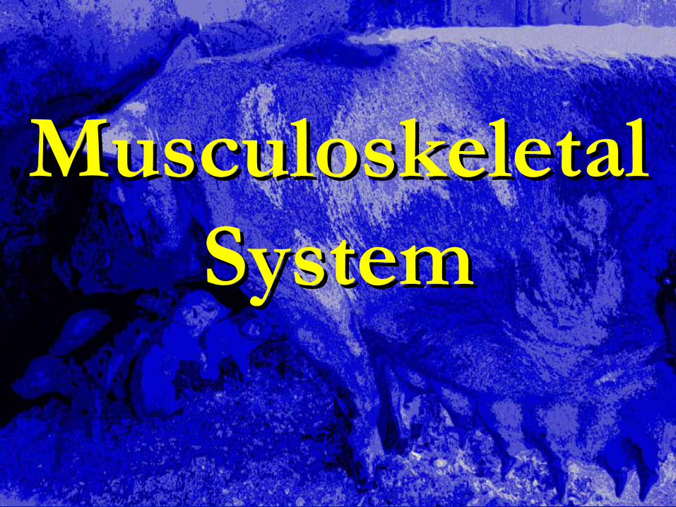
ArcanobacteriumArcanobacterium pyogenespyogenes
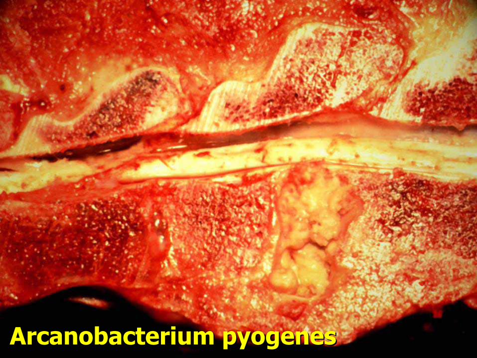
ArcanobacteriumArcanobacterium pyogenespyogenes
ArcanobacteriumArcanobacterium pyogenespyogenes

Degenerative Disc Disease Degenerative Disc Disease

Sciatic Nerve Damage Sciatic Nerve Damage

Sciatic Nerve Damage Sciatic Nerve Damage

ApophysiolysisApophysiolysis tuberistuberis ischiiischii
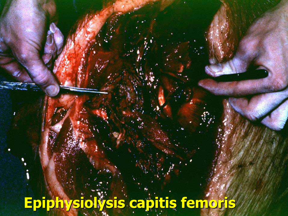
EpiphysiolysisEpiphysiolysis capitiscapitis femorisfemoris

Compartment SyndromeCompartment Syndrome
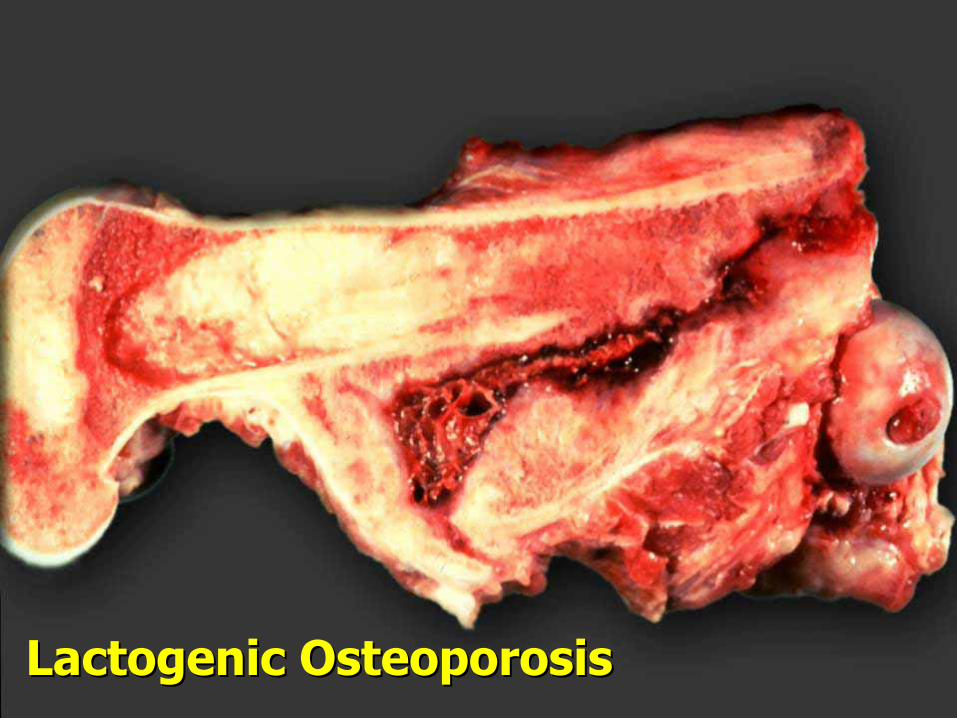
LactogenicLactogenic Osteoporosis Osteoporosis

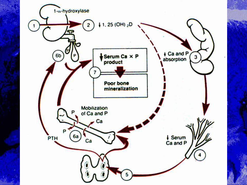
HypovitaminosisHypovitaminosis DD
•• Causes Causes hypocalcemiahypocalcemia•• Homeostatic mechanisms lead to Homeostatic mechanisms lead to
normocalcemianormocalcemia, mobilization of bone , mobilization of bone calciumcalcium
•• HypophosphatemiaHypophosphatemia persists, thus adds to persists, thus adds to impaired bone mineralizationimpaired bone mineralization–– Rickets in young growing animalsRickets in young growing animals–– OsteomalaciaOsteomalacia in adult animalsin adult animals

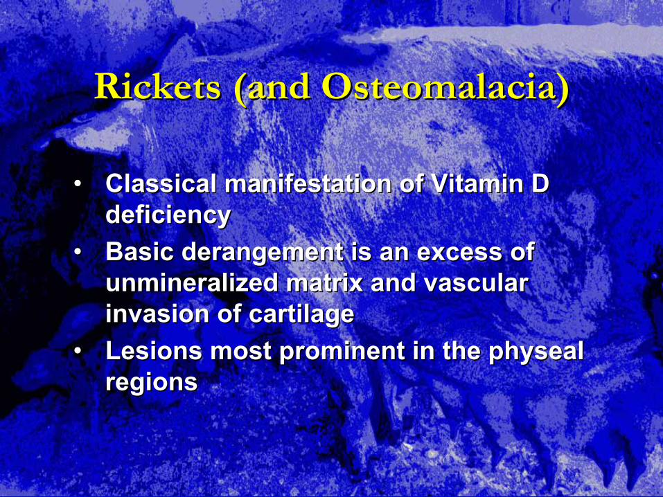
Rickets (and Rickets (and OsteomalaciaOsteomalacia))
•• Classical manifestation of Vitamin D Classical manifestation of Vitamin D deficiencydeficiency
•• Basic derangement is an excess of Basic derangement is an excess of unmineralizedunmineralized matrix and vascular matrix and vascular invasion of cartilageinvasion of cartilage
•• Lesions most prominent in the Lesions most prominent in the physealphysealregionsregions

Rickets (and Rickets (and OsteomalaciaOsteomalacia) ) HistopathologyHistopathology
•• Increased thickness of zone of hypertrophyIncreased thickness of zone of hypertrophy•• Disorderly arrangement of the zone of Disorderly arrangement of the zone of
hypertrophyhypertrophy•• Disorderly penetration of cartilage by blood Disorderly penetration of cartilage by blood
vesselsvessels•• Excess of Excess of uncalcifieduncalcified osteoidosteoid in the in the
metaphysismetaphysis ((osteoidosteoid seamsseams))

EpihysealEpihyseal growth plategrowth plate
CartilageCartilage
Calcified cartilageCalcified cartilage
(primary (primary spongiosaspongiosa))
Mature boneMature bone
((secundarysecundary spongiosaspongiosa))

OsteomalaciaOsteomalacia Long BoneLong Bone
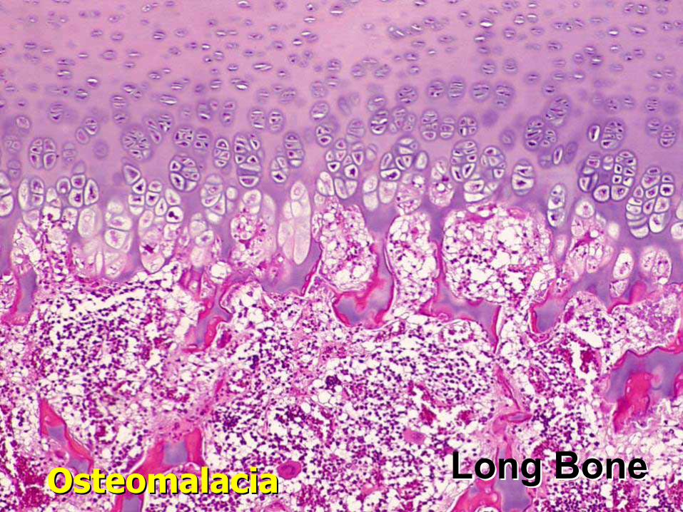
OsteomalaciaOsteomalacia Long BoneLong Bone

Osteoporosis Osteoporosis
•• Caused by deficiencies of Ca, P, or Vitamin DCaused by deficiencies of Ca, P, or Vitamin D•• Results when Results when resorptionresorption rates exceed rates exceed
formation due to increased PTHformation due to increased PTH•• Mainly affects flat bones of skull, scapula, Mainly affects flat bones of skull, scapula,
ileum, and ileum, and metaphysismetaphysis of long bonesof long bones•• HypocalcemiaHypocalcemia complicated by Vitamin D complicated by Vitamin D
deficiency produces more severe lesionsdeficiency produces more severe lesions–– Uncomplicated Uncomplicated hypocalcemiahypocalcemia is rareis rare–– Impairment of homeostatic mechanismsImpairment of homeostatic mechanisms

Osteoporosis HistopathologyOsteoporosis Histopathology
•• Cortical bone is thin due to increased Cortical bone is thin due to increased endostealendosteal or or intracorticalintracortical (severe (severe hypocalcemiahypocalcemia) ) resorptionresorption
•• OsteoidOsteoid seams are of normal width and seams are of normal width and frequencyfrequency
•• Mineralization usually normalMineralization usually normal•• Decreased and or thinned Decreased and or thinned trabeculaetrabeculae

Osteoporosis HistologyOsteoporosis Histology
•• Cortical thinning Cortical thinning •• TrabecularTrabecular fragmentation fragmentation •• Abnormal mineralization with retention of Abnormal mineralization with retention of
cartilage corescartilage cores•• Retained Retained osteoidosteoid in in epiphysealepiphyseal growth plate growth plate
of few bonesof few bones•• PeriostealPeriosteal fibrosisfibrosis

OsteoporosisOsteoporosis Long BoneLong Bone
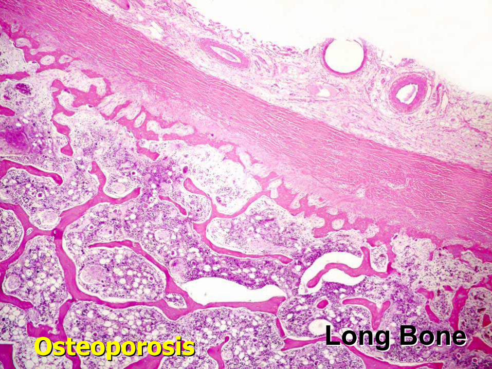
OsteoporosisOsteoporosis Long BoneLong Bone
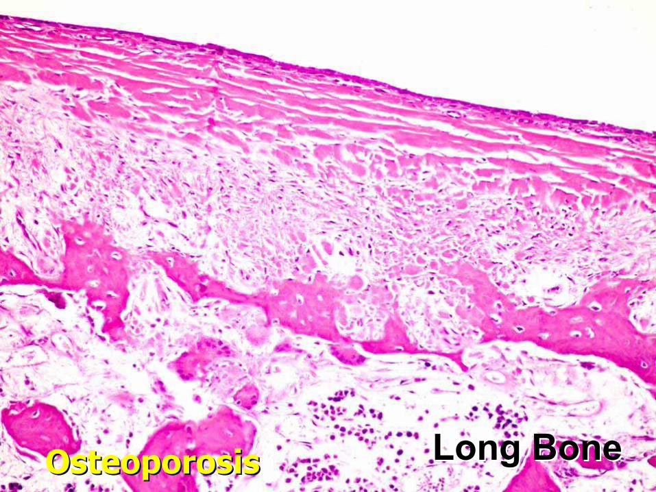
OsteoporosisOsteoporosis Long BoneLong Bone

ArcanobacteriumArcanobacterium pyogenespyogenes
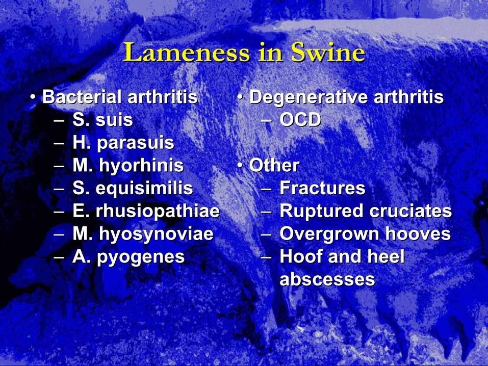
Lameness in SwineLameness in Swine
•• Bacterial arthritisBacterial arthritis–– S. S. suissuis–– H. H. parasuisparasuis–– M. M. hyorhinishyorhinis–– S. S. equisimilisequisimilis–– E. E. rhusiopathiaerhusiopathiae–– M. M. hyosynoviaehyosynoviae–– A. A. pyogenespyogenes
•• Degenerative arthritisDegenerative arthritis–– OCDOCD
•• OtherOther–– FracturesFractures–– Ruptured Ruptured cruciatescruciates–– Overgrown hoovesOvergrown hooves–– Hoof and heel Hoof and heel
abscessesabscesses

PododermalPododermal AbrasionsAbrasions
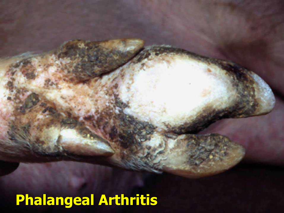
PhalangealPhalangeal ArthritisArthritis
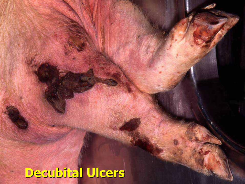
DecubitalDecubital UlcersUlcers
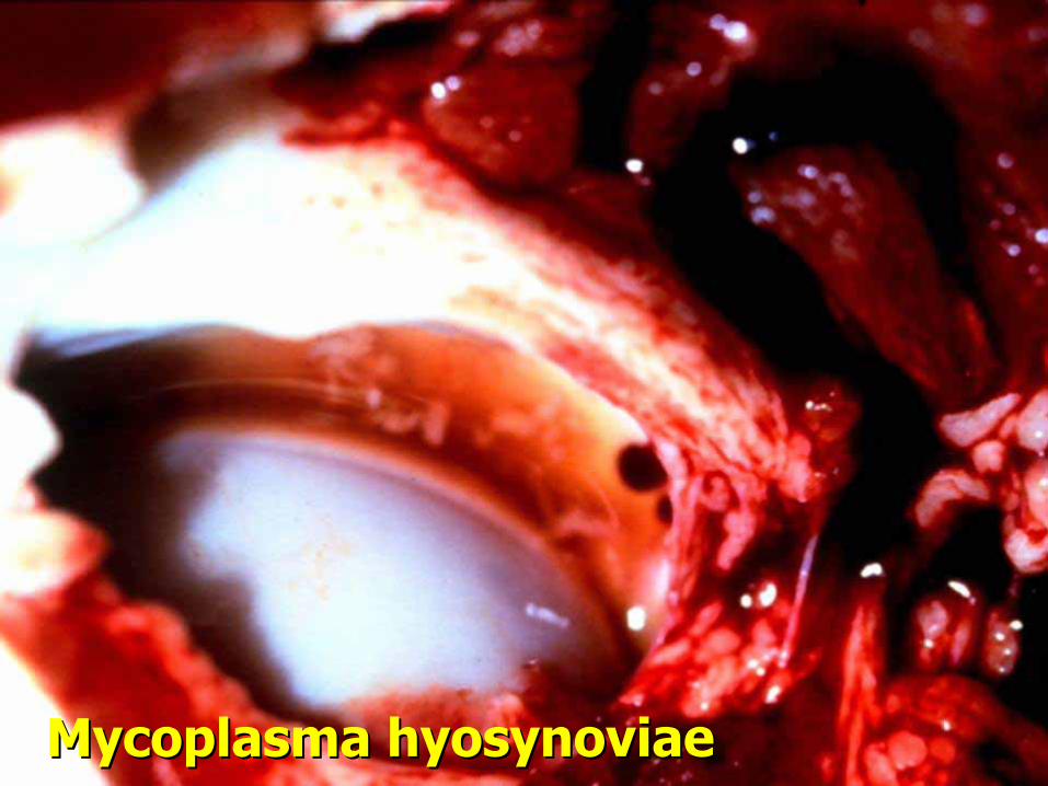
MycoplasmaMycoplasma hyosynoviaehyosynoviae
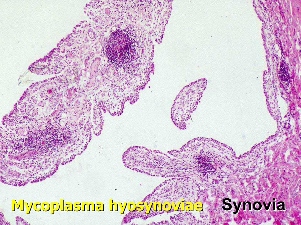
SynoviaSynoviaMycoplasmaMycoplasma hyosynoviaehyosynoviae
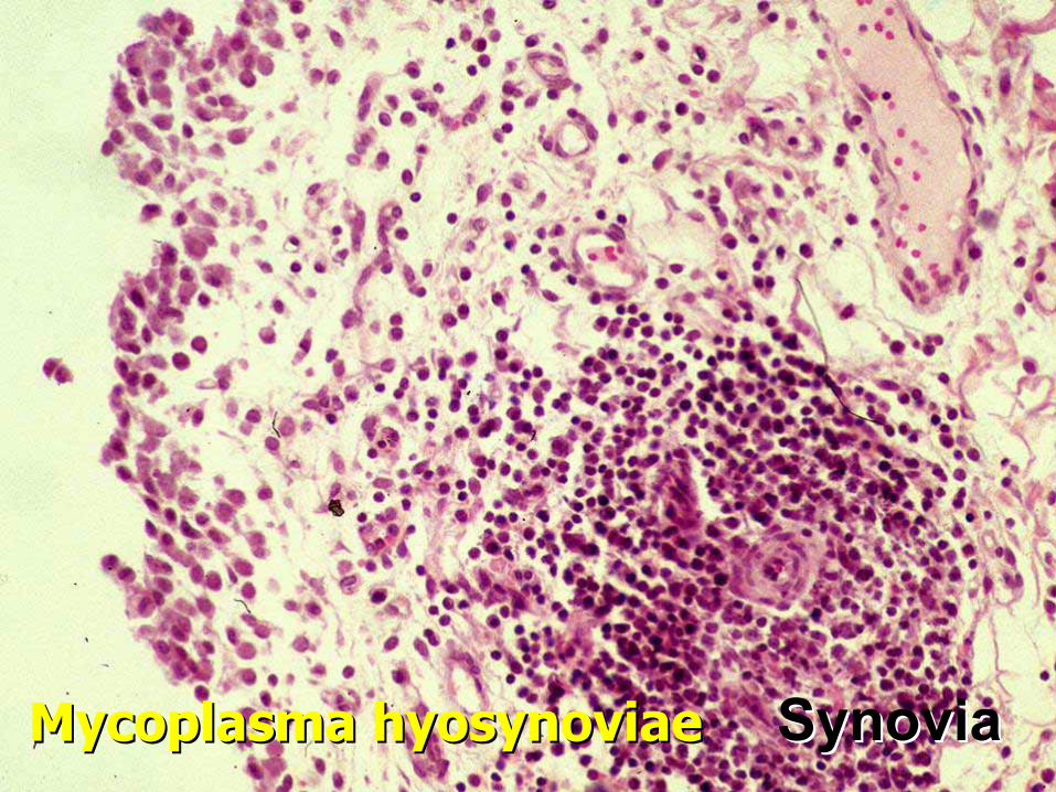
SynoviaSynoviaMycoplasmaMycoplasma hyosynoviaehyosynoviae

MycoplasmaMycoplasma hyosynoviaehyosynoviae

Erysipelas Erysipelas
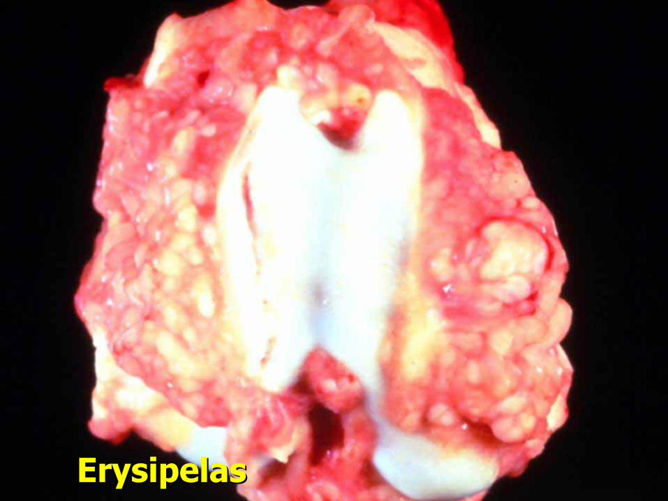
Erysipelas Erysipelas
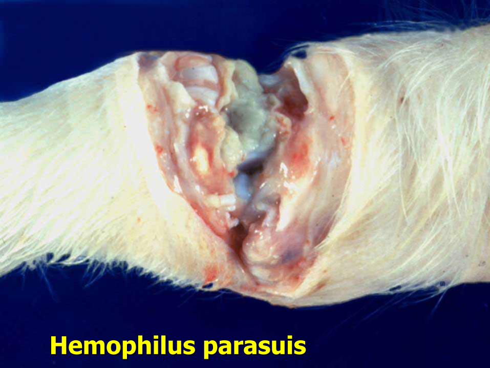
HemophilusHemophilus parasuisparasuis
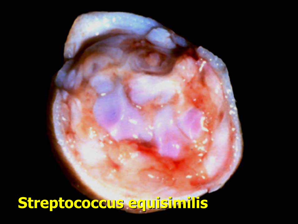
Streptococcus Streptococcus equisimilisequisimilis

ArcanobacteriumArcanobacterium pyogenespyogenes

OCDOCD

OCDOCD
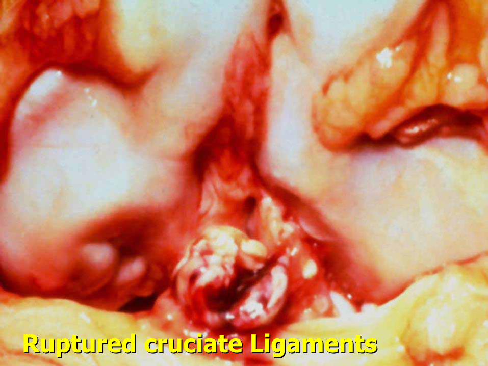
Ruptured Ruptured cruciatecruciate LigamentsLigaments

HygromaHygroma

AnthraxAnthrax
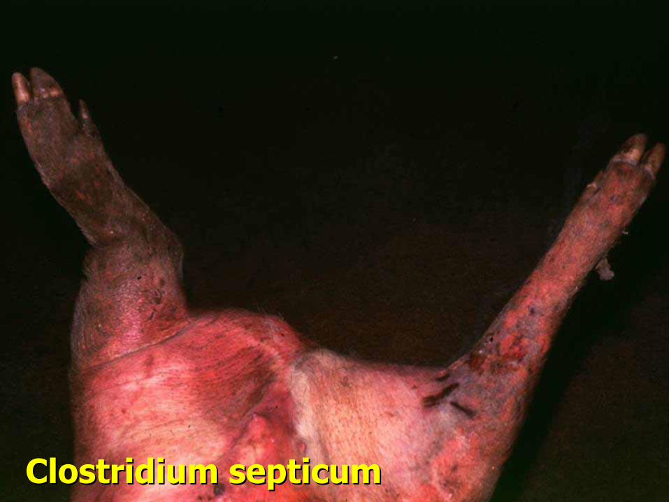
Clostridium Clostridium septicumsepticum

Clostridium Clostridium septicumsepticum
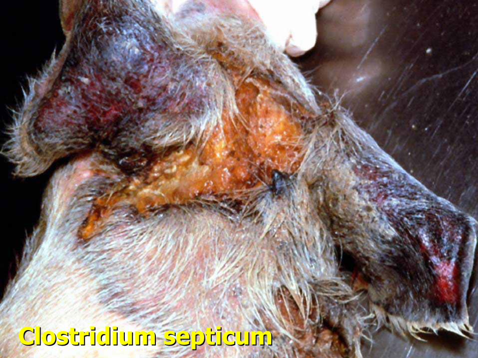
Clostridium Clostridium septicumsepticum

Clostridium Clostridium septicumsepticum
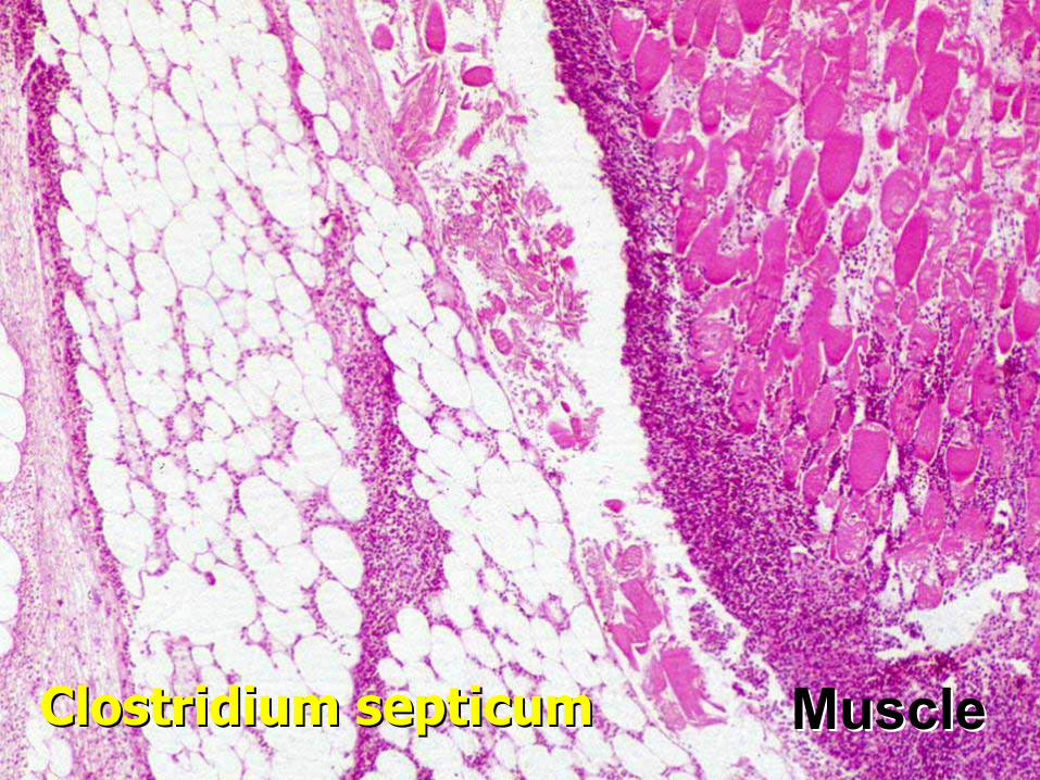
MuscleMuscleClostridium Clostridium septicumsepticum

Porcine Stress SyndromePorcine Stress Syndrome
•• halothane gene (halothane gene (napolenapole gene gene -- Hampshire)Hampshire)•• DNA testDNA test•• single (?) recessive genesingle (?) recessive gene•• calcium transport deficitcalcium transport deficit•• pig becomes red, splotchypig becomes red, splotchy diesdies•• hyperthermia, acidosishyperthermia, acidosis•• rapid postrapid post--mortem autolysismortem autolysis•• associated with extreme musclingassociated with extreme muscling

Porcine Stress Syndrome Porcine Stress Syndrome

Porcine Stress Syndrome Porcine Stress Syndrome
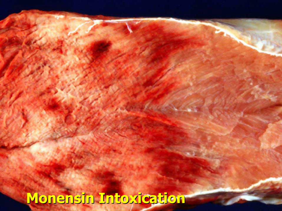
MonensinMonensin Intoxication Intoxication
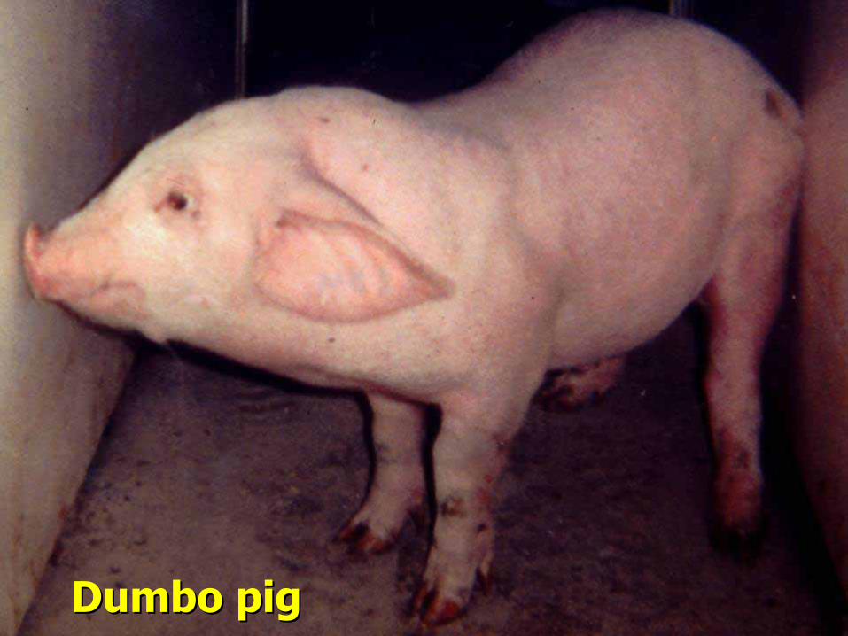
DumboDumbo pig pig