Pathogenic POGZ mutation causes impaired cortical ...
16
ARTICLE Pathogenic POGZ mutation causes impaired cortical development and reversible autism-like phenotypes Kensuke Matsumura et al. # Pogo transposable element derived with ZNF domain (POGZ) has been identified as one of the most recurrently de novo mutated genes in patients with neurodevelopmental disorders (NDDs), including autism spectrum disorder (ASD), intellectual disability and White-Sutton syndrome; however, the neurobiological basis behind these disorders remains unknown. Here, we show that POGZ regulates neuronal development and that ASD-related de novo mutations impair neuronal development in the developing mouse brain and induced plur- ipotent cell lines from an ASD patient. We also develop the first mouse model heterozygous for a de novo POGZ mutation identified in a patient with ASD, and we identify ASD-like abnormalities in the mice. Importantly, social deficits can be treated by compensatory inhi- bition of elevated cell excitability in the mice. Our results provide insight into how de novo mutations on high-confidence ASD genes lead to impaired mature cortical network function, which underlies the cellular pathogenesis of NDDs, including ASD. https://doi.org/10.1038/s41467-020-14697-z OPEN *A full list of authors and their affiliations appears at the end of the paper. NATURE COMMUNICATIONS | (2020)11:859 | https://doi.org/10.1038/s41467-020-14697-z | www.nature.com/naturecommunications 1 1234567890():,;
Transcript of Pathogenic POGZ mutation causes impaired cortical ...
ARTICLE
Pathogenic POGZ mutation causes impairedcortical development and reversible autism-likephenotypesKensuke Matsumura et al.#
Pogo transposable element derived with ZNF domain (POGZ) has been identified as one of
the most recurrently de novo mutated genes in patients with neurodevelopmental disorders
(NDDs), including autism spectrum disorder (ASD), intellectual disability and White-Sutton
syndrome; however, the neurobiological basis behind these disorders remains unknown.
Here, we show that POGZ regulates neuronal development and that ASD-related de novo
mutations impair neuronal development in the developing mouse brain and induced plur-
ipotent cell lines from an ASD patient. We also develop the first mouse model heterozygous
for a de novo POGZ mutation identified in a patient with ASD, and we identify ASD-like
abnormalities in the mice. Importantly, social deficits can be treated by compensatory inhi-
bition of elevated cell excitability in the mice. Our results provide insight into how de novo
mutations on high-confidence ASD genes lead to impaired mature cortical network function,
which underlies the cellular pathogenesis of NDDs, including ASD.
https://doi.org/10.1038/s41467-020-14697-z OPEN
*A full list of authors and their affiliations appears at the end of the paper.
NATURE COMMUNICATIONS | (2020) 11:859 | https://doi.org/10.1038/s41467-020-14697-z | www.nature.com/naturecommunications 1
1234
5678
90():,;
Neurodevelopmental disorders (NDDs), including autismspectrum disorder (ASD) and intellectual disability (ID),are characterized by early life onset with aberrant brain
development, leading to social and cognitive abnormalities thatspan a wide range of functions and are highly heterogeneousamong individuals1,2. Since the prevalence rate of NDDs hasrecently been increasing dramatically, NDDs are becoming asignificant medical and social burden. Among NDDs, the pre-valence rate of ASD is considerable, estimated to be ~1 in 40children3,4. Despite the high heritability of ASD, recent studieshave suggested that the disease is highly genetically hetero-geneous, with rare genetic variants as well as common variants5–7,and that the genetic cause is unidentified in ~90% of patients withthe condition8,9. Accordingly, the molecular etiology of ASDremains largely unclear. There are no pharmacological medica-tions to treat the core symptoms of ASD; mechanism-based drugdevelopment and therapeutic strategies are imperative.
Genetic and epidemiological studies have suggested that denovo mutations, spontaneous rare mutations that appear in an
affected child but not in the unaffected parents, significantlycontribute to ASD10–18 and that ~3–10% of ASD risk is explainedby de novo mutations in exons6,16,19. Importantly, a recent,comprehensive exome analysis has identified 65 high-confidenceASD genes20. Although, among these high-confidence ASD genes,recent studies have found that haploinsufficiency of ARID1B orCHD8 causes ASD-related abnormalities in mice21–27, furtherdirect assessments of the biological significance of ASD-associatedde novo mutations are necessary to fully understand the con-tribution of de novo mutations to the core features of ASD.
We and other groups have found that Pogo transposable elementderived with ZNF domain (POGZ) is one of the most recurrentlymutated genes in patients with NDDs, particularly ASD and ID; thenumber of reported mutations continues to increase (see Fig. 1a andSupplementary Table 1; we classified patients into ASD, ID, andWhite–Sutton syndrome according to the original diagnosis in eachreport)12,14–16,20,28–34. POGZ mutations are also recurrently foundin patients with White–Sutton syndrome, characterized by ID andspecific facial features30,31. POGZ interacts with the SP1
Moc
k
C N C N C N C N C N
WT
R997X
R1004
X
E1043
X
Moc
k
C N C N C N C N
WT
E1036
K
Q1038
R
Moc
k
C N C N C N C N C N
WT
R1001
H
F1047
L
H1080
R
Myc-mPOGZ
Lamin ALamin C
α-Tubulin
Myc-mPOGZ
α-Tubulin
Lamin ALamin C
Myc-mPOGZ
α-Tubulin
Lamin ALamin C
ASD-related mutants
ASD-related mutants
Control mutants
Cytosol Nucleusa
b
c
d
e f
g h
i j
De novo mutation: Missense Nonsense Frameshift TotalASD case 5 4 4 13ID case 0 6 8 14ASD/ID case 1 2 4 7Unclassified NDDs case 1 4 6 11NDDs case (Total case) 7 16 22 45Control 19 0 0 19
WT
R1001
H
F1047
L
H1080
R
1410aa
N C
Missense mutation Nonsense mutation Frameshift mutation
N CHPZC2H2 Zn fingerCENP-DB
domainRve
superfamily
NDDs-related de novo mutations
Control de novo mutations
Mouse POGZ
Mouse POGZ
Mouse POGZ
Human POGZ
Human POGZ
80%
60%
40%
20%
100%
0%
% o
f tot
al p
rote
ins
WT
R997W
R1004
X
E1043
X
WT
E1036
K
Q1038
R
1410aa
N CHPZC2H2 Zn finger CENP-DBdomain
Rvesuperfamily
HPZC2H2 Zn finger CENP-DBdomain
Rvesuperfamily
1501007550
50
250
10075
50
50
15075
50
250
(kDa)
(kDa)
(kDa)
80%
60%
40%
20%
100%
0%
% o
f tot
al p
rote
ins
80%
60%
40%
20%
100%
0%
% o
f tot
al p
rote
inD23
G
N136S
N341S
R374Q
R379Q
P446L
R502K
V591I
N632S
R674C
R797Q
D828N
R1005
H
F1051
L
H1084
R
L110
3H
A1288
V
D1404
N
N159D
Q180X
S314N
S278X
Y404X
Y597C
H641Q
P764Lfs*27
S799N
F822Yfs*43
N941Efs*3
R1008X
Q1042R
E1047XE1203Vfs*28
R674Vfs*9
H858Qfs*13
L927Pfs*17
D946Mfs*12
R1001X
E1040K
R385Sfs*4
E755Sfs*36
S774Cfs*16
K801Qfs*7
L834Wfs*20
E1154Tfs*4 Q1283X
T922Hfs*22
R864X
R979X
Q1014X
V733del
E427XE604X
M394Ifs*76
V700Nfs*7
S839Lfs*25
L1119Cfs*3
T1210P
P918Tfs*26
Q1014Rfs*5R1001X
Q1011X
L904X
Y770X
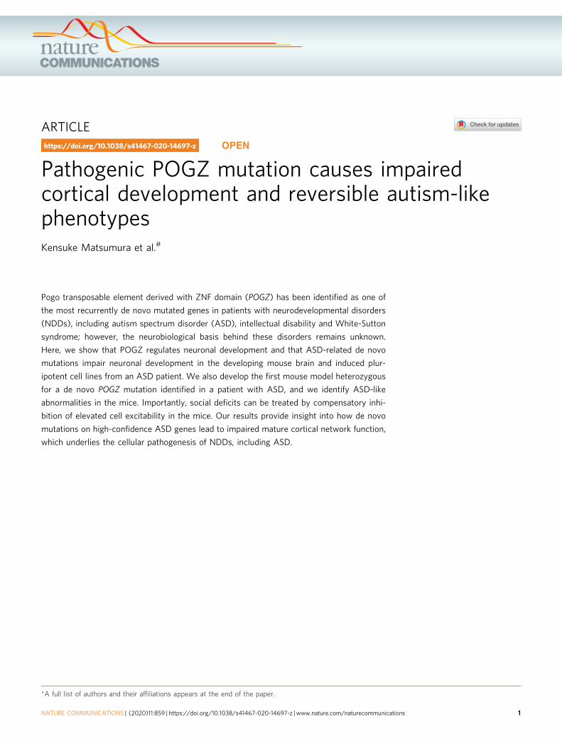
Fig. 1 ASD-related de novo mutations in POGZ impair the nuclear localization of the POGZ protein. a De novo mutations in POGZ identified in patientswith NDDs (blue, ASD; green, ID; red, both ASD and ID; purple, unclassified NDDs). b De novo mutations in POGZ identified in unaffected controls. c Thenumber of missense, nonsense, and frameshift mutations identified in patients with NDDs and unaffected controls. d Distribution of POGZ mutationsidentified in patients with NDDs and unaffected controls (blue, missense mutations; red, nonsense mutations; green, frameshift mutations).e, g ASD-related nonsense e or missense g mutations disrupted the nuclear localization of Myc-tagged overexpressed mouse (m) POGZ in Neuro2a cells.C cytosolic fraction; N nuclear fraction; WT wild-type. Note that Q1038R mutation in mouse POGZ corresponds to Q1042R mutation in human POGZ.f, h, j Quantification of POGZ in cytosolic and nuclear fractions (each n= 4). i Control mutations identified in unaffected healthy controls did not affect thenuclear localization of Myc-tagged overexpressed mouse POGZ in Neuro2a cells. C cytosolic fraction; N nuclear fraction; WT wild-type. Note that theamino acid numbers are based on the human protein (NP_055915.2) a, b and mouse protein (NP_766271.2) e, g, i. f, h, j One-way ANOVA withBonferroni–Dunn post hoc tests, f F3, 12= 23.12; h F2, 9= 67.45; j F3, 12= 0.491. ***P < 0.001. Data are presented as the mean ± s.e.m. The source dataunderlying figures e, g and i are provided as a Source Data file.
ARTICLE NATURE COMMUNICATIONS | https://doi.org/10.1038/s41467-020-14697-z
2 NATURE COMMUNICATIONS | (2020) 11:859 | https://doi.org/10.1038/s41467-020-14697-z | www.nature.com/naturecommunications

transcription factor, heterochromatin protein 1 (HP1), and chro-modomain helicase DNA-binding protein 4 (CHD4)35–37, whichsuggests that POGZ functions as a chromatin regulator; however,the role of POGZ in brain development and the biological sig-nificance of ASD-associated de novo POGZ mutations in theetiology of ASD are largely unknown.
In this study, we developed the first mouse model that carried apathogenic de novo mutation of POGZ identified in an ASDpatient. From the same patient, we established induced plur-ipotent stem cell (iPSC) lines with the same de novo POGZmutated (Q1042R) as the model mouse. Comprehensivelyexamining these human and mouse materials, we determined thatthe de novo mutation in POGZ impaired the cellular localizationof the POGZ protein and hindered cortical neuronal develop-ment. We also determined that this de novo mutation in POGZcaused ASD-related behavioral abnormalities and that theseabnormalities were pharmacologically treatable even in adult-hood. Importantly, de novo POGZ mutations identified in unaf-fected controls had no damaging effect on POGZ function inneuronal development. Together, these observations provide thefirst in vivo evidence suggesting that ASD-associated de novomutations in a high-confidence ASD gene are critical for a widerange of processes involved in ASD pathogenesis.
ResultsDe novo mutations in POGZ impair its nuclear localization.POGZ has been identified as one of the most recurrently denovo-mutated genes in patients with NDDs (Fig. 1a, c, d, Sup-plementary Table 1; the amino acid numbers are based on thehuman protein). The vast majority of de novo POGZ mutationsidentified in patients with NDDs are nonsense and frameshiftmutations and distributed between the C2H2 Zn finger andcentromere protein-B-like DNA-binding (CENP-DB) domainsand in the CENP-DB domain itself (Fig. 1a, c). In contrast, all denovo POGZ mutations identified in unaffected controls (controlde novo mutations) are missense mutations (Fig. 1b, c, Sup-plementary Table 1). Interestingly, while control de novo mis-sense mutations are uniformly distributed in POGZ, manyNDDs-related nonsense and frameshift mutations are posi-tioned just upstream of the CENP-DB domain, implying thatimpaired CENP-DB domain function could contribute to therisk of NDDs, including ASD (Fig. 1d). We examined thedeleterious effect of sporadic-ASD-associated de novo missensemutations within the CENP-DB domain and nonsense muta-tions resulting in the elimination or truncation of the CENP-DBdomain. Since the amino acid sequences of the human andmouse POGZ are very similar (93.9% identified in amino acidsequence) (Supplementary Fig. 1), we think that each mousemutation is likely to correspond to the respective humanmutation. Previous studies have suggested that POGZ is loca-lized to the nucleus and functions as a chromatin regulator, wetherefore assume that the ASD-related de novo mutations mayalter the nuclear localization of POGZ. To examine this possi-bility, we firstly conducted immuno-cytochemical experimentsusing ASD-related missense mutants, E1036K (E1040K inhuman POGZ)- and Q1038R (Q1042R in human POGZ)-mutated POGZ, as well as E1043X (E1047X in human POGZ)-mutated POGZ, the longest nonsense-mutated POGZ, andfound that these mutations partially impaired the nuclearlocalization of POGZ (Supplementary Fig. 2). We next per-formed cellular fractionation experiments and determined that,in contrast to the nuclear localization of overexpressed wild-type(WT)-mouse (m) POGZ, R997X (R1001X in human POGZ)-,R1004X (R1008X in human POGZ)-, and E1043X (E1047X inhuman POGZ)-mPOGZ mutants, which entirely or partially
lack the CENP-DB domain exhibited aberrant distribution in thecytoplasm (Fig. 1e, f; the amino acid numbers are based on themouse protein). We also observed that the E1036K (E1040K inhuman POGZ)- and Q1038R (Q1042R in human POGZ)-mPOGZmutants also exhibited aberrant distribution in the cytoplasm(Fig. 1g, h; the amino acid numbers are based on the mouseprotein). Interestingly, in contrast to the ASD-related mutants, thede novo R1005H-, F1051L-, and H1084R-mPOGZ mutants, whichharbor missense mutations within or adjacent to the CENP-DBdomain identified in unaffected controls (Fig. 1b, d), showedsimilar protein expression patterns to WT-mPOGZ (Fig. 1i, j; theamino acid numbers are based on the mouse protein). Addition-ally, we performed cellular fractionation experiments using humanSH-SY5Y cells and the human Q1042R-mutated POGZ andobtained essentially the same results as the results with the mouseQ1038R mutation in Fig. 1g, h (Supplementary Fig. 3). Theseresults suggest that ASD-related de novo mutations but not controlde novo mutations identified in unaffected controls impair thenuclear localization of POGZ in cells.
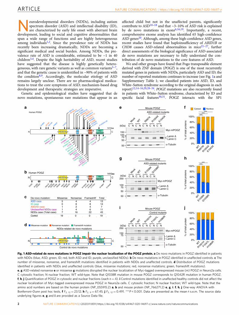
POGZ regulates the development of mouse neural stem cells(NSCs). To elucidate the function of POGZ in the brain, we firstinvestigated the temporal, regional, and cell type-specificexpression pattern of Pogz in the mouse brain. Temporally,expression of Pogz gradually increased during embryonic neu-rogenesis from embryonic day 14.5 (E14.5) to E18.5 and began todecrease after birth (Fig. 2a). At E16.5, Pogz was highly expressedin the cortical NSCs and intermediate progenitor cells (IPs) in theventricular and subventricular zones (VZ/SVZ) (Fig. 2b, c). Theseexpression patterns suggest that POGZ plays an important role incortical neuronal development. To determine the role of POGZ incortical neuronal development, we knocked down the expressionof Pogz using four distinct commercial shRNAs (MISSION TRCshRNA library SP1, SIGMA-Aldrich) and a miR30-based shRNA(shRNAmiR30) targeting Pogz (Supplementary Fig. 4a, Supple-mentary Tables 2 and 3). Plasmids encoding each shRNA againstPogz and GFP were coelectroporated into the lateral ventricle ofE14.5 mouse forebrains. The electroporated embryos wereallowed to develop until E18.5 and histologically analyzed formigration of GFP+ cells in the developing somatosensory cortex.We determined that the migration of GFP+ cells was significantlyinhibited by Pogz knockdown, which was roughly proportional tothe knockdown efficiency of each construct (SupplementaryFig. 4b–i). The impaired migration was rescued by forcedexpression of WT-mPOGZ (Fig. 2d, e). Using antibodies againstcortical layer markers, we then immunostained GFP+ cells whosemigration was delayed by Pogz knockdown, and we determinedthat Pogz knockdown had little effect on the proportion of SATB2+
GFP+ (layer II/III), CTIP2+ GFP+ (layer V) or TBR1+ GFP+
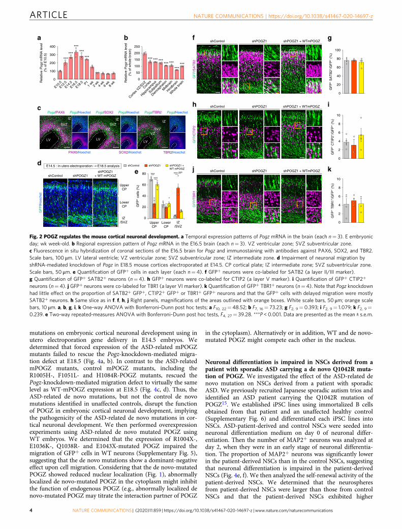
(layer VI) neurons and that the GFP+ cells with delayed migrationwere mostly SATB2+ neurons, representing neurons with youngupper cortical characteristics (Fig. 2f–k). Considering the highexpression of Pogz in NSCs during cortical neuronal development(Fig. 2b, c), the delaying of migration by Pogz knockdown may bedue to impaired neuronal differentiation. We analyzed the pro-portion of GFP+ NSCs, IPs, and neurons at E16.5 (2 days after inutero electroporation) and determined that Pogz knockdownincreased the proportion of PAX6+ NSCs and decreased the pro-portion of TBR2+ IPs and SATB2+ young neurons without sig-nificantly affecting migration in the somatosensory cortex within2 days (Fig. 3a–h). These data suggest that POGZ regulates corticalneuronal development by promoting neuronal differentiation.
POGZ mutations impair POGZ function in neuronal devel-opment. We investigated the effect of ASD-related de novo
NATURE COMMUNICATIONS | https://doi.org/10.1038/s41467-020-14697-z ARTICLE
NATURE COMMUNICATIONS | (2020) 11:859 | https://doi.org/10.1038/s41467-020-14697-z | www.nature.com/naturecommunications 3
mutations on embryonic cortical neuronal development using inutero electroporation gene delivery in E14.5 embryos. Wedetermined that forced expression of the ASD-related mPOGZmutants failed to rescue the Pogz-knockdown-mediated migra-tion defect at E18.5 (Fig. 4a, b). In contrast to the ASD-relatedmPOGZ mutants, control mPOGZ mutants, including theR1005H-, F1051L- and H1084R-POGZ mutants, rescued thePogz-knockdown-mediated migration defect to virtually the samelevel as WT-mPOGZ expression at E18.5 (Fig. 4c, d). Thus, theASD-related de novo mutations, but not the control de novomutations identified in unaffected controls, disrupt the functionof POGZ in embryonic cortical neuronal development, implyingthe pathogenicity of the ASD-related de novo mutations in cor-tical neuronal development. We then performed overexpressionexperiments using ASD-related de novo mutated POGZ usingWT embryos. We determined that the expression of R1004X-,E1036K-, Q1038R- and E1043X-mutated POGZ impaired themigration of GFP+ cells in WT neurons (Supplementary Fig. 5),suggesting that the de novo mutations show a dominant-negativeeffect upon cell migration. Considering that the de novo-mutatedPOGZ showed reduced nuclear localization (Fig. 1), abnormallylocalized de novo-mutated POGZ in the cytoplasm might inhibitthe function of endogenous POGZ (e.g., abnormally localized denovo-mutated POGZ may titrate the interaction partner of POGZ
in the cytoplasm). Alternatively or in addition, WT and de novo-mutated POGZ might compete each other in the nucleus.
Neuronal differentiation is impaired in NSCs derived from apatient with sporadic ASD carrying a de novo Q1042R muta-tion of POGZ. We investigated the effect of the ASD-related denovo mutation on NSCs derived from a patient with sporadicASD. We previously recruited Japanese sporadic autism trios andidentified an ASD patient carrying the Q1042R mutation ofPOGZ15. We established iPSC lines using immortalized B cellsobtained from that patient and an unaffected healthy control(Supplementary Fig. 6) and differentiated each iPSC lines intoNSCs. ASD-patient-derived and control NSCs were seeded intoneuronal differentiation medium on day 0 of neuronal differ-entiation. Then the number of MAP2+ neurons was analyzed atday 2, when they were in an early stage of neuronal differentia-tion. The proportion of MAP2+ neurons was significantly lowerin the patient-derived NSCs than in the control NSCs, suggestingthat neuronal differentiation is impaired in the patient-derivedNSCs (Fig. 4e, f). We then analyzed the self-renewal activity of thepatient-derived NSCs. We determined that the neurospheresfrom patient-derived NSCs were larger than those from controlNSCs and that the patient-derived NSCs exhibited higher
UpperCP
LowerCP
IZ/SVZ
GF
P/H
oech
st
shControl shPOGZ1shPOGZ1
+ WT mPOGZ
a
80
60
40
20
0Lower
CP
GF
P+ c
ells
(%
)
UpperCP
IZ/SVZ
b
ed
cPogz/SOX2Pogz/PAX6 Pogz/TBR2
LV
VZ
SVZ
IZ
LV
VZSVZ
IZ
VZSVZ
IZ
LV
Pogz/Hoechst Pogz/Hoechst Pogz/Hoechst
PAX6/Hoechst SOX2/Hoechst TBR2/Hoechst
shControl shPOGZ1 shPOGZ1 + WT mPOGZ
E14.5 : in utero electroporation E18.5 analysis
300
200
100
0
400
Rel
ativ
e Pog
z m
RN
A le
vel
(%
of E
10.5
)
E10.5
E12.5
E14.5
E16.5
E18.5
1 wk
2 wk
4 wk
6 wk
8 wkP1
200
150
100
50
250
0
Corte
x VZ/S
VZ
Hippoc
ampu
s
Subpa
llium
Dience
phalo
n
Hindbr
ain
Who
le br
ain
Corte
x
Midb
rainR
elat
ive Pog
z m
RN
A le
vel
(%
of w
hole
bra
in)
shControl shPOGZ1
GF
P/S
AT
B2
shPOGZ1 + WTmPOGZ
shControl shPOGZ1
GF
P/C
TIP
2
shPOGZ1 + WTmPOGZ
shControl shPOGZ1
GF
P/T
BR
1
shPOGZ1 + WTmPOGZ
GF
P+ S
AT
B2+
/GF
P+ (
%)
GF
P+ C
TIP
2+/G
FP
+ (
%)
2
4
6
8
0
10
GF
P+ T
BR
1+/G
FP
+ (
%)
h
k
i
f
j
g
80
60
40
20
100
0
2
4
6
8
0
10
Fig. 2 POGZ regulates the mouse cortical neuronal development. a Temporal expression patterns of Pogz mRNA in the brain (each n= 3). E embryonicday; wk week-old. b Regional expression pattern of Pogz mRNA in the E16.5 brain (each n= 3). VZ ventricular zone; SVZ subventricular zone.c Fluorescence in situ hybridization of coronal sections of the E16.5 brain for Pogz and immunostaining with antibodies against PAX6, SOX2, and TBR2.Scale bars, 100 μm. LV lateral ventricle; VZ ventricular zone; SVZ subventricular zone; IZ intermediate zone. d Impairment of neuronal migration byshRNA-mediated knockdown of Pogz in E18.5 mouse cortices electroporated at E14.5. CP cortical plate; IZ intermediate zone; SVZ subventricular zone.Scale bars, 50 μm. e Quantification of GFP+ cells in each layer (each n= 4). f GFP+ neurons were co-labeled for SATB2 (a layer II/III marker).g Quantification of GFP+ SATB2+ neurons (n= 4). h GFP+ neurons were co-labeled for CTIP2 (a layer V marker). i Quantification of GFP+ CTIP2+
neurons (n= 4). j GFP+ neurons were co-labeled for TBR1 (a layer VI marker). k Quantification of GFP+ TBR1+ neurons (n= 4). Note that Pogz knockdownhad little effect on the proportion of SATB2+ GFP+, CTIP2+ GFP+ or TBR1+ GFP+ neurons and that the GFP+ cells with delayed migration were mostlySATB2+ neurons. h Same slice as in f. f, h, j Right panels, magnifications of the areas outlined with orange boxes. White scale bars, 50 μm; orange scalebars, 10 μm. a, b, g, i, k One-way ANOVA with Bonferroni–Dunn post hoc tests; a F10, 22= 48.52; b F7, 16= 73.23; g F2, 9= 0.393; i F2, 9= 1.079; k F2, 9=0.239. e Two-way repeated-measures ANOVA with Bonferroni–Dunn post hoc tests, F4, 27= 39.28. ***P < 0.001. Data are presented as the mean ± s.e.m.
ARTICLE NATURE COMMUNICATIONS | https://doi.org/10.1038/s41467-020-14697-z
4 NATURE COMMUNICATIONS | (2020) 11:859 | https://doi.org/10.1038/s41467-020-14697-z | www.nature.com/naturecommunications
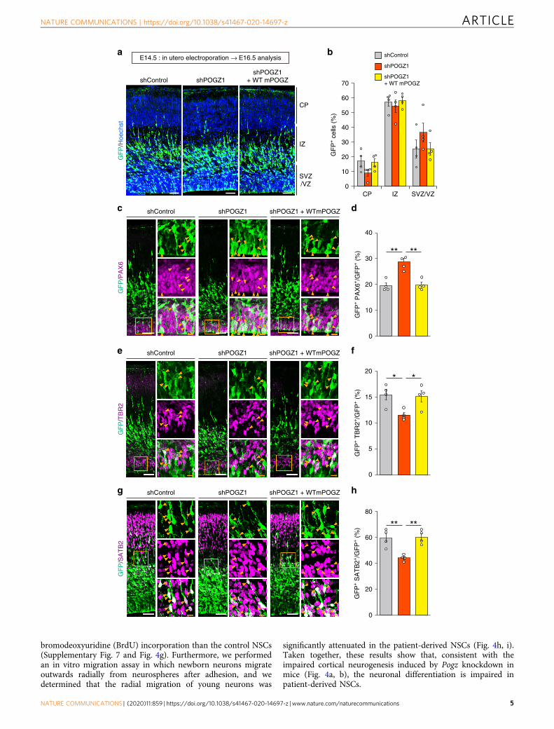
bromodeoxyuridine (BrdU) incorporation than the control NSCs(Supplementary Fig. 7 and Fig. 4g). Furthermore, we performedan in vitro migration assay in which newborn neurons migrateoutwards radially from neurospheres after adhesion, and wedetermined that the radial migration of young neurons was
significantly attenuated in the patient-derived NSCs (Fig. 4h, i).Taken together, these results show that, consistent with theimpaired cortical neurogenesis induced by Pogz knockdown inmice (Fig. 4a, b), the neuronal differentiation is impaired inpatient-derived NSCs.
shControl
CP
IZ
SVZ/VZ
shPOGZ1shPOGZ1
+ WT mPOGZ
CP IZ
GF
P+ c
ells
(%
)
SVZ/VZ
e
h
dc
f
g
a b
GF
P/H
oech
st
shControl shPOGZ1
GF
P/P
AX
6
shPOGZ1 + WTmPOGZ
shControl shPOGZ1
GF
P/T
BR
2
shPOGZ1 + WTmPOGZ
shControl shPOGZ1
GF
P/S
AT
B2
shPOGZ1 + WTmPOGZ
GF
P+ P
AX
6+/G
FP
+ (
%)
GF
P+ S
AT
B2+
/GF
P+ (
%)
GF
P+ T
BR
2+/G
FP
+ (
%)
shControl
shPOGZ1
shPOGZ1+ WT mPOGZ
E14.5 : in utero electroporation → E16.5 analysis
60
50
40
30
20
10
70
0
30
20
15
10
5
20
0
10
40
0
60
40
20
80
0
NATURE COMMUNICATIONS | https://doi.org/10.1038/s41467-020-14697-z ARTICLE
NATURE COMMUNICATIONS | (2020) 11:859 | https://doi.org/10.1038/s41467-020-14697-z | www.nature.com/naturecommunications 5

Generation of POGZWT/Q1038R mice using CRISPR-Cas9 geneediting. To reveal the functional significance of the ASD-relatedQ1042R mutation in brain development and behavioral char-acteristics, we generated POGZWT/Q1038R mice heterozygous forthe Q1038R mutation, corresponding to the human Q1042Rmutation, using CRISPR-Cas9 gene editing (SupplementaryFig. 8a, b). Heterozygous POGZWT/Q1038R mice were born in theratio predicted by Mendelian genetics (Supplementary Fig. 8c),and they exhibited reduced body size and brain in adulthoodcompared to WT mice (Supplementary Fig. 8d–h). We next
measured the thicknesses of cortical layers in the somatosensorycortex and found that the thickness of layers II–IV and V inPOGZWT/Q1038R mice were slightly decreased and increased,respectively (Supplementary Fig. 8i–n). Although POGZWT/
Q1038R mice exhibited decreased brain size, we did not find anydrastic histological abnormalities, such as heterotopias, in thecortex of POGZWT/Q1038R mice. We histologically examinedpatient-related non-neurological abnormalities in adultPOGZWT/Q1038R mice and found that POGZWT/Q1038R mice didnot exhibit any significant changes in peripheral organs, including
Fig. 3 POGZ regulates the neuronal differentiation of mouse cortical neural stem cells. a Slight, non-significant migration defects caused by shRNA-mediated knockdown of Pogz in E16.5 mouse cortices electroporated at E14.5. CP cortical plate; IZ intermediate zone; SVZ subventricular zone; VZventricular zone; E embryonic day. Scale bars, 50 μm. b Quantification of GFP+ cells in each layer (each n= 4). CP cortical plate; IZ intermediate zone; SVZsubventricular zone; VZ ventricular zone. c Increased number of PAX6+ NSCs caused by Pogz knockdown in E16.5 mouse cortices electroporated at E14.5.d Quantification of PAX6+ cells (each n= 4). e Decreased number of TBR2+ differentiated IPs caused by Pogz knockdown in E16.5 mouse corticeselectroporated at E14.5. f Quantification of TBR2+ cells (each n= 4). g Decreased number of SATB2+ differentiated neurons caused by Pogz knockdown inE16.5 mouse cortices electroporated at E14.5. h Quantification of SATB2+ cells (each n= 4). c, e, g Right panels, magnifications of the areas outlined withorange boxes. Arrowheads indicate co-labeled cells. White scale bars, 50 μm; orange scale bars, 10 μm. b Two-way repeated-measures ANOVA withBonferroni–Dunn post hoc tests, F4, 27= 1.861. d, f, h One-way ANOVA with Bonferroni–Dunn post hoc tests; d F2, 9= 18.37; f F2, 9= 5.710; h F2, 9= 11.91.*P < 0.05, **P < 0.01. Data are presented as the mean ± s.e.m.
a b
c d
shControl shPOGZ1shPOGZ1 +WT mPOGZ R1004X E1036K Q1038R E1043X
shPOGZ1 + ASD-related de novo mutated mPOGZ
Upper
CP
Lower
CP
IZ
/SVZ
shControl shPOGZ1shPOGZ1 +WT mPOGZ R1001H F1047L H1080R
shPOGZ1 + Control de novo mutated mPOGZ
UpperCP
LowerCP
IZ/SVZ
GF
P/H
oech
stG
FP
/Hoe
chst
GF
P+ c
ells
(%
)
shControlshPOGZ1shPOGZ1 + WT mPOGZ
shPOGZ1 + R1004X mPOGZshPOGZ1 + E1036K mPOGZshPOGZ1 + Q1038R mPOGZshPOGZ1 + E1043X mPOGZ
GF
P+ c
ells
(%
)
shControlshPOGZ1shPOGZ1 + WT mPOGZ
shPOGZ1 + R1001H mPOGZshPOGZ1 + F1047L mPOGZ
shPOGZ1 + H1080R mPOGZ
Rel
ativ
e ab
sorb
ance
(% o
f con
trol
)
Mig
ratio
n le
ngth
from
neur
osph
ere
(μm
)
MA
P2+
cel
ls (
%)
MA
P2/
Hoe
chst
ControlPatientControl Patient Control Patient
Upper CP Lower CP IZ/SVZ
e f g h i
E14.5 : in utero electroporation → E18.5 analysis
E14.5 : in utero electroporation → E18.5 analysis
25
100
50
150
0
20
15
10
5
0
ControlPatient
400
300
200
100
500
0
ControlPatient
60
40
20
80
0
Upper CP Lower CP IZ/SVZ
60
40
20
80
0
Fig. 4 ASD-related POGZ mutations impair neuronal development. a Forced expression of the ASD-related POGZ mutants failed to rescue the Pogz-knockdown-mediated migration defect in the developing mouse cortex. E embryonic day. Scale bars, 50 μm. b, d Quantification of GFP+ cells in eachlayer (each n=4). CP cortical plate; IZ intermediate zone; SVZ subventricular zone. c Forced expression of the control POGZ mutants rescued the Pogz-knockdown-mediated migration defect. Scale bars, 50 μm. e Low number of MAP2+ differentiated neurons in the patient-derived NSCs harboring theQ1042R POGZ mutation. Scale bars, 100 μm. f Quantification of MAP2+ neurons (each n= 9). g Increased BrdU incorporation in the patient-derived NSCs(each n= 9). h Impaired radial migration of the patient-derived neurons. Scale bars, 50 μm. i Quantification of the migration distance (control, n= 25;patient, n= 20). b, d Two-way repeated-measures ANOVA with Bonferroni–Dunn post hoc tests; b F12, 63= 54.55; d F10, 54= 9.426. f, g, i Student’s t-test.**P < 0.01, ***P < 0.001, ###P < 0.001 (vs. shPOGZ1+WT mPOGZ in b). Data are presented as the mean ± s.e.m.
ARTICLE NATURE COMMUNICATIONS | https://doi.org/10.1038/s41467-020-14697-z
6 NATURE COMMUNICATIONS | (2020) 11:859 | https://doi.org/10.1038/s41467-020-14697-z | www.nature.com/naturecommunications
eye, cochlea, trachea, stomach, duodenum, ileum, cecum andcolon, compared to WT mice (Supplementary Fig. 9). Addition-ally, we did not find diaphragmatic hernia in adult POGZWT/
Q1038R mice. Furthermore, we performed micro-CT scanning ofadult POGZWT/Q1038R mice. We did not find any significantabnormalities in the skull of POGZWT/Q1038R mice (Supple-mentary Fig. 10).
In addition, no homozygous point mutant (POGZQ1038R/Q1038R)offspring were produced by mating male and female POGZWT/Q1038R
mice (0 of 186 pups; Supplementary Fig. 11a). We performed micro-CT scanning of mouse embryos and found that POGZQ1038R/Q1038R
mouse embryos (E15.5) showed a ventricular septal defect, whichlikely results in embryonic lethality (n= 4) (Supplementary Fig. 11b).
Embryonic cortical neuronal development is impaired inPOGZWT/Q1038R mice. We examined embryonic cortical neuronaldevelopment and determined that the density of the SATB2+
neurons (layer II/III) was decreased in the upper layer andincreased in the lower layer in the developing somatosensory cortex(Supplementary Fig. 12a, b), indicating the abnormal distribution ofSATB2+ cortical excitatory neurons in POGZWT/Q1038R mice atE18.5. To confirm the impairment of cortical neuronal developmentin POGZWT/Q1038R mice, we labeled the new-born neurons withBrdU at E14.5. We determined that POGZWT/Q1038R mice exhib-ited a decreased number of SATB2+ BrdU+ cells in the upper layerand an increased number of SATB2+ BrdU+ cells in the lower layerin the developing cortex (Supplementary Fig. 12c, d). These resultssuggest that POGZWT/Q1038R mice exhibit impaired embryoniccortical neuronal development, which is consistent with theimpaired neuronal development in the NSCs derived from the ASDpatient carrying the de novo Q1042R mutation in POGZ (Fig. 4e–i).We also histologically examined the distribution of CUX1+ corticalneurons (layer II/III) and determined that CUX1+ excitatory neu-rons were still abnormally distributed in the adult POGZWT/Q1038R
mice (Supplementary Fig. 12e, f). There were no significant changesin the average density of the CUX1+ neurons (WT, 1126 ± 36.86cells per mm2; POGZWT/Q1038R, 1105 ± 52.64 cells per mm2). Incontrast to excitatory neurons, the average density and distributionof GABA+ interneurons were indistinguishable between WT andPOGZWT/Q1038R mice (Supplementary Fig. 12g, h; the averagedensity of GABA+ interneurons, WT, 121.6 ± 3.710 cells per mm2;POGZWT/Q1038R, 120.9 ± 2.527 cells per mm2).
Transcriptional networks underlying neuronal development isaltered in NSCs derived from both the ASD patient carryingthe Q1042R mutation of POGZ and POGZWT/Q1038R Mice.Given that POGZ interacts with HP1 and CHD435 and is sug-gested to bind DNA, POGZ may modulate the neuronal differ-entiation of NSCs through regulation of gene expression. Toexamine this possibility, we performed RNA-sequencing on NSCsderived from the ASD patient carrying the de novo Q1042Rmutation of POGZ and E16.5 embryonic cortex of POGZWT/
Q1038R mice (significant results are shown in SupplementaryTables 4 and 5). We then analyzed gene ontology (GO) annota-tion of the differentially expressed genes between the unaffectedhealthy control and patient, and WT and POGZWT/Q1038R miceand found that the differentially expressed genes in human andmice were commonly enriched for GO annotations involvingcellular and organismal development, particularly neuronaldevelopment (Supplementary Fig. 13a). In particular, 78 out of913 and 251 genes annotated to neurogenesis (GO: 0022008) inhuman and mouse, respectively, showed commonly differentialexpression between human and mouse (Supplementary Tables 4and 5). Considering that POGZ represses gene transcription in
hematopoietic cells37,38, we focused on the upregulated genes inNSCs derived from the patient and POGZWT/Q1038R mice. Wefound that, among these differentially expressed genes involvingneuronal development, a Notch ligand, Jagged canonical Notchligand 2 (JAG2), was expressed approximately two-fold higher inNSCs derived from both the patient (fold change= 1.970) andPOGZWT/Q1038R mice (fold change= 2.175) compared to eachcorresponding control NSCs (Supplementary Tables 4 and 5). Toinvestigate whether POGZ binds to the Jag2 promoter in NSCs,we performed chromatin immunoprecipitation (ChIP) assaysusing cortical NSCs derived from E16.5 WT mice. We found thatchromosome containing the Jag2 promoter was enriched by anti-POGZ antibodies, suggesting that POGZ binds to the Jag2 pro-moter (Supplementary Fig. 13b, c). Together with the fact thatNotch signaling negatively regulate neuronal differentiation ofNSCs39,40, these results suggest that POGZ may facilitate neu-ronal development by inhibiting gene expression, including JAG2.
POGZWT/Q1038R mice show ASD-related behavioral abnorm-alities. We performed a series of behavioral tests, including open-field, home-cage activity, light/dark transition, Y-maze, fearconditioning, novel object recognition and prepulse inhibition(PPI) tests, in adult POGZWT/Q1038R mice. We observed thatPOGZWT/Q1038R mice showed slightly increased home-cageactivity in the light phase and impaired cognitive function inthe novel object recognition and fear conditioning tests (Sup-plementary Fig. 14a–g). In the open-field test, although thelocomotion of WT and POGZWT/Q1038R mice was indis-tinguishable in quantity, POGZWT/Q1038R mice spent more timein the center zone than their WT littermates did (Fig. 5a, b). Inthe other tests, including the light/dark transition, Y-maze andPPI tests, POGZWT/Q1038R mice showed behavioral featuressimilar to those of their WT littermates (SupplementaryFig. 14h–k; latency for entering into light chamber in the light/dark transition test, WT, 119.4 ± 69.1 s, POGZWT/Q1038R, 47.4 ±13.7 s, P > 0.05, Student’s t-test). We then investigated social andrepetitive behaviors in adult POGZWT/Q1038R mice. In the reci-procal social interaction test, POGZWT/Q1038R mice spent lesstime in sniffing novel mice than their WT littermates did, sug-gesting impaired social interaction (Fig. 5c). In the self-groomingtest, POGZWT/Q1038R mice spent more time in repetitive self-grooming than their WT littermates did, suggesting aberrantrepetitive behavior (Fig. 5d). These data collectively suggest thatadult POGZWT/Q1038R mice show behavioral abnormalitiesassociated with NDDs, including ASD. We next investigatedwhether the behavioral abnormalities of POGZWT/Q1038R miceare present early in development; we determined that juvenilePOGZWT/Q1038R mice spent less time than their WT littermatesin active interactions, such as sniffing, allogrooming, chasing, andplaying, suggesting impaired social interactions in juvenilePOGZWT/Q1038R mice (Fig. 5e). Finally, we assessed the com-munication ability of POGZWT/Q1038R pups at postnatal day 4(P4) by measuring isolation-induced ultrasonic vocalization(USV) responses emitted by mouse pups separated from theirmothers. After separation from their mothers, POGZWT/Q1038R
pups emitted more and longer USV calls than their WT litter-mates did (Fig. 5f, g). POGZWT/Q1038R pups exhibited an alteredcall pattern, such as increased proportions of two-syllable and flatcalls and decreased composite calls and frequency steps, sug-gesting a disturbance in communications from POGZWT/Q1038R
pups to their mothers (Fig. 5h). Thus, POGZWT/Q1038R miceexhibit social deficits even in early development, which mayeffectively reflect the behavioral characteristics of NDDs,including ASD.
NATURE COMMUNICATIONS | https://doi.org/10.1038/s41467-020-14697-z ARTICLE
NATURE COMMUNICATIONS | (2020) 11:859 | https://doi.org/10.1038/s41467-020-14697-z | www.nature.com/naturecommunications 7

The activity of excitatory cortical neurons is increased inPOGZWT/Q1038R mice. Previous studies have suggested that analtered cellular balance of excitation and inhibition (E/I balance)within neural circuitry may cause the social and cognitive deficitsthat characterize ASD41,42. We next focused on whole-brainneuronal activation and suppression patterns during the socialinteraction test in POGZWT/Q1038R mice crossed with Arc-dVenus reporter mice that expressed the destabilized form of thefluorescent protein Venus (dVenus) driven by the promoter ofthe immediate early gene Arc43. Using FAST (block-FAce Serialmicroscopy Tomography), a high-speed serial-sectioning imagingsystem that we previously developed44,45, we examined dVenusexpression in the whole brains of 10-week-old WT and POGZWT/
Q1038R mice in an unbiased manner; we determined that theactivation of excitatory neurons in the anterior cingulate cortex(ACC), which is suggested to be involved in ASD46, was higher inPOGZWT/Q1038R mice than in WT mice after social interaction(Supplementary Fig. 15a–d). Principal component (PC) analysisof the normalized numbers of dVenus+ cells in WT andPOGZWT/Q1038R mice revealed that WT and POGZWT/Q1038R
mice were separated by PC 2 and that the ACC was a majorcontributor to PC2 (Supplementary Fig. 15d). We also observedthat POGZWT/Q1038R mice, in comparison with WT mice,
had an increased density of dendritic spines in the pyramidalneurons in layer II/III of the ACC (Supplementary Fig. 15e, f).We then evaluated excitatory neurotransmission in 10-week-oldPOGZWT/Q1038R mice using whole-cell patch-clamp recordingsfrom pyramidal neurons in layer II/III of the ACC. Whereas therewere no changes in amplitude of miniature excitatory post-synaptic currents (mEPSCs) between WT and POGZWT/Q1038R
neurons, the frequency of mEPSCs was drastically increased inPOGZWT/Q1038R neurons compared to WT neurons (Supple-mentary Fig. 15g–k). Despite the decrease in the number of theexcitatory pyramidal neurons in the upper cortical layer (Sup-plementary Fig. 12e, f), these results suggest that the excitatoryneurons in the cerebral cortex is hyperactivated during the socialinteraction task in POGZWT/Q1038R mice.
Treatment with an anti-epileptic agent, perampanel, improvesthe social deficits in POGZWT/Q1038R mice. Given thatPOGZWT/Q1038R mice showed the elevated activation ofexcitatory neurons after social interaction and abnormally acti-vated excitatory synaptic transmission, we investigated whetherpharmacological inhibition of AMPA-mediated synaptic trans-mission could rescue the impaired social interaction typical ofPOGZWT/Q1038R mice. According to the previous studies, we
0
500
1000
1500
2000
2500
Tim
e in
the
cent
er z
one
(s)
400
300
200
100
0
Dis
tanc
e tr
avel
led
(m)
Sni
ffing
tim
e (s
)
Gro
omin
g tim
e (s
)
Con
tact
ing
time
(s)
Num
ber
of c
alls
Tot
al d
urat
ion
of c
alls
(s)
**
Pro
babi
lity
of e
ach
call
cate
gorie
s (%
)
a b d ec
f g h
WT POGZWT/Q1038R Open field
Social interaction Self-grooming Juvenile playing
USV
400 200
150
100
50
0
300
250
200
350
150
100
50
0
300
200
100
0
400
300
200
100
0
15
20 60
50
40
30
20
10
0H T U D Ch S Cp Fs FICx
10
5
0
Fig. 5 NDDs-related behavioral abnormalities in POGZWT/Q1038R mice. a Distance traveled in the open-field test (each n= 12). b Time spent in thecenter zone in the open-field test (each n= 12). c Time spent sniffing in the reciprocal social interaction test (each n= 13). d Time spent grooming in theself-grooming test (each n= 10). e Time spent contacting in the juvenile playing test (each n= 10). f Numbers of ultrasonic calls made by WT andPOGZWT/Q1038R mice at postnatal day 4 (each n= 19). USV ultrasonic vocalization. g Total duration of ultrasonic calls (each n= 19). h Altered ultrasoniccall patterns in POGZWT/Q1038R mice (WT, n= 17; POGZWT/Q1038R, n= 18) (Cx complex; H harmonics; T two-syllable; U upward; D downward; Chchevron; S shorts; Cp composite; Fs frequency steps; Fl flat). WT wild-type. a–g One-way ANOVA; a F1, 22= 0.277; b F1, 22= 5.771; c F1, 24= 13.02;d F1, 18= 5.914; e F1, 18= 13.62; f F1, 36= 10.64; g F1, 36= 5.465. h Two-way repeated-measures ANOVA with Bonferroni–Dunn post hoc tests,F9, 330= 3.376. *P < 0.05, **P < 0.01. Data are presented as the mean ± s.e.m.
ARTICLE NATURE COMMUNICATIONS | https://doi.org/10.1038/s41467-020-14697-z
8 NATURE COMMUNICATIONS | (2020) 11:859 | https://doi.org/10.1038/s41467-020-14697-z | www.nature.com/naturecommunications

determined the minimum doses of NBQX and perampanel forantiepileptic activity. With these doses, we found that 10 mg/kg ofNBQX did not affect the locomotor activity and that 3 mg/kg ofperampanel tended to slightly decrease the locomotoractivity in the open field, which is not statistically significant(Supplementary Fig. 16a, c). We intraperitoneally administered10 mg/kg of NBQX, a competitive AMPA receptor antagonist, toPOGZWT/Q1038R mice 30 min prior to the reciprocal socialinteraction test and determined that NBQX treatment effectivelyrescued the time spent that POGZWT/Q1038R mice spent sniffingintruder mice without affecting sniffing time in WT mice (Sup-plementary Fig. 16b). We also administered perampanel, anegative allosteric modulator of the AMPA receptor approved bythe European Medicines Agency (EMA), the US Food and DrugAdministration (FDA), and Japanese Pharmaceuticals and Med-ical Devices Agency (PMDA) for epilepsy treatment. Interest-ingly, oral administration of 3 mg/kg of perampanel successfullyrescued sniffing time in POGZWT/Q1038R mice (SupplementaryFig. 16d). These data suggest that the impaired social interactionobserved in POGZWT/Q1038R mice is likely to be caused byhyperactivation of excitatory synaptic transmission.
DiscussionAnalyzing functional mutations in individual putative causativegenes for ASD is important for gaining mechanistic and phar-macological insights into ASD. Recent genetic and epidemiolo-gical studies suggest that the compromising of POGZ function byde novo mutations is likely to be involved in ASD; however, thecontribution of de novo POGZ mutations to ASD onset remainslargely unclear. In silico prediction shows that 17 out of 19missense mutations identified in unaffected controls as well as 6out of 7 NDD-related missense mutations are suggested to bedamaging by at least one out of four predictive tools, namely,PROVEAN, SIFT, PolyPhen2, MutationTaster, CADD score, andThe American College of Medical Genetics and Genomics(ACMG) classification (Table 1); in silico prediction of thepathogenicity of missense mutations is thus challenging andbiologically assessing the pathogenicity of de novo mutations isimportant for understanding the etiology of ASD. Here, weassessed the pathogenicity of de novo POGZ missense mutationsin vitro and in vivo and showed that the ASD-related de novomutants, but not the control de novo mutants identified inunaffected controls, disrupt the nuclear localization of POGZ andembryonic cortical development. We also developed a new mousemodel carrying a de novo POGZ mutation identified in an ASDpatient. In addition to the model mouse, we established iPSC linesfrom the ASD patient with de novo-mutated POGZ. By com-prehensively examining these human and mouse materials, weprovide the first in vivo evidence suggesting that ASD-associatedde novo mutations in a high-confidence ASD gene cause a widerange of aspects of the ASD phenotype.
In this study, using knockdown approaches, we provided thefirst in vivo evidence suggesting that POGZ regulates corticalexcitatory neuron development by promoting neuronal differ-entiation (Figs. 2 and 3). Importantly, we determined that theneuronal developmental gene expression, including Jag2, is sug-gested to be directly regulated by POGZ (Supplementary Fig. 13).Considering that POGZ is suggested to form part of a nuclearcomplex with CHD4 as well as SP1 and HP135,36,47, POGZ islikely to regulate transcriptional networks controlling neuronaldifferentiation through chromatin remodeling.
We determined that the ASD-associated de novo mutations ofPOGZ decreased the nuclear localization of the POGZ protein,impairing its function and compromising cortical excitatoryneuron development (Figs. 1e–h and 4a, b). Post-mortem studies
have found that developmental abnormalities associated withneuronal migration can occur in ASD2; several ASD-associatedgene products, such as CHD8, RELN, CNTNAP2, AUTS2,WDFY3, and TBR1, are differentially involved in excitatoryneuron development2,48. Further studies will be important forelucidating the molecular link between altered excitatory neurondevelopment and ASD phenotypes.
POGZWT/Q1038R mice sufficiently recapitulated the pathogenicabnormalities in patients with NDDs (Fig. 5, SupplementaryFigs. 8, 12). Developmentally, consistent with the observation that13 out of 34 patients with NDDs who carry de novo POGZmutations are diagnosed with microcephaly16,28,30–34,POGZWT/Q1038R mice showed smaller brain size than their WTlittermates (Supplementary Fig. 8e–h). In the adult stage, inaddition to behavioral abnormalities, excitatory neurons in thecerebral cortex were hyperactivated during the social interactiontask in POGZWT/Q1038R mice, leading to altered cellular E/Ibalance within neural circuitry. During development, decreasednumber of excitatory neurons may induce a compensatoryincrease in the activity of excitatory neurons in the adult brain.Given that POGZ is expressed at high levels in the embryonic brainboth in humans (http://hbatlas.org/hbtd/images/wholeBrain/POGZ.pdf http://hbatlas.org/hbtd/images/nctxBrain/POGZ.pdf)and in mice (Fig. 2a–c), the disease-associated deficit in the adultstage may begin prenatally with impaired neuronal developmentnot only in POGZWT/Q1038R mice but also in patients with sporadicASD who carry de novo mutations in POGZ49. The POGZWT/
Q1038R mouse is a good model for studying the links betweenprenatal deficits in cortical excitatory neuron development andclinically relevant abnormalities in adult brain function50,51.
Consistent with the finding that the activity of excitatoryneurons in the cerebral cortex was increased, the social deficitswere rescued by administration of NBQX, a selective AMPAreceptor antagonist in POGZWT/Q1038R mice (SupplementaryFig. 16b). Furthermore, inspired by works showing that ASD andepilepsy have partial clinical and biological overlaps and sharecommon molecular pathogenic mechanisms52,53, we appliedperampanel, an anti-epileptic negative allosteric modulator of theAMPA receptor and determined that the administration of per-ampanel also rescued the social deficits in POGZWT/Q1038R mice(Supplementary Fig. 16d). Together with the fact an excitatoryshift in the cellular E/I balance of the cerebral cortex is suggestedto be associated with social and cognitive deficits54,55, pharma-ceutical modulation of glutamate signaling could be an effectivetherapeutic strategy for ASD patients with POGZ mutation.
In summary, by comprehensively examining these human andmouse materials, we biologically demonstrated POGZ-mediatedregulation of transcriptional networks controlling neuronal differ-entiation and the damaging effects of de novo POGZ mutationsidentified in ASD patients. Importantly, despite the prenatal originof impaired brain development in POGZWT/Q1038R mice, impairedsocial interaction, a core symptom of ASD is pharmacologicallytreatable in adult POGZWT/Q1038R mice. Together with iPSC linesobtained from the patient carrying the Q1042R POGZ mutation,POGZWT/Q1038R mice are versatile tools for developing ther-apeutics for ASD as well as analyzing the molecular pathogenesisof ASD.
MethodsEthics statement. This study was carried out in accordance with the WorldMedical Association’s Declaration of Helsinki and was approved by the ResearchEthics Committee in Osaka University (#28-8-1). All recombinant DNA experi-ments were reviewed and approved by the Gene Modification Experiments SafetyCommittee at Osaka University (#04389). The animal experiments were performedin accordance with the guidelines for animal use issued by the Committee ofAnimal Experiments, Osaka University, Jikei University School of Medicine andRIKEN Tsukuba Branch, and were approved by the Committee in Osaka
NATURE COMMUNICATIONS | https://doi.org/10.1038/s41467-020-14697-z ARTICLE
NATURE COMMUNICATIONS | (2020) 11:859 | https://doi.org/10.1038/s41467-020-14697-z | www.nature.com/naturecommunications 9

University, Jikei University School of Medicine and RIKEN Tsukuba Branch,respectively (Osaka University, #28-1-15; Jikei University School of Medicine,#2017-083; RIKEN Tsukuba Branch, #T2019-007).
Antibodies. The primary antibodies used for immunoblotting were rabbit anti-POGZ (SIGMA-Aldrich, #AV39172, 1:1000), mouse anti-Myc (9E10) (Santa CruzBiotechnology, CA, USA, #sc-40, 1:400), rabbit anti-Lamin A/C (Cell SignalingTechnology, MD, USA, #2032, 1:1000), and mouse anti-α-Tubulin (DM1A)(SIGMA-Aldrich, #T9026, 1:5000); the secondary antibodies used for immuno-blotting were horseradish peroxidase (HRP)-conjugated goat anti-rabbit IgG (SantaCruz Biotechnology, #sc-2004, 1:1000), HRP-conjugated goat anti-mouse IgG(Santa Cruz Biotechnology, #sc-2005, 1:1000), alkaline phosphatase (AP)-con-jugated goat anti-rabbit IgG (Santa Cruz Biotechnology, #sc-2007, 1:1000), and AP-conjugated goat anti-mouse IgG (Santa Cruz Biotechnology, #sc-2008, 1:1000). Theprimary antibodies used for immunostaining were rabbit anti-GFP (MBL, Aichi,Japan, #598, 1:200), chicken anti-GFP (Abcam, Cambridge, UK, #ab13970, 1:500),rabbit anti-PAX6 (BioLegend, CA, USA, #901301, 1:50), rat anti-SOX2 (MolecularProbe, OA, USA, #A-24339, 1:50), rabbit anti-TBR2 (Abcam, #ab23345, 1:50),mouse anti-SATB2 (Abcam, #ab51502, 1:50), rabbit anti-CUX1 (Santa Cruz Bio-technology, #sc-13024, 1:50), rat anti-CTIP2 (Abcam, #ab18465, 1:50), rabbit anti-TBR1 (Proteintech, IL, USA, #20932-1-AP, 1:50), rat anti-BrdU (Abcam, #ab6326,1:40), rabbit anti-MAP2 (Merck Millipore, MA, USA, #AB5622, 1:200), and mouseanti-NESTIN (Merck Millipore, #MAB5326, 1:1000). The secondary antibodiesused for immunostaining were biotinylated goat anti-rabbit IgG (Vector Labs, CA,USA, #BA-1000, 1:200), biotinylated goat anti-mouse IgG (Vector Labs, #BA-9200,1:200), Alexa Fluor 488-conjugated goat anti-rabbit IgG (Life Technologies, CA,USA, #A-11008, 1:200), Alexa Fluor 488-conjugated goat anti-chicken IgY (LifeTechnologies, #A-11039, 1:500), Alexa Fluor 647-conjugated goat anti-rat IgG (LifeTechnologies, #A-21247, 1:200), and Alexa Fluor 594-conjugated donkey anti-mouse IgG (Jackson ImmunoResearch, PA, USA, #715-585-150, 1:250). The pri-mary antibodies used for immunocytochemistry of ASD patient-derived andcontrol iPSC lines were mouse anti-TRA-1-60 (1:1000), mouse anti-TRA-1-81(1:500), rabbit anti-SOX2 (1:800), and rabbit anti-OCT-4A (1:800), which wereincluded in the Stem Light Pluripotency Antibody Kit (Cell Signaling Technology,#9656S). The antibody used for fluorescence in situ hybridization (FISH) wasperoxidase (POD)-conjugated sheep anti-Digoxigenin (DIG; Fab fragments; RocheLife Sciences, Basel, Switzerland, #11207733910, 1:250). The antibodies used forChIP were rabbit anti-POGZ (Bethyl Laboratories, TX, USA, A302-509A), rabbitanti-POGZ (Bethyl Laboratories, A302-510A) and normal rabbit IgG (MerckMillipore, 12-370).
Neuro2a cell culture and transfection. Mouse neuroblastoma Neuro2a cells(ATCC CCL-131) were cultured in Dulbecco’s modified Eagle’s medium (DMEM)supplemented with high glucose, GlutaMAX (Life Technologies), and 10% fetalbovine serum. Neuro2a cells were transfected using GenJet In Vitro TransfectionReagent for Neuro-2A Cells (Ver. II) (SignaGen Laboratories, MD, USA). The cellswere fixed with 4% PFA in PBS for 10 min at room temperature or harvested andlysed with radio-immunoprecipitation assay buffer or a Cytoplasmic & NuclearProtein Extraction Kit (101Bio, CA, USA) 3 days after transfection.
SHSY-5Y cell culture and transfection. Human neuroblastoma SHSY-5Y cells(ATCC CRL-2266) were cultured in DMEM with low glucose (Nissui, Tokyo,Japan) supplemented with 4 mM L-glutamine and 10% fetal bovine serum. SHSY-5Y cells were transfected using Lipofectamine 3000 Reagent (Thermo Fisher Sci-entific, MA, USA). The cells were harvested and lysed with a Cytoplasmic &Nuclear Protein Extraction Kit (101Bio) 2 days after transfection.
Immunocytochemistry. Immunocytochemistry were performed as previouslydescribed56. Briefly, cells were fixed with 4% PFA in PBS for 10 min at roomtemperature. Cells were permeabilized with PBS containing 0.1% Triton X-100(Wako, Osaka, Japan) and incubated with blocking solution containing 4% normalgoat serum (Thermo Fisher Scientific) in PBS for 1 h at room temperature, andthen incubated with the blocking solution combined with primary antibodiesovernight at 4 °C. The following day, the cells were incubated with the blockingsolution combined with fluorescent-dye-conjugated secondary antibody andHoechst 33258 dye (Calbiochem, CA, USA) for 1 h at room temperature56. For theimmunocytochemistry of Neuro2a cells transfected with Myc-tagged mouse POGZ,Alexa Fluor 546-phalloidin (Molecular Probe) were used for staining of F-Actin.Images of the stained cells were acquired using an Olympus FluoView FV1000confocal microscope (Olympus, Tokyo, Japan) and a BZ-9000 microscope (Key-ence, Osaka, Japan). The images were then analyzed with ImageJ software (NIH,MD, USA) and Adobe Photoshop CS (Adobe Systems, CA, USA).
Immunoblotting. Lysates were resolved on 6–7.5% polyacrylamide–SDS gel bySDS–PAGE and transferred to polyvinylidene difluoride membranes56. Subse-quently, these membranes were probed with the indicated primary antibodiesovernight at 4 °C, followed by incubation with the indicated secondary antibodiesfor 1 h at room temperature. Proteins were visualized by AP-reaction using CDPstar (Roche Life Sciences) and HRP-reaction using Western Lightning Plus ECLT
able
1In
silicopred
iction
oftheeffect
ofde
novo
POGZmissensemutations
iden
tified
inpa
tien
tswithNDDsan
dun
affected
controls.
Amino-acid
chan
gePROVEA
NSIFT
PolyP
hen2
MutationT
aster
CADD
score
ACMG
classification
Case
S314N
−0.70(neu
tral)
0.211
(tolerated
)0.000(ben
ign)
0.853
(polym
orph
ism)
17.25
Likely
pathog
enic
ASD
Y59
7C−7.01(deleterious)
0.000(dam
aging)
0.999(probablydamaging)
0.997(disease
causing)
26.60
Likely
pathog
enic
ASD
H641Q
−7.66(deleterious)
0.001(dam
aging)
0.989(probablydamaging)
1.000(disease
causing)
25.30
Likely
pathog
enic
ASD
S799N
−1.81(neu
tral)
0.014
(dam
aging)
0.985(probablydamaging)
0.999(disease
causing)
26.10
Likely
Pathog
enic
ASD
E1040K
−0.94(neu
tral)
0.003(dam
aging)
0.999(probablydamaging)
1.000(disease
causing)
31.00
Pathog
enic
ASD
/ID
Q1042R
−0.92(neu
tral)
0.004(dam
aging)
0.991(probablydamaging)
1.000(disease
causing)
27.20
Pathog
enic
ASD
T1210P
−0.85(neu
tral)
0.038
(dam
aging)
0.077
(ben
ign)
1.000(polym
orph
ism)
13.48
Likely
pathog
enic
Unclassified
NDDs
D23
G−1.75
(neu
tral)
0.001(dam
aging)
0.997(probablydamaging)
1.000(disease
causing)
26.90
Uncertain
sign
ificance
Con
trol
N136S
−0.17(neu
tral)
0.009(dam
aging)
0.118
(ben
ign)
0.899(disease
causing)
21.00
Uncertain
sign
ificance
Con
trol
N159D
−1.39
(neu
tral)
0.004(dam
aging)
0.993(probablydamaging)
0.994(disease
causing)
26.30
Uncertain
sign
ificance
Con
trol
N34
1S−0.47(neu
tral)
0.054
(tolerated
)0.384(ben
ign)
0.923
(polym
orph
ism)
16.72
Uncertain
sign
ificance
Con
trol
R37
4Q
−0.70(neu
tral)
0.01(dam
aging)
0.993(probablydamaging)
0.989(disease
causing)
27.20
Uncertain
sign
ificance
Con
trol
R37
9Q
−1.40(neu
tral)
0.031
(dam
aging)
0.997(probablydamaging)
0.955
(disease
causing)
26.20
Uncertain
sign
ificance
Con
trol
P446L
−1.25
(neu
tral)
0.204(tolerated
)1.000(probablydamaging)
1.000(disease
causing)
24.40
Uncertain
sign
ificance
Con
trol
R50
2K−0.17(neu
tral)
0.641(tolerated
)0.064(ben
ign)
0.866(disease
causing)
22.00
Uncertain
sign
ificance
Con
trol
V59
1I−6.15(deleterious)
0.003(dam
aging)
0.994(probablydamaging)
0.999(disease
causing)
24.20
Uncertain
sign
ificance
Con
trol
N632
S0.21(neu
tral)
0.137
(tolerated
)0.882(possiblydamaging)
0.897(polym
orph
ism)
22.10
Uncertain
sign
ificance
Con
trol
R674
C−2.53
(deleterious)
0.03(dam
aging)
1.000(probablydamaging)
1.000(disease
causing)
32.00
Uncertain
sign
ificance
Con
trol
R79
7Q−1.70
(neu
tral)
0.04(dam
aging)
0.997(probablydamaging)
0.996(disease
causing)
26.40
Uncertain
sign
ificance
Con
trol
D828
N−3.68(deleterious)
0.247(tolerated
)0.835
(possiblydamaging)
1.000(disease
causing)
23.80
Uncertain
sign
ificance
Con
trol
R1005H
−0.75(neu
tral)
0.018
(dam
aging)
0.998(probablydamaging)
0.921
(disease
causing)
28.20
Likely
benign
Con
trol
F1051L
−0.62(N
eutral)
0.452
(tolerated
)0.573
(possiblydamaging)
0.998(disease
causing)
18.31
Likely
benign
Con
trol
H1084R
−1.05(neu
tral)
0.319
(tolerated
)0.666(possiblydamaging)
0.999(disease
causing)
24.90
Likely
benign
Con
trol
L1103H
−0.46(neu
tral)
0.024
(dam
aging)
0.875
(possiblydamaging)
0.573
(polym
orph
ism)
22.30
Uncertain
sign
ificance
Con
trol
A1288V
−0.45(neu
tral)
0.013
(dam
aging)
0.261(ben
ign)
0.967(disease
causing)
23.10
Uncertain
sign
ificance
Con
trol
D1404N
−0.38(neu
tral)
0.001(dam
aging)
0.990(probablydamaging)
0.582(disease
causing)
27.60
Uncertain
sign
ificance
Con
trol
ARTICLE NATURE COMMUNICATIONS | https://doi.org/10.1038/s41467-020-14697-z
10 NATURE COMMUNICATIONS | (2020) 11:859 | https://doi.org/10.1038/s41467-020-14697-z | www.nature.com/naturecommunications

(PerkinElmer, MA, USA). Data acquisition and analysis were performed using anLAS4000 image analyzer (GE Healthcare, NJ, USA). We have provided theuncropped blots in the Source Data file.
Assay for nuclear localization of mutant POGZ. Cytosolic and nuclear fractionsfrom Neuro2a cells expressing Myc-tagged WT or mutant POGZ were preparedusing a Cytoplasmic & Nuclear Protein Extraction Kit (101Bio) according to themanufacturer’s protocol. Those fractions were subjected to immunoblotting withantibodies against Myc, Lamin A/C (a nuclear marker), and α-Tubulin (a cytosolicmarker). The nuclear localization of POGZ was calculated as the ratio of the bandintensity of Myc-POGZ in the nuclear fraction to that of total Myc-POGZ in thecytosolic and nuclear fractions combined.
Reverse transcription and real-time PCR. Total RNAs from cultured cells andtissues were isolated using the PureLink RNA Micro Kit (Thermo Fisher Scientific)and PureLink RNA Mini Kit (Thermo Fisher Scientific) according to the manu-facturer’s instructions. The total RNAs were reverse transcribed with SuperscriptIII (Life Technologies). Real-time PCR was performed with SYBR Premix Ex Taq(Takara Bio Inc., Shiga, Japan) using CFX96 real-time PCR detection system (Bio-Rad Laboratories, CA, USA) as described previously15. The expression levels ofPogz (forward primer sequence: 5′-CCCTACCTATGTGCATTGTTCTC-3′;reverse primer sequence: 5′-TCCGTGGAACATGATTGTTG-3′) were normalizedto those of Gapdh and were determined according to the 2−ΔΔCt method.
Immunohistochemistry. E16.5, E17.5, and E18.5 mouse brains were fixed with 4%paraformaldehyde (PFA) in PBS overnight at 4 °C. The brains were sectioned at a20 μm thickness by using a cryostat (Leica, Wetzlar, Germany, CM1520). Brainsfrom 10-week-old adult male mice were perfused with 4% PFA in PBS and post-fixed with 4% PFA in PBS overnight at 4 °C. The brains were sectioned at a 20 μmthickness by using a cryostat (Leica) for CUX1 staining after antigen retrievalmethods or sectioned at a 50 μm thickness with a LinearSlicer PRO7N (DOSAKAEM CO.,LTD., Kyoto, Japan) for GABA staining. The brain slices were permea-bilized with blocking solution containing 0.25% Triton X-100 (Wako), 1% normalgoat serum (Thermo Fisher Scientific), and 1% bovine serum albumin (SIGMA-Aldrich) in PBS for 1 h at room temperature, and then incubated with the blockingsolution combined with primary antibodies. The following day, the slices wereincubated with the blocking solution combined with biotin-dye or fluorescent-dye-conjugated secondary antibody and Hoechst 33258 dye (Calbiochem) for 1 h atroom temperature. The biotinylated secondary antibody was labeled with TexasRed-conjugated streptavidin (Vector Labs, #SA-5006). Three coronal sections perbrain were imaged for quantification. Images of the stained brain slices wereacquired using an Olympus FluoView FV1000 confocal microscope (Olympus) anda BZ-9000 microscope (Keyence). The images were then analyzed with ImageJsoftware (NIH) and Adobe Photoshop CS (Adobe Systems). The distributions ofSATB2+ cells in E18.5 embryo brains and of CUX1+ cells and GABA+ cells in 10-week-old mice brains were quantified by dividing the cerebral wall into 10 equalbins (cortical plate (CP) 1 to intermediate zone (IZ)/SVZ 10).
Fluorescence in situ hybridization. For fresh samples, E16.5 mouse embryos wererapidly frozen using dry ice. The samples were cut at a thickness of 20 μm by usinga cryostat (Leica) and collected on Matsunami adhesive silane (MAS)-coated glassslides (Matsunami Glass Ind., Ltd, Osaka, Japan). A cRNA probe sequence tar-geting Pogz (NCBI Reference Sequence: NM_172683.3) from base 1062 to base1563 was amplified from mPOGZ cDNA (DNAFORM; clone ID: 30745658) viaPCR and subcloned into a pBluescript II KS (+/−) vector. A 5′-digoxigenin (DIG)-labeled probe for Pogz was prepared by transcribing the BamHI-linearized plasmidusing T3 RNA polymerase (Roche Life Sciences). FISH for Pogz was performed asfollows. The sections were fixed with 4% PFA in PBS for 30 min at room tem-perature. The slices were incubated with hybridization buffer containing the probe,and hybridization was allowed to take place overnight at 48 °C. On day 2, the sliceswere washed and incubated with 1% blocking buffer (Roche Life Sciences) con-taining anti-DIG-POD antibody at 4 °C overnight. On day 3, the anti-DIG-PODantibody was labeled with fluorescein using TSA Plus Fluorescein System (Perki-nElmer). Immunostaining for each cell marker, including PAX6, SOX2, and TBR2,was performed as described above after FISH for Pogz at day 3. Images of thestained brain slices were acquired using an Olympus FluoView FV1000 confocalmicroscope (Olympus) and a BZ-9000 microscope (Keyence). The images werethen analyzed with ImageJ software (NIH) and Adobe Photoshop CS (AdobeSystems).
In utero electroporation. In utero electroporation was performed on E14.5embryos from timed-pregnant WT ICR mice (SLC, Shizuoka, Japan)57. Thepregnant mice were anesthetized by intraperitoneal injections with a solutioncontaining 0.3 mg medetomidine (Domitor, Zenoaq Nippon Zenyaku Kogyo,Fukushima, Japan), 4 mg midazolam (Dormicum, Astellas Pharma Inc., Tokyo,Japan), and 5 mg butorphanol (Bettlefar, MP AGRO Co., Ltd., Hokkaido, Japan)per kg. The uterine horns were exposed and the plasmid (2.5 μg/μL) mixed withFast Green (0.1 mg/mL, SIGMA-Aldrich) were injected into the lateral ventricles.For the knockdown of Pogz and overexpression of WT-POGZ or POGZ mutants,
MISSION shRNA constructs or miR30-based shRNA constructs (1 μg/μL) andpcDNA3 expression constructs encoding WT-POGZ or POGZ mutants (1 μg/μL)were injected into the lateral ventricles together with a pCAG-GFP vector (0.5 μg/μL) expressing GFP at a 2:2:1 ratio. For the overexpression of WT-POGZ or POGZmutants, pcDNA3 expression constructs encoding WT-POGZ or POGZ mutants(2 μg/μL) were injected into the lateral ventricles together with a pCAG-GFP vector(0.5 μg/μL) expressing GFP at a 4:1 ratio. Subsequently, electric pulses (35 V, 4cycles; 50 ms on, 950 ms off) were applied to the head of the embryos targeting thedorsal-medial cortex using Square Wave Electroporator (NEPA GENE, Chiba,Japan, CUY21SC). Each pool of constructs was injected into three or four differentembryos in at least two independent operations. The embryos were harvested 48 or96 h later. Three non-adjacent coronal sections per brain were imaged for quan-tification. The images were acquired with an Olympus FluoView FV1000 (Olym-pus) confocal microscope and analyzed with ImageJ software (NIH) and AdobePhotoshop CS (Adobe Systems). In order to analyze the migration of GFP+ cells,embryos were dissected at E16.5, E17.5, or E18.5, and brain samples from theembryos were immunostained for GFP. Hoechst 33258 dye (Calbiochem) was usedto stain the nuclei. The distribution of the GFP+ cells was quantified by dividingthe cerebral wall into the CP, IZ, and SVZ/VZ at E16.5 or the upper CP, lower CP,and IZ/SVZ at E18.5. In order to assess the neuronal differentiation of GFP+ cells,the percentages of GFP+ PAX6+, GFP+TBR2+, and GFP+ SATB2+ cells wasquantified.
MISSION shRNA, miR30-based shRNA and POGZ expression constructs. TheMISSION shRNA TRC1 vectors (Supplementary Table 2) used in this study werepurchased from Sigma-Aldrich (MO, USA). The sequence of shPOGZmiR30
(Supplementary Table 3) was designed using an online design tool (http://cancan.cshl.edu/RNAi_central/RNAi.cgi?type= shRNA). The template oligonucleotidecarrying the shPOGZmiR30 sequence was obtained from Eurofins Genomics(Tokyo, Japan), amplified via PCR and subcloned into a pCAG-miR30 vector. Inorder to generate a plasmid expressing mPOGZ, mPOGZ cDNA (DNAFORM,Kanagawa, Japan, clone ID: 30745658) was amplified via PCR and subcloned into apcDNA-6Myc vector. Plasmids expressing mPOGZ mutants were generated usinga KOD mutagenesis kit (Toyobo, Osaka, Japan) according to the manufacturer’sprotocol.
Subjects. A patient with ASD was recruited from outpatient services at OsakaUniversity Hospital. She had been diagnosed by at least two trained child psy-chiatrists according to the criteria in the Diagnostic and Statistical Manual ofMental Disorders, fourth edition, text revision (DSM-IV-TR)58 based on interviewswith the patient and unstructured or semi-structured observations of her behavior.During the interview, the pervasive developmental disorders Autism Society JapanRating Scale (PARS)59 and the Japanese version of the Asperger’s Questionnaire(AQ)60 were used to assist in the evaluation of ASD-specific behaviors andsymptoms. The PARS is a semi-structured interview that is composed of 57questions in eight domains corresponding to the characteristics of children withpervasive developmental disorders (PDDs), which was developed by the AutismSociety Japan. The clinicians who diagnosed the subject were trained in the use ofthe PARS. Intelligence quotient data were collected using the full-scale WechslerAdult Intelligence Scale (WAIS)-III61. Written informed consent was obtainedfrom the subject after the procedures were fully explained.
P1381: The patient was a 16-year-old Japanese female. She met the criteria forPDD-Not Otherwise Specified (NOS), which refers to ASD. The patient had nophysical disease. There was no specific abnormality in the results of her brain MRIor blood test. The patient’s PARS and AQ scores were both above the cut-off point.Her scores on the WAIS-III were as follows: Full-scale IQ, 65; Verbal IQ, 70;Performance IQ, 65; Verbal Comprehension Index, 73; Working Memory Index,90; Perceptual Organization Index, 65; and Processing Speed Index, 78. A delay inthe patient’s communication skills had been noted at a check-up when she was18 months old. She could not communicate well with others of similar age andscreeched if she was forced to go to school. She had grammatical languageimpairment, specifically in subject–predicate relations, and had sound sensitivity aswell. She persisted in asking about specific subjects or activities that sheinterested her.
P1399: The subject was a 49-year-old Japanese male, the father of the patient.
Generation of iPSC lines. iPSC lines were generated using immortalized B cellsobtained from the ASD patient carrying Q1042R POGZ mutation and from herunaffected father62. Plasmid vectors for induction of pluripotency, including0.63 μg pCE-hOCT3-4, 0.63 μg pCE-hSK, 0.63 μg pCE-hUL, 0.63 μg pCE-mp53DD, and 0.50 μg pCXB-EBNA1 (Addgene, MA, USA) were electropoartedinto the immortalized B cells using the Nucleofector 2b Device (Lonza, Basel,Switzerland) with the Amaxa Human T-cell Nucleofector Kit (Lonza). Theimmortalized B cells introduced with the reprogramming factors were cultivated inRoswell Park memorial Institute (RPMI) medium (Wako) containing 10% fetalbovine serum for 1 week. Subsequently, the electroporated immortalized B cellswere seeded onto mitomycin C-treated mouse SL10 feeder cells (ReproCELL Inc.,Kanagawa, Japan) and cultured for 20–30 days in DMEM/F12 (Thermo FisherScientific) containing 20% KnockOut Serum Replacement (Thermo Fisher
NATURE COMMUNICATIONS | https://doi.org/10.1038/s41467-020-14697-z ARTICLE
NATURE COMMUNICATIONS | (2020) 11:859 | https://doi.org/10.1038/s41467-020-14697-z | www.nature.com/naturecommunications 11
Scientific), 1% non-essential amino acid solution (SIGMA-Aldrich), 2 mM L-glu-tamine (Thermo Fisher Scientific), 0.1 mM 2-mercaptoethanol (Wako), and 4 ng/mL bFGF (PeproTech, NJ, USA). Colonies of cells similar to human embryonicstem (ES) cells were clonally isolated, morphologically selected, subjected to PCR-based analysis of episomal vector loss and evaluated for expression of pluripotentmarkers using immunocytochemistry (Oct-4A, Sox2, TRA-1-60, and TRA-1-81).
Neural induction of iPSC lines and expansion of NSCs. The neural induction ofiPSC lines into NSCs was performed with PSC Neural Induction Medium (ThermoFisher Scientific) according to the manufacturer’s instructions. For neural induc-tion, iPSC lines were cultured in Essential 8 medium (Thermo Fisher Scientific)under feeder-free conditions on Matrigel (Corning, NY, USA). On day 0 of neuralinduction, ~24 h after the cells were split, the culture medium was replaced withPSC neural induction medium containing neurobasal medium and PSC neuralinduction supplement. The neural induction medium was exchanged every otherday from day 0 to day 4 of neural induction and every day after day 4 of neuralinduction. On day 7 of neural induction, NSCs (P0) were harvested and expandedin neural expansion medium containing 50% neurobasal medium (Thermo FisherScientific), 50% advanced DMEM/F12 (Thermo Fisher Scientific), and neuralinduction supplement (Thermo Fisher Scientific) on Matrigel. Expanded NSCsafter passage 6 were used for subsequent assays.
Neuronal differentiation assay of NSCs derived from iPSC lines. ASD-patient-derived and control NSCs were seeded in Brain Phys basal medium (NacalaiTesque, Kyoto, Japan)-based neuronal differentiation medium containing 1%N2 supplement (Wako), 2% B27 supplement (Thermo Fischer Scientific), 200 μML-ascorbic acid (Sigma-Aldrich), 1 mM dBcAMP (Wako), 20 ng/mL human brain-derived neurotrophic factor (BDNF; R&D Systems, MN, USA), 20 ng/mL humanglial-cell-line-derived neurotrophic factor (GDNF; R&D Systems), 500 ng/mLmouse laminin (Thermo Fisher Scientific), and 1 μM (3,5-difluorophenylacetyl)-L-alanyl-L-2-phenylglycine tert-butyl ester (Wako) on a 24-well plate coated withpoly-L-ornithine (Sigma-Aldrich), 6.67 μg/mL human fibronectin (Thermo FisherScientific), and 6.67 μg/mL mouse laminin (Thermo Fisher Scientific). On day 2,which was in an early stage of neuronal differentiation, the cells were fixed with 4%PFA for 15 min at room temperature. Immunocytochemistry was performed asdescribed previously56. Briefly, the cells were permeabilized with 0.1% Triton X-100 (Wako) in PBS for 10 min and incubated with blocking solution containing 1%normal goat serum in PBS for 1 h at room temperature. Then, the cells wereincubated with the blocking solution combined with primary anti-MAP2 antibodyovernight at 4 °C. The following day, the cells were incubated with secondary AlexaFluor 488 conjugated antibody for 1 h at room temperature. The images wereacquired using a ToxInsight automated microscope (Thermo Fisher Scientific). Theproportion of MAP2+ neurons was automatically analyzed using the samemicroscope.
BrdU ELISA. ASD-patient-derived and control NSCs were seeded in neuralexpansion medium. The following day, the culture medium was replaced withneural expansion medium containing 10 μM BrdU. BrdU+ proliferating cells werelabeled with POD-conjugated anti-BrdU antibody using a Cell Proliferation ELISABrdU Kit (Roche Life Sciences) 3 h after the addition of the BrdU. The absorbancewas measured using an iMARK Microplate Reader (Bio-Rad Laboratories) 15 minafter treatment with a substrate of POD (3,3′5,5′-Tetramethylbenzidine).
Proliferation and migration assays of neurospheres. ASD-patient-derived andcontrol NSC suspension in DMEM/F-12 (Sigma-Aldrich) containing 15 mMHEPES buffer (Sigma-Aldrich), 2% B27 supplement (Thermo Fisher Scientific),20 ng/mL human epidermal growth factor (EGF; PeproTech), 20 ng/mL humanbasic fibroblast growth factor (bFGF; PeproTech), 10 ng/mL human leukemiainhibitory factor (LIF; Merck Millipore), and 0.1% heparin sodium solution1000 IU/5 mL (Nippon Zenyaku Kogyo, Fukushima, Japan) were placed into a96-well V-bottom plate. The NSCs proliferated to form neurospheres in suspensionculture. In order to analyze the proliferation of NSCs, the diameter of neurospherescultured for 5 days was measured. For the in vitro migration assay, neurospherescultured for 3 days were transplanted into neuronal differentiation medium on asix-well plate coated with poly-L-ornithine (Sigma-Aldrich), 6.67 μg/mL humanfibronectin (Thermo Fisher Scientific), and 6.67 μg/mL mouse laminin (ThermoFisher Scientific). On day 2 of neuronal differentiation, cell migration was eval-uated by measuring the distance from the edge of the neurosphere to the nucleus ofthe most distant cell.
Generation of POGZWT/Q1038R mice. The POGZWT/Q1038R mouse strain wasgenerated using a genome-editing technique as described previously63 with somemodifications. The sequence 5′-GCAGCAGCTCCCTGTAAATG-3′ was selectedas the target for single guide RNA (sgRNA). Cas9 mRNA and sgRNA were pro-duced using linearized T7-NLS hCas9-pA (RIKEN BioResource Research Center(BRC) #RDB13130, a gift from Tomoji Mashimo)64 and DR274 (Addgene #42250,a gift from Keith Joung)65, respectively. A 117-nt single-stranded oligodeox-ynucleotide (ssODN) donor coding the POGZ Q1038R mutation was ordered asUltramer DNA oligos from Integrated DNA Technologies (Skokie, IL, USA). The
Cas9 mRNA, sgRNA, and ssODN were dissolved in nuclease-free water andmicroinjected into the cytoplasm of pronuclear-stage mouse zygotes obtained fromC57BL/6NJcl mice (CLEA Japan, Tokyo, Japan). The genotype was determined byPCR analysis using genomic DNA derived from the mice with the mouse WT Pogz(forward primer sequence: 5′-TCTGTGAAGAAGCTTCGGGTAGTAC-3′; reverseprimer sequence: 5′-GTCTCCTCATTTACAGGGAGCT-3′); and mouse Q1038RPogz (forward primer sequence: 5′-GCAGCGGCTCCCCGTTAAC-3′; reverseprimer sequence: 5′-AGCGCACAGCCCACTCATAG-3′). POGZWT/Q1038Rmice will be available from BRC as strain number RBRC09544.
Hematoxylin–eosin (H&E) staining. Brains from 10-week-old adult male micewere perfused with 4% PFA in PBS and post-fixed with 4% PFA in PBS overnightat 4 °C. The brains were sectioned at a 50 μm thickness. The brain slices at Bregma0 mm were stained by HE staining using Mayer’s Hemalum Solution (MerckMillipore) and Eosin Y-solution 0.5% aqueous (Merck Millipore) according to themanufacturer’s instructions. Peripheral organs, including eye, cochlea, trachea,stomach, duodenum, ileum, cecum, and colon, from 10-week-old adult male micewere perfused with 10% formalin neutral buffer solution (Wako). HE staining forthe slices of the peripheral organs from WT and POGZWT/Q1038R mice was per-formed by New Histo. Science Laboratory Co., Ltd.
Micro-CT analysis. Mouse skulls and embryos were scanned using a micro-CTscanner (ScanXmate-E090S, Comscantecno Co., Ltd., Kanagawa, Japan). For skullanalysis, the head region of 22-week-old male mice were scanned at a tube voltageof 80 kV and a tube current of 50 μA. We used TRI/3D-BON (RATOC Systems,Osaka, Japan) and Image J software (NIH) for reconstructing the three-dimensional images of skulls and measuring the linear distances between land-marks, respectively. Before embryo scanning, E15.5 mouse embryos were fixed withBouin solution (Wako) at room temperature, stored in 70% ethanol and thentreated with 1% phosphotungstic acid. These samples were scanned at a tubevoltage of 40 kV and a tube current of 100 μA. Section images were analyzed usingthe OsiriX (www.osirix-viewer.com) software.
BrdU labeling in POGZWT/Q1038R mice. New born neurons were labeled withBrdU at E14.5 by intraperitoneal administration of BrdU to pregnant female miceat a dosage of 50 mg/kg. In order to analyze the migration of BrdU+ cells, embryoswere dissected at E18.5 and brain samples from the embryos were immunostainedfor BrdU and SATB2. Three non-adjacent coronal sections per brain were imagedfor quantification. The images were acquired with an Olympus FluoView FV1000(Olympus) confocal microscope and analyzed with ImageJ software (NIH) andAdobe Photoshop CS (Adobe Systems). The distribution of the BrdU+ SATB2+
cells was quantified by dividing the cerebral wall into 10 equal bins (CP 1–IZ/SVZ 10).
Mouse cortical NSCs cultures. The suspension of cortical cells prepared fromE16.5 embryonic mouse cortex in NSCs expansion medium containing DMEM/F-12 (Thermo Fisher Scientific), 1% N2 supplement (Wako), 20 ng/mL EGF(PeproTech), and 20 ng/mL bFGF (PeproTech) was placed into a six-well plate.The NSCs were expanded to form primary neurospheres in suspension culture fora week. The neurospheres were dissociated into single cells using trypsin (ThermoFisher Scientific) and seeded in the NSCs expansion medium on Matrigel. Thesecondary NSCs were expanded in adhesion. When the cells reached 80% con-fluency 2 days after plating, the cells were harvested and used in the followingexperiments.
RNA sequence. RNA sequence and the following analysis of the aligned reads wasperformed as described previously62. Total RNAs isolated from NSCs differentiatedfrom the iPSC lines and mouse cortical secondary NSCs were sequenced using theIllumina HiSeq2000 system (BGI, Beijing, China) and the IlluminaHiSeq2500 system (Genome Information Research Center, Osaka University,Osaka, Japan), respectively. The expression levels of each gene were analyzed basedon Fragments Per Kilobase of exon per Million mapped (FPKM).
GO and pathway analysis. The ToppGene Suite (https://toppgene.cchmc.org/)was used for GO annotation-based functional classification of differentiallyexpressed genes. The genes with |fold change| ≥ 1.2 were included for the analysisof Biological Process (gene in annotation: 1000 ≤ n ≤ 100,000). The GO annotationswere cut off at P < 0.05 and FDR < 0.05.
ChIP assay. Chromatin was isolated from secondary NSCs derived from E16.5cortex of WT mice. ChIP was performed using ChIP-IT Express EnzymaticMagnetic ChIP Kit & Enzymatic Shearing Kit (Active Motif, CA, USA) accordingto the manufacturer’s instructions. Briefly, NSCs at 80% confluency on 15 cm dishwere crosslinked in DMEM/F12 containing 1% formaldehyde. Following nuclearextraction, chromatin was sheared by enzyme for 30 min at 37 °C. The shearedchromatin was immunoprecipitated with the antibody in a solution containingprotein-G magnetic beads overnight at 4 °C. The immunoprecipitated chromatinwas eluted from the beads and the crosslinking was reversed. Following ChIP, the
ARTICLE NATURE COMMUNICATIONS | https://doi.org/10.1038/s41467-020-14697-z
12 NATURE COMMUNICATIONS | (2020) 11:859 | https://doi.org/10.1038/s41467-020-14697-z | www.nature.com/naturecommunications

DNA samples were purified using Chromatin IP DNA Purification Kit (ActiveMotif) and amplified via PCR using GenoMatrix Whole Genome Amplification Kit(Active Motif). Quantified PCR (qPCR) was performed using the amplified DNAwith the primers for the mouse Jag2 promoter region (forward primer 1 sequence:5′-GGATGGCCTGATTGTGTGT-3′; reverse primer 1 sequence: 5′-TTCGGAG-GAGGGGTCTTC-3′; forward primer 2 sequence: 5′-AGATGGGAAGACCCCTCCT-3′; reverse primer 2 sequence: 5′-CAGTGCCACAGAGGGTTACA-3′; for-ward primer 3 sequence: 5′-GTCATGGGGATCCAGCTTT-3′; reverse primer3 sequence: 5′-TCCCAGGCCTTTATACCACA-3′)66,67. The levels of enrichmentof each amplicon by ChIP were normalized to the amount of amplified DNAfragments immunoprecipitated with each antibody.
Behavioral tests. All behavioral tests were carried out on male C57BL/6NJcl miceat 1.5–4 months of age, except the ultrasonic vocalization test and the juvenile playtest. All behavioral experiments were performed during the light period byexperimenters who were blind to the genotypes and treatments of the mice.
Home cage activity: Each mouse was placed in a test cage under a 12 h:12 hlight–dark cycle (light on at 8:00). After 1 day of habituation, spontaneous activityin the light and dark phases, total activity and the ratio of activity in the light phaseto total activity were measured for 5 days using an infrared activity sensor (O’Hara& Co., Ltd., Tokyo, Japan)68.
Fear conditioning test: The fear-conditioning test was performed using anImage FZ4 (O’Hara & Co., Ltd.), automated fear contextual and tone-dependentfear conditioning apparatus68. On the training day (day 1), each mouse was placedinto a shock chamber (Box A; 10 × 10 × 10 cm, white polyvinyl chloride boards,stainless steel rod floor, O’Hara & Co., Ltd.) for 120 s, immediately followed by thepresentation of four tone–shock pairs at 90 s intervals. Each tone–shock pairincluded a tone (70 dB, 10 kHz) for 30 sc and a 0.5 mA foot shock lasting for thefinal 0.5 s of the tone. On day 2, in order to quantify contextual freezing, eachmouse was placed back in box A for 6 min of measurement. On day 3, each mousewas placed in a white transparent chamber (Box B) for 120 s, and then the 30 s tonewas presented four times at 90 s intervals. Freezing during the first 120 s wasmeasured as pre-tone freezing, and freezing during the tone presentations wasmeasured as cued freezing.
Novel object recognition test: Prior to each session, mice were acclimatized tothe test room for at least 30 min. After 10 min of habituation to the experimentalbox under dim lighting conditions (10–20 lx) for 3 consecutive days, the test mousewas allowed to freely explore two novel objects (A and B) in the box for 10 min.Twenty-four hours after the training session, the retention session was conducted.In the retention session, object B was replaced with novel object C, and the mousewas allowed to move freely for 10 min in the same box. The exploration time foreach object was measured. The discrimination index (%) was the differencebetween the exploration time for the novel object and that for the familiar objectdivided by total exploration time. This index was used to calculate values forrecognition memory. This test was conducted between 10:00 and 14:00.
Open field test: Locomotor activity was measured using the open field. Eachmouse was placed in the center of the open-field apparatus (45 × 45 × 30 cm). Thetotal distance traveled and time spent in the center area (25 × 25 cm) wererecorded. Data were collected for 90 min per mouse.
Light/dark transition test: The light/dark transition test was performed using anapparatus that consisted of two sections of equal size (20 × 20 × 25 cm, O’Hara &Co., Ltd.)68. The illumination was 353 lx in the light chamber and 0.1 lx in the darkchamber. Each mouse was placed in the middle of the light chamber and allowed tomove freely. The total distance traveled, latency to enter the light chamber, timespent in the light chamber and number of transitions between chambers wereautomatically measured using Image LD4 (O’Hara & Co., Ltd.) for 10 min.
Y-maze test: Each mouse was placed on the center of an apparatus consisting ofthree arms (arm length: 40 cm, arm bottom width: 3 cm, arm upper width: 10 cm,height of wall: 12 cm, O’Hara & Co., Ltd.)68. The distance traveled and thealteration ratio were measured.
PPI test: Each mouse was habituated to a sound-proof box (33 × 43 × 33 cm,O’Hara & Co., Ltd.) with 65 dB background noise for 5 min. In order to acclimatizemice to the startle pulse, 110 dB per 40 ms of white noise was presented during the5 min habituation period, and the startle response to this white-noise stimulus wasexcluded from the statistical analysis. A prepulse sound of 70, 75, 80, and 85 dB waspresented for 20 ms, followed by the presentation of 110 dB for 50 ms. Theseprepulse and startle sounds were presented 10 times in pseudorandom order.Startle amplitude was measured 50 ms after presentation of the prepulsesound. The percentage of PPI was calculated as [(startle amplitude withoutprepulse)−(startle amplitude of trial with prepulse)]/(startle amplitude withoutprepulse) × 10068.
Reciprocal social interaction test: A male intruder mouse was placed in the testcage after habituation of the test mouse to the same cage for 60 min. Over the fullexperimental period (20 min), the total duration of time that the resident mousespent sniffing the intruder mouse was measured. The drug administration wasperformed 30 min after the beginning of habituation. This test was carried outbetween 10:00 and 14:00.
Self-grooming test: Prior to the self-grooming test, mice were acclimatized tothe testing room for 60 min. This test was conducted in a new mouse cage without
bedding. The cumulative time that the test mouse spent grooming itself wasmeasured for 10 min following 10 min period of habituation to the test cage.
Juvenile playing test: The juvenile playing test was carried out on juvenilemale mice at 3 weeks of age, before weaning. Each test mouse was placed alone inthe test cage for a 60 min habituation period. A male non-sibling intruder mousefrom a different litter but similar in body weight to the test mouse (±1.0 g) wasplaced in the test cage after habituation. Over the full experimental period(10 min), the total durations of various behavioral events including sniffing,allogrooming, chasing, and playing were measured. This test was carried outbetween 10:00 and 14:00.
USV test: On postnatal day 4, the pups and their mothers were acclimatized tothe test room for at least 30 min. Each male pup was removed from the home cagecontaining the mother and littermates and placed in a clean glass 500 mL Pyrexbeaker with paper-towel bedding. The USVs of each pup were detected for 3 minusing an UltraSoundGateCM16/CMPA microphone (Avisoft Bioacoustics,Glienicke, Germany) in a sound attenuation chamber under light control (90 lx),and recorded with Avisoft-SASLab Pro software (Avisoft Bioacoustics). Themicrophone was placed in the box ~20 cm above the pup, and USVs between 40and 150 kHz were measured. The waveform patterns of the calls were analyzed byclassifying the calls into 10 distinct categories, based on internal pitch changes,lengths, and shapes according to a previous report69.
FAST whole-brain imaging. Ten-week-old adult male WT- and POGZWT/Q1038R-Arc-dVenus mice were perfused with 4% PFA in PBS 5 h after the reciprocal socialinteraction test, at the peak of the dVenus expression driven by Arc promoter. Themouse brain was embedded in 4% agarose gel (Nacalai Tesque) dissolved in PBS.Subsequently, serial whole-brain imaging was performed using FAST44,45. TheFAST whole-brain images were obtained at a resolution of 1.0 × 1.0 × 5 μm3 withan sCMOS camera (Andor Technology, Belfast, UK) with a 2 × 2 binning mode, a×16 NA 0.8 objective lens (Nikon instruments, Tokyo, Japan), and a ×0.83 inter-mediate magnification lens (Yokogawa Electronic, Tokyo, Japan). Semiautomaticanatomical parcellation of the brain and quantification of dVenus-positive neuronsin each brain region were performed using TRI/FCS-NUC64 software (TatocSystem Engineering, Tokyo, Japan).
Principal component analysis (PCA). PCA was performed in R (https://www.r-project.org/) using the preinstalled prcomp function. The data were normalizedbefore PCA by dividing the number of dVenus-positive cells in each brain regionby the total number of dVenus-positive cells in all brain regions.
Golgi impregnation and dendritic spine analysis. Golgi impregnation was per-formed on 10-week-old WT and POGZWT/Q1038R mice with the FD Rapid Gol-giStain Kit (FD Neuro Technologies, MD, USA), according to the manufacturer’sprotocol. Fully focused images were obtained using a BZ-9000 microscope (Key-ence) and quantitatively analyzed using ImageJ software (NIH).
Electrophysiology. WT and POGZWT/Q1038R mice (10–11 week-old, male) weredecapitated under isoflurane anesthesia (5% in 100% O2) and the brains werequickly removed and secured on the cutting stage of a vibrating blade slicer(VT1200S, Leica). Coronal slices (300 μm thick) containing the ACC were cut in anice-cold cutting solution composed of (in mM) 2.5 KCl, 0.5 CaCl2, 10 MgSO4, 1.25NaH2PO4, 2 thiourea, 3 sodium pyruvate, 92 N-methyl-D-glucamine, 20 HEPES, 12N-acetyl-L-cysteine, 25 D-glucose, 5 L-ascorbic acid and 30 NaHCO3 equilibratedwith 95% O2+ 5% CO2 (pH ~ 7.4; osmolality, ~280 mOsm/kg) and incubated inthe cutting solution at 34 °C for 15–20 min. The slices were then kept at roomtemperature (20–25 °C) in the standard artificial cerebrospinal fluid (ACSF)composed of (in mM) 125 NaCl, 3 KCl, 2 CaCl2, 1.3 MgCl2, 1.25 NaH2PO4, 10 D-glucose, 0.4 L-ascorbic acid, and 25 NaHCO3 (pH 7.4 bubbled with 95% O2+ 5%CO2; osmolality, ~310 mOsm/kg) until the electrophysiological recording. Eachslice was transferred to a recording chamber (~0.4 mL volume) and fixed withnylon grids attached to a platinum frame. The slice was submerged and con-tinuously superfused at a rate of 1.5–2.5 mL/min with the standard ACSF at 30–32°C. Whole cell membrane current was recorded from the pyramidal neurons in thelayer II/II of the ACC visually identified under an upright microscope (BX-51WI,Olympus) with oblique illumination. Patch-clamp electrodes (4–6MΩ) were madefrom borosilicate glass pipettes (1B150F-4, World Precision Instruments) and filledwith internal solution containing (in mM) 122.5 potassium gluconate, 10 HEPES,17.5 KCl, 0.2 EGTA, 8 NaCl, 2 MgATP, 0.3 NaGTP (pH, 7.2; osmolarity, 290–300mOsm). The membrane potential was held at −60 mV. The membrane current wasrecorded with a MultiClamp 700B amplifier (Molecular Devices), filtered at 2 kHzand digitized at 10 kHz with a 16-bit resolution using a PowerLab interface (ADInstruments). mEPSCs were recorded in the presence of picrotoxin (100 μM) andtetrodotoxin (1 μM) and analyzed by Igor Pro 7 (WaveMetrics). 100 events in eachneuron were quantified. All experiments were performed in a manner blinded tothe mouse genotype during the experiments and analyses.
Drug administration. NBQX disodium salt hydrate (Abcam) dissolved in salinewas intraperitoneally administered to 10-week-old male mice at a dosage of 10 mg/kg 30 min before the open field test or reciprocal social interaction test. Perampanel
NATURE COMMUNICATIONS | https://doi.org/10.1038/s41467-020-14697-z ARTICLE
NATURE COMMUNICATIONS | (2020) 11:859 | https://doi.org/10.1038/s41467-020-14697-z | www.nature.com/naturecommunications 13

powder (Toronto Research Chemicals, ON, Canada) was suspended in a 0.5%weight-per-volume methyl cellulose (400 cP, Wako) solution. Perampanel wasadministered via oral gavage to 10-week-old male mice at a dose of 3 mg/kg in avolume of 20 μL/g 30 min before the open field test or reciprocal socialinteraction test.
Statistical analysis. The quantified data from the western blots and qRT-PCRwere statistically analyzed using one-way ANOVA followed by Bonferroni–Dunnpost hoc tests. The quantified data from the immunohistochemistry and HEstaining analysis were statistically analyzed using two-way ANOVA with repeatedmeasures followed by Bonferroni–Dunn post hoc tests and Student’s t-test. Thequantified data from in utero electroporation were statistically analyzed using one-way ANOVA and two-way ANOVA with repeated measures followed byBonferroni–Dunn post hoc tests. The quantified data from the neuronal differ-entiation assay, the BrdU ELISA on NSCs and the proliferation and migrationassays on neurospheres were statistically analyzed using Student’s t-test. The bodyweights of WT and POGZWT/Q1038R mice were statistically analyzed using two-wayANOVA with repeated measures followed by Bonferroni–Dunn post hoc tests. Themicro-CT data were statistically analyzed using Welch’s t-test. The behavioral datawere statistically analyzed using one-way ANOVA and two-way ANOVA followedby Bonferroni–Dunn post hoc tests. The Golgi staining data were statisticallyanalyzed using Student’s t-test. The electrophysiological data were statisticallyanalyzed using Mann–Whitney U test and Kolmogorov–Smirnov test. For details,see the description in each figure legend. The significance level was set at P < 0.05.Statistical analyses were conducted using Stat-View (SAS Institute, NC, USA) andR (version 3.4.1).
In silico prediction of the effect of missense mutations. The pathogenicity ofNDD-related and control de novo missense mutations and their effect on thePOGZ function were predicted using PROVEAN, SIFT (http://provean.jcvi.org/protein_batch_submit.php?species= human), PolyPhen2 (http://genetics.bwh.harvard.edu/pph2/), MutationTaster (http://www.mutationtaster.org/), CADDscore (https://cadd.gs.washington.edu/snv), and The American College of MedicalGenetics and Genomics (ACMG) classification70.
Reporting summary. Further information on research design is available inthe Nature Research Reporting Summary linked to this article.
Data availabilityRNA sequencing data have been deposited to the DDBJ Sequence Read Archive (DRA)and are available at the accession number DRA009486. The source data underlying Fig.1e, g and i and Supplementary Figs. 3a, 4b, 4f, 7b, and 8d are provided as a Source Datafile. All data supporting the finding of this study are available with the Article andits Supplementary Information or from the corresponding author upon the reasonablerequest.
Received: 5 July 2019; Accepted: 28 January 2020;
References1. de la Torre-Ubieta, L., Won, H., Stein, J. L. & Geschwind, D. H. Advancing the
understanding of autism disease mechanisms through genetics. Nat. Med. 22,345–361 (2016).
2. Courchesne, E. et al. The ASD Living Biology: from cell proliferation toclinical phenotype. Mol. Psychiatry 24, 88–107 (2018).
3. Xu, G., Strathearn, L., Liu, B. & Bao, W. Prevalence of autism spectrumdisorder among US children and adolescents, 2014–2016. JAMA 319, 81–82(2018).
4. Christensen, D. L. et al. Prevalence and characteristics of autism spectrumdisorder among children aged 8 years—Autism and DevelopmentalDisabilities Monitoring Network, 11 Sites, United States, 2012. MorbidityMortal. Wkly. Rep. Surveill. Summ. (Wash., DC: 2002) 65, 1–23 (2018).
5. Tick, B., Bolton, P., Happe, F., Rutter, M. & Rijsdijk, F. Heritability of autismspectrum disorders: a meta-analysis of twin studies. J. Child Psychol.Psychiatry 57, 585–595 (2016).
6. Gaugler, T. et al. Most genetic risk for autism resides with common variation.Nat. Genet. 46, 881–885 (2014).
7. Abrahams, B. S. et al. SFARI Gene 2.0: a community-driven knowledgebasefor the autism spectrum disorders (ASDs). Mol. Autism 4, 36 (2013).
8. Carter, M. T. & Scherer, S. W. Autism spectrum disorder in the genetics clinic:a review. Clin. Genet. 83, 399–407 (2013).
9. Tammimies, K. et al. Molecular diagnostic yield of chromosomal microarrayanalysis and whole-exome sequencing in children with autism spectrumdisorder. JAMA 314, 895–903 (2015).
10. Turner, T. N. et al. Genomic patterns of de novo mutation in simplex autism.Cell 171, 710–722 (2017).
11. Janecka, M. et al. Advanced paternal age effects in neurodevelopmentaldisorders—review of potential underlying mechanisms. Transl. Psychiatry 7,e1019 (2017).
12. Deciphering Developmental Disorder Study. Prevalence and architecture of denovo mutations in developmental disorders. Nature 542, 433–438 (2017).
13. Wilfert, A. B., Sulovari, A., Turner, T. N., Coe, B. P. & Eichler, E. E. Recurrentde novo mutations in neurodevelopmental disorders: properties and clinicalimplications. Genome Med. 9, 101 (2017).
14. De Rubeis, S. et al. Synaptic, transcriptional and chromatin genes disrupted inautism. Nature 515, 209–215 (2014).
15. Hashimoto, R. et al. Whole-exome sequencing and neurite outgrowth analysisin autism spectrum disorder. J. Hum. Genet. 61, 199–206 (2016).
16. Iossifov, I. et al. The contribution of de novo coding mutations to autismspectrum disorder. Nature 515, 216–221 (2014).
17. Takata, A. et al. Integrative analyses of de novo mutations provide deeperbiological insights into autism spectrum disorder. Cell Rep. 22, 734–747 (2018).
18. Li, J. et al. A comparative study of the genetic components of three subcategoriesof autism spectrum disorder. Mol. Psychiatry 24, 1720–1731 (2019).
19. Krumm, N. et al. Excess of rare, inherited truncating mutations in autism. Nat.Genet. 47, 582–588 (2015).
20. Sanders, S. J. et al. Insights into autism spectrum disorder genomicarchitecture and biology from 71 risk loci. Neuron 87, 1215–1233 (2015).
21. Gompers, A. L. et al. Germline Chd8 haploinsufficiency alters braindevelopment in mouse. Nat. Neurosci. 20, 1062–1073 (2017).
22. Platt, R. J. et al. Chd8 mutation leads to autistic-like behaviors and impairedstriatal circuits. Cell Rep. 19, 335–350 (2017).
23. Durak, O. et al. Chd8 mediates cortical neurogenesis via transcriptionalregulation of cell cycle and Wnt signaling. Nat. Neurosci. 19, 1477–1488 (2016).
24. Katayama, Y. et al. CHD8 haploinsufficiency results in autistic-likephenotypes in mice. Nature 537, 675–679 (2016).
25. Jung, E. M. et al. Arid1b haploinsufficiency disrupts cortical interneurondevelopment and mouse behavior. Nat. Neurosci. 20, 1694–1707 (2017).
26. Shibutani, M. et al. Arid1b haploinsufficiency causes abnormal brain geneexpression and autism-related behaviors in mice. Int. J. Mol. Sci. 18, E1872(2017).
27. Celen, C. et al. Arid1b haploinsufficient mice reveal neuropsychiatric phenotypesand reversible causes of growth impairment. eLife 6, e25730 (2017).
28. Dentici, M. L. et al. Expanding the phenotypic spectrum of truncating POGZmutations: association with CNS malformations, skeletal abnormalities, anddistinctive facial dysmorphism. Am. J. Med. Genet. A 173, 1965–1969 (2017).
29. Yuen, R. et al. Whole genome sequencing resource identifies 18 new candidategenes for autism spectrum disorder. Nat. Neurosci. 20, 602–611 (2017).
30. White, J. et al. POGZ truncating alleles cause syndromic intellectual disability.Genome Med. 8, 3 (2016).
31. Stessman, H. A. F. et al. Disruption of POGZ is associated with intellectualdisability and autism spectrum disorders. Am. J. Hum. Genet. 98, 541–552(2016).
32. Tan, B. et al. A novel de novo POGZ mutation in a patient with intellectualdisability. J. Hum. Genet. 61, 357–359 (2016).
33. Fukai, R. et al. A case of autism spectrum disorder arising from a de novomissense mutation in POGZ. J. Hum. Genet. 60, 277–279 (2015).
34. Ye, Y. et al. De novo POGZ mutations are associated withneurodevelopmental disorders and microcephaly. Cold Spring Harb. Mol. CaseStud. 1, a000455 (2015).
35. Ostapcuk, V. et al. Activity-dependent neuroprotective protein recruits HP1and CHD4 to control lineage-specifying genes. Nature 557, 739–743 (2018).
36. Gunther, M., Laithier, M. & Brison, O. A set of proteins interacting withtranscription factor Sp1 identified in a two-hybrid screening. Mol. Cell.Biochem. 210, 131–142 (2000).
37. Gudmundsdottir, B. et al. POGZ is required for silencing mouse embryonicbeta-like hemoglobin and human fetal hemoglobin expression. Cell Rep. 23,3236–3248 (2018).
38. Vermeulen, M. et al. Quantitative interaction proteomics and genome-wideprofiling of epigenetic histone marks and their readers. Cell 142, 967–980 (2010).
39. Lui, J. H., Hansen, D. V. & Kriegstein, A. R. Development and evolution of thehuman neocortex. Cell 146, 18–36 (2011).
40. Rusanescu, G. & Mao, J. Notch3 is necessary for neuronal differentiation andmaturation in the adult spinal cord. J. Cell. Mol. Med. 18, 2103–2116 (2014).
41. Nelson, S. B. & Valakh, V. Excitatory/inhibitory balance and circuithomeostasis in autism spectrum disorders. Neuron 87, 684–698 (2015).
42. Paterson, C. & Law, A. J. Toward better strategies for understanding disruptedcortical excitatory/inhibitory balance in schizophrenia. Biol. Psychiatry 83,632–634 (2018).
43. Eguchi, M. & Yamaguchi, S. In vivo and in vitro visualization of geneexpression dynamics over extensive areas of the brain. Neuroimage 44,1274–1283 (2009).
ARTICLE NATURE COMMUNICATIONS | https://doi.org/10.1038/s41467-020-14697-z
14 NATURE COMMUNICATIONS | (2020) 11:859 | https://doi.org/10.1038/s41467-020-14697-z | www.nature.com/naturecommunications

44. Seiriki, K. et al. High-speed and scalable whole-brain imaging in rodents andprimates. Neuron 94, 1085–1100 e1086 (2017).
45. Seiriki, K. et al. Whole-brain block-face serial microscopy tomography atsubcellular resolution using FAST. Nat. Protoc. 14, 1509–1529 (2019).
46. Mundy, P. A review of joint attention and social-cognitive brain systems intypical development and autism spectrum disorder. Eur. J. Neurosci. 47,497–514 (2018).
47. Waye, M. M. Y. & Cheng, H. Y. Genetics and epigenetics of autism: a review.Psychiatry Clin. Neurosci. 72, 228–244 (2018).
48. Gilbert, J. & Man, H. Y. Fundamental elements in autism: from neurogenesisand neurite growth to synaptic plasticity. Front. Cell. Neurosci. 11, 359 (2017).
49. Nithianantharajah, J. & Grant, S. G. Cognitive components in mice andhumans: combining genetics and touchscreens for medical translation.Neurobiol. Learn. Mem. 105, 13–19 (2013).
50. Hiroi, N. Critical reappraisal of mechanistic links of copy number variants todimensional constructs of neuropsychiatric disorders in mouse models.Psychiatry Clin. Neurosci. 72, 301–321 (2018).
51. O’Tuathaigh, C. M. P., Moran, P. M., Zhen, X. C. & Waddington, J. L.Translating advances in the molecular basis of schizophrenia into novelcognitive treatment strategies. Br. J. Pharmacol. 174, 3173–3190 (2017).
52. Sundelin, H. E. et al. Autism and epilepsy: a population-based nationwidecohort study. Neurology 87, 192–197 (2016).
53. Tuchman, R. & Rapin, I. Epilepsy in autism. Lancet Neurol. 1, 352–358 (2002).54. Selimbeyoglu, A. et al. Modulation of prefrontal cortex excitation/inhibition
balance rescues social behavior in CNTNAP2-deficient mice. Sci. Transl. Med.9, eaah6733 (2017).
55. Yizhar, O. et al. Neocortical excitation/inhibition balance in informationprocessing and social dysfunction. Nature 477, 171–178 (2011).
56. Nakazawa, T. et al. Emerging roles of ARHGAP33 in intracellular traffickingof TrkB and pathophysiology of neuropsychiatric disorders. Nat. Commun. 7,10594 (2016).
57. Okamoto, M. et al. DBZ regulates cortical cell positioning and neuritedevelopment by sustaining the anterograde transport of Lis1 and DISC1 throughcontrol of Ndel1 dual-phosphorylation. J. Neurosci. 35, 2942–2958 (2015).
58. American Psychiatry Association. Diagnostic and Statistical Manual of MentalDisorders. 4th edn (DSM-IV. American Psychiatric Association, Washington,DC, 1994).
59. Yamada, A. et al. Emotional distress and its correlates among parents ofchildren with pervasive developmental disorders. Psychiatry Clin. Neurosci.61, 651–657 (2007).
60. Wakabayashi, A., Tojo, Y., Baron-Cohen, S. & Wheelwright, S. [The Autism-Spectrum Quotient (AQ) Japanese version: evidence from high-functioningclinical group and normal adults]. Shinrigaku kenkyu : Jpn. J. Psychol. 75,78–84 (2004).
61. CommitteeJW-IP. Japanese Wechsler Intelligence Scale for Children. 3rd edn.(Nihon Bunka Kagakusha, Tokyo, 1998).
62. Nakazawa, T. et al. Differential gene expression profiles in neurons generatedfrom lymphoblastoid B-cell line-derived iPS cells from monozygotic twincases with treatment-resistant schizophrenia and discordant responses toclozapine. Schizophr. Res. 181, 75–82 (2017).
63. Ayabe, S., Nakashima, K. & Yoshiki, A. Off- and on-target effects of genomeediting in mouse embryos. J. Reprod. Dev. 65, 1–5 (2018).
64. Yoshimi, K. et al. ssODN-mediated knock-in with CRISPR-Cas for largegenomic regions in zygotes. Nat. Commun. 7, 10431 (2016).
65. Hwang, W. Y. et al. Efficient genome editing in zebrafish using a CRISPR-Cassystem. Nat. Biotechnol. 31, 227–229 (2013).
66. Pietras, A., von Stedingk, K., Lindgren, D., Pahlman, S. & Axelson, H. JAG2induction in hypoxic tumor cells alters Notch signaling and enhancesendothelial cell tube formation. Mol. Cancer Res. 9, 626–636 (2011).
67. Esteves de Lima, J., Bonnin, M. A., Birchmeier, C. & Duprez, D. Musclecontraction is required to maintain the pool of muscle progenitors via YAPand NOTCH during fetal myogenesis. eLife 5, e15593 (2016).
68. Kim, K. et al. Autophosphorylation of F-actin binding domain of CaMKIIbetais required for fear learning. Neurobiol. Learn. Mem. 157, 86–95 (2018).
69. Scattoni, M. L., Gandhy, S. U., Ricceri, L. & Crawley, J. N. Unusual repertoireof vocalizations in the BTBR T+tf/J mouse model of autism. PLoS One 3,e3067 (2008).
70. Richards, S. et al. Standards and guidelines for the interpretation of sequencevariants: a joint consensus recommendation of the American College of
Medical Genetics and Genomics and the Association for Molecular Pathology.Genet. Med. 17, 405 (2015).
AcknowledgementsWe thank Dr. Masataka Kikuchi for helpful discussion. We acknowledge the NGS corefacility of the Genome Information Research Center at the Research Institute forMicrobial Diseases of Osaka University for the support in RNA sequencing and dataanalysis. This work was supported in part by JSPS KAKENHI, grant numbersJP15H04645 (T.N.), JP18H02574 (T.N.), JP17K19488 (H.H.), JP17H03989 (H.H.),JP16K07004 (A.M.W.) and JP17H05960 (A.M.W.); the JSPS Research Fellowships forYoung Scientists, grant number JP17J00152 (K.M.); JST CREST, grant numberJPMJCR1751 (A.M.W.); MEXT KAKENHI, grant numbers JP18H05416 (H.H.),JP19H05217 (A.K.), JP19H04909 (T.N.) and JP19H05218 (T.N.); AMED, grant numbersJP19gm1310003 (T.N.), JP18dm0107122 (H.H.), JP18dm0207061 (H.H.), andJP18am0101084; Nagai Memorial Research Scholarship, Pharmaceutical Society of Japan(K.M.); and grants from the Takeda Science Foundation (T.N.), the Japan Foundation forPediatric Research (T.N.), the Asahi Glass Foundation (T.N.), and the PharmacologicalResearch Foundation, Tokyo (T.N.). This study was also supported in part by Center forMedical Research and Education, Graduate School of Medicine, Osaka University.
Author contributionsK.M., R.H., H.H., and T.N. designed the experiments and wrote the manuscript. K.M., S.O.,K.N., K.Y., K.K., H.M., N.G-N., M.B., M.K., K.U., A.H-T., N.S., T.I., M.S., H.O., K.T., H.H.and T.N. designed and performed the biochemical and cellular experiments and analyzedthe data. M.N. and A.M.W. performed the electrophysiological experiments and analyzedthe data. S.A., I.Y., T.F., M.T., S.W. and A.Y. generated POGZWT/Q1038R mice, performedthe behavioral analyses and analyzed the data. H.S. and M.T. performed micro-CT analysis.K.M., S.H., Y.A. and T.N. designed and performed the behavioral analyses and analyzed thedata. K.S., H.I., M.H., A.K., S.Y., and H.H. designed and performed the imaging andanalyzed the data. Y.Y., H.Y., M.F. and R.H. recruited and characterized the patient withASD and performed the experiments with clinical samples. All authors read and approvedthe manuscript.
Competing interestsH.O. is a founding scientist of SanBio Co. Ltd. and K Pharma Inc. The other authorsdeclare no competing interests.
Additional informationSupplementary information is available for this paper at https://doi.org/10.1038/s41467-020-14697-z.
Correspondence and requests for materials should be addressed to H.H. or T.N.
Peer review information Nature Communications thanks Amanda Law and V Sutton fortheir contribution to the peer review of this work. Peer reviewer reports are available.
Reprints and permission information is available at http://www.nature.com/reprints
Publisher’s note Springer Nature remains neutral with regard to jurisdictional claims inpublished maps and institutional affiliations.
Open Access This article is licensed under a Creative CommonsAttribution 4.0 International License, which permits use, sharing,
adaptation, distribution and reproduction in any medium or format, as long as you giveappropriate credit to the original author(s) and the source, provide a link to the CreativeCommons license, and indicate if changes were made. The images or other third partymaterial in this article are included in the article’s Creative Commons license, unlessindicated otherwise in a credit line to the material. If material is not included in thearticle’s Creative Commons license and your intended use is not permitted by statutoryregulation or exceeds the permitted use, you will need to obtain permission directly fromthe copyright holder. To view a copy of this license, visit http://creativecommons.org/licenses/by/4.0/.
© The Author(s) 2020
NATURE COMMUNICATIONS | https://doi.org/10.1038/s41467-020-14697-z ARTICLE
NATURE COMMUNICATIONS | (2020) 11:859 | https://doi.org/10.1038/s41467-020-14697-z | www.nature.com/naturecommunications 15

Kensuke Matsumura1,2,3, Kaoru Seiriki1,2, Shota Okada1, Masashi Nagase4, Shinya Ayabe 5, Ikuko Yamada6,
Tamio Furuse6, Hirotoshi Shibuya6, Yuka Yasuda7,8, Hidenaga Yamamori7,9, Michiko Fujimoto7,10,
Kazuki Nagayasu1, Kana Yamamoto1, Kohei Kitagawa1, Hiroki Miura1, Nanaka Gotoda-Nishimura1,
Hisato Igarashi1, Misuzu Hayashida1, Masayuki Baba1, Momoka Kondo1, Shigeru Hasebe11, Kosei Ueshima11,
Atsushi Kasai 1, Yukio Ago 1,12, Atsuko Hayata-Takano 1,13, Norihito Shintani 1, Tokuichi Iguchi14,
Makoto Sato 14,15,16, Shun Yamaguchi17,18, Masaru Tamura 6, Shigeharu Wakana6,19, Atsushi Yoshiki5,
Ayako M. Watabe 4, Hideyuki Okano20, Kazuhiro Takuma 11,13, Ryota Hashimoto7,21,
Hitoshi Hashimoto 1,13,22,23,24✉ & Takanobu Nakazawa 1,11✉
1Laboratory of Molecular Neuropharmacology, Graduate School of Pharmaceutical Sciences, Osaka University, Suita, Osaka 565-0871, Japan.2Interdisciplinary Program for Biomedical Sciences, Institute for Transdisciplinary Graduate Degree Programs, Osaka University, Suita, Osaka 565-0871, Japan. 3Research Fellowships for Young Scientists of the Japan Society for the Promotion of Science, Chiyoda-ku, Tokyo 102-0083, Japan.4Institute of Clinical Medicine and Research, Jikei University School of Medicine, Kashiwa, Chiba 277-8567, Japan. 5Experimental Animal Division,RIKEN BioResource Research Center, Tsukuba, Ibaraki 305-0074, Japan. 6Technology and Developmental Team for Mouse Phenotype Analysis,RIKEN BioResource Research Center, Tsukuba, Ibaraki 305-0074, Japan. 7Department of Pathology of Mental Diseases, National Institute ofMental Health, National Center of Neurology and Psychiatry, Kodaira, Tokyo 187-8553, Japan. 8Life Grow Brilliant Clinic, Osaka, Osaka 530-0012,Japan. 9Japan Community Health care Organization Osaka Hospital, Osaka, Osaka 553-0003, Japan. 10Department of Psychiatry, Graduate Schoolof Medicine, Osaka University, Suita, Osaka 565-0871, Japan. 11Department of Pharmacology, Graduate School of Dentistry, Osaka University, Suita,Osaka 565-0871, Japan. 12Laboratory of Biopharmaceutics, Graduate School of Pharmaceutical Sciences, Osaka University, Suita, Osaka 565-0871,Japan. 13Molecular Research Center for Children’s Mental Development, United Graduate School of Child Development, Osaka University,Kanazawa University, Hamamatsu University School of Medicine, Chiba University and University of Fukui, Suita, Osaka 565-0871, Japan.14Department of Anatomy and Neuroscience, Graduate School of Medicine, Osaka University, Suita, Osaka 565-0871, Japan. 15United GraduateSchool of Child Development, Osaka University, Kanazawa University, Hamamatsu University School of Medicine, Chiba University and Universityof Fukui, Suita, Osaka 565-0871, Japan. 16Research Center for Child Mental Development, University of Fukui, Fukui, Fukui 910-1193, Japan.17Department of Morphological Neuroscience, Gifu University Graduate School of Medicine, Gifu 501-1194, Japan. 18Center for Highly AdvancedIntegration of Nano and Life Sciences, Gifu University, Gifu 501-1194, Japan. 19Department of Gerontology, Institute of Biomedical Research andInnovation, Kobe, Hyogo 650-0047, Japan. 20Department of Physiology, Keio University School of Medicine, Shinjuku-ku, Tokyo 160-8582, Japan.21Osaka University, Suita, Osaka 565-0871, Japan. 22Division of Bioscience, Institute for Datability Science, Osaka University, Suita, Osaka 565-0871, Japan. 23Transdimensional Life Imaging Division, Institute for Open and Transdisciplinary Research Initiatives, Osaka University, Suita, Osaka565-0871, Japan. 24Department of Molecular Pharmaceutical Science, Graduate School of Medicine, Osaka University, Suita, Osaka 565-0871,Japan. ✉email: [email protected]; [email protected]
ARTICLE NATURE COMMUNICATIONS | https://doi.org/10.1038/s41467-020-14697-z
16 NATURE COMMUNICATIONS | (2020) 11:859 | https://doi.org/10.1038/s41467-020-14697-z | www.nature.com/naturecommunications