PAK1’S REGULATION OF EOSINOPHIL MIGRATION AND …
212
PAK1’S REGULATION OF EOSINOPHIL MIGRATION AND IMPLICATIONS FOR ASTHMATIC INFLAMMATION Muithi Mwanthi Submitted to the faculty of the University Graduate School in partial fulfillment of the requirements for the degree Doctor of Philosophy in the Department of Microbiology and Immunology, Indiana University June 2013
Transcript of PAK1’S REGULATION OF EOSINOPHIL MIGRATION AND …
PAK1’S REGULATION OF EOSINOPHIL MIGRATION AND
IMPLICATIONS FOR ASTHMATIC INFLAMMATION
Muithi Mwanthi
Submitted to the faculty of the University Graduate School in partial fulfillment of the requirements
for the degree Doctor of Philosophy
in the Department of Microbiology and Immunology, Indiana University
June 2013
ii
Accepted by the Faculty of Indiana University, in partial fulfillment of the requirements for the degree of Doctor of Philosophy.
_____________________________
D. Wade Clapp, MD, Chair
_____________________________
Janice S. Blum, PhD
Doctoral Committee
_____________________________
Susan Gunst, PhD
July, 6th, 2012 _____________________________
David S. Wilkes, MD
_____________________________
Feng-Chun Yang, MD/ PhD
iii
ACKNOWLEDGEMENTS
First, I sincerely thank Dr. Wade Clapp for his mentorship. Dr. Clapp aggressively
recruited me to Indiana University’s MSTP program, frequently checked on my
school progress, kindly drafted me to work on a project that matched my
scientific interest, defended me, and consistently provided career-building
opportunities, as well as modeled the physician-scientist I aspire to become. Dr.
Clapp selflessly and liberally invested in my career and always set a high yet fair
standard that focused entirely on my development. I look forward to our
interactions for years to come. I also deeply thank my thesis committee of
distinguished scientists with diverse expertise who generously co-mentored my
development. They often identified potential in my project and career before I
saw it and that was only eventually realized by their nurturing guidance.
I thank past and present members of the Clapp, Yang and Hanenberg labs for
their support. I especially am grateful to Dr. Su Jung Park who patiently
monitored and mentored my progress in the laboratory. I am indebted to many
individuals for the collaborative effort and support culminating in the findings of
this thesis research project. I thank all our collaborators for their tireless efforts
towards this project. I also thank the faculty, staff, and students in the
Microbiology and Immunology department as well as the MSTP program who
have encouraged my pursuit of this degree and with whom I have fostered many-
a-lifelong relationship.

iv
I also thank my parents, Japheth and Sarah, as well as my siblings, Wambua,
Caroline, John, and Mwende for their love, guidance, and great examples of
industry. I cannot thank my loving wife, Hannah, enough for her forbearance,
kind disposition, and unselfish support toward my graduate school experience.
Last and most importantly, I thank God for orchestrating every circumstance that
brought about the successful completion of this thesis project “for in Him we live
and move and have our being” (Acts 17:28) and for His guidance throughout
graduate school “for his mercy endures forever” (1 Chronicles 16:34).

v
ABSTRACT
Muithi Mwanthi
PAK1’S REGULATION OF EOSINOPHIL MIGRATION AND IMPLICATIONS
FOR ASTHMATIC INFLAMMATION
More than 300 million people world-wide suffer from breathlessness, wheezing,
chest tightness, and coughing characteristic of chronic bronchial asthma, the
global incidence of which is on the rise. Allergen-sensitization and challenge
elicits pulmonary expression of chemoattractants that promote a chronic
eosinophil-rich infiltrate. Eosinophils are increasingly recognized as important
myeloid effectors in chronic inflammation characteristic of asthma, although few
eosinophil molecular signaling pathways have successfully been targeted in
asthma therapy. p21 activated kinases (PAKs), members of the Ste-20 family of
serine/threonine kinases, act as molecular switches in cytoskeletal-dependent
processes involved in cellular motility. We hypothesized that PAK1 modulated
eosinophil infiltration in an allergic airway disease (AAD) murine model. In this
model, Pak1 deficient mice developed reduced inflammatory AAD responses in
vivo with notable decreases in eosinophil infiltration in the lungs and broncho-
alveolar lavage fluids (BALF). To test the importance of PAK1 in hematopoietic
cells in AAD we used complementary bone marrow transplant experiments that
demonstrated decreased eosinophil inflammation in hosts transplanted with Pak1
deficient bone marrow. In in vitro studies, we show that eotaxin-signaling through
vi
PAK1 facilitated eotaxin-mediated eosinophil migration. Ablating PAK1
expression by genetic deletion in hematopoietic progenitors or siRNA treatment
in derived human eosinophils impaired eotaxin-mediated eosinophil migration,
while ectopic PAK1 expression promoted this migration. Together these data
suggest a key role for PAK1 in the development of atopic eosinophil inflammation
and eotaxin-mediated eosinophil migration.
Wade Clapp, MD, Chair

vii
TABLE OF CONTENTS
ABBREVIATIONS ................................................................................................ ix
BACKGROUND AND SIGNIFICANCE ................................................................. 1
Asthma ....................................................................................................... 1
Development of murine models of allergic airway inflammation ............... 3
Development of murine allergic airway inflammation ................................. 4
The Eosinophil and the chronic inflammatory environment in asthma ..... 10
Identifying novel eosinophil targets .......................................................... 16
p-21 activated Kinases (PAKs) ................................................................ 17
PAK1 in cytoskeletal remodeling and cell migration ............................... 21
PAK1 as a potential target in allergic airway inflammation ...................... 26
THESIS OVERVIEW .......................................................................................... 28
MATERIALS AND METHODS ............................................................................ 29
Animals used ........................................................................................... 29
Mx-1 Cre Induction .................................................................................. 29
Eosinophil infiltration murine models ....................................................... 30
OVA-induced allergic airway disease murine model ................................ 31
Histological analysis and inflammation scores ......................................... 32
Fluorescence cytometry analysis of cell populations ............................... 33
Adoptive bone marrow transplantation model .......................................... 34
Orthotopic left-lung transplantation model ............................................... 34
Cell suspension preparation from murine lungs ....................................... 35
viii
Multiplex ELISAs ...................................................................................... 36
Bone marrow isolation of low density mononuclear cells (LDMNCs) ....... 37
Bone marrow IL-5 methylcellulose cultures ............................................. 37
Pcl11PAK1eGFP mutant generation ....................................................... 38
Lentivirus generation ............................................................................... 38
Lentivirus transduction ............................................................................. 39
Eosinophil culture .................................................................................... 40
Assessment of eosinophil culture purity ................................................... 41
Eosinophil adhesion assay ..................................................................... 42
Eosinophil migration assays .................................................................... 43
Eosinophil degranulation assay ............................................................... 44
HL-60 human eosinophil culture and differentiation ................................. 45
In vitro HL-60 eos PAK1 kinase assay ..................................................... 45
PAK1 RNA interference in HL-60 eos ..................................................... 46
HL-60 eos migration assays ................................................................... 47
Immunoblotting ........................................................................................ 47
Deconvolution confocal microscopy ......................................................... 48
Statistical analysis ................................................................................... 49
RESULTS ...................................................................................................... 50
PAK1 in murine allergic airway disease inflammation .............................. 50
Pak1 genetic disruption ................................................................. 50
ix
A Pak1-/- OVA-albumin allergic airway disease model ................. 53
Decreased airway eosinophil inflammation with Pak1 deletion
in a murine model of AAD ............................................................. 56
Decreased BAL eosinophil numbers with Pak1 deletion in a
murine allergic airway disease model ........................................... 60
Normal OVA-specific T-helper cell cytokine secretion in
Pak1-/- OVA-sensitized mice ........................................................ 65
Decreased eosinophil inflammation in AAD-induced mice with
hematopoeitic Pak1 deletion in an adoptive bone marrow
transplant model ............................................................................ 68
Decreased eosinophil inflammation in AAD-induced mice with
hematopoeitic Pak1 deletion in an orthotopic lung transplant
model ........................................................................................... 74
PAK1 in Eotaxin- mediated Eosinophil function ....................................... 83
Eotaxin-mediated murine eosinophil infiltration in vivo is
PAK1-dependent ........................................................................... 84
IL-5 LDMNC colony formation is not PAK1-dependent ................. 89
Eosinophil adhesion to fibronectin or expression of adhesive
molecules is not PAK1-dependent ................................................ 92
Decreased eotaxin-mediated chemotaxis of murine
eosinophils in vitro with Pak1 deletion ......................................... 102
Decreased eotaxin-mediated chemotaxis of HL-60 derived
human eosinophils with PAK1 RNA Interference ........................ 111
x
Restored and enhanced eotaxin-mediated eosinophil
migration with ectopic expression of full length PAK1 but
not kinase-dead mutant PAK1 in Pak1-/- eosinophils ................. 117
Eosinophil F-actin reorganization by Eotaxin: CCR3 signaling
through PAK1 .............................................................................. 128
Eotaxin-induced eosinophil phosphorylation changes of
cofilin and filamin A as well as F-actin colocalization with
p-34 Arc are PAK1-dependent ................................................... 137
Eotaxin-induced phosphorylation of PAK2,
Ezrin/moeisin/Radixin, Op18/Stathmin, and p38 MAPK in
eosinophil migration is PAK1-independent .................................. 142
Eotaxin-induced eosinophil degranulation in vitro is PAK1-
independent ................................................................................ 158
DISCUSSION AND FUTURE DIRECTIONS .................................................... 168
REFERENCES ................................................................................................. 178
CURRICULUM VITAE

xi
ABBREVIATIONS Αxβy: Integrins (in the form alpha x and beta y) AAD: Allergic airway disease AAI: Allergic airway inflammation ADF: Actin depolymerizing factor FLNA: Filamin AHR: Airway hyperresponsiveness ANOVA: Analysis of Variance APC: Allophycocyanin ARC: Activity-regulated cytoskeleton-associated protein ARP2/3: Actin related protein 2/3 complex ASM: Airway Smooth muscle ATP: Adenosine Triphosphate BALF: Broncho-alveolar Lavage Fluid BCA: Bicinchoninic acid bmEos: Bone marrow derived eosinophils BSA: Bovine serum albumin CCL: Chemokine (C-C motif) ligand CCR3: C-C chemokine receptor type 3 CD: Cluster of Differentiation CDC: Center for Disease Control CDC42: Cell division control protein 42 cDNA: complementary DNA C-ERMAD: COOH-terminal ERM association domain c/EBP: CEBPA CCAAT/enhancer binding protein CLC: Charcot Layden Crystal CRIB: Cdc42/Rac-interactive binding DAPI: 4',6-diamidino-2-phenylindole DC: Dendritic cell DMEM: Dulbecco’s Modified Eagle Medium DMSO: Dimethyl Sulfoxide DNP: 2,4-Dinitrophenol ECP: Eosinophil Cationic Protein EDN: Eosinophil-derived neurotoxin ELISA: Enzyme Linked ImmunoSorbent Assay EPO: Erythropoietin ERK: Extracellular regulated kinase ERM: Ezrin/Radixin/Moiesin ESC: Embryonic Stem Cell EZ KO: Ezrin knockout EZ WT: Ezrin wildtype FACS: Flourescence Activated Cell Sorting F-actin: Filamentous actin FBS: Fetal Bovine Serum
xii
FERM: 4.1 protein, ezrin, radixin and moesin FcεRI: Fc epsilon receptor I/ high-affinity IgE receptor FITC: Fluorescein isothiocyanate Flt3L: Fms-like receptor tyrosine kinase 3 ligand G-CSF: Granulocyte-colony stimulating factor GATA1: GATA binding protein 1 GM-CSF: Granulocyte-macrophage-colony stimulating factor GAP: GTPase activating protein GAPDH: Glyceraldehyde 3-phosphate dehydrogenase GDP: Guanosine diphosphate GFP: Green Fluorescent Protein GMP: Granulocyte-macrophage progenitor GTP: Guanosine triphosphate H&E: Hematoxylin and Eosin HEPES: 4-(2-hydroxyethyl)-1-piperazineethanesulfonic acid HL-60: Human promyelocytic leukemia cells HPGK: Human phosphoglycerate kinase 1 HPPC: High proliferation potential cell HSC: Hematopoietic stem cell Hst: Hoechst ICAM: Intercellular adhesion molecule 1 IgE: Immunoglobulin E IL-: Interleukin- IMDM: Iscove’s Modified Dulbecco’s Medium I.P.: Intraperitoneal I.V.: Intravenous (tail vein) KO: Knockout LDMNC: Low density mononuclear cell LFA-1: Lymphocyte function-associated antigen 1/ CD11a:CD18 LIMK: Lim Kinase Mac1: Macrophage-1 antigen/ αmβ2/ CD11b:CD18/ CR3 MadCAM: Mucosal vascular addressin cell adhesion molecule MAPK: Mitogen activated protein kinase MBP: Major basic protein MCS: Multiple cloning site M-CSF: Macrophage-colony stimulating factor MCP-1: Monocyte chemotactic protein 1 Mek: MAP kinase / Erk Kinase MEP: Megakaryocyte-erythroid progenitor MLCK: Myosin light-chain kinase MW-U: Mann-Whitney U test NaN3: Sodium azide NF1: Neurofibromatosis type 1 NIH: National Institutes of Health OVA: Ova-albumin PAKs: p-21 activated kinases
xiii
PAMP: Pathogen-associated molecular patterns PAS: Periodic acid-Schiff PBD: p-21 binding domain PBS: Phosphate buffered saline PCR: Polymerase chain reaction PE: Phycoerythrin PEI: Polyethyleneimines PerCP: Peridinin chlorophyll protein PU.1/SPI1: Spleen focus forming virus (SFFV) proviral integration oncogene
spi1 PVDF: Polyvinylidene fluoride RAC1: Ras-related C3 botulinum toxin substrate 1 RBC: Red blood cell ROCK: Rho-associated protein kinase RPMI: Roswell Park Memorial Institute (culture medium) SCF: Stem cell factor SDS: Sodium dodecyl sulfate SDS-PAGE: Sodium dodecyl sulfate polyacrylamide gel electrophoresis SNARE: SNAP (Soluble NSF Attachment Protein) REceptor TGF: Transforming growth factor Th2: T-helper 2 TLSP: Thymic stromal lymphopoietin TNF-α: Tumor necrosis factor alpha VAMP: Vesicle-associated membrane protein VCAM: Vascular cell adhesion protein VLA-4: Very Late Antigen-4 aka α4β1/CD49d:CD29 WHO: World Health Organization WT: Wild-type

1
BACKGROUND AND SIGNIFICANCE
Asthma
Approximately 250 million people world-wide suffer from breathlessness,
wheezing, chest tightness, and coughing characteristic of chronic bronchial
asthma. Asthma’s staggering 12% US population prevalence continues to rise,
paralleling hospitalizations for severe asthmatic attacks, especially in women,
children, minority, and low-income populations [1, 2]. Asthma increases
morbidity and health care costs, and, in severe cases, causes death. Chronic
asthma also synergizes with comorbid cardiopulmonary diseases, significantly
compromising the patient’s respiratory system [3]. While patients need essential
education on allergen–avoidance, the management of asthma primarily centers
on pharmacotherapy. The 2009 Asthma Insight and Management survey of
treatment modalities in the US reveals unsatisfactory asthma control assessment
and implementation of current treatment guidelines [4]. Current global
management of asthma symptoms with corticosteroids and β2 agonists has done
little to stop disease morbidity or thousands of annual asthma attack fatalities
(WHO) and clinical asthmatic care and outcomes in the US have idled in the past
decade [5]. General immunosuppressive strategies which cause adverse
systemic effects or novel therapies with narrow target population specificity, fail
to effectively deal with the heterogeneity among asthma patients. This
therapeutic stagnation warrants focused investigation into deregulated
inflammatory pathways within effector cells central to asthma pathogenesis. The
2
mitogen-activated protein kinases and their effector molecules like the p-21
activated kinases (PAKs) are molecular switches that amplify intracellular
proinflammatory signals in immune cells that may provide promising targets to
modulate asthmatic inflammation.
Bronchial asthma is a clinically heterogeneous disease with broad impact, multi-
factorial origin, and varied manifestation. Clinically, asthma is characterized by
airway hyper-responsiveness (AHR) to muscarinic agonists, reversible airway
obstruction, chronic airway inflammation, and excessive mucus production [6, 7].
Atopic asthma is triggered by exogenous antigens. Clinicians delineate atopic
asthmatic subtypes based on the cell infiltrate nature, triggering factors, and
response to corticosteroid treatment [6, 7]. Asthmatic inflammatory processes
occur in three recognized phases. On allergen exposure the patient experiences
rapid onset of local early-phase chest tightness, wheezing, and breathlessness
(1-30 minutes) mediated by mast cell degranulation in airway epithelium and
smooth muscle. The allergen-challenged individual subsequently suffers
prolonged obstructive and pruritic late-phase symptoms (2-9 hours) provoked by
systemic lipid mediators [8]. Repeated and continuous allergen-exposure
establishes a chronic inflammatory lung environment exhibiting pathognomonic
structural changes: distended airways with mucus plugs, eosinophil infiltration,
charcot-laden crystals (CLCs), basement membrane fibrosis, airway smooth
muscle (ASM) hypertrophy, hyperplasia, and mucus airway epithelium
metaplasia (weeks, months, and years) [6]. These morpho-pathological findings
3
implicate deregulated intercellular interactions among multiple hematopoietic cell
lineages and pulmonary environment cell types critical to pathogenesis [9, 10].
Development of murine models of allergic airway inflammation
Animal model design mimics the recognized features of human disease,
highlights disease mechanisms, and drives the development of therapeutics.
Several animal species including rats, guinea pigs, and even several non-human
primate species have provided informative models for allergic airway disease.
However, most current field-advancing asthma research is performed in mice
primarily because of the ease of genetic manipulation, availability of a broad
variety of immunological assays, and practical merits of murine colony
management [11]. Initial murine allergic airway disease models developed to
investigate hyper-reactive smooth muscle mediated bronchoconstriction, as well
as hyperpnea promoted the use of β2 agonists for asthma treatment [12]. For the
last three decades, allergic airway disease (AAD) animal models reflect the
increased recognition of the role chronic inflammation plays in the pathogenesis
of the human disease of asthma. This recognition initiated the dichotomy of AHR
versus inflammatory murine models of allergic airway inflammation (AAI).
Murine ovalbumin (OVA)-sensitization and challenge provides a model to study
specific cell and cytokine regulation of allergic airway inflammation and has
informed the development of many specific therapies. The OVA murine model
has been the linchpin in studying the initial development of allergic airway
4
inflammation [7]. Lessons learned via the OVA model of allergic airway
inflammation have provided insights resulting in oral and inhaled corticosteroids
complementing β2 agonists in human bronchial asthma management [13-16].
Currently β2 agonist monotherapy without corticosteroids is uncommon practice
[17]. On the other hand, in murine models corticosteroids have also been shown
to cause significant immunosuppression and osteoporosis [16, 18]. Moreover,
corticosteroids do not alter the natural course of the disease [19]. The OVA
model, however, is limited by inaccuracy in recapitulating chronic inflammation
and tissue remodeling, along with an allergen-induced tolerance induced by
chronic OVA challenge that are not characteristic of human asthma [20, 21].
However, the OVA murine AAD model has several limitations as a tool for
studying human asthma. Unlike many human allergens, pure OVA lacks intrinsic
protease activity and contaminating environmental adjuvants like house dust mite
extracts; therefore OVA is commonly used in conjunction with the adjuvant
aluminum hydroxide/ALUM or low LPS levels in mice to bolster the inflammatory
AAD response [22-25]. Similarly age, gender, strain, and OVA-dose concerns
also beleaguer the utility of any murine AAD model [26-29].
Development of murine allergic airway inflammation
Allergic airway inflammation, an adaptive immune response, entails antigenic
sensitization and subsequent challenge of the immune system. Allergens,
innocuous environmental antigens, sensitize the immune system of atopic
individuals. In airway sensitization, allergens degrade epithelial tight junctions,
5
enhance epithelial blood flow or penetrate the airway epithelial barrier by other
mechanisms [30-32]. Tobacco smoke, other air pollutants, genetic factors, and
infectious agents may also compromise the integrity of the airway epithelial
barrier exposing immunosurveillant airway dendritic cells (DCs) to higher doses
of environmental allergens [22, 33-35]. At the basolateral surface of the
epithelium, airway DCs usually sample and induce immune tolerance to airway-
invasive allergens in adjacent lymph nodes. However, some antigens can
activate airway DCs by pattern recognition receptors and protease-activated
receptors to induce a potent adaptive immune response [36-38]. Activated airway
DCs secrete IL-4 that directs the maturation of naïve T cells to a Th2 phenotype
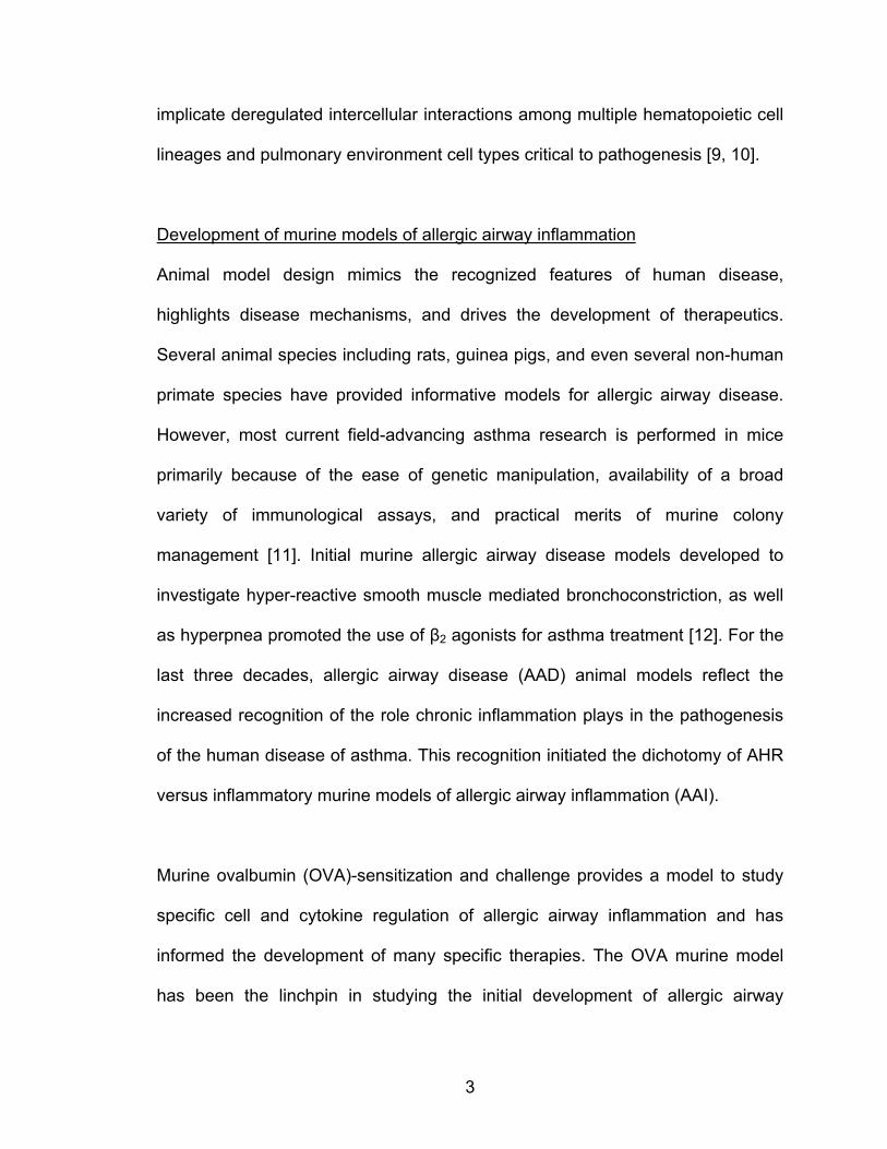
[39]. Airway DCs also secrete chemokines like CCL17 and CCL22 that attract
Th2 cells to the lung to orchestrate AAI [40]. A schematic of cellular interactions
in the development of human asthmatic inflammation is illustrated in Figure 1.
For decades, researchers studying atopy have considered CD4+ T-helper II
(Th2) cell polarization as the central cell dysregulation in allergic diseases and
asthma although the crucial role of the inflammatory environment is increasingly
gaining recognition [7, 8, 25, 41, 42]. The mature Th2 cell synthesizes and
secretes IL-3, IL-4, IL-5, IL-9, IL-13, and granulocyte-macrophage colony
stimulating factor (GM-CSF); these molecules direct asthmatic inflammation [43].
Moreover, depletion of murine CD4+ T cells prevents development of AAI and
only adoptive transfer of Th2 cells restores allergic inflammatory phenotype [44-

6
47]. However, specific IL-4, IL-5, and IL-13 anti-therapies have shown varied and
limited efficacy in clinical trials [41].
Atopic asthma, a type I hypersensitivity, classically centers around the Th2 cell
regulation of B cell antibody isotype switching to antigen-specific IgE molecules
[41, 48]. Th2 derived IL-4 and IL-13 promote B cell maturation to allergen-
specific IgE secreting plasma cells and memory cells, while, IL-3, IL-4, and IL-13
govern basophil maturation as well as mast cell maturation and pulmonary
recruitment [7, 49, 50] . Allergen-specific IgE can then bind to the high affinity Fc
epsilon receptor type I (FcεRI) on basophils and mast cells and subsequent
allergen exposure crosslinks the IgE-bound receptors inducing immediate
degranulation [8]. The mast cell preformed degranulation mediators, histamine,
proteoglycans, and serine proteases rapidly increase vascular permeability and
bronchoconstriction provoking the characteristic obstructive symptoms of the
early phase of asthma [8]. IgE also negatively feeds back on IgE production and
signaling via CD23 (FcεRII) crosslinking on B and non-B cells [49-51]. Moreover,
AAD studies in immunoglobulin deficient (JHD) mice show B cells play an
immunotolerizing role to allergens [52]. Additionally, anti-IgE treatment in mice
reduces several hallmarks of AAI [53]. Mast cell deficient mice (KitW/W-v & KitW-
sh/W-sh) have a significantly attenuated overall airway inflammatory response
indicating mast cells are not only crucial in the early-phase but also the late and
chronic phases of asthma [54]. Similarly, c-kit silencing in mice ablated this
airway inflammation [55]. IgE-bound FcεRI crosslinking also initiates mast-cell

7
and basophil synthesis of lipid mediators important in the late phase of an
asthmatic attack [8].
Another player, the airway epithelium fuels asthmatic inflammation by secreting
critical factors to chronic inflammation [25]. Airway epithelium–Th2 interactions
are pivotal in chronic airway remodeling characteristic of human bronchial
asthma. Th2 IL-13 induces goblet cell metaplasia, airway epithelial hyperplasia,
increased epithelial secretions, and ASM hyper-responsiveness [56-60]. In
contrast to its barrier function, the airway epithelium also secretes cytokines and
growth factors that actively contribute to the development of chronic asthmatic
inflammation. Epithelial cell-derived cytokines and growth factors IL-25, IL-33,
thymic stromal lymphopoeitin (TLSP), GM-CSF, and eotaxins promote the
development of chronic asthmatic inflammation [25]. Epithelial TLSP secreted in
response to ligation of pathogen-associated molecular pattern (PAMP) receptors
amplifies the Th2 response directly, by enhancing antigen presentation, or by
acting on other hematopoietic cells [25, 61-64]. Epithelial IL-25 stimulates Th2
cytokine secretion from both T cells and non-T cells and induces goblet cell
metaplasia and eosinophilia [65-68]. Similarly, IL-33 activates hematopoietic cells
and can generate Th2 cells by toll-like receptor, ST2, ligation independent of
STAT 6. Furthermore, OVA-sensitized and challenged ST2 deficient mice have
decreased parameters of inflammation [69-71]. Critically, epithelial GM-CSF and
eotaxins promote development, trafficking, and lipid mediator synthesis of
eosinophils
8
Figure 1
9
Figure 1: Schematic illustrating cellular interactions promoting chronic
inflammation and remodeling in asthma. Th2 cytokines regulate the
inflammatory changes in multiple hematopoietic and airway cell types. Th2-
eosinophil-airway tissue interactions critically promote chronic asthmatic
inflammation and promote airway hyper-reactivity, cytokine expression, injury,
smooth muscle hypertrophy and consequent airway remodeling. This figure
summarizes content from Galli, Nature reviews, 2008 [8].

10
The eosinophil and the inflammatory environment in asthma
As current understanding of allergic mechanisms does not fully account for the
great variety in asthmatic phenotype, human asthma persists as a clinically-
defined disease of elusive mechanistic complexity. Traditionally asthmatic
inflammation was regarded primarily as a Th2 cell mediated hypersensitivity.
Mounting evidence, however, suggest fundamental modulatory roles for other
myeloid cells, and the airway environment [7, 25, 41, 72]. Granulopoiesis
accelerates because of cytokine secretion during asthmatic inflammation [73,
74]. All major myelocytes (neutrophils, mast cells, and especially eosinophils) are
reported to modulate asthmatic inflammation [25, 75]. Clinically, atopic asthma
with predominant eosinophil lung infiltration and inflammation is the most
common asthmatic subtype [6] in which eosinophils are the most numerous cell
type recruited to the asthmatic lung [76].
Tissue eosinophils are found in the gastrointestinal tract, bone marrow, blood,
and other tissues, excluding the lung under non-inflammatory homeostatic
conditions. Recent reports, however, demonstrate eosinophil-lineage committed
cells undergo proliferation locally in the inflamed asthmatic lung [74]. However,
eosinophils have classically been considered effectors of Th2 cell inflammation.
Th2 eosinophilopoietins (GM-CSF, IL-3, and critically IL-5) direct differentiation
and maturation of eosinophil progenitor cells in the bone marrow as well as
mobilization of eosinophils and eosinophil progenitors into the blood [77-79].
Bone marrow of the eosinophil lineage is coordinately controlled by GATA-1 and
11
PU.1 [80]. GATA-1 drives CCR3, major basic protein (MBP) and IL-5Rα
eosinophil-specific gene expression, and functional deletion of GATA (dlb GATA)
or transgenic co-expresssion of GATA with diphtheria toxin (PHIL) ablates the
eosinophil lineage and AAD hallmarks in allergen-sensitized mice [81-83].
Similarly, PU.1 regulates expression of many eosinophil cationic proteins [84].
Repeated allergen-challenge promotes a chronic eosinophil-rich pulmonary
infiltrate in humans and mice. GM-CSF, IL-3, and IL-5 in concert with allergen-
induced Th2 cell, ASM, and airway epithelial-derived eotaxins elicit rapid
eosinophil vascular egression and selective pulmonary trafficking of eosinophils
via CCR3 G-protein signaling [76, 78, 85-87]. Moreover, IL-5 deficient mice and
patients with a genetic polymorphism decreasing eotaxin expression
demonstrate decreased allergen–induced eosinophilic lung infiltration [84, 88].
On the contrary, CCR3 ligation by inhibitory ligand can inhibit migration [89].
Emerging evidence further suggests that allergen incited inflammatory cytokines
recruit hematopoietic progenitor cells from bone marrow to the lung where they
can undergo further proliferation and final maturation, intensifying the degree of
tissue inflammation [74]. While the genetic evidence for IL-5 and eotaxin control
of eosinophil lung accumulation in AAD is compelling, anti-IL-5 therapy in asthma
patients reduced eosinophilia and eosinophil TGFβ expression in the lung, but
only modestly reduced AHR [90]. In contrast to these studies, other evidence
suggests eosinophil infiltration of tissues can be T-helper cell-independent.
Helper T cells were dispensable in eosinophil infiltration in a non-AAD murine
12
model [91]. Rather, eosinophil transfers were necessary to restore Th2 cytokine
secretion in IL-5 and eotaxin deficient mice [92].
Granulocyte trafficking from bone marrow to sites of inflammation involves
relative eosinophil-endothelial rolling, adhesion, and transmigration. CCR3:
chemokine-induced eosinophil adhesion and transmigration depends on
interactions between eosinophil and the vessel wall. Expression and ligation of
eosinophil alpha class of integrins LFA-1, VLA-4, α4β7, and Mac1 by endothelial
ICAM, VCAM, and MadCAM are implicated in eosinophil accumulation in vivo
[93-95]. In addition, Siglec F seems critical to egression and survival of
eosinophils at sites of eosinophil inflammation [96]. A coordinated balance
among adhesive, motile, and structural forces in the eosinophil are responsible
for its consequent diapedesis through vascular epithelium and infiltration of local
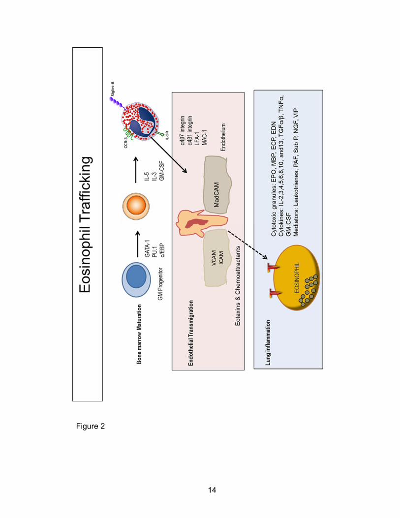
sites of airway inflammation. These processes are illustrated in Figure 2. While
eosinophil infiltration is crucial to the development of asthmatic inflammation, the
exact intrinsic eosinophil mechanisms by which eosinophils traffic to the lung in
asthma and AAD remain incompletely understood.
Accumulating evidence suggests eosinophils can regulate production and
secretion of a variety of immunomodulatory molecules. Critically, eosinophil
mediators, cytokines, and growth factors modulate asthmatic late phase and
chronic airway inflammation. Eosinophils are the main source of cysteinyl
leukotrienes that amplify the late phase response [97]. In concert with Th2
13
cytokines, lipid mediators promote exacerbated systemic vascular leak and
pulmonary inflammation [8]. Moreover, currently prescribed cysteinyl leukotriene
receptor blockers and phosphodiesterase inhibitors have been moderately
successful at attenuating asthma [41]. Eosinophil granules contain toxic cationic
proteins that cause tissue injury, remodeling and chronic inflammation in the
asthmatic lungs [90, 98]. Airway tissue samples from asthmatic patients
frequently demonstrate eosinophil degranulation products in CLCs [99]. The
cationic granule proteins, MBP, eosinophil peroxidase (EPO), eosinophil cationic
protein (ECP) and eosinophil-derived neurotoxin (EDN) wound airway epithelial
cells, parasympathetic nerve endings, basement membrane, and smooth muscle
promoting airway remodeling [100-104]. Degranulation products discovered
around vagal nerve ganglia that innervate smooth muscle are hypothesized to
cause AHR [105]. A piece-meal exocytotic mechanism governs release of human
eosinophil preformed granule proteins and mediators, which allows for selective
mobilization of granule proteins [106, 107]. Eosinophil degranulation and
mediator release is controlled by cytoskeleton-facilitated Soluble N-
ethylmaleimide-sensitive factor Attachment Protein Receptor (SNARE)/Vesicle-
associated membrane protein (VAMP) interactions and intracellular trafficking
[103]. Additionally on allergen-sensitization, eosinophils synthesize and secrete
cytokines, chemokines, and growth factors. These pro-inflammatory molecules
including IL-25, TGFα, TGFβ, metalloproteinases, pro-fibrotic, and angiogenic
factors establish a chronic inflammatory milieu in the airway epithelium and
facilitate injury and remodeling in asthma [90, 108-112].
14
Figure 2
15
Figure 2: Schematic illustrating selective eosinophil interactions in bone
marrow eosinophilopoiesis, egress into the blood, transmigration into the
asthma-inflamed lung, and inflammation. Eosinophil lineage differentiation
and maturation is coordinately controlled by GATA-1, PU.1, and c/EBP driven
eosinophil gene expression as well as IL-3, IL-5, and GM-CSF signaling (top
panel). Mature eosinophils or committed progenitors are mobilized in response to
IL-5. Eotaxin concentration gradients induce the relative adhesion and
diapedesis of eosinophils through the leaky endothelial wall (middle panel).
Eotaxin-stimulated eosinophils release cationic proteins, cytokines, and
inflammatory mediators that modulate chronic airway inflammation and
remodeling. This figure was adapted and simplified from a review published by
Rothenberg and Hogan in Annu. Rev. Immunol. 2006 [103, 113].
16
Identifying novel asthma targets
While we are also beginning to understand the intercellular cytokine and
mediator signaling cascades in asthma, no new drugs have been developed to
replace corticosteroids in managing bronchial asthma. This stagnation in
effective new asthma therapy warrants a broader investigative approach that
seeks to identify putative molecular instigators of inflammation in multiple
inflammatory processes. The mitogen-activated protein kinases (MAPKs) are
kinases that modulate and amplify many intracellular signals important to the
inflammatory process in multiple cell lineages. Moreover, mice treated with
inhaled inhibitors of p38 MAPK have shown promising attenuation of eosinophilic
inflammation [39, 114, 115]. Furthermore p38 MAPK, ERK, and JNK inhibitors
have shown some efficacy at inhibiting eosinophil chemotaxis and pro-
inflammatory mediator synthesis and release [116-118]. p38 MAPK are known to
modulate cytoskeletal remodeling and cell motility [119, 120]. p38 MAPK
transduces migratory signals from multiple upstream molecular switches. PAKs,
one such group of kinases, indirectly activate p38 MAPK and induce
hematopoietic and endothelial cell migration [121, 122]. PAKs, modulators of p38
MAPK and other MAPK proteins, may also promote chronic asthmatic
inflammation and provide attractive alternative therapeutic targets.
The Rho-family of GTPase proteins, the classic activators of PAKs, are
implicated in the pathophysiology of bronchial asthma. Rac GTPase levels and
activity and Rho-kinase (ROCK) activity are both elevated in smooth muscle
17
hyper-responsiveness and eosinophilia in models of chronic AAI [123-125].
Furthermore, Simvastatin decreases human AHR by inhibiting Rac1 [126]. In a
murine model of AAD, ROCK small molecule inhibitors attenuated asthmatic
eosinophilia and AHR [127]. Racs also modulate SCF-kit induced mast cell
migration [128, 129]. Moreover, expression of a dominant negative construct of
Rac 1 decreases mast cell synthesis and secretion of cysteinyl-leukotrienes to
IgE stimulation [130].
p-21 activated Kinases (PAKs)
PAKs, members of the Ste-20 family of serine/threonine kinases, regulate many
cellular physiological processes. Human tissues vary widely in their expression of
the six isoforms of PAKs [131]. PAK1-3, Group 1 PAKs are structurally
comprised of an N-terminal p21-binding domain, an overlapping regulatory region
that includes an autoinhibitory domain, and a C-terminal catalytic kinase domain.
They exist as auto-inhibited trans-homodimers under basal conditions [132-135].
First described as binding partners of the Rho-family proteins CDC42 and Racs
in murine brain tissue, Group 1 PAKs were found at 68, 65, and 62 kDa and
were later designated PAK1 (α-PAK), PAK3 (β-PAK), and PAK2 (γ-PAK)
respectively [134]. Their activation requires the CDC42/Racs-dependent
dismantling of the inhibited dimeric complex into active monomeric units [136].
Classically, the active GTP-bound Rho-family GTPases, CDC42 or Rac, bind and
18
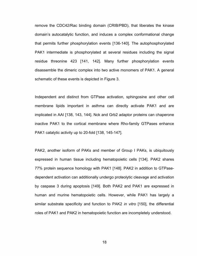
remove the CDC42/Rac binding domain (CRIB/PBD), that liberates the kinase
domain’s autocatalytic function, and induces a complex conformational change
that permits further phosphorylation events [136-140]. The autophosphorylated
PAK1 intermediate is phosphorylated at several residues including the signal
residue threonine 423 [141, 142]. Many further phosphorylation events
disassemble the dimeric complex into two active monomers of PAK1. A general
schematic of these events is depicted in Figure 3.
Independent and distinct from GTPase activation, sphingosine and other cell
membrane lipids important in asthma can directly activate PAK1 and are
implicated in AAI [138, 143, 144]. Nck and Grb2 adaptor proteins can chaperone
inactive PAK1 to the cortical membrane where Rho-family GTPases enhance
PAK1 catalytic activity up to 20-fold [138, 145-147].
PAK2, another isoform of PAKs and member of Group I PAKs, is ubiquitously
expressed in human tissue including hematopoietic cells [134]. PAK2 shares
77% protein sequence homology with PAK1 [148]. PAK2 in addition to GTPase-
dependent activation can additionally undergo proteolytic cleavage and activation
by caspase 3 during apoptosis [149]. Both PAK2 and PAK1 are expressed in
human and murine hematopoietic cells. However, while PAK1 has largely a
similar substrate specificity and function to PAK2 in vitro [150], the differential
roles of PAK1 and PAK2 in hematopoietic function are incompletely understood.
19
Figure 3. adapted from Eswaran, J et al., Trends Biochem Sci, 2008

20
Figure 3: Schematic showing the activation of p-21 activated Kinase 1
protein. A. PAK1 exists as a trans-inhibited homodimer associated at their CRIB
domains (shown in orange). GTP-bound CDC 42 or Rac binding activates this
complex by reorientating the dimeric complex and disinhibiting the kinase activity
of the catalytic subunits (shown in brown). The activated catalytic domains are
phosphorylated at many residues including the signal pT423 residue. This figure
was adapted from the version first published by Eswaran et al in Trends Biochem
Sci, 2008 [151].
21
PAK1 in cytoskeletal regulation and cell motility
PAKs modulate many cellular processes by regulating cytoskeletal function.
PAK1 modulates remodeling of the F-actin cytoskeleton involved in cell
migration. On cell stimulation, PAK1 relocates to various F-actin cytoskeletal
structures. PAK1 localizes to focal adhesion complexes at leading cell edges
[133, 152-154], lamellipodia [155], filopodia, membrane ruffles, retracting cell
edges [156], and vesicles [157]. Furthermore, PAK1 assists in recycling focal
adhesions and actin stress fibers [156, 158]. In lower organisms, embryological,
neurogenic, and metabolic evidence supports a role for PAKs homologs in
modulating the actin cytoskeleton [159-165].
PAK1 modulates cytoskeletal remodeling by both kinase-dependent and -
independent mechanisms. In general, PAK1 binds CDC42/Racs, mediating
activation of its catalytic activity, whereas PAK1 binding of adaptor proteins like
PI mediates its translocation and binding to several transmembrane receptors
[166]. Reports utilizing ectopically expressed activating and deactivating mutant
constructs of PAK1 have clarified some of these effects [133, 142, 167]. Full
length PAK1 as well as a constitutively active kinase mutant, L107F, promote F-
actin turnover and cell motility, whereas a kinase dead mutant of PAK1, K299R,
suppressed these phenotypes [156, 158, 168].
PAK1 catalytically modulates F-actin remodeling in events essential to cell
motility through multiple downstream effectors as summarized in Figure 4. PAK1
activates the LIM Kinase inhibiting ADF/cofilin (cofilin) and allowing for the
22
persistence of polymerized actin filaments in cell protrusions and other actin sub-
cellular structures [169]. Cofilin severs F-actin filaments, generating free actin
barbed ends, the building blocks for polymerizing new filaments of actin [170].
Genetic and overexpression studies support a role of cofilin in F-actin dependent
cell motility [171-173]. PAK1 regulates cofilin activity both directly and by
downstream effectors; however, this temporal-spatial cofilin control is
incompletely understood [174-176].
Importantly this kinase also localizes with, binds, phosphorylates, and activates
p41-ARC, a subunit of the ARP2/3 complex in vivo and in vitro [177]. This
complex directs nucleation and branching of new filaments of actin by using
filaments with free barbed ends [178, 179]. Additionally ARP2/3 contributes to F-
actin motility structures and force-generation at the cell’s cortical membrane, and
consequent cell migration; however, its exact regulation of motility is incompletely
understood [145, 177, 180, 181].
Recent work that we have done suggests that PAK1 regulates Ezrin’s actin
regulation. Ezrin/Radixin/Moeisin (ERM) proteins act as linkers for polymerizing
F-actin to multiple transmembrane receptors [182, 183]. Ezrin and the other ERM
proteins exist under homeostatic conditions in a closed autoinhibited
conformation with tight binding between its N-terminal plasma membrane
associated (FERM) and C-terminal ERM-association (C-ERMAD) domains.
Receptor tyrosine kinase induced signaling events break the association
between these domains and facilitate binding of the underlying actin cytoskeleton
[182]. Ezrin has no intrinsic enzymatic activity but draws the actin cytoskeletal

23
signaling complexes in close apposition to membrane-bound receptors facilitating
signal transduction [182, 183]. As such, Ezrin’s importance has been
demonstrated in actin reorganization necessary for apical-basal organization of
epithelia, T-cell immunological synapse, and hematopoietic cell adhesion to the
extracellular matrix [184-187]. Moreover, tissue-specific murine Ezrin deficiency
has been linked to defects in epithelial morphogenesis [188, 189]. Several
studies link Ezrin deregulated expression and activation with metastatic invasion
in human epithelial-derived and hematopoietic cancers [190, 191]. Our work
demonstrates for the first time that PAK1 in mast cells regulates F-actin
depolymerization in IgE/DNP-mediated degranulation [192].
PAK1 also phosphorylates and activates Filamin A, an actin binding protein.
Filamin A crosslinks F-actin into networks and anchors the actin cytoskeleton to
transmembrane proteins in cellular adhesion, morphology, and motility. Studies
in Filamin A/FLNA deficient embryos and human cell-lines demonstrate an
important role for Filamin A in cellular migration and maintenance of intercellular
junctions in organogenesis [193, 194]. PAK1 can elicit cortical actin assembly by
direct phosphorylation of Filamin A [195]. In cancer cell-lines, PAK1 transduces
signals through Filamin A crucial for lamellipodia formation and cell migration
[196, 197]. In muscle, PAK1 additionally phosphorylates and inhibits myosin light
chain kinase decreasing the actin-myosin filament mediated cell tension [198,
199]. PAK1 can further modify the tubulin cytoskeleton by phosphorylating and
deactivating Op18/Stathmin at Serine 16, stabilizing microtubules [200, 201].
24
Figure 4
25
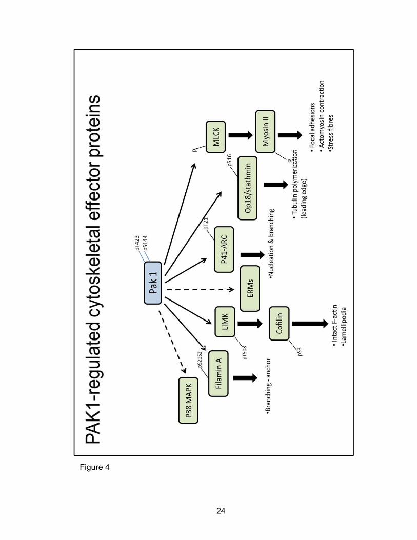
Figure 4: PAK1 regulation of cytoskeletal processes by downstream
cytoskeletal binding and remodeling effector proteins. PAK1 directly
phosphorylates p41ARC of the ARP2/3 complex, LIM Kinases (LIMKs), Filamin A
Myosin Light Chain Kinase (MLCK), and Op18/Stathmin. PAK1 also indirectly
induces phosphorylation changes in cofilin, p38 MAPK and Ezrin/Moeisin/Radixin
(ERM) proteins.
26
PAK1 as a potential target in allergic airway disease
PAK1 and other isoforms regulate chemoattractant-directed cell migration, a
process that heavily involves the actin cytoskeleton. Inhibiting PAK1 kinase
activity in NIH-3T3 fibroblasts alters cell motility, whereas enhancing PAK1
activity by expression of a PAK1 L107F plasmid construct increased fibroblast
migration to collagen [155]. Moreover, other constitutively active Pak1 constructs
rescue decreased chemoattractant-induced formation of lamellipodia and
cytoskeletal rearrangement in Akt-/- fibroblasts [202]. PAK1 also modulates
human endothelial cell, macrophage, and neutrophil motility [203, 204].
Dysregulated PAK1, which promotes cell motility and tissue invasion in cancer
metastasis, localizes at the protrusions on migrating COS-7 cancer cells in situ
[205].
PAK1 is a well-known regulator of the F-actin cytoskeleton and cell migration,
although work has not been done in eosinophils. p38 MAPK one putative
downstream effector of PAK1 has been implicated in migration and F-actin
modulation and motility in multiple hematopoietic cells. In contrast, although
cofilin, ARP2/3, and Ezrin are individually implicated in F-actin remodeling in
other cell systems, their role in eosinophil F-actin-mediated processes remains
unexplored.
PAKs dysregulation in multiple human cancer types is well-documented.
Uncontrolled PAK1 signaling in human breast adenocarcinoma confers tamoxifen
resistance [206-208]. PAK1 levels are also elevated in colonic adenocarcinoma
27
[209], ovarian carcinoma [210, 211], and renal cell carcinoma [212]. Similarly,
Pak1 gene amplification has been detected in transitional cell carcinoma [168],
high-grade ovarian carcinoma [211, 213, 214] and T cell lymphoma [215, 216].
Our multi-disciplinary research group has recently shown that PAK1 modulates
mast cell pro-inflammatory functions in neurofibromatosis 1 [121]. Few studies,
however, have focused on the effect PAK1 has in chronic AAD.
Our collaborators and others have delineated the PAKs-F-actin cell-physiological
changes in contracting ASM [217-219]. Their data suggest that dysregulated
PAK1 signaling permits cellular processes culminating in AHR [219-222]. We
have recently established that genetic deletion of Pak1 attenuates the SCF/c-kit
induced migration of mast cells [121]. Furthermore, PAK1 is important in
immediate allergen/IgE induced calcium-flux changes and F-actin cytoskeletal-
mediated depolymerization in mast cell degranulation [223].These reports taken
together support studies investigating the role PAK1 plays in allergic airway
disease. We hypothesized that a Racs/CDC42/PAK1 signaling axis modulates
the development of chronic AAI by modifying cytoskeletal dependent cellular
processes.
28
THESIS OVERVIEW
In sum, existing therapeutic modalities with adverse systemic effects or new
drugs with narrow target population specificity, fail to effectively impact the
increasing asthma incidence and morbidity. Current management of asthma
symptoms with corticosteroids and β2 agonists has done little to stop the natural
progression and lung remodeling of asthma. In response to allergen-induced
eotaxins and other chemoattractants, eosinophils infiltrate the allergen-sensitized
and challenged airways en masse and promote this remodeling. Furthermore,
Th2-airway tissue-eosinophil reciprocal signaling and interactions orchestrate the
development of asthmatic inflammation. Directed research into these synergistic
cell signaling networks can identify specific targets involved in multiple processes
in this inflammatory cell environment.
PAK1 classical activators Racs, as well as PAK1-interacting MAPK proteins both
regulate eosinophilia in animal models as well as eotaxin-mediated eosinophil
migration in vitro. However, genetic studies testing the mechanism by which
PAK1 may regulate eotaxin-mediated eosinophil pro-inflammatory functions have
not been explored. In these studies, we formally assess PAK1 as a novel anti-
eosinophil inflammation target. We use a Pak1 deficient AAD model to test
PAK1’s role in eosinophil inflammation characteristic of asthma. Furthermore, we
utilize bone-marrow derived eosinophils to evaluate the consequence of Pak1
deletion on eotaxin-mediated eosinophil adhesion, migration, and degranulation.
29
MATERIALS AND METHODS
Animals used
Pak1-/- mice were generated by Dr. Jonathan Chernoff’s laboratory at Fox Chase
as described [223] and were backcrossed at an Indiana University Laboratory
Animal Research Center facility for nine generations onto a C57BL/6 strain
background using mice obtained from Jackson Laboratories.
Ezrin flox/flox mice on a 129/SV strain background were generated by Dr. Andrea
McClatchey’s laboratory at Harvard Medical School as described [188] and bred
with Mx1Cre transgenic (MxCre+) mice on a C57BL/6J strain background to yield
Mx1Cre+ and Mx1Cre- Ezrin flox/+ mice. These F1 mice were bred to yield stable
Mx1Cre+ and Mx1Cre- Ezrin flox/flox breeders and experimental mice. The Ezrin
flox/flox and Mx1Cre alleles are genotyped to yield 251 and 217 base pair bands
respectively in reactions as previously described [188, 224, 225].
Mice were housed in pathogen-free conditions at Indiana University School of
Medicine according to the Institutional Animal Care and Use Committee and
Institutional Review Board guidelines.
Mx1Cre induction
Poly I poly C (PolyIC), a synthetic dsRNA analog, induces IFN-β production and
release in vivo. Downstream JAK-STAT signaling acting at the Mx1 gene
promoter induces Cre expression in Mx1Cre+ mice. Hematopoietic stem and
30
progenitor cells from Mx1Cre+ mice express Cre following polyIC administration
[226]. Ezrin deletion (Ezrin-/-) was achieved in Mx1Cre+ Ezrin flox/flox mice after
the administration of five intraperitoneal injections of polyIC (Sigma, Saint Louis,
MO) dissolved in sterile PBS. Minimal in vivo adverse effects from PolyIC
treatment were achieved using a tapered dosing scheme as follows; two 15 µg,
two 20 µg, and one 25 µg injections per gram of body weight Poly IC injections
on alternate days over a ten day period. MxCre- Ezrin flox/flox control mice for these
experiments also received the same doses of PolyIC prior to experimentation.
Eosinophil infiltration murine models
Cohorts of age and sex matched Pak1-/- and Pak1+/+ mice were injected with a
1 mL intraperitoneal dose of 2 µg murine recombinant eotaxin (‘mreotaxin’, R&D
systems) or PBS. Two hours later, mice were euthanized by CO2 asphyxiation
and their peritoneal cavities lavaged with 5 mL PBS to retrieve peritoneal cells as
described [227]. Total cells were counted by hemocytometer, cell populations
visualized by giesma-stained slides, and eosinophils quantified by fluorescent
cytometry of CCR3.
Alternatively, cohorts of age and sex matched anesthetized Pak1-/- and Pak1+/+
mice were injected with a 25µL intratracheal dose containing 10µg mreotaxin
(Peprotech) or saline as described [228, 229]. The mice were maintained under
anesthesia for four hours [228].The mice were euthanized by CO2 asphyxiation
31
and broncho-alveolar lavage fluid (BALF) collected at 4°C for hemocytometer
counts, giesma staining, and fluorescence cytometric analysis of eosinophils.
OVA-induced allergic airway disease model
Cohorts of male 10-14 week Pak1-/- and Pak1+/+ mice were sensitized and
challenged in a murine model of allergic airway inflammation (AAI). Mice of both
genotypes were sensitized by two intraperitoneal injections, seven days apart
with 100 μg purified Ovalbumin (‘OVA’/ Sigma) adsorbed to 2 mg of the adjuvant
Aluminum hydroxide (‘Alum’/ Sigma). On the 14th day, OVA-sensitized mice
were anesthetized using aerosolized isoflurane (Webster Veterinary) daily for five
consecutive days to prepare them for intranasal OVA challenge. Anesthetized
mice were intranasally challenged with 50 μg of OVA in solution with saline for
five consecutive days. Twenty-four hours after the last intranasal challenge (day
19), AAD mice were euthanized by a 60 μg/g body weight lethal intramuscular
injection of pentobarbital, their tracheas cannulated using a 20G 11/4 inch
veterinary I.V. catheter, and broncho-alveolar lavage (BAL) samples collected at
4°C. Some of these murine tracheas and lungs for histological analysis were
inflated to 20-25 cm of H2O and fixed in formalin while tissue for cellular analysis
was washed and collected in saline at 4°C.
All BAL were centrifuged at 1500 X g for 5 minutes and the first 0.5 mL rinse
stored at -80°C for cytokine analysis by ELISA. The BAL cells from the rinses
were resuspended and pooled then red blood cells lysed in 300 μl of RBC lysis
32
buffer (Qiagen) until the cell pellet blanched at room temperature. Cell pellets
recovered from each mouse were resuspended in 100 µL of FACS buffer (2%
BSA solution with PBS and 0.09% NaN3) and 10 µL aliquots taken for
hemocytometer BAL cell counts and slide preparation. Resuspended cell aliquots
were counted in duplicate in solutions of 1 part cells diluted in 10 or 20 parts
trypan blue (Mediatech, Inc.) by hemocytometer. Pak1-/- and Pak1+/+ total BAL
cell counts were compared for significant differences. BAL cell aliquots for slide
preparation were diluted in PBS, spun onto glass slides at 1200 rpm for 10
minutes, and Giesma-stained (HEMA-TEK 2000, Bayer). Representative digital
pictures per sample were taken by a digital camera slide microscope system
(SPOT, Diagnostic Instruments, Inc.).
Peripheral immune responses in Pak1+/+ and Pak1-/- OVA- sensitized and
challenged mice were evaluated by OVA-specific splenocyte cytokine analysis.
Splenocytes were harvested from mice of both genotypes and challenged with
100 µg/ml OVA for 72 hours as previously described [230]. Cell-free
supernatants were collected and analyzed for cytokines by multiplex ELISA
(reagents from Millipore).
Histological analysis and inflammation scores
Fixed lung tissue was embedded in paraffin, and cut into 4-8 µm sections by our
histology core. Paraffin-embedded tissue sections were mounted onto glass
slides and stained with Hematoxylin and Eosin (‘H&E’, Sigma), Masson's
33
Trichrome (Fisher Scientific), and Periodic acid-Schiff using standard histological
techniques. Slides were visualized by light microscopy, and representative 20
and 40X images captured with a digital camera (SPOT, Diagnostic Instruments,
Inc.). In addition, blinded observers assigned inflammation scores to H&E tissue
sections from 0-4 with 0 being uninflamed lung and 4 severely inflamed lung.
Inflammation scores were compared for both genotypes and treatments and
differences determined.
Fluorescence cytometric analysis of cell populations
Aliquots from BAL samples were pooled to provide at least 106 cells per cell
sample for control staining. At least 105 cells for each sample were probed with
leukocyte antigen conjugate fluorescent antibodies at final concentrations of 5
µg/mL anti-CD45, a generic leukocyte antigen, 2 µg/mL B220-FITC, 10 µg/mL
CD3-FITC, 0.625 µg/mL CCR3-PE (R&D systems), 2.5 µg/mL MHCII-PerCP, and
2 µg/mL CD11c-APCcy7 as well as 6 μg/mL anti mouse CD16/32, Fc receptor
blocking antibody [230, 231]. Alternatively, 10 µg/mL CD4-FITC, and 10 µg/mL
CD8-FITC were used instead of CD3-FITC, 10 µg/mL SiglecF-PE instead of
CCR3-PE, and 10 µg/mL APCcy7 CD11b instead of CD11c-APCCy7. All
antibodies were obtained from BD pharmingen unless otherwise indicated in
parenthesis. All control and test samples were incubated with antibodies for at
least 30 minutes in the dark at 4°C before a wash and resuspension in FACS
buffer for cell population data acquisition (BD FACSCalibur) using CellQuest (BD
Immunocytometry Systems). On analysis, leukocyte population gates (all CD45+)

34
were set to distinguish the following populations: Eosinophils, FSClo B220/CD3 (-
), CCR3/SiglecF (+), Neutrophils, FSClo B220/CD3 (-) CCR3/SiglecF(-),
Lymphocytes FSClo B220/CD3 (+), CCR3/SiglecF (-), and alveolar macrophages,
FSChi, CDIIc or CD11b (+), MHCII (+). Total leukocyte population numbers were
computed from the analysis percentages and significant differences (p<0.05)
between Pak1-/- and Pak1+/+ populations compared.
Adoptive bone marrow transplantation model
For bone marrow transplantation assays, low density bone marrow mononuclear
cells were harvested from CD45.2 Pak1-/- and Pak1+/+ mice on a C57BL/6J
strain background as described in “Bone marrow isolation of LDMNCs”. These
cells were transplanted into Pak1+/+ F1 mice which expressed both CD45.1 and
CD45.2 common leukocyte antigens (CD45.1/2+) obtained from the Indiana
University School of Medicine In Vivo Therapeutics Core as described [232].
Briefly, approximately two million viable Pak1-/- and Pak1+/+ bone marrow cells
were injected with a 30 gauge 0.5 inch needle into the lateral tail vein of lethally-
irradiated (1100 cGy, split dose [700cGy and 400cGy with a 5 hour interval],
cesium isotope source) CD45.1/2+ Pak1+/+ mice. Transplanted mice were
housed in a pathogen-free environment to allow for stem and progenitor cell
engraftment. Mice were assessed for hematopoietic reconstitution at six months
post-transplantation by quantifying peripheral blood CD45.2 versus CD45.1/2
chimerism. Mice were utilized in an OVA-induced allergic airway disease model.
35
Orthotopic left-lung transplantation model
For lung transplantation assays, whole left lungs from donor mice were
transplanted into 8-12 week old male recipient mice using non-suture
microvascular anastomoses which reduces granulation tissue as previously
described [233-235]. These state-of-the-art transplantation experiments were
done in collaboration with Dr. David Wilkes’ lab, where this model is
discriminately available. Pak1+/+ recipient mice received Pak1-/- lungs while
Pak1-/- mice received Pak1+/+ lung grafts. Following a 7-day recovery period,
transplanted mice were utilized in an OVA-induced AAD model as described
above. The lung grafts were procured for cell suspension preparation and
analyzed for eosinophil percentages by fluorescent cytometry for murine siglec
F+ eosinophil in contrast to other populations. Total eosinophils were computed
and compared between transplanted groups.
Cell suspension preparation from murine lungs
Whole lungs were collected from sensitized Pak1-/- and Pak1+/+ mice or lung
transplant mice in cold PBS (4°C). Single cell suspensions were prepared for
MACS separation using a gentle-MACS dissociation protocol (Miltenyi Biotec).
Briefly, single mouse lungs or left-lungs were individually washed, and
dissociated in HEPES buffer ph 7.4 (Sigma) supplemented with 2mg/mL
collagenase D (Sigma) and 10U/mL DNAse I (Sigma) in 10mL C-tubes (Miltenyi).
Total cell in these lungs were dissociated in a gentle-MACS dissociator (Miltenyi)
using proprietary programming, hybridized while rotating in an oven at 37°C, and
36
further dissociated from parenchymal tissue. The solution was filtered through a
70µm mesh, washed in PBS, cells labelled with anti-CD45 magnetic beads, and
leukocytes positively selected by passing these cells through a magnetic column
using standard Miltenyi protocol (#130-052-301). These cells were stained with
antibodies against lineage antigens and analyzed by fluorescence cytometry.
Multiplex ELISA assays
Several allergen-induced murine cytokines and growth factors were assayed with
multiplex simultaneous quantification of fluorescent antibody capture beads
directed against eotaxin-1/CCL11, RANTES, IL-4, IL-5, IL-10, IL-13, IL-17, and
IFNγ. BAL samples from OVA-sensitized and challenged Pak1-/- and Pak1+/+
mice and supernatants from OVA-stimulated ex vivo T-cell cultures were thawed,
vortexed, and 25 µL transferred to custom ordered multiplex plates with analyte
capture antibodies (Millipore Milliplex, Billerica, MA). The multiplex assay was
performed according to the manufacturer’s protocol with each sample tested in
duplicate. Briefly, samples were incubated with analyte antibody for two hours,
washed three times, incubated with streptavidin conjugated fluorescent
secondary antibody, washed three additional times and suspended in 100 μL
saline, and analyzed on a Luminex 200 cytometer with StarStation software
(Luminex Corp, Austin, TX). Standard curves were computed using a cubic
spline fit, according to the manufacturer’s instruction. The quantities of cytokines
were computed relative to the standard curve generated, replicates averaged,
and cytokines for each group tested compared.

37
Bone Marrow Isolation of low density mononuclear cells (LDMNCs)
Whole bone marrow was harvested from pelves, femurs, and tibias of cohorts of
10-16 week old mice in Iscove’s Modified Dulbecco’s Media (IMDM,
Gibco/Invitrogen), supplemented with 2% fetal bovine serum (FBS, Hyclone,
ThermoScientific) using a 20-gauge one inch needle. The flushed bone marrow
was resolved to obtain LDMNCs on a density gradient (Histopaque Sigma
11191) as described [121, 223]. Briefly, harvested cells were carefully layered
onto an equivalent volume of Histopaque (Sigma) and centrifuged for 30 minutes
at 1750 rpm on a gh-3.8 rotor (Beckman Coulter). Cells in the intermediate layer
were collected washed in IMDM or other media, and enumerated as appropriate,
before experimentation.
Bone Marrow IL-5 methylcellulose cultures
Non-adherent LDMNCs Pak1+/+ and Pak1-/- (N=3) were cultured in 35 X 10 mm
diameter tissue culture dishes (Fisher Scientific) in culture medium, which was
made up of 0.9% methylcellulose (Methocult H4100, Stem Cell Technology)
enriched with 20% FBS and Iscove’s Dulbecco’s medium (with 1% penicillin-
streptomycin, 0.35% 2-ME, and 0.1% BSA) supplemented with rmIL-5 (1, 10
ng/ml/ R&D systems) with 2 × 10⁵ LDMNCs/ml of culture media at 37°C. After 5
and 10 days, total IL-5 colonies were counted using inverse microscopy as
previously described [236]. Each mouse was assessed in triplicate for each
cytokine concentration and the total number of IL-5 colonies was compared
between Pak1+/+ and Pak1-/- groups.

38
pCL11eGFP PAK1 mutant generation
The pCL11eGFP lentiviral construct was a kind gift of Helmut Hannenberg at the
Indiana University School of Medicine. This construct contains an eGFP cassette
and a multiple cloning site (MCS) downstream of the (HPGK) promoter, which
drives transcription in eukaryotic cells. The kinase-dead PAK1 mutant, K299R
and the constitutively active mutant, L107F, (Addgene) were NOT1-adapted and
inserted into the NOT1-cut and Klenow-blunted pCL11eGFP vector in frame at
the C-terminus of the eGFP cassette using molecular cloning techniques. The
integrity and orientation of the pCL11eGFPPAK1 mutant plasmid constructs were
confirmed by restriction enzyme digestion and sequencing.
Lentivirus generation
To generate virus, NIH 293T cells at 60-70% confluence on 10 cm dishes were
transfected with 10 µg of either pcl1PAK1eGFP, or pcl11PAK1K299R, or
pcl11PAK1L107, 5 µg gag-pol expressing helper plasmid (pCD/NL-BH), and 1 µg
foamy viral envelope plasmid (pcoPE01) in 6 mL Dulbecco’s Modified Eagle
Media (DMEM, Gibco/Invitrogen) containing 10% FBS and 0.0075 mg/mL
polyethyleneimine (PEI, Sigma). After overnight transfection at 37°C, transfection
media was aspirated and replaced with 5 mL fresh DMEM containing 10% FBS,
50 U/mL penicillin, 50 μg/mL streptomycin, and 2 mM L-Glutamine. After 24
hours, all supernatants were collected, pooled (to 50 mL maximum), filtered
through a 0.22 μm polyethersulfone (PES) membrane Stericup unit (Millipore,
Billerica, MA), and centrifuged at 16,000 x g at 4°C for 2 hours in a polycarbonate
39
Oak Ridge centrifuge tube (Nalgene, Rochester, NY). Supernatant was
decanted, bleached, and disposed in biohazardous waste, and the viral pellet
resuspended in 1 mL IMDM containing 20% FBS. Virus was stored in 1mL
aliquots at -80°C and all thawed samples were either immediately used or
appropriately discarded.
Viral titer (infectious particles per mL) was determined by percent GFP-positivity
of serially transduced HT1080 cells, plated at 100,000 per well of a six-well plate
in 1 mL DMEM/10% FBS. The serial dilution started at 10-2 dilution from viral
frozen stock and ended with 10-8 dilution. Viral titer was determined by the
following equation:
% 10
1 10
Lentiviral transduction
CD117/c-kit + cells were magnetically isolated in a column following labeling
Pak1+/+ and Pak1-/- LDMNCs with anti-c-kit/CD117 microbeads (Miletenyi
Biotec). These cells enriched for stem and progenitor cells were transduced with
viral supernatants as described [223]. Briefly, each well of a six-well plate was
coated with 10 µg/cm2 of recombinant fibronectin (Retronectin, Takara) in 1 mL
PBS, overnight at 4°C or for four hours at room temperature. This
fibronectin/PBS solution was aspirated and replaced with 50-150 infectious
particles per target c-kit+ cell, diluted in a 1 mL solution of IMDM containing 20%
40
FBS, 50 U/mL penicillin, 50 μg/mL streptomycin, 2 mM L-glutamine
supplemented with either 100 ng/mL SCF, and 10 ng/mL IL-6. After one hour of
virus/fibronectin incubation at 37°C, 1-2 million target cells were added in a 100
μL volume of the aforementioned transduction media to each 1 mL solution
containing well. After 16 to 24 hours of incubation at 37°C, the cells were
harvested, washed in IMDM, and resuspended in eosinophil culture media.
Eosinophil culture
Bone marrow eosinophils (bmEos) were derived by culturing bone-marrow
mononuclear cells ex vivo in eosinophil polarizing conditions. Briefly, LDMNCs
were enumerated and cultured ex vivo in IMEM media (Gibco) with 20% fetal calf
serum (Sigma), 1 U/mL penicillin/streptomycin (Lonza), glutamine (Lonza),
supplemented with 1 ng/mL IL 5 (BD pharmingen), 0.25 ng/mL IL 3 (Peprotech),
and 1 ng/mL GM-CSF(R&D systems) for 14 days at 37°C and 5% CO2 as
described [87, 237]. Cells were pelleted by centrifugation at 1500rpm for five
minutes, and the media was changed every three days.
Alternatively LDMNCs or c-kit+ virus-transduced cells were cultured at high IL-5
concentrations to increase cell yields as described [238]. Cells were cultured at
approximately 106 cells/mL in media containing RPMI 1640 (Gibco) with 20%
FBS (Cambrex), 1 U/mL penicillin/streptomycin (Lonza), 2 mM glutamine
(Lonza), 25 mM HEPES, 1 mM sodium pyruvate (Gibco), and 50 μM β-
mercaptoethanol (Sigma) and supplemented with 100 ng/mL stem-cell factor
41
(SCF/ PeproTech) and 100 ng/mL FLT3-Ligand (FLT3-L/ PeproTech) from day 0
to day 4. On day 4, the media was replaced with eosinophil culture media
supplemented with 10 ng/mL recombinant mouse interleukin-5 (rmIL-5/ R&D
Systems) as the only growth factor. On day 8, the cells were moved to new flasks
and maintained in fresh media supplemented with rmIL-5. On alternate days,
from this point, one-half of the media was replaced with fresh media containing
rmIL-5, and the cell concentration adjusted to 106 cells/mL. Cells were
enumerated on day 0 and days requiring media replacement by hemocytometer.
Assessment of eosinophil culture purity
Eosinophil purity of ex vivo LDMC cultures was assessed on days 10, 12, 14 and
16 by cell cytospin and flow cytometric analysis. Approximately 200,000 cells
were washed with PBS and loaded onto a plastic slide funnel mounted on a glass
slide and spun for 10 minutes at 1200 rpm (Beckmann Coulter). The cells affixed
to these slides were Giemsa stained (Hema-Tek 2000, Bayer) and eosinophil
percentage purity determined by counts under a light microscope.
Approximately 500,000 cells from eosinophil cultures were washed and
resuspended in 100 µL FACS buffer and incubated with 0.5 µg/mL PE-
conjugated anti-murine CCR3 (R&D Systems) or 0.5 µg PE-conjugated anti-
murine Siglec F (BD Biosciences) antibody for 30 minutes on ice as described
[238]. Cells were washed in FBS/PBS and Phycoethrin (PE) intensity detected by
fluorescent cytometer (FACS Calibur, BD). The data was analyzed in comparison
42
with PE conjugated isotype control (BD Biosciences) treated cells by Flowjo
v7.6.5 software (Tree Star Inc.).
Eosinophil adhesion assay
Microfluidic 24-well plates (Fluxion Biosciences Inc.) were coated with 50 µg/mL
fibronectin (Invitrogen) or gelatin for one hour at 37°C. Coating solution from
each well was aspirated gently to avoid air bubble introduction and washed with
300 µL of plain RPMI media (Gibco) with 0.5% BSA (Sigma). The wash solution
was steadily pumped through the microfluidic chambers at 5 dynes/cm2 using a
Bioflux 200 controller (Fluxion Biosciences Inc.). Fluid from both input and output
channels was gently aspirated and cells added to each input well. bmEos were
washed and resuspended in RPMI/0.5% BSA media at a cell concentration of 2 x
106 cells/mL. Approximately 4 x 10⁶ eotaxin-treated or untreated Pak1+/+ and
Pak1-/- bmEos were loaded at each input and flow initiated at 2 dynes/cm2 for
two minutes to introduce a bolus of cells into the adhesion chambers, after which
this volume rate was adjusted to 0.5 dynes/cm2 for 20 minutes to mimic capillary
blood flow. The input well was cleared, washed with PBS, and flow initiated for
two minutes at 2 dynes/cm2 to wash away non-adherent cells. Plates were fixed
with paraformaldehyde (PFA) for imaging or stained with Coomassie blue for cell
counts. 40X images of PFA stained cells were taken using (SPOT, Diagnostic
Instruments, Inc.) and stained cells were counted by hemocytometer, and
compared relative to genotype, treatment-group, and plate coating type.

43
Eosinophil migration assays
Eosinophil migration to recombinant murine eotaxin-1/mCCL11 (‘rmEotaxin’/
Peprotech) was assessed using a transwell assay system. During the 10th-14th
days of culture, bmEos were resuspended at cell concentrations of 2 x 10⁶
cells/mL in RPMI/0.5% BSA media. To assess if this transwell setup could
distinguish eotaxin-induced chemotaxis from random chemokinesis a
checkerboard scheme was followed where concentrations of rmEotaxin were
loaded in top or bottom chambers alone, both chambers, or neither chamber
preceding the migration experiment. 100 μl of resuspended bmEos were then
seeded in the top three micron-meshed polycarbonate chamber of a 24-well
transwell plate (Costar 3415) and the cells allowed to migrate to the bottom
chamber where they were collected and enumerated by hemocytometer. All
conditions of this checkerboard migration assay format were tested in duplicate
or triplicate and the cell counts averaged per experiment. To calculate the
chemotactic index, the number of cells that migrated in response to chemokines
was divided by the number of spontaneously migrated cells [239]. Migrating cells
were compared between the two genotypes and four conditions. bmEos cultures
from Pak1+/+ mice were also pretreated for 1 hour at 37°C with small molecule
inhibitors of PAKs, IPA3 (Chernoff, Fox Chase Cancer Center), and p38 MAPK,
SB22025 (Sigma) then assayed for rmEotaxin-mediated chemotaxis. As a
control, a trypan-blue exclusion count was performed on cell aliquots of bmEos
pretreated with inhibitors to check for dead cell percentages.
44
Eosinophil degranulation assay
Eosinophil degranulation to recombinant murine rmEotaxin (Peprotech) was
assessed by a colorimetric assay measuring EPO activity in cell supernatants in
lieu of eosinophil degranulation. On the 14th day of culture, bmEos were
resuspended at cell concentrations of 1 x 10⁶ cells/mL in HBSS media (Gibco).
Cells were stimulated with 100-1000 ng/mL rmEotaxin for one hour at 37°C. Cells
were gently centrifuged to separate supernatant and cell pellet and kept on ice.
The cell pellet was permeabilized in 0.3% Triton-X (Sigma) for five minutes at
room temperature and the cell lysate supernatant collected for each sample.
100μl of cell supernatants and cell-pellet lysates were seeded in a round-bottom
96-well plate. 100 µL of EPO substrate (consisting of a solution of 1 mM H2O2, 1
mM O-phenylenediamine, and 0.1% Triton-X (all from Sigma) in 55 mM Tris
buffer at pH of 8.0) was added to the seeded fluid and the reactants incubated in
the dark for 30 minutes at 37°C. The reaction was terminated by adding 50 µL of
4 M H2SO4, and absorbance read on a microplate luminometer at 490 nm ().
Background absorbance readings (no eosinophil products) was subtracted from
the absorbance readings of each well and the EPO degranulation of each
supernatant expressed as a percentage of total cell EPO content according to
the following equation:
%
100%
45
HL-60 human eosinophil culture and differentiation
HL-60 clone 15 cells (CRL-1964, American Type Culture Collection) were
maintained in 1 U/mL Penicillin/Streptomycin (Lonza) and 2 mM glutamine
(Lonza) containing RPMI-1640 medium (Gibco) supplemented with 10% FBS
(Sigma) and 25 mM HEPES pH7.6 (Sigma), at cell concentrations between 0.5-2
x 106 cells/mL at 37°C and 5% CO2. Approximately 0.5 x 10⁶ cells/mL were
differentiated to eosinophil-like cells (HL-60 Eos) by supplementing the media
with 0.5mM n-butyrate (Sigma) dissolved in ethanol and cultured for five to seven
days as described [240, 241]. HL-60 Eos were evaluated for differentiation by
Giesma staining (HEMA-TEK 2000, Bayer) of cells affixed to a glass slide.
In vitro HL-60 Eos PAK1 kinase assay
HL-60 Eos were stimulated with 100 ng/mL human recombinant eotaxin
(hmEotaxin/ R&D systems) in RPMI media (Gibco) with 0.5% BSA (Sigma) at
37°C for 5, 15, and 30 minutes and the reaction terminated by addition of 1 mM
Na3VO4 in 4°C PBS. 400 µg of whole cell lysate was prepared as previously
described [242] reserving 10 µL aliquot of each sample for GAPDH detection as
a loading control. The remainder of the lysate was immunoprecipitated with 2
µg/mL α-PAK1 antibody (N20, Santa Cruz Biotechnology, Inc.) at 4°C for 18
hours before incubation with protein A/G plus beads (Santa Cruz Biotechnology)
for two hours. PAK1 phosphotransferase activity was assayed by incubating the
immunobeads with 1 µg/reaction Mek (Millipore) and 250 µM ATP (Sigma
Chemical) in 30 µL kinase buffer described [243]. Samples were separated by
46
10% SDS-PAGE, transferred to nitrocellulose, and probed with anti-Mek-
phospho-serine 298 (1:1000, Cell signaling). Phosphorylated Mek was used in
lieu of PAK1 activation.
PAK1 RNA interference in HL-60 eos
PAK1 knockdown in HL-60 Eos was achieved by 72 hour incubation with human
PAK1 siRNA constructs in siPORT NeoFx reagent (Ambion). siPORT™
NeoFX™ Transfection Agent was diluted (1:20) in 37°C Opti-Minimal Essential I
medium (Opti-MEM) and the resulting solution incubated for 10 minutes at room
temperature. Three PAK1 siRNA constructs (SASI_Hs01_00087968,
SASI_Hs02_00334074, and SASI_Hs01_00087970 (Sigma MISSION® siRNA)
were chosen based on rank order of efficacy postulated by a siRNA design
algorithm (Rosetta InPharmatics) and tested. PAK1 siRNA or scramble siRNA
constructs was diluted in Opti-MEM to 30nM and mixed with incubated siPORT
NeoFx reagent. After a 10 minute incubation that permitted formation of
transfection complexes, 2.3 mL of transfection solution was dispensed to each
well of a 6-well plate. Approximately 2 x 10⁵ HL-60 cells in eosinophil
differentiation media were overlaid on the transfection complexes and incubated
at 37°C for 72 hours. HL-60 eos were enumerated, numbers standardized, lysed,
and whole cell lysates prepared in SDS page for western blot analysis. Following
optimization transfection experiments, SASI_Hs01_00087970 was chosen for
further experimentation.

47
HL-60 Eos migration assay
HL-60 Eos, siRNA-treated HL-60 Eos, or NeoFx-only treated HL-60 eos were
utilized in migration assays to no stimulus or 100 ng/mL eotaxin. On the fifth day
of differentiation in n-butyrate supplemented media, HL-60 eos were
resuspended at cell concentrations of 1 x 10⁶ cells/mL in RPMI/0.5% BSA media.
Approximately 100 μL of these cells were seeded in the insert of an eight micron-
meshed polycarbonate chamber of a 24-well transwell plate (Costar 3422,
Invitrogen) and the cells allowed to migrate to the bottom chamber where they
were collected and enumerated by hemocytometer. All conditions of this
checkerboard migration assay format were tested in duplicate and the cell counts
averaged per experiment. To calculate the chemotactic index, the number of
cells migrated in response to chemokines was divided by the number of
spontaneously migrated cells. Migrating cells were compared among stimulus-
treated, siRNA-treated, and non-treated groups.
Immunoblotting
Whole cell bmEos lysates were prepared by addition of 1x SDS sample buffer
supplemented with 1% 2-mercaptoethanol to 1 x 10⁶ cells. Cell lysates were
sonicated, clarified by centrifugation, and denatured at 100°C for 5 mins.
Alternatively, cells lysed with ProteoJET Mammalian Cell Lysis Reagent
(Fermentas), and clarified as above, were quantified by a BCA reagent assay
(ThermoScientific). Samples were separated by SDS-PAGE on a 4-12% gradient
or 6% acrylamide gels (Invitrogen) and transferred to PVDF membrane (Roche
Diagnostics Corporation, IN). Membrane blots were probed with primary

48
antibodies to p-T423 PAKs, p-S3 cofillin, cofilin, p-S2152 filamin A, p-T567
ERMs, Ezrin, p-S298 MEK, p-S16 Op/18 Stathmin (Abcam), and p34-ARC
(Millipore) as well as GAPDH. All antibodies were obtained for Cell Signaling
Technology, Inc. unless otherwise indicated. All blots were visualized using an
HRP-conjugated anti-rabbit secondary antibody (1:5000, Amersham), and films
developed using the West Dura luminal system (ThermoScientific).
Immunoblotting was performed on at least three independent bmEos cultures
and phosphorylated protein levels from representative blots quantified by
subjecting representative autoradiographs to densitometry (NIH ImageJ).
Deconvolution confocal microscopy
For visualization, 10-14 day old cultures of Pak1-/- and Pak1+/+ bmEos were
resuspended in RPMI with 0.5% BSA, 50 U/mL penicillin, 50 µg/mL streptomycin,
and 2 mM L-Glutamine. Cells were standardized to a concentration of 2 x10⁶
cells/mL, and stimulated with 500 ng/mL rmeotaxin or no stimulation from 0-5
minutes at 5% CO2/37°C. Cells were aliquoted to eppendorf tubes and the
stimulation terminated by addition of 4% PFA for 15 minutes at room
temperature. After fixation, cells were resuspended in 1 mL PBS. Approximately
200,000 cells were then loaded in PBS onto a cytospin funnel mounted to a glass
slide and spun for 10 minutes at 1200rpm. Following cytospin, cells affixed on
slides were extracted by washing in 0.3% Triton-X (Sigma) for five minutes,
blocked in 5% FBS for at least one hour, and incubated with primary antibody in
0.1% BSA/PBS overnight at 4°C. The following morning, these cells were

49
washed three times in PBS, and subsequently incubated for one hour at room
temperature with fluorophore-conjugated anti-mouse or anti-rabbit secondary
antibodies (Molecular Probes, Invitrogen). After washing, one drop of a DAPI
mounting solution (Molecular Probes, Invitrogen) was applied and a cover slip
sealed over the samples using lacquer. Data were acquired and analyzed on a
DeltaVision deconvolving microscopy system (Applied Precision, Washington).
Typical controls included cells incubated with secondary (fluorophore-
conjugated) antibody but not primary antibody. Intensity and localization of cells
from both genotypes were compared.
Statistical analyses
All statistical analyses were performed with GraphPad Prism 5.4. One- or two-
way ANOVA was performed, as appropriate, with Bonferroni post-hoc
corrections. For two variable comparisons, unpaired two-tailed student’s t-tests
were used. All tests, numbers of independent replicates, and significance levels
are found within the figure legends in the results section.
50
RESULTS
PAK1 in murine allergic airway inflammation
We tested the role PAK1 played in a model of allergic airway murine
inflammation using Pak1-/- mice. We used a murine allergic airway disease
model employing OVA-albumin sensitization and challenge with adjuvant
treatment. We supplemented these studies with two chimeric models. Using both
adoptive bone marrow and orthotopic lung transplantation models, we tested the
relative roles of hematopoietic and pulmonary contributions to this inflammation.
Pak1 genetic disruption
Our multi-disciplinary research group has previously generated a novel Pak1
global knockout mouse and demonstrated that PAK1 notably modulates mast cell
inflammatory processes. A schematic illustrating disruption of PAK1 in ES cells is
shown in Figure 5. Southern blot analysis of murine Pak1-/- ES cells showed a
disrupted Pak1 allele while western blot analysis of multiple tissues from these
mice failed to detect PAK1 protein [223]. The resulting Pak1-/- mice are viable,
undergo normal growth and development, are fertile, and have a normal lifespan.
The mice used in our studies were backcrossed onto a C57BL/6J background for
at least seven generations to achieve genetic uniformity with the wild-type
(Pak1+/+) mice. The peripheral blood taken from these mice demonstrate normal
blood cell composition and many cytokine levels were comparable to Pak1+/+
mice [223].
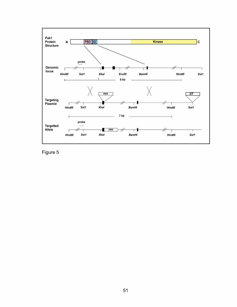
51
Figure 5
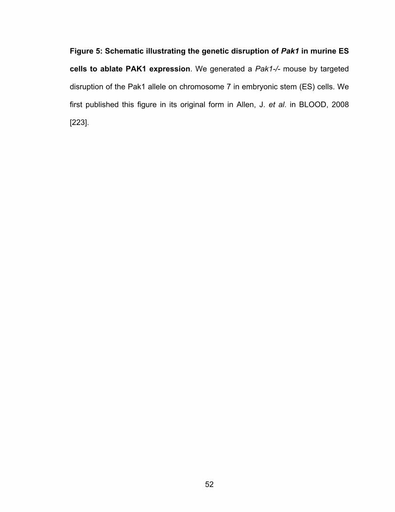
52
Figure 5: Schematic illustrating the genetic disruption of Pak1 in murine ES
cells to ablate PAK1 expression. We generated a Pak1-/- mouse by targeted
disruption of the Pak1 allele on chromosome 7 in embryonic stem (ES) cells. We
first published this figure in its original form in Allen, J. et al. in BLOOD, 2008
[223].
53
The Pak1-/- OVA-albumin allergic airway disease model
Our initial goal was to establish a model to study PAK1’s effect on the
development of allergen-induced airway inflammation. We utilized an ovalbumin
(OVA)-sensitized and challenged murine model with aluminum hydroxide (Alum)
adjuvant supplementation to elicit a potent Th2-mediated inflammatory airway
response. Following optimization of the model in our mice, we employed the
sensitization and challenge schedule illustrated in Figure 6. We utilized age-
matched, male mice in these experiments. We sensitized mice of both genotypes
using two intraperitoneal injections of an amalgam of OVA and Alum and
subsequently challenged the mice by intranasal inhalation for five days. OVA-
sensitized and challenged mice were utilized either for pulmonary function tests
or for serum, BALF, and lung procurement to assess the inflammatory infiltrate.
54
Figure 6

55
Figure 6: Schematic illustrating the Pak1-/- murine model of allergic airway
inflammation. Age-matched male Pak1+/+ and Pak1-/- mice were OVA-
sensitized and challenged in a 19-day protocol and utilized for either pulmonary
function tests for evaluation of AHR or cellular analysis for airway inflammation.
Mice were sensitized to OVA by intraperitoneal injections on day 1 and day 8 and
subsequently challenged by intranasal inhalation for five consecutive days before
lungs tissue was harvested for H&E staining and analysis of leukocytes whereas
BALF was assessed for leukocyte populations by fluorescent cytometry and
cytokine content by ELISA.
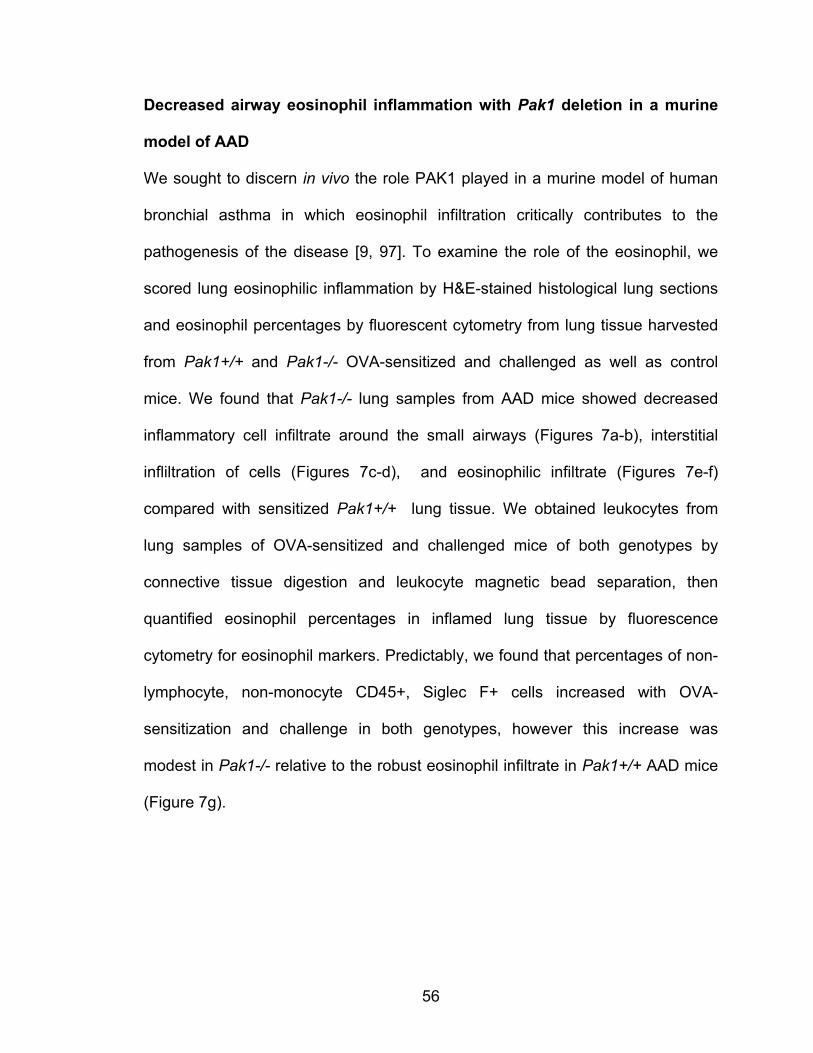
56
Decreased airway eosinophil inflammation with Pak1 deletion in a murine
model of AAD
We sought to discern in vivo the role PAK1 played in a murine model of human
bronchial asthma in which eosinophil infiltration critically contributes to the
pathogenesis of the disease [9, 97]. To examine the role of the eosinophil, we
scored lung eosinophilic inflammation by H&E-stained histological lung sections
and eosinophil percentages by fluorescent cytometry from lung tissue harvested
from Pak1+/+ and Pak1-/- OVA-sensitized and challenged as well as control
mice. We found that Pak1-/- lung samples from AAD mice showed decreased
inflammatory cell infiltrate around the small airways (Figures 7a-b), interstitial
infliltration of cells (Figures 7c-d), and eosinophilic infiltrate (Figures 7e-f)
compared with sensitized Pak1+/+ lung tissue. We obtained leukocytes from
lung samples of OVA-sensitized and challenged mice of both genotypes by
connective tissue digestion and leukocyte magnetic bead separation, then
quantified eosinophil percentages in inflamed lung tissue by fluorescence
cytometry for eosinophil markers. Predictably, we found that percentages of non-
lymphocyte, non-monocyte CD45+, Siglec F+ cells increased with OVA-
sensitization and challenge in both genotypes, however this increase was
modest in Pak1-/- relative to the robust eosinophil infiltrate in Pak1+/+ AAD mice
(Figure 7g).

57
Pak1+/+ Pak1-/-
Figures 7a-b
Figures 7c-f
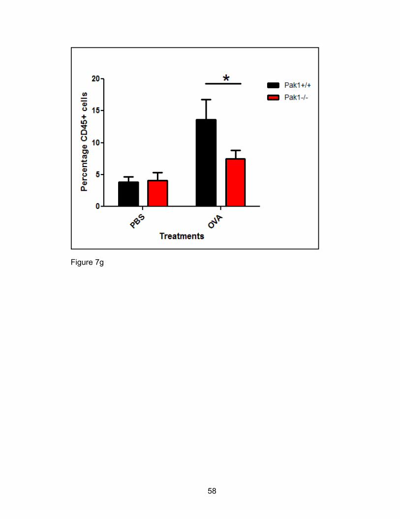
58
Figure 7g

59
Figure 7: PAK1 promotes eosinophil infiltration of lung tissue in a murine
model of AAD. Pak1+/+ and Pak1-/- mice were sensitized and challenged with
OVA to induce allergic airway inflammation or PBS for control. Lungs from these
mice were fixed, sectioned, and stained with H & E. Micrographs are
representative for at least five experiments (Figures 7a-f). Lung leukocytes from
these AAD mice were obtained following enzymatic digestion, mechanical
dissociation, and magnetic separation from lung tissue. Leukocytes were
assessed for lineage markers by multicolor fluorescence cytometry. On analysis,
leukocyte population gates (all CD45+) were set to distinguish the following
populations: Eosinophils, FSClo B220/CD3 (-), CCR3/SiglecF (+), Neutrophils,
FSClo B220/CD3 (-) CCR3/SiglecF(-), Lymphocytes FSClo B220/CD3 (+),
CCR3/SiglecF (-), and alveolar macrophages, FSChi, CDIIc or CD11b (+), MHCII
(+). Population percentages were compared for significant differences (p<0.05)
between Pak1-/- and Pak1+/+ samples N=6 (Figure 7g). Results are averaged
from three experiments and compared. * p<0.05, using a two-tailed student’s t-
test.
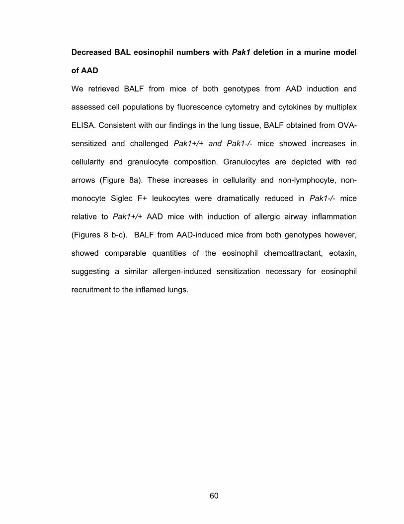
60
Decreased BAL eosinophil numbers with Pak1 deletion in a murine model
of AAD
We retrieved BALF from mice of both genotypes from AAD induction and
assessed cell populations by fluorescence cytometry and cytokines by multiplex
ELISA. Consistent with our findings in the lung tissue, BALF obtained from OVA-
sensitized and challenged Pak1+/+ and Pak1-/- mice showed increases in
cellularity and granulocyte composition. Granulocytes are depicted with red
arrows (Figure 8a). These increases in cellularity and non-lymphocyte, non-
monocyte Siglec F+ leukocytes were dramatically reduced in Pak1-/- mice
relative to Pak1+/+ AAD mice with induction of allergic airway inflammation
(Figures 8 b-c). BALF from AAD-induced mice from both genotypes however,
showed comparable quantities of the eosinophil chemoattractant, eotaxin,
suggesting a similar allergen-induced sensitization necessary for eosinophil
recruitment to the inflamed lungs.

61
Figure 8a
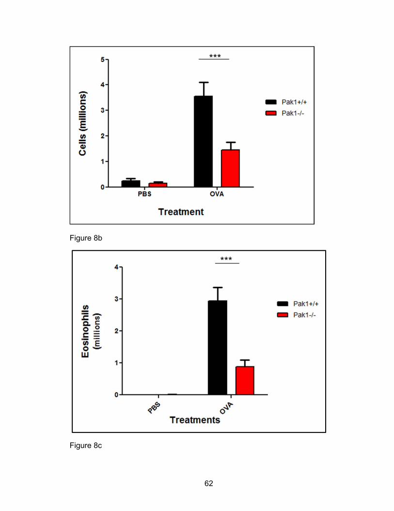
62
Figure 8b
Figure 8c
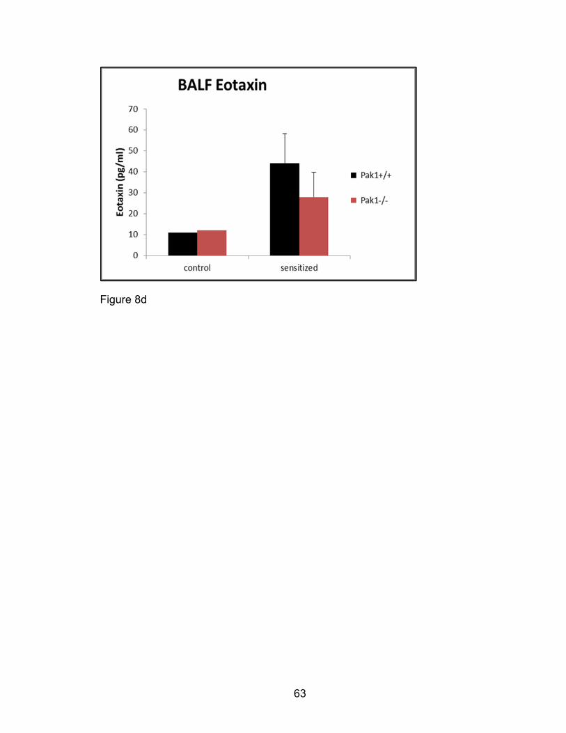
63
Figure 8d
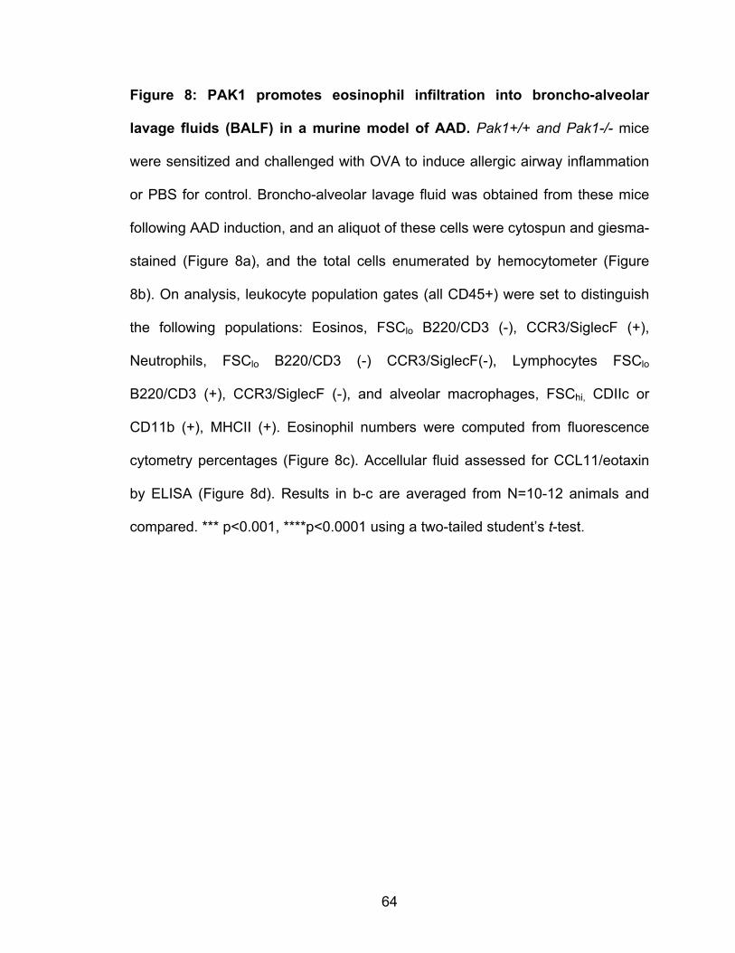
64
Figure 8: PAK1 promotes eosinophil infiltration into broncho-alveolar
lavage fluids (BALF) in a murine model of AAD. Pak1+/+ and Pak1-/- mice
were sensitized and challenged with OVA to induce allergic airway inflammation
or PBS for control. Broncho-alveolar lavage fluid was obtained from these mice
following AAD induction, and an aliquot of these cells were cytospun and giesma-
stained (Figure 8a), and the total cells enumerated by hemocytometer (Figure
8b). On analysis, leukocyte population gates (all CD45+) were set to distinguish
the following populations: Eosinos, FSClo B220/CD3 (-), CCR3/SiglecF (+),
Neutrophils, FSClo B220/CD3 (-) CCR3/SiglecF(-), Lymphocytes FSClo
B220/CD3 (+), CCR3/SiglecF (-), and alveolar macrophages, FSChi, CDIIc or
CD11b (+), MHCII (+). Eosinophil numbers were computed from fluorescence
cytometry percentages (Figure 8c). Accellular fluid assessed for CCL11/eotaxin
by ELISA (Figure 8d). Results in b-c are averaged from N=10-12 animals and
compared. *** p<0.001, ****p<0.0001 using a two-tailed student’s t-test.
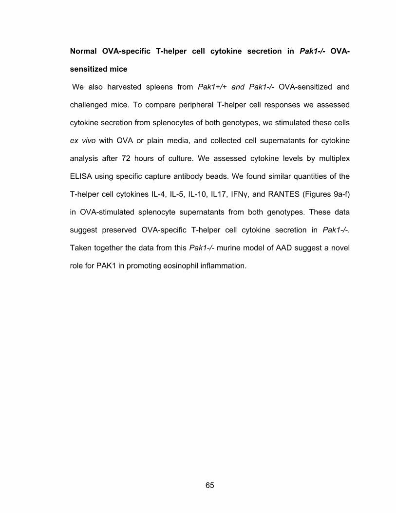
65
Normal OVA-specific T-helper cell cytokine secretion in Pak1-/- OVA-
sensitized mice
We also harvested spleens from Pak1+/+ and Pak1-/- OVA-sensitized and
challenged mice. To compare peripheral T-helper cell responses we assessed
cytokine secretion from splenocytes of both genotypes, we stimulated these cells
ex vivo with OVA or plain media, and collected cell supernatants for cytokine
analysis after 72 hours of culture. We assessed cytokine levels by multiplex
ELISA using specific capture antibody beads. We found similar quantities of the
T-helper cell cytokines IL-4, IL-5, IL-10, IL17, IFNγ, and RANTES (Figures 9a-f)
in OVA-stimulated splenocyte supernatants from both genotypes. These data
suggest preserved OVA-specific T-helper cell cytokine secretion in Pak1-/-.
Taken together the data from this Pak1-/- murine model of AAD suggest a novel
role for PAK1 in promoting eosinophil inflammation.
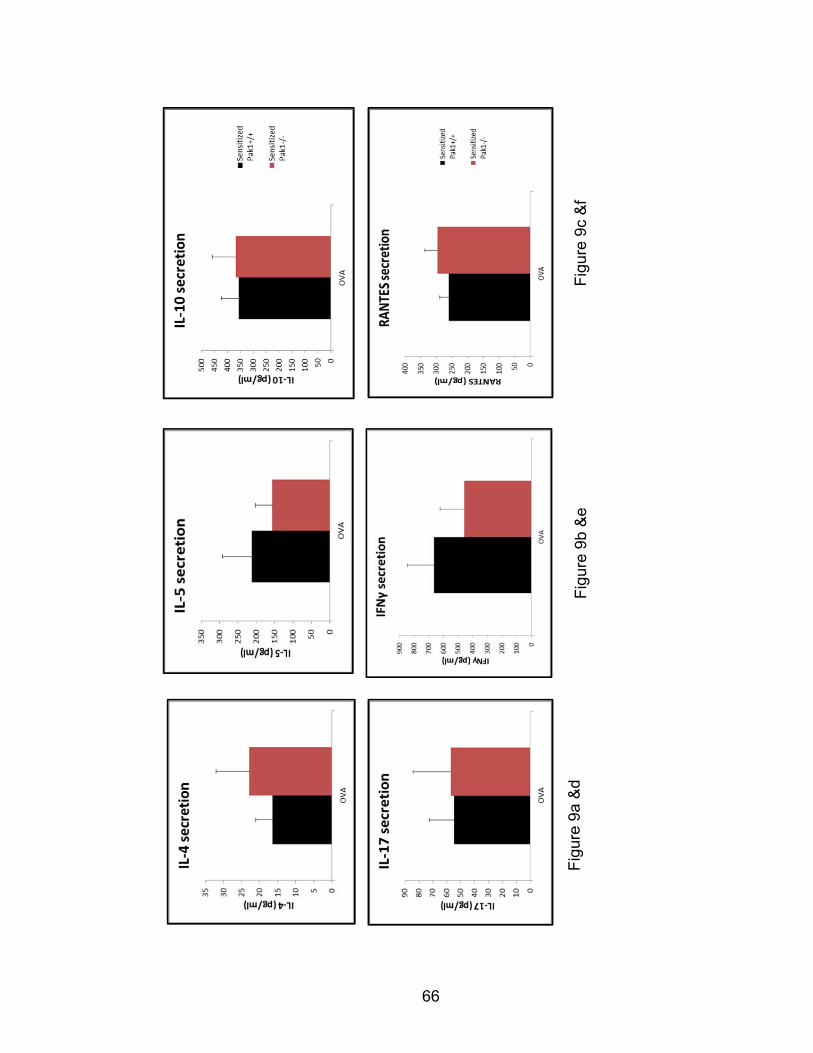
66
Fig
ure
9c &
f F
igur
e 9b
&e
Fig
ure
9a &
d

67
Figure 9: Pak1 genetic deletion does not affect OVA-specific splenocyte
secretion of T-helper cell cytokines. Total splenocytes from OVA-sensitized
and challenged Pak1+/+ and Pak1-/- mice were stimulated with OVA or plain
media and cultured ex vivo for 72 hours. Cell-free supernatants were collected
and stored for multiplex ELISA cytokine measurement of the following analytes:
IL-4, IL-5, IL-10, IL-17, IFNγ, and RANTES (Figures 9a-f). Data from N=7-8 mice
from each group were analyzed and compared between genotypes by a
student’s t-test.
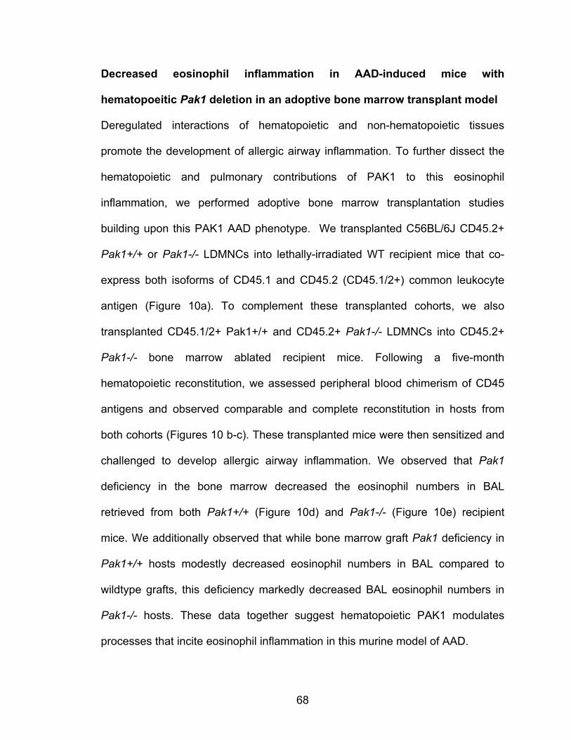
68
Decreased eosinophil inflammation in AAD-induced mice with
hematopoeitic Pak1 deletion in an adoptive bone marrow transplant model
Deregulated interactions of hematopoietic and non-hematopoietic tissues
promote the development of allergic airway inflammation. To further dissect the
hematopoietic and pulmonary contributions of PAK1 to this eosinophil
inflammation, we performed adoptive bone marrow transplantation studies
building upon this PAK1 AAD phenotype. We transplanted C56BL/6J CD45.2+
Pak1+/+ or Pak1-/- LDMNCs into lethally-irradiated WT recipient mice that co-
express both isoforms of CD45.1 and CD45.2 (CD45.1/2+) common leukocyte
antigen (Figure 10a). To complement these transplanted cohorts, we also
transplanted CD45.1/2+ Pak1+/+ and CD45.2+ Pak1-/- LDMNCs into CD45.2+
Pak1-/- bone marrow ablated recipient mice. Following a five-month
hematopoietic reconstitution, we assessed peripheral blood chimerism of CD45
antigens and observed comparable and complete reconstitution in hosts from
both cohorts (Figures 10 b-c). These transplanted mice were then sensitized and
challenged to develop allergic airway inflammation. We observed that Pak1
deficiency in the bone marrow decreased the eosinophil numbers in BAL
retrieved from both Pak1+/+ (Figure 10d) and Pak1-/- (Figure 10e) recipient
mice. We additionally observed that while bone marrow graft Pak1 deficiency in
Pak1+/+ hosts modestly decreased eosinophil numbers in BAL compared to
wildtype grafts, this deficiency markedly decreased BAL eosinophil numbers in
Pak1-/- hosts. These data together suggest hematopoietic PAK1 modulates
processes that incite eosinophil inflammation in this murine model of AAD.

69
Figure 10a
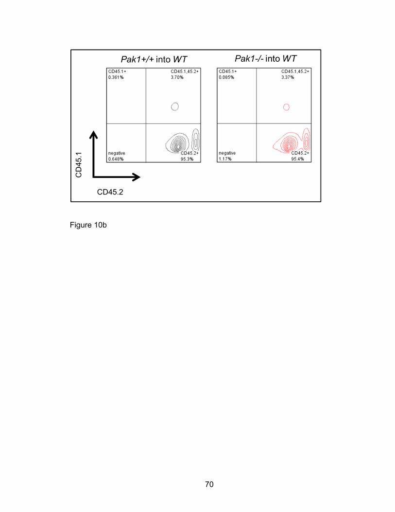
70
Figure 10b
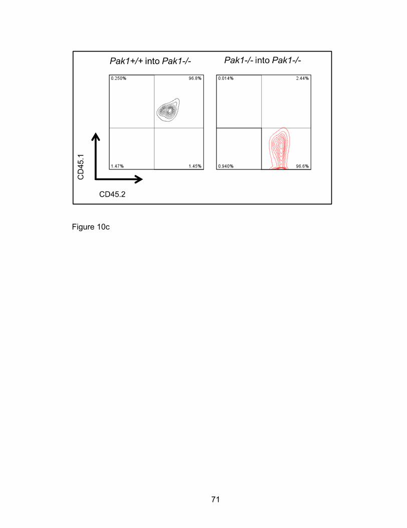
71
Figure 10c
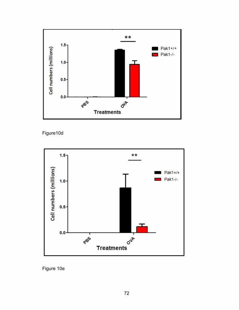
72
Figure10d
Figure 10e
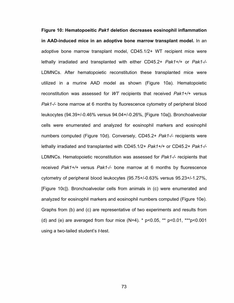
73
Figure 10: Hematopoeitic Pak1 deletion decreases eosinophil inflammation
in AAD-induced mice in an adoptive bone marrow transplant model. In an
adoptive bone marrow transplant model, CD45.1/2+ WT recipient mice were
lethally irradiated and transplanted with either CD45.2+ Pak1+/+ or Pak1-/-
LDMNCs. After hematopoietic reconstitution these transplanted mice were
utilized in a murine AAD model as shown (Figure 10a). Hematopoietic
reconstitution was assessed for WT recipients that received Pak1+/+ versus
Pak1-/- bone marrow at 6 months by fluorescence cytometry of peripheral blood
leukocytes (94.39+/-0.46% versus 94.04+/-0.26%, [Figure 10a]). Bronchoalveolar
cells were enumerated and analyzed for eosinophil markers and eosinophil
numbers computed (Figure 10d). Conversely, CD45.2+ Pak1-/- recipients were
lethally irradiated and transplanted with CD45.1/2+ Pak1+/+ or CD45.2+ Pak1-/-
LDMNCs. Hematopoietic reconstitution was assessed for Pak1-/- recipients that
received Pak1+/+ versus Pak1-/- bone marrow at 6 months by fluorescence
cytometry of peripheral blood leukocytes (95.75+/-0.63% versus 95.23+/-1.27%,
[Figure 10c]). Bronchoalveolar cells from animals in (c) were enumerated and
analyzed for eosinophil markers and eosinophil numbers computed (Figure 10e).
Graphs from (b) and (c) are representative of two experiments and results from
(d) and (e) are averaged from four mice (N=4). * p<0.05, ** p<0.01, ***p<0.001
using a two-tailed student’s t-test.
74
Decreased eosinophil inflammation in AAD-induced mice with
hematopoietic Pak1 deletion in an orthotopic lung transplant model
Bone marrow transplants, usually used to study hematopoiesis require ionizing
radiation that irreparably disrupts the pulmonary resident cells and architecture in
transplant recipient mice. To further investigate the hematopoietic and pulmonary
contributions of PAK1 to allergic airway inflammation, we collaborated with Dr.
David Wilkes’ lab to transplant left lung grafts into Pak1+/+ and Pak1-/- mice.
We separately transplanted Pak1-/- lungs into Pak1+/+ mice and conversely
Pak1+/+ left lungs into Pak1-/- recipient mice. Following a recovery period, we
OVA-sensitized and challenged these transplanted mice in a murine AAD model
(Figure 11a).
We found that Pak1-/- transplanted lungs in Pak1+/+ recipient mice
demonstrated more eosinophilic inflammation than Pak1+/+ transplanted lungs in
Pak1-/- recipients on H&E stained sections (Figures 11b-e) despite comparable
goblet cell metaplastic changes characteristic of AAD between the two groups
(Figures 11f-g). In these preliminary studies, three out of four (3/4) transplants in
the groups of Pak1+/+ mice transplanted with Pak1-/- lungs had peribronchiolar
eosinophil inflammation on histological inspection. Conversely, none of four (0/4)
transplants in the group of Pak1-/- mice transplanted with Pak1+/+ lungs had
these lesions. Likewise, slides from Pak1+/+ mice transplanted with Pak1+/+
lungs, but not Pak1-/- mice transplanted with Pak1-/- lungs demonstrated
eosinophil lesions (Table 1). The chi-square association between transplanted

75
mice with Pak1+/+ versus Pak1-/- hematopoietic cells and peribronchiolar
eosinophil lesions was statistically significant.
In subsequent studies with separate cohorts of mice, we transplanted Pak1-/- left
lung into Pak1+/+ mice and conversely Pak1+/+ left lungs into Pak1-/- recipient
mice. We procured lung grafts from these mice, and enzymatically digested
whole lung tissue to quantify eosinophils by fluorescence cytometry. Consistent
with results from our AAD model, we saw a trend towards decreased total BAL
eosinophil numbers (Figure 11h) and percentages (Figure 11i) in Pak1-/- hosts
relative to Pak1+/+ hosts. Leukocytes retrieved from native right lung (Figure 11j)
and engrafted left lung (Figure 11k) revealed a trend towards decreased non-
lymphocyte, non-monocyte Siglec F+ cells in Pak1-/- hosts compared with
Pak1+/+ hosts. Future studies are needed to resolve these trends by increasing
the number of animals and the experimental power. However, taken together
these histological and quantitative data suggest a role for host/hematopoietic
PAK1 modulation in eosinophil inflammation in a murine model of AAD.
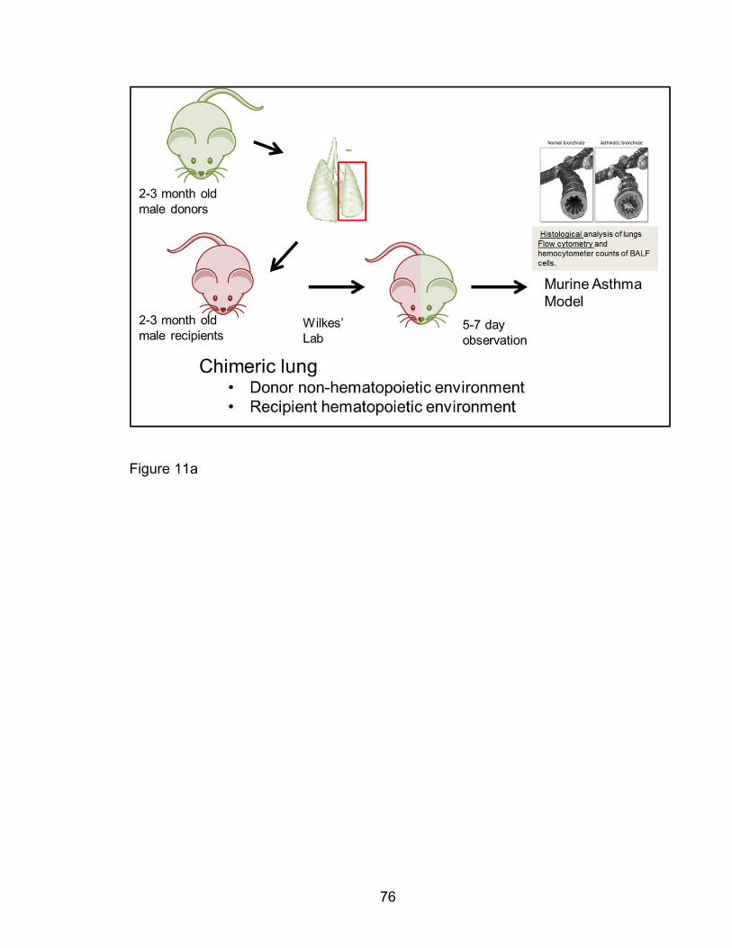
76
Figure 11a
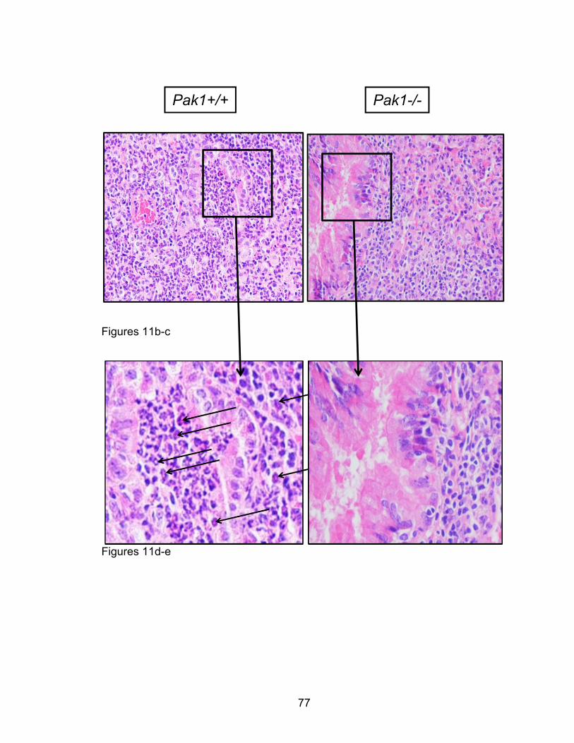
77
Figures 11b-c
Figures 11d-e
Pak1+/+ Pak1-/-
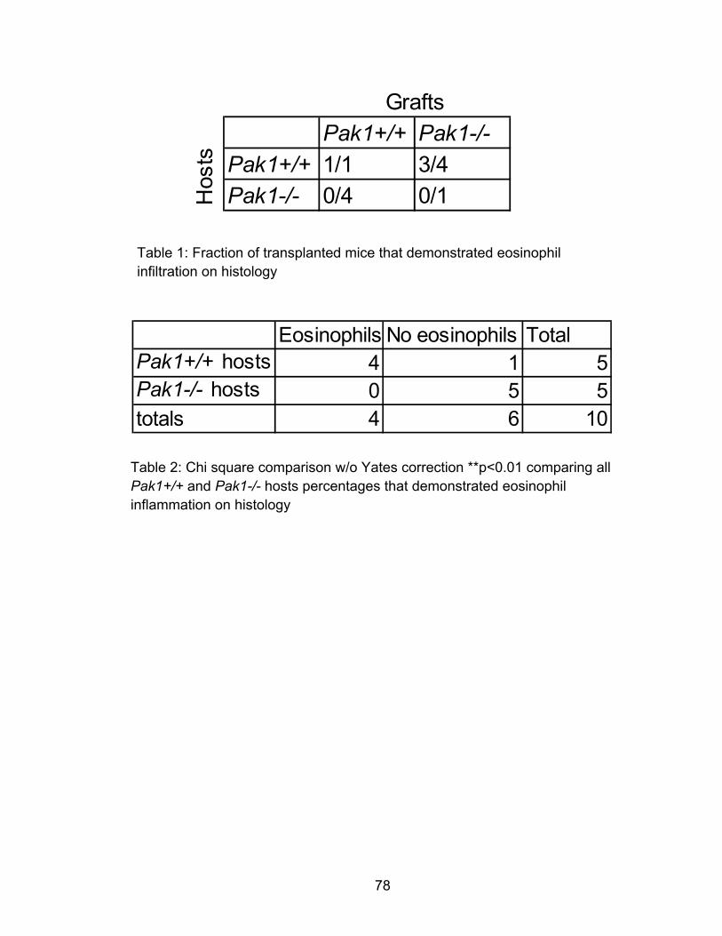
78
Pak1+/+ Pak1-/-Pak1+/+ 1/1 3/4
Pak1-/- 0/4 0/1
Grafts
Hos
ts
Table 1: Fraction of transplanted mice that demonstrated eosinophil infiltration on histology
Table 2: Chi square comparison w/o Yates correction **p<0.01 comparing all Pak1+/+ and Pak1-/- hosts percentages that demonstrated eosinophil inflammation on histology
Eosinophils No eosinophils TotalPak1+/+ hosts 4 1 5Pak1-/- hosts 0 5 5totals 4 6 10
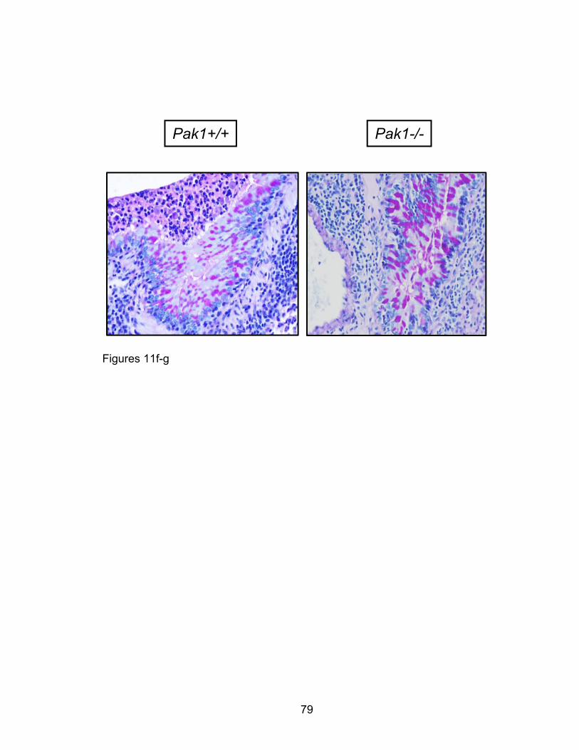
79
Figures 11f-g
Pak1+/+ Pak1-/-
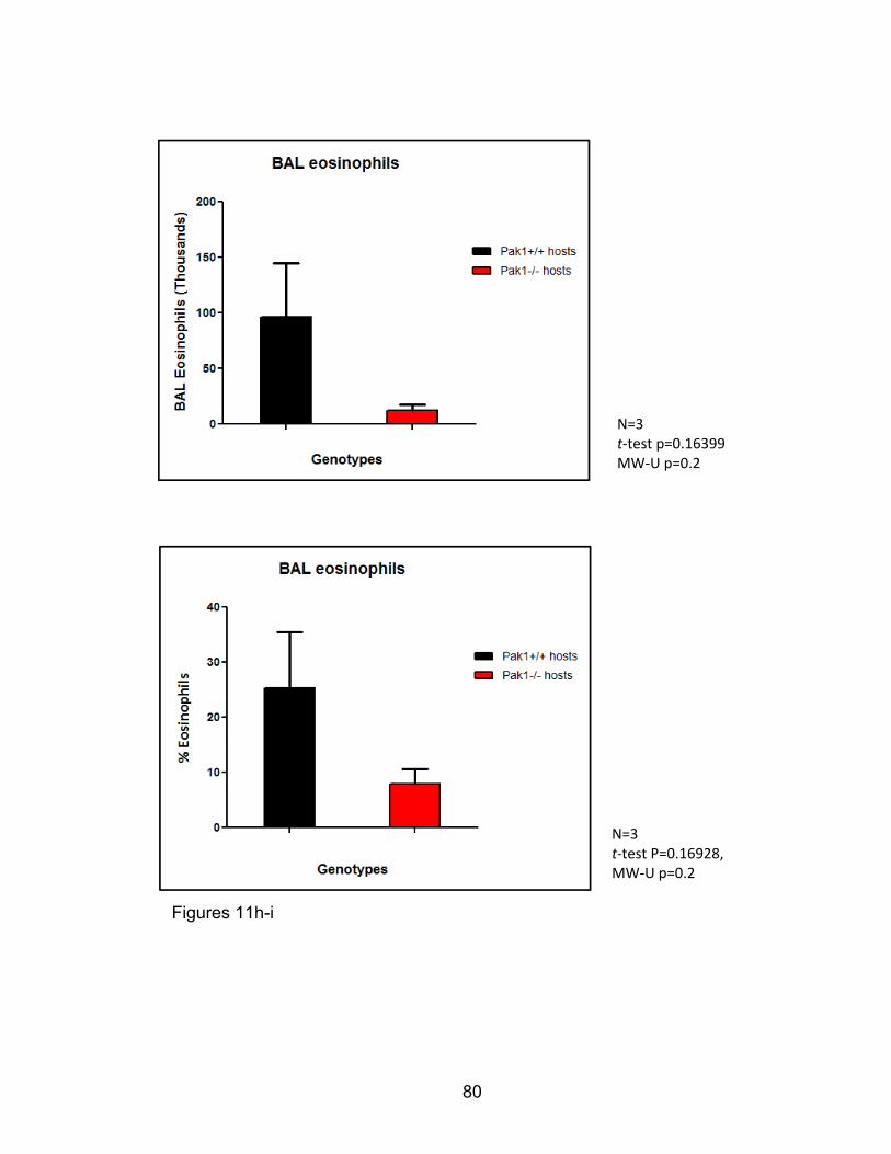
80
N=3 t‐test P=0.16928, MW‐U p=0.2
N=3 t‐test p=0.16399
MW‐U p=0.2
Figures 11h-i

81
N=3 t‐test p=0.129 MW‐U p=0.2
N=3 t‐test p=0.08356 MW‐U p=0.1
Figures 11j-k
82
Figure 11: Hematopoeitic Pak1 deletion may decrease eosinophil
inflammation in AAD-induced mice in an orthotopic lung transplant model.
In an orthotopic lung transplant model, we transplanted lungs from Pak1-/- mice
into Pak1+/+ host mice. Conversely we transplanted Pak1+/+ left lungs into
Pak1-/- recipient mice as shown. Following a five day recovery, we utilized these
mice in a murine model of allergic airway inflammation. We prepared H&E
(Figures 11b-e) and PAS (Figures 11f-g) sections of left lung grafts from these
mice. Micrographs from (b-g) are representative of four mice (N=4). On
histological inspection of H&E stained micrographs presence of peribronchiolar
eosinophilic lesions was scored (Table 1) and chi-square association computed
for Pak1+/+ versus Pak1-/- hosts (Table 2) p<0.01.
In a separate cohort, we collected, enumerated, and stained BAL cells from test
transplanted and OVA-sensitized and challenged Pak1+/+ and Pak1-/- mice for
eosinophil markers and obtained eosinophil percentages by fluorescence
cytometry (Figures 11h-i). We also obtained total leukocytes from native right
lungs and the engrafted left lungs and stained BAL cells from these mice for
eosinophil markers and obtained eosinophil percentages by fluorescence
cytometry (Figures 11j-k). Results from (h) through (k) are from three mice (N=3)
and do not yet show statistical significance.

83
PAK1 in Eotaxin- mediated Eosinophil function
Building upon our whole animal studies, we sought to determine PAK1’s
functional and molecular role in regulating eosinophil function in allergic airway
inflammation. Allergic sensitization induces expression and secretion of
chemokines and cytokines that recruit hematopoietic cells eliciting inflammation
in airways. Eotaxin expression in allergic airway inflammation potently recruits
eosinophils en masse to the lung effecting chronic inflammation. Eotaxins also
elicit eosinophil degranulation and cationic protein release mediating damage
and remodeling in lung tissue. We hypothesized that PAK1 modulates eotaxin-
mediated eosinophil infiltration and degranulation. To investigate the role PAK1
plays in these eotaxin-mediated eosinophil functions, we used two in vivo eotaxin
infiltration murine models. We further evaluated the mechanism of PAK1-
dependent eosinophil recruitment in vitro using bone-marrow derived eosinophils.

84
Eotaxin-mediated Eosinophil infiltration in vivo is PAK1-dependent
Given our previous findings in whole and chimeric animal AAD studies, we
assessed isolated eotaxin recruitment of eosinophils in vivo to explain the
apparent defect in eosinophil recruitment to the lungs in AAD Pak1-/- mice as
compared to their wild-type counterparts. We injected a dose of intraperitoneal or
intratracheal eotaxin in sedated Pak1+/+ and Pak1-/- mice and collected
peritoneal or broncho-alveolar lavage fluids for cell population evaluation by
fluorescent cytometry. We observed from giesma-stained slides of peritoneal
lavage cells obtained from eotaxin-treated Pak1-/- mice an increase in eosin-
staining cells. Similarly, giesma-stained and quantified peritoneal lavages cells
(Figures 12c-d) and fluorescence cytometry quantified CD45+, CD3/B220,
SiglecF+ broncho-alveolar lavage fluid cells (Figures 12e-f) from Pak1-/- mice
were increased with eotaxin treatment but decreased compared to cells in fluids
retrieved from eotaxin-treated Pak1+/+ mice (Figures 12a-d).
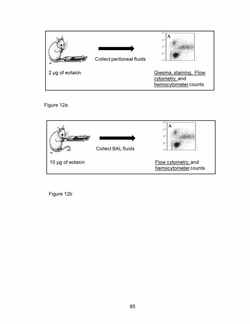
85
Figure 12a
Figure 12b

86
Figure 12c
Figure 12d
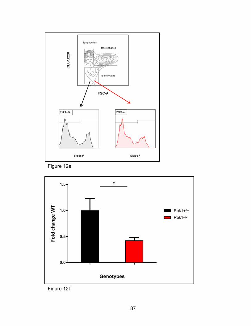
87
Figure 12e
Figure 12f
88
Figure 12: Eotaxin-mediated infiltration in vivo is PAK1-dependent. Pak1+/+
and Pak1-/- mice were treated with PBS and eotaxin by intraperitoneal or
intratracheal injections. Leukocytes from the respective cavities were lavaged
and assessed for eosinophil numbers after 2 and 4 hours according to the
schemes above (Figures 12a-b). Eosinophils were identified by giesma-staining
and total CCR3+ cells back calculated and compared between genotypes
(Figures 12c-d). Similarly, non-lymphocyte and non-monocyte populations of BAL
cells were analyzed for SiglecF expression and SiglecF+ cells calculated and
compared between genotypes (Figures 12e-f). Results in (d) and (f) are
averaged from 4-6 mice and representative of at least three independent
experiments. p<0.05, ** p<0.01, using a two-tailed student’s t-test.
89
IL-5 LDMNC colony formation in vitro is not PAK1-dependent
In order to study eosinophil function in vitro, we chose to differentiate eosinophils
from bone marrow stem and progenitor cells as previously published [244, 245].
The impact of Pak1 deletion on stem and progenitor cell differentiation is
incompletely understood and remains unexplored in eosinophil differentiation. IL-
5 critically coordinately controls murine eosinophil differentiation in vivo and in
vitro and reproducibly drives the formation of murine eosinophil colonies in
methocellulose agar [245, 246]. To assess the impact of Pak1 deletion on stem
and progenitor capacity to form IL-5 colonies we performed an IL-5 colony
forming assay using Pak1 sufficient and deficient bone marrow derived
mononuclear cells. We plated mononuclear cells in methocellulose semi solid
agar medium enriched with high and low concentrations of IL-5 as the only
growth and differentiation factor, incubated these cells, and counted total
colonies on the tenth day of culture as previously described [236]. We showed
that IL-5 stimulation resulted in growth of both Pak1+/+ and Pak1-/- colonies.
However we also observed no difference in total number of colonies between the
two genotypes (Figure 13). These results suggest that IL-5 induced bone marrow
mononuclear cell colony formation does not depend on PAK1.

90
Figure 13
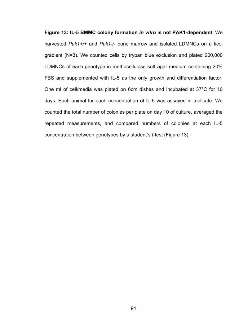
91
Figure 13: IL-5 BMMC colony formation in vitro is not PAK1-dependent. We
harvested Pak1+/+ and Pak1-/- bone marrow and isolated LDMNCs on a ficol
gradient (N=3). We counted cells by trypan blue exclusion and plated 200,000
LDMNCs of each genotype in methocellulose soft agar medium containing 20%
FBS and supplemented with IL-5 as the only growth and differentiation factor.
One ml of cell/media was plated on 6cm dishes and incubated at 37°C for 10
days. Each animal for each concentration of IL-5 was assayed in triplicate. We
counted the total number of colonies per plate on day 10 of culture, averaged the
repeated measurements, and compared numbers of colonies at each IL-5
concentration between genotypes by a student’s t-test (Figure 13).

92
Eosinophil expression of transmigration integrins as well as adhesion to
fibronectin is not PAK1-dependent
Reorganization of the cell cytoskeleton facilitates rolling, adhesion, and
transmigration of leukocytes as they infiltrate sites of tissue inflammation.
Effective transmigration requires coordination between adhesive and motile
forces, which are controlled by extracellular matrix signals acting on the cell
cytoskeleton. CDC42/Racs, activators of PAKs, are implicated in optimally
balancing between these processes [247, 248]. Eotaxin acts as a potent
chemoattractant for eosinophil infiltration in vivo as well as adhesion of
eosinophils to fibronectin [249, 250]. We sought to determine the effect of Pak1
deletion on expression of adhesive proteins and eotaxin-mediated eosinophil
adhesion. We cultured Pak1+/+ and Pak1-/- bmEos and assessed the
expression of eosinophil adhesive molecules important for endothelial
transmigration by fluorescence cytometry. We observed no difference in the
frequency of cells expressing these proteins, with greater than 95% of cells
expressing CD11a/αL, Mac1/αMβ2, α4β7, and VLA4/α4β1 in both genotypes
(Figures 14a-d). This global surface integrin expression was maintained following
eotaxin stimulation (data not shown). Likewise, MFIs denoting surface expression
of these integrins was comparable between genotypes. We used Pak1+/+ and
Pak1-/- bmEos in a microfluidic chamber adhesion assay to fibronectin. We
coated microfluidic plates with gelatin or recombinant fibronectin fragments for
one hour. Eosinophils showed preferential eotaxin-induced adherence to
fibronectin in contrast to gelatin (Figure 14e). A micro-fluid pressure-gradient

93
maintained steady-flow of Pak1+/+ and Pak1-/- bmEos in media through the
fibronectin-coated chambers for 30 minutes. Micrographs (Figure 14f) and
hemocytometer enumeration (Figure 14g) of total adherent cells both
demonstrate indistinguishable eotaxin-mediated eosinophil adhesion between
the two genotypes. These data warrant further functional characterization of
PAK1’s role in modulating eotaxin-mediated eosinophil adhesion via CD11a/LFA-
1, Mac1, α4β7, and VLA4 interactions with recombinant fragments of the
endothelium-expressed ICAM, VCAM, and MadCAM adhesive molecules [251-
253].

94
Figure 14a
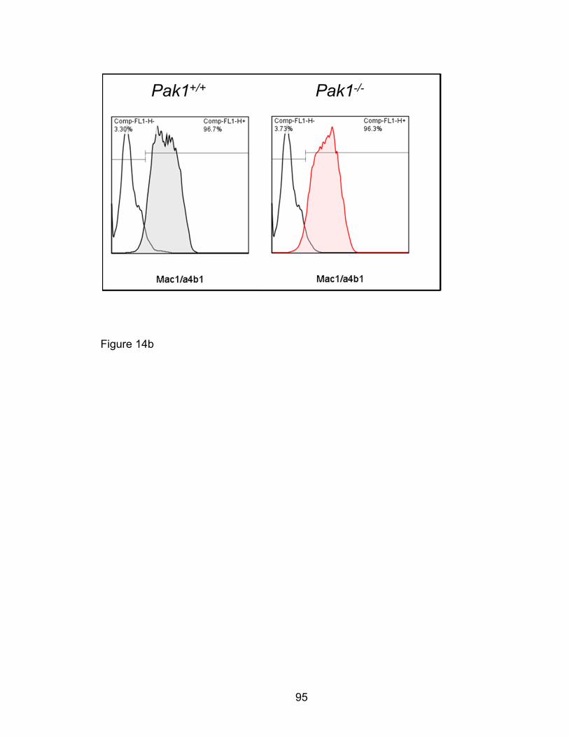
95
Figure 14b

96
Figure 14c
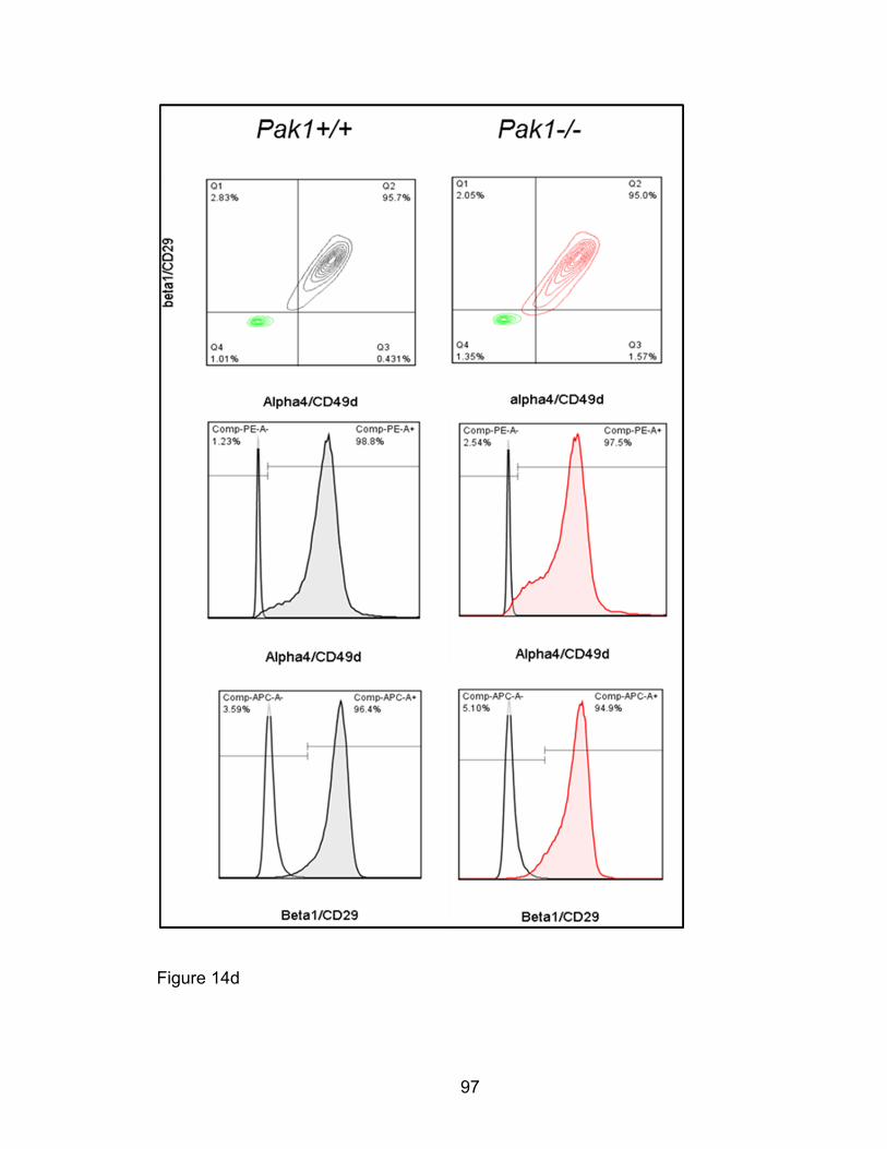
97
Figure 14d

98
Figure 14e
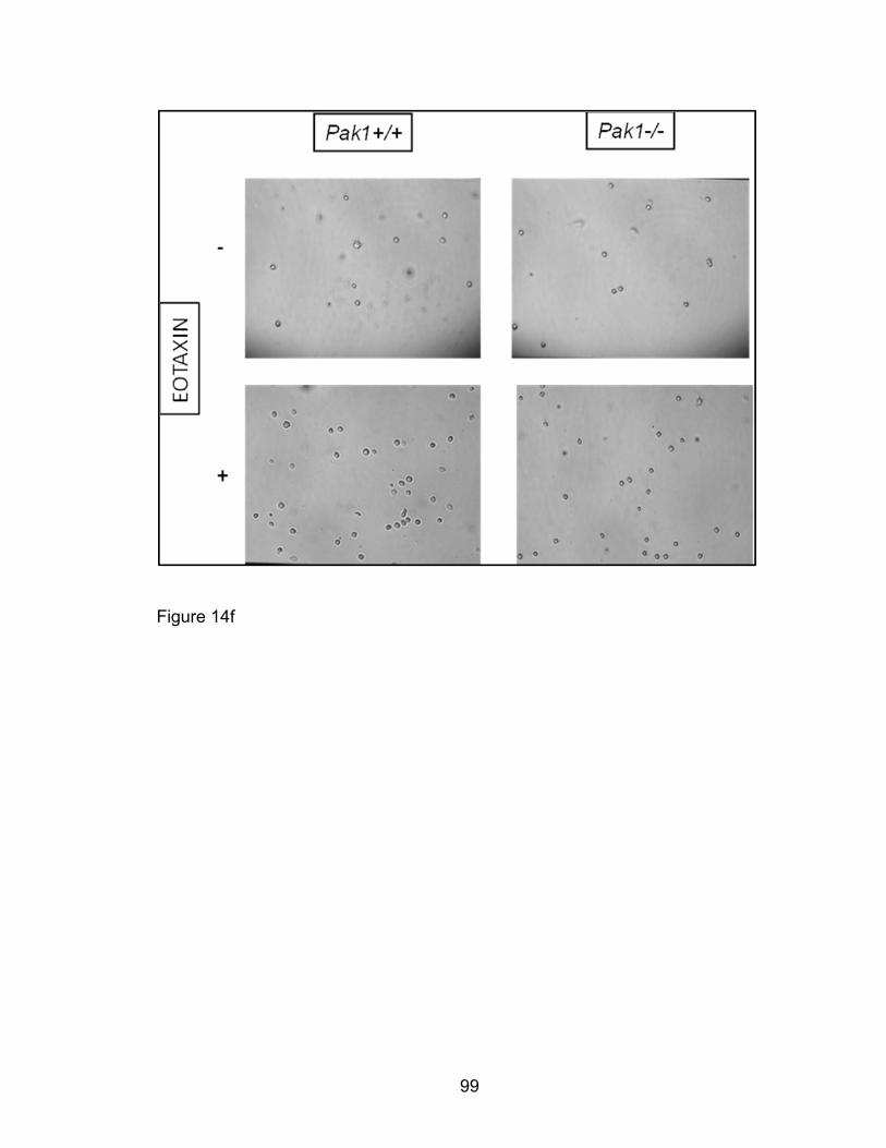
99
Figure 14f

100
Figure 14g
Eotaxin
Fold
cha
nge
non-
stim
adh
esio
n
- +0
1
2
3
4Pak1+/+
Pak1-/-
NS
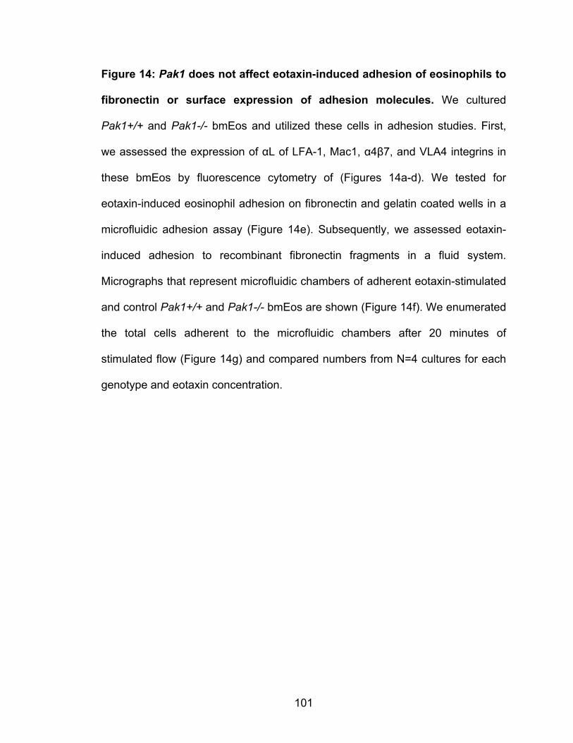
101
Figure 14: Pak1 does not affect eotaxin-induced adhesion of eosinophils to
fibronectin or surface expression of adhesion molecules. We cultured
Pak1+/+ and Pak1-/- bmEos and utilized these cells in adhesion studies. First,
we assessed the expression of αL of LFA-1, Mac1, α4β7, and VLA4 integrins in
these bmEos by fluorescence cytometry of (Figures 14a-d). We tested for
eotaxin-induced eosinophil adhesion on fibronectin and gelatin coated wells in a
microfluidic adhesion assay (Figure 14e). Subsequently, we assessed eotaxin-
induced adhesion to recombinant fibronectin fragments in a fluid system.
Micrographs that represent microfluidic chambers of adherent eotaxin-stimulated
and control Pak1+/+ and Pak1-/- bmEos are shown (Figure 14f). We enumerated
the total cells adherent to the microfluidic chambers after 20 minutes of
stimulated flow (Figure 14g) and compared numbers from N=4 cultures for each
genotype and eotaxin concentration.
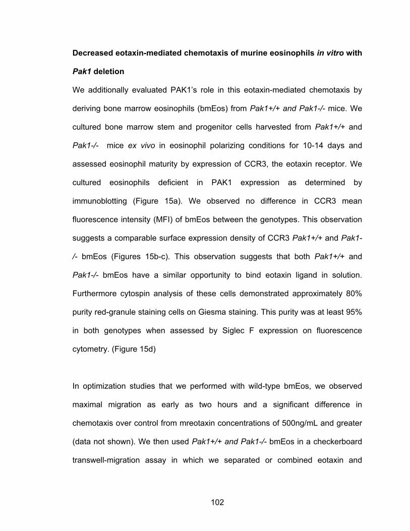
102
Decreased eotaxin-mediated chemotaxis of murine eosinophils in vitro with
Pak1 deletion
We additionally evaluated PAK1’s role in this eotaxin-mediated chemotaxis by
deriving bone marrow eosinophils (bmEos) from Pak1+/+ and Pak1-/- mice. We
cultured bone marrow stem and progenitor cells harvested from Pak1+/+ and
Pak1-/- mice ex vivo in eosinophil polarizing conditions for 10-14 days and
assessed eosinophil maturity by expression of CCR3, the eotaxin receptor. We
cultured eosinophils deficient in PAK1 expression as determined by
immunoblotting (Figure 15a). We observed no difference in CCR3 mean
fluorescence intensity (MFI) of bmEos between the genotypes. This observation
suggests a comparable surface expression density of CCR3 Pak1+/+ and Pak1-
/- bmEos (Figures 15b-c). This observation suggests that both Pak1+/+ and
Pak1-/- bmEos have a similar opportunity to bind eotaxin ligand in solution.
Furthermore cytospin analysis of these cells demonstrated approximately 80%
purity red-granule staining cells on Giesma staining. This purity was at least 95%
in both genotypes when assessed by Siglec F expression on fluorescence
cytometry. (Figure 15d)
In optimization studies that we performed with wild-type bmEos, we observed
maximal migration as early as two hours and a significant difference in
chemotaxis over control from mreotaxin concentrations of 500ng/mL and greater
(data not shown). We then used Pak1+/+ and Pak1-/- bmEos in a checkerboard
transwell-migration assay in which we separated or combined eotaxin and

103
bmEos in the top transwell chamber and allowed the cells to migrate for two
hours according to the schematic below (Figure 15e). We collected the cells
migrating to the bottom well in each condition and expressed migration as a
chemotactic index of background. We only observed a significant difference in
migration over background levels when we applied an eotaxin-gradient to the
bmEos of both genotypes. Remarkably, Pak1-/- bmEos demonstrated 40%
decreased chemotaxis compared with Pak1+/+ cells (Figure 15d). These data
taken together implicate eosinophil PAK1 in regulating infiltration by intrinsically
promoting eotaxin- mediated eosinophil chemotaxis.
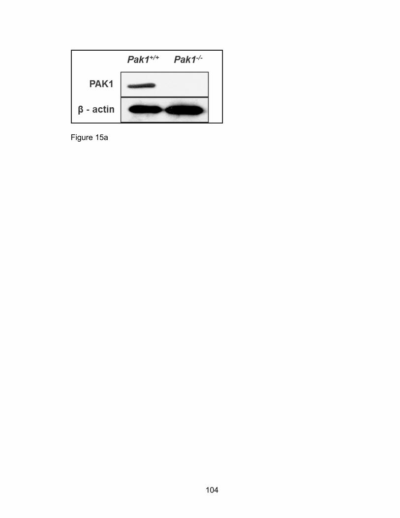
104
Figure 15a

105
Figure 15b

106
Figure 15c
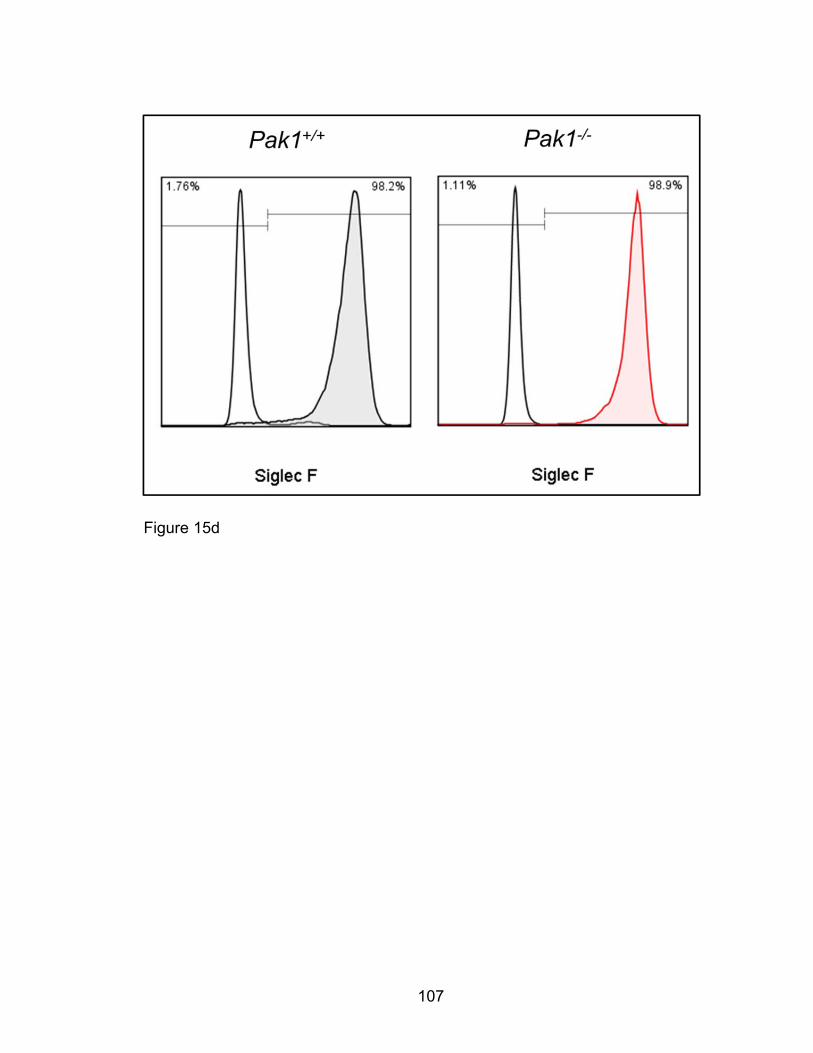
107
Figure 15d

108
Figure 15e
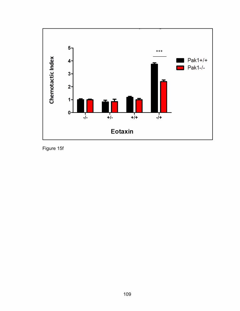
109
Figure 15f
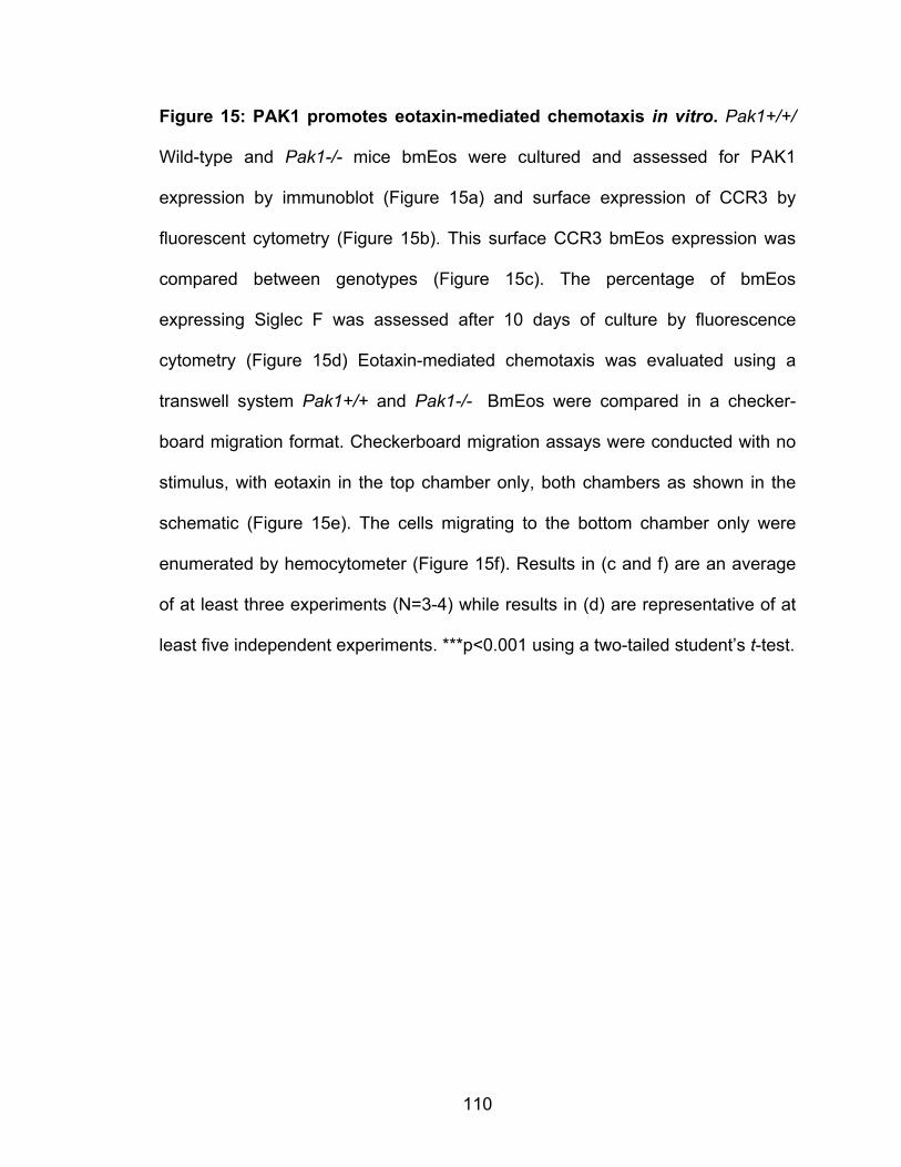
110
Figure 15: PAK1 promotes eotaxin-mediated chemotaxis in vitro. Pak1+/+/
Wild-type and Pak1-/- mice bmEos were cultured and assessed for PAK1
expression by immunoblot (Figure 15a) and surface expression of CCR3 by
fluorescent cytometry (Figure 15b). This surface CCR3 bmEos expression was
compared between genotypes (Figure 15c). The percentage of bmEos
expressing Siglec F was assessed after 10 days of culture by fluorescence
cytometry (Figure 15d) Eotaxin-mediated chemotaxis was evaluated using a
transwell system Pak1+/+ and Pak1-/- BmEos were compared in a checker-
board migration format. Checkerboard migration assays were conducted with no
stimulus, with eotaxin in the top chamber only, both chambers as shown in the
schematic (Figure 15e). The cells migrating to the bottom chamber only were
enumerated by hemocytometer (Figure 15f). Results in (c and f) are an average
of at least three experiments (N=3-4) while results in (d) are representative of at
least five independent experiments. ***p<0.001 using a two-tailed student’s t-test.
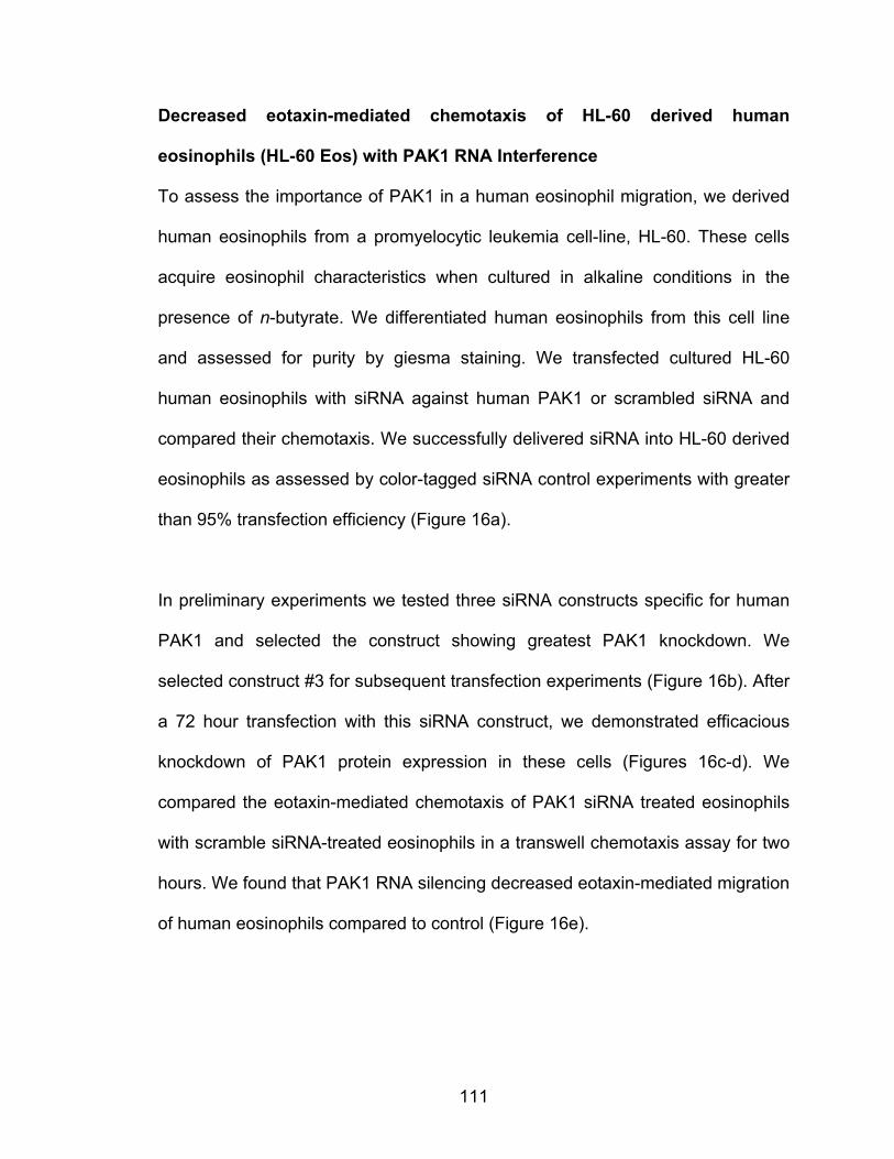
111
Decreased eotaxin-mediated chemotaxis of HL-60 derived human
eosinophils (HL-60 Eos) with PAK1 RNA Interference
To assess the importance of PAK1 in a human eosinophil migration, we derived
human eosinophils from a promyelocytic leukemia cell-line, HL-60. These cells
acquire eosinophil characteristics when cultured in alkaline conditions in the
presence of n-butyrate. We differentiated human eosinophils from this cell line
and assessed for purity by giesma staining. We transfected cultured HL-60
human eosinophils with siRNA against human PAK1 or scrambled siRNA and
compared their chemotaxis. We successfully delivered siRNA into HL-60 derived
eosinophils as assessed by color-tagged siRNA control experiments with greater
than 95% transfection efficiency (Figure 16a).
In preliminary experiments we tested three siRNA constructs specific for human
PAK1 and selected the construct showing greatest PAK1 knockdown. We
selected construct #3 for subsequent transfection experiments (Figure 16b). After
a 72 hour transfection with this siRNA construct, we demonstrated efficacious
knockdown of PAK1 protein expression in these cells (Figures 16c-d). We
compared the eotaxin-mediated chemotaxis of PAK1 siRNA treated eosinophils
with scramble siRNA-treated eosinophils in a transwell chemotaxis assay for two
hours. We found that PAK1 RNA silencing decreased eotaxin-mediated migration
of human eosinophils compared to control (Figure 16e).

112
Figure 16a

113
Figure 16b
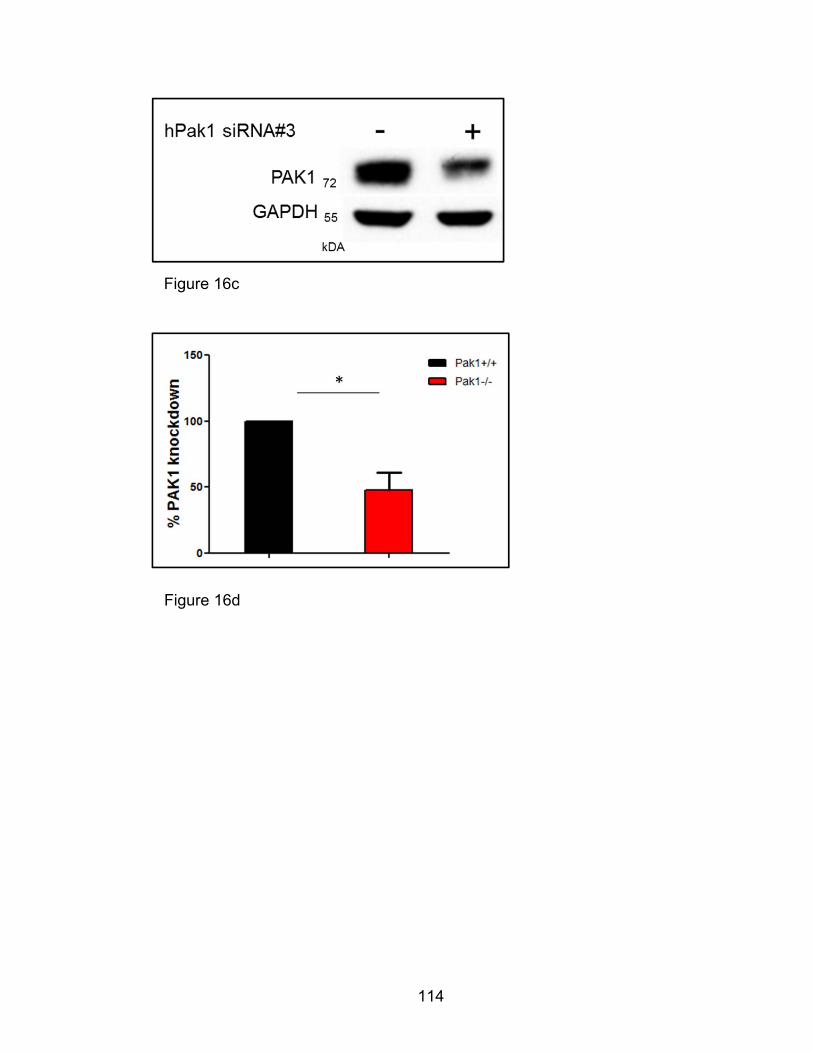
114
Figure 16d
Figure 16c
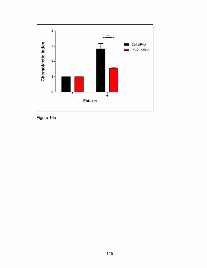
115
Figure 16e
116
Figure 16: PAK1 promotes decreases eotaxin-mediated chemotaxis in HL-
60 derived human eosinophils. We cultured HL-60 clone 15 cells in eosinophil
differentiating conditions for three days and tranfected them with color-tagged
sequence as a positive control, scramble siRNA, or three siRNA constructs
against human PAK1. Transfection efficiency was assessed using color-tagged
siRNA (Figure 16a). Three PAK1 siRNA constructs were screened for PAK1
knockdown by western blot and construct marked #3 selected for subsequent
studies (Figure 16b). PAK1 siRNA (#3) transfected cells used in migration
studies were assessed for PAK1 expression 72 hours after siRNA treatment by
western blot (Figure 16c) and percentage knockdown quantified by densitometry
(Figure 16d) for N=3 independent cultures. Migration of human eosinophil
derived from HL-60 clone 15 cell line transfected with scramble or PAK1 siRNA
were compared from N=5 independent cultures for each siRNA construct (Figure
16e). Results in (d) are representative of at least three experiments. ** p<0.01,
***p<0.001 using a two-tailed student’s t-test.

117
Restored and enhanced eotaxin-mediated eosinophil migration with
ectopic expression of full length PAK1 but not kinase-dead mutant PAK1 in
Pak1-/- eosinophils
Gene ablation by homologous recombination can occasionally result in the
disruption of surrounding genes. Since preliminary studies demonstrate that
Pak1-/- eosinophils have impaired migration, we validated the specificity of this
knockout by ectopically expressing PAK1 to restore eosinophil migration. We
utilized a state-of-the-art lentiviral-mediated transgene expression system to
overexpress PAK1, and a kinase-dead PAK1 mutant construct, K299R. We
adapted and ligated PAK1 cDNA into the pCL1eGFPGwo lentiviral backbone
developed, optimized and kindly given to us by our collaborator Dr. Helmut
Hanenberg as described [223].
Likewise, we obtained the pCMV6M-Pak1 K299R deposited at Addgene as a
generous gift by Dr. Jonathan Chernoff (Figure 17a). Using molecular techniques
we NOTI-adapted and PCR-amplified Pak1 K299R yielding approximately a 1620
base pair insert by gel analysis (Figure 17c). We used the PCL11eGwo lentiviral
vector for this construct, another generous gift from Dr. Hanenberg. This vector
utilizes an HPGK promoter that expresses both PAK1K299R and EGFP
efficiently in myeloid cells. Following a NotI restriction enzyme digestion of the
PCL11eGwo lentiviral vector, we blunted this vector and NotI-pak1K299R-NotI
insert by Klenow treatment. We ligated vector and insert, transformed competent
cells, and picked colonies for plasmid restriction digests. To confirm the 5’->3’
118
orientation after ligation, we amplified these plasmids and subjected them to a
restriction digest with AgeI and BstbI that yielding a DNA fragment spanning the
eGFPPAK1 constructs. We obtained a DNA fragment of about 2000bp (Figure
17d). To confirm translation of our construct in mammalian cells, we transfected
HEK293T cells with these constructs and demonstrated the expression of eGFP
fusion proteins approximately 85kDA (Figure 17e). We sequenced the lentiviral
plasmid to confirm that Pak1 K299R cDNA is encoded, in frame and without
further mutations.
We generated viral particles in a HEK293T packaging cell line by co-transfecting
our Pak1 plasmid constructs and a recombinant foamy virus envelope plasmid
that enhances entry into both quiescent stem cells and proliferating progenitor
cells as described [15]. We filtered and concentrated viral supernatant fifty times
to yield viral titers of between 5 X10⁷ to 5 X10⁸ particles per milliliter.
We magnetically sorted for c-Kit+ cells from LDMNCs for Pak1+/+ and Pak1-/-
bone marrow to enrich for stem and progenitor cells. We transduced these cells
with PCL11eGwo, PCL1Pak1, and PCL11Pak1K299R at multiplicities of infection
(MOIs) of between 50 and 100 infectious particles per cell. The transduced cells
were cultured in eosinophil polarizing conditions and allowed to express these
transgenes. Seven days post-transduction, these cells were collected and sorted
according to GFP or GFP-fusion protein expression. We obtained yields of
approximately 10 to 20% GFP+ cells (Figure 17f). We continued culturing these
119
eosinophil precursors for three to five days post-FACS after which they were
utilized in transwell migration assays. These cells variably expressed Gr-1/Ly-6,
but expressed CCR3 and almost invariably, SiglecF, surface markers of
eosinophil maturity (Figure 17g).
We noted that eGFPPAK1 ectopic expression in Pak1-/- eosinophils enhanced
their eotaxin-induced chemotaxis (Figure 17h). This chemotactic index was
greater than Pak1+/+ eosinophils transduced with PCL11eGFP. However,
eGFPPAK1K299R ectopic expression did not change the chemotactic potential
of Pak1-/- eosinophils (Figure 17h). These data complement and validate our
findings in Pak1-/- eosinophils. Furthermore, these studies suggest that eotaxin-
mediated eosinophil migration is at least partly PAK1 kinase-dependent.
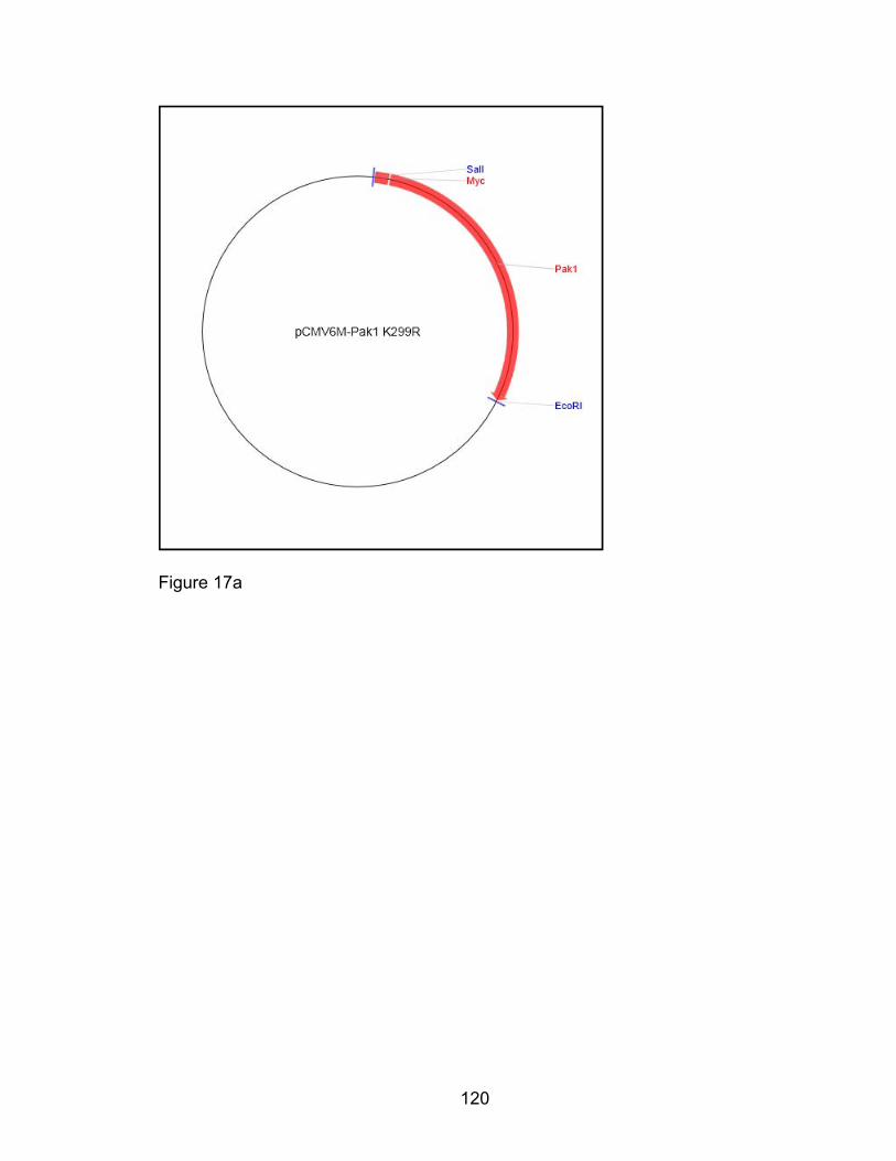
120
Figure 17a
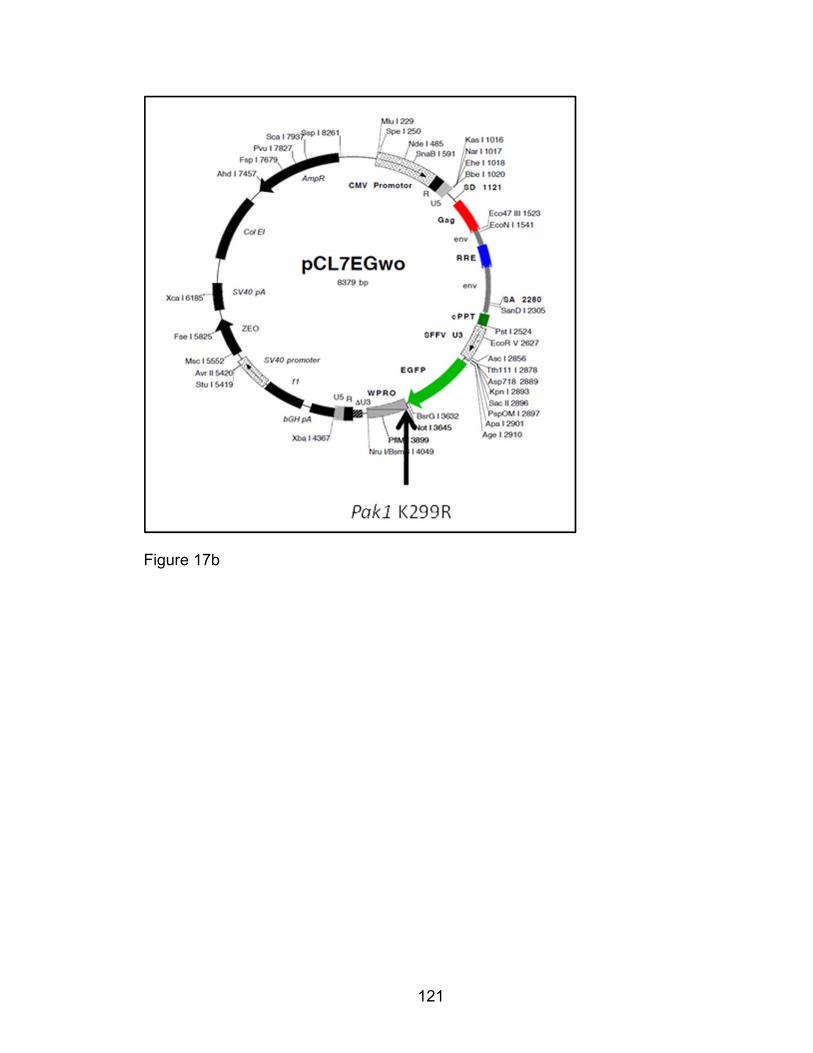
121
Figure 17b

122
Figure 17c
Figure 17d
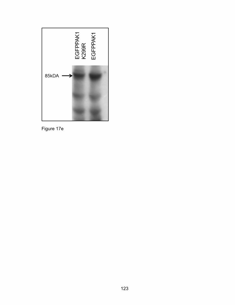
123
Figure 17e
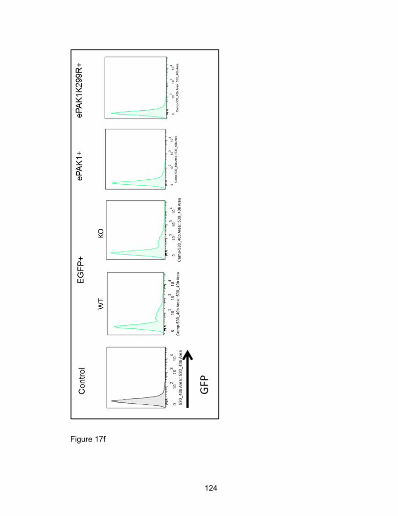
124
Figure 17f
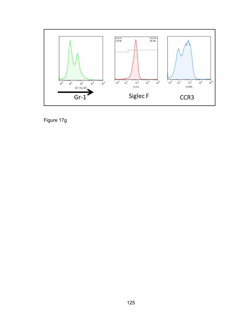
125
Figure 17g
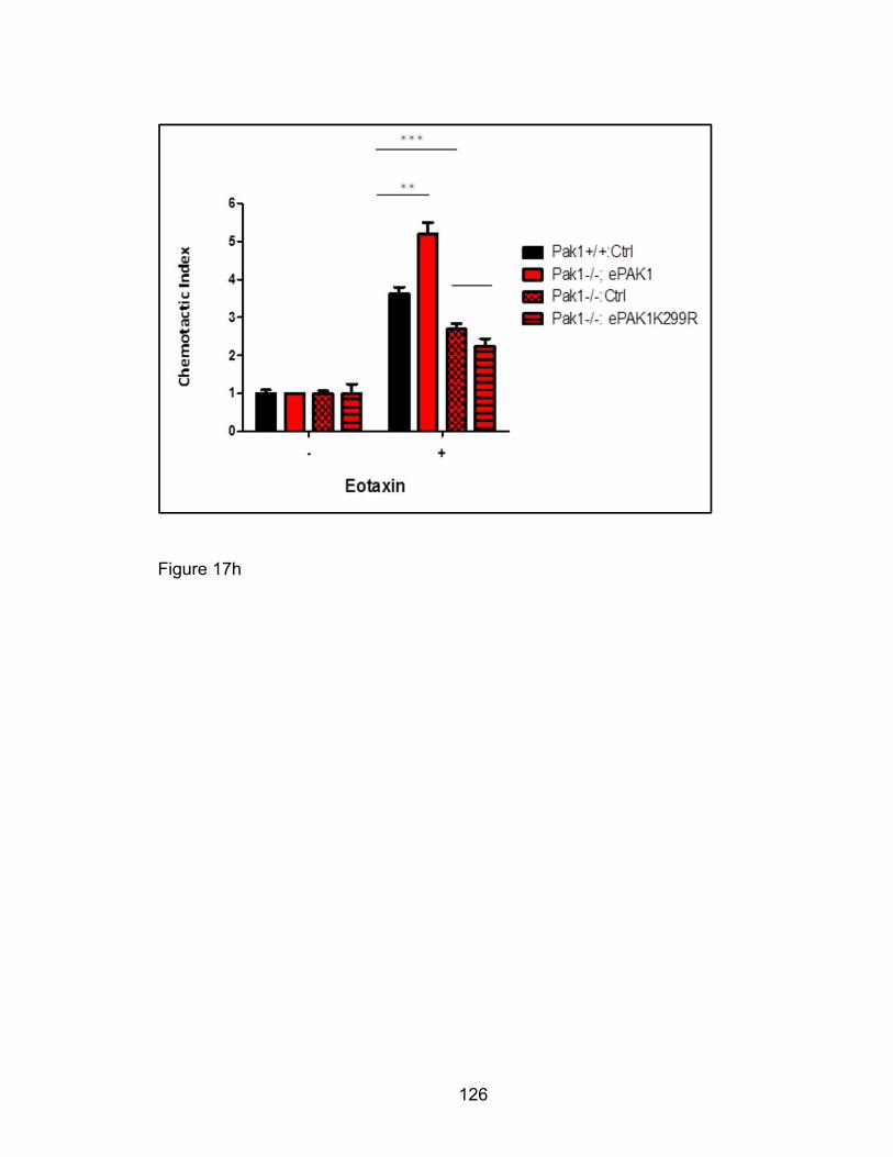
126
Figure 17h

127
Figure 17: Ectopic expression of full length PAK1 but not kinase-dead
PAK1 mutant in Pak1-/- eosinophils restores enhanced eotaxin-mediated
migration. We Not1-adapted and amplified ~1620bp Pak1 K299R insert (Figure
17c) from the PCMV6M-Pak1K229R vector (Figure 17a) and blunted it by
klenow-fragment treatment. We digested PCL11 vector with Not1 and similarly
blunted and CIAP treated it to prevent self-ligation. We ligated the Pak1 K299R
insert into the cut PCL11eGwo vector to form an eGFPPak1K299R construct as
shown (Figure 17b). We confirmed orientation of inserts in both PCL1eGFPPAK1
and PCL11eGFPPAK1K299R by restriction enzyme digestion that would
generate ~2000bp eGFPPAK1 and eGFPPAK1K299R DNA fragments (Figure
17d). As expected, these plasmid constructs ectopically expressed ~85kDA
eGFPPAK1 and eGFPPAK1K299R fusion proteins when transfected into
HEK293T packaging cells. Pak1+/+ and Pak1-/- c-kit+ LDMNCs were transduced
with PCL11eGwo only, PCL1eGFPPAK1 and PCL11eGFPPAK1K299R,
fluorescently sorted (Figure 17f), cultured in eosinophil conditions and assessed
for Gr-1, Siglec F and CCR3 surface expression (Figure 17g). We used cells
transduced with these viral expressed constructs for eotaxin-mediated transwell
chemotaxis studies (Figure 17h).

128
Eosinophil F-actin reorganization by Eotaxin: CCR3 signaling through
PAK1
Eotaxin signaling pathways through PAK1 in eosinophils have not been studied
to date. Since our data indicate Pak1 deletion decreases eotaxin-mediated
eosinophil F-actin polymerization and consequent migration, we sought to
determine PAK1’s downstream molecular targets responsible for these changes.
To elucidate F-actin dynamics in the eotaxin-stimulated eosinophil, we examined
the phosphorylation of putative PAK1-dependent actin-regulating proteins by
immunoblot from total cell extracts obtained from eotaxin-stimulated Pak1-/- and
Pak1+/+ bmEos. First to show that eotaxin ligation of CCR3 induced PAK1
activation, we used an in vitro kinase assay where we immunoprecipitated PAK1
from HL-60 derived human eosinophils. Eotaxin-stimulation increased PAK1’s
phosphorylation of MEK1 at its serine 298 residue (Figure 18a). PAK1’s kinase
activation is a complex process that critically requires autophosphorylation of its
threonine 423 (T423) residue [139]. Eotaxin-treatment of Pak1+/+ murine bmEos
phosphorylated PAKs at threonine 423, their signal activation residue (Figure
18b). Although work has not yet been done in eosinophils, PAK1 is known to
regulate cell migration via the cytoskeleton. [155, 202, 205]. To test the role of
PAK1 in cytoskeletal rearrangement, we cultured Pak1-/- and Pak1+/+ bmEos
and examined F-actin dynamics by fluorescence cytometry. Eotaxin stimulation
in bmEos of both genotypes caused initial polymerization and reorganization of
F-actin, but this eotaxin-induced F-actin polymerization at 30 seconds was
significantly diminished in Pak1-/- bmEos compared to Pak1+/+ (Figure 18c).
129
Furthermore eotaxin-stimulated Pak1-/- bmEos had altered cortical F-actin
arrangement compared to WT bmEos (Figure 18f). The F-actin and the tubulin
cytoskeleton are coordinately controlled in migration. We examined tubulin and
F-actin polymerization in eotaxin-treated bmEos of both genotypes and likewise
observed perturbed tubulin polymerization. These data together suggest that
eotaxin-mediated F-actin polymerization and organization in eosinophils is PAK1-
dependent. We hypothesized that these cytoskeletal dynamics perturbed in
Pak1-/- eosinophils were associated with eotaxin-induced migration (Figure 18g).
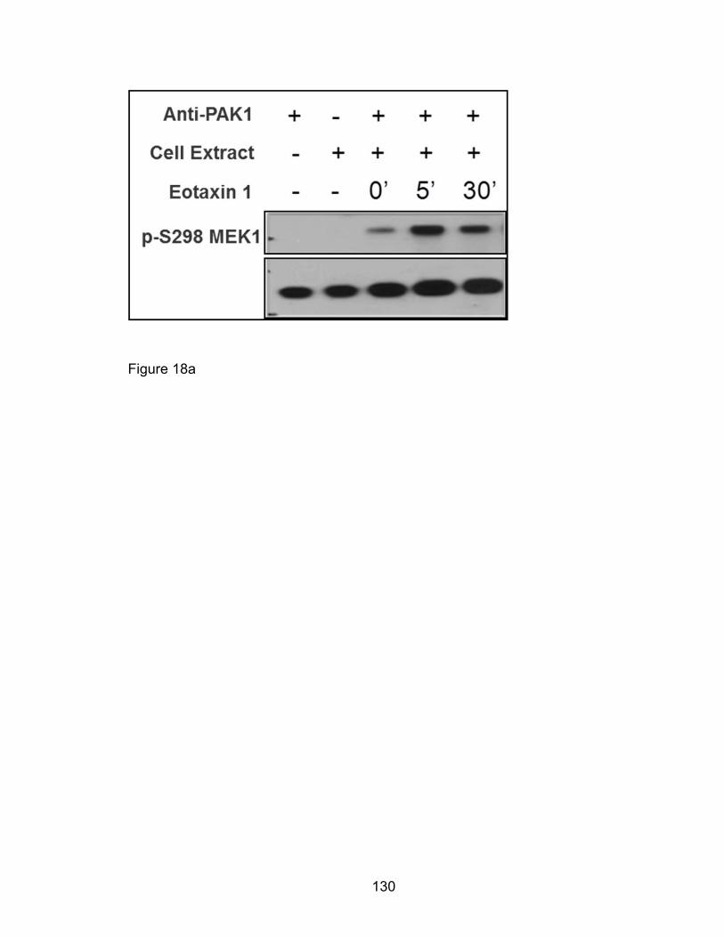
130
Figure 18a
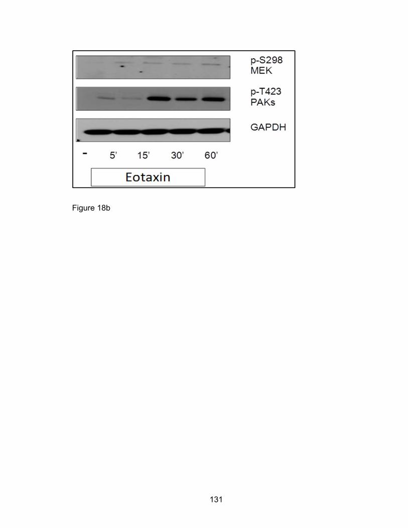
131
Figure 18b
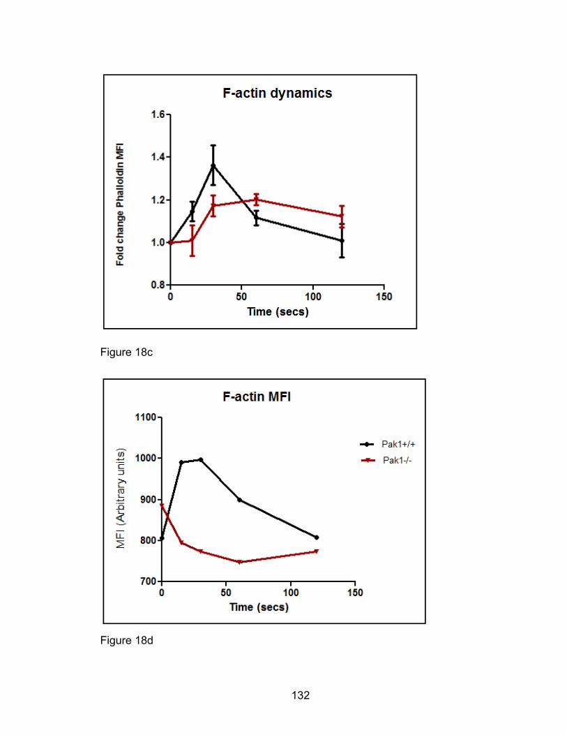
132
Figure 18c
Figure 18d
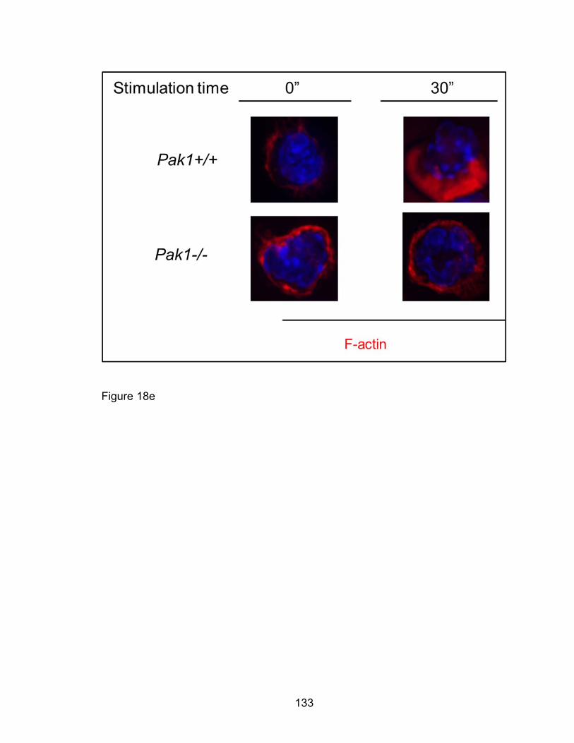
133
Figure 18e
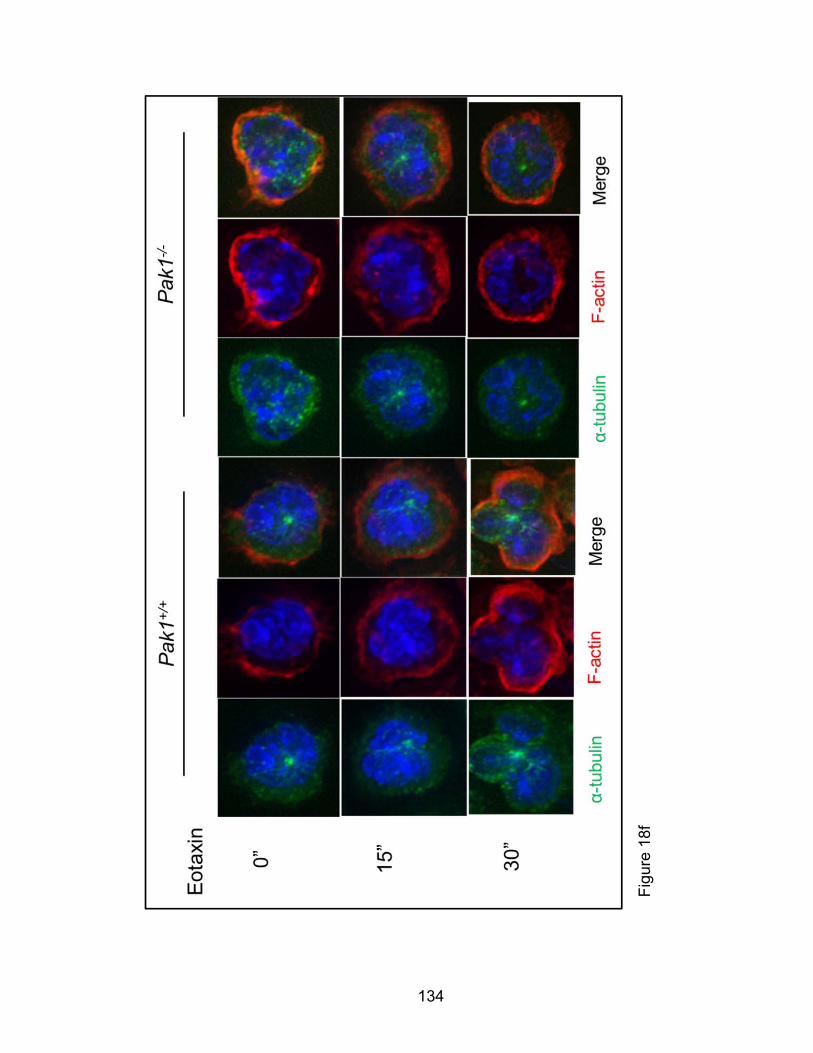
134
Fig
ure
18f
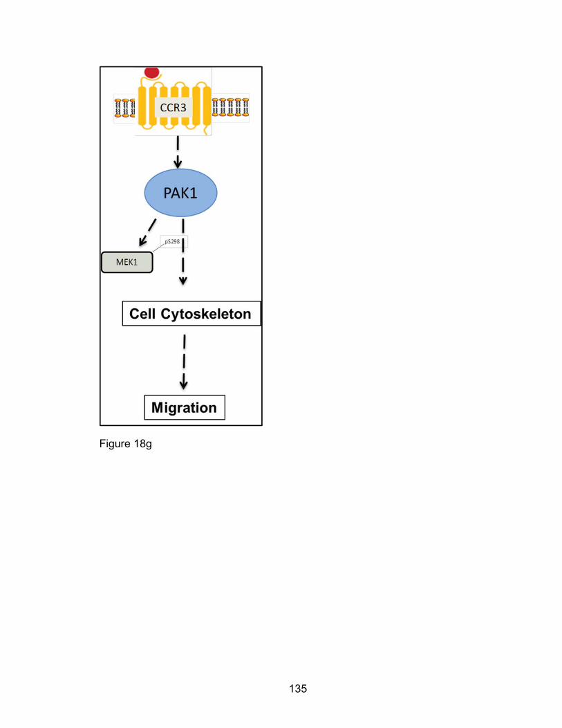
135
Figure 18g
136
Figure 18: PAK1 promotes F-actin reorganization induced by eotaxin
signaling in eosinophils. HL-60 derived human eosinophils were generated in
vitro and human PAK1 immunoprecipitated from eotaxin treated HL-60 Eos.
PAK1’s kinase activity was assessed to its substrated purified MEK1 (Figure
18a). Pak1+/+ and Pak1-/- bmEos were cultured in vitro, treated with eotaxin,
total cell extracts prepared and immunoprobed for PAKs and MEK1
phosphorylation (Figure 18b). Results in (a-b) are representative of at least three
independent experiments. Pak1+/+ and Pak1-/- BmEos were also cultured and
stimulated with eotaxin over a two-minute duration for cytoskeletal studies.
Eotaxin treated Pak1+/+ and Pak1-/- bmEos were fixed and relative (Figure 18c)
and representative arbitrary (Figure 18d) mean fluorescent intensities (MFI) of
phalloidin-tagged F-actin polymerization quantified by fluorescence cytometry,
visualized by confocal microscopy (Figure 18e) and compared. Eotaxin-treated or
untreated bmEos of both genotypes were also stained for tubulin and F-actin and
visualized by deconvolution microscopy (Figure 18f). Results in (c) are averaged
from five experiments (N=5) while results in (d-f) are representative from at least
five experiments. A schematic summarizing conclusions from this study is
illustrated (Figure 18g) * p<0.05 using a two-tailed student’s t-test.
137
Eotaxin-induced eosinophil phosphorylation changes of cofilin as well as
F-actin colocalization with p-34 Arc are PAK1-dependent
Eotaxin signaling pathways through PAK1 in eosinophils have not been studied
to date. PAK1 in many cell systems has been known to regulate several F-actin
regulating proteins [150, 176, 254]. To elucidate F-actin dynamics in the eotaxin-
stimulated eosinophil, we examined the phosphorylation of putative PAK1-
dependent actin-regulating proteins by immunoblot from total cell extracts
obtained from eotaxin-stimulated Pak1-/- and Pak1+/+ bmEos. We showed that
eotaxin induced dephosphorylation of cofilin in bmEos occurs in a PAK1-
dependent manner (Figures 19a-b). Remarkably, Pak1-/- bmEos demonstrated
impaired dephosphorylation of cofilin at serine 3 relative to Pak1+/+ bmEos
(Figures 19a-b). PAK1 regulates cofilin in signaling events essential for F-actin
cytoskeletal dynamics in cell migration [176, 195].
Pak1 deficiency perturbed spatial eosinophil F-actin localization/reorganization
30 seconds after eotaxin stimulation (Figure 18c-d). To further examine F-actin
cytoskeletal events at this time point, we fluorescently tagged p34-ARC a subunit
of the ARP2/3 protein complex. ARP2/3 is a known substrate of PAK1 that
initiates new F-actin polymerization sites [177]. We found decreased
colocalization of F-actin with this PAK1-regulated component of ARP2/3 in Pak1-
/- bmEos although p34ARC expression is comparable between the two
genotypes (Figures 19c-d). These data taken together suggest decreased de
novo polymerization of actin-filaments in eotaxin-stimulated Pak1-/- bmEos.
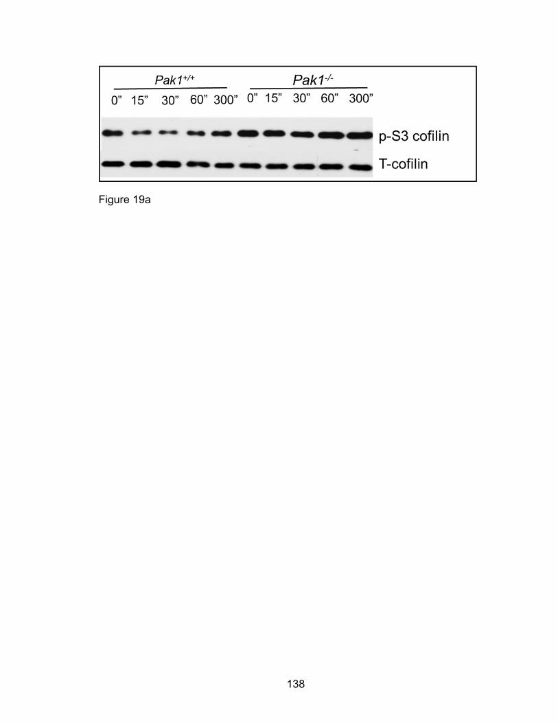
138
Figure 19a

139
Figure 19b
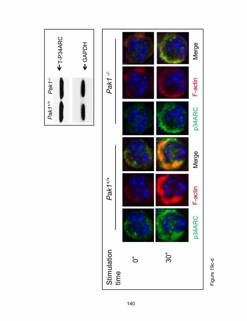
140
Fig
ure
19c-
d
141
Figure 19: Eotaxin induces PAK1-dependent phosphorylation changes of
cofilin, filamin A and F-actin localization with p-34 ARC. Pak1+/+ and Pak1-/-
bmEos were treated with eotaxin and fixed for immunocytochemical analysis or
total cell extracts prepared for western blot. Cell extracts were immunoprobed for
pS3-cofilin and total cofilin (Figure 19a) and levels of these proteins compared by
densitometry (Figure 19b). Cell extracts from bmEos were immunoprobed for p-
34 ARC levels (Figure 19c). Fixed and permeabilized cells were probed for total
p-34 ARC and F-actin (Figure 19d). Results in (a-c) are representative from at
least three experiments. * p<0.05 using a two-tailed student’s t-test.
142
Eotaxin-induced phosphorylation of PAK2, Ezrin/ moeisin/ Radixin, Op18/
Stathmin, and p38 MAPK in the eosinophil is PAK1-independent
To expand upon and explain PAK1-dependent F-actin polymerization, we also
immunoblotted for phosphorylated PAK2, Ezrin/moeisin/Radixin, Op18/Stathmin,
and p38 MAPK in total cell extracts from eotaxin-treated Pak1+/+ and Pak1-/-
bmEos. PAK2, an isoform of PAK1, has been shown to have some redundant
substrate specificities and function in vitro and in vivo [150]. PAK1 and PAK2 are
both expressed in hematopoietic tissues although their redundant and disparate
role in eosinophil function remain to be explored. We show that eotaxin
stimulation induced PAK2 phosphorylation at the threonine 423 residue that is
independent of PAK1 expression (Figures 20a-b). To further investigate the role
of these two isoforms of PAKs in eosinophil migration we used, IPA3, an
allosteric inhibitor of PAK1 that also inhibits PAK2. We pretreated Pak1+/+
eosinophils with IPA3 for 20 minutes prior to using these cells in a transwell
migration assay. We observed that PAKs inhibition prevented eotaxin-mediated
eosinophil migration (Figure 20c). Furthermore, IPA3 treatment significantly
decreased spontaneous non-chemokine driven chemokinesis of eosinophils
(Figure 20d) despite no profound effect on cell membrane compromise as
assessed by trypan blue exclusion counts two hours after IPA3-incubation (not
depicted). These findings suggest both PAK1 and PAK2 may have additive or
synergistic effects on eotaxin-mediated eosinophil migration.

143
ERM, p38 MAPK, Filamin A, and Op18/Stathmin have been shown in other cell
systems to regulate cytoskeletal remodeling downstream of PAK1 [121, 195,
196, 200, 255]. We have previously observed that Pak1 modulates F-actin
depolymerization and mast cell degranulation through Ezrin [192]. We show for
the first time that eotaxin-treatment induced phosphorylation changes of p-T567
ERM (Figures 20i-j). Phospho-T567 ERM also colocalized with F-actin in the
eotaxin-polarized Pak1+/+ eosinophil (Figure 20e) so we hypothesized that a
PAK1-ERM signaling axis is involved in eotaxin-induced F-actin changes and
migration. To test this hypothesis, we generated mice with conditional
hematopoietic Ezrin deficiency. We crossed Ezrin floxed mice with mice
expressing the Mx-1 Cre transgene that is expressed in hematopoietic and liver
tissues in an inducible fashion (Figure 20f). Approximately 25% of the second
generation of pups yielded the desired Ezrin flox/flox Mx1Cre- (Ez-WT) and
Ezrinflox/flox Mx1Cre+ (Ez-KO). We induced Mx-1 Cre expression by
intraperitoneal administration of a tapered dose of PolyIC on alternative days
standardized by mouse weight. We demonstrated hematopoietic ablation of Ezrin
expression by western blot analysis of Ez-KO LDMNCs (Figure 20g). We
cultured Ez-WT and Ez-KO bmEos and used them in eotaxin migration assays.
We observed no difference between migration of Ez-WT and Ez-KO eosinophils
(Figure 20h). These data together suggest eotaxin-induced phosphorylation of
Ezrin is neither necessary for eotaxin-mediated eosinophil F-actin polymerization
nor migration. Furthermore, these Ezrin changes are not PAK1-dependent.
144
p38 MAPK is known to regulate the cell cytoskeleton and cell migration via its
downstream effector HSP27 [122, 245]. In hematopoietic cells, SCF-mediated
mast-cell migration is in part PAK1-dependent [121]. We postulated that PAK1-
p38-dependent signaling may also be important in eotaxin-mediated eosinophil
migration. To examine p38 MAPK’s role in PAK1-dependent eotaxin-mediated
eosinophil migration, we used chemical inhibition of p38 MAPK activity in
transwell migration assays as well as western blot analysis of eotaxin-treated
bmEos. p38 MAPK inhibition by SB 203580 decreased eotaxin-mediated wild-
type eosinophil migration by greater than 40% in a similar manner as PAK1
(Figure 20k). We also measured and quantified eotaxin-induced phosphorylation
of p38 MAPK and observed phosphorylation of T180/Y182 p38 MAPK. However,
the eotaxin-mediated phosphorylation of p38 MAPK did not appear to be PAK1-
dependent in the initial eotaxin-mediated PAK1 signaling (Figures 20i-j).
PAK1 also phosphorylates Filamin A at its serine 2152 residue. Filamin A
anchors integrins and transmembrane receptor complexes to the cell
cytoskeleton modulating cell shape. Moreover, studies in FLNA knockout
systems demonstrate multiple defects with migrational etiology [193, 194]. We
show that eotaxin-induces phosphorylation of Filamin A comparably in both
Pak1+/+ and Pak1-/- bmEos (Figures 20l-m). Additionally, PAK1 is known to
phosphorylate Op18/stathmin at its serine 16 residue mediating tubulin and
consequently F-actin reorganization. In our studies, while we show a novel

145
eotaxin-induced phosphorylation of Op18/Stathmin in eosinophils, this
phosphorylation does not appear to be PAK1-dependent (Figures 20i-j).
From the biochemical and microscopic data we report in Figures 19 and 20, we
propose a model of eotaxin signaling in the eosinophil (Figure 20n). We show
that eotaxin:CCR3 independently phosphorylates PAK1 and PAK2. We also
show that PAK1 in the eosinophil mediates eotaxin-induced depolymerization of
cofilin in modulating F-actin temporal-spatial polymerization. In so doing, PAK1
may increase de novo polymerization of F-actin at new sites that are ARP2/3-
mediated. However, the intital eotaxin-induced phosphorylation of ERM, p38
MAPK, filamin A, and Op18/Stathmin is PAK1-independent.
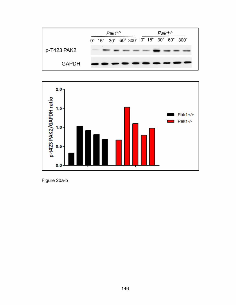
146
Figure 20a-b

147
Figure 20c
Figure 20d
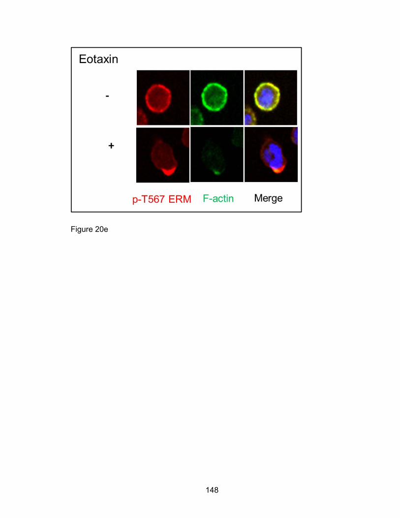
148
Figure 20e
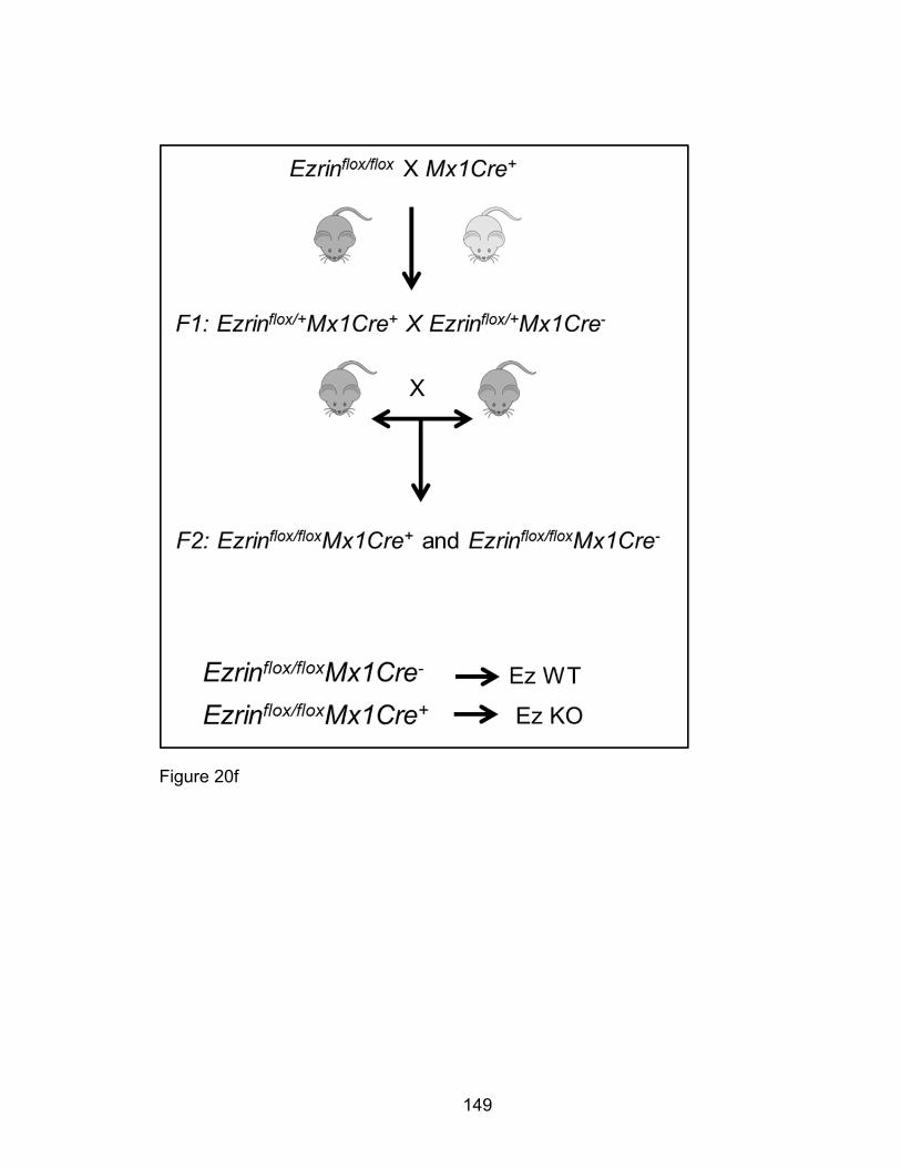
149
Figure 20f
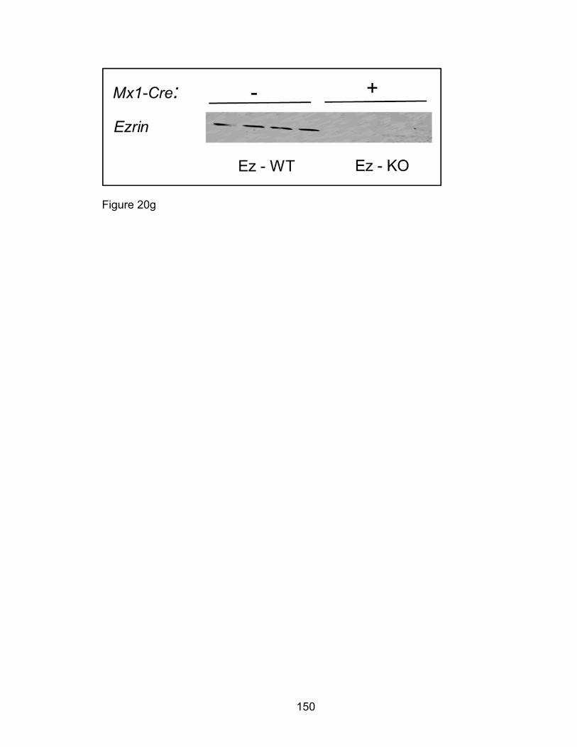
150
Figure 20g
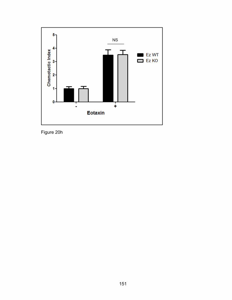
151
Figure 20h

152
Figure 20i
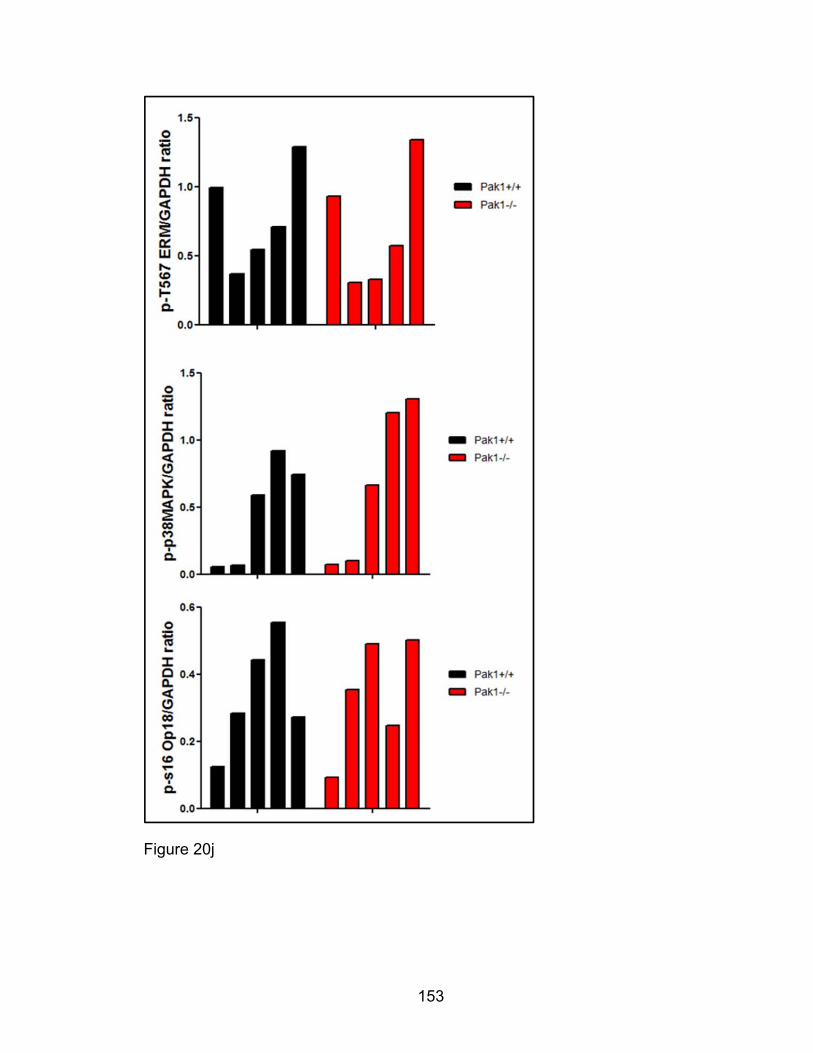
153
Figure 20j
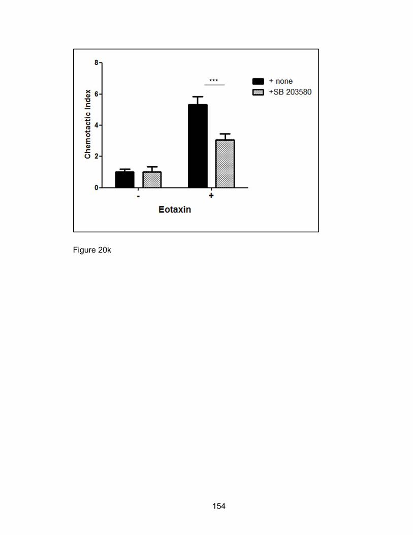
154
Figure 20k
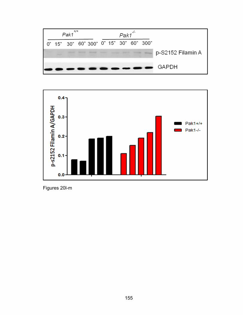
155
Figures 20l-m
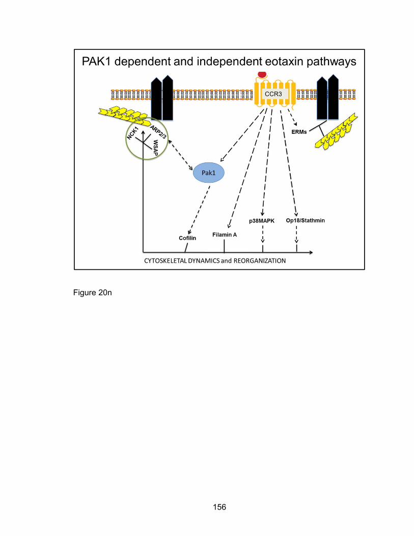
156
Figure 20n

157
Figure 20: PAK1-independent Eotaxin-induced phosphorylation of PAK2,
Ezrin/moeisin/Radixin, Op18/Stathmin, and p38 MAPK in the eosinophil.
Pak1+/+ and Pak1-/- bmEos were treated with eotaxin and total cell extracts
prepared for western blot or fixed for immunocytochemical analysis. Cell extracts
were blotted for p-T423 PAKs (Figures 20a-b), p-T567 ERM, p-T180/Y182 p38
MAPK, Fillamin A, p-S16 Op18/Stathmin as well as GAPDH as a loading control
(Figures 20i-m). Paraformaldehyde fixed bmEos were permeabilized and
immunoprobed for p-T567 Ezrin and phalloidin-treated for F-actin visualization
(Figure 20e). To assess migration inhibitor treated cells and cell deficient in Ezrin
were utilized in transwell migration assays where bmEos migrating to the bottom
chamber were enumerated by hemocytometer. Pak1+/+ bmEos were pretreated
with inhibitors against PAKs, IPA3, and p38 MAPK, SB 203580, before being
utilized in an eotaxin transwell migration assays, N=3-6 (Figures 20c-d, k). Ezrin
floxed mice were crossed with mice expressing the inducible Mx-1 Cre
transgene, to yield the desired F2 descendants according to this schematic
(Figure 20f). Ez WT and Ez KO mice were injected with five doses of PolyIC on
alternate days to induce expression of Mx-1 Cre in Ez KO bone marrow. Cell
extracts from LDMNCs from four Ez WT and Ez KO mice, post-PolyIC treatment
was immunoblotted for Ezrin (Figure 20g). Ez WT and Ez KO eotaxin-mediated
chemotaxis was evaluated using a transwell system, N=5 (Figure 20h). Our
eosinophil migration studies, suggest a generalized schematic of PAK1-
dependent and -independent eotaxin-induced pathways (Figure 20n). *p<0.05, **
p<0.01, ***p<0.001 using a two-way ANOVA with Bonferroni post hoc.
158
Eotaxin-induced eosinophil degranulation in vitro is PAK1- independent
Eosinophil degranulation and mediator release promote airway remodeling [90].
Based on the results from our murine asthma model that demonstrated
decreased eosinophils in the BAL and lung tissue of Pak1-/- mice, we wanted to
examine Pak1’s role in eotaxin-mediated murine eosinophil degranulation.
Eotaxin is known to induce degranulation and granule protein release from
human and murine eosinophils [117, 256, 257]. In a peritoneal eotaxin-mediated
eosinophil infiltration model, we show eotaxin induces potent eosinophil
recruitment and degranulation in vivo. Furthermore, this degranulation seems to
be dependent on PAK1 with fewer degranulated eosinophils in Pak1-/- mouse
peritoneal lavage Giesma-stained cytospins compared with Pak1+/+ (Figures
21a-b). We sought to characterize the peritoneal lavage cell populations by
FACS. We found consistent with previous studies that whereas granulated
eosinophils were predominantly CCR3+, prolonged exposure to eotaxins cause
degranulation and receptor internalization [236, 258]. Hence degranulated
eosinophils were predominantly CCR3- (like neutrophils) and appeared to have
without red granules on giesma staining (Figure 21c). This ablated CCR3
expression makes fluorescence cytometric CCR3 assessment of eosinophils
from peritoneal lavage fluid variable and unreliable. Our previous work supports a
role for PAK1 in mast cell degranulation. Pak1-/- mast cell degranulation is
decreased measured by β-hexoaminidase enzymatic activity [223]. Furthermore,
transgenic lentiviral expression of PAK1 restored IgE/DNP-mediated mast cell
degranulation in vitro (Figure 21d). Moreover, we observed that this PAK1-
159
dependent degranulation is mediated in part by Ezrin [192]. Similar to Pak1
deletion, Ezrin deficiency in mast cells decreased IgE/DNP-mediated
degranulation (Figure 21e) and F-actin depolymerization (Figure 21f). We
consequently hypothesized that PAK1 regulates eotaxin-mediated eosinophil
degranulation and used a well-documented colorimetric assay quantifying
supernatant EPO activity in lieu of granule release. We found that we could not
elicit eotaxin-induced EPO degranulation over baseline despite strong in vivo
degranulation. Furthermore, we could not distinguish between relative
degranulation levels of Pak1-/- versus Pak1+/+ bmEos (Figure 21g). Similarly,
further degranulation studies using Ez-KO bmEos revealed no difference in
degranulation compared with Ez-WT (Figure 21h). Our inconclusive
degranulation results suggest a need for a more sensitive system to study
eotaxin-mediated degranulation in vitro.

160
Fig
ures
21a
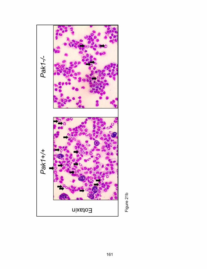
161
Fig
ure
21b

162
Figure 21c
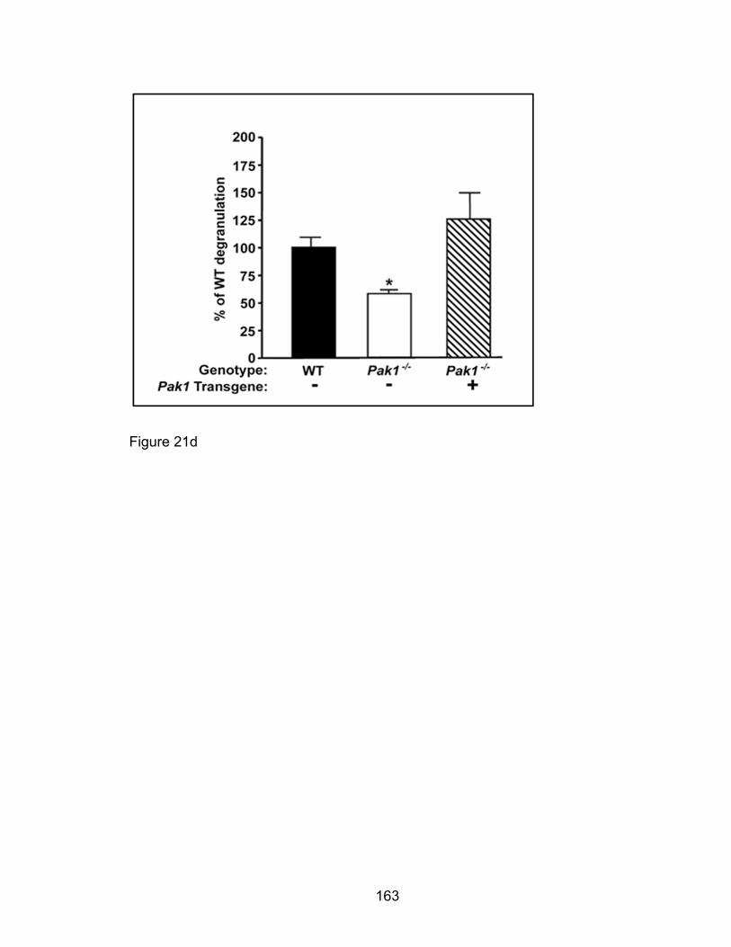
163
Figure 21d
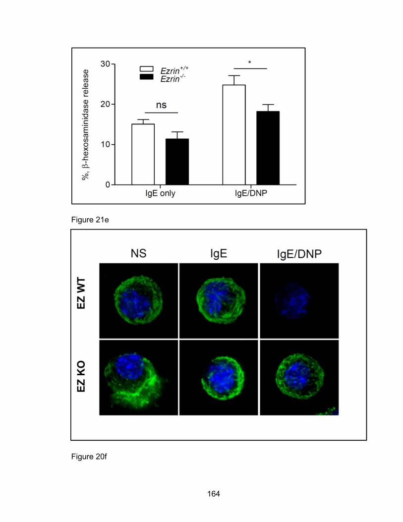
164
Figure 21e
Figure 20f
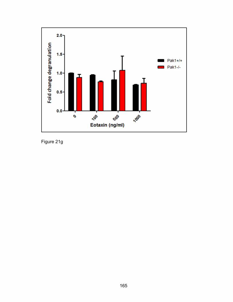
165
Figure 21g

166
Figure 21h
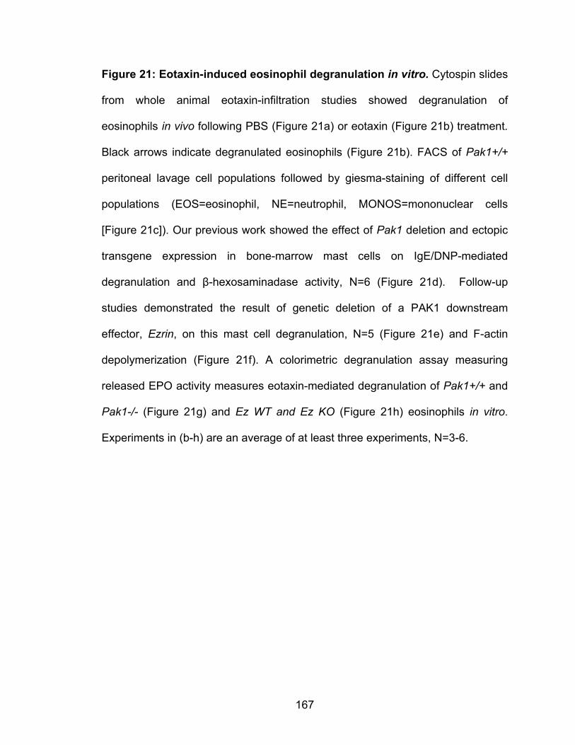
167
Figure 21: Eotaxin-induced eosinophil degranulation in vitro. Cytospin slides
from whole animal eotaxin-infiltration studies showed degranulation of
eosinophils in vivo following PBS (Figure 21a) or eotaxin (Figure 21b) treatment.
Black arrows indicate degranulated eosinophils (Figure 21b). FACS of Pak1+/+
peritoneal lavage cell populations followed by giesma-staining of different cell
populations (EOS=eosinophil, NE=neutrophil, MONOS=mononuclear cells
[Figure 21c]). Our previous work showed the effect of Pak1 deletion and ectopic
transgene expression in bone-marrow mast cells on IgE/DNP-mediated
degranulation and β-hexosaminadase activity, N=6 (Figure 21d). Follow-up
studies demonstrated the result of genetic deletion of a PAK1 downstream
effector, Ezrin, on this mast cell degranulation, N=5 (Figure 21e) and F-actin
depolymerization (Figure 21f). A colorimetric degranulation assay measuring
released EPO activity measures eotaxin-mediated degranulation of Pak1+/+ and
Pak1-/- (Figure 21g) and Ez WT and Ez KO (Figure 21h) eosinophils in vitro.
Experiments in (b-h) are an average of at least three experiments, N=3-6.

168
DISCUSSION AND FUTURE EXPERIMENTS
Eosinophils whose major function had largely been regarded as T-helper 2 cell
effectors, are increasingly gaining recognition for their integral role in modulating
airway hyperresponsiveness, inflammation, and remodeling through toxic
degranulation products, lipid mediators, growth factors, and cytokines. In an
OVA-albumin murine AAD model, eosinophil T-helper cell airway epithelial
signaling networks establish self-perpetuating chronic inflammation in the airway
that recapitulates human asthma. Identifying the mechanisms through which
eosinophils infiltrate the lung in asthma would offer novel specific targets for
therapeutic intervention. In these studies we have identified PAK1 as a key
regulator of allergic airway eosinophil inflammation in vivo as well as eotaxin-
mediated eosinophil migration in vitro.
PAK1 in murine allergic airway inflammation
We have previously demonstrated that genetic ablation of Pak1 does not alter
murine survival or in vivo hematopoietic lineage differentiation as assessed by
peripheral blood differential leukocyte assessment [223]. Studies done with these
Pak1-/- mice implicate PAK1 in migration and degranulation of mast cells [121,
192, 218]. These pro-inflammatory processes drive allergic airway inflammation
and depend heavily on stimulus-induced F-actin dynamics [192, 223]. While
several studies propose a role for PAK1’s in force generation in airway smooth
muscle hyperresponsiveness in AAD, no reports discuss PAK1’s potential role in
allergic airway inflammation [218].

169
The data presented in our studies support a PAK1 kinase-dependent mechanism
for eosinophil infiltration in vivo. Several Rho-family GTPase pathways have
been associated with promoting allergic airway inflammation [125, 259]. Our data
implicate PAK1, a known Rho-GTPase molecular substrate in eosinophil airway
inflammation characteristic of AAD. We show for the first time an attenuated
inflammatory cell response in a Pak1-/- OVA-albumin murine AAD model that
recapitulates human eosinophil-rich airway inflammation in tissue (Figure 7) and
BALF (Figure 8). The levels of eotaxin in the BAL and other T-helper cell derived
cytokines from OVA-stimulated splenocytes of Pak1+/+ and Pak1-/- sensitized
mice were indistinguishable (Figures 8d and 9) implying a preservation of T-cell
responses in this Pak1-/- AAD model.
This markedly depressed airway inflammation seems primarily hematopoietically-
derived and intrinsic to the eosinophil as both Pak1+/+ and Pak1-/- recipient mice
transplanted with Pak1-/- bone marrow showed decreased eosinophil
inflammation compared to their counterpart recipients transplanted with Pak1+/+
bone marrow (Figure 10). However, our data cannot rule out the possible role of
other factors in the inflammatory environment in recruiting eosinophils to the lung
in these sensitized mice as may be suggested by the adoptive bone marrow
transplant results. We saw markedly suppressed eosinophil recruitment in BAL
fluids from Pak1-/- mice transplanted with Pak1-/- bone marrow compared to BAL
fluids from Pak1+/+ mice transplanted with Pak1-/- bone marrow (Figure 10d-e).
Similar to our bone marrow transplantation studies, orthotopic lung transplants
demonstrated a trend towards greater lung eosinophil inflammation in host mice
170
with a Pak1+/+ hematopoietic system as compared to Pak1-/- hosts (Figure 11),
however, this trend needs to be clarified by increasing the number in subsequent
cohorts of mice that are transplanted.
The exact contribution of PAK1 to allergic airway inflammation remains largely
unexplored. Our studies demonstrate PAK1’s integral role in eosinophil infiltration
of the lungs following allergen-sensitization and challenge. However, PAK1 role
in other cells in this allergic process remains understudied. Mast cell
degranulation and SCF-mediated migration are impaired in Pak1-/- mice [121,
223]. A role of PAK1 in neutrophil chemotaxis is also reported however studies in
Pak1-/- mice are indicated to clarify its mechanism [260, 261]. One possibility is
that PAK1 is selectively effective in promoting allergen-induced infiltration of
inflammatory cells. The other possibility is that PAK1 promotes the motility of
cells of multiple hematopoietic cell lineages in which case further studies are
critical to fully characterizing the immune responses of Pak1-/- mice.
Our data in this section are obtained from an OVA-Alum sensitization model that
elicits a potent Th2-driven eosinophil response in vivo. While this model is a very
useful tool in studying eosinophil infiltration, the model does not fully recapitulate
human disease. In human bronchial asthma, mast cells inflammation plays a
major role in eosinophil infiltration as well as the other hallmarks of asthma while
the adjuvant Alum potentiates the Th2 response to override the mast cell effect
on development of this allergic airway inflammation [54, 262]. Similarly, here we
only examine acute eosinophil inflammation recruitment [21]. Future studies
utilizing a chronic sensitization and challenge protocol for Pak1-/- mice would
171
complement and supplement our findings in a model that mimics the mast-cell
involvement and chronic accumulation of eosinophils in vivo. Since the exact
effect of ionizing-radiation on the lung parenchyma from bone marrow
transplanted mice is also unknown [259, 263] we also utilized complementary
orthotopic lung transplantation studies. Our results from these studies indicate a
need for increased numbers in cohorts used in future studies. Likewise, these
prospective studies should include groups of isogeneic transplant animals as
controls for the surgical procedure. Ultimately, AAD studies in genetic crosses
between Pak1-/- and KitW-sh/W-sh mice are warranted to study eosinophil
inflammation in a mast-cell deficient context. These studies would clarify the
distinct roles of PAK1 in mast cells, T-helper cell and eosinophils in AAD.
The process of eosinophil migration and infiltration is critical to the development
of chronic airway inflammation in asthma. Eosinophils take up residence in the
lungs and modulate chronic inflammatory processes that cause airway tissue
injury and remodeling. Our studies have identified PAK1 as a potential
transducer of allergen-induced chemotactic signals in the migrating eosinophil.
Although PAK1 is likely not the exclusive regulator of eotaxin-mediated migration,
it is a promising targetable molecular switch for therapeutic intervention. Our
work is a basis for future studies testing PAK1’s role in allergen recruitment of
eosinophils.

172
PAK1 in Eotaxin- mediated Eosinophil function
No published reports have examined the role of PAK1 in eosinophil function to
date. Building upon our robust in vivo AAD model of eosinophil recruitment, we
sought to understand the mechanism by which PAK1 modulated eosinophil
function. We hypothesized based on our transplant data, that PAK1 plays a role
in intrinsic eosinophil activity. We test this question in two in vivo murine
infiltration models where we standardized the concentration of eotaxin, a potent
eosinophil chemoattractant. Our results suggest a role for PAK1 in eotaxin-
mediated infiltration of eosinophils in vivo (Figure 12). In previous eotaxin
infiltration studies, eotaxin preferentially recruited eosinophils relative to other cell
lineages to the peritoneum [227, 228]. Both studies identify the importance of a
readily available pool of circulating or tissue eosinophils to respond to this
eotaxin signal, consequently the degree of eosinophil accumulation in vivo was
dependent on allergen-sensitization or IL-5 in transgenic mice. Our studies in the
non-sensitized animal could therefore be complemented by repeating these
studies in the OVA-sensitized mice.
We obtained further evidence on PAK1’s role in eotaxin-mediated eosinophil
function from bmEos and HL-60 Eos. We demonstrated for the first time that
eotaxin phosphorylated and activated PAK1 in murine and HL-60 derived
eosinophils (Figures 19a-b). We subsequently sought to generate Pak1 deficient
eosinophils and were able to demonstrate a comparable potential of Pak1-/- stem
and progenitor cells to generate colonies from IL-5 the major eosinophilopoietic
cytokine (Figure 14). Pak1-/- bmEos generated using IL-5 also demonstrated
173
indistinguishable expression of CCR3, siglec F as well as many adhesive
molecules compared to Pak1+/+ bmEos. Since eosinophil infiltration involves
margination, adhesion, and transmigration we assessed PAK1’s role in adhesive
and motive eosinophil processes. First we assessed PAK1’s effect on eotaxin-
mediated eosinophil adhesion. Previous studies have shown the importance of
eotaxin-mediated adhesion to eosinophil migration to sites of inflammation.
These studies have frequently used recombinant fibronectin to mimic adhesion of
eosinophils to the endothelial wall [192, 250, 264]. Adhesion in eotaxin-
stimulated eosinophils is mediated by integrin association with neighboring cell
integrins. We examined the effect Pak1 deletion had on surface expression of
CD11a/αL, Mac1/αMβ2, α4β7, and VLA4/α4β1, integrins well-documented to
play important roles in eosinophil trafficking in various body organs [265]. We
found comparable percentages of cells expressing similar densities of these
integrins (Figures 16a-d). These data suggest that Pak1 deletion does not affect
the surface expression of the important adhesive eosinophil integrins. We
subsequently performed functional adhesive studies on fibronectin coated plates
in a microfluidic system that recapitulates capillary flow. We confirmed in our
studies, that eosinophil preferentially adhered to fibronectin compared to gelatin
coated plates, and this adhesion was eotaxin-mediated (Figures 16e-f). However,
the adhesion was not different between Pak1+/+ and Pak1-/- eosinophils (Figure
14g). Our studies in adhesion are limited in that they are largely correlative in that
functional adhesion of each integrin is not tested. In future studies, we will
expand upon these adhesion studies utilizing recombinant ICAM, VCAM, and

174
MadCAM, the interacting integrins on the endothelium that eosinophils can bind
[265, 266].
Our studies also propose a model of eotaxin-mediated eosinophil chemotaxis
that depends on PAK1’s modulation of F-actin polymerization. Our data indicate
that eotaxin: CCR3 signaling phosphorylates and activates PAK1 driving
eosinophil chemotaxis (Figure 14). While Pak1-/- eosinophils ectopically
expressing a full length PAK1 construct had enhanced chemotaxis, ectopic
expression of catalytically inactive PAK1, K299R, did not affect Pak1-/-
eosinophil migration (Figure 16). PAK1 regulates F-actin dynamics via distinct
mechanisms [133, 167]. These results suggest an eotaxin-mediated PAK1
kinase-dependent mechanism as opposed to a kinase-independent interaction
with downstream proteins in the F-actin changes needed for eosinophil
chemotaxis (Figure 16).
Chemokine-induced hematopoietic cell movement depends on cycles of F-actin
polymerization, depolymerization and reorganization [267-270]. Our results
demonstrate that PAK1 regulates eotaxin-induced eosinophil F-actin
polymerization. PAK1 can catalytically control cell motility via cofilin-dependent
actin cytoskeletal changes as well as ARP2/3-mediated nucleation and branching
of F-actin in different cell types. PAK1’s regulation of cofilin in migrating cells is
complex [174, 176] and cofilin phosphorylation in eotaxin-stimulated murine
eosinophils appears to be spatial-temporally determined. Our data indicate that
PAK1 facilitates dephosphorylation of cofilin which may be necessary for F-actin
remodeling around the cell (Figure 19). PAK1 directly regulates the p41-Arc
175
subunit of the ARP2/3 F-actin nucleating complex independent of cofilin [177,
271]. We show that Pak1 deletion decreases the co-localization of F-actin and
the p-34-Arc subunit of this complex suggesting a role for PAK1 in ARP2/3-
mediated de novo F-actin polymerization. In contrast to these two F-actin binding
protein partners of PAK1, p38MAPK, Ezrin/Radixin/Moesin, and Op18/Stathmin
do not seem critical to PAK1-dependent eotaxin-mediated eosinophil chemotaxis
despite their phosphorylation in the eotaxin-treated eosinophil (Figure 20).
Furthermore, migration studies using Ezrin-/- eosinophils that showed no
attenuated migration make Ezrin an unlikely downstream PAK1 effector critical to
eotaxin-mediated eosinophil migration (Figure 20). These data taken together
provide initial evidence for a model where eotaxin-induced activation of PAK1’s
kinase activity results in the catalytic downstream regulation of cofilin and
ARP2/3-mediated F-actin changes and eosinophil chemotaxis.
One implication our studies have on the field is the use of small molecule
inhibitors to target PAK1 kinase activity to attenuate eosinophil inflammation. The
disparate roles of PAK1 and PAK2 in hematopoietic cells, however, remains
incompletely understood. PAK1 and PAK2 are highly homologous and of similar
substrate specificity in vitro [150]. We show that similar to PAK1, PAK2 is
phosphorylated at its activation residue with eotaxin-stimulation (Figure 10).
However, this activation does not seem critical to eotaxin-mediated eosinophil
migration as is shown by our migration studies in Pak1-/- ectopically expressing
K299R, a dominant negative form of PAKs (Figure 17). Nonetheless, we show in
migration studies using IPA3 a pan-PAKs inhibitor severely hampered migration
176
(Figure 20). These data suggest that loss of Pak2 may potentiate the loss of
eotaxin-mediated migration potential exhibited by Pak1-/- eosinophils. However,
studies using ectopic expression of K299R and IPA3 are hindered by
uncertainties of in vitro manipulation and non-selectivity. Other current PAK1
selective inhibitors developed to target cell invasion and metastasis in cancer
treatment are unable to select for PAK1 over its group I PAK isoforms PAK2 and
PAK3 [244, 272]. Our studies identify a need to evaluate the differential versus
redundant roles of PAK1 and its isoform PAK2 in eotaxin-mediated eosinophil
migration and infiltration. To adequately address this issue in future studies, we
would utilize Pak1-/- and Pak2 floxed mice crossed with Mx1Cre + mice we can
begin to assess the differential role of these isoforms in eosinophil inflammation
in vivo. The results of this study warrant further exploration of the anti-
inflammatory attributes of PAK1 inhibition on eosinophil inflammation in a mouse
model of AAD.
Lastly, we tested the effect PAK1 has on eotaxin-mediated eosinophil
degranulation and mediator release, based on our definitive data in mast-cell
degranulation [223]. In vivo, in our eotaxin mediated eosinophil peritoneal
infiltration model we demonstrate an increased number of degranulated Pak1+/+
eosinophils compared to Pak1-/- eosinophils (Figures 21b-c) [273]. To build upon
this in vivo phenotype, we used a colorimetric EPO quantifying assay.
Surprisingly, we could not elicit increased degranulation over baseline using
increasing doses of eotaxin alone (Figure 21g) neither could we demonstrate a
difference in degranulation between Pak1+/+ and Pak1-/- nor EZ-WT and EZ-KO

177
eosinophils (Figures 21g-h). The in vivo system is significantly different for
eosinophils in contact with other cells and the extracellular matrix when
responding to eotaxin [250]. EPO is extremely adhesive and may not readily
solubilize during the colorimetric reaction [274, 275]. Consequently, newer
studies on eotaxin-mediated eosinophil degranulation utilize multiple
degranulation assays [256]. In future degranulation studies we will utilize multiple
end-point analysis including MBP, EPO, and β-hexosaminidase to assess
degranulation. We will also include adhesive recombinant integrins like ICAM and
VCAM or cytokines like IL-5 to potentiate eotaxin-mediated degranulation
response.
PAK1 acts as a molecular switch in many physiological cellular processes. In
eosinophils our studies demonstrate that PAK1 modulates eosinophil function by
transducing the eotaxin:CCR3 signal. We provide novel data implicating PAK1 in
regulating actin-mediated inflammatory processes in eosinophil inflammation
characteristic of AAD. Our findings have opened up a novel front exploring PAK1
as a potential asthmatic therapeutic. These studies broaden the understanding of
the molecular mechanisms underpinning eosinophil inflammation in AAD. Due to
relatively innocuous consequences of Pak1 deletion coupled with its critical role
in eosinophil migration, our findings warranted broader investigative exploration
of PAK1’s role in allergic disease.
178
REFERENCES 1. CDC/P. Asthma: A Presentation of Asthma Management and Prevention
(Slide Presentation and Speaker Notes). 2009 [cited 2012 01/01]; Available from: http://www.cdc.gov/asthma/healthcare.html.
2. WHO. Asthma: Factsheet No. 307. 2011; Available from: http://www.who.int/mediacentre/factsheets/fs307/en/index.html.
3. CDC/P. Flu and People with Asthma. [Webpage] 2011 [cited 2012 01/01]; Available from: http://www.cdc.gov/flu/asthma/index.htm.
4. Murphy, K.R., et al., Asthma management and control in the United States: results of the 2009 Asthma Insight and Management survey. Allergy and asthma proceedings : the official journal of regional and state allergy societies, 2012. 33(1): p. 54-64.
5. Nathan, R.A., et al., Comparison of the Asthma in America and Asthma Insight and Management surveys: did asthma burden and care improve in the United States between 1998 and 2009? Allergy and asthma proceedings : the official journal of regional and state allergy societies, 2012. 33(1): p. 65-76.
6. Robbins, S.L., V. Kumar, and R.S. Cotran, Robbins and Cotran pathologic basis of disease. 8th ed. 2010, Philadelphia, PA: Saunders/Elsevier. xiv, 1450 p.
7. Buc, M., et al., Immunopathogenesis of bronchial asthma. Arch Immunol Ther Exp (Warsz), 2009. 57(5): p. 331-44.
8. Galli, S.J., M. Tsai, and A.M. Piliponsky, The development of allergic inflammation. Nature, 2008. 454(7203): p. 445-54.
9. Costa, J.J., P.F. Weller, and S.J. Galli, The cells of the allergic response: mast cells, basophils, and eosinophils. JAMA, 1997. 278(22): p. 1815-22.
10. Bloemen, K., et al., The allergic cascade: review of the most important molecules in the asthmatic lung. Immunol Lett, 2007. 113(1): p. 6-18.
11. Corry, D.B. and C.G. Irvin, Promise and pitfalls in animal-based asthma research: building a better mousetrap. Immunol Res, 2006. 35(3): p. 279-94.
12. Zuyderduyn, S., et al., Treating asthma means treating airway smooth muscle cells. Eur Respir J, 2008. 32(2): p. 265-74.
13. Bates, J.H., M. Rincon, and C.G. Irvin, Animal models of asthma. Am J Physiol Lung Cell Mol Physiol, 2009. 297(3): p. L401-10.
14. Kroegel, C. and H. Wirtz, History of guidelines for the diagnosis and management of asthma: from opinion to control. Drugs, 2009. 69(9): p. 1189-204.
15. Expert Panel Report 3 (EPR-3): Guidelines for the Diagnosis and Management of Asthma-Summary Report 2007. J Allergy Clin Immunol, 2007. 120(5 Suppl): p. S94-138.
16. Barnes, P.J. and I.M. Adcock, How do corticosteroids work in asthma? Ann Intern Med, 2003. 139(5 Pt 1): p. 359-70.
179
17. McIvor, R.A., et al., Potential masking effects of salmeterol on airway inflammation in asthma. Am J Respir Crit Care Med, 1998. 158(3): p. 924-30.
18. Weldon, D., The effects of corticosteroids on bone growth and bone density. Ann Allergy Asthma Immunol, 2009. 103(1): p. 3-11; quiz 11-3, 50.
19. Guilbert, T.W., et al., Long-term inhaled corticosteroids in preschool children at high risk for asthma. N Engl J Med, 2006. 354(19): p. 1985-97.
20. Yiamouyiannis, C.A., et al., Shifts in lung lymphocyte profiles correlate with the sequential development of acute allergic and chronic tolerant stages in a murine asthma model. Am J Pathol, 1999. 154(6): p. 1911-21.
21. Bates, J.H., et al., The synergistic interactions of allergic lung inflammation and intratracheal cationic protein. Am J Respir Crit Care Med, 2008. 177(3): p. 261-8.
22. Nygaard, U.C., A. Aase, and M. Lovik, The allergy adjuvant effect of particles - genetic factors influence antibody and cytokine responses. Bmc Immunology, 2005. 6: p. -.
23. Ng, N., et al., House dust extracts have both T(H)2 adjuvant and tolerogenic activities. Journal of Allergy and Clinical Immunology, 2006. 117(5): p. 1074-1081.
24. Horner, A.A., et al., House dust extracts elicit toll like receptor dependent dendritic cell responses. Journal of Allergy and Clinical Immunology, 2005. 115(4): p. 893-893.
25. Barrett, N.A. and K.F. Austen, Innate cells and T helper 2 cell immunity in airway inflammation. Immunity, 2009. 31(3): p. 425-37.
26. Melgert, B.N., et al., Female mice are more susceptible to the development of allergic airway inflammation than male mice. Clinical and Experimental Allergy, 2005. 35(11): p. 1496-1503.
27. Hamada K, G.C., Goldman A, Kobzik L, Resistance of very young mice to inhaled allergen sensitization is overcome by coexposure to an air-pollutant aerosol. Am J Respir Crit Care Med, 2000 Apr: p. 1285-93.
28. Sakai, K., et al., Effect of different sensitizing doses of antigen in a murine model of atopic asthma. Clin Exp Immunol, 1999. 118(1): p. 9-15.
29. Morokata, T., et al., C57BL/6 mice are more susceptible to antigen-induced pulmonary eosinophilia than BALB/c mice, irrespective of systemic T helper 1/T helper 2 responses. Immunology, 1999. 98(3): p. 345-51.
30. Jeong, S.K., et al., Mite and cockroach allergens activate protease-activated receptor 2 and delay epidermal permeability barrier recovery. J Invest Dermatol, 2008. 128(8): p. 1930-9.
31. Shakib, F., A.M. Ghaemmaghami, and H.F. Sewell, The molecular basis of allergenicity. Trends Immunol, 2008. 29(12): p. 633-42.
32. Antony, A.B., R.S. Tepper, and K.A. Mohammed, Cockroach extract antigen increases bronchial airway epithelial permeability. J Allergy Clin Immunol, 2002. 110(4): p. 589-95.
180
33. Bhalla, D.K., et al., Cigarette smoke, inflammation, and lung injury: a mechanistic perspective. J Toxicol Environ Health B Crit Rev, 2009. 12(1): p. 45-64.
34. Samuelsen, M., U.C. Nygaard, and M. Lovik, Allergy adjuvant effect of particles from wood smoke and road traffic. Toxicology, 2008. 246(2-3): p. 124-31.
35. Vercelli, D., Discovering susceptibility genes for asthma and allergy. Nat Rev Immunol, 2008. 8(3): p. 169-82.
36. Suarez, C.J., N.J. Parker, and P.W. Finn, Innate immune mechanism in allergic asthma. Curr Allergy Asthma Rep, 2008. 8(5): p. 451-9.
37. Sandor, F. and M. Buc, Toll-like receptors. III. Biological significance and impact for human medicine. Folia Biol (Praha), 2005. 51(6): p. 198-203.
38. Kiss, A., et al., A new mechanism regulating the initiation of allergic airway inflammation. J Allergy Clin Immunol, 2007. 120(2): p. 334-42.
39. Barnes, P.J., Role of GATA-3 in allergic diseases. Curr Mol Med, 2008. 8(5): p. 330-4.
40. Perros, F., et al., Blockade of CCR4 in a humanized model of asthma reveals a critical role for DC-derived CCL17 and CCL22 in attracting Th2 cells and inducing airway inflammation. Allergy, 2009. 64(7): p. 995-1002.
41. Holgate, S.T. and R. Polosa, Treatment strategies for allergy and asthma. Nat Rev Immunol, 2008. 8(3): p. 218-30.
42. Robinson, D.S., et al., Predominant TH2-like bronchoalveolar T-lymphocyte population in atopic asthma. N Engl J Med, 1992. 326(5): p. 298-304.
43. Cousins, D.J., T.H. Lee, and D.Z. Staynov, Cytokine coexpression during human Th1/Th2 cell differentiation: direct evidence for coordinated expression of Th2 cytokines. J Immunol, 2002. 169(5): p. 2498-506.
44. Cohn, L., J.S. Tepper, and K. Bottomly, IL-4-independent induction of airway hyperresponsiveness by Th2, but not Th1, cells. J Immunol, 1998. 161(8): p. 3813-6.
45. Gavett, S.H., et al., Depletion of murine CD4+ T lymphocytes prevents antigen-induced airway hyperreactivity and pulmonary eosinophilia. Am J Respir Cell Mol Biol, 1994. 10(6): p. 587-93.
46. Cohn, L., et al., IL-4 promotes airway eosinophilia by suppressing IFN-gamma production: defining a novel role for IFN-gamma in the regulation of allergic airway inflammation. J Immunol, 2001. 166(4): p. 2760-7.
47. Cohn, L., et al., Induction of airway mucus production By T helper 2 (Th2) cells: a critical role for interleukin 4 in cell recruitment but not mucus production. J Exp Med, 1997. 186(10): p. 1737-47.
48. Ishizaka, K., T. Ishizaka, and M.M. Hornbrook, Physicochemical properties of reaginic antibody. V. Correlation of reaginic activity wth gamma-E-globulin antibody. J Immunol, 1966. 97(6): p. 840-53.
49. Aubry, J.P., et al., CD23 interacts with a new functional extracytoplasmic domain involving N-linked oligosaccharides on CD21. J Immunol, 1994. 152(12): p. 5806-13.
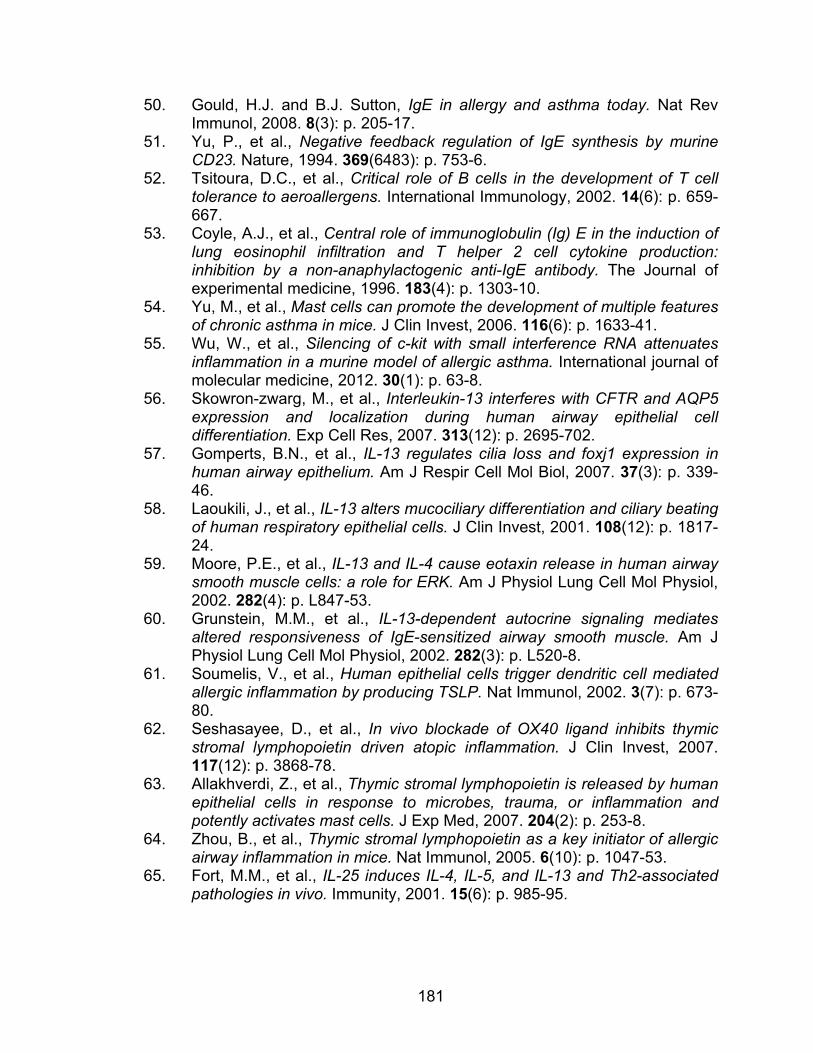
181
50. Gould, H.J. and B.J. Sutton, IgE in allergy and asthma today. Nat Rev Immunol, 2008. 8(3): p. 205-17.
51. Yu, P., et al., Negative feedback regulation of IgE synthesis by murine CD23. Nature, 1994. 369(6483): p. 753-6.
52. Tsitoura, D.C., et al., Critical role of B cells in the development of T cell tolerance to aeroallergens. International Immunology, 2002. 14(6): p. 659-667.
53. Coyle, A.J., et al., Central role of immunoglobulin (Ig) E in the induction of lung eosinophil infiltration and T helper 2 cell cytokine production: inhibition by a non-anaphylactogenic anti-IgE antibody. The Journal of experimental medicine, 1996. 183(4): p. 1303-10.
54. Yu, M., et al., Mast cells can promote the development of multiple features of chronic asthma in mice. J Clin Invest, 2006. 116(6): p. 1633-41.
55. Wu, W., et al., Silencing of c-kit with small interference RNA attenuates inflammation in a murine model of allergic asthma. International journal of molecular medicine, 2012. 30(1): p. 63-8.
56. Skowron-zwarg, M., et al., Interleukin-13 interferes with CFTR and AQP5 expression and localization during human airway epithelial cell differentiation. Exp Cell Res, 2007. 313(12): p. 2695-702.
57. Gomperts, B.N., et al., IL-13 regulates cilia loss and foxj1 expression in human airway epithelium. Am J Respir Cell Mol Biol, 2007. 37(3): p. 339-46.
58. Laoukili, J., et al., IL-13 alters mucociliary differentiation and ciliary beating of human respiratory epithelial cells. J Clin Invest, 2001. 108(12): p. 1817-24.
59. Moore, P.E., et al., IL-13 and IL-4 cause eotaxin release in human airway smooth muscle cells: a role for ERK. Am J Physiol Lung Cell Mol Physiol, 2002. 282(4): p. L847-53.
60. Grunstein, M.M., et al., IL-13-dependent autocrine signaling mediates altered responsiveness of IgE-sensitized airway smooth muscle. Am J Physiol Lung Cell Mol Physiol, 2002. 282(3): p. L520-8.
61. Soumelis, V., et al., Human epithelial cells trigger dendritic cell mediated allergic inflammation by producing TSLP. Nat Immunol, 2002. 3(7): p. 673-80.
62. Seshasayee, D., et al., In vivo blockade of OX40 ligand inhibits thymic stromal lymphopoietin driven atopic inflammation. J Clin Invest, 2007. 117(12): p. 3868-78.
63. Allakhverdi, Z., et al., Thymic stromal lymphopoietin is released by human epithelial cells in response to microbes, trauma, or inflammation and potently activates mast cells. J Exp Med, 2007. 204(2): p. 253-8.
64. Zhou, B., et al., Thymic stromal lymphopoietin as a key initiator of allergic airway inflammation in mice. Nat Immunol, 2005. 6(10): p. 1047-53.
65. Fort, M.M., et al., IL-25 induces IL-4, IL-5, and IL-13 and Th2-associated pathologies in vivo. Immunity, 2001. 15(6): p. 985-95.
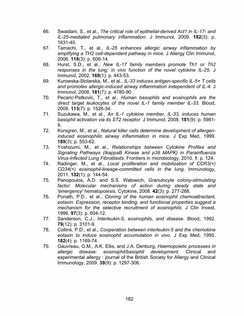
182
66. Swaidani, S., et al., The critical role of epithelial-derived Act1 in IL-17- and IL-25-mediated pulmonary inflammation. J Immunol, 2009. 182(3): p. 1631-40.
67. Tamachi, T., et al., IL-25 enhances allergic airway inflammation by amplifying a TH2 cell-dependent pathway in mice. J Allergy Clin Immunol, 2006. 118(3): p. 606-14.
68. Hurst, S.D., et al., New IL-17 family members promote Th1 or Th2 responses in the lung: in vivo function of the novel cytokine IL-25. J Immunol, 2002. 169(1): p. 443-53.
69. Kurowska-Stolarska, M., et al., IL-33 induces antigen-specific IL-5+ T cells and promotes allergic-induced airway inflammation independent of IL-4. J Immunol, 2008. 181(7): p. 4780-90.
70. Pecaric-Petkovic, T., et al., Human basophils and eosinophils are the direct target leukocytes of the novel IL-1 family member IL-33. Blood, 2009. 113(7): p. 1526-34.
71. Suzukawa, M., et al., An IL-1 cytokine member, IL-33, induces human basophil activation via its ST2 receptor. J Immunol, 2008. 181(9): p. 5981-9.
72. Korsgren, M., et al., Natural killer cells determine development of allergen-induced eosinophilic airway inflammation in mice. J Exp Med, 1999. 189(3): p. 553-62.
73. Yoshizumi, M., et al., Relationships between Cytokine Profiles and Signaling Pathways (IkappaB Kinase and p38 MAPK) in Parainfluenza Virus-Infected Lung Fibroblasts. Frontiers in microbiology, 2010. 1: p. 124.
74. Radinger, M., et al., Local proliferation and mobilization of CCR3(+) CD34(+) eosinophil-lineage-committed cells in the lung. Immunology, 2011. 132(1): p. 144-54.
75. Panopoulos, A.D. and S.S. Watowich, Granulocyte colony-stimulating factor: Molecular mechanisms of action during steady state and 'emergency' hematopoiesis. Cytokine, 2008. 42(3): p. 277-288.
76. Ponath, P.D., et al., Cloning of the human eosinophil chemoattractant, eotaxin. Expression, receptor binding, and functional properties suggest a mechanism for the selective recruitment of eosinophils. J Clin Invest, 1996. 97(3): p. 604-12.
77. Sanderson, C.J., Interleukin-5, eosinophils, and disease. Blood, 1992. 79(12): p. 3101-9.
78. Collins, P.D., et al., Cooperation between interleukin-5 and the chemokine eotaxin to induce eosinophil accumulation in vivo. J Exp Med, 1995. 182(4): p. 1169-74.
79. Gauvreau, G.M., A.K. Ellis, and J.A. Denburg, Haemopoietic processes in allergic disease: eosinophil/basophil development. Clinical and experimental allergy : journal of the British Society for Allergy and Clinical Immunology, 2009. 39(9): p. 1297-306.
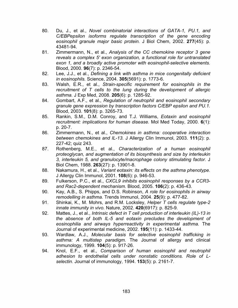
183
80. Du, J., et al., Novel combinatorial interactions of GATA-1, PU.1, and C/EBPepsilon isoforms regulate transcription of the gene encoding eosinophil granule major basic protein. J Biol Chem, 2002. 277(45): p. 43481-94.
81. Zimmermann, N., et al., Analysis of the CC chemokine receptor 3 gene reveals a complex 5' exon organization, a functional role for untranslated exon 1, and a broadly active promoter with eosinophil-selective elements. Blood, 2000. 96(7): p. 2346-54.
82. Lee, J.J., et al., Defining a link with asthma in mice congenitally deficient in eosinophils. Science, 2004. 305(5691): p. 1773-6.
83. Walsh, E.R., et al., Strain-specific requirement for eosinophils in the recruitment of T cells to the lung during the development of allergic asthma. J Exp Med, 2008. 205(6): p. 1285-92.
84. Gombart, A.F., et al., Regulation of neutrophil and eosinophil secondary granule gene expression by transcription factors C/EBP epsilon and PU.1. Blood, 2003. 101(8): p. 3265-73.
85. Rankin, S.M., D.M. Conroy, and T.J. Williams, Eotaxin and eosinophil recruitment: implications for human disease. Mol Med Today, 2000. 6(1): p. 20-7.
86. Zimmermann, N., et al., Chemokines in asthma: cooperative interaction between chemokines and IL-13. J Allergy Clin Immunol, 2003. 111(2): p. 227-42; quiz 243.
87. Rothenberg, M.E., et al., Characterization of a human eosinophil proteoglycan, and augmentation of its biosynthesis and size by interleukin 3, interleukin 5, and granulocyte/macrophage colony stimulating factor. J Biol Chem, 1988. 263(27): p. 13901-8.
88. Nakamura, H., et al., Variant eotaxin: its effects on the asthma phenotype. J Allergy Clin Immunol, 2001. 108(6): p. 946-53.
89. Fulkerson, P.C., et al., CXCL9 inhibits eosinophil responses by a CCR3- and Rac2-dependent mechanism. Blood, 2005. 106(2): p. 436-43.
90. Kay, A.B., S. Phipps, and D.S. Robinson, A role for eosinophils in airway remodelling in asthma. Trends Immunol, 2004. 25(9): p. 477-82.
91. Shinkai, K., M. Mohrs, and R.M. Locksley, Helper T cells regulate type-2 innate immunity in vivo. Nature, 2002. 420(6917): p. 825-9.
92. Mattes, J., et al., Intrinsic defect in T cell production of interleukin (IL)-13 in the absence of both IL-5 and eotaxin precludes the development of eosinophilia and airways hyperreactivity in experimental asthma. The Journal of experimental medicine, 2002. 195(11): p. 1433-44.
93. Wardlaw, A.J., Molecular basis for selective eosinophil trafficking in asthma: A multistep paradigm. The Journal of allergy and clinical immunology, 1999. 104(5): p. 917-26.
94. Knol, E.F., et al., Comparison of human eosinophil and neutrophil adhesion to endothelial cells under nonstatic conditions. Role of L-selectin. Journal of immunology, 1994. 153(5): p. 2161-7.
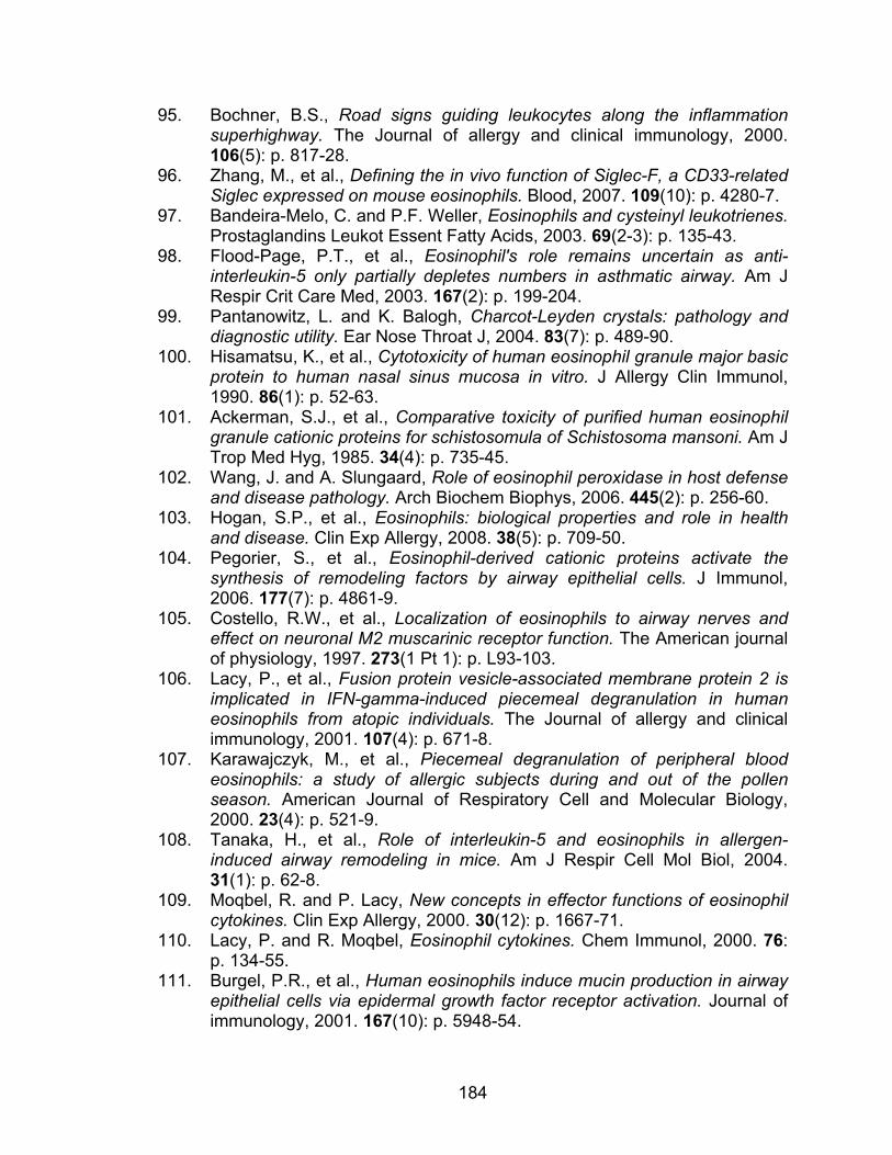
184
95. Bochner, B.S., Road signs guiding leukocytes along the inflammation superhighway. The Journal of allergy and clinical immunology, 2000. 106(5): p. 817-28.
96. Zhang, M., et al., Defining the in vivo function of Siglec-F, a CD33-related Siglec expressed on mouse eosinophils. Blood, 2007. 109(10): p. 4280-7.
97. Bandeira-Melo, C. and P.F. Weller, Eosinophils and cysteinyl leukotrienes. Prostaglandins Leukot Essent Fatty Acids, 2003. 69(2-3): p. 135-43.
98. Flood-Page, P.T., et al., Eosinophil's role remains uncertain as anti-interleukin-5 only partially depletes numbers in asthmatic airway. Am J Respir Crit Care Med, 2003. 167(2): p. 199-204.
99. Pantanowitz, L. and K. Balogh, Charcot-Leyden crystals: pathology and diagnostic utility. Ear Nose Throat J, 2004. 83(7): p. 489-90.
100. Hisamatsu, K., et al., Cytotoxicity of human eosinophil granule major basic protein to human nasal sinus mucosa in vitro. J Allergy Clin Immunol, 1990. 86(1): p. 52-63.
101. Ackerman, S.J., et al., Comparative toxicity of purified human eosinophil granule cationic proteins for schistosomula of Schistosoma mansoni. Am J Trop Med Hyg, 1985. 34(4): p. 735-45.
102. Wang, J. and A. Slungaard, Role of eosinophil peroxidase in host defense and disease pathology. Arch Biochem Biophys, 2006. 445(2): p. 256-60.
103. Hogan, S.P., et al., Eosinophils: biological properties and role in health and disease. Clin Exp Allergy, 2008. 38(5): p. 709-50.
104. Pegorier, S., et al., Eosinophil-derived cationic proteins activate the synthesis of remodeling factors by airway epithelial cells. J Immunol, 2006. 177(7): p. 4861-9.
105. Costello, R.W., et al., Localization of eosinophils to airway nerves and effect on neuronal M2 muscarinic receptor function. The American journal of physiology, 1997. 273(1 Pt 1): p. L93-103.
106. Lacy, P., et al., Fusion protein vesicle-associated membrane protein 2 is implicated in IFN-gamma-induced piecemeal degranulation in human eosinophils from atopic individuals. The Journal of allergy and clinical immunology, 2001. 107(4): p. 671-8.
107. Karawajczyk, M., et al., Piecemeal degranulation of peripheral blood eosinophils: a study of allergic subjects during and out of the pollen season. American Journal of Respiratory Cell and Molecular Biology, 2000. 23(4): p. 521-9.
108. Tanaka, H., et al., Role of interleukin-5 and eosinophils in allergen-induced airway remodeling in mice. Am J Respir Cell Mol Biol, 2004. 31(1): p. 62-8.
109. Moqbel, R. and P. Lacy, New concepts in effector functions of eosinophil cytokines. Clin Exp Allergy, 2000. 30(12): p. 1667-71.
110. Lacy, P. and R. Moqbel, Eosinophil cytokines. Chem Immunol, 2000. 76: p. 134-55.
111. Burgel, P.R., et al., Human eosinophils induce mucin production in airway epithelial cells via epidermal growth factor receptor activation. Journal of immunology, 2001. 167(10): p. 5948-54.
185
112. Puxeddu, I., et al., Human peripheral blood eosinophils induce angiogenesis. The international journal of biochemistry & cell biology, 2005. 37(3): p. 628-36.
113. Rothenberg, M.E. and S.P. Hogan, The eosinophil. Annu Rev Immunol, 2006. 24: p. 147-74.
114. Duan, W., et al., Inhaled p38alpha mitogen-activated protein kinase antisense oligonucleotide attenuates asthma in mice. Am J Respir Crit Care Med, 2005. 171(6): p. 571-8.
115. Chopra, P., et al., Therapeutic potential of inhaled p38 mitogen-activated protein kinase inhibitors for inflammatory pulmonary diseases. Expert Opin Investig Drugs, 2008. 17(10): p. 1411-25.
116. Eynott, P.R., et al., Allergen-induced inflammation and airway epithelial and smooth muscle cell proliferation: role of Jun N-terminal kinase. British journal of pharmacology, 2003. 140(8): p. 1373-80.
117. Kampen, G.T., et al., Eotaxin induces degranulation and chemotaxis of eosinophils through the activation of ERK2 and p38 mitogen-activated protein kinases. Blood, 2000. 95(6): p. 1911-7.
118. Underwood, D.C., et al., SB 239063, a potent p38 MAP kinase inhibitor, reduces inflammatory cytokine production, airways eosinophil infiltration, and persistence. The Journal of pharmacology and experimental therapeutics, 2000. 293(1): p. 281-8.
119. Gamell, C., et al., The p38/MK2/Hsp25 pathway is required for BMP-2-induced cell migration. PLoS One, 2011. 6(1): p. e16477.
120. Birukov, K.G., et al., Shear stress-mediated cytoskeletal remodeling and cortactin translocation in pulmonary endothelial cells. American Journal of Respiratory Cell and Molecular Biology, 2002. 26(4): p. 453-64.
121. McDaniel, A.S., et al., Pak1 regulates multiple c-Kit mediated Ras-MAPK gain-in-function phenotypes in Nf1+/- mast cells. Blood, 2008. 112(12): p. 4646-54.
122. Rousseau, S., et al., CXCL12 and C5a trigger cell migration via a PAK1/2-p38alpha MAPK-MAPKAP-K2-HSP27 pathway. Cellular signalling, 2006. 18(11): p. 1897-905.
123. Chiba, Y. and M. Misawa, The role of RhoA-mediated Ca2+ sensitization of bronchial smooth muscle contraction in airway hyperresponsiveness. J Smooth Muscle Res, 2004. 40(4-5): p. 155-67.
124. Schaafsma, D., et al., Allergic sensitization enhances the contribution of Rho-kinase to airway smooth muscle contraction. Br J Pharmacol, 2004. 143(4): p. 477-84.
125. Taki, F., et al., Effects of Rho-kinase inactivation on eosinophilia and hyper-reactivity in murine airways by allergen challenges. Clin Exp Allergy, 2007. 37(4): p. 599-607.
126. Takeda, N., et al., Role of RhoA inactivation in reduced cell proliferation of human airway smooth muscle by simvastatin. Am J Respir Cell Mol Biol, 2006. 35(6): p. 722-9.

186
127. Henry, P.J., T.S. Mann, and R.G. Goldie, A rho kinase inhibitor, Y-27632 inhibits pulmonary eosinophilia, bronchoconstriction and airways hyperresponsiveness in allergic mice. Pulm Pharmacol Ther, 2005. 18(1): p. 67-74.
128. Tan, B.L., et al., Genetic evidence for convergence of c-Kit- and alpha4 integrin-mediated signals on class IA PI-3kinase and the Rac pathway in regulating integrin-directed migration in mast cells. Blood, 2003. 101(12): p. 4725-32.
129. Yang, F.C., et al., Rac2 stimulates Akt activation affecting BAD/Bcl-XL expression while mediating survival and actin function in primary mast cells. Immunity, 2000. 12(5): p. 557-568.
130. Cho, S.H., et al., Rac and protein kinase C-delta regulate ERKs and cytosolic phospholipase A2 in FcepsilonRI signaling to cysteinyl leukotriene synthesis in mast cells. J Immunol, 2004. 173(1): p. 624-31.
131. Jaffer, Z.M. and J. Chernoff, p21-activated kinases: three more join the Pak. Int J Biochem Cell Biol, 2002. 34(7): p. 713-7.
132. Zhao, Z.S., et al., A conserved negative regulatory region in alphaPAK: inhibition of PAK kinases reveals their morphological roles downstream of Cdc42 and Rac1. Mol Cell Biol, 1998. 18(4): p. 2153-63.
133. Frost, J.A., et al., Differential effects of PAK1-activating mutations reveal activity-dependent and -independent effects on cytoskeletal regulation. J Biol Chem, 1998. 273(43): p. 28191-8.
134. Manser, E., et al., A brain serine/threonine protein kinase activated by Cdc42 and Rac1. Nature, 1994. 367(6458): p. 40-6.
135. Bagrodia, S. and R.A. Cerione, Pak to the future. Trends Cell Biol, 1999. 9(9): p. 350-5.
136. Lei, M., et al., Structure of PAK1 in an autoinhibited conformation reveals a multistage activation switch. Cell, 2000. 102(3): p. 387-97.
137. Buchwald, G., et al., Conformational switch and role of phosphorylation in PAK activation. Mol Cell Biol, 2001. 21(15): p. 5179-89.
138. Bokoch, G.M., et al., A GTPase-independent mechanism of p21-activated kinase activation. Regulation by sphingosine and other biologically active lipids. J Biol Chem, 1998. 273(14): p. 8137-44.
139. King, C.C., et al., p21-activated kinase (PAK1) is phosphorylated and activated by 3-phosphoinositide-dependent kinase-1 (PDK1). J Biol Chem, 2000. 275(52): p. 41201-9.
140. Morreale, A., et al., Structure of Cdc42 bound to the GTPase binding domain of PAK. Nat Struct Biol, 2000. 7(5): p. 384-8.
141. Gatti, A., et al., Multisite autophosphorylation of p21-activated protein kinase gamma-PAK as a function of activation. J Biol Chem, 1999. 274(12): p. 8022-8.
142. Chong, C., et al., The mechanism of PAK activation. Autophosphorylation events in both regulatory and kinase domains control activity. J Biol Chem, 2001. 276(20): p. 17347-53.
187
143. Oskeritzian, C.A., S. Milstien, and S. Spiegel, Sphingosine-1-phosphate in allergic responses, asthma and anaphylaxis. Pharmacol Ther, 2007. 115(3): p. 390-9.
144. Rivera, J., R.L. Proia, and A. Olivera, The alliance of sphingosine-1-phosphate and its receptors in immunity. Nat Rev Immunol, 2008. 8(10): p. 753-63.
145. Buday, L., L. Wunderlich, and P. Tamas, The Nck family of adapter proteins: regulators of actin cytoskeleton. Cell Signal, 2002. 14(9): p. 723-31.
146. Puto, L.A., et al., p21-activated kinase 1 (PAK1) interacts with the Grb2 adapter protein to couple to growth factor signaling. J Biol Chem, 2003. 278(11): p. 9388-93.
147. Lu, W., et al., Activation of Pak by membrane localization mediated by an SH3 domain from the adaptor protein Nck. Curr Biol, 1997. 7(2): p. 85-94.
148. Mayhew, M.W., et al., Identification of phosphorylation sites in betaPIX and PAK1. J Cell Sci, 2007. 120(Pt 22): p. 3911-8.
149. Rudel, T. and G.M. Bokoch, Membrane and morphological changes in apoptotic cells regulated by caspase-mediated activation of PAK2. Science, 1997. 276(5318): p. 1571-4.
150. Bokoch, G.M., Biology of the p21-activated kinases. Annu Rev Biochem, 2003. 72: p. 743-81.
151. Eswaran, J., et al., UnPAKing the class differences among p21-activated kinases. Trends Biochem Sci, 2008. 33(8): p. 394-403.
152. Sells, M.A., A. Pfaff, and J. Chernoff, Temporal and spatial distribution of activated Pak1 in fibroblasts. J Cell Biol, 2000. 151(7): p. 1449-58.
153. Dharmawardhane, S., et al., Localization of p21-activated kinase 1 (PAK1) to pinocytic vesicles and cortical actin structures in stimulated cells. J Cell Biol, 1997. 138(6): p. 1265-78.
154. Dharmawardhane, S., et al., Localization of p21-activated kinase 1 (PAK1) to pseudopodia, membrane ruffles, and phagocytic cups in activated human neutrophils. J Leukoc Biol, 1999. 66(3): p. 521-7.
155. Sells, M.A., J.T. Boyd, and J. Chernoff, p21-activated kinase 1 (Pak1) regulates cell motility in mammalian fibroblasts. J Cell Biol, 1999. 145(4): p. 837-49.
156. Sells, M.A., et al., Human p21-activated kinase (Pak1) regulates actin organization in mammalian cells. Curr Biol, 1997. 7(3): p. 202-10.
157. Dharmawardhane, S., et al., Regulation of macropinocytosis by p21-activated kinase-1. Mol Biol Cell, 2000. 11(10): p. 3341-52.
158. Manser, E., et al., Expression of constitutively active alpha-PAK reveals effects of the kinase on actin and focal complexes. Mol Cell Biol, 1997. 17(3): p. 1129-43.
159. Hing, H., et al., Pak functions downstream of Dock to regulate photoreceptor axon guidance in Drosophila. Cell, 1999. 97(7): p. 853-63.
188
160. Chen, W., et al., The Caenorhabditis elegans p21-activated kinase (CePAK) colocalizes with CeRac1 and CDC42Ce at hypodermal cell boundaries during embryo elongation. J Biol Chem, 1996. 271(42): p. 26362-8.
161. Leberer, E., et al., Virulence and hyphal formation of Candida albicans require the Ste20p-like protein kinase CaCla4p. Curr Biol, 1997. 7(8): p. 539-46.
162. Weiss, E.L., et al., Chemical genetic analysis of the budding-yeast p21-activated kinase Cla4p. Nat Cell Biol, 2000. 2(10): p. 677-85.
163. Arias-Romero, L.E., et al., EhPAK2, a novel p21-activated kinase, is required for collagen invasion and capping in Entamoeba histolytica. Mol Biochem Parasitol, 2006. 149(1): p. 17-26.
164. Klopocka, W., J. Moraczewska, and M.J. Redowicz, Characterisation of the Rac/PAK pathway in Amoeba proteus. Protoplasma, 2005. 225(1-2): p. 77-84.
165. Nagel, M., et al., Role of p21-activated kinase in cell polarity and directional mesendoderm migration in the Xenopus gastrula. Dev Dyn, 2009. 238(7): p. 1709-26.
166. Manser, E., et al., PAK kinases are directly coupled to the PIX family of nucleotide exchange factors. Molecular cell, 1998. 1(2): p. 183-92.
167. Adam, L., et al., Regulation of microfilament reorganization and invasiveness of breast cancer cells by kinase dead p21-activated kinase-1. J Biol Chem, 2000. 275(16): p. 12041-50.
168. Ito, M., et al., P21-activated kinase 1: a new molecular marker for intravesical recurrence after transurethral resection of bladder cancer. J Urol, 2007. 178(3 Pt 1): p. 1073-9.
169. Delorme, V., et al., Cofilin activity downstream of Pak1 regulates cell protrusion efficiency by organizing lamellipodium and lamella actin networks. Dev Cell, 2007. 13(5): p. 646-62.
170. Chan, A.Y., et al., Role of cofilin in epidermal growth factor-stimulated actin polymerization and lamellipod protrusion. The Journal of cell biology, 2000. 148(3): p. 531-42.
171. Kumar, G., et al., Over-expression of S4D mutant of Leishmania ADF/cofilin impairs flagellum assembly by affecting actin dynamics. Eukaryotic cell, 2012.
172. Tang, W., et al., A PLCbeta/PI3Kgamma-GSK3 signaling pathway regulates cofilin phosphatase slingshot2 and neutrophil polarization and chemotaxis. Developmental cell, 2011. 21(6): p. 1038-50.
173. Vartiainen, M.K., et al., The three mouse actin-depolymerizing factor/cofilins evolved to fulfill cell-type-specific requirements for actin dynamics. Molecular biology of the cell, 2002. 13(1): p. 183-94.
174. Coniglio, S.J., S. Zavarella, and M.H. Symons, Pak1 and Pak2 mediate tumor cell invasion through distinct signaling mechanisms. Molecular and cellular biology, 2008. 28(12): p. 4162-72.
189
175. Starinsky-Elbaz, S., et al., The pre-GAP-related domain of neurofibromin regulates cell migration through the LIM kinase/cofilin pathway. Molecular and cellular neurosciences, 2009. 42(4): p. 278-87.
176. Edwards, D.C., et al., Activation of LIM-kinase by Pak1 couples Rac/Cdc42 GTPase signalling to actin cytoskeletal dynamics. Nature cell biology, 1999. 1(5): p. 253-9.
177. Vadlamudi, R.K., et al., p41-Arc subunit of human Arp2/3 complex is a p21-activated kinase-1-interacting substrate. EMBO Rep, 2004. 5(2): p. 154-60.
178. Pantaloni, D., et al., The Arp2/3 complex branches filament barbed ends: functional antagonism with capping proteins. Nature cell biology, 2000. 2(7): p. 385-91.
179. Machesky, L.M., et al., Scar, a WASp-related protein, activates nucleation of actin filaments by the Arp2/3 complex. Proceedings of the National Academy of Sciences of the United States of America, 1999. 96(7): p. 3739-44.
180. Wu, C., et al., Arp2/3 is critical for lamellipodia and response to extracellular matrix cues but is dispensable for chemotaxis. Cell, 2012. 148(5): p. 973-87.
181. Mullins, R.D., J.A. Heuser, and T.D. Pollard, The interaction of Arp2/3 complex with actin: nucleation, high affinity pointed end capping, and formation of branching networks of filaments. Proceedings of the National Academy of Sciences of the United States of America, 1998. 95(11): p. 6181-6.
182. Fehon, R.G., A.I. McClatchey, and A. Bretscher, Organizing the cell cortex: the role of ERM proteins. Nat Rev Mol Cell Biol, 2010. 11(4): p. 276-87.
183. McClatchey, A.I. and R.G. Fehon, Merlin and the ERM proteins--regulators of receptor distribution and signaling at the cell cortex. Trends Cell Biol, 2009. 19(5): p. 198-206.
184. Shaffer, M.H., et al., Ezrin and moesin function together to promote T cell activation. J Immunol, 2009. 182(2): p. 1021-32.
185. Shaffer, M.H., et al., Ezrin is highly expressed in early thymocytes, but dispensable for T cell development in mice. PLoS One, 2010. 5(8): p. e12404.
186. Belkina, N.V., et al., LOK is a major ERM kinase in resting lymphocytes and regulates cytoskeletal rearrangement through ERM phosphorylation. Proceedings of the National Academy of Sciences of the United States of America, 2009. 106(12): p. 4707-12.
187. Brown, K.L., et al., Regulation of hyaluronan binding by F-actin and colocalization of CD44 and phosphorylated ezrin/radixin/moesin (ERM) proteins in myeloid cells. Experimental cell research, 2005. 303(2): p. 400-14.
188. Saotome, I., M. Curto, and A.I. McClatchey, Ezrin is essential for epithelial organization and villus morphogenesis in the developing intestine. Dev Cell, 2004. 6(6): p. 855-64.
190
189. Bonilha, V.L., et al., Microvilli defects in retinas of ezrin knockout mice. Exp Eye Res, 2006. 82(4): p. 720-9.
190. Curto, M. and A.I. McClatchey, Ezrin...a metastatic detERMinant? Cancer Cell, 2004. 5(2): p. 113-4.
191. Monni, R., et al., Ezrin is a target for oncogenic Kit mutants in murine erythroleukemia. Blood, 2008. 111(6): p. 3163-72.
192. Staser, K., et al., A Pak1-PP2A-ERM signaling axis mediates F-actin rearrangement and degranulation in mast cells. Exp Hematol, 2012.
193. Feng, Y., et al., Filamin A (FLNA) is required for cell-cell contact in vascular development and cardiac morphogenesis. Proceedings of the National Academy of Sciences of the United States of America, 2006. 103(52): p. 19836-41.
194. Cunningham, C.C., et al., Actin-binding protein requirement for cortical stability and efficient locomotion. Science, 1992. 255(5042): p. 325-7.
195. Vadlamudi, R.K., et al., Filamin is essential in actin cytoskeletal assembly mediated by p21-activated kinase 1. Nature cell biology, 2002. 4(9): p. 681-90.
196. Maceyka, M., et al., Filamin A links sphingosine kinase 1 and sphingosine-1-phosphate receptor 1 at lamellipodia to orchestrate cell migration. Molecular and cellular biology, 2008. 28(18): p. 5687-97.
197. Rider, L. and M. Diakonova, Adapter protein SH2B1beta binds filamin A to regulate prolactin-dependent cytoskeletal reorganization and cell motility. Molecular endocrinology, 2011. 25(7): p. 1231-43.
198. Dummler, B., et al., Pak protein kinases and their role in cancer. Cancer Metastasis Rev, 2009. 28(1-2): p. 51-63.
199. Sanders, L.C., et al., Inhibition of myosin light chain kinase by p21-activated kinase. Science, 1999. 283(5410): p. 2083-5.
200. Takahashi, K. and K. Suzuki, Membrane transport of WAVE2 and lamellipodia formation require Pak1 that mediates phosphorylation and recruitment of stathmin/Op18 to Pak1-WAVE2-kinesin complex. Cellular signalling, 2009. 21(5): p. 695-703.
201. Wittmann, T., G.M. Bokoch, and C.M. Waterman-Storer, Regulation of microtubule destabilizing activity of Op18/stathmin downstream of Rac1. The Journal of biological chemistry, 2004. 279(7): p. 6196-203.
202. Somanath, P.R. and T.V. Byzova, 14-3-3beta-Rac1-p21 activated kinase signaling regulates Akt1-mediated cytoskeletal organization, lamellipodia formation and fibronectin matrix assembly. J Cell Physiol, 2009. 218(2): p. 394-404.
203. Kiosses, W.B., et al., A role for p21-activated kinase in endothelial cell migration. J Cell Biol, 1999. 147(4): p. 831-44.
204. Smith, S.D., et al., PAK1-mediated activation of ERK1/2 regulates lamellipodial dynamics. J Cell Sci, 2008. 121(Pt 22): p. 3729-36.
205. Parrini, M.C., et al., Dissecting activation of the PAK1 kinase at protrusions in living cells. J Biol Chem, 2009. 284(36): p. 24133-43.
191
206. Balasenthil, S., et al., p21-activated kinase-1 signaling mediates cyclin D1 expression in mammary epithelial and cancer cells. J Biol Chem, 2004. 279(2): p. 1422-8.
207. Holm, C., et al., Association between Pak1 expression and subcellular localization and tamoxifen resistance in breast cancer patients. J Natl Cancer Inst, 2006. 98(10): p. 671-80.
208. Mira, J.P., et al., Endogenous, hyperactive Rac3 controls proliferation of breast cancer cells by a p21-activated kinase-dependent pathway. Proc Natl Acad Sci U S A, 2000. 97(1): p. 185-9.
209. Carter, J.H., et al., Pak-1 expression increases with progression of colorectal carcinomas to metastasis. Clin Cancer Res, 2004. 10(10): p. 3448-56.
210. Davidson, B., M. Shih Ie, and T.L. Wang, Different clinical roles for p21-activated kinase-1 in primary and recurrent ovarian carcinoma. Hum Pathol, 2008. 39(11): p. 1630-6.
211. Schraml, P., et al., Combined array comparative genomic hybridization and tissue microarray analysis suggest PAK1 at 11q13.5-q14 as a critical oncogene target in ovarian carcinoma. Am J Pathol, 2003. 163(3): p. 985-92.
212. O'Sullivan, G.C., et al., Modulation of p21-activated kinase 1 alters the behavior of renal cell carcinoma. Int J Cancer, 2007. 121(9): p. 1930-40.
213. Brown, L.A., et al., Amplification of EMSY, a novel oncogene on 11q13, in high grade ovarian surface epithelial carcinomas. Gynecol Oncol, 2006. 100(2): p. 264-70.
214. Brown, L.A., et al., Amplification of 11q13 in ovarian carcinoma. Genes Chromosomes Cancer, 2008. 47(6): p. 481-9.
215. Mao, X., et al., Genomic alterations in blastic natural killer/extranodal natural killer-like T cell lymphoma with cutaneous involvement. J Invest Dermatol, 2003. 121(3): p. 618-27.
216. Mao, X., et al., Amplification and overexpression of JUNB is associated with primary cutaneous T-cell lymphomas. Blood, 2003. 101(4): p. 1513-9.
217. Gerthoffer, W.T., Actin cytoskeletal dynamics in smooth muscle contraction. Can J Physiol Pharmacol, 2005. 83(10): p. 851-6.
218. McFawn, P.K., et al., Calcium-independent contraction and sensitization of airway smooth muscle by p21-activated protein kinase. Am J Physiol Lung Cell Mol Physiol, 2003. 284(5): p. L863-70.
219. Gunst, S.J. and W. Zhang, Actin cytoskeletal dynamics in smooth muscle: a new paradigm for the regulation of smooth muscle contraction. Am J Physiol Cell Physiol, 2008. 295(3): p. C576-87.
220. Tang, D.D. and S.J. Gunst, The small GTPase Cdc42 regulates actin polymerization and tension development during contractile stimulation of smooth muscle. J Biol Chem, 2004. 279(50): p. 51722-8.
221. Tang, D.D., Y. Bai, and S.J. Gunst, Silencing of p21-activated kinase attenuates vimentin phosphorylation on Ser-56 and reorientation of the vimentin network during stimulation of smooth muscle cells by 5-hydroxytryptamine. Biochem J, 2005. 388(Pt 3): p. 773-83.
192
222. Wu, Y., et al., Integrin-linked kinase regulates smooth muscle differentiation marker gene expression in airway tissue. Am J Physiol Lung Cell Mol Physiol, 2008. 295(6): p. L988-97.
223. Allen, J.D., et al., p21-activated kinase regulates mast cell degranulation via effects on calcium mobilization and cytoskeletal dynamics. Blood, 2009. 113(12): p. 2695-705.
224. Sauer, B., Inducible gene targeting in mice using the Cre/lox system. Methods, 1998. 14(4): p. 381-92.
225. Kuhn, R., et al., Inducible gene targeting in mice. Science, 1995. 269(5229): p. 1427-9.
226. Kiel, M.J., et al., Hematopoietic stem cells do not depend on N-cadherin to regulate their maintenance. Cell Stem Cell, 2009. 4(2): p. 170-9.
227. Das, A.M., R.J. Flower, and M. Perretti, Eotaxin-induced eosinophil migration in the peritoneal cavity of ovalbumin-sensitized mice: mechanism of action. Journal of immunology, 1997. 159(3): p. 1466-73.
228. Rothenberg, M.E., et al., Eotaxin triggers eosinophil-selective chemotaxis and calcium flux via a distinct receptor and induces pulmonary eosinophilia in the presence of interleukin 5 in mice. Molecular medicine, 1996. 2(3): p. 334-48.
229. Fukuyama, S., et al., Effect of eotaxin and platelet-activating factor on airway inflammation and hyperresponsiveness in guinea pigs in vivo. American journal of respiratory and critical care medicine, 2000. 161(6): p. 1844-9.
230. Chang, H.C., et al., The transcription factor PU.1 is required for the development of IL-9-producing T cells and allergic inflammation. Nat Immunol, 2010. 11(6): p. 527-34.
231. van Rijt, L.S., et al., A rapid flow cytometric method for determining the cellular composition of bronchoalveolar lavage fluid cells in mouse models of asthma. J Immunol Methods, 2004. 288(1-2): p. 111-21.
232. Yang, F.C., et al., Nf1-dependent tumors require a microenvironment containing Nf1+/-- and c-kit-dependent bone marrow. Cell, 2008. 135(3): p. 437-48.
233. Jungraithmayr, W.M., et al., A mouse model of orthotopic, single-lung transplantation. J Thorac Cardiovasc Surg, 2009. 137(2): p. 486-91.
234. Krupnick, A.S., et al., Orthotopic mouse lung transplantation as experimental methodology to study transplant and tumor biology. Nat Protoc, 2009. 4(1): p. 86-93.
235. Okazaki, M., et al., A mouse model of orthotopic vascularized aerated lung transplantation. American journal of transplantation : official journal of the American Society of Transplantation and the American Society of Transplant Surgeons, 2007. 7(6): p. 1672-9.
236. Saito, H., et al., Pathogenesis of murine experimental allergic rhinitis: a study of local and systemic consequences of IL-5 deficiency. J Immunol, 2002. 168(6): p. 3017-23.
193
237. Sanderson, C.J., D.J. Warren, and M. Strath, Identification of a lymphokine that stimulates eosinophil differentiation in vitro. Its relationship to interleukin 3, and functional properties of eosinophils produced in cultures. J Exp Med, 1985. 162(1): p. 60-74.
238. Dyer, K.D., et al., Functionally competent eosinophils differentiated ex vivo in high purity from normal mouse bone marrow. J Immunol, 2008. 181(6): p. 4004-9.
239. Iellem, A., et al., Unique chemotactic response profile and specific expression of chemokine receptors CCR4 and CCR8 by CD4(+)CD25(+) regulatory T cells. J Exp Med, 2001. 194(6): p. 847-53.
240. Tiffany, H.L., F. Li, and H.F. Rosenberg, Hyperglycosylation of eosinophil ribonucleases in a promyelocytic leukemia cell line and in differentiated peripheral blood progenitor cells. Journal of leukocyte biology, 1995. 58(1): p. 49-54.
241. Fischkoff, S.A., et al., Eosinophilic differentiation of the human promyelocytic leukemia cell line, HL-60. The Journal of experimental medicine, 1984. 160(1): p. 179-96.
242. Ingram, D.A., et al., Hyperactivation of p21(ras) and the hematopoietic-specific Rho GTPase, Rac2, cooperate to alter the proliferation of neurofibromin-deficient mast cells in vivo and in vitro. The Journal of experimental medicine, 2001. 194(1): p. 57-69.
243. Hiatt, K.K., et al., Neurofibromin GTPase-activating protein-related domains restore normal growth in Nf1-/- cells. The Journal of biological chemistry, 2001. 276(10): p. 7240-5.
244. Lu, L., et al., Detection of luxol-fast-blue positive cells in human promyelocytic leukemia cell line HL-60. Exp Hematol, 1981. 9(9): p. 887-92.
245. Johnson, G.R. and D. Metcalf, Detection of a new type of mouse eosinophil colony by Luxol-fast-blue staining. Exp Hematol, 1980. 8(5): p. 549-61.
246. Clutterbuck, E.J., E.M. Hirst, and C.J. Sanderson, Human interleukin-5 (IL-5) regulates the production of eosinophils in human bone marrow cultures: comparison and interaction with IL-1, IL-3, IL-6, and GMCSF. Blood, 1989. 73(6): p. 1504-12.
247. Lozano, E., et al., PAK is required for the disruption of E-cadherin adhesion by the small GTPase Rac. J Cell Sci, 2008. 121(Pt 7): p. 933-8.
248. Delorme-Walker, V.D., et al., Pak1 regulates focal adhesion strength, myosin IIA distribution, and actin dynamics to optimize cell migration. The Journal of cell biology, 2011. 193(7): p. 1289-303.
249. Bates, M.E., et al., Human airway eosinophils respond to chemoattractants with greater eosinophil-derived neurotoxin release, adherence to fibronectin, and activation of the Ras-ERK pathway when compared with blood eosinophils. Journal of immunology, 2010. 184(12): p. 7125-33.
194
250. Lintomen, L., et al., Human eosinophil adhesion and degranulation stimulated with eotaxin and RANTES in vitro: lack of interaction with nitric oxide. BMC pulmonary medicine, 2008. 8: p. 13.
251. Jia, G.Q., et al., Selective eosinophil transendothelial migration triggered by eotaxin via modulation of Mac-1/ICAM-1 and VLA-4/VCAM-1 interactions. International Immunology, 1999. 11(1): p. 1-10.
252. Nakajima, H., et al., Role of vascular cell adhesion molecule 1/very late activation antigen 4 and intercellular adhesion molecule 1/lymphocyte function-associated antigen 1 interactions in antigen-induced eosinophil and T cell recruitment into the tissue. J Exp Med, 1994. 179(4): p. 1145-54.
253. Walsh, G.M., et al., Integrin alpha 4 beta 7 mediates human eosinophil interaction with MAdCAM-1, VCAM-1 and fibronectin. Immunology, 1996. 89(1): p. 112-9.
254. Wittmann, T., G.M. Bokoch, and C.M. Waterman-Storer, Regulation of leading edge microtubule and actin dynamics downstream of Rac1. The Journal of cell biology, 2003. 161(5): p. 845-51.
255. Adyshev, D.M., et al., Differential involvement of ezrin/radixin/moesin proteins in sphingosine 1-phosphate-induced human pulmonary endothelial cell barrier enhancement. Cellular signalling, 2011. 23(12): p. 2086-96.
256. Shamri, R., et al., CCL11 elicits secretion of RNases from mouse eosinophils and their cell-free granules. FASEB J, 2012. 26(5): p. 2084-93.
257. El-Shazly, A., et al., Human eotaxin induces eosinophil-derived neurotoxin release from normal human eosinophils. International Archives of Allergy and Immunology, 1998. 117 Suppl 1: p. 55-8.
258. Zimmermann, N., J.J. Conkright, and M.E. Rothenberg, CC chemokine receptor-3 undergoes prolonged ligand-induced internalization. The Journal of biological chemistry, 1999. 274(18): p. 12611-8.
259. Ro, J.Y., et al., Effects of 7,8-dihydro-8-oxo-deoxyguanosine on antigen challenge in ovalbumin-sensitized mice may be mediated by suppression of Rac. British journal of pharmacology, 2009. 158(7): p. 1743-52.
260. Carstanjen, D., et al., Rac2 regulates neutrophil chemotaxis, superoxide production, and myeloid colony formation through multiple distinct effector pathways. Journal of immunology, 2005. 174(8): p. 4613-20.
261. Reutershan, J., et al., Blocking p21-activated kinase reduces lipopolysaccharide-induced acute lung injury by preventing polymorphonuclear leukocyte infiltration. American journal of respiratory and critical care medicine, 2007. 175(10): p. 1027-35.
262. Nakae, S., et al., Mast cell-derived TNF contributes to airway hyperreactivity, inflammation, and TH2 cytokine production in an asthma model in mice. J Allergy Clin Immunol, 2007. 120(1): p. 48-55.
195
263. McGlade, J.P., et al., Suppression of the asthmatic phenotype by ultraviolet B-induced, antigen-specific regulatory cells. Clinical and experimental allergy : journal of the British Society for Allergy and Clinical Immunology, 2007. 37(9): p. 1267-76.
264. Sung, K.P., et al., Eotaxin induces a sustained reduction in the functional adhesive state of very late antigen 4 for the connecting segment 1 region of fibronectin. The Journal of allergy and clinical immunology, 2000. 106(5): p. 933-40.
265. Barthel, S.R., et al., Roles of integrin activation in eosinophil function and the eosinophilic inflammation of asthma. Journal of leukocyte biology, 2008. 83(1): p. 1-12.
266. Croker, B.A., et al., Rac2-deficient mice display perturbed T-cell distribution and chemotaxis, but only minor abnormalities in T(H)1 responses. Immunology and cell biology, 2002. 80(3): p. 231-40.
267. Insall, R.H. and L.M. Machesky, Actin dynamics at the leading edge: from simple machinery to complex networks. Developmental cell, 2009. 17(3): p. 310-22.
268. Voermans, C., et al., SDF-1-induced actin polymerization and migration in human hematopoietic progenitor cells. Exp Hematol, 2001. 29(12): p. 1456-64.
269. Kim, C.H. and H.E. Broxmeyer, SLC/exodus2/6Ckine/TCA4 induces chemotaxis of hematopoietic progenitor cells: differential activity of ligands of CCR7, CXCR3, or CXCR4 in chemotaxis vs. suppression of progenitor proliferation. J Leukoc Biol, 1999. 66(3): p. 455-61.
270. Weiner, O.D., et al., Spatial control of actin polymerization during neutrophil chemotaxis. Nat Cell Biol, 1999. 1(2): p. 75-81.
271. Ichetovkin, I., W. Grant, and J. Condeelis, Cofilin produces newly polymerized actin filaments that are preferred for dendritic nucleation by the Arp2/3 complex. Current biology : CB, 2002. 12(1): p. 79-84.
272. Yi, C., et al., Development of small-molecule inhibitors of the group I p21-activated kinases, emerging therapeutic targets in cancer. Biochemical pharmacology, 2010. 80(5): p. 683-9.
273. Fujisawa, T., et al., Chemokines induce eosinophil degranulation through CCR-3. The Journal of allergy and clinical immunology, 2000. 106(3): p. 507-13.
274. Ochkur, S.I., et al., The development of a sensitive and specific ELISA for mouse eosinophil peroxidase: assessment of eosinophil degranulation ex vivo and in models of human disease. Journal of Immunological Methods, 2012. 375(1-2): p. 138-47.
275. Adamko, D.J., et al., The induction of eosinophil peroxidase release: improved methods of measurement and stimulation. Journal of Immunological Methods, 2004. 291(1-2): p. 101-8.

CURRICULUM VITAE
Muithi Mwanthi
Current Position:
2006 Student, NIH Medical Scientist Training Program Indiana University School of Medicine (IUSOM) Department of Microbiology and Immunology Mentor/Advisor - D. Wade Clapp, MD Education:
2005 B.S. - Pre-medicine Summa cum laude, Clearwater Christian College, Clearwater FL
2013 Ph.D. - Microbiology and Immunology, Indiana University,
Indianapolis IN 2014 M.D. - Indiana University School of Medicine, expected
Academic and Professional Honors and Fellowships:
Undergraduate 2002 Presidents’ Academic Fellowship, 2002-04 2003 Alpha Chi academic chapter, National College Honor
Scholarship Society Membership, 2003-04 2003 Bailey Family Foundation Scholarship, 2003-04
Graduate
2006 University Fellowship, 2006-07 2009 Edwin T. Harper Scholarship (R25 GM079657), 2009-10 2010 Joint Best first-time presenter, Research in Progress,
Department of Microbiology and Immunology, 2010 1st place, Sigma Xi Graduate Biomedical Research
Competition 2010 Harold Raidt Department of Microbiology and Immunology
Graduate Student Teaching Award

2011 Ruth L. Kirschstein NRSA for Individual Predoctoral Fellowship to Promote Diversity in Health-Related Research - F31HL108569 PI: “Pak1's Role in Eotaxin-Mediated Murine Eosinophil Migration” National Heart Lung and Blood Institute, National Institutes of Health (NHLBI/NIH), 2011-13
2011 Southern Regional Education Board (SREB) Doctoral
Scholarship, Institute of Teaching and Mentoring, 2011-12 2011 Abstract achievement travel award (MGSAAA), American
Society of Hematology (ASH) Professional Experience:
2004 Kaplan Test Prep and Admissions, Class Instructor and
Tutor Indianapolis IN, and Tampa, Fl, 2004-09 2005 Florida Department of Citrus, Research assistant, Lake
Alfred, FL 2005 Sun Orchards of Florida, Quality Assurance Technician
Haines City, FL 2008 IU School of Medicine MSMS MCAT Summer Prep Program,
Tutor/Facilitator, Indianapolis, IN 2009 IU School of Medicine Teaching Assistant & Lecturer J210,
Principles in Microbiology, 2009-11 Publications:
1. Mwanthi, M., S. Chen, E. Michaels, K. Staser, Z. Sun, S. Sehra, D.
Wilkes, M. Kaplan, S.Park, and D. W. Clapp. 2012. PAK1 regulates eosinophil migration in vivo and in vitro (In submission)
2. Staser, KW, M. Shew, E. Michaels, M. Mwanthi, D. Clapp, and S.
Park. 2011. A Pak1-PP2A-Ezrin signaling axis mediates IgE-dependent F-actin rearrangement and degranulation in mast cells. Exp Hematology, 2012
3. Zeng, Y., K. Staser, K. Menon, S. Park, M. Mwanthi, L. Jiang, and
D. W. Clapp. 2012. Ezrin Regulates Hematopoietic Stem/Progenitor Cell Motility (In preparation)

Abstracts and Presentations:
1. Mwanthi MM, Micheals GE, Park SJ, Clapp DW. “PAK1’s regulation of eotaxin-mediated murine eosinophil migration” IUSM Medical Scientist Training Program (MSTP) retreat, July 2009, Lafayette, IN (Abstract and Poster)
2. Mwanthi MM, Micheals GE, Yongqi Y, Tepper RS, Park SJ, Clapp DW. “PAK1 Modulates F-Actin Dynamics in Murine Eosinophil Migration and Tissue Infiltration”. Annual Biomedical Research Conference for Minority Students (ABRCMS), Nov 4-7 2009, Phoenix, AZ (Abstract and Poster)
3. Mwanthi MM, Micheals GE, Yongqi Y, Tepper RS, Park SJ, Clapp DW. “PAK1 regulates eotaxin-mediated eosinophil migration and inflammation” American Society of Clinical Investigation (ASCI/AAP) 2010: April 17, 2010, Chicago, IL (Abstract and Poster)
4. Mwanthi MM, Micheals GE, Yongqi Y, Tepper RS, Park SJ, Clapp DW. “PAK1 Regulates Eotaxin-Mediated Murine Eosinophil Migration and Tissue Infiltration”. Sigma Xi Research Day May 10, 2010, Indianapolis, IN (Abstract and Oral Presentation)
5. Mwanthi MM, Micheals GE, Staser KW, Yongqi Y, Tepper RS, Park SJ, Clapp DW. “PAK1 regulates eotaxin-mediated eosinophil migration” Regional conference, National Organization for the Professional Advancement of Black Chemists and Chemical Engineers (NOBCCHE), November 4, 2011, Indianapolis, IN (Abstract and Poster)
6. Mwanthi MM, Micheals GE, Staser KW, Sehra S, Jander M, Chen S, Wilkes DS, Kaplan, MH, Park SJ, Clapp DW. Annual conference for American Society of Hematology: “PAK1 regulates eotaxin-mediated eosinophil migration in vivo and in vitro” December 9, 2011, San Diego, CA (Abstract and Oral Presentation)
7. Mwanthi MM, Chen S, Micheals GE, Staser KW, Yongqi Y, Sehra
S, Tepper RS, Kaplan, MH, Park SJ, Clapp DW. “PAK1 regulates eotaxin-mediated eosinophil migration” 10th Annual Midwest Blood Club Symposium, March 15, 2012, Indianapolis, IN (Abstract and Poster)
8. Walline C, Benson H, Sehra S, Mwanthi M, Fisher A, Clapp DW, Brutkiewicz R, Kaplan M, Wilkes DS, and Blum, J. “Alterations in pulmonary immunity in response to vaccinia virus” Immunology 98th Annual Meeting May 14-15, 2011, San Francisco, CA (Abstract, Poster and Oral Presentation)
9. Zeng, Yi, Staser KW, Menon KM, Park SJ, Mwanthi MM, Jiang L,
and Clapp DW. “Ezrin Regulates Hematopoietic Stem/Progenitor Cell Motility” Annual conference for American Society of Hematology, December 10, 2011, San Diego, CA (Abstract and Poster)