p5Ihdependent Growth Arrest of Human Astrocytomas by pl4- · Abstract p53-Independent Growth Arrest...
121
p5Ihdependent Growth Arrest of Human Astrocytomas by pl4- by Craig Leslie Stewart A thesis submitted in confomüty with the requirements for the degree of Master of Science Department of Laboratory Medicine and Pathobiology University of Toronto O Copyright by Craig L. Stewart (2000)
Transcript of p5Ihdependent Growth Arrest of Human Astrocytomas by pl4- · Abstract p53-Independent Growth Arrest...

p5Ihdependent Growth Arrest of Human Astrocytomas by pl4-
by
Craig Leslie Stewart
A thesis submitted in confomüty with the requirements for the degree of
Master of Science
Department of Laboratory Medicine and Pathobiology
University of Toronto
O Copyright by Craig L. Stewart (2000)

National Library Bibliothèque nationale du Canada
Acquisitions and Acquisitions et Bibliographie Services services bibliographiques
395 Wellington Street 395, nie Wellington OttawaON K1AON4 ûttawa ON KI A ON4 Canada Canada
The author has granted a non- L'auteur a accorde une licence non exclusive licence allowing the exclusive permettant à la National Library of Canada to Bibliothèque nationale du Canada de reproduce, loan, distribute or sell reproduire, prêter, distribuer ou copies of this thesis in microform, vendre des copies de cette thèse sous paper or electronic formats. la forme de microfiche/nlm, de
reproduction sur papier ou sur format électronique.
The author retains ownership of the L'auteur conserve la propriété du copyright in this thesis. Neither the droit d'auteur qui protège cette thèse. thesis nor substantial extracts fiorn it Ni la thèse ni des extraits substantiels may be p ~ t e d or otherwise de celle-ci ne doivent être imprimés reproduced without the author's ou autrement reproduits sans son permission. autorisation.

Abstract
p53-Independent Growth Arrest of Human Astrocytomas by ~ 1 4 ~ ~
Master of Science, 2000.
Craig Stewart, Department of Laboratory Medicine and Pathobiology,
University of Toronto
The role of the two distinct tumour suppressor proteins expressed frorn the CDKIV2A
locus, ~ 1 6 ' " ~ ~ ~ and pllAE, has been intensely studied due, in part, to their frequent inacti-
vation in human maiignancies such as astrocytornas. p 1 6WKJ"unction~ as an inhibitor of
CDK4, thereby activating pRB and resulting in ceII cycle arrest. In contrat, growth inhibi-
tion by p 14ARF OCCU~S through the p53 pathway due to its interaction with MDM2 and resul-
tant stabilization of p53. In order to assess the effect of expression of ~ 1 4 ~ " or ~ 1 6 ~ " "
on the growth of astrocytomas, we have employed a series of human ce11 lines harbounng
distinct mutation in the p53 andor pRB pathways, We observed that, as expected, growth
arrest mediated by p161NK4" was dependent on an intact pRB pathway. In transient assays,
we also observed that arrest due to expression of ~ 1 4 " ~ ~ required wild-type p53. However,
in colony-forming assays, ~ 1 4 ~ ~ ~ potently reduced the number of colonies in al1 ceIl lines
regardIess of their p53 stanis. Furthemore, dones that were recovered cycled at a greatly
reduced rate and exhibited a significant increase in senescence-associated P-gdactosidase
activity. Thrse data indicate a p53-independent mechanism whereby pl JAN can mediate
growth arrest in astrocytomas leading to apparent ce11 senescence.

TABLE OF CONTF,NTS
AB STRACT ........................ 2
TABLE OF CONTENTS ........................ 3
ACKNOWLEDGEMENTS ......................... 6
........................ LIST OF FIGURES 7
LIST OF TABLES ....................... -8
........................ AB B REVIATIONS 9
INTRODUCTION ........................ I l
........................ The "pRB Pathway" 12
The pRB F d l y Proteins ........................ 13
........................ The E2F-Family Proteins 14
........................ The "p53 Pathway" 17
The p53 Protein ....................... 17
........................ The p53 Inhibitor. MDM2 18
........................ The Cip/Kip Farnily of CDK Inhibitors 20
The Ce11 Growth and Ce11 Death Pathways are Controlled by a Single Genetic Locus.,.24
The PX4 Farnily Proteins ........................ 24
The ARF Turnour Suppressor ........................ 26
Interactions between the pRB and p53 Ce11 Cycte Control Pathways ...... ,. ....... ,., ...... 30
ARF Regulates the Ce11 Cycle through bath the pRB and p53 Pathways ............... 30
Regdation of the p53 Pathway by pRB ........................ 32
INK4a.ARF Inactivation in Cancer .............. ... ....... 33
Human Asmcytomas .......................... 35
INK4dARF Inactivation in Astrocytomas ........................... 36
RESEARCH HYPOTHESIS AND OBJECTIVES ........................ 39
MAI'ERIALS AND METHODS ...... ............,... 41
Production of GST-Fusion Proteins ........................ 41

........................ Preparation of Mamrnalian Ce11 Lysates 42
........................ Site-Directed Mutagenesis 42
........................ Sequencing of CDK4-R24C Mutant 43
........................ GST Pull-Dom Assays 44
Subcellular Localization of GFP-Fusion Proteins ........................ 44
........................ Gelatin Zymography 44
Ce11 Lines and Culture Conditions .......................... 45
........................ Ionizing Radiation Assays 45
Western Blotting Andysis ........................ 45
Mammalian and Bacterid Expression Constructs ........................ 46
Transfection of Mamrnaiian Expression Plasmids ........................ 48
Colony-Formation Assays and Generation of Stable Clones ........................ 48
........................ Adenovims Infections 49
Co-lmmunoprecipitation Assays ........................ 49
FACS Analysis ........................ 50
Senescence-Associated p-Galactosidase Staining ........................ 50
........................ RESULTS 51
................. .... CDK4-TNK4 Interaction Assays ,., 51
Sub-CelIular Locaiization of INK4 and ARF Proteins ....................... 53
Analysis of p53 Functional Status in Glioma Ce11 Lines ........................ 54 INK4a Adenovinl-Mediated Expression of p 16 and 1 4 ~ in Glioma Cell Lines.35
p14m Inhibits Colony-Formation of Cells Deficient in p53 andor pRB .. .58
Reconstituted Expression of can O C C U ~ without Perturbation of pRB
Pathway in GIiomas Lacking Functional p53 ...................... 60
Stable Expression of 1 4 ~ is Associated with Cellular Senescence in Gliornas ... 63
[NKh Affect of pl6 Expression on Invasion of Human Gliomas ............ 65
........................ DISCUSSION 67


1 wish to thank Dr. Paul Hamel for giving me the opportunity to conduct this research in his
lab, and for dl his much appreciated instruction, scientific knowledge and generousity. 1 also
wish to thank Dr. Jim Rutka for his clinical expertise, inspiration and support.
Tfianks to Dr. Suzanne Kamel-Reid, Dr, Irene Andrulis and Dr. Rod Bremner for providing
thoughtfbl insights and instructive suggestions as members of my committee. I would also
like to thank Dr. Minta and Dr. Sarma for their encouragement and counsel.
To ai1 the members of the lab, 1 wish to extend rny sincere appreciation for dl oftheir techni-
cal assistance, constructive criticism, and humourous moments. You will d l be missed.
Most of all, 1 ;un eternally grateful to my parents for supporthg me while 1 puaued this work.
Your unceasing labour will be repaid.

List of Fipures
1) Schematic diagram of the eukaryotic ce11 cycle ..................... 11
2) Schematic diagram of the G. restriction point .................... 16
3) p53 regulation under normal conditions and in the presence of
DNA damage . schematic diagram .......................... 19
4) Structure of the IM(4a/ARF locus . schematic diagram ............................. 27
5) Integration of the pRB and p53 pathways . schematic diagnm ......................... 31
6) GST-INK4 pull-down assays with wild-type CDK4 ........................... 51
73) Sequencing of the CDK4-R24C mutant ......................... 52
7b) GST-IM<4 pull-down assays with the CDK4-R24C mutan r .......................... 52
............................... 8 4 Subcellular localization of INK4 proteins in marnmalian cells 53
8b) Subceiluiar localization of ARF proteins in marnmaiian cells ........................ 53
9) p53-mediated response of human glioma ce11 Lines to ionizing radiation ............... 55
.............. 1Oa) Adenovid transfer of p 1 61NK4= and p 14"" to human glioma ce11 lines 56
lob) Co-immunoprecipitation of exogenous p 16MK4a and p IJARF with endogenous
....................... substrates in human giiorna ce11 lines 56
.... 1 Oc) Molecular changes induced by transient expression of p 16NK4a in human gliomas 56
1Od) Molecular changes induced by transient expression of p 1JARF in human gliomas ..... 57
1 la) Colony-formation assays with U343 cells .............................. 59
1 Ib) Colony-formation assays with U25 1 cells .......................... .... 59
1 lc) Colony-formation assays with SF126 cells ................................ 59
1 ld) Quantitation of results from colony-formation assays with U343. U25 1 and SF126 ... 59
123) Expression of ce11 cycle factors in U25 1 cells stably-expressing p 14m ............... 61
12b) Expression of celI cycle factors in SF126 cells stably-expressing ~ 1 4 ~ ................ 61
13a) MMP-2 activity in a panel of human glioma ceE h e s ............................ 66

13b) MMP-2 activity in U2S 1 and SFI26 cells transiently expressing
............................ or ~ 1 4 ~ 66
........................... 13c) MMP-2 activity in U25 1 cells stably-expressing p MhRF 66
13d) MMP-2 activity in U25 1 cells stably-overexpressing CDK4 ............................ 66
List of Tables
......................... 1) DNA constmcts 47
2) Summary of growth m s t induced by transient or stable expression of p 16MK4a or
p 14ARF in human glioblastoma ce11 lines .......................... .60
3) FACS anaiysis of p14ARf-expressing U25 1 clones infected with ~ 1 6 " ~ ~ ~ ....... 62
4) FACS anaiysis of p 14ARf-expressing U25 1 clones exposed to ionizing radiation ........ 63
5) Proliferation rate of p 14ARf-expressing U25 1 clones ......................... 64
6) Senescence-associated P-galactosidase activity in p 14A"7expressing U25 1 clones ..... 64

Abbreviations
Ad-CMV
Ad- p 1 4""
Ad- p 1 PK"
ARF
BSA
C2
C4
CDK
Cip
CO2
cDNA
D n
E1S
Ela
EDTA
FACS
FPLB
G4t8
GBM
GFP
GST
GY
HBS
IgG
'INK4
control adenovirus (non-encoding)
adenovims encoding p 14Aw
adenovirus encoding p 1 61N"'
altemate reading f m e (protein)
bovine serum albumin
U2S 1 CDK4-stable clone
U25 1 CDK4-stable clone
cyclin dependent kinase
CDK inhibitory protein
carbon dioxide
copy deoxyribonucleic acid
dithiothreiol
exon i P
exon la
ethy lenediaminetetrmcetate
fluorescence-activated ce11 sorting
fusion protein lysis buffer
geneticin
glioblastoma multiforme
green Ruorescent protein
glutathione S-tramferase
Gray
hepes buffered saline
immunoglobutin G
inhibitor of CDK4

IPTG
K ~ P
LB
mg
ml
m M
M
MDM2
MEFs
MEM
MMP
P3
PAGE
PBS
PCR
PMSF
PRB
RPM
SA P-gal
SDS
TE
TBS
w/v
isopropyl p-D-thiogalactopyranoside
Kinase inhibitory protein
Luria-Bertani medium
mi lligram
milliliter
millimoIar
Molar
murine double minute-:! protein
murine embryonic fibroblasts
minimal essential media
matrix metailoproteinase
U25 1 p 14ARF-stable clone P3
polyacrylarnide gel eiectrophoresis
phosphate buffered saline
polymeme chain reaction
phenyImethanesuIfony1 fluoride
Retinoblastorna protein
revolutions per minute
senescence-associated P-galactosidase
sodium dodecyl sulfate
Tris-EDTA
tris buffered saline
weight per volume

Introduction
Progression through or exit from the eukaryotic ce11 division cycle is regulated by a
series of stringent control mechanisms. The ce11 cycle most commonly depicted (see Figure
1) consists of two major phases. one where replication of genome occurs (S phase) and
another responsible for segregation of the duplicated genome into daughter cells (mitosis
or M phase). These two phases are typically sepamted by gaps: G, between M and S and
G2, between S and M. While not the subject of this introduction. variations of this general
scheme are employed by many ce11 types where. for example. the gaps are absent. mitosis
proceeds in the absence of DNA synthesis or where a ce11 cornpletes additional rounds of
DNA synthesis without passing through M. It is also clear that the regulation of the ce11 cycle
involves mechanisms which are highly conserved among al1 eukaryotes. This conservation
is particularly evident for rnitosis where replacement of defective proteins conuolling M in
Saccharomyces cerevisiae with their human courtterparts restores normal mitosis (for review
see ( 1)).
For this introduction, we restrict consideration of the ce11 cycle to control of
Figure I The Eukaryoac Ceii Cycle Schernatic diagram of the eukaryotic ceii cycle showing stages of DNA synthesis (S phase) and mitosis (M phase) separafed by gaps (G1 and G2). Molecules which regulate the transition between each respective stage are shown outside the cycle.
the GJG, to S transi-
tion. Cells in Go or
G, c m be stimulated
by mitogens (growth
factors) to progress
through G, towards S
phase. This transition
is mitogen-dependent
until the ceIls reach
the "restriction point"
(2) in late G, just
prior t~ S phase entry.
11

After passage through the restriction point, celIs are irreversibly committed to DNA synthesis
regardless of the presence of the mitogenic signal. Thus. the restriction point represents a
cntical checkpoint in the ce11 cycle. Here, integration of an a m y of endogenous and exog-
enous signals leads either to ce11 cycle arrest or continuation through the ceIl cycle to mitosis.
Due to its irreversible nature, the restriction point is tightly regulated. Passage through this
checkpoint is governed by both positive and negative ce11 cycle regulatory factors. It is dso
evident that additional checkpoinis are important regdators of progression after the restric-
tion point has been passed. These checkpoints ensure the proper timing of specific events in
the ce11 cycle and assess the fidelity of DNA synthesis. When these checkpoints are invoked,
celIs can be halted from progressing further through the ce11 cycle until, for example, DNA
repair can be completed. or can be instructed to undergo prognmmed ce11 death if DNA
damage is too extensive.
In its simplest sense, then, the ce11 cycle appears to be regulated by a ce11 growth
and a ceil death pathway. In this context, we review two fundamental pathways, the "pRB
pathway" and the "p53 pathway". respectively, which govem these two processes. As is dis-
cussed in greater detail in other reviews on this issue, the importance of these pathways in
cellular growth control is underscored by the observation that members of these pathways are
found mutated in al1 human cancers, As will also become evident in the discussion below,
whiIe these pathways are typically studied and discussed independently, recent data have
revealed an intimate molecular and genetic interaction between these pathways.
The '$RB Pathwuy "
As has been reviewed in detail elsewhere, the rate limiting step for progression fiom
Gd G, to S phase is the appearance of the ciass of proteins, known as cyclins ((3.4); see (5)
for review). For the ce11 cycle described in Figure 1, these inctude the D-type cyclins (cycrins
Dl, D2, and D3) and cyciin E. The a p p e m c e of these cyclins following a mitogenic stimu-
lus generdy occurs in a highly regulated manner. Failure to express these cyclins resulü

in arrest of the ce11 cycle at specific points in the ce11 cycle (6-10). The early portion of G,
appears to be govemed by the expression of the D-type cyclins (7.1 1 - 1 3). In fibroblasts, their
levels tend to increase significantly, peaking 6 to 8 hours following the mitogenic stimulus.
As cells pass through the point govemed by cyclin D expression. a second checkpoint at the
G,-S boundq is encountered. this one determined by the expression of a distinct cyciin.
cyclin E (9,10,14).
Cyciins D and E are CO-factors for a class of kinases known as the cyclin dependent
kinases (CDK's). These serinelthreonine-specific kinases phosphorylate these residues in the
general context of the amino acid sequence Ser/Thr-Pro-x- ArgLys (( 15.16)). Importmtly,
the cyclins exhibit distinct affinities towards specific members of the CDK family. The
D-type cyclins are typically associated with CDW (17,18) and CDK6 (1 7.19) while cyclin E
binds CDK2 exclusively (20-22). Since the peak of associated kinase activity of the D-type
cyciins coincides with the restriction point, th& expression has been considered to be an
essential aspect of the mechanism regulating passage through this point (for example see
(23)). This notion is somewhat complicated by the observation that the different D-type
cyclins are expressed in distinct but overlapping sets of ce11 types during embryogenesis and
in adult tissues (24-29). These unique expression patterns account for the defects which are
observed in animais deficient for some of these. specifically cyclins DI (30) and D2 (31).
That these expression patterns refiect unique biological activities is supported by the observa-
tions that different sets of D-type cyclins can block ce11 cycle exit during cellular differentia-
tion in distinct cells (32,33). It is also clear that the D-type cyclins have other cellular roles
distinct from ceil cycle regulation, suggested, for exarnple, by the requirement of cyclin D3
for ce11 exit frorn the ce11 cycle during myogenesis (24,28,34,35).
The pRB Famgy Proteins.
Cloning and sequencing of the gene responsible for the pediatric childhood retinal
malignancy, retinoblastoma (36), reveded the most important target for cyclin D/CDK4
activity and a critical mediator of cell cycle progression. The human retinoblastoma protein

(PM). harboun 16 distinct cyclin/CDKconsensus sequences. Furthermore. the pRB pro-
tein becornes highly phosphorylated at the same point in G, that kinase activity associated
with the D-type cyclins begins to peak (37-40). Experiments using both in vitro and in vivo
systems strongly support the notion that the principle target of cyclin DlCDK4 activity is
the pRB protein (41-46) and that phosphorylation-dependent inactivation of pRB is required
for ce11 cycle progression (45,4749). Whether pRB is important for regulation of ce11 cycle
progression or is more fundamental to ce11 cycle exit, when. for example, cells terminally
differentiate, is unresolved. The latter role is supported by observations where, for many ce11
types both in vivo and in vitro, loss of pRB leads to apoptosis during differentiation (50-53).
Compensation for the lack of pRB by the related pRB-family members, p IO7 and p 130, ha
been proposed for pRB-deficient cells which escape this fate (50) (54) (53.55.56). Support-
ing this mode1 are snidies employing chirneric mice deficirnt for pRB andor pl07 (57-59).
Normal retinal development is seen in animals lacking either pRB or p107. In contrast, chi-
meric animals where both pl07 and plU3 were absent exhibited hyperplasia, disorganized
growth and tumours in the retina, consistent with a compensatory role for p107. However,
while pl07 and pl30 exhibit activities sirnilar to pRB (60-67), their role as tumour suppres-
sors has not been fully resolved since tumours hahouring mutations in p 107 or p 130 are rare
(68-70). Furthemore, mice lacking one or both functional alleles of pl07 or pl30 are not
prone to tumour formation (58,59,7 1 J2).
The E2F-Family Proteins.
Many pRB-interacting factors have been reported (for examples see (73-88)). In the
context of the ce11 cycle, the ESF-family of bHLH transcription factors are the most impor-
tant if not the best characterizcd. The six members of this farnily, E2F-1 to -6, form heterodi-
mers with the DP-family proteins. DP-1, -2 or -3 (for reviews see (89-91)). Their DNA-
binding site consensus sequence, first defined in the genome of adenovims (92), occurs in
the promoter region of a large number of factors involved in ceil cycle progression or DNA
synthesis. Some of these factors include cyclin E (93). dihydrofolate reductase ( D m ;

(94-96)). thymidylate synthetase (97), cdc25A (98), cdc2 (99), cyciin A (97), E2F1 itself
(lOOtlO1), as well as pRB (102-104) and pl07 (105). With the exception of E2F6, al1 of
the E2F-farnily rnembers have a transcriptional activation domain at their C-termini. The
tramactivation domain harbours sequences responsible for mediating binding to the pRB-
family proteins. Rather than rnerely repressing the activity of the E2F's, binding of pRB to
E2F converts E2F from being an active transcriptional xtivator to a transcriptional repressor
(106,107). This repressor activity is further enhanced by the association of histone deacety-
lase (108,109) (HDAC). Thus, active repression also occurs by inducing a "closed" structure
for chrornatin at a particular locus. Like the pRB-family and D-type cyclins. the E2F-fam-
ily proteins also exhibit tissue-specific expression, at l e s t during embryogenesis (1 10,111).
Animais deficient for specific E2F members have indicated further that E2F3, nther than
E2F1, is the pnnciple family member required for ce11 proliferation during ernbryogenesis
((1 12-1 14)). This observation is consistent with antibody microinjection experiments dem-
onstrating that E2FI is involved in regulation of the fint ce11 cycle immediately following a
mitogenic stimulus while E2F3 is required for subsequent ce11 cycles as cells continue to pro-
lifente ( 1 15.1 16). It has also been recently shown that, in quiescent cells, pRB is associated
prirnarily with a novel fom of E2F3, E2F3b (1 17), supporting the superiority of this E2F-
family mernber in pRB-dependent ce11 cycle control. It is clear, however, that EZFI activity
must be carefully regulated. Overexpression of E2Fl or loss of pRB Ieading to uncontrolled
E2Fl activity strongly induces programmed ce11 death (1 18-1 2 1). E2F3, in contmt, does not
appear to drive apoptosis when its activity is deregulated (1 16,120,122). These data suggest
iùrther that the different E2F's may have distinct transcriptional targets. This notion is s u p
ported by analyses dernonstrating distinct perturbations in the expression pattern of differ-
ent ce11 cycle regulatory factors using mouse embryo fibroblasts (MER) denved fiom either
pRB, p 107 or p 130-deficient embryos (123).
Thus, whiIe a great number of important details remain to be defined, a fundamenta1
cell cycle regulatory pathway involving the D-type cyclins, pRB-family and the E2F-famiiy

proteins has emerged (see Figure 2). In resting or quiescent cells, complexes of E2F4 associ-
ated with p 130 appear to be the predominant complex bound to promoten with EZF-consen-
sus sequences (1 24- 126). pRBE2FI CO-complexes are also found in quiescent cells, but,
given the presence of E2F-binding sites in their promoter regions. their levels tend to be
decreased in resting cells via an autoregulatory mechanism . It has also been revealed that
pRB- or p130-containing complexes exhibit cytoplasmic compartmentalization in specific
quiescent cells and in terminally differentiated cells both in vitro and in vivo (127-129). This
comparîmentalization further prevents activated transcription by the E2F-farnily proteins.
Transcriptional repression of E2F target genes by pRB/EZF complexes is further enhanced
by pRB-dependent binding of histone deacetylase (HDAC).
As cells are stimulated to enter the ce11 cycle. the p107iE2F4 complexes replace the
pl 30fE2F4 complexes (130) while complexes containing pRB and EIF1. E1F2 or E2F3.
bound to DNA. become more abundant (126). The inhibitory rffects of the pRB-family pro-
teins on E2F- dii-tbn
Figure 2 Activity a l the pRB pathway during progression throagh G1. In resting or quiescent cells, repression of transcription of S-phase promoting genes is mediated by pRB recruitment of histone deacetylase to pmmoters condning E2F sites. Additionatly, repression crui also occur via p13kE2F4 complexes. In the ptesence of growth stimulus, leveb of cyclin D increase, and cyclin D/CDK4 complexes phosphorylate pRB, releasing repression and ailowing transcription by ESFI. Repression mediated by p130:E2F4 complexes is overcome by a rnechanism that involves phosphorylation of p l30 which targets p 130 for degradation.
dependent acti-
vated mscrip-
tion are then
Iost due to the
up-regulation
of cyclin D. In
combination
with CDK4/6,
phosphoryla-
tion of the
pRB - f a m i l y
proteins liber-
ates E2F h m

these repressors, resulting in the activated transcription of E2F target genes. It has been
suggested further that another checkpoint involves E2F1 (and presumably E2R and E2F3)
during S phase (1 3 1,132). Specificaily, following entry into S. E2F activity appears to be
inhibited by binding of E2F1 to cyclin A, whose levels begin to increase dunng S. The cyclin
AKDK2 complex phosphorylates the DP proteins and possibly E2F, decreasing ESFIDP
a n i t y for DNA and. thereby, causing its release. This mode1 for regdation of E2F1 activ-
ity during S was supported using a mutant E2F1, deficient for the cyclin A binding site.
Retrovial-mediated transferof this mutant, E2FI(A 24), to NIH 3T3 fibroblasts promoted ce11
cycle progression in resting cells but blocked their exit from S phase.
Thus. while specific temporal aspects of this pathway are under active investigation, it
is cIear that the "pRB-pathway" represents an important growth control mechanism involving
the antagonistic ce11 cycle replatory activities of the D-type cyclins, the pRB-farnily proteins
and the E2F-family of transcription factors.
The '$53 Pathway"
The p53 Protein.
The transcription factor, p53, acts as a fundamental regulator of ceIl cycle arrest and
apoptosis in the normal cell. It's cenûal role in these processes is supported by the fact that
p53 is the most frequent target for inactivation in malignantly transformed cells. Since its
discovery in 1979, p53 mutations have ken described in more than 50% of human cancers
(1 33). While its complete role continues to be elucidated, it is clear is that p53 integrates
signals fiom intemal and extemai stimuli, dowing the ce11 to respond to a variety of stresses.
These responses a . ~ generated, in part, by p53-mediated transcriptional activation of genes
possessing a p53-response element in their promoter.
Stnicturally, the hurnan p53 transcription factor is 393 amino acids long and consists
of five domains (for review, see (134))- The first 42 amino acids at the N-terminus consti-
tute the transactivation domain which interacts and co-operates with components of the basal

transcription machinery such as TAFs (TATA-binding protein Associated Factor). Further-
more, inhibition of p53-mediated transcription is achieved through binding of proteins to the
p53 transactivation domain. These negative regulaton of p53 activity inhibit anscription
by both interaction with the p53 transactivation domain and direct inhibition of the transcrip-
tional machinery assembled at the promoter.
In conjunction a proline-rich domain (Pm) located between the transactivation
domain and the sequence-specific DNA-binding domain, the C-terminal domain (CTD) regu-
lates the growth arrest and apoptotic pmmoting activities of p53. The CTD harbours basic
residues rhat bind preferentially to specific DNA and RNA sequences and to DNA ends.
In addition, this dornain mediates the reassociation of double-stranded DNA or RNA from
single strands. The CTD in conjunction with the PRD maintains the p53 tetramer in a con-
formation that has low-affhity for binding its consensus sequence (5'-PuPuPuC(Mï)-3'
arnnged as a pair of inverted repeats). Phosphorylation by protein kinase C or casein
kinase iI activates sequence-specific DNA binding of p53 resulting in activated transcription
of, for exmple, p21aplNAF11Wi"C*i1 (p21). MDM2, GADDJS, Cyclin G, Bax, and IGF-BP3
(135-140). In addition to its transactivation activity, p53 c m aIso repress the expression
of several cellular and viral genes whose promoters do not contain a p53-response element
(111) through a rnechanisrn which involves direct interaction with components of the basal
transcription machinery, possibly TBP (TATA-Binding Protein). Among these genes are
c-fos and SV40 large T antigen (142-144).
The p53 inhibitor, MDM2.
Under normal conditions, pS3 is a latent, short-lived protein with a hdf-Life of 5-20
minutes. Protein levels and activity are kept low through various regdatory mechanisms.
One of the p n m q regulaton of p53 fùnction in the ce11 is the Murine Double Mnute-2
protein, or MDM2. MDM2 was originally discovered as a gene overexpressed in the
tumourigenic 3T3DM mouse ceIl line that stabIy maintains double minute chromosomes
(145). The N-terminus of MDM2 binds to the transactivation domain of p53 (146,147).

DNA DAMAGE
t CeIl Cycle hrrut
Figure 3 The p53 autoregdatory loop. Under normal conditions, MDM2 binds to pS3 and inhibits p53-mediated transactivation. Nuclear export of p53 and subsequent degradrition in the cytoplasm is dso mediated by MDMZ In the presence of DNA dmage. enzymes such 3s DNA-PK are activated and phosphorylate both p53 and MDMZ thereby prcvenung the intenction between the two proteins. As p53 IeveIs and rictivity rise. p53 transcriptional t q e t s such as p21 are upregulated to arrest the ce11 cycle. Once the damage to DNA is repaired, DNA-PK levels drop, thereby pennitting dom-regulation of p53 levels and rictivity by MDM2 once again.
This interaction inhibits the
transcriptionai activity of
p53 through masking of
the transactivation domain,
and direct inhibition of
the basal transcription
rnachinery at the promoter,
possibly TFIIE ( 148-1 50).
The transrepression func-
tion (ix, the ability to
repress transcription frorn a
particular prornoter) of p53
is also impaired as a con-
sequence of MDM2 bind-
ing (15 1). The critical role
of MDM2 in the modula-
tion of p53 activity is most
evident in the fact that the embryonic lethal phenotype seen in MDM2-nul1 mice c m be over-
corne by CO-deletion of p53 (1 52).
In addition to rnodulating p53 activity, MDM2 regulates p53 protein levels. Binding
of MDM2 to p53 targets p53 for nuclear export and subsequent degndation in the cytoplasrn
(153-155). MDM2 contains a nuclear export signal that alIows it to CO-transport p53 out
of the nucleus into the cytoplasm (156-258). There, MDM2 functions as an E3 ubiquitin
ligase, directly targeting p53 for destruction via the ubiquitin-proteosome degradation path-
way (159-162). However, as noted above, MDM2 is a transcriptional target of p53. Thus,
an autoregdatory loop exists where high p53mediated transactivation is countered by p53-
dependent up-replation of the p53 inhibitor, MDM2 (263)(see Figure 3).
19

The cellular response to DNA damage illustrates the intimate relationship between
p53 and its regulator MDM2 in the presence of stressful stimuli. Genetic insuits denved from
ionizing radiation compromise the integrity of genomic DNA structure, catdyzing breakage
of DNA double-strands. The free DNA ends created as a result give nse to a senes of events,
one of which is activation of a nuclear kinase known as DNA-PK. Both p53 and MDM2
undergo phosphorylation mediated by DNA-PK (or enzymes with similar specificity) at their
respective N-terminal regions dter DNA damage (164,165). As a result. p53 and MDM2 fail
to bind each another, leading to stabilization of p53 protein in the nucleus and thereby caus-
ing activated transcription of target genes that induce either ceIl cycle anest (137) or apop-
tosis (165-168). When p53 elicits ce11 cycle arrest through activation of genes such as p21
(see below) the proliferative block is overcome only when damaged DNA is repaired. Once
repaired, DNA-PK activity decreases due to the loss of DNA ends (164). Consequently,
newly-synthesized p53 and MDM2 would remain unphosphorylated leading to decreased
p53 stability, protein levels and overall activity as the ce11 cycle progresses once again.
The Cip/Kip Family of CDK Inhibitors.
A criticai mediator of the p53 response to DNA damage is the CDK inhibitor p21
(cloned variously as Cip 1 (169), Cdi 1 (170). Sdi 1 (1 7 1) and WAFl (172)). MEFs derived
from p2 1-nul1 animals fail to undergo normal G, arrest in response to DNA darnage (173).
Moreover, p21 has been impiicated in protecting cells from apoptosis initiated from stress
or p53 induction, dthough the mechanisms remain obscure (174.175). p53 directly transac-
tivates expression of p2 1 via p53 binding sites in the p21 promoter (172,176). That p21 is
transcriptionally regulated by p53 provided an important Link between the function of the
major human tumor suppressor and negative cell cycIe controI. However, a basai level of p21
can be found in cells denved from p53-deficient mice indicating that p21 expression is dso
reguIated in a p53-independent manner (1 77- 179). One of these p53-independent pathways
involves TGF-p. Here, ceil cycle arrest is mediated, at lest in part, by the induction of p21
mediated by Smad3 and Smad4 regulation of the p2i promoter ( 180,18 1).

p2 1 can hinction as a dual specific inhibitor of ce11 proliferation by two independent
and iünctiondly distinct mechanisms. In addition to its ability to bind and inhibit CDKs, p21
also associates with the DNA replication factor PCNA (Prolifemting ce11 nucIear antigen)
via the unique carboxyl-terminal domain in PCNA ( 182). PCNA is an auxillary protein to
DNA polymerase-8 required for DNA synthesis (1 83). Overexpression of the C-terminal
domain of p21 in marnmdian cells reduces the fraction of celIs found in S phase (184).
Furthemore, in vitro, the p21FCNA interaction blocks DNA replication catalyzed by the
poL8/RFC/PCNA complex ( 182) but does not inhibit PCNA-mediated DNA repair (185).
There are six binding sites for p2 1 per PCNA himer ( 186). therefore p2 1 cm form either a
quaternary complex with PCNA. cyclin and CDK or c m bind to PCNA directly. The fact
that complexes containhg p2 1 and cyclinKDK's also include PCNA suggests that p21 may
coordinate CDK-dependent ce11 cycle progression with processes regulating DNA repiica-
tion andor repair.
p21 is one member of the Cip/Kip family of CDK inhibitors, a family that includes
p27"' (p27) and ~ 5 7 ~ Q " (p57). In conmt to the INK4 proteins, the CipIKip family membea
inhibit a wide range of CDKs which include CDK4. CDK6 and CDK2 (187). In addition,
the two families also differ in their mechanism of binding to CDKs. Cip/Kip proteins
inhibit kinase activity by making contact with both the cyclin and CDK subunits (186490).
Stnictudly, ai1 three membea of the CiplKip family have a 65-amino-acid ngion with
homology (38-442 identity) at their N-terminal portions, which is necessary and sufficient
for binding and inhibition of G, cyclin1CDK complexes (19 1) as well as cyclin B-containing
complexes (1 88). However. unlike the INK4 proteins, which demons~ te extensive sequence
similarity and functional redundancy, each Cip/Kip family mernber has distinct functional
properties, attributable to stmctural differences at their C-termini. While only p2l is actually
in the "p53 pathway", we briefly descnbe here the other CiplKip family memben for
completeness.
p27mr (p27), hke other members of the Cip/Kip family, has a CDK-binding domain

at its N-terminus, which binds to and inhibits cyclin D-. E-, A-, and B-dependent kinases
(192495). This inhibitor shares 47% amino acid identity with p2 1. In contrast to p21, p27
does not bind PCNA and its expression is not regulated by p53. The expression of p27 is
controiled, in part. post-translationally (196.197). p27 mRNA is also induced by vitamin D3
in U937 cells (198) and by [FNP and IFNa ( 199,200) suggesting that transcriptional regula-
tion of the p27 gene is important dunng cellular differentiation and inhibition of ceil growth.
The expression of p27 is high in cells inhibited by ce11 contact. semm deprivation and by a
CAMP activated pathway (20 1-204).
In proliferating cells. p27 is found predominandy in complexes with cyclin DlCDK416
(194.205). These complexes are active, perhaps as a result of p27 being bound to the cyclin
subunit without establishing an inhibitory interaction with the CDK subunit. TGFP mat-
ment of these cells does not increase the total level of p27, but induces a redistribution of p27
from cyclin D/CDK4/6 complexes to cyclin UCDK2. thereby inhibiting CDK? kinase activ-
ity (206,207). As will be discussed in more detail below. this redistribution occurs as a result
of a rapid induction of p1SNKJb by TGFP (207). In contnst to its effect on cyclin DlCDK416
complexes, p27 has a potent inhibitory influence on CDK2containing complexes. The crys-
td structure of p27 bound to the cyclin A/CDK2 complex revealed that p27 invades the
catalytic subunit and dismantles its ATP binding site (19 1). Hence. cyclin DfCDK416 cm
sequester p27 without being subjected to inhibition. whereas the catdytic activities of com-
plexes containing CDKZ are eKciently aboüshed by the same CDK inhibitor. Since cell
cycle progression requires cyciin E- and A-associated kinase activi ty, reduction of p27 levels
is required. Loss of pz7 occun analogously to other ce11 cycle regulatory factors. specificaiiy
via a ubiquitin-mediated pathway in late G, (208-210).
It is iikely that the weak inhibition of cyciin DfCDK4 by p21 or p27 is due to the
role of these CKIs in formation of these cyclin DICDK4f6 complexes (21 1). As Figure 2
depicts, p21 and p27 promote interactions between the D-type cyclios and their CDK part-
ners by stabilizing the complexes and acting as chaperones for their transport to the nucleus

(212,213). Assembly of cyclin DllCDK4 and cyclin DUCDK4 complexes is impaired in
primary MEFs taken fiom animals lacking p2 1, p27 or both (21 1). Moreover, lack of CDK4
in CDK4deficient mice coincides with increased binding of p27 to cyclin WCDK2, dimin-
ished activation of CDK2 and impaired pRB phosphorylation (214). These data suggest
that one rate-limiting CDK4dependent mechanism controlling the the Go to S transition
involves regulation of p27 activity. Thus. the CipiKip proteins act as positive ce11 cycle regu-
lators, facilitating cyclin DKDK complex formation, They are dso potent inhibitors of ce11
cycle progression when they block kinase activity associated with cyclin WCDK2 or cyclin
AKDK2 complexes.
The most recendy identified member of the CiplKip family is p57" (p57; (215.2 16)).
It harbours an N-terminal CDK inhibitory domain and has sequences similar to p27 at its
C-terminus. Like p21, p57 contains a PCNA-binding dornain within its C terminus that,
when separated from its N-terminai CDKîyclin binding domain, can prevent DNA replica-
tion in vitro and S phase entry Nt vivo (217). Disruption of either cyclin/CDK or PCNA
binding partially reduces the ability of p57 to suppress myc/RAS-rnediated transformation
in primary cells. while loss of both inhibitory functions completely elirninates it's suppres-
sive activity. p57 is a potent inhibitor of the G,- and S-phase CDKs (cyclin EICDU, cyclin
D2KDK4. and cyclin AKDK.2) and, to lesser extent, of the mitotic cyclin Bkdc2 (215.216).
The ability of p57 to inhibit cyclin D/CDK4 complexes as well as CDK2-containing com-
plexes appears to be due to the utilization in p57 of a 3,,,, helix region for its inhibitory activ-
ity (218). Mutations within the 3,,,, helix region of the p57 molecule completely abolish its
ability to arrest the ceU cycle at G, in vivo. whereas deletion of the analogous structure in
either p21 or p27 has no effect on their ability to inhibit CDK2-associated kinase activity.
Thus, the "p53 pathway" appem to be a fundamental pathway which regulates ce11
cycle progression in response to cellular @NA) damage. While it appears to have a limited
d e in the normal controi of progression, it is clearly fundamental for maintaining the integ-
rity of the genome and, in the event of a catastrophic insuit, essential for dnving cells into the

prognmmed ce11 death pathway.
The Cell Growth and Cell Death Pathways are Controlled &y a Single Genetic Locus.
As described in the introduction above, the "plU3-pathway" and the "p53 pathway"
have typically been descnbed and studied independently. However, the discovery that a
single genetic locus, the CDKIVSA locus, which produces two unrelated proteins, one of
which regulates the pRB pathway and the other the p53 pathway, provided the first evidence
of the inter-relationship between these ce11 growth and ce11 death pathways (see Figure 4).
We now discuss the INKJ family of CDK4- and CDK6-specific inhibitors and the ARF pro-
tein which is responsible for regulating MDM2 activity.
The INK4 Family Proteins.
The INK4 farnily of CDK inhibitors are 15- to 1 9-kilodalton proteins which specifi-
cdly inhibit CDK4 and CDK6 kinase activity (hence the nomenclature ENKd-mibitor of
Cyclin Dependent Kinase 4). The prototype inhibitor. p161xK'a, was isolated in a yeast two
hybrid screen of a HeLa ce11 cDNA Iibrary with CDK4 (219) and was identified as the
candidate gene mutated in familial melanoma (220). ?le INKJ farnily currently includes
four mernbers: p 1 SINKab (22 1,222). p 1 6INKaa, p 1 8MK*. (223). and p 1 gmKM (223.224). Stmctur-
ally, the INK4 inhibitors are closely related. sharing 40% amino acid identity between them.
The important functional motif common to these proteins is their ankyrin-Iike repeats which
mediate protein-protein interactions specificdly with CDK4 or CDK6. plSMJb and ~ 1 6 ~ ~
have four of these repeats while pl 8INKk and p 1gM" have five repeats. Inhibition of kinase
activity by the INK4 farnily rnembers is mediated by direct binding of the inhibitor, particu-
Iarly via the third ankyrin-like repeat, to CDK4 or CDK6. The solved crystallographic struc-
ture of the p 19mK4dlCDK6 (225) and p16WK4VCDK6 (226) binary complexes revealed that the
IM(4 proteins bind to the side opposite the cyclin binding face of the CDK. Binding induces
significant distortion between the N- and C-terminai lobes of the CDK and hirther prevents
the "PSTAIRE" a-helix h m participating in formation of the catdytic cleft. These distor-

tions prevent binding of CDM or CDK6 to the cyclin and block any possibility of the kinase
having cataiytic activity. Blocking CDK association with the cyciin was, in fact, predicted
based on biochemical analyses pnor to generation of the crystallographic data (227-229).
In vivo, the INK4 proteins are found in complexes containing CDK4 or CDK6
unbound by cyclin D, The INK4 proteins are also capable of inhibiting pre-assernbled cyclin
D/CDK4/6 complexes as we11 (207,230). In vitro, these trimeric structures are devoid of
kinase activity, consistent with significant distortion of the catalytic cleft of the kinase due to
INK4 binding (225,226).
Andogous to other ce11 cycle regulatory protein families, the INK4 proteins have
distinct expression patterns in developing mice despite their similarities in structure and
hinction. Transcripts encoding plSLYKJb and p l6INK* are not detected during embryogeeneesis,
but low levels of plSNKJb and p16INKh mRNA are discretely expressed in adult lung, testis.
spleen and kidney (23 1,232). Furthemore, expression of p 1 61NKJa transcript and protein
increases as mice grow older, implicating a role for this particular M 4 protein in cellular
senescence. In conaast to p 1 SINub and p 1 6lNKk, &anscripts encoding p 1 81HKk and p 1 gwU
are detectable during embryonic development and in a wide variety of postnatal tissues
which include the heart, testis, spleen, lung and skeletal muscle (23 1). Interestingly, pl ElmK*
expression in murine brain is restricted to dividing neurons. while p 19wKW is pmsent primar-
ily in post-mitotic neurons (232). Amongst the INK4 pmteins, ~ 1 9 ' ~ ~ ~ expression levels
predorninate in the murine adult brain.
In the context of normal embryonic development, only p 19wKW-nuiI mice show a
significant phenotype, specifically exhibiting testicular atrophy (233). Other INK4-knockout
mice have revealed that, biologicdly, the M 4 proteins are not completely redundant, but
rather may have lineage-specific functions in vivo (233-235). However, the results from
INK4-bock out animals suggest that the functions of the INKS proteins can be compensated
for by other proteins during development.
While the celi cycle arrest followuig expression of plfimh occurs specifically

through inactivation of the cyclin D/CDK4/6 complexes. binding of the INK4 proteins to
CDK4 and CDK6 indirectly leads to inhibition of cyciin EKDK2 and cyciin A/CDK2 com-
plexes. This activity of the INK4 proteins is apparently mediated by rnobilization of the
Cip/Kip family of CDK inhibiton (see Figure 4; (236)). Specifically, as descnbed above, the
Cip/Kip proteins chaperone the formation of cyclin D/CDK4/6 complexes. apparently asso-
ciating with active cyciin D/CDK4/6 complexes in a 1: 1 ratio without impairment of kinase
activity (2 13,237). However, induction the INK4 inhibitors competes with Cip/Kip proteins
for binding to CDK4/6. The INK4 proteins bind CDK4/6 in the cytoplasm. blocking subse-
quent CipKip association to these kinases. The inability to bind CDK4/6 then mobilizes
the once latent pool of CiptKip inhibiton, re-distributing hem to cyclin EKDK.2 and cyclin
A/CDK2 complexes (236). These latter cyclin/CDK complexes are more sensitive to inhi-
bition by CipIKip proteins than the cyclin D/CDK4/6 complexes. Thus. expression of the
INK4 proteins leads to concomitant loss of virtually al1 G, CDK activity and thereby effec-
tively induces cet1 cycle arrest.
These findings bring into question the relative importance of CDK4 versus CDK2
kinase activity dunng proliferation. Does direct inhibition of CDK4/6 by the M 4 pmteins,
or the subsequent loss of CDIU activity mediated by CipKip proteins elicit ce11 cycle arrest?
A number of data support the latter possibility. For example. a cataiytically inactive version
of CDK2 acts in a dominant manner, blocking proliferation (238) while the andogous CDK4
mutant has no effect on the ce11 cycle progression (18). Furthemore, ectopic expression
of p 16m4a fails to arrest cells prognmmed to overexpress cyclin E (239). Findly, p 16MKh
expression in the U2-OS osteosarcorna ceil iine induces ce11 cycle arrest that is associated
with a corresponding induction of p21 protein levels and subsequent inhibition of CDK2
activity (240). These results suggest that the Cip/Kip proteins may play a more prominent
role as negative cell cycle regulators than their INK4 counterparts.
The ARF Tumour Suppressor
Genetically, the ~ 1 6 ~ ~ protein encoded by the CDKNîA locus exerts its influence

E l a €2 O at the "top" of the pRB
pathway. Northern anaIy-
~ 1 9 - sis revealed, however, that
a related transcript was also
produced from this locus.
This message encoded a
second unrelated protein,
p lJAw (21 1). The relation-
ship between p 161NK4' and
~ 1 4 ~ ~ is depicted in Figure
Figure 4 The ïNK4a/ARF locus. 4. p16MK4"-transcripts are The INK4dARF locus encodes two unrelated nirnour suppressors in over- 1;ipping reading frimes. Spiicing of exon e h t0 exons 2 Xld 3 3f the genented by spücing of locus generates p\61NK4a. Spücing of exon el b CO uons 2 and 3 gm- entes p19ARF, which ir read in m nitemate re!ilding fnme of exan 2. exon 1 or to exons 2 and 3 of
the locus. Through the use
of a distinct first exon, exon 1 pl located 20 kilobase pairs upstream of exon la, CDKNZA
also encodes for a 14 kilodalton ce11 cycle inhibitor, the expression of which is regulated by
a separate promoter (242) and trmslated in an altemate reading fnme of exon 2 (243). In
humans. this protein is known as the pl4-Altemate Reading &une product. or pl4*? while
the larger murine homologue is referred to as ~ 1 9 ~ ~ ( 2 4 3 , 2 4 4 ) . For simplicity, we will refer
CO the rnurine and human proteins collectively as ARF unless they need be distinguished. It
shouid be noted that recent studies have shown that the hurnan CDKN2A locus encodes a
third transcript using exon la and 274 base pairs of intron 1 which generates a 12 kilodaiton
protein expressed specifically in the hurnan pancreas referred to as p 12 (245). It has been
suggested that p 12-dependent ceii cycle arrest may be independent of both the pRB and p53
pathways, based on its effect when expressed in the pRB- and p53-deficient, human cervical
Stmcturally, ARF is a highly basic protein that shows no smicturai similarities to

known proteins in searchable databases. To date, al1 known growth suppressive hinctions
of ARF are encoded by exon Ip. In vivo, ARF is a nuclear protein localized specificaily
to the nucleolus (241). A consensus nucleolar localization signal is present in exon Ip of
p1gAW (246) while such a signai is found in both exon 1 p and exon 2 of ~ 1 4 " ~ ~ (217). ARF
ce11 cycle inhibitory activity is mediated through the "p53 pathway" by indirectly stabilizing
and activating p53 (244). Specifically, ARF binds and sequesters MDM2 in the nucleotus,
preventing MDM2-rnediated export of p53 to the cyroplasrn for degradation (248-253). This
sequestration of MDM2 may be promoted in part by a nucleolar localization signai within the
MDMZ C-terminal RING domain, which is unmasked upon ARF binding (346). The interac-
tion of ARF with MDM2 dso inhibits the ubiquitin ligase activity of MDM2, allowing p53
to escape ubiquitin-mediated proteosomal degradation (249). In addition to stabilizing p53
pmtein levels, ARF activates p53dependent transcription by impairing the ability of MDM2
to inhibit p53 rransactivation of iargets (248,253). Consequently, expression of p53 target
genes, such as p21, is up-reguiated, inducing ce11 cycle arrest in both G, and GJM. Further-
more, ectopic over-expression of plgARF in ceils containing wild-type p53 blocks ce11 cycle
progression in G, and at the GJM boundary (243,254). This ARF-mediated p53 activation
cm be regulated through modulation of the activity of the ARF promoter. Wild-type p53 c m
dom-regulate transcription from the p 14Aw prornoter despite the fact that this promoter does
not appear to have p53 binding sites (242). Thus, an autoregulatory feedback loop is forrned
in which ~ 1 4 ~ ~ ~ activates p53, the latter of which c m then down-regulate ~ 1 4 ~ tmscrip-
tion to ensure that p53 IeveIs remain in check (242,244). Findly, the Bmi- 1 Polycomb-group
transcriptionai repressor also functions as a negative reguiator of ARF (and p 16MK47 expres-
sion. Overexpression of Bmi-1, a conserved protein required to maintain stable expression
of target genes such as homeobox-cluster genes dunng developmeat, dom-regulates pl 9-
expression, while levels of protein increase in the absence of Bmi-1 (255,256).
The participation of ARF in specific signaling pathways upstream of p53 requires
furiher elucidation. While it is clear that ARF expression is induced in response to hyperp-

roliferative signals, the roIe of ARF in the p53-mediated cellular response to DNA damage is
being challenged. DNA damage induced in mouse embryo fibroblasts (MER) derived f'om
a p19ARF-specific nullizygous mouse (p 16wK4a expression is intact in this animal) causes p53
activation. p21 accumulation and subsequent ce11 cycle arrest in a manner identicai to that
of their wild-type counterparts (243). More recent experirnents examining the DNA damage
response of p Nm-nul1 MEFs over a substantiaily longer time course have demonstrated that
these cells continue cycling 24 hours post-exposure to ionizing radiation relative to untreated
control celIs (257). The sustained induction of p53 observed up to 48 hours after radiation
exposure of wild-type MEFs was not observed in plgARF-nul1 MEFs. Instead, induction of
p53 protein expression in p 1 gARF-nul1 MEFs transiently increased 2- 10 hours post-irradiation.
but decreased to undetectable levels after 24 hours. This correlates with the finding that
levels of p21 protein increased 2- to 5-fold in wild-type MEFs. while the maximal increase
seen in p19Aw-null MEFs was only 2-fold. The participation of p 19"" in the p53-mediated
response to DNA dmage is further substantiated by the induction of p lgARF protein levels in
wild-type MEFs 2- 10 hours following exposure to ionizing radiation. Thus, the role of ARF
as an upstrearn activator of p53 in ce11 cycle regulation may include the cellular response to
DNA damage.
The absence of AEW expression during murine embryogenesis suggests that ARF
does not play a role in development. Instead, ARF mRNA. like that of pl6""'. is detected
postnatally in Limited tissues such as the testis and lung (23 1). As mice grow older. levels of
pl gAE transcript increase in the brain. but are unchanged in most other tissues. This would
suggest that ARF might participate primarily in maintaining the arrested state of specific ce11
lineages in vivo. In accordance with this, induction of ARF expression, p53 stabilization and
subsequent growth arrest occurs in wild-type MEFs, but not p53-nul1 MEFs, in response to
oncogenic signals such as EIA, Myc, v-Abl, and Ras (258-26 1). Following Myc transforma-
tion, ARF enhances the apoptotic response of Myc-expressing wild-type MEFs foliowing
withdrawd of serum. In addition, it appears that ARF cari dso CO-opente with other growth

inhibitors to combat turnourigenesis as is evident in the ability of the BRCAl breast tumour
suppnssor to induce p14Awexpression in the H460 human lung non-smail ce11 carcinoma
ce11 line (262).
Interactions beîween the pRB and p53 CeU Cycle Control Pcrthwuyx.
.MF Replatps the Cell Cjrk thmugh bo!h the pRB andp53 Pathways
To date, the ability of ARF to m s t the ce11 cycle through p53-dependent mechanisms
has been well characterized. ARF antagonizes the negative regulatory function of MDM2
to stabilize and activate p53, and thereby inhibits proliferation through the p53 pathway.
MDM2, however, in addition to its interaction with p53, binds to the C-terminus of pRB
(263). Binding of MDM2 to pRB inhibits the regulation of E2F t activity by pRB, and cm
overcome a pRB-induced G, arrest in U2-OS cells. Furthemore, MDM2 directly stimulates
the transcriptional activity of E2F1 through contacts made with the activation domain of
E2FI (264). In vivo, binary complexes containing MDM2 bound to either E2F1 or DP-1
are seen, and the direct interaction of MDM2 with E2Fl is necessary for MDM2-mediated
stimulation of E2Fl transcriptional activity. In addition to this, MDM2 may indirectly stimu-
late E2F1 as a consequence of the nature of the MDM2:pRB interaction. MDMZ binds to
the pRB C-terminal domain, which is the region also required, dong with the pRB "srnall
pocket". for cornplex formation with E2FI (265-267). Therefore, interaction of MDM2 with
pRB would maintain E2F uncomplexed with pRB, and thereby dlow transcription of factors
required for ce11 cycle progression (250). The auioregulatory aspect of E2F1 described above
is consistent with the ability of MDM2 to increase E2F1 transcriptional activity. This activity
may be further enhanced by the apparent ability of MDM2 to participate in the stabilization
of E2F1 protein under certain conditions (268). TGF-P treatment of Mv 1 Lu cells induces
a decrease in E2F1 activity and protein expression, both of which cm be prevented by ecto-
pic expression of MDM2 in these same ceiis. Therefore MDM2, in addition to being a pri-
mary inhibitor of p53, enhances EîF activity ttirough both direct and indirect mechanisms.
The abiiity of MDM2 to act on both the p53 and pRB turnour suppressor proteins makes it
30
functionally analogous
to SV40 Large T anti-
gen.
Further evi-
dence supporting the
notion that ARF inter-
acts with the pRB
pathway continues has
been recently published.
induction of p1ghRF
expression in NIH 3T3
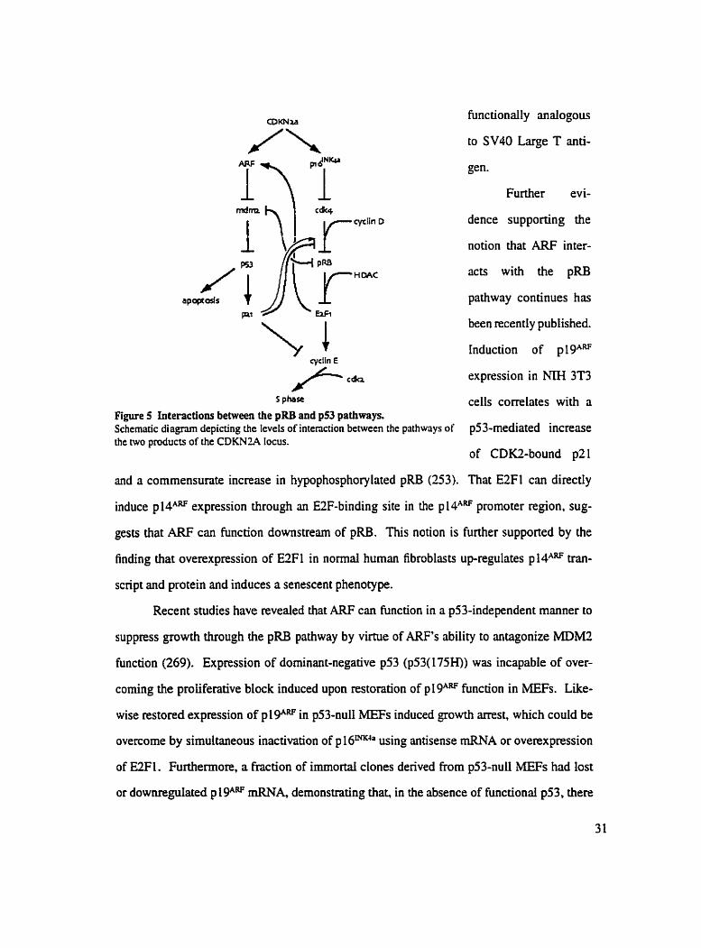
s p h s ~ cells cordates with a figure 5 Interactions between the pRB and p53 pathways. Schematic diagnm depicting the leveis of interaction becween the pathways of p53-mediated increase the nvo products of the CDKN7L4 locus.
of CD=-bound p21
and a cornmensunte increase in hypophosphorylated pRB (253). That E3Fl cm directly
induce ~ 1 4 ~ " expression through an E2F-binding site in the ~14"" promoter region, sug-
gests that ARF c m hinction downstream of pRB. This notion is further supported by the
finding that overexpression of E2F1 in normal human fibroblasu up-regulates ~ 1 4 " ~ trm-
script and protein and induces a senescent phenotype.
Recent studies have revealed that ARF cm function in a p53-independent manner to
suppress growth through the pRB pathway by virtue of ARF's ability to antagonize MDM2
fûnction (269). Expression of dominant-negative p53 (p53(175H)) was incapable of over-
coming the proliferative block induced upon restoration of plgARF f'unction in MEFs. Like-
wise restored expression of p l W in p53-nul1 MEFs induced growth arrest, which could be
overcome by sirnultaneous inactivation of p 16mh using antisense mRNA or overexpression
of =FI. Furthemore, a fraction of irnrn0rta.I clones derived from p53-nul1 MEFs had lost
or downregulated p lgM mRNA. demonstrating that, in the absence of hinctiond p53, there

is shll selective pressure to inactivate plgARF dunng the process of immortalization. This
apparent p53-independent pRB-dependent mechanism for ARF-mediated growth arrest is
contrary to the initial hypotheses sunounding ARF hnction.
Regulation of the p53 path way by pRB
The biological consequences of the pRB:MDM2 interaction can be viewed from two
perspectives. As discussed previously, binding of MDM2 to the C-terminal domain of pRB
inhibits the ability of pRB to negatively regulate E2F activity. However, recent evidence sug-
gests that pRB impairs certain hinctions of MDM2 during the process of forming a trimeric
complex with p53. Specifically, pRB overcomes the ability of MDM2 to inhibit p53-medi-
ated apoptosis (270). In the pRB- and pS3-deficient human osteosrucoma, Saos-2. the per-
centage of cells containing a subG, DNA content (an indicator of apoptosis) was decreased
by 50% by the addition of MDM2 to p53-transfected cells. This decrease in apoptotic cells
was reversed by CO-expression of pRB, suggesting that pRB binding to MDM2 could block
MDM2 anti-apoptotic activity. This notion was supported by the ability of pRB expression to
maintain p53 stability despite expression of MDM2. These results suggest that pRB inhib-
its MDM2-mediated p53 degrridation. However, pRB does not impair dl of the inhibitory
effects of MDMZ on p53. With respect to p53 transcriptionai activity for example, pRB
blocks the ability of MDM2 to impair transcnptiond repression mediated by p53 but does
not appear to alter MDM2's inhibition of p53-mediated transcnptiond activation (270). The
latter observation may be explained by the nature of the trimeric complex which is formed
through binding of pRB and p53 to non-overlapping regions of MDM2. Binding of pRB
to MDM2 does not promote the dissociation of p53 from the latter (270). Thus, even in
the trimeric complex, MDM2 remains bound to the p53 tramactivation domain and thereby
continues to inhibit the transcriptional activity of p53 by masking this domain despite the
binding of pRB.
That the pRB pathway has a significant interaction with the p53 pathway is funher
substantiated by the finding that MDM2 binds preferentially to hypo-phosphorylated pRB

(270,271). This implies that events which function to activate pRB, such as expression
of ~ 1 6 ~ ~ or mitogen depletion, cause subsequent activation of p53 through inhibition of
MDM2 function in addition to down-regulation of E2F activity. We suggest that the ability of
the pRB pathway to regulate the activity of p53 may provide an explanation for the induction
of p21 pmtein levels following expression of p161NK43 in U2-OS cells described previously.
Furthemore, the extensive interactions between the two pathways permits each pathway to
compensate for defects in the other. In the absence of functional p53, levels of p2 1 diminish
such that the formation of cyclin D/CDK4/6 complexes and their subsequent transport to the
nucleus would be perturbed. leading to activation of pRB. Sirnilarly. the loss of pRB function
would be compensated for by E2F1-mediated up-regulation of ARF expression which would
induce p53 activation. However. the disruption of only one of these pathways cm lead to
malignant transformation. and thus the degree to which these compensatory measures extend
warrants funher study and points to the existence of other growth regulatory pathways which
are important in the maintenance of ce11 cycle control.
To conclude. it is clear that two fundamentai pathways, the pRB and the p53 path-
ways. regulate ce11 growth and ce11 death. As is evidenced by the continous publication of
important papea in this ma, only very broad outiines of the mechanisms controlling these
pathways have been defined. It is dso clear that both pathways must be simuItaneously con-
sidered during discussion of ce11 cycle control given the recent data which clearly demon-
strate the molecular and genetic interactions between these two pathways.
INKMARF Inactivation in Cancer
The locus encoding ~ 1 6 ~ & , first referred to as MTS l (Multiple Tumour Suppressor
l), was ongindly identified as a homozygous deletion in human melanoma ce11 lines (272).
Since then, disruption of ~ 1 6 ~ ~ activity has been frequendy associated with NmOUr forma-
tion in human tissues. ~ 1 6 ~ ~ ~ hinction is disabled in a wide range of human malignancies,
which include glioblastornas (i.e, the most malignant form of astrocytoma, see below), lym-

phomas, as well as tumours of the colon, Lung and bladder (273-277). So significant is the
role of p161m4" in tumour suppression that a variety of methods are utilized dunng tumouri-
genesis to impair its activity. Inactivation of p16'Nmn occurs by at least three mechanisms:
( 1) homozygous dele tion, (2) Iack of transcription due to hypermethy lation of the prornoter,
and (3) point mutation (278,279). The frequency with which each of these mechanisms is
employed during rnalignant transformation varies in different types of turnours. Homozy-
gous deletion of p16INK4' occun in 48% of non-small ce11 lung cancers and 35% of acute lym-
phobiastic leukernias (280,28 1). In contrast, only 17% of esophageal squamous ce11 carcino-
mas contain homozygous deletions of pI6INK4", while 38% of these tumoun show evidence
of ~ 1 6 ~ " " promoter hypermethylation (282). Similarly, the majority of hepatocellular car-
cinomas (62%) contain hypermethylated ~ 1 6 ~ ~ ~ " promoten, while the frequency of ~ 1 6 ' ~ ~ ~ '
homozygous deletion (10%) is significantly lower in these tumours (283). Inactivation of
p161NK4' via deletion or mutation is observed in one-fourth of sporadic melanomas and 38%
of head and neck squamous ce11 carcinomas (284.285). Loss of p16 rNK4~xpre~~ ion also
occurs frequently in adrenocortical and pituitary turnours (286,287). Thus, it appears t!!at the
various mechanisms by which p161NK4= cm be inactivated contributes to the observation of
non-functional p 16'NK4a in a broad range of tumours.
Abrogation of ARF activity, while less frequent than that of p 161NKda in human can-
cers, has been reported in a Iimited set of malignancies. Specific disruption or deletion of
sequences encoding plPRF that do not affect ~ 1 6 ~ ~ ~ have been observed in 100% of T-ce11
acute Lymphoblastic leukemia cases in which chromosomal reamngements have occurred
in the locus (288). Inactivation of pMARF by CO-dehtion with ~ 1 6 ~ " ha been reponed
in 8% of non-Hodgkin's Iymphomas (289) and 70% of human mesotheliomas (290-292),
while a reduction in the expression levels of both and ~ 1 6 ~ ~ ~ is seen in half of non-
small ceil lung cancers (293). In the case of pancreatic carcinomas, loss of both pl 6wK4a and
~ 1 4 ~ coding regions is observed at a fkquency approaching 100% (294-296). Up to 33%
of esophageal squamous ceil carcinomas have homozygous deletions of E l e, and another

15% contain hypermethylated p 14AM promoters (282). .4s is the case with p 1 6'NK4a, inactiva-
tion of p lPRF via promoter hypermethylation is associated with malignant transformation,
which is evident in 28% of primary colorectal carcinomas and 32% of colorectai adenornas
(297). However, uniike p16'NK4a, additional regulation of ~ 1 4 " ~ ~ activity h a been shown to
occur at the translational Ievel. Tumour ce11 lines of B-type lymphoid origin express high
levels of ~ 1 4 " ~ transcript but no ARF protein. suggesting that inactivation of ~ 1 4 ~ ~ ~ may
occur through disruption of a translational mechanism (298). Likewise. p 14ARF protein is lost
in 65% of srnall ce11 lung cancer and in 25% of non-smdl ce11 lung cancer without deletion
of exons 1 and 2 despite the presence of abundant ARF tnnscnpt in these cells (299).
Human Astrocytomas
Turnours of the human centrai nervous system arising from astrocytes represent the
most common form of primary brain cancer. Each year, 50 000 North Americans are diag-
nosed with an astrocytoma As one type of tumour of the centnl netvous system, astro-
cytomas are collectively referred to with other rnalignancies of the neuroglia as gliomas.
Amongst the four chnical grades recognized by the World Health Organization, grade 4 or
glioblastoma multiforme (GBM), is the most prevdent in humans. Appropnately narned.
GBMs are complex tumours with multiple forms. Stmcturally, the tumour mass is a diverse
growth consisting of areas of necrosis, hemorrhage, and ceI1ula.r proliferation. At the genetic
level, deIetions, ampiifications, and point mutations give rise to inappropriate activation of
oncogenic signaling cascades or disniption of ce11 cycle arrest pathways during the progres-
sion From low grade to high grade neoplasrns. Ultirnately, this hetemgeneity culminates in
the generation of uncontrollable malignant growths which are uniform in their lethality to
humans. Patients diagnosed with GBM have Little hope of long-term survival as they follow a
course towards profound neurological deficit consisting of dementia, behavioral disturbance,
language loss or paralysis. Death usualIy ensues within 2 yem of diagnosis.
In addition to npid proliferation, GBMs aiso present a diffuse localization pattern in
the brain. Glioma ceils commody migrate away h m the tumour mass through the brain

parenchyma dong white matter tracks, surrounding neurons and blood vessels in the process
of invading vitai areas of the bmin. The invasion process is aided, in part, by over-expression
of degradative enzymes such as matrix metalloproteinases which break-down the surround-
ing tissue. As a result, individual glioma cells which have infiltrated various regions of the
brain c m form new foci of neoplastic growth. thereby creating multicentnc GBMs. It is
estimated that approximately 25% of GBM patients have multiple GBMs. Thus the invasive
behavior, coupled with rapid cellular prolifention of GBMs, contributes to the aggressive
nature of this rnaiignancy.
Current ciinical treatrnent for GBM involves surgical removal of as much of the
tumour as possible, followed by extensive radiation thenpy and chemothenpy. However,
the difise localization of tumour cells wiihin the brain makes complete surgical resection of
GBMs very difficult, and thus recurrence of the tumour occurs frequently. Furthennore, once
tumour cells from the GBM infiltrate vital areas of the brain, surgical treatment no longer
becomes an option. Conventional chemotherapy frequentl y fails as well because most agents
ridministered systernically will not cross the blood-brain barrier. On average, these proce-
dures extend survivai of GBM patients from 2 months to 1 year. Given the poor prognosis of
GBM patients. the need for the development of thenpies to combat this disease is both urgent
and of the utmost significance. One of the more ment strategies makes use of intergeneric
recombinant poliovirus chimeras PVl(RIP0) to specifically target malignant glioma cells
(300). Initial studies conducted in mice carrying intracerebral gliorna xenografts have shown
that PVI(RIP0) can mediate growth inhibition, Iytic destruction and uftimately elimination
of malignant gliomas without propagation in normal neuronal cells. Thus. it appears that the
transfer of growth-inhibitory properties to malignant gliomas may provide a viable strategy
for clinical treatrnent of these tumours as the poor prognosis of gliorna patients continues to
increase the urgency of pursuing al1 possible cures.
INKWARF Inactivation in Astrocytomas
Inactivation of the CDKhr2A gene products or their downstrearn effectors is a common

occurrence in human gliomas (301-305). Lesions found in such malignancies which con-
tribure to ~ 1 6 ~ ~ " loss of function include homozygous deletions, which occur in 41% of
primary glioblastomas, while point mutations and promoter hypennethylation are observed
to a lesser extent (306-308). Analysis of primary gliomas has not revealed the presence of
germline or somatic p mutations to-date. aithough gross deletions of the CDKNZA locus
which encompass the pl4- coding region have been reported (309). Homozygous deletion
of exon Ip of ~ 1 4 ~ ~ ha. been observed in as much as 30% of glioblastomas (3 10). Com-
pared to the lack of ~ 1 4 " ~ ~ mutations, this latter finding implies that homozygous deletion
rather than mutation of the p 14A'<F gene is the more effective way to inactivate ARF during
the process of gliomagenesis. In support of this notion, mutations in exon 2 have been shown
to disrupt the ability of ~ 1 4 ~ ~ ~ to localize to the nucleolus. however such lesions occur with
very low frequency in glioblastomas (247,303).
The contribution of ARF-incapacitation to gliomagenesis is underscored further by
the development of astrocytic malignancies in p 19ARF-specific nullizygous mice (3 1 1). These
tumours, which are rare in normal mice (3 1 1). infiltrated the brain parenchyma in a diffuse
manner similar to that observed in human gliomas. In contras?, p 1 61NKh/p 1 gARF-double nul1
mice. generated by disruption of exons 2 and 3 of the CDKNZA locus, do not develop spon-
taneous gliomas, despite the fact that the tumour spectrum is similar to that of mice Iacking
p 19ARFaI~ne (234.3 1 1). It is worth noting that expression of constitutively active epidermal
growth factor receptor in glial precursor cells induces the formation of malignant gliomas
in these same double-nul1 mice (305). However, the failure of p16'NK4/plF double-nul1
mice to develop spontaneous astrocytornas may be attributed to the incompIete inactivation
of plgAW in these animais. While the double-null mice lack exons 2 and 3 of the CDfUVîA
locus, exon l p is not deleted and remains intact Recently, aberrant mscnpts encoding
exon iP have been detected in the atrocytes denved kom these rnice (305). Since exon l e
encodes ail of the known growth suppressive functions of p 19*(248,3 13 , it is possible that
ARF retains enough huiction to mediate tumour suppression in the astrocytes of the double-
nul1 mice, accounting for the lack of gliomas. However, given that p 1 6wK4a function, at least,
is disabled in the double-nul1 mice, the lack of giiomas in these animals may reflect the rela-
tive importance of the pRB pathway versus the p53 pathway in asûocytomas. In humans,
deletion of pRB occurs in 22% of astrocytomas, while 30-5046 are nul1 or mutant for p53
(3 13-3 15)- suggesting that inactivation of the latter is more effective in malignant transfoma-
tion of astrocytes. Despite the arnbiguity surrounding the p l6INK4'/p 19*IIF double-nul1 mice,
which underscores the need for a pure, p16'NK4-n~11 animal via exon l a deletioo to accu-
ntely define the role of p16'Nw" in güomagenesis, the development of astrocytic maügnan-
cies in p19ARF-null mice suggesü that ARF plays a critical mle in the ceIl cycle regulation of
astrocytes.
Given that the known efFects of ARF are mediated by p53, a logical expectation is that
the spectrum of tumours in p53-nul1 mice would be similar to that of mice lacking p lgARF
However, dthough p53-nul1 mice develop a wide range of spontmeous tumours. no evidence
of astrocytic malignancies in these rnice has been observed (3 l6,3 17). This may reflect the
fact that rnice lacking p53 survive only 6 months, while plgARF-nul1 animals live considenbly
longer. up to 15 months. The shorter survival time may preclude the development of gliomas
in the p53-nul1 mice. However. astrocytomas in p19ARF-null mice did not have a long latency
period, arising within 12-15 weeks of birth (31 1). Furthemore, given the ability of p53 to
negatively regulate ARF transcription, one would expect that ARF is over-expressed in the
astrocytes of p53-nul1 rnice. Thus, an alternative possibility is that ARF has growth suppres-
sive effects in astrocytes that are independent of its known downstream effector, p53, which
may be manifested in the failure of p53-nu11 mice to develop astrocytomas.
Studies conducted by other investigators examining the ce11 cycle effects of ~ 1 6 ~ ~
or p MAW expression in human gliomas have been performed. Expression of p 1 6MKh inhibits
proliferation of giiomas by causing a potent G, arrest, which is accompanied by the loss of
anchorage-independent growth in soft agar (3 18,3 19). In addition. the induction of pl 6'NR4a-
mediated arrest in gliomas is associated with senescent features such as cytoskeletal re-

arrangements of actin and vimentin, ultimately giving rise to a Battened cellular phenotype
(320,32 1). On the other hand, comprehensive studies andyzing the complete growth inhibi-
tory effects of ARF in human gliomas are Iacking. Arap et al observed that expression of
ARF in human glioma ce11 lines inhibited proliferation, however these findings fail to charric-
terize the mechanisms by which ARF mediates growth arrest (322). Given that both ~ 1 6 ~ "
and ARF are bonajïde turnour suppressors, it is tempting to speculate that expression of ARF
in human gliomas may also induce senescent features when expressed in these malignan-
cies.
Given the role of CDKNZA in tumour suppression and evidence of its disruption in
astrocytomas by deletion or mutation. expression of p 16NK'a or p MARF in astrocytomas would
be expected to block proliferation of these malignancies, provided that the integrity of their
respective growth-inhibitory pathways rernained intact. To test this hypothesis. p161NK4' or
p 14ARF were expressed in a panel of malignant astrocytoma ceIl lines in which the functional
status of pRB and p53 was distinct for each ce11 line. Our data reved that growth inhibition
can be mediated by plVRF in the absence of an intact p53 and. simultaneously. pRB path-
way.
Research Hmothesis and Obiectives
The products of the CDKNlA locus, ~ 1 6 " ~ ~ . and p14*5 act as potent ce11 cycle
inhibitors through the pRB and p53 pathways, respectively. Knockout mice lacking expres-
sion of either ~ 1 6 ~ ~ ~ ' or p 19"" deveiop a wide range of spontaneous tumours, supporting
the characterization of these two proteins as bona jide tumour suppressors. Inactivation of
~ 1 6 ~ ~ or p 14ARF is frequently associated with human gliomas, suggesting that these tumour
suppressors play a critical d e in blocking the maîignant transformation of astrocytes. This
is further supported by the finding that p53 or, to a lesser extent, pRB are deleted or mutated
in human astrocytomas. Thus, it is reasonable to expect that expression of p 16MC.4J or p MARF

in CDKN2A-nul1 human gliomas will arrest the growth of these tumours. Furthemore, given
that p19ARF-null mice, but not p16'NK4n-null mice. develop astrocytic malignancies, the ARF
NmOUr suppressor may be the more effective of the CDKN2A products in mediating growth
arrest of human gliomas.
Hmothesis
Ectopic expression of the INK4dARF tumour suppressor products will induce growth
arrest of malignant CDKNZA-nul1 human glioblastoma ce11 ünes in a manner which is depen-
dent on the integrity of their respective downstream effectors in these tumours, That is,
will arrest the growth of glioblastomas harbounng a wild-type pRB pathway. while
p14*" will inhibit the proliferation of glioblastomas which have retained a functional p53
pathway.
Obiectives
1) To determine if p16'NK4n cm inhibit the growth of human glioblastomas in a pRB-depen-
dent manner and characterize the inhibitory ce11 cycle rffects, if any, of p 16LYK4a expres-
sion in human glioblastomas that are independent of the pRB pathway.
2) To determine if p 14"" can inhibit the growth of human glioblastomas in a manner which
is dependent on the presence of functiond p53 in these tumours and characterize the ce11
cycle effects, if any, of p 14ARF expression in humm glioblastomas tfiat are independent of
the p53 pathway.
3) To examine whether or not ectopic expression of p 16MKh or ~ 1 4 " ~ ~ in human giioblasto-
mas has an effect on the invasive property of these tumours.
To achieve these objectives, p 1 6MK4a or p l 4ARF will be expressed via adenoviral-trans-
fer to a panel of human glioblastorna ce11 lines which Vary in the functiond status of their
respective pRB and p53 pathways. The molecular changes induced by expression of these
tumour suppressors wiii be evaluated by western analysis, and the accompanying ceil cycle

changes will be evaluated by FACS andysis. The ability of ~ 1 6 ~ ~ ~ " or ~ 1 4 ~ ~ to arrest
these human glioblastomas stably-transfected with vectors encoding these proteins will be
assessed in colony-forming assays. Clones of p53-deficient human glioblastomas stably-
expressing p MA" will be isolated fiom the colony-forming assays and analyzed for perturba-
tions in the pRB pathway. The expression of proteins which function in the p53 a d o r pRB
pathway will be examined by western detection. The functional status of the pRB pathway in
these p 14A'v-expressing clones will be assessed by FACS analysis of these cells post-infec-
tion with adenovirus encoding pl 6NK4. TO investigale whether p l P R F induces a senescent
phenotype in p53-deficient human glioblastomas. the senescence-associated p-galactosidase
activity of the p14ARf-expressing clones will be measured. To ensure that our ~ 1 6 ~ ~ ~ and
~14"" pmteins function as expected, the in vitro and in vivo properties of p 14ARF and p 1 6mK4a
will be observed. Specificdly. the binding of ~16""" to wild-type or mutant CDM (cre-
ated by site-directed mutagenesis) will be assessed in GST pull-down assays, while CO-
immunoprecipitation of P I ~ ~ ~ ~ ~ with CDK4 or ~14"" with MDM2 will be conducted using
lysates from infected human glioblastomas. In addition, the sub-cellular localization pat-
tern of p16INK4' and p will be observed in marnmalian cells (COS) using GFP-fusion
proteins. The p53-mediated response of these glioblastoma ce11 lines to ionizing radiation
will be used to ver@ the functional status of the p53 pathway in these ce11 iines. Finally,
gelatin zymography will be used to examine MMP-2 activity in the human glioblastoma ce11
lines as an indicator of invasion post-expression of p16INKIa or ~ 1 4 ~ ~ ~ in these cells.
Materials and Methods
Production of GST-frtsion proteins
TOPP3 bacteria transformed with the appropriate pGEX vector (Phmacia) were
grown in a 100 ml culture of LE3 containing 50 pg/ml of ampicillin (Roche) at 37 Celsius with
agitation ovemight. This culture was added to 500 ml of LI3 and grown for an additional 2
hours. To induce GST protein synthesis, 1 ml of 200 mM IPTG was added to the cultute and

incubated at 30" Celsius for 3 hours with agitation. The culture was placed in ice water for
10 minutes pnor to centrifugation in a GSA rotor at 4' Celsius. 5000 RPM for 15 minutes.
The bacteriai pellet was resuspended in 10 ml of Fusion Protein Lysis Buffer (FPLB; 500
mM NaCl. 1% Triton X. 50 m M Hepes, 2 m M EDTA. 10% glycerol) plus protease inhibi-
tors (aprotinin (10 pg/ml) and leupeptin (10 pglml)) in a 15 ml polypmpylene tube. Sonica-
tion was performed 4 x 1 minute (output setting = 4) on ice. The bacterial lysate was trans-
ferred to an SS34 tube and centrifuged at 10 MIO RPM for 10 minutes at 4 Celsius. Lysate
supematant was transfemd to a fresh 14 ml polystyrene tube and incubated with 100 pl of
glutathione-sepharose beads (Phmacia) pnviously washed 5 times in FPLB. The washed
beads and supematant were incubated ovemight at 4 Celsius with rocking. The beads were
removed by centrifugation at 1500 RPM for 5 minutes at 4' Celsius. transferred to an eppen-
dorf tube and washed 5 times with 1 ml of FPLB. Proteins were analyzed and quantified by
Coomassie gel staining using BSA standards.
Preparaîion of Mammahn Cell Lysates
The media was removed from the cells which were then washed three times with cold
P M on ice. 0.2-1 ml of NP40 lysis butTer (50 rnM Tris-CI pH 8.0. 120 m M NaCl, 0.5%
NP40) plus protease inhibitors (aprotinin (10 pg/ml). leupeptin (10 pg/ml). PMSF (0.1 mM))
was added to the cells. which were scnpped into the buffer and transferred to an eppendorf
tube for 15 minutes on ice. Cellular debris was removed by centrifugation at 13 000 RPM
for 15 minutes ai 4 Celsius. The supematant containing whole ce11 lysate was transfemd
to a fresh eppendod tube and quantified by spectrophotometry at 595 nm using the BioRad
protein assay reagent.
Site- Directed Mutagenesis
To determine if pMHKAa bound to mutant forms of CDM. the CDK4-R24C mutant
(which does not bind INK4 proteins) was created. To generate the CDK4-lU4C mutant, the
N-terminai 200 base pairs of CDK4 were arnptified by PCR using pBSK-CDK4 and the T3
primer dong with the following mutagenic primer which contains the Arg24 to Cys24 muta-

tion: 5' CAC AAA GTG GCC ACT GTG GGG ATC GCA TGC ïTï G 3'. Similady, the
C-terminal 700 base pairs of CDK4 were amplified using a primer compIementary to the
mutagenic primer 5' AAA GCA TGC GAT CCC CAC AGT GGC CAC TIT GTG 3' ; dong
with a C-terminal primer that mutated the stop codon to an EcoN site: 5' GAA TTC CTC
TGC GTC GCT ïTC CTC CTT 3'. The two PCR fragments were purified frorn 0.7% aga-
rose gel electrophoresis, and rnixed with 10X PCR buffer (500 m M KCI, 100 mM Tris-HCI,
pH 8.3) in a finai volume of 100 pl. The fragments were denatured at 94 Celsius for 5 min-
utes, after which the 30 cornptimentary base pairs in both fragments containing the R24C
mutation were annealed by slow cooling to 42 Ceisius. 2 pl of 10 mM dNTPs and 1 pl of
Vent Polymerase (New England BioLabs) were added and DNA extension was performed at
72 Celsius for 10 minutes, The T3 and C-terminus primers were then added and PCR was
performed in a Thermocycler for 25 cycles to ampli@ the CDK4-R24C mutant.
Sequencing of CDK4- R2QC Mutant
10 pg of pGEM-T-CDK4-R24C in a 100 pl volume was incubated with 4 pl of 2
mglm1 RNase A at 37 Celsius for 1 hour. extracted with equai volume pheno1:chloroform and
then incubated with 4 pl of 5M NaOH at 37 Celsius for 30 minutes. DNA was precipitated
with 0.4 volume of 100% ethanol at -20 Celsius for 30 minutes, washed with 70% ethanol
and resuspended in 7 pl of TE. Sequencing reactions were performed with the T7 Sequenc-
ing Kit (Pharmacia Biotech) according to manufacturer's pmtocol. Briefly, 1 pl of primer
and 2 pl of annealing buffer were added to the DNA solution which was incubated at 80
Celsius for 3 minutes, then cooled to 20 Celsius before being placed on ice. 3 pl of labeling
mix A, 2.5 pl 3SS-dATP, and 2 pl of 1.5 U/pi T7 polymerase were added to the DNA solu-
tion which was then incubated at room tempemure for 5 minutes. 4.5 pl of this sample was
added to each of four tubes containing 2.5 pl ddATP, ddCTP, ddGTP, or ddTTP and incubated
at 37 Celsius for 5 minutes. 5 pl of stop solution was added, and the sarnple was boiled for 3
minutes, then placed on ice before loading onto an 8 2 SDS polyacrylamide gel. 2 pl of each
of the four stopped sarnples was Ioaded per lane. Gels were run at constant current, fixed,

dned and exposed to film at 4 0 Celsius overnight.
GS T Pull-dow n Assays
1 pg of GST-hision protein bound to glutathione sephamse beads (Pharmacia) washed
three times with 0.5 ml of NP40 lysis buffer was incubated with 1 mg of NP40 whole ceIl
lysate at 4 Celsius for 2 hours in a final volume of 1 ml. The beads were spun down at 5000
RPM for 1 minute and washed 5 times with 180 pl of NP40 lysis buffer. Beads were boiled
in IX sample buffer, subjected to 10% SDS-PAGE, and andyzed by Western blotting.
Subcelluhr Lucalization of GFP-kion Proteins
To determine the subcellular localization of the INK4 and ARF proteins, COS ceiis
seeded on covealips were transfected with 20 pg of vector encoding GFP-p15WK4b, GFP-
p 16WK4a, GFP-p 1 VNKk1 GFP-p 1 9INKWl GFP-p 1 4ARF or GFP-p l gA? 77 houn post-transfec-
tion, slips were washed three times in PBS, fixed for three minutes in ice-cold methanol. air-
dried, mounted, and analyzed by confocal microscopy.
Gelatin Zymograp hy
MMP-2 activity of human giioblastomacell Iines was assayed as an indicator of inva-
sion. MMP activity was analyzed using sodium dodecyl sulfate-polyacrylamide gels impreg-
nated with 0.1% gelatin (wfv) and 10% polyacrylamide. Glioma cells were grown in 60
mm2 tissue culture plates in alpha-MEM containing 10% FBS until they reached 80% conBu-
ency, at which time the cells were washed three times in PBS and normal culture media was
replaced with serum-free media Conditioned media was collected after 48 houa. 50 pl
of conditioned media was mixed with IX sample buffer (minus DTT) pnor to electrophore-
sis. Gels were run at constant curent, washed several times in 2.5% Triton-X to =-nature
the proteins and then incubated for 48 houa at 37' Celsius in 50 mM Tns-Cl, pH 7.6, 10
mM CaCL, - 150 m M NaCl. and 0.05% NaN,. Gels were then stained with Coomassie Blue
(0.25% Coomassie Brilliant Blue R250, 10% glacial acetic acid, 30% methanol) for I hour
and hen de-stained ( IO% giacid acetic acid, 30% methanol).

Cet1 tines and culture conditions
The astrocytoma ce11 lines. U343MG, U25lMG and SF126MG were grown in alpha-
MEM supplemented with 10% fetal bovine semm (Sigma) at 37" Celsius and 5% CO,. Al1
ce11 lines were allowed to grow to no more than 80% confluence, at which time the cells were
washed two times with PBS, trypsinized and re-plated in fresh media at a dilution no lower
than 15. COS cells were maintained in Dulbecco's H21 media supplemented with 10% fetal
bovine serum at 37" Celsius and 5% CO,.
IonMng radîution assays
U343, U251 and SF126 cells were exposed to 7 Gy of ionizing radiation (Gamma-
Cell), and returned to normal culture conditions. TotaI ce11 lysates were made in NP40 lysis
buffer 2 and 4 hours post-irradiation, subjected to SDS-PAGE and analyzed by westem bIot-
ting.
Western blornhg analysk
50 pg of NP40 total ce11 Iysate from each sarnple was subjected to 8-14% SDS-
polyacrylarnide gel electrophoresis and transfemd to nitrocellulose membrane. These mem-
branes were blocked in 5% skim rn iWBS for at l e s t one hour and probed with mouse
monoclonal anti-p53 (PAL 180 1) or mouse monoclonal anti-MDM2 (Santa Cruz, catalogue
#SC-965, 1:250 dilution) or mouse monoclonal anti-p21Gpl (Pharmingen 6595 1A) or mouse
monoclonal anti-p161NKJa (K2. a kind gift from Dr. Jim Koh. University of Vermont) or
rabbit polyclonal anti-CDM (Santa Cruz, C-22) or rabbit polyclonal anti-EX1 (Santa Cruz,
C-20) or mouse monoclonal anti-pRB (Pharmingen, 1400 1 A, or IF8 supernatant) or nbbit
polyclonal anti-CD= (Santa Cruz. SC-163) or mouse monoclonal anti-HA (12CAS) IgG at
1: 1000 dilution. The membranes were washed three times with 5% skim miWO. 1% Tween
20/PBS. The secondary antibodies were horseradish peroxidase-conjugated donkey anti-
mouse and anti-rabbit IgG (Jackson Immunoresearch). The membranes were then washed
twice with 5% skim miIk/û.l% Tween 20/PBS, foliowed by three washes with O. 1% Tween
20PBS. For westem analysis of pMm, the nitrocellulose was blocked with 10% BSAiTBS
for 1 hour, then probed with goat polyclonai anti-p 14ARF (Santa Cmz, C- 18) diluted 1:200 in
5% B S m S for 2 hours. The membrane was washed three times with TBS/O. 1 % Tween 20,
after which the membrane was probed with horseradish peroxidase-conjugated anti-goat IgG
(Santa Cruz, SC-2020) diluted 1:15 000 in 5% skirn rnilk/O.l% Tween 20RBS and washed
three times with O. 1 % Tween 20/TBS- Al1 membranes were developed using LumiGLO ECL
reagents (Kirkegaard and Perry) and exposed to hyperfilm (Amersharn).
Mammalian and bacterid expression constructs
Full-length hurnan plSLNKJb and p161NK4= cDNAs were obtained from Dr. Manuel
Serrano (Madrid, Spain). Full-length ~14"" cDNA was obdned from Dr. Gordon Peten
(Imperia1 Cancer Research Fund Laboratones, London. UK). Full-length cDNAs encoding
mouse p 1 8INK", p 1 9INKu. p 1 gARF and CDM were obtained from Dr. Charles J. S hem (Si. Jude
Research Hospital, Memphis, Tennessee). The pGU[-2T-p 1 SLYKqb and pGEX-2T-p 1 6INKJn
constructs were created by ligation of the respective cDNAs between the BarnHI and EcoRI
sites in pGEX-2T (Pharmacia). The pECE-CDK4-HA and pECE-CDK4-R24C-HA con-
structs were created by ligation of the respective cDNAs between the HindiII and EcoRI sites
of the pECE-HA vector. The pEGFP-CI expression plasmid (Clontech) was used for con-
struction of al1 GFP-fusion proteins. The pEFGP-p 1 51NKJb, pEGFP-p 1 6fxK4a. pEGFP-p 1 SMK&,
pEGFP-p191NKW, and P E G F P - ~ ~ ~ ~ ~ constructs were created by excision of the respective
cDNAs with BamHI and EcoRI and ligation between the BglII and EcoRI cloning sites in
pEGFP-C 1. The pEGFP-p 1 gARF constmct was created by ligation of the p 1 gARF cDNA into
the EcoRi site of pEGFP-Cl. The pcDNA3.l expression plasmid (Sûatagene) was used
for al1 transfections of human glioblastoma ce11 lines. The pcDNA3-p161m4' and pcDNA3-
~ 1 4 ~ ~ were constructed by ligation of the respective cDNAs between the BamHI and EcoRI
cloning sites in pcDNA3. The ~ C D N A ~ - H A - ~ ~ ~ ~ constmct was built by tigating HA-
tagged pl9* into the BamHI and XhoI sites. The pcDNA3-CDK4 constmct was built by
ligation of the CDK4 gene into the BamHI and EcoRV sites. (See Table 1 for complete iist-
ing of DNA constructs). Expression of a i l constructs was verified by transfection into COS

cells and observed by western blotting before use.
Table 1. DNA Coastructs
Sub-céllular loçalaation of WT CDK4 GSFINK4 pu[-dwn asays GSFINK4 pull-dwn assys Sub-cbning precursor Sub-cell~.dm Iccalkation of CDK4-R24C GSTIM(4 pull-dcrwn assays Sequencmg of CDK4-F124C mutant Sequencmg of CDK4-R24G N-terminus Sequencmg of CDK4-RS4S N-terminus Sequencmg of CDK4-R24C Nterminus Te t-induci ble pl 4ARF expresim in giiomas Sub-cdiular îccaîiration of ~ 1 4 ~ ~ ~ Sub-cbning precursor GSTpi4ARF pulCdown of MDM2 in gliomas Cobny-forming w s w i î h gliomacell Iines ktrccytespecifc expression of p14ARF Homologxu recombinatbn for adenouirus Tat-protein mediated transduction of ~ 1 4 ~ ~ ~ Tat-medinted transduction of GFP-pl 4*F Sub-celluiar fccalization of pl^^^^^^ GSTlNK4 pull-dwn assnyswith CDK4 Cobwforming assays w l h glioma cell lines
DNA Conçtmct Name pcDNA3-CDK4
Experimentat Putpose Cobny-forrning xsays with gtomacell lines
~ E C E - G F A P - ~ ~ C - ~ ~ G ~ ~ pEGFP-nyc-pl eMca pRdTikCMVmÿcp1 6NKB pECE-myc-pl 6'NCQ p lNT-GFP-myc-p l~~~~ pGEX-2Tmyc-pi eENCs pSP7BGFAP-rnyc-pl 6M4 pEGFP-pl aaUWc ~MINT-~I p EGFP-pi gound ~MIM-~I 9INw pEGFP-HA-pl #RF
pGM-3X-HA-pi9MF pECE-HAmcs-pl #RF p c ~ N M - p 1 9 * ~ ~ p ~ d ~ i m ~ - ~ ~ - p l SARF pTRE- HA- pl eF plNT-GFP-pl fiRF
Suù-cloning pre-cursor Sub-cellular localkation of p l elNKk t-brnobgous recombinalion for adenouirus Marndan expression of p l 6l NK4a
Tat-mediatedtran~ductionofGFP-pl6~~~~~ QSFINK4 pull-down zesays w l h CDK4 Astrocytespecifc expression of p161NK4a Sub-cellulnr Iccalkation of p18INKk Tabprot ein mediated transduction of p l 8IN Sub-cellulai Iccalkation of p191NK4d Tat-protein mediateci transduction of pl9INwd Sub-cellular tocdiration of Irrminkationf0rpi9~~~mmclonalAht Mammaiian expression of plgARF Cobnpfoming assaus w l h gliomacell lines Homolopus rembimtion for ademvirus Tet-irducible expression of pl SARF Tat-mediated transduction of G FP- pl RF

Transfection of mummalian express ion phsrnids
Human glioblastoma cells or COS cells were grown to 80% confluency. 20 pg of
DNA was mixed with dH,O in a find volume of 125 pl in a sterile eppendorf tube. 125
pi of 0.33 M CaCl, was added to the DNA, and the mixture was transferred drop-wise to
250 pl of 2X Hepes Buffered Saiine (HBS), pH 7.1 (280 mM NaCl, 50 m M Hepes, 1.5 mM
NqHP0,.7H20) in a 14 ml polystyrene tube while bubbling air through the HBS with a 1
ml disposable pipette. The mixture was allowed to sit for 10 minutes at roorn temperature.
after which it was added drop-wise ont0 the cells. The precipitate was left on the cells for
16 hours and removed with thne washes in PBS. The media was replaced and the cells were
retumed to the incubator for rit least 48 hours.
Colony forming assays and Generation of SJable Clones
Human glioblastoma cells grown to 80% confluence were split 1: 10 and plated
on 100 mm2 plates one day prior to transfection. On the day of transfection, 20 pg of
either pcDNA3. pcDNA3-p 161NK4', pcDNA3-p l4"Y pcDNA3-p 1 gAW. or pcDNA3-CDK4
was transfected using the calcium phosphate precipitate method (see Trmsfections). Selec-
tion was camed out using media supplemented with 200 @ml of G418 (i.e, Geneticin)
(Gibco BRL). G418 is an aminoglycoside antibiotic which functions by inhibiting ribosomal
protein synthesis in both prokaryotic and eukaryotic cells. A concentration of 200 pg/ml
was chosen because this amount of antibiotic killed 100% of untransfected human glioblas-
toma cells while ailowing colony-formation by cells msfected with vector encoding G418-
resistance. Concentrations of 400 pgfrnl or greater impaired the growth of colonies in these
assays, and thus a lower concenaation of G418 was used. One week d e r transfection. cells
were washed twice in PBS, and fresh media containhg G4I 8 was added. Individuai colonies
were isolated and grown in G418containing media for 2 weeks to generate stable clones.
Two weeks post-transfection, ceUs were washed twice with PBS, rnethanol-fixed, and stained
with 0.125% toluidine blue. Colony numbers were quantified. Assays for each ce11 iine were
conducted at least four times for accuracy.

Adenovirus infections
Adenovirus encoding human p 161NK4' was obtained from Dr. Frank Graham (McMas-
ter University, Hamilton. Ontario). Adenovirus encoding ~ 1 4 ~ ~ was obtained from Dr. Yue
Xiong (University of North Carotina, Chape1 Hill). Adenoviruses were chosen for these
infections rather than retroviruses because adenoviruses infect both proliferating and non-
prolifenting cells, while retrovimses only infect prolifenting cells. As well. adenoviruses
express their genetic material transiently in the host cell. while retroviruses integrate their
genetic material into the genome of the host cell. Thus, to achieve transient expression of
~ 1 6 ~ ~ ~ or ~ 1 4 " ~ ~ in both non-proliferating and proliferating human glioblastoma cells, ade-
noviruses were employed. Approximately 5 x l(r human glioblastoma cells were plated on
60 mm2 plates the day before infection. Cells were washed three times with PBS and infected
with control adenovirus (Ad-CMV). or adenovirus encoding either p16'NKJa or ~ 1 4 " ~ ~ at an
MOI of 100 (titers determined using lysis of the human kidney cell line, 293) in 1.5 ml of
alpha-MEM containing 0% semm. Infection was carried out at 37' Celsius and 5% CO, for
2 houa, with a brief agitation every 20 minutes. Afier infection. virus was removed and fksh
media with serum was added. Cells were incubated for 2-3 days before harvesting.
Co-lmmunoprecr'pircttion assays
To ensure that exogenously expressed p 16ImAa and p 14ARF were binding their endog-
enous substrates in human glioblastorna cells infected with adenoviruses encoding these pro-
teins, CO-immunoprecipitations were petformed using infected lysates. 200 pg of NP40 total
ce11 lysate was incubated with 5 pg of mouse monoclonal anti-p 16mK4' (JC4. a kind gifi from
Dr. Jim Koh. University of Vermont) or goat polyclonal mti-p 1 4"" (Santa C m , C- 18) with
rocking at 4 Celsius for 4 hours. For p 1 4 Y 5 pg of rabbit a-goat IgG (Jackson Immunore-
search) was added, and the incubation was continued for an additional 2 hours. Protein-G
sepharose beads (Pharmacia) were used for ~ 1 6 ~ ~ ' irnrnunoprecipitations, while protein-A
sepharose beads (Pharmacia) were used to bind ~14"" complexes. Beads were washed 3
times in NP40 lysis buffer, boiled in 1X SDS loading buffet, and the irnrnunoprecipitated pro-

teins were separated by SDS-PAGE. Proteins were transferred to nitrocellulose membrane
and analyzed by western blotting.
FACS analysis
To determine the ceil cycle profile of human gliobiastoma cells, fluorescence-acti-
vated ce11 sorting was used. Human glioblastoma cells were trypsinized, washed with excess
PBS and resuspended in 50 pi of 2% FBSFBS pnor to fixation in ice-cold 70% ethanol for
at least 24 hrs. The cells were cenû-ifuged at 1500 g for 10 minutes at 4" Celsius and washed
with PBS. O. 1 m g / d of propidium iodide solution (O. 1 mg/ml propidium iodide in PBS con-
taining 0.6% NP40) with 1 m g h l of Ribonuclease A (Sigma) was used to stain cellular DNA
for 30 minutes at 4" Celsius in the dark. dter which cells were anaiyzed by ffow cytometry
(Flow cytometry facility, The Hospital for Sick Children, Toronto, Ontario).
Seneseence-Associated ~Gaiùctosidase Sfaining
To determine if human glioblastoma cells stably-expressing plJARF were entenng
senescence, the senescence-associated P-gdactosidase activity of these cells was assayed.
Incubation of fixed cells with ihis P-galactosidase staining solution stains senescent cells
blue. Senescence-associated p-galactosidase staining was done as described previously by
Judith Carnpisi (323). Briefly, human giioblastoma cells seeded on covenlips were washed
twice with PBS and fixed for 5 minutes in PBS containing 2% formaldehyde and 0.2% glu-
taraidehyde. Cells were then washed 3 times with PBS and incubated in staining solution (1
m g h l X-Ga1 in dimethylformamide, 40 mM citric acicüsodium phosphate buffer pH 6.0,5
mM potassium ferrocyanide. 5 m M potassium ferricyanide, 150 mM sodium chloride, 2 m M
magnesium chioride) at 37" Celsius without CO, for 14 hrs. Blue cells were visualized by
light microscopy and quantified.

Results
CD K4INK4 Interaction Assays
A The BK4 family of kinase inhibitors induce cell cycle arrest by inhibiting
Figure 6 Binding of CDK4 to the INK4 proteins. GST uI1-down assays with CDK4 using GST-
( b G S T - ~ ~ ~ ' ~ ~ ~ ~ (ch or GST- ::9.w4d (ci). Binding to HA-ragged CDKJ transfected into COS cells was determined by west- ern blotting. with the exception of b) where binding 'O endogenous CDK4 in U343 cells was asayed.
cyclin DlCDK4 kinase activity, thereby acti-
vating pRB. Inhibition of cyclin DKDK4
kinase activity mediated by the INK4 kinase
inhibitors occurs as a resuIt of the direct inter-
action of the iNK4 proteins with the CDK4
catalytic subunit. To veriQ that ~ 1 6 ~ ~ "
and its related INK4 family members bound
CDK4, the abiiity of GST-INK4 fusion pro-
teins to bind transfected wild-type CDK4 was
assessed in GST pull-down assays. As shown
in Figure 6a. the HA-tagged CDK4 bound
specifically to the GST-p 1 6INKh fusion pro-
tein, while no binding was evident in the con-
troI where GST alone was incubated with the
same lysate. When the quantity of transfected
CDM bound by the GST-p 16wK4a fusion pro-
tein is compared to the amount present in
the lysate, a substantial quantity of the input
CDK4 was pulled-down by ~ 1 6 " ~ ~ . When compared to binding assays using endogenous
CDK4 in U343 and COS celis, again ~ 1 6 " ~ " bound the large majority of the input protein
(Figure 6b and data not shown). Two additional bands (Figure 6b, G S T - P I ~ " ~ ~ plus U343
lysate, lower two bands) appeared below the CDK4 band (Figure 6b, GST-p IdLwh plus U343
lysate, upper band) in the lane where G S T - P I ~ ~ ~ was incubated with U343 lysate. How-
ever, these bands were also observed with the G S T - P ~ ~ ~ ~ protein not incubated with lysate

wt mut
Figure 7 Binding of CDK4R24C to LNK4 proteins 9) Sequencing of the CDKCR24C mutant, with a silient mutation in codon 23 includcd to genente a novel Sphl restriction site for identification b) GST pull-dom assay with transfccted wild-type and mutant CDK4 wiih G S T - ~ ~ ~ ~ ~ ~ ~ ~ protein Y in figure 1. CDKbR24C failed to bind p151NK4b, while binding of wild-type CDK4 wsts readiiy observed
(Figure 6b, with no lysate), and thus were not attributable to the U343 lysate.
Analogous results were obtained fiom pull-down assays conducted with GST-p 1 gmKk and
GST-plgMK4 fusion proteins (Figure 6c and 6d). These data confinn previous observations
that the W 4 family members have similar binding qualities for CDK4 in vitro (2 13). These
assays demonstrate that p16ENK4" and its reiated INK4 family membea to be used in subse-
quent experiments can form stabIe complexes with CDK4.
A mutation of CDK4 that renders it insensitive to binding of pldmK"" and the iNK4
family proteins was identified in a sporadic human melanoma (324,325). This mutation
changes arginine-24 to a cysteine residue. thereby generating a functional CDK4 molecule
that fails to form stable compIexes with the M 4 proteins (324,326). To further verify that
the INK4 proteins behaved accotding to published reports, the CDK4R24C mutant was cre-
ated by PCR-based site-directed mutagenesis. Selection of the mutation was determined by

the presence of a novel SphI restriction site created by the inclusion of a silent mutation in
codon 23, and the sequence of the entire amplicon verified by forward and reverse di-deoxy
sequencing (Figure 7a). Transfected lysates containing the CDK4-R24C mutant were then
used in pull-down assays with GST-INK4 fusion proteins as descnbed above, and the abiiity
of the INK4 proteins to pull down the mutant was compared to binding of wild-type CDM.
As show in Figure 7b. GST-pIS1NK4b pulled-down wild-type CDM, but failed to bind the
CDK4-R24C mutant. Both the wild-type and mutant CDK4 proteins were expressed at simi-
lar levels in both sets of lysate (Figure 7b). Sirnilar results were obtained with p16'NK4a.
p1fIwK" and plgMKW pmteins (data not show). The inability of the iNK4 proteins to bind
the CDK4-R24C mutant further supports the notion that the behavior of these proteins is in
Figure 8 Sub-celIuiar locaüzation of INK4 and ARF proteins COS c c b were trausfectcd with GFP-MC4 (a) or GFP-ARF (b) M o n proteins and visualized by direct fluorescence. AU iNK4 pro- teins were found in bath the nuciear and cytoplasmic compartments, whiie both human and murine ARF were observeci in the nucleoii.
accord with previously pub-
Iished reports.
Sub-cellular Locaiization
of iNK4 and ARF Proteins
The iNK4 and A R ' pro-
teins Iocalize to distinct
compartments of the
marnmalian cell. iNK4
proteins are present in the
cytoplasm where they bind
CDK4 and, as result of this
binding, are translocated to
the nucIeus (207). Thus,
the INK4 proteins reside in
both nuclear and cytoplas-
mic compartments. In con-

trast, the ARF proteins are found strictIy in the nucleus, prirnarily associated with the nucleoli
(246,247,327-329). To confirm that these localization patterns were exhibited by the INK4
and ARF proteins to be used in subsequent expenments, the INK4 and ARF cDNAs were
cloned in-fmne with the cDNA for Green Fluorescent Protein (GFP). Vectors encoding
these N-terminal tagged GFP-fusion proteins were individually transfected into COS cells
and the sub-cellular localization of each protein was observed by direct fluorescent rnicros-
copy. Figure 8a shows that p 1 5 WK", p l6INK': p WK4. and p 1 gLNYW proteins are found in both
the nuclear and cytoplasmic cornparimenu of the cell. Both ~14~" and p19*" were found
specifically in the nucleoli (Figure 8b) which was confirmed by staining of transfected cells
with Hoechst dye (data not shown).
Analysis of pS3 Functional Siatm in Glioma Cell Lines
The effects of ~16"~~' and ~ 1 4 ~ ~ ~ on ceIl growth are predicted to be highly depen-
dent on the fidelity of the pRB and p53 pathways, respectively. Thus, in order to determine
the potentid growth inhibitory effects on glioblastomas. we employed a series of lines that
do not express p 16M4' or p 14ARF (320.330) and that also harbour distinct mutations in the
pRB and p53 pathways (see Table 2). Specifically, U343 expresses wild type pRB and p53.
while U251 expresses wild type pRB but a vanscriptionally inactive mutant p53 protein
(320,33 1 ,332). Finally, SF126 is defective for both growth control pathways, lacking expres-
sion of both pRB and p53 (330) (see below).
We first verified the functional status of the p53 pathway in these glioma lines by test-
ing their p53-mediated growth arrest response to DNA damage. Cells were exposed to 7 Gy
of ionizing radiation and levels of p53 protein, and the p53 transcriptionai targets MDM2 and
p21"p1 were monitored post-irradiation (Figure 9). In all cases. the expected changes in the
expression of the factors in the p53 pathway were observed. Specificdy, p53 Ievels increase
within two hours following exposure of U343 to ionizing radiation (Figure 9, upper panel,
Ianes 2 vs. 1) which is accompanied by a corresponding increase in p21aQ1 (Figure 9, Iower

Figure 9 p53 activity in response to DNA damage in human gliorna ce11 lines. U343, U25 1 or SF126 cells were exposed to 7 Gy of ionizing radiation, rifter which lysates were harvested at 2 and 4 hours post-irradiation. Levels of p53 (top panels), MDM2 (middle panels), and p2l (bottom panels) were monitored by Western detection and cornpared to non-irradirtted conuol. The ce11 cycle profile of cells andyzed by FACS 24 hours post-irradiation as well as that of non-irradiated control cells is shown below.
panel, lanes 2 vs. 1) and a decrease in MDM2 levels (Figure 9, middle panel, lanes 2 vs.
1). Four hours after exposure, levels of p53 protein begin to decrease back towards control
levels due presumably to the increase in MDM2 levels at this time point (Figure 9, upper
and middle panels, lanes 3 vs. 1). These observations are consistent with a wild type p53
response (164,333). FACS analysis reveals arrest of U343 ceHs 24 hours post-irradiation in
both GdG, and GJM. Both U25 1 and SF126, respectively, also behave as pteviously docu-
mented (330,332). Here, as is expected in cells deficient for functional p53, MDM2 and
p21aPl levels remain unchanged following irradiation. We noted chat U251 cells exposed
to ionizing radiation accumulated in GJM, consistent with previous observations that cells
lacking functional pS3 cm undergo radiation-induced arrest in this stage of the ce11 cycle
(334,335).
Adenovùal-Mediarod Erpression of p161NKk and p l W in GIiomn Cen &es
The U343, U251 and SF126 ce11 Lines allowed us to determine the effect of expres-
55


Figure 10 h n s i e n t expression of p16m4a/p14ABF io hvman gliomas. a) Adenovinl-tramfer of p 16 WK4a (left) or p14ARF ("ght) detected by western blotting b) Co-irnmlinopmipitacion of p161NK4a with CDK4 (right) or MARF w i l MDM2 (left) fmm infccted human gliornas derected by western bloning with antibod- ies rpecific to CDKJ or MDM2 c) Western analyris of pRB (top panels . E2FI (middle panels) in p161NKJa- 4 infected human gliorna cell lines 72 hmrs past-infection wirh p161NK %ncoding adenovirus. C D K levels (bottom panels) were included as a loading control. d) Western maiysis of p53 (top panels), MDM2 (middle panels) and p2l (bonom panels) in human gliom ce11 lines 72 hours pst-infection with p14ARF-encoding adenovims.
sion of exogenous p16INwa and p 14ARF in glioma cells with different defecu in the p53 and
pRB pathways. ~ 1 6 ~ ~ ~ ' and p14*" were introduced by adenovirus-mediated infection and
their subsequent expression determined by western analysis (Figure 1Oa). Both p16 'NK4~nd
~ 1 4 ~ were expressed at high levels in al1 three lines post-infection (Figure IOa). To verify
proper function of ~ 1 6 ~ ~ ~ ' and ~ 1 4 ~ ~ in the hurnan glioma ceIl lines, the ability of these
exogenously expressed proteins to bind endogenous targets was assessed by co-immunopre-
cipitation assays. Figure 1 Ob shows that adenovird p 1 6Im4 readily CO-immunoprecipitated
CDK4 in dl three ce11 tines, while p l4* was found in complexes with MDM2 in U343 and
U251 cells. Binding of pMm to MDM2 in SF126 was not observed. most likely attributable
to the low Ievel of MDM2 protein in these ceiis.
The molecular changes induced by expression of ~ 1 6 ~ ~ ~ and ~ 1 4 ~ ~ ~ in each glioma
cell line were also examined 72 hours post-infection as an indicator of ce11 cycle arrest
1 Oc and 1 Od, ~spectively). For both U343 and U25 1. which have intact pRB path-
ways. ~ 1 6 ~ ~ expression induced signifiant decreases in pRB and E2F1 levels, which

agrees with the defined mechanism of p 161NK4%nduced ceIl cycle arrest (320). For SF126,
where pFü3 is not expressed, E2F1 levels were unafTected. In the case 0 f p l 4 ~ ~ , the expected
changes in p53 pathway pmteins were observed. p53. MDM2 ruid p2 lap' levels al1 increased
in U343 cells while no effects were seen for these factors in U251 or SF126 cells, which
indicates that ~ 1 4 ~ induced ceIl cycle arrest of U343 cells only (243,254).
Thus, when examined 72 hours following adenovid-rnediated expression of either
p l6INKIa or p l4*5 the distinct glioma ünes respond in a predictable manner according to the
integrity of the pRB and p53 pathways, respectively.
~ 1 4 ~ ~ inhibiis colony fonnation of celk deficient in p53 undior pRB
The effects of pl6""' and ~ 1 4 ~ ~ ~ on U343. U25 I and SF126 seen in transient assays
predicted that both p161NKla and ~ 1 4 ~ ~ would block colony fonnation of U343. and that
p l 6'NKU. but not p MAw, would negatively affect U25 1 growth in this assay. Figure 1 1 illus-
trates the effect of or ~ 1 4 " ~ ~ expression on formation of colonies by U343 (Figure
1 la) and U251 (Figure 1 lb). These results were quantified and presented in Figure 1 ld.
As expected p16MK4a strongly reduced the number of colonies in the pRB-expressing U343
and U25 1 lines relative to that obtained by cells transfected with empty vector, which was
in accord with our previous findings in the transient assays. However, in addition to the pre-
dicted growth inhibition of U343 by ~ 1 4 " ~ . surprisingly the colony forming assays revealed
that sustained reconstituted expression of ~ 1 4 ~ was able to repress growth of the U25 1 ceIl
üne. Specificaily, the nurnber of U343 colonies was reduced to 20% of the vector conwl
when p MAW or its murine homologue, p l a ( was expressed. For U25 1, which expresses
mutant p53, pl4- and p lgW also strongly reduced the number of colonies (Figure 1 Id.
middle panel). In contrasf cells transfected with vector encoding CDK4 showed no evidence
of inhibited colony growth.
Growth inhibition mediated by p l w has been chmcterized as being p53depen-
dent, and involves pRB activation. Induction of plPRF expression in MEFs containing wild-

Figure 11. Colony formation assays wiîh human giioma c d Unes. U343 a . U25 1 (b or SF126 (c) ceils were stably transfected with anpty vector. or vector encoding 1 61NK4a,
l & @ , or 19~' and gmwn for two weeks in selectable media Colony nurnben wen quantified and nor- malized to that of empty vector (set at 103%). CelIs stibly transfected with vector encoding CDK4 served as a negative control for inhiiition of colony formation. The results were quantifed and shown in (d) as the average of four independent assays. Error bars represent the standard deviation of the mean.
59

type p53 has been shown previously to result in an increase of hypophosphorylated pRB.
which is mediated by p21ap1 inhibition of CDK2 activity (253). However, a m e n t study has
s h o w that expression of plgARF in p53-nul1 MEFs inhibits proliferation through a mecha-
nism which may involve p lgARf-rnediated impairment of MDM2 inhibition of pRB (269). To
detennine if pRB was responsible for the p53-independent inhibition of U25 1 colony growth
by ~ 1 4 ~ ~ . colony formation assays were performed with the p53deficientlpRBdeficient
SF126 cell üne. As shown in Figure I lc. p14*" expression in SF126 cells lead to a sub-
stantial reduction in colonies. Expression of p16mK4"n these cells had no effect on colony
formation (data not shown). Once again, no inhibition of ce11 growth was seen upon transfec-
tion of CDK4-encoding vector. The results obtained from both transient or stable expres-
sion of or ~ 1 4 ~ ~ ~ in the human glioblastoma ceil lines are summarized in Table 2.
These findings suggest that p MAW expression in these glioma lines inhibits proliferation via
a mechanism which is independent of both the p53 and pRB pathways.
Table 2. Summary of growth m s t induced by transient or stable expression of pl 6mK4a or p UARF in hurnan glioblastoma ceIl iines.
Cell Une pRB Status 7- p53 Status Growth Arrest by Transient ExpressionGmwth Arrest by Stable Expressior p161NK4a 1 pl 4ARÇ p161NK4a ) pl4ARF
Wild-type + + + +
Mutant + + +
Reconstitued Expression of p14ARF can occur without Perturbation of the pRB Pathway in
Gliobhtomas lacking Functio~lp53
In order to investigate the effect of pl4- on U251 and SF126, individuai colonies
stably transfected with empty vector or vector encoding pl4- were randomly isolated and
60

A Vector 2 4 5 6 7 8 10 11 13 15
. - F I & c i -
- - - - - - - ~ X D M G & - i E - e
B Vector
Figure 12 Recoiisotuted expression 0 f ~ 1 4 * ~ in humon glioblastomas lacking funciionai p53. Colonies from Figure I I were isoIated to generate clones of U25 1 (upper panels) or SF126 (lower panels) cells stably- expressing empty vector (left panels) or 1 4 ~ ~ ~ - e n c o d i n ~ vector ("ght panels). The cloner were gmwn for ai Ieast three weeks. and levels of ~14'. p u . MDM2. p2l. and E2Fl were malyzed by western bloning. CDK4 protein levels served as a Ioading control.
anaiyzed for expression of p 14ARP and rnembers of the pRB pathway such as MDM2. p2 l a p l ,
pRB and E2F1 (Figure 12). In the case of UIi1, 70% of the colonies expressed p Mm
figure 12a). Expression of ail other factors was not significantly altered relative to colonies
isolated from the vector control with the exception of p2laP1. In this case. a single colony
(Figure 12a, colony #24) expressed relatively high levels of this CKI in contrat to control
colonies, a i l of which expressed p21CiQ1, However, no correlation was observed between the
expression of and the absence of p2lCPL protein in these clones. In similar assays
conducted with SF126 ceus, three of 10 clones expressed hi@ ievels of while two

Clone #5 Uninfecteci Ad-CMV ~ d - p 1 etNK4
Clone #6 Uninfected Ad-CMV ~ d - p l 6INM
Clone #7 Uninfected Ad-CMV ~ d - p l 61NK4
GWG1 S G2/M Clone n29 GûiGf 89.63% 5.35% 5.03% Uninfected 71 .73% 90.35% 3.87% 5.78% Ad-CMV 73.82% 9214% 1 278% 1 5.W% 1 Ad-plbYulal 9û.737'0
Table 3. FACS Analyds of p l4"-~x~msin~ U25 I Clones Tmsiently Expressing p l 6WK4a.
othea expressed low but detectable levels (Figure 12b). As illustrated in Figure 12b. no
significant alteration in levels of MDM2. p2 1Q1 or E2F1 could be detected. These data sug-
gest bat the ability of the U25 1 and SF126 colonies to grow in the presence of reconstituted
p 14"" expression did not involve alteration of the expression of ce11 cycle factors in the pRB
pathway.
To further verify that the pRB pathway was not disnipted in colonies that expressed
p MAW, the U25 1 -derived clones described in Figure 12a that expressed p 14"" were infected
with p16mJil-encoding adenovirus ac in Figure 1Oa. Analysis of the ce11 cycle response. pre-
sented in Table 3 reveds that p1 6[NKSa blocked proliferation of clones expressing p MA" in
GJG,. Given the significant decrease in colony formation following p 14A'V expression in
U25 1 cells, we ensured that these cells stiil exhibited characteristics inûinsic to the parent
line. Tbus. we tested these stable lines for response to ionizing radiation as in Figure
9 and found accumulation of celis in G,/M similar to that observed in the parental line
(Table 4). These results strongly argue that the pRB pathway has not been inactivated, but

Clone #4 1 GO/GI 1 S 1 G2/M 1 Control 54.80% 26.80% IR-% hn 1 32.91% 1 7.10%
r Clone #6 l GOlGl I s I
Control 69.3 1% 23.58% tR-24 hrs 37.64% 3.67%
Control 70.33% 12.58%
Clone #7 ControI I R - 2 4 k
Clone #22 Conwl IR-24 hrs
GO/G 1 5 1.45% 32.12%
GOIG 1 53.55% 37.87%
Clone #26 Controt
Clone #29 Control
TPble 4. FACS Analysis of 14"-~x~ressin~ U2S 1 Clones Exposed to Ioniziting Radiation.
S 27.53% 6.15%
S 22.99% 1 1.54%
GOG 1 30.37%
CIonc #3 4 Control
rather is functional in these p53-defi-
cient gIioma ceiis reconstituted with
pl4"Y Considering al1 three ce11
lines. these data suggest that ~ 1 4 " ~ ~
employs a p53- and pRB-indepen-
dent pathway to arrest the growth of
these cells.
G 2 M 21.02% 61.73%
G 2 M 23.46% 50.59%
GO/G 1 63.23%
Stable Expression of pl&" k
Associated with Cellular Senes-
cence in Glionras
p l 4ARF had littie effect on the
growth characteristics of U25 1 three
days following infection but strongly
repressed colony formation. Further-
more, the colonies isolated which
expressed ~ 1 4 ~ ~ grew at approxi-
mately half the rate of the control
colonies (see Table 5). This appar-
ent reduction in growth rate was sup-
ported by a change in the cell cycle
profile of these same colonies (see
Table 3). Specificaliy, ceus in which
S 34.28%
GOIG 1 48.25%
p14ARF was expressed showed an increased proportion of cells in G, which typicaily
approached 80%. 'these results suggested that ~ 1 4 ~ might block ceIl growth in p53-defi-
cient ceUs using a rnechanisrn with relatively slow kinetics, such as celI senescence, Thus,
as an indication of the cells entering ihis state, we measured the number of ceils expressing
63
G2/M 35.34%
S 19.03%
G2M 17.74%
S 29.54%
G2/M 22.21%

Table 5. Growth of U Z 1 Clones S tably-Expressing p 14"
Vector-Transfcctcd Clones v1 v2 v3 v4 .-- v 3
Average CDKJ-Trnnsfcctcd Clones
C 1 C2 C3 CJ C5
Number o f CcIh 7 Days Post-Plating 5 .5x le 4.0xlOJ 4 . 6 ~ tOJ 3.5x1@ 2.5xiü4
4.1 x10~+/-1.0x10~ Numbcr of Cclh 7 Days Post-Piating
2.0~104 7.4xlOJ 3.9xioJ 7 . 4 x l d 7.4x1$
P 1 P2 P3 P4 P5
Average
.4vemge p14(UIF-Transfcctcd Clona
Table 6. Senescence-Associated P-Galactosidase Activity of U15 i Clones Stably-Expressing p 1 4ARF.
5.6x10%/-25x 104 Number of Cclb 7 Days Post-Plnting
U251 Clone #2 #5 #6
#24 #26 #34
% SA-O Gal Positive Cells 1

senescence-associated P-galactosidase (SA p-gal) (323) of the U251 colonies expressing
p MALF and compared them to stable clones with empty vector. As Table 6 illustrates, ~ 1 4 ~ ~ ~ -
expressing U251 clones al1 showed increases in the number of cells expressing SA P-gal.
Thus, our data have demonstrated that expression of p 1 JARF cm block growth of these tumour
ceUs via a p53- and pRB-independent mechanism which may involve the induction of ceIl
senescence.
Affect of p16fNClo/p14Am Expression on Invasion of Humun Gliomas
Human gliomas are chmcterized not only by rapid cellular prolifention, but also
their high invasive capacity as we11(336)(for review see (337)). While gliornas rarely metas-
tasize to other organs, these tumours infiltrate the surrounding b n i n tissue to such an extent
biat complete surgical removai of the tumour is virtually impossible. This invasive behavior
is facilitated by the secretion of matrix metalloproteinases (MMPs) which degrade the extra-
celluiar architecture and thereby aid the movernent of the tumour mass (338). High-grade
astrocytomas such as glioblastomas are highly invasive and often exhibit greater MMP activ-
ity, specifically MMP-2, than lower grade tumoua of the same type (339-34 1 ). The MMP-2
activity of four human glioma ce11 lines was analyzed by gelatin zymognphy and is shown in
Figure 13a As expected, ce11 lines which have been previously characterized as being highly
invasive such as U251, SF126 and U87 show high levels of MMP-2 activity. In contrast, the
U343 cell line shows very Little invasive behavior and, correspondingly, exhibits negligible
MMP-2 activity in this assay.
Experiments conducted by Chintala et al demonstrated that adenoviral-mediated
m s f e r to SNI319 human gliorna cells suppressed invasion of these ceils in vitro through
a mechanism which involved down-regulation of MMP-2 activity (342). To investigate
the e£fects of ~ 1 6 ~ ~ or plqARF expression on the invasive potential of human gliomas,
the MMP-2 activity of U25 1 and SF126 ceils infected with Ad-p 1 6mKJI or Ad-p 14- ade-
novirus was anaiyzed by gelatin zymography. As shown in Figure I3b, infection with

Figure 13. Effect of expression of p16*h or p14ARF on MMP-2 acdvity in human giiomas. a) MMP-2 activity in U343, U251, SFI26 and US7 celIs. b) MMP-2 activity in U251 (left panel) or SF126 (right panel) uninfected celis, or cells infected with conml AdCMV. or virus encading p161NK4a or p[4ARF. C) p14ARF western analysis (upper panels) of U251 cells awbl transfected wiih empty vector ileft) or vector encoding p14 A& (right). MMP-2 activity of these clones is shown below (lower panels). Note: Clone P4 died during ihis assay. accounting for reduced levels of MMP-2 in this clone, d) Western anaiysîs of U Z 1 celis stably transfected with ernpty vector (left) or vector encoding CDK4 (right) with amibodies specific to CDK4 (upper panels) or pRB (middIe panels). The MMP-2 activity of these clones is show below (lower panels).
p 1 6""'- or p 1 4Aw-encoding ade-
novirus had no effect on MMP-2
activity relative to that of cells
infected with control virus. To
investigate further the effects of
pMAW expression in rhis assay.
U25 1 clones stably-transfected
with empty vector or vector encod-
ing p 14Aw were generated. Figure
1 3c reveals that the single p 1 4Aw-
expressing clone (done P3) exhib-
ited a similar level of MMP-2
activity dative to that of clones
transfected with empty vector.
Taken together, these results sug-
gest that expression of p 1 6MKh or
p 1 4ARF in human gliomas does not
affect MMP-2 activity in these
turnours.
The obsentation that
p161NKh expression had no effect
on MMP-2 activity in human
glioma ce11 lines is in direct con-
trast to the results obtained by
Chintala et al. However, studies
conducted by Arato-Ohshirna and
Sawa demoostrated that US7 celis generated to overexpress cyc lin D 1 have increased MMP-2
66

activity (343). Therefore, the primary supposition that is apparent from the results reported
by these two grroups is that high levels of CDM activity will give rise to elevated MMP-2
activity in human gliomas. To determine if this was in fact the case, U251 clones stably
overexpressing CDK4 were generated. As shown in Figure 13d. expression of CDM pro-
tein is highest in clones C2 and C4, which corresponds with higher proiifention rates (see
Table 5) and elevated ieveis of hyperphosphorylated pRB in these ciones reiauve to ciones
transfected with empty vector. Analysis of these cells by gelatin zymography revealed that
MMP-2 activity was slightly reduced in the CDK4-transfected clones. This observation was
confinned by titration of the amount of conditioned media used in the assay (data not shown).
Thus the failure of elevated CDK4 activity to induce a subsequent increase in MMP-2 activ-
ity supports the previous observation that p16INK4' expression in human glioblastoma cells
does not affect MMP-2 activity.
Discussion
Human astrocytomas, consisting of turnours ruising from the glia or their precursors
within the central nervous system collectively refemd to as gliomas, represent the most
common forrn of primary brain cancer. The most mdignant form of astrocytoma, known as
glioblastorna multiforme, is the most prevdent in humans. These turnours of the astrocyte
are characterized by rapid cellular proüferation, which is frequendy mediated by disruption
of the pRB andor p53 ce11 cycle arrest pathways in these cells. In addition, astrocytornas are
also highly invasive turnours which migrate in a difise manner throughout the surrounding
tissue into vitai areas of the brain. The proiiferative and invasive properties of astrocytomas
contribute to the aggressive nature of these tumours and make them very difficult to combat
fiom a chical perspective. Current treatrnents for glioblastoma patients involving surgical
tesection, radiation and chemotherapy modestiy extend the survival of these patients, how-
ever death usually ensues within two years of diagnosis. Given the poor prognosis of glio-
blastoma patients, and evidence for inactivation of the pRB and p53 growth regdatory path-

ways in these tumoun. the urgent need to investigate sfrategies which re-activate these
endogenous mechanisms of growth inhibition is of the utmost significance.
Tumour suppression mediated by the hwo alternative splice products of the CDKN2A
locus occun through two major growth regulatory pathways (344). ~ 1 6 " ~ ~ " functions
upstrearn of pRB by inhibiting CDK4 activity during G, phase (345). B y blocking phosphor-
ylauon of pRB, release of E2F transcription factor bound CO pRB dors noc ocçur. hereby pre-
venting E2F-rnediated transactivation o f S-phase promoting genes (346,347)(see Figure toc).
In this manner, p161NKaa acts through pRB to induce G, arrest. The second product of the
locus, ~ 1 4 * ~ , has been shown, for the most part, to function in a p53-dependent manner
(243,254). To block proliferation. ~ 1 4 ~ inhibits the activity of MDMZ, resulting in stabili-
zation and activation o f p53. Subsequently, p53 up-regulates transcription o f target genes,
which include MDM2 and p21C'p1, leading to arrest in G, and G, (244,252). Thus, the ability
of p 16mK4a and p 14ARF to suppress tumour gmwth appears to be dependent on intact pRB and
p53 pathways, respectively.
Assays conducted to assess the binding capabilities of p16WK4a and the INK4 inhibi-
tors confirmed that these proteins function norrnalty with respect to compIex formation with
CDK4 in vitro. Purified GST-p161NKh was able to pull-down transfected wild-type CDK4
without evidence of background binding to GST protein alone. Likewise, CDK4 interacted
with GSrp 1 gmKk and GST-p 1 9INKW proteins. Furthemore, examination of the quantity of
HA-tagged CDM present in the cellular lysate in cornparison with that pulled-down in the
binding assays reveded that a substantial amount of the input protein was bound by each of
the INK4 proteins. This is consistent with reports that the iNK4 proteins have similar affini-
ties for CDK4 in vitro (21 3). The results obtained with transfected CDK4 were also readily
reproduced in puildown assays conducted using endogenous CDK4 in COS and U343 ceus,
m e r supporting the notion that the ~ 1 6 ~ ~ and INK4 proteins were functioning nomally
with respect to in vitro complex formation with wild-type CDK4.
Crystallographic studies have reveaied that the INK4 proteins bind CDK residues
which induce specific conformational changes in the latter to inhibit kinase activity. The
interaction of ~ 1 6 ~ ~ ~ ' or p1gWK4 with CDK6. a close relative of CDM. occurs next to the
ATP-binding site, thereby disrupting ATP binding and causing distortion of the kinase cata-
Iytic cleft (226). Structural changes that occur as a result of INK4 binding are propagated
CO the cyclin-binding site and thereby prevent the interaction of cycIin with the CDK sub-
unit Furthemore. the N K 4 prcteins prevcnt the cyrlic-indwed s tr~ctud rp-mmgement
of the kinase required for its activation (225). Tumour-derived mutations in CDKLF map to
the INK4:CDK interface region. One such mutation. in which arginine-24 is changed to a
cysteine midue, was found as both a germline and spondic mutation in human melanoma
patients (324.325). The CDK4-R24C mutant is functional and does not bind to the INK4
proteins (324,326). To hirther venQ that ~ 1 6 ~ ~ ~ ' and the INK4 proteins possessed normal
CDK binding propetties, the CDK4-R24C mutant was created to assess the binding of INK4
proteins to this mutant The INK4 proteins did not bind the CDK4-R24C mutant, while bind-
ing of wild-type CDKJ was readily obsrrved in the sarne assay. Lack of binding to CDK4-
R24C strengthens the notion that the LM4 proteins demonstrate normal binding properties
in vitro.
As a preliminary assessrnent of the in vivo fùnction of the INK4 and ARF proteins.
the subceilular localization of each protein was examined. INK4 and ARF proteins Iocaiize
to distinct compartments of the ceIl according to published reports. Newly-synthesized
INK4 proteins are primarily cytoplasrnic, and remain in the cytoplasm where they await
binding to c D K 4 and subsequent tnnslocation to the nucleus (207). In contrast, the ARF
proteins are located strictly in the nucleolus where it is believed that they bind and seques-
ter MDM2 and thereby allow accumulation of p53 in the nucleoplasm (246,247,328,329).
Transfected GFP-tagged fusion proteins showed that p lSmKdb, p 16MK4a. p 1 8MKk, and pl gmKM
were located in both the nucleus and cytoplasm, whereas ~ 1 4 ~ ~ ~ and plgAw were observed
primariIy in the nucleoii. Taken together with the results obtained fiorn the GST pull-down
assays, ~ 1 6 ~ ~ ' and the ARF proteins appear to function as expected with respect to in vitro

binding properties and subcellular localization in vivo.
In contrast to a single previous report showing that ~16"". decreases invasion of
a human glioma ce11 line (342). our assays show that expression of p16rNK4a, or p MAY in
human gliomas does not have an effect on the invasive behavior of these rnalignancies. No
alteration in MMP-2 activity of U25 1 and SF126 cells was seen following transient expres-
$ion of pl4*RF This result was recapitulated in U251 cells stably expressing p In addi-
tion, levels of MMP-2 activity were not aitered following adenoviral-rnediated tram fer of
~ 1 6 ~ ' " to U25 1 and SF126. This latter observation is in disagreement with the report that
~ 1 6 ~ ' " down-regulates MMP-2 activity in SNB 19 cells (342). However, it shouid be noted
that the results of our study were confirmed with two independent glioma ce11 lines. Further-
more. U25 1 cells genented to over-express CDM showed no detectable increase in MMP-2
activity, supporthg the notion that the pRB pathway is independent of control of expression
and activity of MMP-2. Taken together, it appears that ~ 1 6 ~ ~ ~ ' and ~ 1 4 " ~ ~ do not regulate
MMP-2 activity, and rhus their respective ce11 cycle regulatory roles appear unrelated to the
invasive nature of human gliomas (348-35 1).
Given that the ce11 cycle effects of p lPRF have been previously shown to be p53-
dependent, the functional status of p53 in Our human glioblastoma ce11 lines was assessed
prior to expression of pMAW in these sarne cells. The cellular response to DNA damage that
gives rise to the induction of G,-amst is mediated by p53 (166,352)(for review, see (133)).
As described previously. in the presence of a genetic insult, p53 undergoes a stabilizing phos-
phorylation event that permits accumulation of the protein as well as transactivation of cell
cycle inhibitors such as p2laPl (135,353,354). Cells lacking p53 fail to arrest in response
to ionizing radiation, while embryonic fibroblasts derived from p21W1-nul1 mice show sig-
nificant deficiency in G,-arrest in response to DNA damage (173.355.356). hd ia t ed U343
cells showed elevated p53 and p21ap1 protein levels relative to non-irradiated cells. consis-
tent with wild-type p53 activity. Expression of MDM2 initially decreased due to transcrip
tional mechanisms (333), and then increased in a manner that corresponded with elevated

p53 levels. Furthemore. the imdiated U343 ceils underwent arrest in G,, supporting the
notion that this glioblastoma cell iine contained wild-ty pe p53 activity.
Consistent with the fidelity of the p53 pathway, exposure of U25 1 and SF126 cells to
radiation failed to induce changes in the levels of p53, MDM2 or p2IGp[ protein. The lack
of p53 activity in U251 and SF126 correlated with the observation that imdiated ceIls from
both ünes did not arrest in G,, but instead accumulated in GJM. Studies conducted by Anta
et al have s h o w that colon cancer ce11 lines expressing non-functiond p53 underwent GJM
accumulation which preceded the induction of apoptosis in response to ionizing radiation
(335). Likewise, irradiation of pre-B ceIIs containing mutant p53 lead to an alteration in the
proportion of cells that entered GJM following exposure to radiation (357). More recentiy,
prolonged G,-anest was observed in adriarnycin-treated human fores kin fibroblasts lacking
p53, however the exact mechanisms responsibIe for this p53-independent arrest in response
to DNA damage still remain to be elucidated (358). Thus, the lack of cellular response to
ionizing radiation demonstrates that the U25 1 and SF126 glioblastoma ce11 Iines contain non-
functional p53 pathways.
Transient expression of p 1 6mKh via adenoviral-mediated transfer induced growth
arrest of human giioblastomas in a pRB-dependent manner. Expression of p16lNKb in U343
and U25 1 cells lead to a decrease in pRB protein levels. Although such effects are possibly
attributable to the loss of hyperphosphorylated pRB, no change in levels of the hypophos-
phorylated form of pRB was evident. An alternative explanation for this result is that regula-
tion of pRB expression by ~ 1 6 ~ ~ ' is responsible. Fang et al observed an inverse correlation
between high levels of ~ 1 6 ~ ~ ' transcript and low levels of pRB mRNA in human ovarian
cancer cell Lines (359). Furthemore, introduction of ~ 1 6 ~ ~ ~ into p161mJo-negative ovarian
cancer ce11 lines by adenovirai infection lead to a decrease in both pRB protein and mRNA
leveIs. Transcriptional cepression of plü3 was readily reproduced in nuclear mn-off assays
using SKOV-3 and HEY cells infected with ~ 1 6 " ~ ~ . Down-regdation of pRB expression
by adenoviral-transfer of ~ 1 6 ~ ~ has been observed In various ce11 Lines (360). The mecha-

Nsm by which p 16mK4a could regulate pRB transcription may involve the effect of p 16'NK4u on
E2F1-mediated transcription. By binding to CDK4, p 16mK43 blocks phosphorylation of pRB,
thereby alIowing the accumulation of hypophosphorylated pRB. In this active state, pRB
binds E2F1 and actively represses promoters containing E2F-binding sites which include
both the pRB (104,361) and E2F1 (101,362) promoters. As a result. E2F1 levels would
be expected to decrease in response to p161NK4' expression, and such is the case in ~ 1 6 ~ ~ ~ ~ ' -
infected ovarian cancer ce11 ünes (359). In agreement with this. p 16"K4a was found in com-
plex with CDK4 in ail three glioma ceU ünes, and lead to a reduction in E2F1 levrls in U343
and U25 1 cells, but not in the pRB-deficient SF126 ce11 Line. These results support the notion
that ~ 1 6 " ~ ~ " induced arrest of U343 and U25 1, but not SF126 cells.
Cell cycle arrest mediated by transient expression of ~ 1 4 ~ ~ was dependent on the
presence of functional p53 in human gliornas. Infection of U343 cells with adenovirus
encoding ~ 1 4 " ~ ~ induced p53 expression. and a comsponding up-regulation of MDM2 and
p2lap1 protein levels. Given that the exogenous p14"" protein was found in complex with
MDM2 in these cells. it appears that ~14*~-mediated inhibition of MDM2 function acti-
vated the p53 growth regdatory pathway. Thus. transient expression of ~ 1 4 ~ impaired
MDW-mediated degradation and transcriptional silencing of p53, allowing accumulation of
the latter as expected. Furhermore. the stabilized p53 protein was transcriptionally active
to up-ngulate MDM2 and p21aP1 protein levels. consistent with repom that p 14- binds
with p53 on DNA and enhances tramactivation of p53-responsive pmmoters (248). Zhang
et al have reported that expression of ~ 1 4 ~ ~ in HeLa cells destabilizes MDMî by promot-
h g degradation of the latter (250). Pulse-chase analysis revealed that the half-life of MDM2
decreased from 90 minutes to 30 minutes in the presence of ARE However, the observation
of ARF-induced MDM2 up-regulation in U343 is more consistent with results reported by
Honda et ai that ARF stabilizes MDM2 by inhibithg MDM2 auto-ubiquitination (249). Fur-
themore, MDMZ protein levels would be expected to rise given that ~ 1 4 ~ activates -- scription mediated by p53. The transcriptionai activity of p53 in pl4*-infected U343 cells

is distinct from the background stabilization of p53 seen in control infected cells in which
no p53-mediated transactivation is evident. The background induction of p53 has been char-
actenzed previously as being a by-product of adenovinl modification of ubiquitin-specific
enzymes in the ubiquitin-proteasorne pathway (363). The ability of transient ~ 1 4 ~ ~ ~
expression to inhibit proliferation in gliomas appeared to be p53dependent in that p53
stabilization and activation, as expected, were not observed in U251 and SF126 cells
infected with Ad-p14*? despite the presence of readily-observable com-
plexes in U251 cells. These results strongly agree with reports that characterize the
requirement of functional p53 in ARF-induced growth arrest (243,2&,248,250,252,254).
Stable expression of ~ 1 6 ~ " " inhibited prolifention of the glioblastoma ce11 lines in
a rnanner which reflected the pRB-dependent growth arrest observed in transient expression
assays. Specifically, colony-formation by U343 and U25 1 cells was inhibited by p 161NK4",
while growth of SF126, which lacks an intact pRB-pathway, was not affected by ~ 1 6 ~ ~ ~ '
expression. Due to its role as an inhibitor of CDK4 and CDK6. p161NK4' has been well char-
acterized as a potent mediator of ce11 cycle arrest in transient assays (345,364-367). The
finding that stable expression of p16INW is capable of mcdiating growth arrest of human
gliomas is consistent with studies surrounding the ce11 cycle effects of long-term ~ 1 6 ~ ~ ~ '
expression. Studies conducted by Lukas et al using tetncycline-repressible expression of
p 1 6MCLa in U2-OS osteogenic sarcoma cells reveded that p 1 6m4a can impose G, w s t within
24-36 hours which is sustained 6 days after exposure (368). This prolonged expression of
~ 1 6 ~ ' was accompanied by dissociation of cyclin D/CDK4(6) complexes, causing subse-
quent release of both p21aP1 and cyclin D which independently bound to and inhibited the
kinase activity of CDK2. In contrasf some reports claim that p161M<4a-mediated ceIl cycle
arrest is reversible for the most part (321,369) and dependent on its continuous expression
(370). However, the results of this study are consistent with the notion that sustained expres-
sion of pl6- can impose an irreversible proliferabve block in that colony-formation by
p16ENKb-transfected human gliomas containhg an intact pRB-pathway failed to occur over

a two-week penod. Furthermore, despite the high probability that not d l stably-transfected
ciones in this assay maintain expression of the transgene. a ment study has shown that
plfim4" can arrest ce11 pmliferation in a rnanner that does not require its ongoing expression
(371). Induction of ~ 1 6 " ~ ~ in the Tet-repressible 0sp16.1 stable line (denved from U2-OS
parental line) for 1 day followed by repression of p 1 6INK4" expression to undeteciable levels
for an additionai 2 days lead to S-phase progression by a substantial fraction of the cells.
However, this was accornpanied by a negligible increase in DNA synthesis and ce11 number,
suggesting that the cells entered an "inefficient" S-phase in the absence of ~ 1 6 ' " ~ ~ ~ expres-
sion, two days after G, arrest mediated by one-day exposure to p l6INK4' (37 1). Furthermore,
recovery of DNA synthesis was less robust following longer periods of sustained ~ 1 6 ' ~ ~ ' "
induction. as the p 1 61NK4-induced arrest became more durable and cells appeared to lose their
proliferative capacity with longer exposure times. Thus. Our finding that stable expression of
p 1 6MK*a inhibited colony-formation of human glioblastomas extends previous observations
that ~ 1 1 6 ~ " cm initiate molecular events which abolish the cell's potential for growth on a
long-tenn basis.
While the ce11 cycle effects observed with transient expression of pl4- in human
gliomas are in agreement with previous reports addressing the means by which this tumour
suppressor functions, expenments involving stable reconstituted expression demonstrate that
glioblastoma growth inhibition by p Mm or p 1 gAPF does not require an intact p53 pathway.
While the growth-regdatory role of ARF in short-tenn assays has been well examined, char-
acterization of the effects of prolonged ARF expression has not been as thorough by com-
parison. In this snidy, U343 cells stably-transfected with vector encoding ~ 1 4 ~ ~ or plgAW
formed significantly fewer colonies than cells transfected with empty vector or CDK4-encod-
ing vector as expected. given the results of the transient assays. However, a reduction in col-
ony-formation upon stable expression of ARF in U25 1 and SF126 cells was evident, although
the magnitude of this reduction was not as dramatic as that seen with U343 cells. In a direct
cornparison of the efficûcy of the two ARF proteins. it appears that p 14- was stighdy more

effective in mediating growth inhibition than was its murine counterpart plgARF. This dif-
ference may refiect the fact that the ce11 Lines used in these assays were of human origin, and
thus the mouse protein may be less effective in non-murine cells. Stnicturally, ~ 1 4 ~ and
p 1gAW share only 50% homology at the amino acid Ievel(244). In addition to this, the larger
plgARF protein contains one N-terminai nucleolar localization signal. whereas ~ 1 4 ~ ~ ~ con-
tains two such signals distributed amongst the N- and C- tennini (246,247,328). Regardless
of their structurai diferences. sustained expression of ARF. be it p 14Aff or plgAY arrested
the growth of human glioblastomas in a p53-independent manner.
Characterization of the mechanisms underlying ARF-rnediated ce11 cycle arrest has
identified a mle for pRB activation in this process. Induction of p1gAW expression in NM
37'3 cells lead to the accumulation of active hypophosphorylated pRB attributed to inhibition
of CDK4 and CDK2 activity by p2 lap1 (253). A link between ARF and pRB activation was
further established by the finding that ARF is transcriptionaily induced by E2FI ( 1 16,372).
However, a recent study has demonstrated that p lgAW cm inhibit proliferation using the pRB
pathway in a manner that is not dependent on p53 (269). Specifically, restoration of ~ 1 9 ~ ~ ~
expression in wild-type MEFs by CRE recombinase mediated excision of p19AW-antisense
DNA stably transferred by replication defective retrovirus induced growth arrest even in the
presence of dominant-negative p53. Similady, growth arrest by restoration of p lgARF expres-
sion also occurred in p53& MEFs. Evidence supporting the notion that pRB can mediate
p53-independent ARF-induced growth inhibition is given by the finding that inactivation of
p 16mK4a or expression of E2F1 overcomes the arrest of p53' MEFs induced by re-expression
of ~ 1 9 ° F These effects appear to be due to the ability of ARF to inhibit the functional
impairment of pRB by MDM2. Expression of MDM2 can overcome the growth arrest
observed in wild-type MEFs induced by restoration of plgARF expression, while neither
ceil proliferation nor colony-formation is inhibited by p lgm expression in MDM2"-p53"
double-knockout MEFs. These results represent fonnal proof of the only p53-independent
mechanism of ARF-mediated ce11 cycle arrest currently in existence.

The ability of ~ 1 4 ~ ~ ~ to inhibit the growth of glioblastoma cells lacking functional
p53 was not mediated by the pRB pathway. plJARF inhibited colony formation of the pRB-
deficient SF126 glioma Line, further supporthg the notion that growth-inhibition by ~ 1 4 ~ ~ ~
in these glioma cens appeared to be independent of the pRB and pS3 pathways. Alterations
in expression of memben of the pRB or p53 pathway was not evident in p14Aw-expressing
U2S 1 and SF126 clones derived from the colony-forming assays, as levels of MDM2, pRB,
p21ap1 and E2F1 protein were not aitered in these clones. Furthemore. as we have verified
for ourselves that p16MK4a-mediated growth arrest is suictly pRB-dependent, these p MA"-
expressing clones retained a functional pRB pathway that was evident in the ability of these
cells to arrest in response to expression of p161NKJa. The observation that these ~ 1 4 " ~ -
expressing U25 1 clones undergo accumulation in GJM in response to ionizing radiation con-
firms that these cells maintain characteristics of the parentd U251 ce11 line, and thus do not
represent severely mutated clonal derivatives.
Thus. p14*" appears to block cellular proliferation by acting directly through the
p53 pathway and by acting on pathways independent of the p53 and pRB pathways. We
hypothesize further that this p53/pRB-independent pathway Ieads to cellular senescence,
supported by our initial observation that plJARF-expressing cells grow at a slower rate and
exhibit a strong increase in senescence-associated P-galactosidase activity relative to their
control counterpaxts. The abiiity of ~ 1 4 ~ ~ to induce senescence in normal human fibmblasts
has been characterized as being p53-dependent (373). Our data reveal however, that while
robust growth arrest occua in cells expressing wild-type p53, an additional p53-independent
pathway(s) exists which contributes to the growth arrest mediated by this tumour suppres-
sor protein which may ultimately lead to senescence as well. In cells harbounng wild-type
p53, this other pathway may not be detected due to the potent celi cycle block mediated
through p53. However, in the absence of p53, as is seen for the U51 and SF126 ce11 lines,
these obscure effects of p14- activation on this weaker or kineticdy slower pathway are
observed. In this context, we hypothesize that ~ 1 4 ~ rnay act through other factors which

ultimately influence cellular senescence. We expect that activation of these other potential
pathways occurs through binding of novel factors independent of the p53 and pRB path-
ways.
In conclusion, Our results suggest that the two products of the CûKN2A gene cm
mediate growth suppression of malignant hurnan gliomas. In these tumours, ~ 1 6 ~ ~ ~ " func-
tions in a pRB-dependent manner to induce ce11 cycle arrest. In contrast, ~ 1 4 ~ ~ ~ blocks pro-
liferation of astrocytomas harbouring wild-type p53 as well as tumours lacking both p53
and pRB activity. W l e in conflict with previous reports which have characterized ARF-
mediated growth arrest as k ing strictly p53-dependent, our results dernonstrate that ARF cm
block proliferation through a mechanism that is independent of p53 and pRB. Furthemore,
our findings corne at a time when the role of ARF in cellular responses has been questioned.
For example, Kamijo et al have shown that the DNA-damage response in p 1 gARF-null MEFs
does not differ from that of wild-type MEFs, suggesting that p 1 gARF does not play a role in the
p53-mediated response to genetic insults (243,374). However, studies recently conducted by
Khan et ai have demonsmted that examination of the DNA-damage response in p19ARF-null
MEFs over a longer time period shows that the response is in fact defective relative to wild-
type MEFs (257). These observations lend credence to the notion that there is a temporal
factor that must be considered when exarnining the role of ARF in the cell. Our finding that
may have additional, perhaps temporally-dependent, tumour suppressor functions in
astrocytomas which have yet to be observed in malignancies derived from other ce11 lineages
is in accordance with the development of gliomas in p19ARF-null mice (3 11). Most impor-
tantly, our data also suggest that ectopic expression of ARF in astrocytoma patients in order
to facilitate a reduction or halt to the growth of these, as yet, unmatable malignancies rnay
have po tentid clinical applications and thus warrants further investigation.

Future Studies
Future studies for this work would include a complete characterization of the p53-
independent mechanism of growth arrest induced by p 14ARF expression in human glioblasto-
mas. To verify that this mechanism involved the induction of senescence, analysis of the
senescence-associated P-galactosidase activity of a larger nurnber of U25 1, as welt as SF126
clones stably-expressing ~ 1 4 " ~ would be used to provide a more precise measurement of
the fraction of such cells entering a senescent state. Prolifecation rates in this larger pool of
clones would be measured by counting increases in ce11 numbers over a specific penod of
time. Furthemore, additional assays to measure senescence would be employed. Previous
studies have show that expression of ~ 1 6 ' ~ ~ " in human gliomas induces senescence which
is accornpanied by ce11 Rattening and reanangement of cytoskeletal proteins such as vimen-
tin. actin and GFAP (320,321). Thus. the cellular morphology of p14ARf-expressing clones
would be examined by confocal rnicroscopy, and immunostaining of these clones with anti-
bodies specific for the forementioned cytoskeletal proteins would be performed and com-
pared to control clones as an additional assay to venw that p lJARF induced p53-independent
senescence in human gliomas.
The effects of ~ 1 4 ~ ~ ~ expression on human gliomas were observed in cultured ce11
lines. However, these ce11 lines may have acquired additional mutations since their original
estabiishment From human patients. Therefore, to further verifj that ~ 1 4 " ~ cm mediate
p53-independent ce11 cycle regulation in human gliomas, we would cecapitulate our studies
using primary tumours. This would reduce the acquisition of mutations in the tumour cells
due to culturing outside the brain, and thereby confirm that the effects we observed with
~ 1 4 ~ " in cultured human giiomas are in fact representative of what would be observed in a
human patient. Provided that Our results were reproducible in primary glioma cells, rnice
with reconstituted human gliomas would be employed as an in vivo mode1 for studying the
effects of ~ 1 4 ~ . These animals would be treated with intracranial injections of Ad-pl4*,
and the effects on tumour proliferation would be monitored through periodic biopsies post-

administration of the adenovirus.
The ultimate future study for this work is to examine if expression of pl4- is a
viable ciinicai treatment for human glioma patients. Recent studies have shown that recom-
binant poliovimses have specific tropism for glial neoplasms using the Ig superfamily mol-
ecule CD 155 (300). Using such viruses. it may be possible to m s f e r ~ 1 4 ~ ~ to the glioma
of a human patient to induce growth arrest of the tumour cells. lntraturnoural injection of this
ARF-encoding virus rnay inhibit proliferation of gliomas without affecting non-transformed
cells within the brain, however multiple injections may be required for patients with multi-
centric GBMs. The results of our study demonstrate that these growth inhibitory effects of
ARF will occur independent of the functional status of p53 in these maiignancies, suggest-
ing that a large proportion of human glioma patients will be suitable candidates for this treat-
ment.
In summary, p 1 6INm" and p MAV are potent inhibiton of ce11 cycle progression. Sup-
pression of glioma proliferation mediated by p161NK4" OCCU~S in â pRB-dependent manner.
In contrast, the ce11 cycle effects of ~14"" expression in astmcytic rnalignancies involve a
temporal factor. Transient expression of ~ 1 4 ~ ~ ~ requires hnctional p53 to arrest the cycle.
However, inhibition of the growth of human gliomas by stable expression of ~ 1 4 ~ ~ is inde-
pendent of both the p53 and pRB pathways. Thus, while the use of p 16INK4' to suppress the
growth of astrocytomas has gamered much attention, Our results suggest that ~ 1 4 ~ " rnay be
more versatile, and thus more effective, for clinical treatment of human gliomas.

References.
Nurse, P. (1990) Universal control mechanism regulating onset of M-phase. Natwe
344503-508.
Pardee. A. B. (1974) A restriction point for control of normal animal proiiferation.
Pruc. Natl. Acad Sci. (USA) 71: 1286- 1290.
Hatakeyama, M., Brill, J. A.. Fink, G. R., and Weinberg, R. A. (1994) Collaboration
of G1 cyclins in the hinetional inactivation of the retinoblastoma pmtein. Genes Dev
8: 1759-7 1.
Honon, L. E.. Qian, Y.. and Templeton, D. 1. (1 995) G 1 cyclins control the retinoblas-
toma gene product growth regdation ac tivity via upstream mechanisms. Ce11 Growth
Differ 6:395-407.
Sherr, C . J. ( 1993) Mammalian G 1 cyclins. Ceil 73: 1 059-65.
Arber, N., Doki, Y., Han, E. K., Sgambato, A., Zhou, P., Kim, N. H,, Delohery, T.,
Klein, M. G., HoIt, P. R,, and Weinstein, 1. B. (1997) Antisense to cyclin Dl inhibits
the growth and tumorigenicity of human colon cancer cells. Cancer Res 57: 1569-74.
Baiciin, V., Lukas, J., Marcote, M. I., Pagano, M., and Dnetta, G. (1993) Cyclin D 1 is
a nuclear protein required for ceIl cycle progression in G1. Genes and Developrnent
7:8 12-821,
Duronio, R. J., and O'Farrell, P. H. (1995) Developmental control of the GI to S
transition in Drosophila:: cyclin E is a Limiting downstream target of E2F. Genes Dev.
9: 1456-1468.
Duronio, R. J., Brook, A., Dyson, N., and O'Farrell, P. H. (1996) E2F-induced S
phase requires cyclin E. Genes Dev. 10:2505-25 1 3.
Ohtsubo, M., Theodoras, A. M., Schumacher, J., Roberts, I. M., and Pagano, M.
(1995) Human cyclin E, a nuclear protein essential for the G1-to-S phase transition.
Mol. Ce11 Biol. 1526 12-2624.
Hanna, Z., Jankowski, M.. TrernbIay, P., Jiang, X., Milatovitch, A., Francke, U., and

Jolicoeur, P. (1 993) The vin- 1 gene, identified by provirus insertional mutatgenesis, is
the cyclin D2. Oncogene 8: 166 1 - 1666.
Lukas, J., Bartkova, J., Welcker, M., Peterson, O. W., Peters, G., Strauss, M., and
Bartek, J. (1995) Cyclin D2 is a moderately oscillating nucleoprotein required for Gl
phase progression in specific ce11 types. Oncogene 10:2 125-2 1 34.
Lew, D. J., Dulic, V., and Reed, S. 1. (1991) Isolation of three novel human cyclins by
rescue of G 1 cyclin (Ch) function in yeast. Ceil 66: 1 197- 1206.
Duronio, R. J., and O'Farrell, P. H. (1995) Developmental control of the G1 to S tran-
sition in Drusophila; cyclin E is a Limiting downstream target of ESF. Genes & Dev,
9: 1456- 1468.
Kitagawa, M., Higashi, H., Jung, H. K., Suzuki, T. 1.. Ikeda, M., Tamai, K., Kato,
J., Segawa, K., Yoshida, E., Nishimum, S., and Taya, Y. (1996) The consensus motif
for phosphorylation by cyclin D 1 -Cdk4 is different from that for phosphoryIation by
cyclin NE-CdkZ. Embo J 157060-9.
Higashi, H., Suzuki-Takahashi, I., Taya, Y., Segawa, K., Nishimura, S., and Kitagawa,
M. (1995) Differences in substrate specificity between Cdk2-cyclin A and Cdk2-
cyclin E in vitro. Biochem Biophys Res Commun 216520-5.
Bates, S., Bonetta, L., MacAllan, D., Parry, D., Holder, A., Dickson, C., and Peters,
G. (1 994) CDK6 (PLSTIRE) and CDK4 (PSK-13) are a distinct subset of the cyclin-
dependent kinases that associate with cyclin D 1. Oncogene 9:7 1-9.
Matsushime, H., Ewen, M. E., Strorn, D. K., Kato, J. Y., Hanks, S. K., Roussel, M. F.,
and Sherr, C. J. (1992) Identification and properties of an atypical catalytic subunit
(p34PSK-J3/cdk4) for mammalian D type G1 cycüns. Ceil 71:323-334.
Meyerson, M., and Harlow, E. (1994) Identification of G 1 kinase activity for cd.6, a
novel cyclin D partner. Mol. CelL Biol. 14:2077-2086.
Dulic, V., Lees, E., and Reed, S. 1. (1992) Association of humm cyctin E with a pen-
odic Gl -S phase protein kinase. Science 257: 1958- 196 1 -

Koff, A., Cross, F., Fisher, A., Schumacher, J., LeGuellec, K., Phillippe, M., and Rob-
erts, J. (1991) Human cyclin E: a new cyclin that intencts with two rnernbers of the
CDC:! gene familiy. Ce11 66: 12 17- 1228.
Koff, A., Giordano, A., Desai, D., Yamashitii, K., Harper, J. W., Elledge, S., Nishi-
moto. T., Morgan, D. O., Franza, B. R., and Roberts, J. M. (1992) Formation and
activation of a cyclin Ecdk2 compIex during the G1 phase of the human ce11 cycle.
Science 257: 1689- 1694.
Sherr, C. (1996) in Twelfrh Annual Meeting on Oncogenes, Frederick, Maryland
Aguzzi, A., Kiess, M., Rüedi, D., and Hamel, P. A. (1996) Cyclins D 1, D2 and D3 are
expressed in distinct tissues during mouse embryogenesis, Transgenics 229-39.
Sicinski, P., Donaher, I, L., Parker, S. B., Li, T., Fazeli,A., Gardner, H., Haslam, S. Z,,
Bronson, R. T., Elledge, S. J., and Weinberg, R. A. (1995) Cyclin Dl provides a link
between development and oncogenesis in the retina and breast. Cell82:62 1-630.
Sicinski, P., Donaher, J. L., Geng, Y, Parker, S. B., Gardner, H., Park, M. Y., Robker,
R. L., Richards, J. S., McGinnis, L. K., Biggers, J. D., Eppig, J. J., Bronson, R. T.,
Eiledge, S. J., and Weinberg, R. A. (1996) Cyclin D2 is i ~ 1 FSH-responsive gene
involved in gonadal ceIl proliferation and oncogenesis. Nature 384:470474.
Zhao, J. Z., Nomes, H. O., and Neuman, T. (1995) Expression of RB, EZFl, cdc2, and
D-cyclins, and B-cyclins in developing spinal-cord. Neuroscience letters 191:49-52.
Kiess, M., Gi11, R. M., and Hamel, P. A. (1995) Expression of the positive regulator of
cell cycle progression, cyclin D3, is induced during differentiation of myoblasts into
quiescent myotubes. Oncogene 10: 159- 166.
Della, R. F., Bomello, A., Mastropietro, S., Della, P. V., Monno, F., Gabutti, V.,
Locatelli, F., Bonsi, L., Bagnara, G. P., and Iolascon, A. (1997) Expression of Gl-
phase cell cycle genes during hematopoietic Iineage. Biochem Biophys Res Commun
231:73-6.
Sicinski, P., Donaher, f. L., Parker, S. B., Li, T., Fazeli, A., Gardner, H., Haslam, S.

2.. Bronson, R. T., Elkdge, S. J., and Weinberg, R. A. (1995) Cyclin Dl provides a
link between development and oncogenesis in the retina and breast. Ce11 82:621-30.
Sicinski, P., Donaher, J. L., Geng, Y., Parker, S. B., Gardner, H., Park, M. Y., Robker,
R. L., Richards, J. S., McGinnis, L. K., Biggers, J. D., Eppig, J. J., Bronson, R. T.,
Elledge, S. J., and Weinberg, R. A. (1996) Cyclin D2 is an FSH-responsive gene
involved in gonadai ceIl prohieration and oncogenesis, Nature 384:470-4.
Rao, S. S., Chu, C., and Kohtz, D. S. (1994) Ectopic expression of cyclin Dl prevents
activation of gene transcription by myogenic basic helix-loop-helix replators. Mol
Ce11 Biol145259-67.
Kato, J X , and Sherr, C. J. (1993) Inhibition of granulocyte differentiation by Gl
cyclins D2 and D3 but not D 1. Froc. Natl. Acad. Sci. (USA) 90: 1 15 13- 1 15 17.
Rao, S. S., and Kohtz, D. S. (1995) Positive and negative regulation of D-type cyclin
expression in skeletai myoblasts by basic fibroblrist growth factor and transforming
growth factor b. A role for cyclin Dl in contro1 of myoblast differentiation. J. BioL
Chem 270:40934 100.
Cenciarelli, C., De Santa, F., Puri, P. L., Mattei, E., Ricci, L., Bucci, F., Felsani,
A., and Caruso, M. (1999) Critical role played by cyclin D3 in the MyoD-mediated
arrest of ce11 cycle during myoblast differentiation [In Process Citation]. Mol Ce11
Biol l9:5203- 17.
Friend, S. H., Bernards, R., Rogelj, S., Weinberg, R. A., Rapaport, J. M., Albert, D.
M., and Dryja, T. P. (1986) A human DNA segment with properties of the gene that
predisposes to retinoblastoma and osteosarcorna Nature 3233643-6.
Buchkovich, K., Duw, L. A., and Harlow, E. (1989) The retinoblastoma protein is
phosphorylated dunng specific phases of the ce11 cycle. Ce11 58: 1097-1 105.
Chen, P. L., Sculty, P., Shew, J. Y , Wang, J. Y, and Lee, W. H. (1989) Phosphoryla-
tion of the retinoblastoma gene product is modulated during the ceIl cycle and cel1uIa.r
differentiation. CellS8: 1 193-1 198.

Mihara, K., Cao, X. R., Yen, A., Chandler, S., Driscoll, B., Murphree, A. L., TAng,
A., and Fung, Y. K. (1989) Ce11 cycledependent regulation of phosphorylation of the
human retinoblastoma gene product. Science 246: 1 300-3.
DeCaprio, J. A., LudIow, I. W., Lynch, D., Furukawa, Y, Griffin, J., Piwnica, W. H.,
Huang, C. M., and Livingston, D. M. (1989) The product of the retinoblastoma sus-
ceptibility gene has properties of a ce11 cycle regulatory element. Cell58: 1085-95.
Kato, J., Matsushime, H., Kiebert, S. W., Ewen, M. E., and Sherr, C. J. (1993) Direct
binding of cyclin D to the retinoblastoma gene product (pRB) and pRB phosphoryla-
tion by the cyclin D-dependent kinase CDK4, Genes and Development 7:33 1-342.
Ewen, M. E., Siuss, H. K., Sherr, C. J., Matsushime, H., Kato, J., and Livingston,
D. M. ( 1993) Functional interactions of the retinoblastorna protein with mammdian
D-type cyclins. Ce11 73:487397.
Xiao, 2.-X., Ginsberg, D., Ewen, M., and Livingston, D. (1996) Regulation of the
retinoblastoma protein-related protein p 1 O7 by G 1 cyclin-associated kinases. Pmc
Nat1 Acad Sei USA 93:4633-4637.
Hinds, P. W., Mittnacht, S., Duiic, V., Arnold, A., Reed, S. I., and Weinberg, R.
A. (1992) Regulation of retinoblastorna functions by ectopic expression of human
cyclins. Ce11 7U:993- 1006.
Dowdy, S. F., Hinds. P. W., Louie, K., Reed, S. L, Arnold. A., and Weinberg, R. A.
(1993) Physical interaction of the retinoblastoma protein with human D cyciins. Ce11
73:499-5 1 1.
Mittnacht, S., and Weinberg, R. A. (1991) GIIS phosphorylation of the retinoblas-
toma protein is associated with an altered affinity for the nuclear cornpartment. Ce11
65:381-93.
Hamel, P. A., Gill, M., Philiips, R. A., and Gallie, B. L. (1992) Regions controbng
hyper-phosphorylation and conformation of the retinoblastoma gene product are inde-
pendent of domains required for transcriptional repression. Oncogene 7:693-70 1.

Hamel, P. A., Gill, R. M., Phillips, R. A., and Gallie, B. L. (1992) Transcriptional
repression of the E2-containing promoters EIIaE, c-mye and RB1 by the product of
the RB I gene. Mol, Cell. Biol. l2:M 1-3438.
Chang, M. W., Barr, E., Seltzer, I., Jiang, Y.-Q., Nabef, G. J., Nabel, E., Parmacek,
M. S., and Leiden, J. M. (1995) Cytostatic gene therapy for vascular proliferative dis-
orders with a constitutively active fom of the retinoblastoma gene product. Science
2675 18-522.
Jacks, T.. Fazeli. A., Schmitt, E. M.. Bmnson. R. T.. Goodell, M. A., and Weinberg,
R. A, (1992) Effects of an Rb mutation in the mouse. Nature 359:295-300.
Almasan, A., Yin, Y. X.. Kelly, R. E., Lee, E., Bradley, A., Li, W. W., Bertino, J. R.,
and Wahl, G. M. (1995) Deficiency of retinoblastoma protein leads to inappropriate
S-phase entry, activation of E2F-responsive genes, and apoptosis. Proc. Nd. Acad.
Sei. (USA) 925436-5440-
Shan, B.. Durfee, T., and Lee, W.-H. (1996) Disruption of RB/E?F-1 interaction by
single point mutations in E2F-1 enhances S-phase entry and apoptosis. Proc. Natl.
Acad Sei. (USA) 93:679-684.
Lee, E. Y. H. P., Chang, C. Y., Hu, N., Wang, C. J., Lai, C. C., Hemp, K., Lee, W.
H., and Bradley, A. (1992) Mice deficient for Rb are nonviable and show defects in
neurogenesis and haematopoiesis. Nature 359:288-294.
Slack, R. S., Skejanc, 1. S., Lach. B., Cnig, J., Jardine, K., and McBurney, M. W.
(1995) Cells differentiating into neuraiectoderm undergo apoptosis in the absence of
hinctional retinoblastoma family proteins. J. Cell Biol. 129:779-788.
Schneider, J. W., Gu, W., Zhu, L., Mahdavi, V., and NadaKinard, B. (1994) Rever-
sai of terminal differentiation mediated by p107 in Rb-/- muscle cens. Science
264: 1467-1471.
MuUgan, G. J., Wong, J., and Jacks, T. (1998) p l 3 0 is dispensable in peripheral T
lymphocytes: evideace for functiooal compensation by pl07 and pRB. Mol Ce11 Biol

18:206-20.
Robanus-Maandag, E., Dekker, M., van der Valk, M., Carrozza, M. L., Jeanny, J. C.,
Dannenberg. J. H., Bems, A., and te Riele, H. ( 1998) pl07 is a suppressor of retino-
blastoma development in pRb-deficient mice. Genes Dev 12: 1599-609.
Cobrinik, D., Lee, M.-H., Hannon, G., Mulligan, G., Bronson, R. T., Dyson, N.,
Harlow, E., Beach, D., Weinberg, R. A., and Jacks, T. (1996) Shared role of the pRB-
related pl30 and p 107 proteins in limb developmenr Gener & Dev. 10: 1633-1644.
Lee, M.-K., Williams. B. O., Mulligan, G., Mukai, S., Bronson, R. T., Dyson, N.,
Harlow, E., and Jacks, T. (1996) Targeted disruption of p107: functional overlap
between p 107 and RB. Genes & Dev. 10: 162 1 - 1632.
Claudio, P. P., Howard, C. M., Baldi, A., De Luca, A., Fu, Y., Conorelli, G., Sun, Y.,
Colbum, N., Calabretta, B., and Giordano, A. (1994) p1301pRb2 has growth suppres-
sive properties sirniiar to yet distinctive fiom those of retinoblastoma family memben
pRb and p 107. Cancer Res 545556-5560.
Ewen, M. E., Xing, Y. G.. Lawrence, J. B., and Livingston. D. M. (199 1) Molecular
cloning, chromosomal mapping, and expression of the cDNA for p107, a retinoblas-
toma gene product-related protein. Ce11 66: 1 155-64.
Grana, X., Garriga, J., and Mayol, X. (1998) Role of the retinoblastoma protein
family, pRB, pl07 and pl30 in the negative control of ce11 growth. Oncogene
17:3365-83.
Beijeabergen, R. L., Carlee, L., Kerkhoven, R. M., and Bemards, R. (1995) Regula-
tion of the retinoblastoma protein-related pl07 by G1 cyclin complexes. Genes Dev
9: 1340-1 353.
Knudsen, E. S., and Wang, J. Y. (1998) Hyprphosphorylated pl07 and pl30 bind
to T-antigen: identification of a critical regulatory sequrnce present in RB but not in
p107Ip 130. Oncogene 16: 1655-63.
Zhu, L., Enders. G., Lees, J. A., Beijersbergen, R. L., Bernards, R., and Harlow, E.

(1995) The pRB-related protein p 107 contains two growth suppression domains: inde-
pendent interactions with E2F and cyclinkdk complexes. Embo J 14: l9O4- 19 13.
Zrimanian, M., and La Thangue, N. B. (1993) Transcriptional repression by the Rb
related protein p 107. Mol. Biol. Ce11 4389-396.
Zhu, L., van den Heuvel, S., Helin, K., Fattaey, A., Ewen, M., Livingston, D., Dyson,
N., and Harlow, E. (1993) Inhibition of ce11 prolifention by p107, a relative of the
retinobbstoma protein. Genes & Da! 7: 1 1 1 1 - 1 125.
Li, Y., Graham, C., Lacy, S., Duncan, A. M. V., and Whyte, P. (1993) The adenovims
Ela-associated 130-kD protein is encoded by a member of the retinoblastorna gene
family and physicaily interacts with cyclins A and E. Genes and Dm. 72366-2377.
Yeung, R. S., Bell, D. W., Testa, J. R., Mayoi, X., Baidi, A., G m a , X., Klinga, L.
K., Knudson, A. G., and Giordano, A. (1993) The retinoblastorna-related gene, RB2,
maps to human chromosome 16q12 and nt chromosome 19. Oncogene 8:3465-8.
Mayol, X., Grana, X., Baldi, A., Sang, N., Hu, Q., and Giordano, A. (1993) Cloning
of a new member of the retinoblastoma gene family (pRb2) which binds to the E 1 A
transforrning domain. Oncogene 8:256 1-6.
LeCouter, J. E., Kablar, B., Hardy, W. R., Ying, C., Megeney, L. A., May, L. L., and
Rudnic Ici, M. A. ( 1998) S train-dependent myeIoid hyperplasia, growth deficiency,
and accelerated ce11 cycle in rnice lacking the Rb-related pl07 gene. Mol Cell Biol
18:7455-65.
LeCouter, J., Megeney, L. A., Kablar, B., Parker, M., Hardy, R., Singh, G., and Rud-
nicki, M. A. (1997) Severe growth and differentiation deficits in mice lacking pl07
and p 1 30. AACR Tumur Suppressor Meeting, Kctoria, B. C. .
Wiggan, O., Taniguchi-Sidle, A., and Harnel, P. A. (1998) interaction of the pRB-
family proteins with paired-like homeodomains. Oncogene 16:227-236.
Gu, W., Schneider, I. W., Condorelli, G., Kaushai, S., Mahdavi, V., and Nadal-Ginard,
B. (1993) Interaction of myogenic factors and the retinblastoma protein mediates

muscle ce11 cornmitment and differentiation. Cell72:309-324.
Datta, P. K., Raychaudhuri, P.. and Bagchi. S. (1995) Association of pl07 with Sp1:
genetically separable regions of pl07 are involved in regulation of E2F- and Spl-
dependent transcription. Mol Cell Biol15:5444-5452.
Kim. S . I., Onwuta, U. S., Lee, Y. I., Li, R., Botchari, M. R., and Robbins. P. D.
(1 992) The retinoblastoma gene product regulates S p 1 mediated transcription. MOL
CelL Biol. 12:2455-2463.
van Wijnen. A. J.. Cooper. C.. Odgren, P.. Aziz. F.. De Luca A.. Shakoori. R. A..
Giordano, A., Quesenbeny, P. J.. L i a . J. B.. Stein. G. S., and Stein. J. L. (1997)
Ce11 cycle-dependent modifications in activities of pRb-related tumor suppressors
and proliferation-specific CDPkut homeodomin factors in munne hematopoietic
progenitor cells. J Cell Biochem 665 12-23.
Buyse. 1. M.. Shao, G., and Huang, S. (1995) The retinoblastoma protein binds to
RIZ. a zinc-tinger protein that shares an epitope with the adenovirus E1A protein.
Proc. Natl. Acad. Sci. (USA) 92:467-447 1.
Chen, P.-L.. Riley, D. I., Chen-Kiang. S.. and Lee. W.-H. (1996) Retinoblastoma pro-
tein directly interacts with and activates the transcription factor NF-iL6. Pmc. N d .
A c d Sci. (USA) 93:465469.
Chen. P. L., Riley, D. J.. Chen. Y.. and Lee. W. H. (1996) Retinoblastoma pmtein
positively regulates terminai adipocyte differentiation through direct interaction with
C/EBPs. Genes Dev 10:2794-804.
Cvekl, A., Kashanchi, F., Brady, J. N., and Piatigorsky, J. (1999) Pax-6 interactions
with TATA-box-binding pmtein and retinoblastoma protein. Invesr Ophthalmol Vis
Sci 40: 1343-50.
Dunaief, J. L., Strober. B. E., Guha, S., Khavari, P. A.. fin. K.. Luban, I.. Begemann,
M., Crabtree. G. R., and Goff. S. P. (1994) The retinoblastoma protein and BRGl
form a complex and cooperate to induce ce11 cycle arrest. Ceil 79: 1 19-1 30.

Hu. Q., Lees. J. A., Buchkovich. K. J., and Harlow, E. (1992) The retinoblastoma pro-
tein physicaily associates with the human cdc2 kinase. Mol. Cd . Biol. 12:97 1-980.
Iavarone, A., Garg, P.. Lasorella, A., Hsu, J., and Israel, M. A. (1994) The helix-loop-
h e k protein Id-2 enhances ce11 proliferation and binds to the retinoblastorna protein.
Genes & Da) 8: l27O-l284.
houe. A., Torigoe. T.. Sogahata, K., Karniguchi, K., Takahahi, S.. Sawada, Y., Saijo,
M., Taya, Y, Ishii, S., Sato, N., and et al. (1995) 70-kDa heat shock cognate protein
interacts directly with the N- terminal region of the retinoblastoma gene product pRb.
Identification of a novel region of pRb-mediating protein interaction. J Biol Chem
27O:2257 1-6.
Shan, B., Zhu, X., Chen, P. L., Durfee, T., Yang, Y., Sharp, D., and Lee, W. H. (1992)
Molecular cloning of cellular genes encoding retinoblastoma-associated proteins:
identification of a gene with properties of the transcription factor E2F. Mol Ce11 Bi01
12:5620-3 1.
Tevosian, S. G., Shih, H. H., Mendelson, K. G., Shephard. K. A., Paulson, K. E., and
Yee, A. S. (1997) HBP-1: a HMG box transcriptional repressor that is targeted by the
retinoblastoma family. Genes Dm. 11:383-396.
Wang, C., Petryniak, B., Thompson, C. B., Kaelin. W. G., and Leiden, J. M. (1993)
Replation of the Ets-related transcription factor Elf- 1 by binding the retinoblastoma
protein. Science 260: 1330-1335.
Fan, J., and Beriino, J. R. (1997) Functional roles of E2F in ce11 cycle regulation.
Oncogene 14: 1 19 1-200.
Dyson, N. (1998) The reguiation of E2F by pRB-fdly proteins. Genes Dm
12~2245-62.
Nevins, J. R (1 998) Toward an understanding of the hinc tiond complexity of the E2F
and retinoblastoma families. Cell Gmwth Difer 9585-93.
Kovesdi, 1.. Reichel, R., and Nevins, J. R. (1986) EIA transcription induction:

enhanced binding of a factor to upstream promoter sequences. Science 23l:7 19-22.
Ohtani, K., DeGregori, J., ûnd Nevins, J. R. (1995) Regulation of the cyclin E gene
by transcription factor E2F1. Proc Nat1 Acad Sci U S A 92: 12 146-50.
Famharn, P. J., and Schimke, R. T. (1985) Transcriptional regulation of mouse dihy-
drofolate reductase in the ce11 cycle, J Biol Chem 260:7675-80.
Wade, M., Blake, M. C., Jambou, R. C., Hetin, K., Harlow, E., and Azizktian, J. C.
(1 995) An inverted repeat motif stabilizes binding of E2F and enhances transcription
of the dihydrofolate reductase gene. J. Biol. Chem. 270:9783-9791.
Slansky, J. E., Li. Y.. Kaelin, W. G., and Farnham, P. J. (1993) A protein synthesis-
dependent increase in E2F1 mRNA correlates with growth regulation of the dihy-
drohlate reductase promoter. Mol Cell. Biol. 125620-563 1.
DeGregori, J., Kowalik, T., and Nevins, J. R. (1995) Cellular targets for activation by
the E2Fl transcription factor include DNA synthesis and GIIS regulatory genes. Mol.
Cell. Biol. l5:42 15-4224.
Iavarone, A.. and Massague. 1. (1999) E2F and histone deacetylase mediate trans-
forming growth factor beta repression of cdc25A dunng keratinocyte ce11 cycle arrest.
Mol Ce11 Bi01 l9:9 16-22,
Furukawa, Y,, Terni, Y., Sakoe, K., Ohta, M., and Saito, M. (1994) The role of cellu-
lar transcription factor E2F in the regulation of cdc2 messenger-RNA expression and
cellcycle control of human hematopoietic-ceils. J. Biol. Chem 269:26249-26258.
Johnson, D. G., Ohtani, K., and Nevins, J. R. (1994) Autoregulatory control of E2F1
expression in response to positive and negative regulators of ce11 cycle progression.
Genes Dev. 8: 15 14- 1525.
Hsiao, K. M., McMahon, S. L., and Famharn, P. J. (1994) Multiple DNA elements
are required for the growth regulation of the mouse E2F1 promoter. Genes Dev
8: 1526-37.
GU, R. M., HameI, P. A., Zhe, J., Zacksenhaus, E., Gailie, B. L., and Phillips, R.

A. (1 994) Characterization of the human RB 1 promoter and of elements involved in
transcriptional regulation. Cd1 Gmwth Differ 5:467-474.
Zacksenhaus, E., Gill, R. M., Phillips, R. A., and Gallie, B. L. (1993) Molecular don-
ing and charactenzation of the rnouse RB 1 promoter. Oncogene 8:2343-51.
Shan, B., Chang, C. Y., Jones, D., and Lee, W. H. (1994) The transcription
factor E2F-1 mediates the autoregulation of RB gene expression. Mol Cell Biol
14:299-309.
Zhu, L., Zhu, L., Xie, E., and Chang, L. S. (1995) Differential roles of two tandem
E2F sites in repression of the human pl07 promoter by retinoblastoma and p 107 pro-
teins. Mol Ce12 Bi01 193552-3562.
Weintraub, S . J., Prater, C. A., and Dean, D. C. (1992) Retinolastoma protein switches
the E2F site from a positive to a negative element. Nnrure 358:259-26 1.
Bremner, R., Cohen, B. L., Sopta, M., Hamel, P. A., Ingles, C. J., Gallie, B. L., and
Phillips, R. A. (1995) Direct transcriptional repression by pRB and its reversal by
specific cyclins. Mol. Ce11 Biol. 153256-3265.
Magnaghi-Jaulin, L., Groisman, R., Naguibneva, I., Robin, P., Lorain, S., Le Villain,
J. P., Troalen, F., Trouche, D., and Harel-Bellan, A. ( 1998) Retinobiastoma protein
represses transcription by recruiting a histone deacetylase. Nature 391:6Ol-5.
Luo, R. X., Postigo, A. A., and Dean, D. C. (1998) Rb interacts with histone deacety-
lase to repress transcription. Ce11 92:463-73.
Dagnino, L., Fry, C. J., Bartley, S. M., Famham, P., Gallie, B. L., and Phillips, R. A.
(1997) Expression patterns of the E2F family of transcription factors during mouse
nemous system developnient. Mech. Dev. 66: 13-25.
Dagnino, L., Fry, C. J., Bartiey, S. M., Famham, P., Gallie, B. L., and Philiips, R. A.
(1997) Expression patterns of the E2F family of transcription factors during murine
epithelial development. Ceil Gmwth D@èt 8:553-63.
Humbert, P. O., Verona, R., Trimarchi, I. M., Rogers, C., Dandapani, S., and Lees,

J . A. (2000) E20 is critical for normal cellular proliferation F Process Citation].
Genes Dev 14:690-703.
Yamasaki, L., Bronson, R., Williams, B. O., Dyson, N. J., Harlow, E., and Jacks,
T. ( 1998) Loss of E2F- 1 reduces tumorigenesis and extends the tifespan of Rb 1 (+/-
)mice. Nat Genet 18:360-4.
Yarnasaki, L., Jacks, T., Bronson, R., Goillot, E., Harlow, E., and Dyson, N. (1996)
Tumor induction and tissue atrophy in mice lacking ES-1 . Ceii85:537-548.
Leone, G., DeGregori, J., Yan, Z., Jakoi, L., Ishida, S., Williams, R. S., and Nevins,
J. R. (1998) E2D activity is regulated dunng the cell cycle and is required for the
induction of S phase. Genes Dev 12:2120-30.
DeGregori. J., Leone, G., Miron, A., Jakoi, L., and Nevins, J. R. (1997) Distinct roles
for E2F proteins in cell growth control and apoptosis. Pmc Nafi Acad Sci U S A
94:7245-50.
Leone, G., Nuckolls, F., Ishida, S., Adams, M., Sem, R., Jakoi, L., Miron, A., and
Nevins, 1. R. (2000) Identification of a Novel E2F3 Product Suggests a Mechanism
for Detemining Specificity of Repression by Rb. Moi. Ceii. Bioi. 20:3626-3632.
Logan, T. J . , Evans, D. L., Mercer, W. E., Bjomsti, M. A., and HalI, D. J. (1995)
Expression of a deletion mutant of the E2FI transcription factor in fibroblasts
lengthens S phase and increases sensitivity to S phase- specific toxins. Cancer Res
55:2883-289 1.
Pan, H.,Yin, C., Dyson, N. J., Harlow, E., Yamasaki, L., and Van Dyke, T. (1998) Key
Roles for E2F1 in Signaling p53-Dependent Apoptosis and in Cell Division within
DeveIoping Tumors. Mol. Cell2:283-292.
Kowalik, T. F., DeGregori. J., Leone, G., Jakoi, L., and Nevins, J. R. (1998) E2F1-
specific induction of apoptosis and p53 accumulation, which is blocked by Mdm2.
Cell Gmwth Differ 9: t 13-8.
Zacksenhaus, E., Jiang, S., Chung, D., Marth, J. D., Phiilips, R. A., and Gallie, B. L.

(1996) pRb controls proliferation, differentiation, and death of skeletal muscle cells
and other lineages during embryogenesis. Genes Dev 10:305 1-64.
Dirks, P. B., Rutka, J. T., Hubbard, S. L., Mondal, S., and Hamel, P. A. (1998)
The E2F-family proteins induce distinct ce11 cycle regdatory factors in p16-arrested,
U343 astrocytoma ce1Is. Oncogene 17:867-76.
Hurford, R. K., Jt, Cobnnik, D., Lee, M. H., and Dyson, N. (1997) pRB and
p107/p130 are required for the regulated expression of different sets of E2F respon-
sive genes. Genes Dev 11: 1447-63.
Sardet, C., Vidai, M., Cobrinik, D., Geng, Y., Onufryk, C., Chen, A., and Weinberg,
R. A, (1995) E2F-4 and E2F-5, two members of the E2F family, are expressed in the
early phases of the ceIl cycle. Proc. Natl. Acad. Sci. USA 92:2303-2407.
Kiess, M., Gill, R. M., and Hamel, P. A. (1995) Expression and activity of the reti-
noblastorna protein (pRB)-farnily pmteins, pl07 and pl 30, dunng Lg myoblast dif-
ferentiation. Ce11 Growrh & Diflel: 6: 128% 1298.
Takahahi, Y., Rayman, J. B., and Dynlacht, B. D. (2000) Analysis of promoter bind-
ing by the E2F and pRB families in vivo: distinct E2F proteins mediate activation and
repression. Genes Dev 14:804-8 16.
Gill, R. M., and Hamel. P. A. (2000) Subcellular compartmentalization of E2F Family
membea is required for maintenance of the post-mitotic state in tenninally differenti-
ated muscle. J. Ceil Biol. 148: 1 187- 120 1.
Verona, R., Moberg, K., Estes, S., S m , M., Vernon, J. P., and Lees, J. A. (1997) E2F
activity is regulated by ce11 cyciedependent changes in subcellular locaiization. Mol
Ce11 Biol 17:7268-82.
Yen, A,, Coder, D., and Vawayanis, S. (1997) Concentration of RB protein in nucleus
vs. cytoplasrn is stable as phosphorylation of RB changes during the ce11 cycle and
differentiation. Eur J Ce11 Bi01 72: 159-65.
Moberg, K., Starz, M. A., and Lees, J. A, (1996) E2F-4 switches from p 130 to pl07

and pRB in response to ce11 cycle reentry. Mol. Cd. Biol. 16: 1436-1449.
Krek, W., Ewen, M. E., Shirodkar, S., Arany, Z,, Kaelin Jr., W. G., and Livingston, D.
M. (1994) Negative regdation of the growth-promoting transcription factor E2F-I by
a stably bound cyciin Adependent protein kinase. Ce11 78: 16 1 - 172.
Krek, W., Xu, G., and Livingston, D. M. (1995) Cyclin A-kinase regulation of
E2F-1 DNA binding function underlies suppression of an S phase checkpoint. Celi
83: 1149-1 158.
Levine, A. J. (1997) p53, the cellular gatekeeper for growth and division, Ce11
88:323-3 1,
Giaccia, A. J., and Kastao, M. B. (1998) The complexity of p53 modulation: emerg-
ing patterns fiom divergent signals. Genes Dev 122973433.
el-Deiry, W. S., Tokino, T., Velculescu, V. E.. Levy, D. B., Parsons, R., Trent, J. M.,
Lin, D., Mercer, W. E., Kinzler, K. W., and Vogelstein, B. (1993) WAF1, a potential
mediator of p53 tumor suppression. Ce11 7 5 8 17-25.
B a d , Y., Juven, T., HafFner, R., and Oren, M. (1993) mdm2 expression is induced
by wild type p53 activity. Embo J 12:461-8.
Kastan, M. B., Zhao, Q., el-Deiry, W. S., Carrier, F., Jacks, T., Walsh, W. V., Plunkett,
B. S., Vogelstein, B., and Fomace, A. J., Jr. (1 992) A mammdian ce11 cycle check-
point pathway utilizing p53 and GADD45 is defective in ataxia-teiangiectasia Celi
71587-97.
Okamoto, K., and Beach, D. (1994) Cyclin G is a transcriptional target of the p53
tumor suppressor protein. Embo J 13:48 16-22.
Miyashita, T., and Reed, I. C. (1995) Tumor suppressor p53 is a direct transcriptional
activator of the human bax gene. Cell80:293-9.
Buckbinder, L., Talbott, R., Velasco-Miguel, S., Takenaka, I., Faha, B., Seizinger, B.
R., and Kley, N. (1995) Induction of the growth inhibitor IGF-binding protein 3 by
p53. Nature 377646-9.

Ko, L. J., and Prives, C. (1996) p53: puzzie and p d i g m . Genes Dev 10: 1054-72.
Ginsberg, D., Mechta, F., Yaniv, M., and Oren, M. (199 1) Wild-type p53 can down-
modulate the activity of various promoters. froc Nat1 Acad Sci U S A 88:9979-83.
Mack, D. H., Vartikar, J., Pipas, J. M., and Laimins, L. A. (1993) Specific repression
of TATA-mediated but not initiator-rnediated transcription by wild-type p53. Natzire
363:281-3.
Subler, M. A., Martin, D. W., and Deb, S. (1992) Inhibition of virai and cellular pro-
moters by human wiid-type p53. J Km1 66:4757-62.
Fakharzadeh, S. S., Trusko, S. P., and George, D. L. ( 199 1 ) Tumorigenic potential
associated with enhanced expression of a gene that is arnplified in a mouse tumor ce11
Line. Embo J 10: 1565-9.
Chen, J., Marechai, and Levine, A. J. (1993) Mapping of the p53 and rndm-2 inter-
action domains. Mol Cell Bi01 13:4 107- 14.
Kussie, P. H., Gorina, S., Marechai, V., Elenbaas, B., Moreau, J., Levine, A. J., and
Pavletich, N. P. (1996) Structure of the MDM2 oncoprotein bound to the p53 tumor
suppressor transactivation domain [comment]. Science 274:948-53.
Momand, J., Zunbetti, G. P., Olson, D. C., George, D., and Levine, A, 1. (1992) The
mdrn-2 oncogene product forms a complex with the p53 protein and inhibits p53-
rnediated transactivation. Cell69: 1237-45.
Oiiner, J. D., Pietenpol, J. A., Thiagalingam, S., Gyuris, I., Kinzier, K. W., and Vogel-
stein, B. (1993) Oncoprotein MDMZ conceals the activation dornain of tumour sup-
pressor p53. Nature 362:857-60.
Thut, C, J., Goodrich, J. A., and Tjian, R. (1997) Repression of p53-mediated tran-
scription by MDM2: a dual mechanism. Genes Da, 11: 1974-86.
Chen, J., Lin, J., and Levine, A. J. (1995) Regulation of transcription functions of the
p53 tumor suppressor by the mdm-2 oncogene. Mol Med 1: 142-52.
Montes de Oca Luna, R., Wagner, D. S., and Lozano, G. (1995) Rescue of early

embryonic lethality in mdm2-deficient mice by deletion of p53. Nature 378:203-6.
Haupt, Y., Maya, R., Kazaz, A., and Oren, M. (1997) Mdm2 promotes the rapid deg-
radation of p53. Nature 387:296-9.
Kubbutat, M. H., Jones, S. N., and Voosden, K. H. (1997) Regulation of p53 stability
by MdmS. Nature 387:299-303.
Kubbutat, M. H., Ludwig, R. L., Ashcroft, M., and Vousden, K. H. (1998) Regulation
of Mdm2-directed degradation by the C terminus of p53. Mol Cell Biol185690-8.
Tao, W., and Levine. A. J. (1999) Nucleocytoplasmic shuftling of oncoprotein Hdm2
is required for Hdm2-mediated degradation of p53. Pmc Nat1 Acad Sri U S A
96:3077-80.
Freedman, D. A., and Levine, A. J. (1998) Nuclear export is required for degrada-
tion of endogenous p53 by MDM2 and human papillomavirus E6. Mol Cell Bi01
18:7288-93.
Roth, J., Dobbetstein, M., Freedman, D. A., Shenk, T., and Levine, A. J. (1998)
Nucleo-cytoplasmic shuttling of the hdm2 oncoprorein regulates the levels of the p53
protein via a pathway used by the human immunodeficiency virus rev protein. Embo
J 17554-64.
Honda, R., Tanaka, H., and Yasuda, H. (1997) Oncoprotein MDM2 is a ubiquitin
ligase E3 for tumor suppressor p53. FEBS Leu 420:25-7.
Honda, R., and Yasuda, H. (2000) Activity of MDM2, a ubiquitin ligase, toward p53
or itself is dependent on the RING finger domain of the ligase. Oncogene 19: 1473-6.
Fuchs, S. Y., Adler, V., Buschmann, T., Wu, X., and Ronai, Z. (1998) MdmS associa-
tion with p53 targets its ubiquitination. Oncogenr 172543-7.
Fang, S., Jensen, J. P., Ludwig, R. L., Vousden, K. H., and Weissman, A. M. (2000)
Mdm2 is a RING finger-dependent ubiquitin protein ligase for itseif and p53 F Pro-
cess Citation]. J Biol Chem 2758945-5 1.
Wu, X., Bayle, J. H., Olson, D., and Levine, A. J. (1993) The p53-mdm-2 autoregula-

tory feedback loop. Genes Dev 7: 1 126-32.
Shieh, S. Y., Ikeda, M., Taya, Y., and Prives, C. (1997) DNA damage-induced phos-
phorylation of p53 alleviates inhibition by MDM2. Ce11 91:325-34.
Siliciano, J. D., Canman, C. E., Taya, Y., Sakaguchi, K., Appella, E., and Kastan,
M. B. (1997) DNA damage induces phosphorylation of the arnino terminus of p53.
Genes Dev 1 l:M7 1-8 1.
Cox, L. S., and Lane, D. P. ( 1995) Tumour suppressors, kinases and clamps: how p53
regulates the ce11 cycle in response to DNA darnage. Bioessays 17501-8.
Chehab, N. H., Maliktay, A., Stavridi, E. S., and Hdazonetis, T. D. (1999) Phosphor-
ylation of Ser-20 mediates stabilizntion of human p53 in response to DNA darnage.
Proc Natl Acad Sci U S A 96: 13777-82.
Mayo, L. D., Turchi, J. J., and Berberich, S. J. (1997) Mdm-2 phosphorylation
by DNA-dependent protein kinase prevents interaction with p53. Cancer Res
5750 13-6.
Harper, J. W., Adami, G. R., Wei, N., Keyomarsi, K., and Elledge, S. J. (1993)
The p21 Cdk-interacting protein Cipl is a potent inhibitor of G1 cyclin-dependent
kinases. Ce11 75805-8 16.
Gyuris, J., Golemis, E., Chertkov, H., and Brent, R. (1993) Cdil, a human G1 and S
phase protein phosphatase that associates with cdk2. Ce11 7579 1-803.
Nakanishi, M., Robetorye, R. S., Adami, G. R., Pereira-Smith, O. M., and Smith, J.
R. (1995) Identification of the active region of the DNA synthesis inhibitory gene
p21Sdi l/CIPl/WAFl. Emba J 14:555-63.
El-Deiry, W. S., Tokino, T., Velculescu, V. E., Levy, D. B., Parsons, R., Trent, J. M.,
Lin, D., Mercer, W. E., Kinzler, K. W., and Vogelstein, B. (1993) WAFI, a potential
mediator of p53 turnor suppression. Cell75:8 17-8Z.
Brugarolas, J.9 Chandrasekaran, C., Gordon, J. I., Beach, D., Jacks, T., and Hannon,
G J. (1995) Radiation-induced ce11 cycle arrest cornpromised by p21 deficiency.

Nature 377552-7.
Polyak, K., Waidman, T., He, T. C., Kinzler, K. W., and Vogelstein, B. (1996) Genetic
deterrninants of p53-induced apoptosis and growth arrest. Genes Dev 10: 1945-52.
Gorospe, M., Liu, Y., Xu, Q., Chrest, F. J., and Holbrook, N. J. ( 1996) inhibition of
GI cyclin-dependent kinase activity during growth arrest of hurnan breast carcinoma
cells. Mol. Cell. Biol. i6:762-770.
El-Deiry, W. S., Harper, J. W., O'Conner, P. M., Velculescu, V. E., Canman, C. E.,
Jackrnan, J., Pietenpol, J. A., Burrell, M., Hill, D. E., Wang, W., Wiman, K. G.,
Mercer, E. W., Kastan, M. B., Kohn, K. W., EIledge, S. J., Kinzler, K. W., and Vogel-
stein, B. (1994) WAFIKIPI is induced in p53-mediated Gl arrest and apoptosis.
Cancer Res. 54: 1 169- 1 174.
Michieli, P., Chedid, M., Lin, D., Pierce, J. H., Mercer, W. E., and Givol, D.
(1994) Induction of WAFI/CIPl by a p53 independent pathway. Cancer Res.
54:339 1-3395.
Strasberg Rieber, M., Welch, D. R., Miele, M. E., and Rieber, M. (1996) p53-inde-
pendent increase in p21 WAFI and recipmcal down-regulation of cyclin A and prolif-
erating cell nuclear antigen in bromodeoxyuridine-mediated growth arrest of human
melanoma cells. Ce11 Gro wth Difer 7: 197-202.
Loignon, M., Fetni, R., Gordon, A. J., and Drobetsky, E. A. (1997) A p53-independent
paihway for induction of p2 1 waf lcip 1 and concomitant G I arrest in UV-irradiated
human skin fibroblasts. Cancer Res 57:3390-4.
Datto, M. B., Li, Y., Panus, J. F., Howe, D. J., Xiong, Y., and Wang, X. F. (1995)
Transforming growth factor beta induces the cycIin-dependent kinase inhibitor p21
through a p53-independent mechanism. Pmc Narl Acad Sci U S A 925545-9.
Pardaii, K., Kurisaki, A., Moren, A., Dijke, P., Kardassis, D., and Moustakas, A.
(2000) Role of Smad proteins and transcription factor Sp 1 in p2 1 WAF-{ superl )/Cip
{superl ) regulation by transforming growth factor-beta. J Bi01 Chem .

Waga, S., Hannon, G. J., Beach, D., and Stillman, B. (1994) The p21 inhibitor
of cyclindependent kinases controls DNA replication by interaction with PCNA.
Nature 369574-578.
Prelich, G., Tan, C. K., Kostura, M., Mathews, M. B., So, A. G., Downey, K. M.,
and Stillmm, B. (1987) Functional identity of proIifenting ce11 nuclear antigen and a
DNA polymerase-delta awriliary protein. Nature 3265 17-20.
Luo, Y., Hurwitz, J., and Massague, J. (1995) Cell-cycle inhibition by independent
CDK and PCNA binding domains in p2 1 Cip 1. Natiire 375: 159-6 1 .
Li, R., Waga, S., Hannon, G. J., Beach, D., and Stillman, B. (1994) Differential effects
by the p21 CDK inhibitor on PCNA dependent DNA replication and repair. Nature
371534-537.
Chen, J., Jackson, P. K., Kirschner, M. W., and Dutta, A. (1995) Separate domains of
p21 involved in the inhibition of Cdk kinase and PCNA. Nature 374:386-388.
Xiong, Y., Hannon, G. J., Zhang, H., Casso, D., Kobayashi, R., and Beach, D. (1993)
p21 is a universal inhibitor of cyclin kinases. Nature 366:7O 1-704.
Chen, I., Saha, P., Kornbluth, S., Dynlacht, B. D., and Dutta, A. (1 996) Cyclin-bind-
ing motifs are essential for the function of p21CIPI. Moi Cell Biol16:4673-4682.
Fotedar, R., Fitzgedd, P., Rousselle, T., Cannella, D., Doree, M., Messier, H., and
Fotedar, A. (1996) p21 contains independent binding sites for cyclin and cdk2: both
sites are required to inhibit cdk2 kinase activity. Oncogene 122 155-2 164.
Harper, J. W., Elledge, S. J., Keyomarsi, K., Dynlacht. B., Tsai, L. H., Thmg. P.,
Dobrowolski, S., Bai, C., Connell, C. L., Swindell, E., and et, a. 1. (1995) Inhibition
of cyclin-dependent kinases by p21. Mol Biol Cell6:387-400.
Russo, A. A., Jeffrey, P. D., Patten, A. K., Massague, J., and Pavletich, N. P. (1996)
Crystal stucture of the p27Ki~l cyciindependent-kinase inhibitor bound to the cyclin
A-Cdk2 cornplex. Narure 382:325-33 1.
Polyak, K., Kato, I., Solomon, M. J., Shen, C. J., Massague, I., Roberts, I. M., and

Koff. A. (1994) p27Ki~l. a cycün-Cdk inhibitor. links transforming growth factor D
and contact inhibition to ce11 cycle arrest. Genes & Dev. 8:9-22.
Polyak, K., Lee. M.-H., Erdjument-Bromage. H.. Koff. A., Roberts. J. M.. Tempst, P..
and Massagué. J. (1994) Cloning of p27Ki~I. a cyclin-dependent kinase inhibitor and
potential mediator of extracellular antimitogenic signals. Ce11 7859-66.
Toyoshima, H., and Hunter. T. (1994) p27, a novel inhibitor of Gl cyclin-cdk protein
kinase activity, is related to p21. Ceil 78:67-74.
Hengst. L.. Dulic. V., Slingerland, J. M.. Lees. E.. and Reed. S. 1. (1994) A ceIl
cycle regulated inhibitor of cyclin dependent kinases. Pmc. N d . Acad. Sci USA
91 :529 1 -5295.
Mehlen, P., and Amgo, A. P. ( 1994) The serum-induced phosphorylation of m m -
malian hsp27 correlates with changes in its intncellulûr localization and levels of
oligomerization. Eur J Biochem 2213327-34.
Pagano. M.. Tam. S. W.. Theodoras. A. M., Beer-Romero. P.. Del Sal. G., Chau. V..
Yew, P. R., Draetta, G, F., and Rolfe, M. (1995) Role of the ubiquitin-proteosome
pathway in regulating abundance of the cyctin-dependent kinase inhibitor p27. Sci-
ence 269:682-685.
Rots, N. Y., Iavamne, A.. Bromleigh, V., and Freedman. L. P. (1999) Induced differ-
entiation of U937 cells by 1.25-dihydroxyvitamin D3 involves ce11 cycle arrest in G1
that is preceded by a transient proliferative burst and an increase in cyclin expression.
Blood 93:272 1-9.
Kuniyasu, H., Yasui, W., Kitahara, K., Naka, K.. Yokozaki, H.. Akama, Y., Hama-
moto, T., Tahara, H., and Thara, E. (1997) Growth inhibitory effect of interferon-
beta is associated with the induction of cyclin-dependent kinase inhibitor p27Kipl in
a human gastric carcinoma ceU line. Ce11 Growth Differ 8:47-52.
Moro, A., Caiixto, A., Suarez, El, Arana, M. J., and Perea, S. E. (1998) Differential
expression of the p27Kipl mRNA in IFN-sensitive and resistant ceU Lines. Biochem

Biophys Res Commun MS:752-6.
201. Kato, J., Matsuoka, M., Polyak, K., Massague, J., and Shen; C. J. (1994) Cyclic
AMP-induced G1 phase arrest mediated by an inhibitor (p27Ki~f) of cyclin-depen-
dent kinase 4 activation. Ce11 79:487-496.
202. Levenberg, S., Yarden, A., Kam, 2.. and Geiger, B. (1999) p27 is involved in
N-cadherin-mediated contact inhibition of ce11 growth and S-phase entry. Oncogene
18: 869-76.
203. Haddad, M. M., Xu, W., Schwahn, D. J., Liao, F., and Medrano, E. E. (1999) Activa-
tion of a CAMP pathway and induction of melanogenesis correlate with association
of p16(INK4) and p27(KIP1) to CDKs, loss of E2F-binding activity, and premature
senescence of human melanocytes. Exp Cell Res 253561-73.
204. Suzuki. E., Nagata, D., Yoshizumi. M., Kakoki, M., Goto, A.. Ornata, M.. and Hirata.
Y. (2000) Reentry into the ce11 cycle of contact-inhibited vascular endothelid cells by
a phosphatase inhibitor. Possible involvement of extracellular signal-regulated kinase
and phosphatidyiinositol3-kinase. J Bioi Chem 2753637-44.
205. Soos, T. J., Kiyokawa, H., Yan, J. S.. Rubin. M. S., Giordano, A., DeBlasio, A., Bot-
tega, S., Wong, B., Mendelsohn, J., and Koff, A. (1996) Formation of p27-CDK com-
plexes during the human mitotic ce1 cycle. Cell Gmwth Differ 7: 135-46.
206. Reynisdottir, I., Polyak, K., Iavarone, A., and Massague, J. (1995) KipICip and Ink4
Cdk inhibitors cooperate to induce ce11 cycle arrest in response to TGF-beta. Genes
Dm 9: 1831-45.
207. Reynisdottir, I., and Massague, 1. (1997) The subcellular locations of p lS(Ink4b) and
p27(Kipl) coordinate their inhibitory intemctions with cdk4 and cdk2. Genes Dev
11 :492-503.
208. Coats, S., Flanagan, W. M., Nourse, J., and Roberts, J. M. (1996) Requirement
for ~ 2 7 ~ ~ ~ ~ for restriction point control of the fibroblast cell cycle. Science
272:877-880.

Pagano, M., Tarn, S. W., Theodoras, A. M., Beer, R. P., Del, S. G., Chau, V., Yew,
P. R., Draetta, G. F., and Rolfe, M. (1995) Role of the ubiquitin-proteasorne path-
way in regulating abundance of the cyclin-dependent kinase inhibitor p27. Science
269~682-685.
Nourse, J., F i o , E., Hanagan, W. M., Coats, S., Polyak, K., Lee, M. H., Massague,
J., Crabtree, G. R., and Roberts, J, M. (1994) Interleukin-2-mediated elirnination
of the p27Kip 1 cyclin-dependent kinase inhibitor prevented by raparnycin. Namre
372: 5 70-3.
Cheng, M., Olivier, P., DiehI, J. A., Fero, M., Roussel, M. F., Roberts, J. M., and
Shen, C. J. (1999) The p2 1 (Cip 1) and p27(Kip 1) CDK 'inhibitors' are essentiai acti-
vators of cyclin D-dependent kinases in murine fibroblasts. Embo J 18: 1571-83.
LaBaer, J., Garrett, M. D., Stevenson. L. F., Slingerland, J. M., Sandhu, C., Chou, H.
S., Fattaey, A., and Harlow, E. (1 997) New functional activities for the p21 family of
CDK inhibitors. Genes Dev. 113847-862.
Parry, D., Mahony, D., Wills, K., and Lees, E. (1999) Cyclin D-CDK subunit arrange-
ment is dependent on the rtvailability of competing INK4 and p21 chss inhibitors.
Mol Cell Bi01 19: 1775-83.
Tsutsui, T., Hesabi, B., Moons, D. S., Pandolfi, P. P., Hansel, K. S., Koff, A., and
Kiyokawa, H. (1999) Targeted disruption of CDK4 delays cell cycle entry with
enhanced p27(Kipl) activity. Mol Cell Biol l9:7O 1 1-9.
Matsuoka, S., Edwards, M. C., Bai, C., Parker, S., Zhang, P., Baldini, A., Harper,
J. W., and EUedge, S. J. (1995) p57KzP2, a structunlly distinct member of the
p21CIPI Cdk inhibitor family, is a candidate tumor suppressor gene. Genes & Dev.
9:650-662.
Lee, M.-H., Reynisdottir, L, and Massague, J. ( 1995) Cloniog of p57K1p2, a cyclin-
dependent kinase inhibitor with unique domain structure and tissue distribution.
Genes & Dev. 9:639-649.
Watanabe, H., Pan, 2. Q., Schreiber-Agus, N., DePinho, R. A., Hurwitz, J., and
Xiong, Y. (1998) Suppression of ce11 transformation by the cyclin-dependent kinase
inhibitor p57KIP2 requires binding to proliferating ce11 nuclear antigen. Proc Natf
Acad Sci U S A 95: 1392-7.
Hashimoto, Y., Kohri, K., Kaneko, Y., Morisaki, H., Kato, T., Ikeda, K., and Nakani-
shi, M. (1998) Critical role for the 3 IO helix region of p57(Kip2) in cyclin-dependent
kinase 2 inhibition and growth suppression. J Bi01 Chem 273: 16544-50.
Serrano, M., Hannon, G., and Beach, D. (1993) A new regulatory motif in cet1 cycle
control causing specific inhibition of cyclin DlCDK4. Nature 366:704-707.
Kamb, A., Gmis, N. A., Weaver-Feldhaus, J., Liu, Q., Harshman, K., Tavtigian, S. V.,
Stockert, E., Day, R. S., 3rd, Johnson, B. E., and Skolnick, M. H. (1994) A ce11 cycle
regulator potentially involved in genesis of many tumor types. Science 264:436-40.
Hannon, G. J., and Beach, D. (1994) p151nk4b is a potential effector of ce11 cycle
arrest mediated by TGFb. Nature 371:257-26 1 .
Quelle, D. E., Ashmun, R. A., Hannon, G. J., Rehberger, P. A., Trono, D., Richter, K.
H., Walker, C., Beach, D., Sherr, C, J., and Serrano, M. (1995) Cloning and chanc-
teriwtion of munne e l 6 m 4 A and e l 5 M 4 B genes. Oncogene 11:635-645.
Hirai, H., Rouseel, M. F., Kato, J.-Y., Ashmun, R. A., and Sherr, C. J. (1995) Novel
INK4 proteins, p 19 and p18, are specific inhibitors of the cyclein D-dependent
kinases, cdk4 and cdk6. Mol. Celf. Bioi. 152672-268 1.
Guan, K. L., Jenkins, C. W., Li, Y, O'Keefe, C. L., Noh, S., Wu, X., Zariwaia, M.,
Matera, A. G., and Xiong, Y. (1996) Isolation and characterization of pl9INK4d, a
p 16-related inhibitor specific to CDK6 and CDK4 Mol Biof Cefi 757-70.
Brotherton, D. H., Dhanaraj, V., Wick, S., Brimela, L., Domaille, P. J., Volyanik, E.,
Xu, X, Parisini, E., Smith, B. O.,Archer, S. J., Serrano, M., Brenner, S. L., Blundeil,
T. L., and Laue, E. D. (1998) Crystal stnicture of the complex of the cyclin D-depen-
dent kinase Cdk6 bound to the ceU-cycle inhibitor pl9INK4d [published erratum

appears in Nature 1998 Nov 26;396(6709):390]. Nanlre 395244-50.
Russo, A. A., Tong, L., Lee, J. O., Jeffrey, P. D., and Pavletich, N. P. (1998) Structural
basis for inhibition of the cyclin-dependent kinase Cdk6 by the Nmour suppressor
p l6INK4a. Nature 395:237-43.
Ha& M., Bates, S., and Peters, G. (1995) Evidence for different modes of action of
cyclin-dependent kinase inhibitors: p 15 and p 16 bind to kinases, p2 1 and pz7 bind to
cyclins. Oncogene 11: 158 1-1588.
Zariwala, M., Liu, E.. and Xiong, Y. (1996) Mutationai anaiysis of the pl6 farnily
cyclin-dependent kinase inhibitors plSINK4b and p 1 8 i N K k in tumor-derived alleles
and primary tumors. Oncogene l 2 :6 1 -455.
Wick, S. T., Dubay, M. M., Imanil, I., and Brizuela, L. (1995) Biochernicai and
mutagenic analysis of the melanoma Nmor suppressor gene product, p16. Oncogene
11:20 13-20 19.
Himi, H., Roussel, M. F., Kato, J. Y., Ashmun, R. A., and Sherr, C. J. (1995) Novel
INK4 proteins, p 19 and p 18, are specific inhibitors of the cyclin D-dependent kinases
CDK4 and CDK6. Mol Cell Biol15:2672-8 1.
Zindy, F., Quelle, D. E.. Roussel, M. F., and Shen, C. J. (1997) Expression of the
p16INK4a turnor suppressor versus other INK4 family members dunng mouse devei-
opment and aging. Oncogene IWO3- 1 1 .
Zindy, F., Soares, H., Herzog, K. H., Morgan, J., Sherr, C. I., and Roussel, M. F.
(1997) Expression of INK4 inhibitors of cyclin D-dependent kinases during mouse
brain development. CeZZ Gmwth DifJér 8: 1 139-50.
Zindy, F., van Deursen, J., Grosveld, G., Sherr, C. J., and RousseI, M. F. (2000)
INKM-deficient rnice are fertile despite testicular atrophy. Mol Cell Biol20:372-8.
Serrano, M., Lee, H., Chin, L., Cordon-Cardo, C., Beach, D., and DePinho, R.
A. (1996) Role of the INK4a locus in tumor suppression and ce11 rnoaality. Ceil
85:27-37.

Franklin. D. S., Godhy. V. L., Lee, H., Kovalev. G. I., Schoonhoven, R., Chen-
Kiang, S., Su, L., and Xiong, Y. (1998) CDK inhibitors pI8(INK4c) and p27(Kipl)
mediate two separate pathways to colIaboratively suppress pituitary tumongenesis.
Genes Da, 12:2899-9 1 1.
McConnell, B. B., Gregory, F. J., Stott, F. J., Hara, E., and Peters, G. (1999) Induced
expression of p 16(INK4a) inhibits both CDK4- and CD=- associated kinase activity
by reassortment of cyclin-CDK-inhibitor complexes. Mol Ce11 Biol19: 198 1-9.
LaBaer, J., Garrett, M. D., Stevenson, L. F., Slingerland, J. M.. Sandhu, C., Chou, H.
S., Fattaey, A., and Harlow, E. (1997) New functional activities for the p2f family of
CDK inhibitors. Genes Dev 11:847-62.
Tsai, L A . , Lees, E., Faha, B., Harlow, E., and Eiiabowol, K. ( 1993) The cdk2 kinase
is required for the G 1 -to-S transition in marnmalian cells. Oncogene 8: 1593- 1602.
Jiang, H., Chou, H. S., and Zhu. L. (1998) Requirement of cyclin E-Cdk2 inhibition
in p lo(INK4a)-mediated growth suppression. Mol Cell Bi01 185284-90.
Mitra, J., Dai, C. Y., Somasundafam, K., El-Deiry, W. S., Satyamoorthy, K., Herlyn,
M., and Enders, G. K. (1999) Induction of p21(WAFl/CIP 1) and inhibition of Cdk2
mediated by the tumor suppressor p l6(INK4a). Mol Cell Bi01 l9:N 16-28.
Quelie, D. E., Zindy, F., Ashmun, R. A., and Sherr, C. J. (1995) Alternative reading
frames of the ïNK4a tumor suppressor gene encode unrelated proteins capable of
inducing ce11 cycIe arrest. Cell83:993-1000.
Robertson, K. D., and Jones, P. A. (1998) The human ARF ce11 cycle regdatory gene
promoter is a CpG island which c m be silenced by DNA methylation and down-
regulated by wild-type p53. Mol CeIf Bi01 18:6457-73.
Kamijo, T., Zindy, F., Roussel, M. F., Quelle, D. E., Downing, 1. R., Ashmun, R. A.,
Grosveld, G., and Sherr, C. J. (1997) Tumor suppression at the mouse INK4a locus
mediated by the alternative reading frame product pl 9ARF. Ce11 91:649-59.
Stott, F. J., Bates, S., James, M. C., McConnell, B. B.. Starborg, M., Brookes, S.,

Palmero, I., Ryan, K., Hara, E., Vousden, K. H., and Peters, G. (1998) The altema-
tive product from the human CDKN2A locus, p l4(ARF), participates in a regulatory
feedback Ioop with p53 and MDM2. Embo J 17:500I- 14.
Robertson, K. D., and Jones, P. A. (1999) Tissue-specific alternative splicing in the
human iFJK4at'AR.F ce11 cycle regulatory locus. Oncogene 18:38 10-20.
Weber, J. D., Kuo, M. L., Bothner, B., DiGiarnmarino, E. L., Knwacki, R. W., Rous-
sel, M. F., and Sherr, C. J. (2000) Coopentive signais governing ARF-rndrn2 interac-
tion and nucleolar locdization of the cornplex. Mol Ce11 Bi01 20:25 17-28.
Zhang, Y., ruid Xiong, Y. (1999) Mutations in human ARF exon 2 disrupt its nucleolar
localization and impair its ability to block nuclear export of MDM2 and p53. Mol Cet1
3579-9 I .
Kamijo, T., Weber, J. D., Zarnbetti, G., Zindy, F., Roussel, M. F., and Sherr, C. J.
(1998) Functional and physical interactions of the ARF tumor suppressor with p53
and Mdm2. froc Nat1 Acad Sci U S A 958292-7.
Honda, R., and Yasuda, H. (1999) Association of p 19(ARF) with Mdm2 inhibits ubiq-
uitin Ligase activity of Mdm2 for tumor suppressor p53. Embo J 18:22-7.
Zhang, Y., Xiong, Y., and Yarbrough, W. G. (1 998) ARF promotes MDM2 degradation
and stabilizes p53: ARF-INK4a locus deletion impairs both the Rb and p53 tumor
suppression pathways. Ce11 92:725-34.
Tao, W., and Levine, A. J. (1999) P19(ARF) stabilizes p53 by blocking nucleo-cyto-
plasmic shuttling of Md&. PIPC Nat1 Acad Sci U S A 96:6937-4 1.
Pomerantz, J., Schreiber-Agus, N., Liegeois, N. J., Silverman, A., Alland, L., Chin,
L., Potes, J., Chen, K., Orlow, I., Lee, H. W., Cordon-Cardo, C., and DePinho, R. A,
(1998) The Ink4a tumor suppressor gene product, p i9Arf, interacts with MDM2 and
neutrabes MDM2's inhibition of p53. Ce11 92:7 13-23.
Kurokawa, K., Tanaka T., and Kato. S. (1 999) p l9ARF prevents G 1 cyclin-dependent
base activation by interacting with MDM2 and activating p53 in mouse fibmblasts.

Oncogene 18:27 18-27.
Quelle, D. E., Zindy, F., Ashmun, R. A., and Sherr, C. J. (1995) Alternative reading
frames of the INK4a tumor suppressor gene encode two unrelated proteins capable of
inducing ce11 cycle arrest. Cell83:993-1000.
Jacobs, J. J., Kieboom, K., Marino, S., DePinho, R. A., and van Lohuizen, M. (1999)
The oncogene and Polycomb-group gene bmi-1 regulates ce11 proliferation and senes-
cence through the ink4a locus. Natztre 397: 164-8.
Jacobs, J. J., Scheijen, B., Voncken, J. W., Kieboom, K., Berns, A., and van Lohui-
zen, M. ( 1999) Bmi- 1 collaborates with c-Myc in tumorigenesis by inhibiting c-Myc-
induced apoptosis via INK4a/ARF. Genes Da, 139678-90,
Khan, S, H., Montsugu, J., and Wahl, G. M. (2000) Differential requirement for
p19AW in the p53-dependent arrest induced by DNA damage, micronibule disnip-
tion, and ribonucteotide depietion pn Process Citation]. Pmc Nail Acud Sci Cl S A
97:3266-7 1.
de Stanchina, E., McCurrach, M. E., Zindy, F., Shieh, S. Y., Ferbeyre, G., Samuelson,
A. V., Prives, C., Roussel, M. F., Sherr, C. J., and Lowe, S. W. ( 1998) E1A signaling
to p53 involves the pl9(ARF) tumor suppressor. Genes Dev l2:2434 42 .
Zindy, F., Eischen, C. M., Randle, D. Ha, Kamijo, T., Cleveland, J. L., Sherr, C. J., and
Roussel, M. F. (1998) Myc signaling via the ARF tumor suppressor regulates pS3-
dependent apoptosis and irnmortalization. Genes Dev 12:2424-33.
Unnikrishnan, I., Radfar, A., Jenab-Wolcott, J., and Rosenberg, N. (1999) p53 medi-
ates apoptotic crisis in pnmary Abelson virus-transformed pre- B celIs. Mol Ceil Biol
19:4825-3 1,
Pairnero, I., Pantoja, C., and Serrano, M. (1 998) p l9ARF links the tumour suppressor
p53 to Ras [letter]. Nature 395: 125-6.
Somasundaram, K., MacLachlan, T. K., Burns, T. F., Sgagias, M., Cowan, K. H.,
Weber, B. L., and el-Deiry, W. S. (1999) BRCAl signals ARF-dependent stabiiization

and coactivation of p53. Uncogene 18:6605- 14.
Xiao, 2. X., Chen, I,, Levine, A. J., Modjtahedi, N., Xing, J., Sellers, W. R., and Liv-
ingston, D. M. (1995) Interaction between the retinoblastorna protein and the oncop-
rotein MDM2. Nature 375694-698.
Martin, K., Trouche, D., Hagemeier, C., Sorensen, T. S., LaThangue, N. B., and Kou-
zarides, T. (1995) Stimulation of EZFliDPl tnnscriptional activity by MDM2 onco-
protein. Nature 37569 1-4.
Huang, S., Shin, E., Sheppard, K. A., Chokroverty, L., Shan, B., Qian, Y W., Lee. E.
Y., and Yee, A. S. (1992) The retinobIastorna protein region required for interaction
with the E2F transcription factor includes the TE1 A binding and carboxy-terminal
sequences. DNA Cell Bioi 11539-48.
Whitaker, L. L., Su, H,, Baskaran, R., Knudsen, E. S., and Wang, J. Y. (1998) Growth
suppression by an E2F-binding-defective retinoblastorna protein (RB): contribution
f'rom the RB C pocket. Mol Ce11 Bi01 18:4032-42.
Hiebert, S . W. (1993) Regions of the Retinoblastoma gene product required for
its interaction with the E2F transcription factor are necessary for EZpromoter
repression and pRB-rnediated growth suppression. Molecuiar and Cellular Biology
13:3384-339 1.
Sun, P., Dong, P., Dai, K., Hannon, G. J., and Beach, D. (1998) p53-independent role
of MDM2 in TGF-betal resistance. Science 282:2270-2.
Camero, A., Hudson, J. D., Price, C. M., and Beach, D. H. (2000) p161NK4A and
pl9ARF act in overlapping pathways in cellular i m m o ~ z a t i o n [see comments]. Nat
Cell Bi01 2: 148-55-
Hsieh, J. K., Chan, F. S., O'Connor, D. J., Mittnacht, S., Zhong, S., and Lu, X. (1999)
RB regulates the stability and the apoptotic function of p53 via MDM.2. Mal Ce11
3~181-93.
Xiao, Z. X., Chen, J., Levine, A. J., Modjtahedi, N., Xing, I., Sellers, W. R., and Liv-

ingston, D. M. (1995) Interaction between the retinoblastoma protein and the oncop-
rotein MDM2. Nature 3756948.
Kamb, A., Gruis, N. A., Weaver-FeIdhaus, J., Liu, Q., Harshman, K., Tavtigian, S. V.,
Stockert, E., Day, R. S.. 3rd. Johnson, B. E., and Skolnick, M. H. (1994) A ce11 cycle
regulator potentiaily involved in genesis of many tumor types [see comments]. Sci-
ence 2W436-40.
Izumoto, S., Arita, N., Ohnishi, T., Hiraga, S., Taki, T., and Hayakawa, T. (1995)
Hamozygous deletions of p l6INK4A/MTS 1 and p 1 SINK4B/MTS2 genes in giioma
ceUs and primary gIioma tissues. Cancer Lett 97:N 1-7.
MoIler, M. B., Kania, P. W., Ino, Y.. Gerdes, A. M., Nielsen, O., Louis, D. N., Skjodt,
K., and Pedersen, N. T. (2000) Frequent disruption of the RB 1 pathway in difise
large B ce11 lyrnphoma: prognostic significance of E2F- 1 and p 16INK4A. Leukemia
14:898-904.
Hsieh, C . J., Klump, B., Holzrnann, K., Borchard, F., Gregor, M., and Porschen,
R. (1998) Hypermethylation of the pl6INK4a prornoter in colectorny specimens of
patients with long-standing and extensive ulcentive colitis. Cancer Res 58:3942-5.
Shapiro, G. I., Park, 1. E., Edwards, C. D., Mao, L., Merlo, A., Sidransky, D., Ewen,
M. E., and Rollins, B. J. (1995) Multiple mechanisms of p 16TNK4A inactivation in
non-small ce11 Iung cancer ce11 lines. Cancer Res 556200-9.
Dreyling, M. H., Bohlander, S. K., Adeyanju, M. O., and Olopade, O. 1. (1995) Detec-
tion of CDKN2 deletions in nimor ce11 lines and primary gIioma by interphase fluo-
rescence in situ hybridization. Cancer Res 559848.
Liggett, W. H,, Jr., and Sidransky, D. (1998) RoIe of the pl6 tumor suppressor gene
in cancer, J Clin Oncol 16: 1 197-206.
Serrano, M. (1997) The tumor suppressor protein p16INK4a Exp CeiZ Res
237:7-13.
Gazzeri, S., Gouyer, V., Vour'ch, C., Brambilla, C., and Brambilla, E. (1998)

Mechanisms of p16INK4A inactivation in non small-ce11 lung cancers. Oncogene
16:497-504.
Delmer, A., Tang, R., Senamaud-Beaufort, C., Paterlini, P., B rechot, C., and Zittoun,
R. (1 995) Alterations of cyclindependent kinase 4 inhibitor (p16INK4AIMTS 1) gene
structure and expression in acute lymphoblastic leukemias. Leukemia 9: 1240-5.
Xing, E. P., Nie, Y.. Song, Y., Yang, G. Y., Cai, Y. C., Wang, L. D., and Yang, C. S.
(1999) Mechanisms of inactivation of p 14AW plSINK4b, and p16IMC4a genes in
human esophageal squamous ce11 carcinoma. Clin Cancer Res S:27O4- 1 3,
Liew, C. T., Li, H. M., Lo, K. W., Leow, C. K., Chan, J. Y., Hin, L. Y., Lau, W.
Y, Lai, P. B., Lim, B. K., Huang, J., Leung, W. T,, Wu, S., and Lee, J, C. (1999)
High frequency of p l6INK4A gene alterations in hepatocellular carcinoma. Onco-
gene 18:789-95.
Flores, J. F., Walker, G. J., Glendening, J. M., Hduska, F. G., Castresana, J. S., Rubio,
M. P., Pastorfide, G. C., Boyer, L. A., Kao, W. H., Bulyk, M. L., Barnhill, R. L.,
Hayward, N. K., Housman, D. E., and Fountain, J. W. (1996) Loss of the p l 6 M 4 a
and p 15INK4b genes, as well as neighboring 9p21 markers, in spondic melanoma
Cancer Res 565023-32.
Papadimitrakopoulou, V., Izzo, J., Lippman, S. M., Lee, J. S., Fan, Y. H., Clayman,
G., Ro, J. Y., Hittelman, W. N., Lotan, R., Hong, W. K., and Mao, L. (1997) Frequent
inactivation of p l6INK4a in oral premalignant Iesions. Oncogene 14: 1799-803.
Pilon, C., PistoreHo, M., Moscon, A., Altavilla, G., Pagotto, U., Boscaro, M., and
Fallo, F. (1999) Inactivation of the p l 6 turnor suppressor gene in adrenocorticd
tumors. J Clin Endocrinul Metab 84:2776-9.
Woloschak, M., Yu, A., Xiao, I., and Post, K. D. (1996) Frequent Ioss of the
P 16INK4a gene product in human pituitary tumors. Cancer Res 56:2493-6.
Gardie, B., Cayuela, J. M., Martini. S., and Sigaux. E (1998) Genornic alterations
of the pl9ARF encoding exons in T-cell acute lyrnphoblastic leukemia Biuod

91: 10 16-20.
289. Pinyol, M., Hernandez, L., Martinez, A., Cobo, F., Hernandez, S., Bea, S., Lopez-
Guillermo, A., Nayach, I., Palacin, A., Nadd, A., Fernandez, P. L., Montserrat, E.,
Cardesa, A., and Campo, E. (2000) INK4dARF locus aiterations in human non-
Hodgkin's lymphomas mainly occur in tumors with wild-type p53 gene. Am J Pathol
156: 1987-96.
290. Cheng, J. Q., Jhanwar, S. C., Klein, W. M., Bell, D. W., Lee, W. C., Altomare, D. A.,
Nobori, T., Olopade, O. I., Buckler, A. I., and Testa, 1. R. ( 1994) p 16 altentions and
deIetion rnapping of 9p21-p22 in mdignant mesothelioma. Cancer Res 545547-5 1.
291. Prins, J. B., Williamson, K. A., Kamp, M. M., Van Hezik, E. J., Van der Kwast, T.
H., Hagemeijer, A., and Versnel, M. A. (1998) The gene for the cyclin-dependent-
kinase-4 inhibitor, CDKN2A. is preferentially deIeted in mdignant rnesothelioma.
Int J Cancer 75649-53.
292. Yang, C. T., You, L., Yeh, C. C., Chang, J. W., Zhang, F., McCormick, F., and Jablons,
D. M. (2000) Adenovirus-mediated p l4(ARF) gene transfer in human rnesothelioma
cells. J Nat1 Cancer Inst 923636-4 I .
293. Vonlanthen, S., Heighway, J., Tschan, M. P., Borner, M. M., Altermatt, H. J., Kap-
peler, A., Tobler, A., Fey, M. F., Thatcher, N., Yarbrough, W. G., and Betticher, D. C.
(1998) Expression of p 16IM(4a/p 16aipha and p l9ARF/p 16beta is frequently aitered
in non-small ce11 lung cancer and correlates with p53 overexpression. Oncogene
17:2779-85.
294. Caldas, C., Hahn, S. A., da Costa, L. T., Redston, M. S., Schutte, M., Seymour, A. B.,
Weinstein, C. L., Hmban, R. H., Yeo, C. J., and Kern, S. E. (1994) Frequent somatic
mutations and homozygous deletions of the pl6 (MTS 1) gene in pancreatic adeno-
carcinoma [published erratum appears in Nat Genet 1994 Dec;8(4):410]. Nat Genet
8:27-32.
295. RozenbIum, E., Schutte, M., Goggins, M., Hahn, S. A., Panzer, S., Zahurak, M.,

Goodman, S. N., Sohn, T. A., Hruban, R. H., Yeo, C. J., and Kern, S. E. (1997) Tumor-
suppressive pathways in pancreatic carcinoma Cancer Res 57: 173 1-4.
Schutte, M., Hruban, R. H., Geradts, J., Maynard, R., Hilgers, W., Rabindran, S. K.,
Moskduk, C. A., Hahn, S. A., Schwarte-Wddhoff, I., Schmiegel, W., Baylin, S. B.,
Kem, S. E., and Herman, J. G. (1997) Abrogation of the Rb/pf6 tumor-suppressive
pathway in virtually all pancreatic carcinomas. Cancer Res 57:3 126-30.
Esteller, M., Tortola, S., Toyota, M., Capella, G., Peinado, M. A., Baytin, S. B.,
and Herrnan, J. G. (2000) Hypermethylation-associated ifiactivation of plJ(ARF')
is independent of p16(INK4a) methylation and p53 mutational status. Cancer Res
60: 129-33.
Della Valle, V., Duro, D., Bernard, O., and Larsen, C. J. (1997) The human protein
pl9ARF is not detected in hemopoietic human ce11 lines that abundantly express the
alternative betn transcript of the p 16INK4dMTS 1 gene [published emnim appears
in Oncogene 1998 Feb 26; 16(8): 10951. Oncogene lS:2475-8 1 .
Gazzeri, S., Della Valle, V., Chaussade, L., Brambilla, C., Larsen, C. J., and Bmm-
billa, E. (1998) The human p l9AR.F protein encoded by the beta transcnpt of the
p16ïNK4a gene is frequently Iost in smdl ceil lung cancer. Cancer Res 58:3926-3 1 .
Grorneier, M., Lachmann, S., Rosenfeld, M. R., Gutin, P. H., and Wimmer, E. (2000)
Intergeneric poliovirus recombinants for the ueatment of mdignant glioma [see com-
ment~]. Pmc Nat1 Acad Sci U S A 97:6803-8.
He, J., Allen, J. R., Collins, V. P., Ailalunis-Turner, M. J., Godbout, R., Day, R, S.,
3rd, and James, C. D. (1994) CDK4 amplification is an aiternative mechanism to p l6
gene homozygous deletion in glioma ceil iines. Cancer Res 5458047.
Ichimura, K., Schmidt, E. E., Goike, H. M., and Collins, V. P. (1996) Human giio-
blastomas with no alterations of the CDKN2A (p 16INWA, MTS 1) and CDK4 genes
have fiequent mutations of the retinoblastoma gene. Oncogene 13: 1065-72.
Ichimura, K., Bolin, M. B., Goike, H. M., Schmidt, E. E., Moshref, A., and Collins,

V. P. (2000) Deregulation of the p14ARFMDMYp53 pathway is a prerequisite for
human astrocytic gliomas with G1-S transition control gene abnomahies. Cancer
Res 6O:4 17-24.
Newcomb, E. W., Alonso, M., Sung, T., and Miller, D. C. (2000) Incidence of
p 14ARF gene deletion in high-grade adult and pediatric astrocytomas. Hum Pathol
31: 1 15-9.
Holland, E. C., Hively, W. P., DePinho, R. A., and Varmus, H. E. (1998) A constitu-
tively active epidermal growth factor receptor cooperates with disruption of G1 cell-
cycle arrest pathways to induce glioma-like lesions in mice. Genes Dev 12:3675-85.
Giani, C., and Finocchiaro, G. (1 994) Mutation rate of the CDKN2 gene in malignant
gliomas. Cancer Res 54:6338-9.
Schmidt, E. E., Ichimura, K., Reifenberger, G., and Collins, V. P. (1994) CDKN2
(pl6/MTS 1) gene deletion or CDK4 amplification occurs in the majority of glioblas-
tomas. Cancer Res 54532 1-4.
Moulton, T., Samara, G., Chung, W. Y, Yuan, L., Desai, R., Sisti, M., Bruce, J., and
Tycko, B. (1995) MTSl/pl6/CDKN2 Iesions in primary glioblastorna multiforme.
Am J Pathol146:6 13-9.
Jen, J., Harper, J. W.. Bigner, S. H., Bigner, D. D., Papadopoulos, N., Markowitz, S.,
Willson, J. K., Kinzler, K. W., and Vogelstein, B. (1994) Deletion of p 16 and pl5
genes in brain tumors. Cancer Res 54:6353-8.
Hayashi, Y., Ueki, K., Waha, A., Wiestler, O. D., Louis, D. N., and von Deimling, A.
(1 997) Association of EGFR gene amplification and CDKN2 (p 1 6 W S 1) gene dele-
tion in glioblastoma multiforme. Brain Pathol7:871-5.
Kamijo, T., Bodner, S., van de Karnp, E., Randle, D, H., and Sherr, C, 1. (1999) Tumor
spectrurn in ARF-deficient mice. Cancer Res S9:Z 17-22.
Queue, D. E., Cheng, M., Ashmun, R. A., and Sherr, C. J. (1997) Cancer-associated
mutations at the INK4a locus cancel ce11 cycle arrest by p16INK4a but not by the

alternative reading fnme protein pl9ARE Pmc Nat1 Acad Sci U SA 94:669-73.
Ueki, K., Ono, Y., Henson, J. W., Efird, J. T., von Deirnling, A.. and Louis, D. N.
(1996) CDKNUp16 or RB alterations occur in the majority of glioblastomas and are
inversely correlated. Cancer Res 56: 150-3.
Tenan, M., Colombo, B. M., Polio, B., Cajola, L., Broggi, G., and Finocchiaro, G.
(1994) p53 mutations and microsatellite analysis of loss of heterozygosity in malig-
narit gliomas. Cancer Genet Cytogenet 74: 139-43.
Rasheed, B. K., McLendon, R. E., Herndon, J. E., Friedman, H. S., Friedman, A. H.,
Bigner, D. D., and Bigner, S. H. (1994) Altentions of the TP53 gene in human glio-
mas. Cancer Res 54: 1324-30.
Donehower, L. A., Harvey, M., Slagle, B. L., McArthur, M. J., Montgomery, C. A.,
Ir., Butel, J. S., and Bradley. A. (1992) Mice deficient for p53 are developmentally
normal but susceptible to spontaneous tumours. Nature 356215-21.
Jacks, T., Remington, L., Williams, B. O., Schmitt, E. M., Halachmi, S., Bronson, R.
T., and Weinberg, R. A. (1994) Tumor spectrum anaiysis in p53-mutant mice. Curr
Biol4: 1-7.
Arap, W., Nishikawa, R., Furnari, F. B., Cavenee, W. K., and Huang, H. J. (1995)
Replacement of the p16/CDKN2 gene suppresses human glioma ce11 growth. Cancer
Res 55: 135 1-4.
Higashi, H., Suzuki-Takahahi, I., Yoshida, E., Nishimura, S., and Kitagawa, M.
(1997) Expression of p16INK4a suppresses the unbounded and anchorage- indepen-
dent growth of a glioblastoma ceIl line that lacks p 16INK4s Biochem Biophys Res
Commun 231:743-50.
Dirks, P. B., Patel, K., Hubbard, S. L., Ackerley, C., Hamel, P. A., and Rutka, J. T.
(1997) Retinoic acid and the cyclin dependent kinase inhibitors synergisticaily alter
proliferation and morphology of U343 astrocytoma ceils. Oncogene 15:2037-48.
Uhrbom, L., Nister, M., and Westermark, B. (1997) Induction of senescence in human

malignant glioma cells by p l6INK4A. Oncogene l5:5OS- 14.
Arap, W., Knudsen, E., Sewell, D. A., Sidransky, D., Wang, J. Y., Huang, H. J.,
and Cavenee, W. K. (1997) Functiona! analysis of wild-type and mdignant glioma
derived CDKN2Abeta alleles: evidence for an RB-independent growth suppressive
pathway. Oncogene l5:2O 13-20.
Dimri, G. P., Lee, X., Basile, G., Acosta, M., Scott, G., Roskelley, C., Medrano, E. E.,
Linskens, M., Rubelj, I., Pereira-Smith, O., and et al. (1995) A biomarker that identi-
fies senescent human cells in culture and in aging skin in vivo. froc Nat1 Acad Sci U
S A 92:9363-7.
Wolfel, T., Hriuer, M., Schneider, J., Serrano, M., Wolfel, C., Kiehmann-Hieb, E.,
De Plaen. E.. Hankeln, T., Meyer zum Buschenfelde, K. H., and Beach, D. (1995) A
p l6INMa-insensitive CDM mutant targeted by cytolytic T lymphocytes in a humm
melanoma Science 269: 128 1-4.
Zuo, L., Weger, J., Yang, Q., Goidstein, A. M., Tucker, M. A., Waker, G. J., Hay-
ward, N., and Dracopoli, N. C. (1996) Gerrnline mutations in the pl6INK4a binding
domain of CDK4 in familial meIrnoma. Nat Genet 12:97-9.
Luh, F. Y., Archer, S. J., Domaille, P. J., Smith, B. O., Owen, D., Brotherton, D. H*,
Raine, A. R., Xu, X., Brizuela, L., Brenner, S. L., and Laue, E. D. (1997) Structure of
the cyclin-dependent kinase inhibitor p19Ink4d. Nnture 389:999- 1003.
Lohnim, M. A., Ashcroft, M., Kubbutat, M. H., and Vousden, K. H. (3000) Contribu-
tion of two independent MDM2-binding domains in p 14(ARF') to p53 stabiiization
Process Citation], Curr Biol10539-42-
Rizos, H., Darmanian, A. P., Mann, G. J., and Kefford, R. F. (2000) Two arginine nch
domains in the p l4ARF tumour suppressor mediate nucleoiar localization. Oncogene
19:2978-85.
Lincistrom, M. S., Klangby, U., houe, R., Pisa, P., wman, K. G., and Asker, C. E.
(2000) Immunolocalization of human pl4(ARF) to the granular component of the

interphase nucleolus. Erp Cell Res 256:400- 10.
Costanzi-Strauss, E., Strauss, B. E., Naviaux, R. K., and Haas, M. (1998) Restoration
of growth arrest by p l6INK4, p2 1 WAF1, pRB, and p53 is dependent on the integrity
of the endogenous cell-cycle control pathways in human glioblastoma cell lines. Exp
Cell Res 2385 1-62.
Fueyo, J., Gomez-Manzano, C.. Yung, W. K., Clayman. G. L., Liu, T. J., Bruner, J..
Levin, V. A., and Kyritsis, A. P. ( 1996) Adenovirus-mediated p 16KDKN2 gene trans-
fer induces growth arrest and modifies the transformed phenotype of glioma cells.
Oncogene 12: 103- 10.
Asai, A., Miyagi, Y, Sugiyama, A., Gamanuma, M., Hong, S. H., Takamoto, S.,
Nomura, K., Matsutani, M., Takakura, K., and Kuchino, Y. (1994) Negative effects
of wild-type p53 and s-Myc on cellular growth and tumongenicity of glioma
cells. Implication of the turnor suppressor genes for gene therapy. J Neurooncol
19259-68.
Wu, L., and Levine, A. J. (1997) Differential regulation of the p2 l/WAF- 1 and mdm2
genes after high-dose W irradiation: p53-dependent and p53-independent regulation
of the mdm2 gene. Mol Med 3:44 1-5 1.
Allday, M. J., Inman, G. I., Crawford, D. H., and Farrell, P. J. (1995) DNA
damage in human B cells can induce apoptosis, proceeding from GUS when p53
is transactivation comptent and GUM when it is transactivation defective. Embo J
14:4994-505.
Arita, D., Kambe, M., Ishioka, C., and Kanamaru, R. (1997) Induction of p53-inde-
pendent apoptosis associated with G2M arrest folfowing DNA damage in human
colon cancer ceU iines. Jpn J Cancer Res 88:39-43.
McComb, R. D., and Bigner, D. D. (1984) The biology of malignant gliomas-a com-
prehensive survey. Clin Neuropathul3:93- 106.
Giese, A., and Westphal, M. (1996) Glioma invasion in the central nervous system.

Neurosurgery 39:235-50; discussion 250-2.
Deryugina, E. I., Bourdon, M. A., Luo, G. X., Reisfeld, R. A., and Strongin, A, (1997)
Matrix metdoproteinase-2 activation modulates glioma ce11 migration. J Cell Sci
110 ~2473-82.
Larnpert, K., Machein, W., Machein, M. R., Conca, W., feter, H. H., and Volk, B.
(1 998) Expression of matrix metalloproteinases and their tissue inhibitors in human
brain tumors. Am J Pathol153:429-37.
Uhm, J. H., Dooley, N. P., Villemure, J. G., and Yong, V. W. ( 1996) Glioma invasion in
vitro: regulation by matrix metailoprotease-2 and protein kinase C. Clin Exp Meta-
tusis 14:42 1-33.
Nakano, A., Tani, E., Miyazaki, K., Yamamoto, Y., and Fumyama, J. (1995) Matrix
metalloproteinases and tissue inhibitors of metalloproteinases in human gliomas. J
Neurosurg 83:298-307.
Chintala, S. K., Fueyo, I., Gomez-Manzano, C., Venkaiah, B., Bjerkvig, R., Yung,
W. K., Sawaya, R., Kyritsis, A. P., and Rao, J. S. (1997) Adenovirus-mediated pI6/
CDKN2 gene transfer suppresses glioma invasion in vitro. Oncogene 15:2049-57.
Arato-Ohshima, T., and Sawa, H. (1999) Over-expression of cyclin Dl induces
gliorna invasion by increasing matrix metaHoproteinrise activity and ce11 motility. Inr
3 Cancer 83:387-92.
Chin, L., Pomerantz, J., and DePinho, R. A. (1998) The iNK4aIARF tumor suppres-
sor: one gene-two products-two pathways. Trends Biochem Sci 23:29 1-6.
Serrano, M., Hannon, G. I., and Beach, D. (1993) A new regulatory motif in ce&
cycle control causing specific inhibition of cyclin D/CDK4 [see comments]. Namre
366:704-7.
Schwarz, J. K., Devoto, S. W., Smith, E. J., ChelIappan, S. P., Jakoi, L., and Nevins, J.
R. (1993) Interactions of the p 107 and Rb proteins with E2F during the ceU prolifera-
tion response. Embo J 12: 101 3-20.

Weintraub, S. J., Prater. C. A., and Dean, D. C. (1992) Retinoblastoma protein
switches the E2F site frorn positive to negative element. Nature 358:259-6 1.
Giese, A., Loo, M. A., Tm, N., Haskett, D., Coons, S. W., and Berens, M. E. (1996)
Dichotomy of astrocytoma migration and proliferation. Int J Cancer 67127582.
Schiffer, D., Cavalla, P., Dutto, A., and Botsotti, L. (1997) CeII proliferation and inva-
sion in malignant gliomas. Anticancer Res 1716 1-9.
Terzis, A. J., Pedersen, P. H., Feuerstein, B. G., Arnold, H., Bjerkvig, R., and Deen,
D. F. (1998) Effects of DFMO on glioma ce11 prolifention, migration and invasion in
vitro. J Neurooncol36: 1 13-2 1.
Amar, A. P., D e h o n d , S. J., Spencer, D, R., Coopersmith, P. F., Rarnos, D. M., and
RosenbIum, M. L. (1994) Development of rin in vitro extracellular matrix assay for
studies of brain tumor cell invasion. J Neurooncol20: 1- 15.
Kastan, M. B., Onyekwere, O., Sidransky, D., Vogelstein, B., and Craig, R. W. (1991)
Participation of p53 protein in the cellular response to DNA damage. Cancer Res
Sl:63O4- 1 1.
Harper, I. W., Adami, G. R., Wei, N., Keyornarsi, K., and Elledge, S. J. (1993)
The p21 Cdk-interacting protein Cipl is a potent inhibitor of G1 cyclindependent
kinases. Ce11 7S:805- 1 6.
Xiong, Y., Hannon, G. J., Zhang, H., Casso, D., Kobayashi, R., and Beach, D. (1993)
p2 1 is a universal inhibitor of cyc lin kinases [see comments]. Nature 366:701-4.
Lee, J. M., Abrahamson, J. L., Kandel, R., Donehower, L. A., and Bernstein, A,
(1994) Susceptibility to radiation-carcinogenesis and accumulation of chromosomd
breakage in p53 deficient mice. Oncogene 9:373 1-6.
Deng, C., Zhang, P., Harper, J. W., Eiledge, S. J., and Leder, P. (1995) Mice Iacking
p21CIPl/WAF1 undergo normal development, but are defective in G1 checkpoint
control. Cell82:675-84.
Aloni-Grinstein, R., Schwartz, D., and Rotter, V. (1995) Accumulation of wild-type

p53 protein upon gamma-irradiation induces a G2 arrest-dependent immunoglobulin
kappa üght chain gene expression. Embo J 14: 139240 1.
Passalaris, T. M., Benanti, J. A., Gewin, L., Kiyono, T., and Gdloway, D. A.
(1999) The G(2) checkpoint is maintained by redundant pathways. Mol Cell Biol
195872-8 1.
Fang, X., Jin, X., Xu, H. I., Liu, L., Peng, H. Q., Hogg, D., Roth, J. A., Yu, Y., Xu,
F., Bast, R. C., Jr., and Mills, G. B. (1998) Expression of pl6 induces transcriptional
downregulation of the RB gene. Oncugene 16: 1-8.
Sandig, V., Brand, K., Herwig, S., Lukas, J., Bartek, J., and Strauss, M. ( 1997) Adeno-
virally transferred p 16INK4KDKN2 and p53 genes cooprnte to induce apoptotic
tumor ceil death. Nat Med 3:3 13-9.
Gill, R. M., Hamel, P. A., Zhe, J., Zacksenhaus, E., Gallie, B. L., and Phillips, R.
A. (1994) Characterization of the human RB 1 promoter and of eIements involved in
transcriptional reguiation. Cell Growth Differ 5467-74.
Johnson, D. G., Ohtani, K., and Nevins, J. R. (1994) Autoregulatory contml of E2F1
expression in response to positive and negative regulators of ce11 cycle progression.
Genes Dev 8: 15 14-25,
Nakajima, T., Monta, K., Tsunoda, H., Imajoh-Ohmi, S., Tanaka, H., Yasuda, H., and
Oda, K. (1998) Stabilization of p53 by adenovirus ElA occurs through its amino-
terminal region by modification of the ubiquitin-proteasorne pathway. J Biol Chem
273:20036-45.
Koh, J., Enders, G. H., Dynlacht, B. D., and HarIow, E. (1995) Tumour-denved pi6
alleles encoding pmteins defective in ceIl-cycle inhibition. Namre 375506-10.
Lukas, J., Parry, D., Aagaard, L., Mann, D. J., Bartkova, J., Strauss, M., Peters, G.,
and Bartek, J. (1995) Retinoblastorna-protein-dependent cell-cycle inhibition by the
turnour suppressor p 16. Naiure 375503-6.
Medema, R. H., Herrera, R. E., Lam, E., and Weinberg, R. A. (1995) Growth suppres-

sion by p16ink4 requires functional retinoblastoma pmtein. Proc Nat1 Acad Sci U S
A 92:6289-93.
Serrano, M., Gomez-Lahoz, E., DePinho, R. A., Beach, D., and Bar-Sagi, D. (1995)
Inhibition of ras-induced proliferation and cellular transfomation by p16INK4. Sci-
ence 267:249-52.
Lukas, I., Sorensen, C. S., Lukas, C., Santoni-Rugiu, E., and Bartek, J. (1999)
p16INK4a. but not constitutively active pRb, can impose a sustained G l arrest:
molecular rnechanisms and implications for oncogenesis. Oncogene 18:3930-5.
McConnell, B. B., Starborg, M., Brookes, S., and Peters, G. (1998) Inhibitors of
cyclin-dependent kinases induce feanires of replicative senescence in early passage
human diploid fibroblasts. Curr Biol8:3S 14.
Rossi, F. M., Guichent, O. M., Spicher, A., Kringstein, A. M., Fatyol, K., Blakely, B.
T., and Blau, H. M. (1998) Tetracycline-regulatable factors with distinct dirnerization
domains dlow reversibIe growth inhibition by p 16. Nat Genet 20:389-93.
Dai, C. Y., and Enders, G. H. (2000) pl6 INK4a cm initiate an autonomous senes-
cence program. Oncogene 19: 16 13-33.
Bates, S., Phillips, A. C., Clark, P. A., Ston, F., Peters, G., Ludwig, R. L., and Vous-
den, K. H. (1998) p I4ARF links the Nmour suppressors RB and p53 [letter]. Nature
395: 1 24-5.
Dimri, G. P., Itahana, K., Acosta, M., and Campisi, 1. (2000) Regulation of a senes-
cence checkpoint response by the E2F1 transcription factor and p l4(ARF) tumor sup-
pressor. Mol Cell Biol20:273-85.
Kamijo, T., van de Kamp, E., Chong, M. J., Zindy, F., Diehl, J. A., Sherr, C. J., and
McKinnon, P. J. (1999) Loss of the ARF tumor suppressor reverses premature tep-
iicative arrest but not radiation hypersensitivity arising from disabled atm function.
Cancer Res 59:246$-9.