
oth20
4
CASE STUDY 20 Lateral retinacular over-release What does ‘over-release’ mean? What are the causes for over-release? How can we treat it? Table CS20 Patellofemoral joint examination Diagnostic clues Findings Diagnostic clues Findings Pain Chronic, diffuse, suprapatellar Patellar gliding mechanism Unstable, medialization Tenderness Lateral Patellar apprehension Severely positive to medial Effusion With overload Q angle Normal Swelling With overload Catching Sometimes, medial Patellar position, relaxed, 0 ◦ Centred Locking Sometimes, medial Patellar position, contracted, 0 ◦ Medialization Range of motion Normal Patellar position, 30 ◦ Medialization Radiographs Normal Patellar mobility Increased to medial and lateral Other Excessive defect of the lateral soft tissue structures, quadriceps weakness Patellofemoral Disorders: Diagnosis and Treatment. Edited by Roland M. Biedert 2004 John Wiley & Sons, Ltd ISBN: 0-470-85011-6
-
Upload
jennifer-franklin -
Category
Documents
-
view
2 -
download
0
description
102
Transcript of oth20

CASE STUDY 20
Lateral retinacular over-release
� What does ‘over-release’ mean?
� What are the causes for over-release?
� How can we treat it?
Table CS20 Patellofemoral joint examination
Diagnostic clues Findings Diagnostic clues Findings
Pain Chronic, diffuse,suprapatellar
Patellar glidingmechanism
Unstable, medialization
Tenderness Lateral Patellar apprehension Severely positive to medialEffusion With overload Q angle NormalSwelling With overload Catching Sometimes, medialPatellar position,
relaxed, 0◦Centred Locking Sometimes, medial
Patellar position,contracted, 0◦
Medialization Range of motion Normal
Patellar position, 30◦ Medialization Radiographs NormalPatellar mobility Increased to medial
and lateralOther Excessive defect of the lateral
soft tissue structures,quadriceps weakness
Patellofemoral Disorders: Diagnosis and Treatment. Edited by Roland M. Biedert 2004 John Wiley & Sons, Ltd ISBN: 0-470-85011-6

266 CASE STUDY 20 LATERAL RETINACULAR OVER-RELEASE
History
This 31 year-old patient suffered from chronicpatellofemoral pain, severe weakness and insta-bility on his left knee. At the age of 21 years hehad his first surgical intervention, with open revi-sion and abrasion of the retropatellar cartilage.The second operation was performed 4 yearslater, repeating the abrasion and adding a lat-eral retinacular release. Because of persisting andincreasing pain, the third surgical interventionwas performed, extending the lateral retinacularrelease. After this operation, the condition (pain,weakness and instability) of his left knee contin-ued to worsen.
Comments
Increasing and chronic patellofemoral pain, incombination with weakness and feeling of insta-bility, document a combination of severe symp-toms and a disquieting condition of the knee.Every time, the different surgical procedurescaused additional complaints (weakness, insta-bility). They did not eliminate the initial problem(pain).
Course of action
Physical examination
Visual inspection of the left knee confirmed theexcessive persisting problem (Figure CS20.1). Aparapatellar lateral incision reached from thetibial tuberosity to the vastus lateralis muscleand ended 10 cm proximal to the proximal edgeof the patella. An extensive defect with a holewas present proximal and lateral to the patella(Figure CS20.2). At the site of the defect, thetendon and muscle belly of the vastus lateralismuscle no longer existed. In 30◦ of knee flexion,the defect remained and the quadriceps tendon(anterior) and the posterior part of the iliotib-ial band (posterolateral) became more visible
Figure CS20.1 Anterolateral view showing an impressivedefect of the lateral soft tissue structures after threeoperations
Figure CS20.2 Anterior view with the defect in thedistal part of the musculotendinous unit of the vastuslateralis
PLAN 267
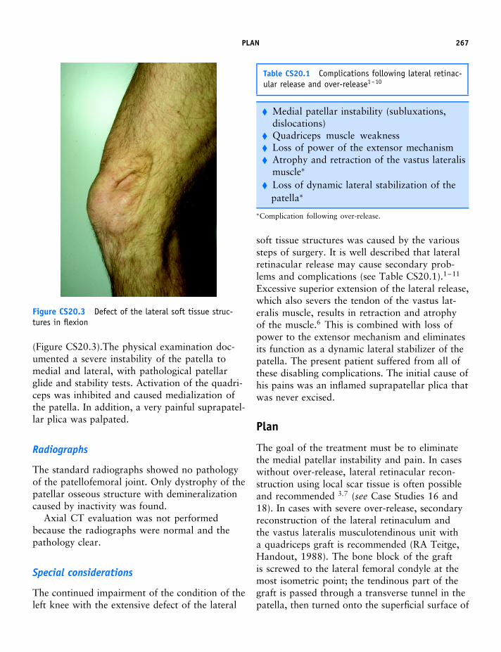
Figure CS20.3 Defect of the lateral soft tissue struc-tures in flexion
(Figure CS20.3).The physical examination doc-umented a severe instability of the patella tomedial and lateral, with pathological patellarglide and stability tests. Activation of the quadri-ceps was inhibited and caused medialization ofthe patella. In addition, a very painful suprapatel-lar plica was palpated.
Radiographs
The standard radiographs showed no pathologyof the patellofemoral joint. Only dystrophy of thepatellar osseous structure with demineralizationcaused by inactivity was found.
Axial CT evaluation was not performedbecause the radiographs were normal and thepathology clear.
Special considerations
The continued impairment of the condition of theleft knee with the extensive defect of the lateral
Table CS20.1 Complications following lateral retinac-ular release and over-release1–10
� Medial patellar instability (subluxations,dislocations)
� Quadriceps muscle weakness� Loss of power of the extensor mechanism� Atrophy and retraction of the vastus lateralis
muscle∗
� Loss of dynamic lateral stabilization of thepatella∗
∗Complication following over-release.
soft tissue structures was caused by the varioussteps of surgery. It is well described that lateralretinacular release may cause secondary prob-lems and complications (see Table CS20.1).1–11
Excessive superior extension of the lateral release,which also severs the tendon of the vastus lat-eralis muscle, results in retraction and atrophyof the muscle.6 This is combined with loss ofpower to the extensor mechanism and eliminatesits function as a dynamic lateral stabilizer of thepatella. The present patient suffered from all ofthese disabling complications. The initial cause ofhis pains was an inflamed suprapatellar plica thatwas never excised.
Plan
The goal of the treatment must be to eliminatethe medial patellar instability and pain. In caseswithout over-release, lateral retinacular recon-struction using local scar tissue is often possibleand recommended 3,7 (see Case Studies 16 and18). In cases with severe over-release, secondaryreconstruction of the lateral retinaculum andthe vastus lateralis musculotendinous unit witha quadriceps graft is recommended (RA Teitge,Handout, 1988). The bone block of the graftis screwed to the lateral femoral condyle at themost isometric point; the tendinous part of thegraft is passed through a transverse tunnel in thepatella, then turned onto the superficial surface of

268 CASE STUDY 20 LATERAL RETINACULAR OVER-RELEASE
the patella and anchored there with sutures (seeCase Study 19). The inflamed suprapatellar plicais removed during the lateral reconstruction.
Postoperative care and rehabilitation
See Case Studies 16, 18 and 19.
Discussion
The lateral retinaculum is a normal anatomicalstructure with possible variations.3,8 It is ‘abnor-mal’ when it is too short, tight or too thick. Thismay cause passive patellar tilt. Lateral retinacu-lar release 12 may not be the only option. Somecomplications cannot always be eliminated, evenwhen using correct surgical techniques. There-fore, instead of release, we recommend a length-ening of the lateral retinaculum (see Case Studies3, 5 and 6).
Over-release with transection of the musculo-tendinous unit of the vastus lateralis is the mostserious complication. The vastus lateralis mus-cle is composed of a vastus lateralis and a vastuslateralis obliquus component.7,13 Over-release ofboth parts change the stability and alignment ofthe patellar balancing and should therefore abso-lutely be avoided.3,6,7 The physiological functionof the vastus lateralis muscle must be respected.
Summary
Over-release is the worst form of lateral retinacu-lar release with the most serious complications. Itabsolutely must be avoided. Although the recon-struction may be structurally possible, the physio-logical function of the lateral structures can neverbe achieved again.
References
1. Kramers-de Quervain IA, Biedert R, Stussi E(1997) Quantitative gait analysis in patientswith medial patellar instability following lateralretinacular release. Knee Surg Sports TraumatolArthrosc 5: 95–101
2. Kolowich PA, Paulos LE, Rosenberg TD, Farns-worth S (1990) Lateral release of the patella:indications and contraindications. Am J SportsMed 18: 359–365
3. Biedert RM, Friederich NF (1994) Failed lateralretinacular release: clinical outcome. J SportsTraumatol 16: 162–173
4. Teitge RA, Faerber WW, Des Madryl P, MatelicTM (1996) Stress radiographs of the patello-femoral joint. J Bone Joint Surg Am 78: 193–203
5. Hughston JC, Walsh WM, Puddu G (1984)Patellar Subluxation and Dislocation. SaundersMonographs in Clinical Orthopaedics, volume V.Philadelphia, PA, WB Saunders
6. Hughston JC, Deese M (1988) Medial subluxationof the patella as a complication of lateral retinac-ular release. Am J Sports Med 16: 383–388
7. Nonweiler DE, DeLee JC (1994) The diagnosisand treatment of medial subluxation of the patellaafter lateral retinacular release. Am J Sports Med22: 680–686
8. Busch MT, DeHaven KE (1989) Pitfalls of thelateral retinacular release. Clin Sports Med 8:279–290
9. Henry JH, Goletz TH, Williamson B (1986)Lateral retinacular release in patellofemoralsubluxation. Indications, results, and comparisonto open patellofemoral reconstruction. Am JSports Med 14: 121–129
10. Marumoto JM, Jordan C, Akins R (1995) Abiomechanical comparison of lateral retinacularreleases. Am J Sports Med 23: 151–155
11. Johnson DP, Wakeley C (2002) Reconstruction ofthe lateral patellar retinaculum following lateralrelease: a case report. Knee Surg Sports TraumatolArthrosc 10: 361–363
12. Arendt EA, Fithian DC, Cohen E (2002) Currentconcepts of lateral patella dislocation. Clin SportsMed 21: 499–519
13. Javadpour DP, Finegan PJ, O’Brien M (1991)The anatomy of the extensor mechanism and itsclinical relevance. Clin J Sport Med 1: 229–235
Suggested reading
Nonweiler DE, DeLee JC (1994) The diagnosis andtreatment of medial subluxation of the patella afterlateral retinacular release. Am J Sports Med 22:680–686


