ORIGINAL RESEARCH Stent alone treatment for …ORIGINAL RESEARCH Stent alone treatment for...
6
ORIGINAL RESEARCH Stent alone treatment for dissections and dissecting aneurysms involving the basilar artery Chuanhui Li, Youxiang Li, Chuhan Jiang, Zhongxue Wu, Yang Wang, Xinjian Yang Department of Interventional Neuroradiology, Beijing Neurosurgical Institute and Beijing Tiantan Hospital, Capital Medical University, Beijing, China Correspondence to Dr X Yang, Department of Interventional Neuroradiology, Beijing Neurosurgical Institute and Beijing Tiantan Hospital, Capital Medical University, No 6, Tiantan Xili, Dongcheng District, Beijing 100050, People’s Republic of China; [email protected] Received 11 September 2013 Revised 6 December 2013 Accepted 15 December 2013 Published Online First 2 January 2014 To cite: Li C, Li Y, Jiang C, et al. J NeuroIntervent Surg 2015;7:50–55. ABSTRACT Background and purpose: Dissections and dissecting aneurysms involving the basilar artery (BA) are rare lesions with a poor prognosis, and controversies exist on treatment strategy. We describe the clinical presentation, radiologic features, and clinical outcomes of 11 consecutive patients with these lesions, treated using stent alone placement. Materials and methods 11 patients were studied (10 men, one woman), with a mean age of 51 years (range 30–68 years). Clinical and angiographic data were reviewed retrospectively. Results It was technically feasible to place stents in all 11 cases, and a multiple stents technique was used in nine patients. Procedure related posterior circulation ischemic complications occurred in two cases. One patient presenting with locked-in syndrome died of a pulmonary embolism 3 months after treatment although the clinical condition was gradually improving after stent placement. In the other eight cases, improvement in initial symptoms or a stable condition was observed on follow-up at 1–48 months. Angiographic follow-up was obtained in nine cases using DSA (n=8) or CT angiography (n=1). Successful occlusion of the aneurysm or dissection sac was noted in two cases, BA occlusion in one case, disappearance of double lumen sign in one case, and delayed retention of contrast medium in one case. For the remaining four patients there was no change in the configuration of the lesions on follow-up angiographic results. Conclusions Stent alone treatment as a potential and disputable alternative therapeutic option for the treatment of BA dissection and dissecting aneurysms, although technically feasible, was effective in only certain lesions, and this treatment carries substantial risks of complications and a high failure rate. The true effect of this strategy is questionable. A study involving larger populations and a longer follow-up is necessary to evaluate the efficacy of this treatment modality. INTRODUCTION Dissections and dissecting aneurysms involving the basilar artery (BA) are rare lesions with a poor prognosis, and controversies exist as to the proper management of these lesions, which most com- monly present with subarachnoid hemorrhage or posterior circulation ischemic stroke, and some- times a mass effect due to brainstem compres- sion. 1–4 Although conservative management has been advocated by some authors, 13 selected cases might require positive surgical or endovascular treatment, especially when they are ruptured or present with progressive ischemic or mass effect symptoms. Because of the morphological character- istics of these lesions and complicated anatomical structures in the posterior cranial fossa, surgical treatment is usually difficult. 56 Hence endovascular treatment has gradually become an important alter- native therapeutic option to manage these lesions. 7–12 Endovascular BA occlusion for these lesions, which may be life threatening as a result of severe ischemic complications unless sufficient collateral is present through the posterior communicating arter- ies, is rather limited compared with lesions of the vertebral artery (VA). Recently, with the improve- ment in modern endovascular techniques and mate- rials, reconstructive therapy using stents for vertebrobasilar dissection and dissecting aneurysms has been increasingly reported. 9–14 However, to the best of our knowledge, the literatures regarding stent alone treatment (SAT) for dissection and dis- secting aneurysms involving the BA are sparse, and relevant case series reports are rare. 7 12 14 Here we present our experience using a stent alone tech- nique for the treatment of 11 consecutive patients with these types of lesions, describing the clinical presentation, angiographic features, and clinical outcome. MATERIALS AND METHODS Patients Between December 2008 and December 2012, 11 consecutive patients diagnosed with dissection or dissecting aneurysms of the BA were treated using a stent alone placement technique in our department. There were 10 men and one woman, with a mean age of 51 years (range 30–68 years). The clinical and imaging characteristics of these 11 patients are presented in table 1. Clinical presentation was pos- terior circulation ischemic stroke in case Nos 2, 3, 4, 5, 8, 9, and 11, mass effect on the brainstem in case Nos 1, 6, and 11, agnogenic headache in case No 7, and in case No 10, the aneurysm was an incidental finding on CT angiography performed for unrelated symptoms. Preoperative radiologic evaluation Every patient underwent preoperative planar image examination, including CT scanning or MRI, or both. Before the treatment procedure, all patients underwent DSA to confirm the diagnosis and angiographic characteristics and to assist in treat- ment planning. Diagnosis was based on clinical manifestations and findings of radiological exami- nations, including CT, MRI, and CT/MR/cerebral angiography. Intramural thrombus on T1 weighted Hemorrhagic stroke 50 Li C, et al. J NeuroIntervent Surg 2015;7:50–55. doi:10.1136/neurintsurg-2013-010967 on May 3, 2020 by guest. Protected by copyright. http://jnis.bmj.com/ J NeuroIntervent Surg: first published as 10.1136/neurintsurg-2013-010967 on 2 January 2014. Downloaded from
Transcript of ORIGINAL RESEARCH Stent alone treatment for …ORIGINAL RESEARCH Stent alone treatment for...
ORIGINAL RESEARCH
Stent alone treatment for dissections and dissectinganeurysms involving the basilar arteryChuanhui Li, Youxiang Li, Chuhan Jiang, Zhongxue Wu, Yang Wang, Xinjian Yang
Department of InterventionalNeuroradiology, BeijingNeurosurgical Institute andBeijing Tiantan Hospital,Capital Medical University,Beijing, China
Correspondence toDr X Yang, Department ofInterventional Neuroradiology,Beijing Neurosurgical Instituteand Beijing Tiantan Hospital,Capital Medical University,No 6, Tiantan Xili, DongchengDistrict, Beijing 100050,People’s Republic of China;[email protected]
Received 11 September 2013Revised 6 December 2013Accepted 15 December 2013Published Online First2 January 2014
To cite: Li C, Li Y, Jiang C,et al. J NeuroIntervent Surg2015;7:50–55.
ABSTRACTBackground and purpose: Dissections anddissecting aneurysms involving the basilar artery (BA) arerare lesions with a poor prognosis, and controversiesexist on treatment strategy. We describe the clinicalpresentation, radiologic features, and clinical outcomesof 11 consecutive patients with these lesions, treatedusing stent alone placement.Materials and methods 11 patients were studied(10 men, one woman), with a mean age of 51 years(range 30–68 years). Clinical and angiographic datawere reviewed retrospectively.Results It was technically feasible to place stents in all11 cases, and a multiple stents technique was used innine patients. Procedure related posterior circulationischemic complications occurred in two cases. Onepatient presenting with locked-in syndrome died of apulmonary embolism 3 months after treatment althoughthe clinical condition was gradually improving after stentplacement. In the other eight cases, improvement ininitial symptoms or a stable condition was observed onfollow-up at 1–48 months. Angiographic follow-up wasobtained in nine cases using DSA (n=8) or CTangiography (n=1). Successful occlusion of the aneurysmor dissection sac was noted in two cases, BA occlusionin one case, disappearance of double lumen sign in onecase, and delayed retention of contrast medium in onecase. For the remaining four patients there was nochange in the configuration of the lesions on follow-upangiographic results.Conclusions Stent alone treatment as a potential anddisputable alternative therapeutic option for thetreatment of BA dissection and dissecting aneurysms,although technically feasible, was effective in onlycertain lesions, and this treatment carries substantialrisks of complications and a high failure rate. The trueeffect of this strategy is questionable. A study involvinglarger populations and a longer follow-up is necessary toevaluate the efficacy of this treatment modality.
INTRODUCTIONDissections and dissecting aneurysms involving thebasilar artery (BA) are rare lesions with a poorprognosis, and controversies exist as to the propermanagement of these lesions, which most com-monly present with subarachnoid hemorrhage orposterior circulation ischemic stroke, and some-times a mass effect due to brainstem compres-sion.1–4 Although conservative management hasbeen advocated by some authors,1 3 selected casesmight require positive surgical or endovasculartreatment, especially when they are ruptured orpresent with progressive ischemic or mass effect
symptoms. Because of the morphological character-istics of these lesions and complicated anatomicalstructures in the posterior cranial fossa, surgicaltreatment is usually difficult.5 6 Hence endovasculartreatment has gradually become an important alter-native therapeutic option to manage theselesions.7–12
Endovascular BA occlusion for these lesions,which may be life threatening as a result of severeischemic complications unless sufficient collateral ispresent through the posterior communicating arter-ies, is rather limited compared with lesions of thevertebral artery (VA). Recently, with the improve-ment in modern endovascular techniques and mate-rials, reconstructive therapy using stents forvertebrobasilar dissection and dissecting aneurysmshas been increasingly reported.9–14 However, to thebest of our knowledge, the literatures regardingstent alone treatment (SAT) for dissection and dis-secting aneurysms involving the BA are sparse, andrelevant case series reports are rare.7 12 14 Here wepresent our experience using a stent alone tech-nique for the treatment of 11 consecutive patientswith these types of lesions, describing the clinicalpresentation, angiographic features, and clinicaloutcome.
MATERIALS AND METHODSPatientsBetween December 2008 and December 2012, 11consecutive patients diagnosed with dissection ordissecting aneurysms of the BA were treated using astent alone placement technique in our department.There were 10 men and one woman, with a meanage of 51 years (range 30–68 years). The clinicaland imaging characteristics of these 11 patients arepresented in table 1. Clinical presentation was pos-terior circulation ischemic stroke in case Nos 2, 3,4, 5, 8, 9, and 11, mass effect on the brainstem incase Nos 1, 6, and 11, agnogenic headache in caseNo 7, and in case No 10, the aneurysm was anincidental finding on CT angiography performedfor unrelated symptoms.
Preoperative radiologic evaluationEvery patient underwent preoperative planar imageexamination, including CT scanning or MRI, orboth. Before the treatment procedure, all patientsunderwent DSA to confirm the diagnosis andangiographic characteristics and to assist in treat-ment planning. Diagnosis was based on clinicalmanifestations and findings of radiological exami-nations, including CT, MRI, and CT/MR/cerebralangiography. Intramural thrombus on T1 weighted
Hemorrhagic stroke
50 Li C, et al. J NeuroIntervent Surg 2015;7:50–55. doi:10.1136/neurintsurg-2013-010967
on May 3, 2020 by guest. P
rotected by copyright.http://jnis.bm
j.com/
J NeuroIntervent S
urg: first published as 10.1136/neurintsurg-2013-010967 on 2 January 2014. Dow
nloaded from

images (case Nos 1, 2, 3, 5, 6, 7, and 10) and intimal flaps onT2 weighted images (case Nos 1, 4, 6, 9, and 11) were majorfindings indicating dissection. Demonstration of these lesions oncerebral angiography included typical results such as doublelumen (case Nos 2, 5, 8, and 9), tapered narrowing (case No 1),pearl-string sign (case No 7), or retention of contrast medium inthe aneurysmal or dissection sac (case Nos 1, 3, 4, 6, and 8).
Endovascular treatment and techniquesAll patients received dual antiplatelet therapy, including 100 mgof aspirin and 75 mg of clopidogrel from at least 3 days prior tothe procedures. Dual antiplatelet therapy was maintained for2 months postoperatively followed by aspirin monotherapylasting indefinitely for cases presenting with ischemic stroke; inthe other cases, aspirin monotherapy lasted for 6 months.
All procedures were performed with the patient undergeneral anesthesia. Anticoagulation during the treatment processincluded an IV bolus of 3000–5000 IU of heparin given at thebeginning of each procedure and 1000 IU bolus administeredintravenously thereafter every hour. Every coaxial catheter flush-ing fluid was mixed with heparin at a concentration of 1000 IUof heparin per liter of saline.
In all cases, vascular access to the BA for stent placement wasobtained via a transfemoral approach using a 6 F guiding cath-eter. Diagnostic angiography and three-dimensional rotational
angiography were performed through a single or bilateral VAcontrast injection. Following the diagnostic angiogram, theguiding catheter was navigated into VA, usually the preponder-ant one. The stents used in this case series included the SolitaireAB (eV3), Enterprise (Codman Cordis), Neuroform (BostonScientific), Wingspan (Boston Scientific), and Leo (Balt). Thechoice of stent was dependent on anatomic geometry, size, andlength of the aneurysmal lumen, and the availability of thedevices. Using the roadmapping technique, a microcatheter(Rebar 027, eV3; Prowler Select Plus, Codman Cordis) or spe-cialized delivery system (Vasco, Balt; Wingspan, BostonScientific) was coaxially, using a microguidewire (Silverspeed 14,eV3; Traxcess-14, Microvention; Transcend-14, BostonScientific), navigated through the guiding catheter into the BAto cover the lesion. In some cases, a single stent was not suffi-cient to remodel the blood flow sufficiently or completely coverthe entire length of the lesion, so subsequent overlapping stentsor cascading stents were deployed. Basilar dissection maydevelop via antegrade progression of unilateral or bilateral VAdissections. In this case, the proximal stent should cover thelesion of the VAs. In lesions involving bilateral VAs, afterdeploying stents into the BA and unilateral VA, the guiding cath-eter should be transferred to the contralateral VA to deploy thestent through the mesh of the stent placed at the vertebrobasilarjunction to cover the lesion at this side. After stent deployment,
Table 1 Clinical and imaging characteristics of 11 patients with dissection or dissecting aneurysm involving the basilar artery
PatientNo Presentation Preoperative CT and MRI findings Preoperative DSA findings Stents
1 Intermittent headache andvertigo
Brainstem compression, intramuralthrombus and intimal flap, noinfarction
Tapered narrowing, aneurysm (9.7×7.7 mm) of middleBA, right AICA involved, retention of contrast medium
Leo stent (3.5×25 mm) (×1)
2 Diplopia, recurrent amaurosis Intramural thrombus Double lumen, fusiform dilation of middle–lower BA,bilateral AICA involved
Enterprise stent(4.5×37 mm +4.5×28 mm)(×1)
3 Blurred vision Intramural thrombus Fusiform dilation involving lower BA and bilateraldistal VA, right AICA involved, retention of contrastmedium
Solitaire stent (6×30 mm)(×4)
4 Locked-in syndrome Intimal flap, brainstem infarction Serpentine dissection involving left distal VA andmiddle–lower BA, retention of contrast medium
Neuroform stent (4×30 mm)(×2)Wingspan stent (4×20 mm)(×1)Enterprise stent(4.5×37 mm) (×1)
5 Sudden dizziness, vomiting Intramural thrombus Fusiform dilation of entire BA, double lumen Enterprise stent(4.5×28 mm) (×2)
6 Right clonic facial spasm Brainstem compression, intramuralthrombus and intimal flap, noinfarction
Fusiform aneurysm (9.8×9.7 mm) of BA, retention ofcontrast medium
Leo stent (5×50 mm) (×1),Enterprise stent(4.5×37 mm) (×1)
7 Long term headache Intramural thrombus Dissection of bilateral distal VA and middle–lower BA,pearl-string sign
Solitaire stent (6×30 mm)(×2)Solitaire stent (6×20 mm)(×1)
8 Sudden vertigo, coma, alalia,and quadriplegia
Brainstem infarction Dissection of entire BA, double lumen, retention ofcontrast medium
Solitaire stent (6×30 mm)(×4)
9 Sudden dizziness and leftweakness
Intimal flap Dissection of middle–lower BA, double lumen Solitaire stent (6×30 mm)(×3),Solitaire stent (6×20 mm)(×1)
10 Incidental finding Intramural thrombus Aneurysm (2.0×3.6 mm) of middle BA Enterprise stent(4.5×37 mm) (×1)
11 Right hemidysesthesia,drinking cough
Intimal flap, brainstem compressionand brainstem infarction
Fusiform aneurysm (15.2×25.6 mm) of lower BA andleft distal VA, left PICA involved
Solitaire stent (6×30 mm)(×3)
Age range of this case series was 30–68 years (mean 51 years); there were 10 men and one woman.AICA, anterior inferior cerebellar artery; BA, basilar artery; PICA, posterior inferior cerebellar artery; VA, vertebral artery.
Hemorrhagic stroke
Li C, et al. J NeuroIntervent Surg 2015;7:50–55. doi:10.1136/neurintsurg-2013-010967 51
on May 3, 2020 by guest. P
rotected by copyright.http://jnis.bm
j.com/
J NeuroIntervent S
urg: first published as 10.1136/neurintsurg-2013-010967 on 2 January 2014. Dow
nloaded from
control angiography was performed to confirm the patency ofthe parent artery and evaluate intra-aneurysmal hemodynamicchanges.
Follow-up protocolPatient followup was performed clinically and with angiographyexamination, preferably DSA. Clinical outcomes were evaluatedaccording to modified Rankin Scale (mRS).
RESULTSIt was technically feasible to place stents in all 11 cases. A mul-tiple stents technique were used in nine cases (case Nos 2, 3, 4,5, 6, 7, 8, 9, and 11), and the true overlapping stents techniquewas used in five (case Nos 2, 5, 6, 9, and 11). In the remainingfour cases (case Nos 3, 4, 7, and 11), multiple stents deployedin tandem or in a partial overlapping fashion were used to com-pletely cover the long lesions involving the distal unilateral VA(case Nos 4 and 11) or bilateral VAs (case Nos 3 and 7).
Immediately after stent angioplasty, control angiography wasperformed to evaluate postprocedural hemodynamic changes inall 11 cases. More retention of contrast medium was observedin the aneurysmal or dissection sac in three cases (case Nos 1, 6,and 8; 27.3%). In case No 4, improvement in blood flow in theparent artery and bilateral posterior cerebral arteries was eobserved after stenting. In another seven cases (63.6%) therewas no obvious hemodynamic change immediately after stent-ing. Angiographic follow-up was obtained in nine cases. In caseNo 1, follow-up DSA obtained 6 months after treatmentrevealed delayed occlusion of the BA after stenting and theaneurysm had also disappeared. This patient did not presentwith any symptoms related to the occluded BA because thehypertrophied bilateral posterior communicating artery suppliedthe distal BA and its branches sufficiently. In case Nos 6 and 8,follow-up DSA obtained 6 months after treatment confirmeddelayed thrombosis of the dissection or aneurysmal sac withpatency of the BA. In case Nos 3, 9, and 11, there was noobvious change on follow-up DSA compared with the angio-gram performed immediately after stent placement during theprocedure. Follow-up DSA 5 month later revealed
disappearance of the double lumen sign in case No 2. In caseNo 7, follow-up DSA 8 months later showed delayed retentionof contrast medium in the dissection sac. In case No 10, CTangiography follow-up obtained 2 months after treatment con-firmed that the morphology and size of the aneurysm wasstable, and there was good patency of the BA. Follow-up angiog-raphy was unavailable for case Nos 4 and 5.
Complications after treatment occurred in three patients(27.3%). Case No 4, who was bed-ridden because of locked-insyndrome before SAT, unfortunately died of a pulmonaryembolism 3 months later although the clinical condition hadbeen gradually improving after treatment. Ischemic complica-tions were encountered immediately after treatment in case Nos5 and 10. Case No 5 presented with right hemiparesis, leftfacial paralysis, and vertigo, and CT scanning confirmed pontineinfarction. Case No 10 presented with left hemiparesis aftertreatment, and CT scanning discovered pontine and cerebellarinfarction in this patient. No other complication was observedprior to patient discharge and during follow-up.
Clinical followup was performed in all 11 patients. Case No6, who presented with right clonic facial spasm, improved grad-ually, and this symptom had disappeared when he was admittedfor DSA followup 6 months after treatment. Symptoms in caseNos 1, 2, 8, and 9 were improved during followup; in case Nos3, 7, and 11, their condition was unchanged compared withtheir baseline condition. Although the condition of two patientspresenting with ischemic complications were gradually improv-ing during follow-up, both still suffer from moderate disability(case No 5, mRS score=4; case 10, mRS score=3). Clinical andangiographic outcomes are summarized in table 2.Representative cases are provided in figures 1–3.
DISCUSSIONAdvances in angiography and MRI have led to increased recog-nition that posterior circulation cranial vessel dissections anddissecting aneurysms are a noteworthy cause of stroke.2 9 12–17
Dissections and dissecting aneurysms involving the BA havebeen reported much less frequently compared with those involv-ing the VA. Currently, reconstructive endovascular treatment
Table 2 Clinical and angiographic outcomes
Patient NoControl angiographyduring procedure
Imaging follow-up(results/modality/time) Initial mRS Complications
Clinical follow-up(mRS/time)
1 Patency of BA, more retention of contrastmedium
No change/DSA/1.5 monthsOcclusion of BA/DSA/6 months
1 None 0/48 months
2 No change Disappearance of double lumen/DSA/5 months
1 None 0/31 months
3 No change No change/DSA/11 months 2 None 2/11 months4 Improvement in blood flow None 5 Pulmonary embolism 4/2 months
died/3 months5 No change None 2 Pontine infarction 4/36 months6 More retention of contrast medium Thrombosis of the aneurysm sac/DSA/
6 months1 None 0/26 months
7 No change Delayed retention of contrast medium/DSA/8 months
1 None 1/8 months
8 More retention of contrast medium Thrombosis of the dissection sac/ DSA/6 months
4 None 3/28 months
9 No change No change/DSA/5 months 2 None 1/6 months10 No change No change/CTA/2 months 0 Pontine and cerebellar
infarction3/2 months
11 No change No change/DSA/6 months 2 None 2/6 months
BA, basilar artery; CTA, CT angiography; mRS, modified Rankin Scale.
Hemorrhagic stroke
52 Li C, et al. J NeuroIntervent Surg 2015;7:50–55. doi:10.1136/neurintsurg-2013-010967
on May 3, 2020 by guest. P
rotected by copyright.http://jnis.bm
j.com/
J NeuroIntervent S
urg: first published as 10.1136/neurintsurg-2013-010967 on 2 January 2014. Dow
nloaded from
with stents has been used as an important therapeutic optionfor dissections and dissecting aneurysms involving the BA.However, apart from a few anecdotal cases,7 9 12 14 case series
reports on using SAT for the management of these lesions haverarely been described. In this study, we have presented ourexperience using SAT for these lesions.
Patient selectionReconstructive strategies utilizing stents for the treatment ofintracranial dissecting aneurysms and dissections consist of stentassisted coiling (SAC) and SAT. Both methods promote throm-bosis of the aneurysm while preserving or even improving bloodflow in the parent vessel. In most circumstances, SAC is prefer-ential because coiling decreases blood flow impingement intothe fragile vessel wall of the lesion and further reduces wallstress of the pseudoaneurysm, which is beneficial for thrombosiswithin the aneurysmal or dissection sac, thereby facilitating longterm healing of the lesions and decreasing the possibility ofbleeding compared with SAT.18 However, dissecting aneurysmsof the basilar trunk may be fusiform or dolichoectatic, whichoften involve the whole circumference of the BA and lack adefined neck. On the other hand, some of these lesions involvevital perforating branches, the occlusion of which can have acatastrophic outcome. Therefore, sometimes SAC is not feasiblefor these lesions due to the morphological features because thecoils may protrude into the parent artery or nearby large per-forators, thus resulting in ischemic complications.
Conservative management using anticoagulation and antipla-telet therapy for these lesions presenting with ischemic strokehas been advocated by some authors. However, conservativemanagement has been followed by disappointing results in some
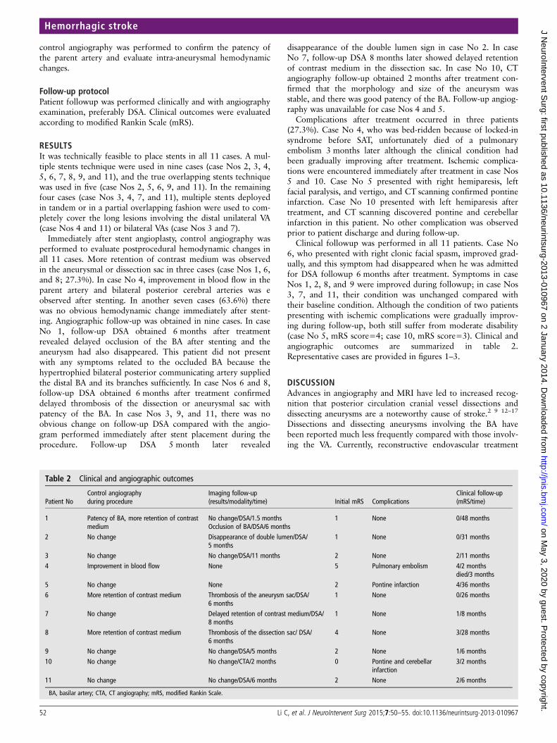
Figure 1 A quadragenarian presenting with blurred vision for2 months (case No 3). (A, B) Bilateral vertebral angiograms, frontalview, show a fusiform basilar trunk aneurysm involving the rightanterior inferior cerebellar artery and distal segments of the bilateralvertebral arteries. Note the irregular distal vertebral arteries and rightposterior inferior cerebellar artery originate from the aneurysm. (C, D)Bilateral control vertebral angiograms performed immediately afterstenting during the procedure. No obvious change in retention wasobserved after stenting. Follow-up DSA obtained 11 months later showsthere is still no hemodynamic changes and no newly formedthrombosis in the aneurysm sac (E, F). Arrows in (C–F) show stentmarkers. The condition of this patient was unchanged compared withbaseline after treatment (modified Rankin Scale score=2).
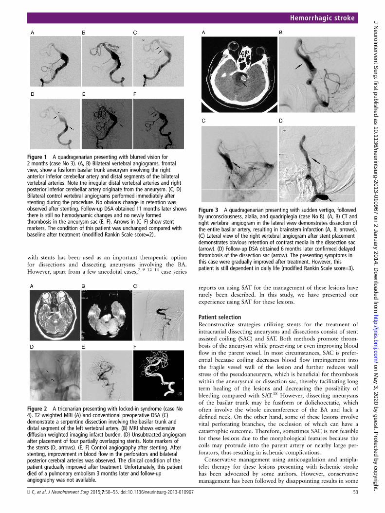
Figure 2 A tricenarian presenting with locked-in syndrome (case No4). T2 weighted MRI (A) and conventional preoperative DSA (C)demonstrate a serpentine dissection involving the basilar trunk anddistal segment of the left vertebral artery. (B) MRI shows extensivediffusion weighted imaging infarct burden. (D) Unsubtracted angiogramafter placement of four partially overlapping stents. Note markers ofthe stents (D, arrows). (E, F) Control angiography after stenting. Afterstenting, improvement in blood flow in the perforators and bilateralposterior cerebral arteries was observed. The clinical condition of thepatient gradually improved after treatment. Unfortunately, this patientdied of a pulmonary embolism 3 months later and follow-upangiography was not available.
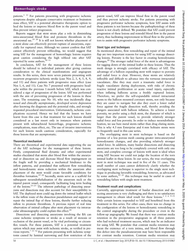
Figure 3 A quadragenarian presenting with sudden vertigo, followedby unconsciousness, alalia, and quadriplegia (case No 8). (A, B) CT andright vertebral angiogram in the lateral view demonstrates dissection ofthe entire basilar artery, resulting in brainstem infarction (A, B, arrows).(C) Lateral view of the right vertebral angiogram after stent placementdemonstrates obvious retention of contrast media in the dissection sac(arrow). (D) Follow-up DSA obtained 6 months later confirmed delayedthrombosis of the dissection sac (arrow). The presenting symptoms inthis case were gradually improved after treatment. However, thispatient is still dependent in daily life (modified Rankin Scale score=3).
Hemorrhagic stroke
Li C, et al. J NeuroIntervent Surg 2015;7:50–55. doi:10.1136/neurintsurg-2013-010967 53
on May 3, 2020 by guest. P
rotected by copyright.http://jnis.bm
j.com/
J NeuroIntervent S
urg: first published as 10.1136/neurintsurg-2013-010967 on 2 January 2014. Dow
nloaded from
patients.1 11 For patients presenting with progressive ischemicsymptoms despite adequate conservative treatment or brainstemmass effect, SAT is a potential alternative therapeutic option toheal the lesions or improve blood flow in the parent vessel andthus prevent infarct progression.
Reports suggest that stent struts play a role in diminishingintra-aneurysmal blood flow and promote thrombosis in theaneurysmal sac.19 20 It is still not known whether placement ofa stent alone can effectively prevent rupture of aneurysms, espe-cially for ruptured ones. Although we cannot confirm that SATcannot effectively prevent rebleeding, we would suggest thatusing SAT for the management of hemorrhagic lesions requiresgreat caution considering the high rebleed rate after SATreported by some authors.10 11
In conclusion, SAT for the management of these lesionsshould be tailored to individual patients after giving cautiousconsideration to the clinical manifestations and angiogramresults. In this series, there were seven patients presenting withrecurrent progressive ischemic stroke (case Nos 2, 3, 4, 5, 8, 9,and 11) and three patients with progressive mass effect (caseNos 1, 6, and 11). Case No 7 presented with progressive head-ache within the previous 1 month before SAT, which was con-sidered a sign of progression of the lesion. SAT was performedwith the aim of preventing progression of the lesions in thesecases. The remaining case No 10, who was incidentally diag-nosed and clinically asymptomatic, developed severe depressionafter knowing the diagnosis and the potential risks, and stronglyrequested procedural intervention. Unfortunately, ischemic com-plications occurred. Hence we feel that the lesson we havelearnt from this case is that treatment for such lesions shouldconsidered as a last resort only in instances where patientspresent with subarachnoid hemorrhage, progressive ischemiccomplications, mass effect, etc. The use of invasive interventionsfor such lesions needs cautious consideration, especially forthose lesions that are asymptomatic.
Theoretical mechanismThere are theoretical and experimental data supporting the useof the SAT technique for the management of these lesions.Firstly, computational fluid dynamic and other experimentalstudies elucidated that stents alter blood flow within the aneurys-mal or dissection sac and decrease blood flow impingement onthe fragile wall by providing a mechanical hindrance to theinflow patterns, and postulated that sufficient flow remodelingand hemodynamic stress reduction within the aneurysm afterplacement of the stent would create favorable conditions forthrombus formation.19 20 Secondly, stents serve as a scaffold forsubsequent neoendothelialization of the aneurysm neck or theinjured parent vessel, consequently facilitating long term healingof the lesions.21–23 The inherent pathology of dissecting aneur-ysms and dissections may also account for their susceptibility toSAT. The deployed stent could tag down the intimal leaflet whichis the inflow route of the dissection pseudolumen and support orrepair the intimal flap of these lesions, thereby further reducinginflow to promote thrombosis. A previous report of real timeobservation of intimal collapse after stent placement via intravas-cular ultrasonography could explain this effect.24
Dissections and dissecting aneurysms involving the BA cancause ischemic symptoms or stroke as a result of stenosis orocclusion of the parent vessel, or by vital perforating or circum-flex vessels. For these patients, SAT is a potential treatmentoption which may assist with ischemic stroke, as verified in pre-vious reports.7 11 12 For patients presenting with ischemic symp-toms caused by luminal narrowing and flow restriction of the
parent vessel, SAT can improve blood flow in the parent vesseland thus prevent ischemic stroke. For patients presenting withprogressive perforator ischemic symptoms, how SAT assists withthese patients is not known because the pathophysiology of theselesion is not clearly defined. We postulate that SAT could preventprogression of these lesions and remodel blood flow in the parentartery, thus facilitating improvement in blood flow in the perfora-tors to assists with progressive perforator ischemic symptoms.
Stent type and techniquesAs mentioned above, flow remodeling and repair of the intimalflap are two important reasons for using SAT to manage dissect-ing lesions. Porosity is the key factor in creating hemodynamicchanges.20 The stronger radial force of the stent is advantageousfor tagging down of the intimal leaflet in these lesions. Thus themesh design is strongly correlated with efficacy of SAT. Theadvantage of a balloon expanding stent with respect to porosityand radial force is clear. However, these stents are relativelyinflexible and difficult to advance into the tortuous intracranialvasculature. Also they may exert a traumatic radial force infragile vasculature during deployment, with risks of chronicreactive intimal proliferation or acute vessel injury, especiallywhen inflating balloons across a freshly ruptured lesions.Therefore, we prefer the self-expandable nitinol intracranialstents to the balloon mounted coronary stents, not only becausethey are easier to navigate but also they exert a lower radialforce against the fragile dissection wall, thereby avoiding thedangers associated with balloon inflation. To achieve a lowerporosity, we prefer using a thick stent, the diameter of which islarger than the parent vessel, to provide relatively strongerradial force and less porosity. In order to induce neoendothelia-lization, we use bare metal stents rather than drug eluting stents.This is why 4.5 mm Enterprise and 6 mm Solitaire stents wereso frequently used in this case series.
The overlapping stent in stent technique is based on thepremise of a less porous mesh causing greater intra-aneurysmalhemodynamic changes, and also providing relatively strongerradial force. In addition, many basilar dissections and dissectinganeurysms are too long to be completely covered with only onestent, and complete coverage of lesions with stent is ideal whenusing SAT because we could not judge the location of the tornintimal leaflet in these lesion. In our series, the true overlappingstent in stent technique was used in five of the 11 cases. Thissmall number of cases cannot definitively clarify whether theoverlapping stent technique is superior to the single stent tech-nique in producing favorable remodeling; however, as advocatedby some authors,12 25 this technique may be useful in cases ofremodeling failure with single stent treatment.
Treatment result and complicationsCurrently, appropriate treatment of basilar dissection and dis-secting aneurysms is very challenging and there is no satisfactorymanagement to obtain ideal result for these lesions.1 2 6 9–11
Only certain lesions responded to SAT and benefitted from thistreatment in this series. For other cases, there was no change inthe configuration of the lesions or hemodynamic remodeling inthe control angiography during the procedure or even in thefollow-up angiography. We found that there was contrast mediaretention in the preoperative angiogram in all three patientswho had technical treatment success, which may be an indicatorfor using SAT. We postulate that contrast media retention maymean the existence of a torn intima, and blood flow throughthis defect into the pseudoaneurysm may have been responsiblefor contrast media retention. Contrast media retention may
Hemorrhagic stroke
54 Li C, et al. J NeuroIntervent Surg 2015;7:50–55. doi:10.1136/neurintsurg-2013-010967
on May 3, 2020 by guest. P
rotected by copyright.http://jnis.bm
j.com/
J NeuroIntervent S
urg: first published as 10.1136/neurintsurg-2013-010967 on 2 January 2014. Dow
nloaded from
disappear with spontaneous healing of the torn intima whichact as the inflow route of the dissection pseudolumen. So theeffect of SAT for lesions without contrast media retention maybe limited, because these lesions are often relatively stable.However, not all the lesions showing contrast media retentionresponded to SAT. In this series, no change in the configurationof the lesions was observed on follow-up angiogram in twocases presenting with postprocedural contrast media retention.From this small series, we cannot definitely explain why certainlesions responded to treatment while others did not. A studyinvolving a larger population and longer follow-up is necessaryto clarify this issue and evaluate the efficacy of this treatmentmodality.
SAT for these lesions carries a substantial risk of complications.Ischemic complications after treatment occurred in two patients.Previous reports indicated that levels of P2Y12, a platelet mem-brane protein receptor which plays a key role in platelet activation,vary in individuals and influence the effect of antiplatelettherapy.26 27 To reduce ischemic complications for these patientsreceiving reconstructive endovascular treatment, P2Y12 plateletfunction tests, to evaluate if there is P2Y12 resistance to antiplate-let therapy, and coagulogram tests, such as thrombelastography tomonitor blood coagulation, may be helpful.27 Unsatisfactoryresults of thrombelastography could prompt a change in the antic-oagulation and antiplatelet strategy to obtain a better effect.
There are many other issues to clarify for this treatmentmodality. Improvement or stable condition of initial symptomswas observed in eight cases, apart from those who encounteredcomplications. However, was the improvement in initial symp-toms in the above cases caused by stenting or the antiplatelettherapy after SAT, or was it a natural course of the disease? Forexample, although comparison of preprocedural and postproce-dural angiograms could confirm whether SAT remodelled bloodflow and improved perfusion of the brainstem perforators andbilateral posterior cerebral arteries in case No 4 (who sufferedfrom locked-in syndrome resulting from spiral dissections of theBA), it is not clear whether this treatment had anything to dowith the gradual clinical improvement. In most of these cases,follow-up time was short. Does SAT improve the prognosis ofpatients with basilar dissections and dissecting aneurysms in thelong-term? Only certain lesions responded to SAT. Are therelessons to be leant in terms of patient selection, stent selection,and use of a single stent or overlapping stent in stent techniquethat may impact on the treatment result? If a flow diverter isavailable for treating these lesions using SAT, what is the result?Could SAT effectively prevent rebleed of ruptured lesions? Astudy with a larger population and longer follow-up is necessaryfor validation of the efficacy of this treatment modality.
CONCLUSIONSSAT as a potential and disputable alternative therapeutic optionfor the treatment of BA dissections and dissecting aneurysms.Although technically feasible, SAT is effective in only certainlesions and carries a substantial risk of complications and a highfailure rate. The true effect of this strategy is questionable. Astudy involving a larger population and longer follow-up isnecessary to evaluate the efficacy of this treatment modality.
Contributors CL: collecting the data, and drafting and revising the article. YL, CJ,ZW, and YW: substantial contributions to conception and design, revising the article,and analysis and interpretation of the data. XY: conception and design, revising thearticle, and final approval of the version to be published.
Funding This work was supported by the National Science Foundation of China(grant Nos 81220108007 and 81171079), National ‘Twelfth Five-Year’ Plan forScience and Technology Support (grant No 2011BAI08B06), High-Level Health
Technique Talent Training Plan of Beijing Health System (grant No 2009-3-22), andNational 973 Basic Research Program of China (grant No 2010CB732605).
Competing interests None.
Ethics approval The study was approved by the ethics committee of BeijingTiantan Hospital.
Provenance and peer review Not commissioned; externally peer reviewed.
REFERENCES1 Pozzati E, Andreoli A, Padovani R, et al. Dissecting aneurysms of the basilar artery.
Neurosurgery 1995;36:254–8.2 Yoshimoto Y, Hoya K, Tanaka Y, et al. Basilar artery dissection. J Neurosurg
2005;102:476–81.3 Ross GJ, Ferraro F, DeRiggi L, et al. Spontaneous healing of basilar artery
dissection: MR findings. J Comput Assist Tomogr 1994;18:292–4.4 Hosoda K, Fujita S, Kawaguchi T, et al. Spontaneous dissecting aneurysms of the
basilar artery presenting with a subarachnoid hemorrhage. Report of two cases.J Neurosurg 1991;75:628–33.
5 Aziz KMA, van Loveren HR, Tew JM Jr, et al The Kawase approach to retrosellarand upper clival basilar aneurysms. Neurosurgery 1999;44:1225–36.
6 Lawton MT, Daspit PD, Spetzler RF. Technical aspects and recent trends in the managementof large and giant midbasilar artery aneurysms. Neurosurgery 1997;41:513–21.
7 Shin YS, Kim HS, Kim SY. Stenting for vertebrobasilar dissection: a possible treatmentoption for nonhemorrhagic vertebrobasilardissection. Neuroradiology 2007;49:149–56.
8 Wenderoth JD, Khangure MS, Phatouros CC, et al. Basilar trunk occlusion duringendovascular treatment of giant and fusiform aneurysms of the basilar artery. AJNRAm J Neuroradiol 2003;24:1226–9.
9 van Oel LI, van Rooij WJ, Sluzewski M, et al. Reconstructive endovascular treatmentof fusiform and dissecting basilar trunk aneurysms with flow diverters, stents, andcoils. AJNR Am J Neuroradiol 2013;34:589–95.
10 Chung J, Park H, Lim YC, et al. Endovascular treatment of basilar artery trunkaneurysms. Acta Neurochir (Wien) 2011;153:2137–45.
11 Kim BM, Suh SH, Park SI, et al. Management and clinical outcome of acute basilarartery dissection. AJNR Am J Neuroradiol 2008;29:1937–41.
12 Yoon WK, Kim YW, Kim SR, et al. Angiographic and clinical outcomes ofstent-alone treatment for spontaneous vertebrobasilar dissecting aneurysm. ActaNeurochir (Wien) 2010;152:1477–86.
13 Cohen JE, Gomori JM, Moscovici S, et al. Successful endovascular treatment of agrowing megadolichoectasic vertebrobasilar artery aneurysm by flow diversion usingthe “diverter-in-stent” technique. J Clin Neurosci 2012;19:166–70.
14 Bain M, Hussain MS, Spiotta A, et al. “Double-barrel” stent reconstruction of asymptomatic fusiform basilar artery aneurysm: case report. Neurosurgery 2011;68:E1491–6.
15 Santos-Franco JA, Zenteno M, Lee A. Dissecting aneurysms of the vertebrobasilarsystem. A comprehensive review on natural history and treatment options.Neurosurg Rev 2008;31:131–40.
16 Schievink WI, Wijdicks EF, Piepgras DG, et al. The poor prognosis of rupturedintracranial aneurysms of the posterior circulation. J Neurosurg 1995;82:791–5.
17 Hosoya T, Adachi M, Yamaguchi K, et al. Clinical and neuroradiological features ofintracranial vertebrobasilar artery dissection. Stroke 1999;30:1083–90.
18 Kim BM, Chung EC, Park SI, et al. Treatment of blood blister-like aneurysm of theinternal carotid artery with stent-assisted coil embolization followed bystent-within-a-stent technique. Case report. J Neurosurg 2007;107:1211–13.
19 Fiorella D, Albuquerque FC, Deshmukh VR, et al. Endovascular reconstruction withthe Neuroform stent as monotherapy for the treatment of uncoilable intraduralpseudoaneurysm. Neurosurgery 2006;59:291–300.
20 Rhee K, Han MH, Cha SH. Changes of flow characteristics by stenting in aneurysmmodels: influence of aneurysm geometry and stent porosity. Ann Biomed Eng2002;30:894–904.
21 Bai H, Masuda J, Sawa Y, et al. Neointima formation after vascular stentimplantation. Spatial and chronological distribution of smooth muscle cellproliferation and phenotypic modulation. Arterioscler Thromb 1994;14:1846–53.
22 Geremia G, Brack T, Brennecke L, et al. Occlusion of experimentally created fusiformaneurysms with porous metallic stents. AJNR Am J Neuroradiol 2000;21:739–45.
23 Mizutani T, Kojima H, Asamoto S. Healing process for cerebral dissecting aneurysmspresenting with subarachnoid hemorrhage. Neurosurgery 2004;54:342–8.
24 Yoon WK, Kim YW, Kim SD, et al. Intravascular ultrasonography-guided stentangioplasty of an extracranial vertebral artery dissection. J Neurosurg2008;109:1113–18.
25 Park SI, Kim BM, Kim DI, et al. Clinical and angiographic follow-up of stent-onlytherapy for acute intracranial vertebrobasilar dissecting aneurysms. AJNR Am JNeuroradiol 2009;30:1351–6.
26 Zhou BR, Shi HT, Wang R, et al. Dynamic changes and associated factors ofclopidogrel resistance in patients after cerebral infarction. J Neurol2013;260:2928–37.
27 Nordeen JD, Patel AV, Darracott RM, et al Clopidogrel resistance by P2Y12 plateletfunction testing in patients undergoing neuroendovascular procedures: incidence ofischemic and hemorrhagic complications. J Vasc Interv Neurol 2013;6:26–34.
Hemorrhagic stroke
Li C, et al. J NeuroIntervent Surg 2015;7:50–55. doi:10.1136/neurintsurg-2013-010967 55
on May 3, 2020 by guest. P
rotected by copyright.http://jnis.bm
j.com/
J NeuroIntervent S
urg: first published as 10.1136/neurintsurg-2013-010967 on 2 January 2014. Dow
nloaded from