Original Article Normalized autofluorescence imaging
9
Int J Clin Exp Pathol 2012;5(9):956-964 www.ijcep.com /ISSN:1936-2625/IJCEP1208025 Int J Clin Exp Pathol 2012;5(9): www.ijcep.com /ISSN:1936-2625/IJCEP1208025 Original Article Normalized autofluorescence imaging diagnostics in upper GI tract: a new method to improve specificity in neoplasia detection Ekaterina Krauss 1* , Abbas Agaimy 2* , Alexandre Douplik 3,4 , Heinz Albrecht 1 , Helmut Neumann 1 , Arndt Hartmann 2 , Ralf Hohenstein 3 , Martin Raithel 1 , Eckhart G Hahn 1 , Markus F Neurath 1 , Jonas Mudter 1 1 Department of Medicine 1, University of Erlangen-Nuremberg, Ulmenweg 18, D-91054 Erlangen, Germany; 2 Institute of Pathology, University of Erlangen-Nuremberg, Krankenhausstr.12, 91054 Erlangen, Germany; 3 Medi- cal Photonic Engineering Group, Chair of Photonics Technologies, Erlangen, Germany; 4 Clinical Photonics Lab, Erlangen Graduate School in Advanced Optical Technologies (SAOT), Erlangen, Germany. * Equal contributors. Received August 28, 2012; Accepted October 2, 2012; Epub October 20, 2012; Published October 30, 2012 Abstract: Background & Aims: This study was performed to improve the autofluorescence imaging (AFI) in the upper GI tract by applying a new method of normalized autofluorescence (NAFI) obtained via tri-modal imaging. Objective: NAFI may provide lower false positive rate to achieve ultimately better specificity at acceptable sensitivity. Patients and methods: This is a prospective, controlled single-centre study. 18 patients with suspected esophagus or stom- ach cancer undergoing esophagogastroduodenoscopy (EGD) were enrolled between February and May 2010. After endoscopy each patient was assigned into one of two groups: (1) non- cancer, including inflammation; (2) cancer group. EGDs were performed using video white light endoscopy, followed by AFI/NAFI. The targeted biopsy samples were taken from the abnormal areas as well as from adjacent mucosa. NAFI was compared versus AFI for cancer diagnostics in terms of specificity and sensitivity. Results: NAFI detected all neoplastic lesions. WLE or NBI detected no additional neoplasia. The AFI displayed mucosal inflammation and carcinomas of esophagus and stomach as dark red color, the normal mucosa background was displayed as light green. The NAFI didn’t differentiate inflamed tissue from normal in majority of cases, but in tumorous mucosa, the cancer areas were detected precisely. AFI shows 100% sensitivity but 50% specificity which correlates with previous literature data. On the other hand, NAFI demonstrated lower sensitivity (88%) but higher specificity compared to AFI (69%). Conclusions: Measuring the NAFI instead of the AFI was found improving the specificity of cancer diagnosis. Use of fiber-optic endoscopes to analyze AFI and possible endoscopic and histological sampling error are the main potential limitations of this method. Keywords: Autofluorescence imaging, NAFI, endoscopic tri-modal imaging, esophagogastroduodenoscopy, cancer diagnostic Introduction Gastrointestinal (GI) neoplasms continue to be one of the leading cancer-related deaths world- wide; therefore early detection of pre-cancer- ous stages in the upper GI tract is subject to extensive research efforts throughout the world. One of the highest shares in GI malignan- cies belongs to the upper GI including esopha- gus and stomach. Furthermore, presence of Barrett’s esophagus (BE) and chronic gastritis increase the risk of malignancy development [1, 2]. BE is a known precursor for development of esophageal cancer, and is frequently linked to the pre-existing GERD [3, 4]. Patients with BE have a 30-125 fold higher risk of developing cancer of the esophagus than the general pop- ulation [5]. The early detection and treatment of oesophagus cancer can significantly improve patient survival. When detected early, the cura- tive endoscopic resection may be an option, without the need for surgery [6, 7]. Unfortunately, the poor detection of pre-cancer and early stage cancer in BE by WLE is a significant limi- tation [8].
Transcript of Original Article Normalized autofluorescence imaging

Int J Clin Exp Pathol 2012;5(9):956-964www.ijcep.com /ISSN:1936-2625/IJCEP1208025Int J Clin Exp Pathol 2012;5(9):www.ijcep.com /ISSN:1936-2625/IJCEP1208025
Original Article Normalized autofluorescence imaging diagnostics in upper GI tract: a new method to improve specificity in neoplasia detection
Ekaterina Krauss1*, Abbas Agaimy2*, Alexandre Douplik3,4, Heinz Albrecht1, Helmut Neumann1, Arndt Hartmann2, Ralf Hohenstein3, Martin Raithel1, Eckhart G Hahn1, Markus F Neurath1, Jonas Mudter1
1Department of Medicine 1, University of Erlangen-Nuremberg, Ulmenweg 18, D-91054 Erlangen, Germany; 2Institute of Pathology, University of Erlangen-Nuremberg, Krankenhausstr.12, 91054 Erlangen, Germany; 3Medi-cal Photonic Engineering Group, Chair of Photonics Technologies, Erlangen, Germany; 4Clinical Photonics Lab,
Erlangen Graduate School in Advanced Optical Technologies (SAOT), Erlangen, Germany. *Equal contributors.
Received August 28, 2012; Accepted October 2, 2012; Epub October 20, 2012; Published October 30, 2012
Abstract: Background & Aims: This study was performed to improve the autofluorescence imaging (AFI) in the upper GI tract by applying a new method of normalized autofluorescence (NAFI) obtained via tri-modal imaging. Objective: NAFI may provide lower false positive rate to achieve ultimately better specificity at acceptable sensitivity. Patients and methods: This is a prospective, controlled single-centre study. 18 patients with suspected esophagus or stom-ach cancer undergoing esophagogastroduodenoscopy (EGD) were enrolled between February and May 2010. After endoscopy each patient was assigned into one of two groups: (1) non- cancer, including inflammation; (2) cancer group. EGDs were performed using video white light endoscopy, followed by AFI/NAFI. The targeted biopsy samples were taken from the abnormal areas as well as from adjacent mucosa. NAFI was compared versus AFI for cancer diagnostics in terms of specificity and sensitivity. Results: NAFI detected all neoplastic lesions. WLE or NBI detected no additional neoplasia. The AFI displayed mucosal inflammation and carcinomas of esophagus and stomach as dark red color, the normal mucosa background was displayed as light green. The NAFI didn’t differentiate inflamed tissue from normal in majority of cases, but in tumorous mucosa, the cancer areas were detected precisely. AFI shows 100% sensitivity but 50% specificity which correlates with previous literature data. On the other hand, NAFI demonstrated lower sensitivity (88%) but higher specificity compared to AFI (69%). Conclusions: Measuring the NAFI instead of the AFI was found improving the specificity of cancer diagnosis. Use of fiber-optic endoscopes to analyze AFI and possible endoscopic and histological sampling error are the main potential limitations of this method.
Keywords: Autofluorescence imaging, NAFI, endoscopic tri-modal imaging, esophagogastroduodenoscopy, cancer diagnostic
Introduction
Gastrointestinal (GI) neoplasms continue to be one of the leading cancer-related deaths world-wide; therefore early detection of pre-cancer-ous stages in the upper GI tract is subject to extensive research efforts throughout the world. One of the highest shares in GI malignan-cies belongs to the upper GI including esopha-gus and stomach. Furthermore, presence of Barrett’s esophagus (BE) and chronic gastritis increase the risk of malignancy development [1, 2].
BE is a known precursor for development of esophageal cancer, and is frequently linked to the pre-existing GERD [3, 4]. Patients with BE have a 30-125 fold higher risk of developing cancer of the esophagus than the general pop-ulation [5]. The early detection and treatment of oesophagus cancer can significantly improve patient survival. When detected early, the cura-tive endoscopic resection may be an option, without the need for surgery [6, 7]. Unfortunately, the poor detection of pre-cancer and early stage cancer in BE by WLE is a significant limi-tation [8].

Normalized autofluorescence imaging diagnostics in upper GI: preliminary results
957 Int J Clin Exp Pathol 2012;5(9):956-964
The sensitivity and positive predictive value of standard EGD for diagnosing BE was reportedly 82% and 34% respectively [9]. The routine pro-tocol of cancer detection at BE prescribes 4-quadrant biopsies taken at regular intervals throughout the BE, and even the most rigorous biopsy protocols may be associated with sam-pling error [10]. The detection of gastric cancer in different studies showed a sensitivity of 84.2-92% and a specificity of 78-89.7% [7, 11-14].
Despite the progressive development of endo-scopic modalities, the early detection of super-ficial neoplasms during routine EGD remains difficult because there are few morphological changes that differentiate malignant from non-malignant lesions [1]. Accurate diagnosis of tumor extent delineation is sometimes difficult because gastric neoplasms occasionally have flat or isochromatic tumor extensions.
To date, a clinical demand is still high for screening methods to highlight especially early lesions. Such methods as Optical Coherence Tomography [15, 16], Laser Scattering Spec-troscopy [17], Raman Spectroscopy [18], Con-focal Laser Endomicroscopy [19], Chromoen-doscopy [20, 21], Magnification Chromoen- doscopy [22], Infrared Endoscopy [23] and Spectral Imaging, particularly Fluorescence Imaging including Autofluorescence Imaging (AFI) [24, 25] and Narrow Band Imaging (NBI) [26] have been successfully applied in the GI tract including esophagus and stomach, for biopsy guidance and microsurgery naviga-tion[27-31]. Recently, WLE, AFI and NBI have been incorporated into one system: endoscopic tri-modal imaging [32, 33].
AFI endoscopy imaging produces real-time pseudo-color images based on natural tissue. Autofluorescence (AF) emitted by light excita-tion from endogenous fluorophores such as col-lagen, elastin, nicotinamide, adenine dinucleo-tide (NADH), flavins (FAD) and porphyrins [34, 35]. This method is able to identify lesions, including malignancies, by detecting differenc-es in tissue fluorescence, thus revealing early carcinomas, not yet detectable by conventional WLE. Because of these properties AFI may potentially improve the identification and char-acterization of the premalignant lesions in oesophageal and gastric mucosa [35, 36]. Despite the reported success for AFI in the
respiratory tract and lower GI tract, these approaches are still not specific enough to dis-cover dysplastic or cancer lesions in the esoph-agus with intestinal metaplastic background [25, 37] or flat lesions in the stomach where specificity for AFI varied from author to author from 21% to 69 % (for BE) to 92% (for stomach) [38].
One of the contemporary approaches to improve the specificity and image quality of the AFI diagnostics is to measure an intrinsic AF, the parameter decoupled from the excitation and emission absorption and scattering by bio-logical tissues, as opposed to the state of the art AF technique where the AF is measured as a product of the intrinsic AF, and emission re-absorption and scattering, i.e. so-called mea-sured AF [39, 40]. Collecting the intrinsic AF instead of measured by means of the spectros-copy was found enhancing the specificity of cancer diagnostics although the improvement of the sensitivity was found insignificant or even reduced.
The AF is a product of emission re-absorption and intrinsic fluorescence; if the measured AF is normalized over the diffuse reflectance and taking into account the difference between the path lengths of the diffusively reflected and fluoresced photons, one can obtain the intrin-sic AF. Receiving a true intrinsic AF image will require spectrally resolved imaging dataset such as hyperspectral imaging collection. Hence, we normalized the green RGB channel of AFI over either the blue or green channels of WLI, or green or blue channels of NBI to finally obtain 4 normalized AF images (NAFI).
The working hypothesis of this study is that such innovation as NAFI may provide lower false positive rate to achieve ultimately better specificity at acceptable sensitivity.
Materials and methods
Patient criteria
Prospectively, 18 patients (12 male, 6 female; median age 63.3 years, age range 51-73 years) undergoing EGD during the period between February and May 2010, were enrolled. The first 6 patients (2 with esophageal adenocarci-noma, 1 with gastric adenocarcinoma, 2 with GERD, 1 with BE with dysplasia) were consid-

Normalized autofluorescence imaging diagnostics in upper GI: preliminary results
958 Int J Clin Exp Pathol 2012;5(9):956-964
ered as a pilot group. The pilot data were used to finalize the data collection and data analysis protocols for NAFI and were not included into the final clinical data analysis.
12 patients were examined using both the new technique NAFI and state of the art technique AFI: 2 patients with squamous cell carcinoma of the esophagus, 3 patients with gastric ade-nocarcinoma, 2 patients with GERD, 2 patients with BE with dysplasia, and 3 patients with nor-mal results of EGD, who were undergoing sur-veillance procedure because of suspected cancer.
The indications for EGD were as following: sus-pected or known esophagus or stomach can-cer, BE; complains suspicious for acute or chronic gastritis or esophagitis. The diagnoses were made by correlation of patient’s medical history, endoscopy, and histological results of targeted biopsy specimens, taken from suspi-cious areas. The Montreal classification was used for definition of BE; Los-Angeles classifi-cation was used for the diagnosis of GERD [41-43].
The following exclusion criteria were applied: Age < 18 years; unable or unwilling to give informed consent. Further, ionizing radiation therapy to the chest or abdomen within the past six months; chromoendoscopy within the past 7 days; significant upper GI bleeding of any etiology; esophageal candidiasis; chemo-therapy for cancer within three months; confo-cal laser endoscopy with fluorescent photosen-sitizing drugs within two months.
A signed informed consent was obtained from all patients included in the study. The study was approved by the Ethic Committee of the University Erlangen-Nuremberg. During the examination the patients received upon requirement sedation with Midazolam and Pethidin.
Endoscopic procedure
EGDs were performed using WLE followed by AFI endoscopy, using conventional fiber-optic gastroscope (GIF H180) with fluorescence endoscopy system (PinPoint™, Novadaq, Canada) described below in Data collection section [44].
Each endoscopy was performed by two experi-enced endoscopists in a single-center setting and all endoscopic findings for each lesion were mutually agreed upon. After obtaining the image set, biopsy samples were taken from the lesion, as well as from normal adjacent muco-sa. The biopsy specimens were evaluated by two pathologists, blinded to the results of AFI and NAFI endoscopy, one of them considered as a gastrointestinal expert. Biopsies were classified according to the Vienna criteria of GI epithelial neoplasia [14]. In case of disagree-ment between the pathologists, discussion led to a consensus diagnosis. Low-grade and high-grade intraepithelial neoplasia, as well as inva-sive neoplasia was defined as neoplasia; lesions diagnosed as indefinite for neoplasia were not considered neoplastic.
The main limitation of our study was that our AFI endoscopy system was applicable only to fiber-optic endoscopes, facilitating reduced spatial resolution as compared to video-endos-copy. Further, the endoscopists were aware of the clinical history of the patients and may have detected the suspicious lesions with WLE that would have been inconspicuous without such awareness. The possibility that certain areas were endoscopically mismatched (endoscopic sampling error) or wrongly sampled (histologi-cal sampling error) also can’t be completely excluded, although all lesions and biopsies were carefully documented during the proce-dure by an assistant on a specially designed scoring sheet.
Data collection
The PINPOINT™ system (Novadaq Technologies Corp, ON, Canada, former Onco-LIFE™, Xillix Technologies Corp, Canada) comprises a switchable light source and dual-camera unit (one 3-color chip CCD and one monochrome ICCD). Illumination for both WLE and fluores-cence endoscopy is provided by a super high-brightness mercury lamp (VIP R 150/P24, Osram, Germany). In fluorescence mode, the green portion of the light source emission is suppressed by an optical notch filter, so that the output is a combination of the remaining blue and red light (purple). The excitation pur-ple light is coupled to a fiberoptic endoscope that projects it onto the tissue and collects the

Normalized autofluorescence imaging diagnostics in upper GI: preliminary results
959 Int J Clin Exp Pathol 2012;5(9):956-964
fluorescence (green) and diffuse reflectance (red) light.
Normal tissue is displayed as cyan or green, while abnormal tissue is shown as a range of red color, depending on the red-to-green ratio [45]. The shadows consequently displayed as dim or no signal dark patches. The central 16 x 12 pixels are averaged over 4 frames and con-tinuously displayed on the fluorescence image as a numerical color value (NCV). The higher the NCV the lower the fluorescence intensity (associated with neoplasia): hence high NCVs (the threshold was set as NCV>0.9) help con-firm the abnormality of lesions seen on the fluo-rescence image.
The image set was acquired in burst “one by one” mode; each set containing 3 still images of WLE, AFI and NBI of the same tissue spot taken from the same distance and angle, i.e. keeping the measurement geometry constant. Monitoring, triggering and capturing of the video frames was provided using original soft-ware created in Matlab (R2007b, Mathworks Inc., USA). Images of 768 x 576 pixel resolution
were obtained from the digital video footage, in TIFF format. The manual switching between conventional and fluorescence modes was car-ried out by the foot pedal.
Image processing
During endoscopy three types of images were taken - WLE, AFI and NBI. The colors of each of these images are encoded via red (R), green (G) and blue (B) channels. Images contained an illuminated circular area of interest (AOI). The approximate radius and coordinates of the cen-ter of the AOI within the images were estimated using a rudimentary level approach, i.e. we averaged over all channels of WLE, AFI and NBI which generated a grey-scale image per channel.
We looked at a variety of fractions between individual channels in the AOI. The nonlinear scaling essentially allows inspecting detail that the human eye would not be able to differenti-ate in the almost unicolor dark images of unscaled pixel wise fractions F.
Figure 1. A-D: Appearance of normal gastric mucosa in WLE, AFI, NAFI and histology.
Figure 2. A-D: Patient with gastric signet ring cell carcinoma. Both AFI and NAFI detected the cancer. Histological examination revealed signet ring cell carcinoma (D).

Normalized autofluorescence imaging diagnostics in upper GI: preliminary results
960 Int J Clin Exp Pathol 2012;5(9):956-964
For the purpose of specifying what looks like cancer to an experienced endoscopist, con-tours were drawn manually into endoscopic images and grey scale images of NAFI. Then we counted the pixels circumferenced by the con-tours drawn by the endoscopist in a given image. We considered the lesion identified cor-rectly by NAFI, if within the lesion margins its grey grade intensity differs by more than 10% as opposed to the adjacent tissue (according to the contrast sensitivity of the human eye) and this gradient is represented by more than 10% of the pixels within the observation spot or AOI. This evaluation has been carried out based on a pixel by pixel sensitivity. The sensitivity and specificity preliminary analysis of this study has been performed on patient by patient or image by image (in case we had more than one image per patient) basis using the 10% rule described
above as the cancer identification threshold for a given tissue observation AOI.
Results
After normalization of AF, in a per patient analy-sis, we evaluated images of 12 patients in total: 2 patients with squamous cell carcinoma of the esophagus, 2 patients with gastric adenocarci-noma, 1 patient with acute ulcerated gastritis, 2 patients with GERD, 2 patients with BE with dysplasia, and 3 patients with normal results of EGD.
In general, mucosal inflammation and carcino-mas of esophagus and stomach appeared as similar shade dark red color on the AFI images, the normal mucosa background was displayed as cyan or light green (Figure 1A-C). We did not evaluate shades of green color with regard to
Figure 3. A-D: Patient with gastric adenocarcinoma removed in early stage using ESD technique 2 months prior to this surveillance EGD. Macroscopically, the pyloric mucosa at the site of the previously excised early gastric cancer showed redness with a prominent edematous pit pattern. WLE shows a red spot at the site of prior cancer. AFI highlighted a wider area. NAFI displayed a much smaller spot as compared to WLE, but suspected neoplasia. Histo-logical examination showed villous glandular architecture with low-grade intraepithelial neoplasia with intervening intestinal metaplasia.
Figure 4. A-D: Patient with reflux esophagitis; Grade C (LA Classification). Inflammation area can be observed around Z-line. WLE and AFI both displayed tongue-shaped patchy inflammation. NAFI recognized the area of inflammation in WLE again as a normal tissue. Histological examination showed superficial erosion with prominent acanthosis of the adjacent mucosa consistent with erosive esophagitis.

Normalized autofluorescence imaging diagnostics in upper GI: preliminary results
961 Int J Clin Exp Pathol 2012;5(9):956-964
chronic inflammation in this study, as this work was done by other groups [36]. NAFI images were displayed in scale of grey. Image sets rep-resentative of those made during the examina-tion are shown in Figures 2-4.
In case of mucosal inflammation, AFI shows wide patchy redness and rarely displays it pre-cisely. NAFI doesn’t differentiate inflamed tis-sue from normal in majority of cases. In tumor-ous mucosa, shown by AFI, NAFI in most cases displays the cancer area more accurately, which was confirmed by histological results of biopsy specimens. However, the quantitative pixel by pixel comparative analysis of NAFI ver-sus AFI diagnostics is not considered in this paper. In terms of precision of tumor recogni-tion by NAFI, there was no significant difference between esophagus and stomach cancer.
In both patients with squamous cell carcinoma of the esophagus we could identify the pres-ence of carcinoma using WLE. AFI showed nor-mal mucosa as light green shade and cancer-ous tissue as dark red in both cases. In case the adjacent to cancer mucosa displayed on WLE inflammation signs, AFI showed it as a similar dark red shade. NAFI delineated tumor borders very precisely in one case and recog-nized beginning of the cancerous area as a nor-mal tissue using WLE in another patient (Figure 2A-C). Histological examination revealed in both cases focal replacement of the normal squamous epithelial cells by atypical crowded squamous epithelial cells that have lost normal polarity and layering extending to the most superficial cell layer indicating high-grade squa-mous intraepithelial neoplasia associated with invasive moderately differentiated squamous cell carcinoma (Figure 2D).
Two further patients exhibited gastric adeno-carcinoma. In one case the tumor could be
identified very easily using WLE; the results were confirmed both by AFI and NAFI (Figure 2A-C). Histological examination revealed dif-fuse infiltration of the lamina propria mucosae by large polygonal cells filled with intracytoplas-mic mucin and having atypical eccentric nuclei. These atypical cells stained strongly positive with the Periodic Schiff (PAS) and Alcian blue stain consistent with signet ring cell carcinoma (Figure 2D).
The next patient (Figure 3A-D) came to the sur-veillance EGD after the early gastric adenocar-cinoma was removed using endoscopic submu-cosa dissection technique 2 months prior to this examination. WLE and AFI recognized big reddish spot where the tumor was removed. In contrast, NAFI displayed a spot, much smaller. Histological examination of the area identified by NAFI showed villous architecture of the glands with elongated hyperchromatic atypical stratified nuclei indicating low-grade intraepi-thelial neoplasia with intervening islands of intestinal metaplasia. The histology of biopsy specimens, taken from the area, delineated by AFI only showed normal mucosa, without any signs of malignancy.
The WLE suspected gastric ulcer in one patient, was not recognized by AFI. However, NAFI con-toured ulcers precisely, but recognized them as a cancer. Histological examination showed fragments of florid peptic ulceration. The intact mucosa exhibited villous architecture and replacement of the foveolar epithelial cells and the mucous glands by mature goblet cells indi-cating intestinal metaplasia. There was no evi-dence of intraepithelial neoplasia or invasive cancer.
In one patient with erosive reflux esophagitis, AFI showed inflammation as patchy redness; in contrast to it, NAFI recognized in this and in general, the inflamed tissue as normal (Figure 4A-C). Histological examination showed focal superficial epithelial loss covered by fibrinous exsudate containing granulocytes. The adja-cent mucosa displayed prominent acanthosis of the squamous epithelium with elongated rete pegs (Figure 4D).
The ROC analyses and statistical classification were performed for control (inflammation plus normal) vs. cancer group, based on per patient/AOI analysis. The results are summarized in
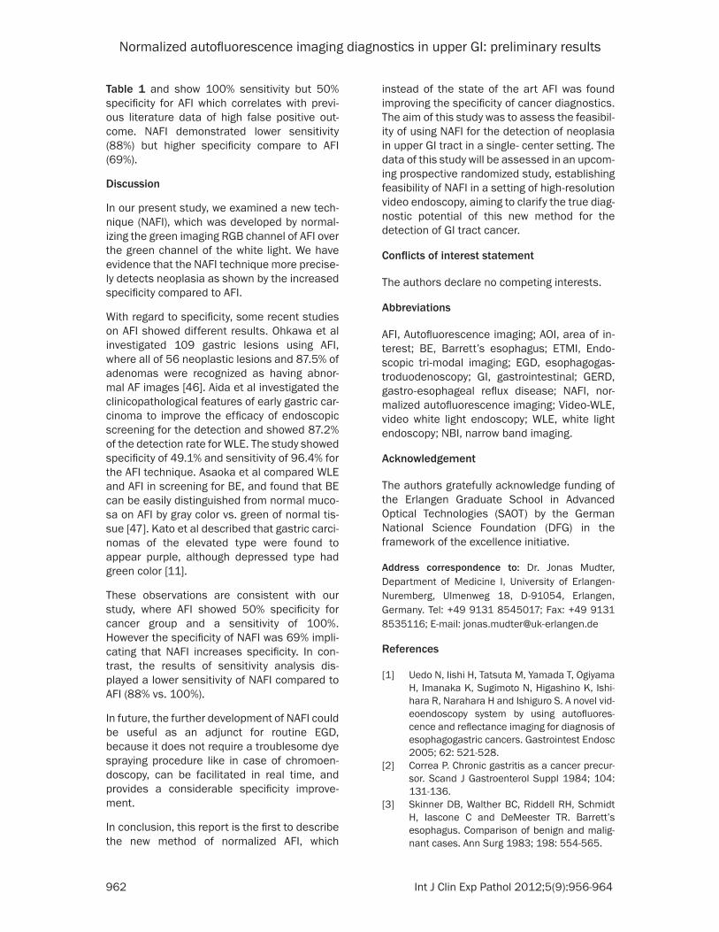
Table 1. Sensitivity and specificity data of both AFI and NAFI methods
TN TP FN FP Sensitivity SpecificityAFI 11 7 0 11 100 % 50 %NAFI 11 7 1 5 88 % 69 %AFI shows 100% sensitivity but 50% specificity which correlates with previous literature data of high false posi-tive outcome of AFI. NAFI demonstrated lower sensitivity (88%) but higher specificity compare to AFI (69%). AFI, Autofluorescence imaging; NAFI, normalized autofluores-cence imaging.
Normalized autofluorescence imaging diagnostics in upper GI: preliminary results
962 Int J Clin Exp Pathol 2012;5(9):956-964
Table 1 and show 100% sensitivity but 50% specificity for AFI which correlates with previ-ous literature data of high false positive out-come. NAFI demonstrated lower sensitivity (88%) but higher specificity compare to AFI (69%).
Discussion
In our present study, we examined a new tech-nique (NAFI), which was developed by normal-izing the green imaging RGB channel of AFI over the green channel of the white light. We have evidence that the NAFI technique more precise-ly detects neoplasia as shown by the increased specificity compared to AFI.
With regard to specificity, some recent studies on AFI showed different results. Ohkawa et al investigated 109 gastric lesions using AFI, where all of 56 neoplastic lesions and 87.5% of adenomas were recognized as having abnor-mal AF images [46]. Aida et al investigated the clinicopathological features of early gastric car-cinoma to improve the efficacy of endoscopic screening for the detection and showed 87.2% of the detection rate for WLE. The study showed specificity of 49.1% and sensitivity of 96.4% for the AFI technique. Asaoka et al compared WLE and AFI in screening for BE, and found that BE can be easily distinguished from normal muco-sa on AFI by gray color vs. green of normal tis-sue [47]. Kato et al described that gastric carci-nomas of the elevated type were found to appear purple, although depressed type had green color [11].
These observations are consistent with our study, where AFI showed 50% specificity for cancer group and a sensitivity of 100%. However the specificity of NAFI was 69% impli-cating that NAFI increases specificity. In con-trast, the results of sensitivity analysis dis-played a lower sensitivity of NAFI compared to AFI (88% vs. 100%).
In future, the further development of NAFI could be useful as an adjunct for routine EGD, because it does not require a troublesome dye spraying procedure like in case of chromoen-doscopy, can be facilitated in real time, and provides a considerable specificity improve- ment.
In conclusion, this report is the first to describe the new method of normalized AFI, which
instead of the state of the art AFI was found improving the specificity of cancer diagnostics. The aim of this study was to assess the feasibil-ity of using NAFI for the detection of neoplasia in upper GI tract in a single- center setting. The data of this study will be assessed in an upcom-ing prospective randomized study, establishing feasibility of NAFI in a setting of high-resolution video endoscopy, aiming to clarify the true diag-nostic potential of this new method for the detection of GI tract cancer.
Conflicts of interest statement
The authors declare no competing interests.
Abbreviations
AFI, Autofluorescence imaging; AOI, area of in- terest; BE, Barrett’s esophagus; ETMI, Endo-scopic tri-modal imaging; EGD, esophagogas-troduodenoscopy; GI, gastrointestinal; GERD, gastro-esophageal reflux disease; NAFI, nor-malized autofluorescence imaging; Video-WLE, video white light endoscopy; WLE, white light endoscopy; NBI, narrow band imaging.
Acknowledgement
The authors gratefully acknowledge funding of the Erlangen Graduate School in Advanced Optical Technologies (SAOT) by the German National Science Foundation (DFG) in the framework of the excellence initiative.
Address correspondence to: Dr. Jonas Mudter, Department of Medicine I, University of Erlangen-Nuremberg, Ulmenweg 18, D-91054, Erlangen, Germany. Tel: +49 9131 8545017; Fax: +49 9131 8535116; E-mail: [email protected]
References
[1] Uedo N, Iishi H, Tatsuta M, Yamada T, Ogiyama H, Imanaka K, Sugimoto N, Higashino K, Ishi-hara R, Narahara H and Ishiguro S. A novel vid-eoendoscopy system by using autofluores-cence and reflectance imaging for diagnosis of esophagogastric cancers. Gastrointest Endosc 2005; 62: 521-528.
[2] Correa P. Chronic gastritis as a cancer precur-sor. Scand J Gastroenterol Suppl 1984; 104: 131-136.
[3] Skinner DB, Walther BC, Riddell RH, Schmidt H, Iascone C and DeMeester TR. Barrett’s esophagus. Comparison of benign and malig-nant cases. Ann Surg 1983; 198: 554-565.

Normalized autofluorescence imaging diagnostics in upper GI: preliminary results
963 Int J Clin Exp Pathol 2012;5(9):956-964
[4] Hameeteman W, Tytgat GN, Houthoff HJ and van den Tweel JG. Barrett’s esophagus: devel-opment of dysplasia and adenocarcinoma. Gastroenterology 1989; 96: 1249-1256.
[5] Bytzer P, Christensen PB, Damkier P, Vinding K and Seersholm N. Adenocarcinoma of the esophagus and Barrett’s esophagus: a popula-tion-based study. Am J Gastroenterol 1999; 94: 86-91.
[6] Ang TL, Khor CJ and Gotoda T. Diagnosis and endoscopic resection of early gastric cancer. Singapore Med J 2010; 51: 93-100.
[7] Tougeron D and Michel P. [Gastric tumors]. Rev Prat 2010; 60: 129-137.
[8] Dacosta RS, Wilson BC and Marcon NE. Spec-troscopy and fluorescence in esophageal dis-eases. Best Pract Res Clin Gastroenterol 2006; 20: 41-57.
[9] Eloubeidi MA and Provenzale D. Does this pa-tient have Barrett’s esophagus? The utility of predicting Barrett’s esophagus at the index en-doscopy. Am J Gastroenterol 1999; 94: 937-943.
[10] Falk GW, Rice TW, Goldblum JR and Richter JE. Jumbo biopsy forceps protocol still misses un-suspected cancer in Barrett’s esophagus with high-grade dysplasia. Gastrointest Endosc 1999; 49: 170-176.
[11] Kato M, Uedo N, Ishihara R, Kizu T, Chatani R, Inoue T, Masuda E, Tatsumi K, Takeuchi Y, Hi-gashino K, Iishi H, Tomita Y and Tatsuta M. Analysis of the color patterns of early gastric cancer using an autofluorescence imaging video endoscopy system. Gastric Cancer 2009; 12: 219-224.
[12] Pohl J, Pech O, May A, Manner H and Ell C. En-doscopic resection of early esophageal and gastric neoplasias. Dig Dis 2008; 26: 285-290.
[13] Kato M, Kaise M, Yonezawa J, Goda K, Toyoi-zumi H, Yoshimura N, Yoshida Y, Kawamura M and Tajiri H. Trimodal imaging endoscopy may improve diagnostic accuracy of early gastric neoplasia: a feasibility study. Gastrointest En-dosc 2009; 70: 899-906.
[14] Schlemper RJ, Riddell RH, Kato Y, Borchard F, Cooper HS, Dawsey SM, Dixon MF, Fenoglio-Preiser CM, Flejou JF, Geboes K, Hattori T, Hi-rota T, Itabashi M, Iwafuchi M, Iwashita A, Kim YI, Kirchner T, Klimpfinger M, Koike M, Lauw-ers GY, Lewin KJ, Oberhuber G, Offner F, Price AB, Rubio CA, Shimizu M, Shimoda T, Sipponen P, Solcia E, Stolte M, Watanabe H and Yamabe H. The Vienna classification of gastrointestinal epithelial neoplasia. Gut 2000; 47: 251-255.
[15] Li XD, Boppart SA, Van Dam J, Mashimo H, Mutinga M, Drexler W, Klein M, Pitris C, Krinsky ML, Brezinski ME and Fujimoto JG. Optical co-herence tomography: advanced technology for
the endoscopic imaging of Barrett’s esopha-gus. Endoscopy 2000; 32: 921-930.
[16] Poneros JM, Brand S, Bouma BE, Tearney GJ, Compton CC and Nishioka NS. Diagnosis of specialized intestinal metaplasia by optical co-herence tomography. Gastroenterology 2001; 120: 7-12.
[17] Wallace MB, Perelman LT, Backman V, Craw-ford JM, Fitzmaurice M, Seiler M, Badizadegan K, Shields SJ, Itzkan I, Dasari RR, Van Dam J and Feld MS. Endoscopic detection of dyspla-sia in patients with Barrett’s esophagus using light-scattering spectroscopy. Gastroenterolo-gy 2000; 119: 677-682.
[18] Pfau PR and Sivak MV Jr. Endoscopic diagnos-tics. Gastroenterology 2001; 120: 763-781.
[19] Kiesslich R and Neurath MF. Endoscopic con-focal imaging. Clin Gastroenterol Hepatol 2005; 3: S58-60.
[20] Connor MJ and Sharma P. Chromoendoscopy and magnification endoscopy in Barrett’s esophagus. Gastrointest Endosc Clin N Am 2003; 13: 269-277.
[21] Wo JM, Ray MB, Mayfield-Stokes S, Al-Sabbagh G, Gebrail F, Slone SP and Wilson MA. Com-parison of methylene blue-directed biopsies and conventional biopsies in the detection of intestinal metaplasia and dysplasia in Bar-rett’s esophagus: a preliminary study. Gastro-intest Endosc 2001; 54: 294-301.
[22] Sharma P, Weston AP, Topalovski M, Cherian R, Bhattacharyya A and Sampliner RE. Magnifica-tion chromoendoscopy for the detection of in-testinal metaplasia and dysplasia in Barrett’s oesophagus. Gut 2003; 52: 24-27.
[23] Lieber CA, Urayama S, Rahim N, Tu R, Sarou-feem R, Reubner B and Demos SG. Multimodal near infrared spectral imaging as an explor-atory tool for dysplastic esophageal lesion identification. Opt Express 2006; 14: 2211-2219.
[24] Haringsma J and Tytgat GN. Fluorescence and autofluorescence. Baillieres Best Pract Res Clin Gastroenterol 1999; 13: 1-10.
[25] Kara MA, Smits ME, Rosmolen WD, Bultje AC, Ten Kate FJ, Fockens P, Tytgat GN and Berg-man JJ. A randomized crossover study compar-ing light-induced fluorescence endoscopy with standard videoendoscopy for the detection of early neoplasia in Barrett‘s esophagus. Gastro-intest Endosc 2005; 61: 671-678.
[26] Kara MA, Peters FP, Fockens P, ten Kate FJ and Bergman JJ. Endoscopic video-autofluores-cence imaging followed by narrow band imag-ing for detecting early neoplasia in Barrett‘s esophagus. Gastrointest Endosc 2006; 64: 176-185.
[27] Adachi Y, Shiraishi N and Kitano S. Early gas-tric cancer: endoscopy, laparoscopy and sur-gery. Asian J Surg 2003; 26: 1-3.

Normalized autofluorescence imaging diagnostics in upper GI: preliminary results
964 Int J Clin Exp Pathol 2012;5(9):956-964
[28] Aida K, Yoshikawa H, Mochizuki C, Mori A, Muto S, Fukuda T and Otsuki M. Clinicopatho-logical features of gastric cancer detected by endoscopy as part of annual health checkup. J Gastroenterol Hepatol 2008; 23: 632-637.
[29] Kadowaki S, Tanaka K, Toyoda H, Kosaka R, Imoto I, Hamada Y, Katsurahara M, Inoue H, Aoki M, Noda T, Yamada T, Takei Y and Kata-yama N. Ease of early gastric cancer demarca-tion recognition: a comparison of four magnify-ing endoscopy methods. J Gastroenterol Hepatol 2009; 24: 1625-1630.
[30] Otani A, Amano Y, Koshino K, Takahashi Y, Mishima Y, Imaoka H, Moriyama I, Ishimura N, Ishihara S and Kinoshita Y. Is autofluorescence imaging endoscopy useful for determining the depth of invasion in gastric cancer? Digestion 2010; 81: 96-103.
[31] Otsuka Y, Niwa Y, Ohmiya N, Ando N, Ohashi A, Hirooka Y and Goto H. Usefulness of magnify-ing endoscopy in the diagnosis of early gastric cancer. Endoscopy 2004; 36: 165-169.
[32] van den Broek FJ, Fockens P, van Eeden S, Re-itsma JB, Hardwick JC, Stokkers PC and Dekker E. Endoscopic tri-modal imaging for surveil-lance in ulcerative colitis: randomised com-parison of high-resolution endoscopy and au-tofluorescence imaging for neoplasia detection; and evaluation of narrow-band im-aging for classification of lesions. Gut 2008; 57: 1083-1089.
[33] Curvers WL, Singh R, Song LM, Wolfsen HC, Ragunath K, Wang K, Wallace MB, Fockens P and Bergman JJ. Endoscopic tri-modal imaging for detection of early neoplasia in Barrett‘s oe-sophagus: a multi-centre feasibility study using high-resolution endoscopy, autofluorescence imaging and narrow band imaging incorporat-ed in one endoscopy system. Gut 2008; 57: 167-172.
[34] Falk GW. Autofluorescence endoscopy. Gastro-intest Endosc Clin N Am 2009; 19: 209-220.
[35] Haringsma J, Tytgat GN, Yano H, Iishi H, Tatsu-ta M, Ogihara T, Watanabe H, Sato N, Marcon N, Wilson BC and Cline RW. Autofluorescence endoscopy: feasibility of detection of GI neo-plasms unapparent to white light endoscopy with an evolving technology. Gastrointest En-dosc 2001; 53: 642-650.
[36] Inoue T, Uedo N, Ishihara R, Kawaguchi T, Kawada N, Chatani R, Kizu T, Tamai C, Takeu-chi Y, Higashino K, Iishi H, Tatsuta M, Tomita Y and Toth E. Autofluorescence imaging videoen-doscopy in the diagnosis of chronic atrophic fundal gastritis. J Gastroenterol 2010; 45: 45-51.
[37] Egger K, Werner M, Meining A, Ott R, Allescher HD, Hofler H, Classen M and Rosch T. Biopsy surveillance is still necessary in patients with
Barrett‘s oesophagus despite new endoscopic imaging techniques. Gut 2003; 52: 18-23.
[38] Kobayashi M, Tajiri H, Seike E, Shitaya M, Tou-nou S, Mine M and Oba K. Detection of early gastric cancer by a real-time autofluorescence imaging system. Cancer Lett 2001; 165: 155-159.
[39] Bard MP, Amelink A, Skurichina M, den Bakker M, Burgers SA, van Meerbeeck JP, Duin RP, Aerts JG, Hoogsteden HC and Sterenborg HJ. Improving the specificity of fluorescence bron-choscopy for the analysis of neoplastic lesions of the bronchial tree by combination with opti-cal spectroscopy: preliminary communication. Lung Cancer 2005; 47: 41-47.
[40] Ntziachristos V, Turner G, Dunham J, Windsor S, Soubret A, Ripoll J and Shih HA. Planar fluo-rescence imaging using normalized data. J Biomed Opt 2005; 10: 064007.
[41] Neumann H, Monkemuller K, Kandulski A and Malfertheiner P. Dyspepsia and IBS symptoms in patients with NERD, ERD and Barrett‘s esophagus. Dig Dis 2008; 26: 243-247.
[42] Vakil N, van Zanten SV, Kahrilas P, Dent J and Jones R. The Montreal definition and classifi-cation of gastroesophageal reflux disease: a global evidence-based consensus. Am J Gas-troenterol 2006; 101: 1900-1920; quiz 1943.
[43] Dent J. Endoscopic grading of reflux oesopha-gitis: the past, present and future. Best Pract Res Clin Gastroenterol 2008; 22: 585-599.
[44] Douplik A, Chen D, Akens MK, Zanati S, Ciroc-co M, Bassett N, Marcon NE, Fengler J and Wil-son BC. Assessment of photobleaching during endoscopic autofluorescence imaging of the lower GI tract. Lasers Surg Med 2010; 42: 224-231.
[45] Douplik A, Leong WL, Easson AM, Done S, Netchev G and Wilson BC. Feasibility study of autofluorescence mammary ductoscopy. J Biomed Opt 2009; 14: 044036.
[46] Ohkawa A, Miwa H, Namihisa A, Kobayashi O, Nakaniwa N, Ohkusa T, Ogihara T and Sato N. Diagnostic performance of light-induced fluo-rescence endoscopy for gastric neoplasms. Endoscopy 2004; 36: 515-521.
[47] Asaoka D, Nagahara A, Oguro M, Kurosawa A, Osada T, Kawabe M, Hojo M, Yoshizawa T, Ota-ka M, Ogihara T and Watanabe S. Utility of au-tofluorescence imaging videoendoscopy in screening for Barrett‘s esophagus. Endoscopy 2009; 41 Suppl 2: E113.