Original Article MicroRNA-21 and microRNA-146a … · could negatively regulate the inflammatory...
9
Int J Clin Exp Pathol 2018;11(7):3348-3356 www.ijcep.com /ISSN:1936-2625/IJCEP0076419 Original Article MicroRNA-21 and microRNA-146a negatively regulate the secondary inflammatory response of microglia after intracerebral hemorrhage Ming Wang, Rajneesh Mungur, Ping Lan, Ping Wang, Shu Wan Department of Neurosurgery, The First Affiliated Hospital, College of Medicine, Zhejiang University, Hangzhou 310003, Zhejiang Province, P.R. China Received March 20, 2018; Accepted April 27, 2018; Epub July 1, 2018; Published July 15, 2018 Abstract: Background: A secondary inflammatory response is the most important mechanism of injury after intra- cerebral hemorrhage (ICH). Previous studies found microRNAs (miRs) expressed abnormally in the perihematomal tissue and blood of patients with ICH and demonstrated that miRs were related to pathophysiological changes and prognosis after ICH, and the development of inflammation. Methods: We induced a microglial inflammatory response by lipopolysaccharide (LPS) to construct a microglial inflammatory model. MiR-21/miR-146a overexpres- sion adenovirus was used to infect microglia to increase miR-21/miR-146a expression. MiR-21, miR-146a, IRAK1, MMP-9, TNF-α, TIMP3 and other inflammatory factors were analyzed. Then, miR-21/miR-146a overexpression ad- enovirus was injected into rats with ICH to modulate the expression. Inflammation, brain edema, and neurological scores were assessed. Results: For in vitro and vivo experiments, overexpression of miR-21/miR-146a decreased the expression of IL-1β, IL-6, IL-8, IRAK1, MMP-9 and TNF-α, meanwhile increased the expression of TIMP3 sig- nificantly (P<0.001), compared with the negative control group. Additionally, miR-21 and miR-146a reduced brain edema and improved the neurological function in ICH rats. Conclusion: Our study proved that miR-21 and miR-146a could negatively regulate the inflammatory response of microglia after ICH and provided a new theoretical basis for the treatment of secondary inflammatory injury after ICH in humans. Keywords: Intracerebral hemorrhage, inflammation, microglia, microRNA-21, microRNA-146a Introduction As a subtype of stroke, intracerebral hemor- rhage (ICH) harm to human health is serious, characterized by high incidence, mortality and disability. The brain damage caused by ICH, includes primary hematoma compression and secondary injury which result from hematoma decomposition products. Studies have demon- strated that secondary injury after ICH is the critical factor in the prognosis of patients with ICH. The mechanism of secondary injury after ICH is complicated, including acute inflamma- tory reaction, local free oxygen release, edema effect, apoptosis, and autophagy. Secondary inflammation is considered to be the most important mechanism of secondary injury after ICH [1, 2], but the detail of its mechanism of action has not been clearly known. Microglia play an important role in the initial inflammatory response after spontaneous ICH. As an important inflammatory effector cell in the nerve system, microglia are activated first, after ICH, and release a series of inflammatory mediators and biological activity factors: such as TNF-α, IL-1β, IL-6, IL-8, and so on, through the process of cell morphological and physio- logical changes, which lead to occurrence of an inflammatory response after ICH and result in further secondary injury [2, 3]. MicroRNAs (miRNAs, miRs) are a group of small non-coding RNAs with a length of 18-25 nucleo- tides, that can accumulate in cells and down- regulate the expression of target genes, by bin- ding the special sequence on the 3’ untrans- lated region (3’UTR) of mRNA, and have the effect of regulating gene transcription [4]. miRs play a very important role in maintaining the biological activity of cells. Studies showed that miRs are expressed abnormally in perihemato- mal tissue and blood of patients with ICH and relate to pathophysiological changes and prog-
Transcript of Original Article MicroRNA-21 and microRNA-146a … · could negatively regulate the inflammatory...

Int J Clin Exp Pathol 2018;11(7):3348-3356www.ijcep.com /ISSN:1936-2625/IJCEP0076419
Original ArticleMicroRNA-21 and microRNA-146a negatively regulate the secondary inflammatory response of microglia after intracerebral hemorrhage
Ming Wang, Rajneesh Mungur, Ping Lan, Ping Wang, Shu Wan
Department of Neurosurgery, The First Affiliated Hospital, College of Medicine, Zhejiang University, Hangzhou 310003, Zhejiang Province, P.R. China
Received March 20, 2018; Accepted April 27, 2018; Epub July 1, 2018; Published July 15, 2018
Abstract: Background: A secondary inflammatory response is the most important mechanism of injury after intra-cerebral hemorrhage (ICH). Previous studies found microRNAs (miRs) expressed abnormally in the perihematomal tissue and blood of patients with ICH and demonstrated that miRs were related to pathophysiological changes and prognosis after ICH, and the development of inflammation. Methods: We induced a microglial inflammatory response by lipopolysaccharide (LPS) to construct a microglial inflammatory model. MiR-21/miR-146a overexpres-sion adenovirus was used to infect microglia to increase miR-21/miR-146a expression. MiR-21, miR-146a, IRAK1, MMP-9, TNF-α, TIMP3 and other inflammatory factors were analyzed. Then, miR-21/miR-146a overexpression ad-enovirus was injected into rats with ICH to modulate the expression. Inflammation, brain edema, and neurological scores were assessed. Results: For in vitro and vivo experiments, overexpression of miR-21/miR-146a decreased the expression of IL-1β, IL-6, IL-8, IRAK1, MMP-9 and TNF-α, meanwhile increased the expression of TIMP3 sig-nificantly (P<0.001), compared with the negative control group. Additionally, miR-21 and miR-146a reduced brain edema and improved the neurological function in ICH rats. Conclusion: Our study proved that miR-21 and miR-146a could negatively regulate the inflammatory response of microglia after ICH and provided a new theoretical basis for the treatment of secondary inflammatory injury after ICH in humans.
Keywords: Intracerebral hemorrhage, inflammation, microglia, microRNA-21, microRNA-146a
Introduction
As a subtype of stroke, intracerebral hemor-rhage (ICH) harm to human health is serious, characterized by high incidence, mortality and disability. The brain damage caused by ICH, includes primary hematoma compression and secondary injury which result from hematoma decomposition products. Studies have demon-strated that secondary injury after ICH is the critical factor in the prognosis of patients with ICH. The mechanism of secondary injury after ICH is complicated, including acute inflamma-tory reaction, local free oxygen release, edema effect, apoptosis, and autophagy. Secondary inflammation is considered to be the most important mechanism of secondary injury after ICH [1, 2], but the detail of its mechanism of action has not been clearly known.
Microglia play an important role in the initial inflammatory response after spontaneous ICH.
As an important inflammatory effector cell in the nerve system, microglia are activated first, after ICH, and release a series of inflammatory mediators and biological activity factors: such as TNF-α, IL-1β, IL-6, IL-8, and so on, through the process of cell morphological and physio-logical changes, which lead to occurrence of an inflammatory response after ICH and result in further secondary injury [2, 3].
MicroRNAs (miRNAs, miRs) are a group of small non-coding RNAs with a length of 18-25 nucleo-tides, that can accumulate in cells and down-regulate the expression of target genes, by bin- ding the special sequence on the 3’ untrans-lated region (3’UTR) of mRNA, and have the effect of regulating gene transcription [4]. miRs play a very important role in maintaining the biological activity of cells. Studies showed that miRs are expressed abnormally in perihemato-mal tissue and blood of patients with ICH and relate to pathophysiological changes and prog-
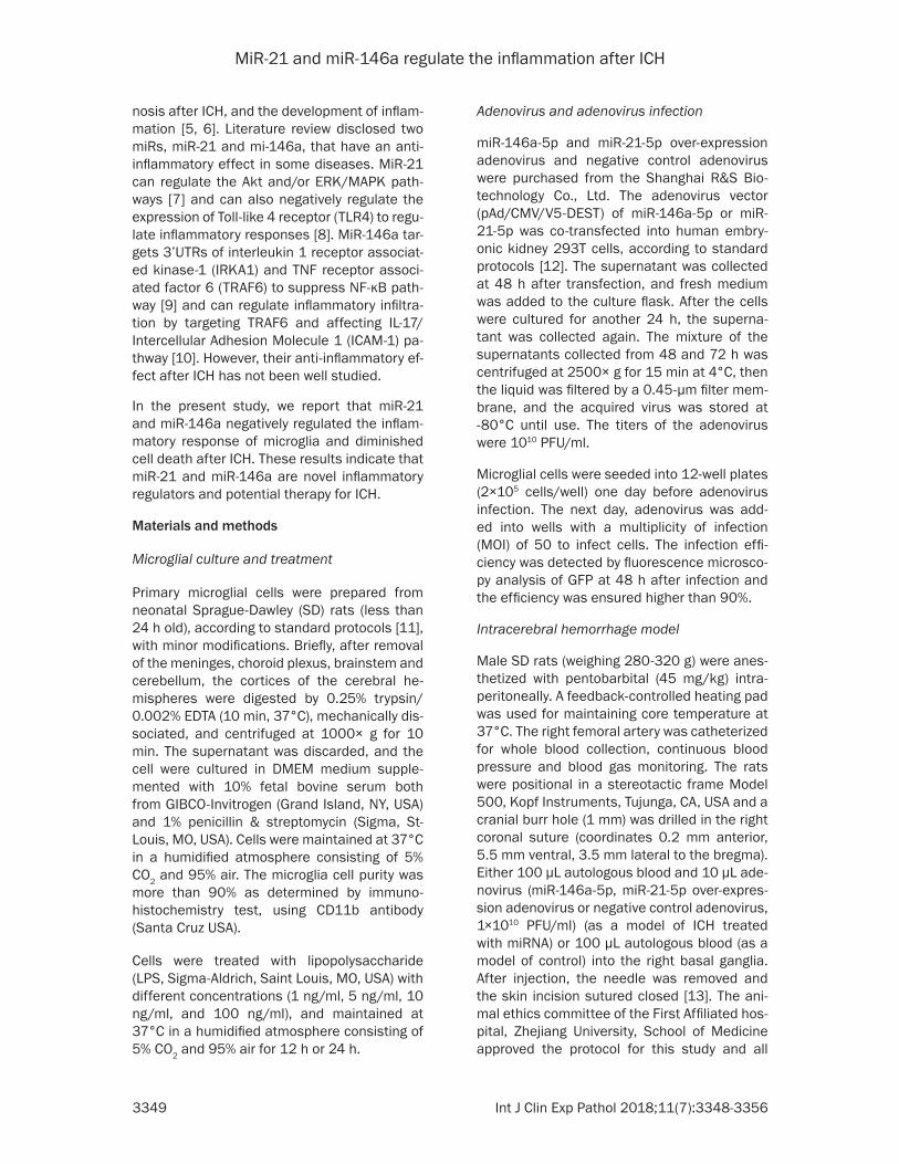
MiR-21 and miR-146a regulate the inflammation after ICH
3349 Int J Clin Exp Pathol 2018;11(7):3348-3356
nosis after ICH, and the development of inflam-mation [5, 6]. Literature review disclosed two miRs, miR-21 and mi-146a, that have an anti-inflammatory effect in some diseases. MiR-21 can regulate the Akt and/or ERK/MAPK path-ways [7] and can also negatively regulate the expression of Toll-like 4 receptor (TLR4) to regu-late inflammatory responses [8]. MiR-146a tar-gets 3’UTRs of interleukin 1 receptor associat-ed kinase-1 (IRKA1) and TNF receptor associ-ated factor 6 (TRAF6) to suppress NF-κB path-way [9] and can regulate inflammatory infiltra-tion by targeting TRAF6 and affecting IL-17/Intercellular Adhesion Molecule 1 (ICAM-1) pa- thway [10]. However, their anti-inflammatory ef- fect after ICH has not been well studied.
In the present study, we report that miR-21 and miR-146a negatively regulated the inflam-matory response of microglia and diminished cell death after ICH. These results indicate that miR-21 and miR-146a are novel inflammatory regulators and potential therapy for ICH.
Materials and methods
Microglial culture and treatment
Primary microglial cells were prepared from neonatal Sprague-Dawley (SD) rats (less than 24 h old), according to standard protocols [11], with minor modifications. Briefly, after removal of the meninges, choroid plexus, brainstem and cerebellum, the cortices of the cerebral he- mispheres were digested by 0.25% trypsin/ 0.002% EDTA (10 min, 37°C), mechanically dis-sociated, and centrifuged at 1000× g for 10 min. The supernatant was discarded, and the cell were cultured in DMEM medium supple-mented with 10% fetal bovine serum both from GIBCO-Invitrogen (Grand Island, NY, USA) and 1% penicillin & streptomycin (Sigma, St- Louis, MO, USA). Cells were maintained at 37°C in a humidified atmosphere consisting of 5% CO2 and 95% air. The microglia cell purity was more than 90% as determined by immuno- histochemistry test, using CD11b antibody (Santa Cruz USA).
Cells were treated with lipopolysaccharide (LPS, Sigma-Aldrich, Saint Louis, MO, USA) with different concentrations (1 ng/ml, 5 ng/ml, 10 ng/ml, and 100 ng/ml), and maintained at 37°C in a humidified atmosphere consisting of 5% CO2 and 95% air for 12 h or 24 h.
Adenovirus and adenovirus infection
miR-146a-5p and miR-21-5p over-expression adenovirus and negative control adenovirus were purchased from the Shanghai R&S Bio- technology Co., Ltd. The adenovirus vector (pAd/CMV/V5-DEST) of miR-146a-5p or miR-21-5p was co-transfected into human embry-onic kidney 293T cells, according to standard protocols [12]. The supernatant was collected at 48 h after transfection, and fresh medium was added to the culture flask. After the cells were cultured for another 24 h, the superna- tant was collected again. The mixture of the supernatants collected from 48 and 72 h was centrifuged at 2500× g for 15 min at 4°C, then the liquid was filtered by a 0.45-µm filter mem-brane, and the acquired virus was stored at -80°C until use. The titers of the adenovirus were 1010 PFU/ml.
Microglial cells were seeded into 12-well plates (2×10⁵ cells/well) one day before adenovirus infection. The next day, adenovirus was add- ed into wells with a multiplicity of infection (MOI) of 50 to infect cells. The infection effi-ciency was detected by fluorescence microsco-py analysis of GFP at 48 h after infection and the efficiency was ensured higher than 90%.
Intracerebral hemorrhage model
Male SD rats (weighing 280-320 g) were anes-thetized with pentobarbital (45 mg/kg) intra-peritoneally. A feedback-controlled heating pad was used for maintaining core temperature at 37°C. The right femoral artery was catheterized for whole blood collection, continuous blood pressure and blood gas monitoring. The rats were positional in a stereotactic frame Model 500, Kopf Instruments, Tujunga, CA, USA and a cranial burr hole (1 mm) was drilled in the right coronal suture (coordinates 0.2 mm anterior, 5.5 mm ventral, 3.5 mm lateral to the bregma). Either 100 µL autologous blood and 10 µL ade-novirus (miR-146a-5p, miR-21-5p over-expres-sion adenovirus or negative control adenovirus, 1×10¹⁰ PFU/ml) (as a model of ICH treated with miRNA) or 100 µL autologous blood (as a model of control) into the right basal ganglia. After injection, the needle was removed and the skin incision sutured closed [13]. The ani-mal ethics committee of the First Affiliated hos-pital, Zhejiang University, School of Medicine approved the protocol for this study and all

MiR-21 and miR-146a regulate the inflammation after ICH
3350 Int J Clin Exp Pathol 2018;11(7):3348-3356
animal experiments were performed in accor-dance with National Research Council guide for the care and use of laboratory animal.
Rats were divided into five groups. Rats re- ceived only intracerebral needle insertion in Sham group orhad intracerebral infusion of 100 µL autologous blood in Control group, or infusion of 100 µL autologous blood and 10 µL adenovirus (miR-21-OE group received miR-21-5p over-expression adenovirus, miR-146a-OE group received miR-146a over-expression ade-novirus, NC group received negative control adenovirus). Rats were killed after 12 h, 24 h or 72 h after surgery.
RNA extraction and quantitative real-time qPCR (qRT-PCR)
Total RNA was extracted from cell lines or brain tissues using Trizol reagent (Invitrogen, USA). The qRT-PCR reactions were performed using All-in-One™ miRNA qRT-PCR Detection Kit (Ge- neCopoeia Inc, USA), and iQ-5 (Bio-Rad) was used to monitor the PCR in real-time. The qPCR cycling profile was denatured at 95°C for 10 min, followed by 40 cycles of annealing at 95°C for 15 seconds and extension at 60°C for 20 seconds with a final extension at 72°C for 20 seconds. The endogenous U6 was chosen as the internal control for miRs and GAPDH for IL-1β, IL-8, and TNF-α. The average cycle thresh-old (CT), from triplicate assays, was used for fur-ther calculations. Relative expression levels
Kits purchased from Cloud-Clone Corp (TX, USA) according to manufacturer’s instruction. IL-8, IRAK1 and TIMP3 activity were measured with assay Kits purchased from R&D Systems (MN, USA) in accordance with the manufactur-er’s instruction.
Histology
Rats were immediately sacrificed by cervical dislocation and their brain tissues were co- llected, weighed, and frozen at -20°C until analysis. Edema and hemorrhage were used as indicators of acute inflammation.
For histological observation, paraffin-embed- ded basal ganglia sections (18 μm thick) were prepared. Slides were deparaffinized and the sections were stained with hematoxylin-eosin (H&E) for histological examination (Nikon EC- LIPSE TS2000-0, Japan).
Brain water content measurement
Brain water content was measured after ICH as described previously [14]. In brief, rats were anesthetized by intraperitoneal injection with pentobarbital (60 mg/kg). Then, the cerebral tissues were removed, and the surface water on the cerebral tissues was blotted with filter paper. Brain samples were divided into five parts: ipsilateral and contralateral basal gan-glia, ipsilateral and contralateral cortex, and
Table 1. RT and qPCR primers used in this studyName Sequence (5’→3’)U6 RT: CGCTTCACGAATTTGCGTGTC
Forward: TCGCTTCGGCAGCACATATACReverse: GCGTGTCATCCTTGCGCAG
miR-21 RT: GTCGTATCCAGTGCAGGGTCCGAGGTATTCGCACTGGATACGACTGTCAGForward: GTTGACTGTTGAATCTCATGGCAACAReverse: ATCCAGTGCAGGGTCCGAGG
miR-146a RT: GTCGTATCCAGTGCAGGGTCCGAGGTATTCGCACTGGATACGACACACGATGForward: ACTGAATTCCATGGGTTGTGTCAGTReverse: ATCCAGTGCAGGGTCCGAGG
GAPDH Forward: CCCCAATGTATCCGTTGTGReverse: CTCAGTGTAGCCCAGGATGC
TNF-α Forward: CTCTTCTGTCTACTGAACTTCGGGReverse: ACGTGGGCTACGGGCTTGT
IL-1β Forward: TGCCACCTTTTGACAGTGATGReverse: TGTGCTGCTGCGAGATTTG
IL-8 Forward: TGGGTGAAGGCTACTGTTGGReverse: TGGAAAGGGAAATATTCTCTGT
were normalized to control. The 2-ΔΔCt me- thod was used to quantify the relative amount of miR-146a, miR-21, IL-1β, IL-6, and TNF-α. The sequ- ences of the primers are listed in Table 1.
ELISA assay
To test the inflamma-tory response after LPS inducement, IL- 1β, IL-6, IL-8, MMP- 9, IRAK1, TIMP3 and TNF-α of cells super-natant activity were measured by ELISA. IL-1β, IL-6, MMP-9 and TNF-α were as- sayed using assay

MiR-21 and miR-146a regulate the inflammation after ICH
3351 Int J Clin Exp Pathol 2018;11(7):3348-3356
Figure 1. Acute inflammation of microglia was induced by LPS. Microglia were treated with LPS of different concen-trations (0 ng/ml, 1 ng/ml, 5 ng/ml, 10 ng/ml, 100 ng/ml) for 12 h or 24 h. The relative expression of IL-1β (A), IL-8 (B), and TNF-α (C) was assessed by qPCR, with data represented as mean ± SD, n=6. *P<0.05, **P<0.01, ***P<0.001 versus control (0 ng/ml of LPS).
Figure 2. miR-21 and miR-146a attenuate the inflammatory response of microglia induced by LPS. After infection of miR-21 overexpression adenovirus (miR-21-OE), miR-146a overexpression adenovirus (miR-146a-OE), or negative control adenovirus (NG), microglia were treated by LPS (10 ng/ml) at 24 h after infection. The relative expression of miR-21-5p (A) or miR-146a-5p (B) was measured by qPCR, with data represented as mean ± SD, n=6. *P<0.05, **P<0.01, ***P<0.001 versus NC. The concentration of IL-1β (C), IL-6 (D), IL-8 (E), IRAK1 (F), MMP-9 (G), TNF-α (H) and TIMP3 (I) were assayed by ELISA, with data represented as mean ± SD, n=6. ***P<0.001 versus control, ###P<0.001 versus LPS+NC group.

MiR-21 and miR-146a regulate the inflammation after ICH
3352 Int J Clin Exp Pathol 2018;11(7):3348-3356
cerebellum. These samples were immediately weighed on an electric analytic balance to obtain the wet weight and then dried at 100°C for 24 h to obtain the dry weight. Brain water content was calculated using the following formula: brain water content (%) = (wet weight-dry weight)/wet weight ×100%.
Evaluation of neurological scores
Animals were tested before and after surgery and scored by investigators who were blind to neurological and treatment condition. Two behavioral assessments were used: corner turn and forelimb use asymmetry tests, as de- scribed before [15].
Statistical analysis
SPSS 22.0 software was used for all statisti- cal analyses. Data are presented as means ± standard deviations (SD). Significant differenc-es in the mean values of two groups were eval-uated by Student’s unpaired t-test. Data need- ing multiple comparisons were evaluated by one-way ANOVA with Bonferroni correction. P-value of 0.05 or less was considered to be statistical significant.
Results
LPS induces microglia inflammation
LPS with different concentrations was used to stimulate microglia for 12 h or 24 h and the relative expression of IL-1β, IL-8 and TNF-α were measured by qPCR to evaluate inflam- matory response. LPS induced IL-1β, IL-8, and TNF-α expression in a dose-dependent increa- se (Figure 1A-C). Compared with control group (LPS 0 ng/ml), 1-100 ng/ml of LPS significantly increased expression of IL-1β, IL-8 and TNF-α at 12 h or 24 h (P<0.001). The expression of IL-1β and IL-8 peaked at 24 h after treatment of LPS with the concentration of 10 ng/ml, while TNF-α peaked at 24 h after treatment of LPS with the concentration of 100 ng/ml. But the expres-sion of TNF-α is much higher than control group at 24 h after treatment of 10 ng/ml LPS.
MiR-21/miR-146a attenuates inflammation response of microglia induced by LPS
MiR-21/miR-146a overexpression adenovirus was used to infect microglia. The relative ex- pression of miR-21/miR-146a was measured by qPCR. The expression of IL-1β, IL-6, IL-8,
IRAK1, MMP-9, TNF-α, and TIMP3 was mea-sured by ELISA. The relative expression of miR-21 in microglia treated with LPS (10 ng/ml) and infected by miR-21 overexpression adenovirus was 1.53 fold and miR-146a expression was 2.10 fold more than cells infected by negative control adenovirus (NG) and treated with LPS (Figure 2A, 2B). The IL-1β, IL-6, IL-8, IRAK1, MMP-9 and TNF-α levels of microglia in the supernatant were significantly up-regulated (P<0.001) and TIMP3 levels down-regulated (P<0.001) after being treated with LPS which confirmed the relative expression of IL-1β, IL-8, and TNF-α measured by qPCR. Overexpression of miR-21 or miR-146a significantly decreased the levels of IL-1β, IL-6, IL-8, IRAK1, MMP-9 and TNF-α and increased the levels of TIMP3 (P<0.001) compared with that of negative con-trol cells (Figure 2C-I).
Intracerebral hematoma induces brain tissue inflammation
H&E was used for histological observation of perihematomal tissue (Basal ganglia). We used qPCR to detect the relative expression of IL- 1β, IL-6, IL-8, IRAK1, TNF-α, miR-21, and miR-146a in the perihematomal tissue at 12 h, 24 h and 72 h post-ICH. Representative sections from perihematomal tissues (Model) and nor-mal contralateral cerebral tissues (Normal) of intracerebral hemorrhage rats are shown in Figure 3A. Hemorrhage distribution tended to disperse from 12 h to 24 h, and decrease at 72 h. Results of qPCR indicated that the relative expression of IL-1β, IL-6, IRAK1, and TNF-α was significantly increased in the perihematomal tissues compared with normal contralateral cerebral tissues (P<0.001) (Figure 3B, 3C, 3E, 3F), whereas IL-8 expression began to increase significantly at 24 h (Figure 3D). The expression of IL-1β, IL-6, and IRAK1 peaked at 12 h, and began to decrease at 24 h after ICH. However, the expression of IL-8 and TNF-α peaked at 24 h and decreased at 72 h.
MiR-21 and miR-146a downregulate inflam-mation response in vivo
The relative expression of IL-1β, IL-6, IL-8, IRAK1, MMP-9, TNF-α, TIMP3, miR-21, and miR-146a in brain tissues of rat at 72 h after ICH or ICH and injection with adenovirus (miR-21-OE, miR-146a-OE or NG adenovirus) were also measured by qPCR. The results indicate that miR-21/miR-146a expression of perihema-

MiR-21 and miR-146a regulate the inflammation after ICH
3353 Int J Clin Exp Pathol 2018;11(7):3348-3356
tomal tissue was significantly increased after ICH and injection with miR-21/miR-146a over-expression adenovirus (P<0.001) (Figure 4A, 4B). The relative expression of IL-1β, IL-6, IL-8, IRAK1, MMP-9 and TNF-α in perihematomal tissues (Model) were significantly upregulat- ed (P<0.05) and TIMP3 levels downregulated (P<0.01) after ICH or ICH and injection nega- tive control adenovirus (NC) which confirmed the relative expression of IL-1β, IL-6, IL-8, IRAK1, MMP-9 and TNF-α measured by qPCR and showed in Figures 3, 4C-I. The expression of IL-1β, IL-6, IL-8, IRAK1, MMP-9, and TNF-α of hematoma tissues after ICH and injection miR-21 or miR-146a overexpression adenovirus (miR-21 or miR-146a-OE) was significantly de- creased (P<0.01) and the expression of TIMP3 was increased (P<0.01) compared with the negative control (Figure 4C-I). This result ma-
tches our observations in rat microglial cells treated with LPS and infected by adenovirus.
miR-21/miR-146a reduces brain damage and improves neurological function
Brain water content was measured in rat cere-bral tissues at 72 h after ICH. The results shown that the water content of ipsilateral basal gan-glia tissue was significantly reduced after in- jection of miR-21 OE/miR-146a OE, compared with control and NC groups (Figure 5A). The behavioral test was performed before surgery and 72 h after surgery. Neurological scores in the miR-21 or miR-146a group was also sign- ificantly higher than NC group (Figure 5B, 5C).
Discussion
Several mechanisms have been proposed for secondary injury after ICH, but there is no
Figure 3. Intracerebral hematoma induces brain tissue inflammation. (A) H&E staining for histological observation of perihematomal tissues (Model) and normal contralateral cerebral tissues (Normal) was performed at 12 h, 24 h and 72 h after ICH (Original magnification ×200). Hemorrhage distribution tended to disperse from 12 h to 24 h and decrease at 72 h. There is no obvious change in the normal tissue. Relative expression of IL-1β (B), IL-6 (C), IL-8 (D), IRAK1 (E) and TNF-α (F) in the perihematomal tissue and normal contralateral cerebral tissues at 12 h, 24 h and 72 h post-ICH was measured by qPCR. Relative expression levels were normalized to endogenous GAPDH, with data represented as mean ± SD, n=6. **P<0.01, ***P<0.001 versus Normal group. The expression of IL-1β, IL-6, and IRAK increased at all time-tested points and peaked at 12 h in the perihematomal tissue. IL-8 expression started to increase at 24 h and peaked at 24 h. The expression of TNF-α also increased at all time-tested points and peaked at 24 h.

MiR-21 and miR-146a regulate the inflammation after ICH
3354 Int J Clin Exp Pathol 2018;11(7):3348-3356
breakthrough in prevention and treatment of ICH clinically. Inflammation response is consid-ered to be the most important mechanism of brain injury after ICH. It is a hot spot to study intracerebral hemorrhage therapy for inflam-mation, but further mechanisms still need ex- ploration. miRs are a group of small non-coding RNAs newly discovered and studied much in recently years. They can regulate the expres-
sion of target genes, by binding to the target gene, and regulate the formation of proteins. The role of miRs in spontaneous ICH have got-ten much recent attention. miR-132 has been proven to ameliorate brain edema, lessen blood-brain barrier (BBB) integrity, and attenu-ate the inflammatory response after ICH in mice [16]. miR-223 has been shown to be ac- rucial regulator of microglial activation, inflam-
Figure 4. miR-21 and miR-146a downregulate inflammatory response in vivo. The relative expression of miR-21 (A) and miR-146a (B) in perihematomal tissue and normal contralateral cerebral tissues at 72 h after ICH or ICH and in-jection with adenovirus (miR-21-OE, miR-146a-OE or NG adenovirus) were also measured by qPCR. Relative expres-sion levels were normalized to U6, with data represented as mean ± SD, n=6. The relative expression of miR-21/miR-146a in perihematomal tissues significantly increased after injection with miR-21/miR-146a overexpression adenovirus (miR-21/miR-146a-OE) compared with negative control adenovirus (NC) (***P<0.001). The relative expression of IL-1β (C), IL-6 (D), IL-8 (E), MMP-9 (F), IRAK1 (G), TNF-α (H) and TIMP3 (I) were measured by qPCR. Relative expression levels were normalized to endogenous GAPDH, with data represented as mean ± SD, n=6. Overexpression of miR-21/miR-146a decreased the expression of IL-1β, IL-6, IL-8, IRAK1, TNF-α and increased the expression of TIMP3 in perihematomal tissue at 72 h after ICH. *P<0.05, **P<0.01, ***P<0.001, perihematomal tissues versus normal contralateral cerebral tissues. ##P<0.01, ###P<0.001, perihematomal tissues in miR-21/miR-146a-OE group versus NC group.

MiR-21 and miR-146a regulate the inflammation after ICH
3355 Int J Clin Exp Pathol 2018;11(7):3348-3356
mation, and neuron injury after ICH by directly targeting HLRP3 [17].
Literature review disclosed two miRs, miR-21 and mi-146a, which have an anti-inflammatory effect in some diseases. miR-21 can target the 3’-untranslated regions (3’UTRs) of PTEN and Smad7 to downregulate PI3K/Akt path- way in human umbilical vein endothelial cells [18], and miR-21 can target Smad7 to regulate the Akt and/or ERK/MAPK pathways, contribut-ing to renal fibrosis [7]. miR-21 can also nega-tively regulate the expression of Toll-like 4 receptor (TLR4) via targeting of the proinflam-matory tumor suppressor programmed cell death protein 4 (PDCD4) to regulate inflamma-tory responses [8]. MiR-146a targets 3’UTRs of IRKA1 and TNF receptor associated factor 6 (TRAF6) to suppress the NF-κB pathway [9] and can regulate inflammatory infiltration by targeting TRAF6 and affecting the IL-17/In- tercellular Adhesion Molecule 1 (ICAM-1) path-way in polymyositis/dermatomyositis [10]. How- ever, their anti-inflammatory effect after ICH has not been well studied.
In the present study, we proved that miR-21 and miR-146a can negatively regulate the sec-ondary inflammatory response of microglia after intracerebral hemorrhage, in vitro and in vivo. It has been reported that LPS could acti-vate microglia and induce the expression of pro-inflammatory mediators [19]. Therefore, we used LPS at different doses to stimulate rat microglia to conduct a model of brain inflam- mation in vitroto explore ICH inflammation-re- lated protein expression. Results demonstrat-ed that the effect of LPS on microglia cells was dose-dependent (0-10 ng/ml) and time-depen-dent (12-24 h). We increased the expression of
miR-21 and miR-146a in microglia by adenovi-rus infection, and found miR-21 and miR-146a can both attenuate the expression of IL-1β, IL-6, IL-8, IRAK1, MMP-9 and TNF-α and incre- ase the expression of TIMP3. Based on experi-mental results in vitro, we injected autologous whole-blood into a rat ICH model and injected miR-21/miR-146a overexpression adenovirus to upregulate the expression of miR-21/miR-146a. As reported, the hematoma developed produces the desired brain injury and neuro-logic deficits [20], and a high level of inflamma-tory cytokines was significantly correlated with cerebral hematoma [21]. Our study also dem-onstrated that the expression of IL-1β, IL-6, IL-8, IRAK1, and TNF-α in the perihematomal tissues significantly increased after ICH. miR-21 and miR-146a could downregulate the ex- pression of these inflammatory cytokines and upregulate TIMP3 expression. Furthermore, our results indicated that miR-21 and miR-146a reduce the edema and improves neurological function after ICH. As described above, the tar-get genes of miR-21 include PTEN, Smad7, and PDCD4, and those of miR-146a include IRKA1 and TRAF6. In further research, we will confirm the target genes of these miRs in ICH, by con-structing vectors of target gene 3’UTR con-struction and luciferase reporter assays.
Acknowledgements
This study was supported by National Natural Science Foundation of China (NSFC) (Grant No. 81371336 awarded to Shu Wan), Project of Medical and Health Technology Development Program of Zhejiang Province (Grant No. 2013- KYA082 awarded to Shu Wan and Grant No. 2018KY044 awarded to Ming Wang) and
Figure 5. miR-21/miR-146a reduces brain damage and improves neurological function. Brain water content (A) was measured in rat cerebral tissues at 72 h after ICH, with data represented as mean ± SD, n=6. *P<0.05, miR-21/miR-146a-OE group versus NC group. Forelimb use asymmetry (B) and corner turn test (C) at 72 h after ICH or sham control. Data represented as mean ± SD, n=10. ***P<0.001 versus SHAM group, #P<0.05 versus NC group.

MiR-21 and miR-146a regulate the inflammation after ICH
3356 Int J Clin Exp Pathol 2018;11(7):3348-3356
Scientific Research Project of Traditional Chi- nese Medicine Administration in Zhejiang Province (Grant No. 2010ZA066 awarded to Shu Wan).
Disclosure of conflict of interest
None.
Address correspondence to: Dr. Shu Wan, Depart- ment of Neurosurgery, The First Affiliated Hospital, College of Medicine, Zhejiang University, 79 Qing- chun Road, Hangzhou 310003, Zhejiang Province, P.R. China. Tel: 86-571-87236803; Fax: 86-571-87236805; E-mail: [email protected]
References
[1] Xi G, Keep RF, Hoff JT. Mechanisms of brain injury after intracerebral ha-emorrhage. Lancet Neurol 2006; 5: 53-63.
[2] Wang J, Dore S. Inflammation after intracere-bral hemorrhage. J Cereb Blood Flow Metab 2007; 27: 894-908.
[3] Felberg RA, Grotta JC, Shirzadi AL, Strong R, Narayana P, Hill-Felberg SJ, Aronowski J. Cell death in experimental intracerebral hemor-rhage: the “black hole” model of hemorrhagic damage. Ann Neurol 2002; 51: 517-524.
[4] Damiani D, Tiribelli M, Franzoni A, Michelutti A, Fabbro D, Cavallin M, Toffoletti E, Simeone E, Fanin R, Damante G. BAALC overexpression re-tains its negative prognostic role across all cy-togenetic risk groups in acute myeloid leuke-mia patients. Am J Hematol 2013; 88: 848-852.
[5] Guo D, Liu J, Wang W, Hao F, Sun X, Wu X, Bu P, Zhang Y, Liu Y, Liu F, Zhang Q, Jiang F. Alteration in abundance and compartmentalization of inflammation-related miRNAs in plasma after intracerebral hemorrhage. Stroke 2013; 44: 1739-1742.
[6] Wang J, Zhu Y, Jin F, Tang L, He Z, He Z. Differ-ential expression of circulation microRNAs in blood and haematoma samples from patients with in-tracerebral haemorrhage. J Int Med Res 2016; 44: 419-432.
[7] Wu G, Xi G, Hua Y, Sagher O. T2* magnetic resonance imaging sequences reflect brain tis-sue iron deposition following intracerebral hemorrhage. Transl Stroke Res 2010; 1: 31-34.
[8] Dang G, Yang Y, Wu G, Hua Y, Keep RF, Xi G. Early erythrolysis in the hemotoma after ex-perimental intracerebral hemorrhage. Transl Stroke Res 2017; 8: 174-182.
[9] Yuan B, Shen H, Lin L, Su T, Zhong L, Yang Z. MicroRNA367 negatively regulates the inflam-matory response of microglia by targeting IRAK4 in intracerebral hemorrhage. J Neuroin-flamm 2015; 12: 206.
[10] Hua Y, Schallert T, Keep RF, Wu J, Hoff JT, Xi G. Behavioral test after intracerebral hemorrhage in the rat. Stroke 2002; 33: 2478-2484.
[11] Zhang Y, Han B, He Y, Li D, Ma X, Liu Q, Hao J. Micro-RNA-132 attenuates neurobehavioral and neuropathological changes associated with intracerebral hemorrhage in mice. Neuro-chemIn 2017; 107: 182-190.
[12] Yang Z, Zhong LN, Xian RH, Yuan BQ. MicroR-NA-223 regulates inflammation and brain in-jury via feedback to NLRP3 inflammasome af-ter intracerebral hemorrhage. Mol Immunol 2015; 65: 267-276.
[13] Rippe C, Blimline M, Magerko KA, Lawson BR, LaRocca TJ, Donato AJ, Seals DR. MicroRNA changes in human arterial endothelial cells with senescence: relation to apoptosis, eNOS and inflammation. Exp Gerontol 2012; 47: 45-51.
[14] Loboda A, Sobczak M, Jozkowicz A, Dulak J. TGF-β1/Smads and miR-21 in renal fibrosis and inflammation. Mediators Inflamm 2016; 2016: 8319283.
[15] Sheedy FJ, Palsson-Mcdermott E, Hennessy EJ, Martin C, O’Leary JJ, Ruan Q, Johnson DS, Chen Y. Negative regulation of TLR4 via target-ing of the proinflammatory tumor suppressor PDCD4 by the microRNA miR-21. Nat Immunol 2010; 11: 141-147.
[16] Zhong X, Jiang YZ, Liu P, He W, Xiong Z, Chang W, Zhu J, Cui Q. Toll-like 4 receptor/NFκB in-flammatory/miR-146a pathway contributes to the ART-correlated preterm birth outcome. Oncotarget 2016; 7: 72475-72485.
[17] Yin Y, Li F, Shi J, Li S, Cai J, Jiang Y. MiR-146a regulates inflammatory infiltration by macro-phages in polymyositis/dermatomyositis by targeting TRAF6 and affecting IL-17/ICAM-1 pathway. Cell Physiol Biochem 2016; 40: 486-498.
[18] Defaux A, Zurich MG, Braissant O, Honegger P, Monnet-Tschudi F. Effects of the PPAR-beta agonist GW501516 in an in vitro model of brain inflammation and antibody-induced de-myelination. J Neuroinflammation 2009; 6: 15.
[19] Wang J. Preclinical and clinical research on in-flammation after intracerebral hemorrhage. Prog Neurobiol 2010; 92: 463-477.
[20] Frati A, Salvati M, Mainiero F, Ippoliti F, Rocchi G, Raco A, Caroli E, Cantore G, Delfini R. Inflam-mation markers and risk factors for recurrence in 35 patients with a posttraumatic chronic subdural hematoma: a prospective study. J Neurosurg 2004; 100: 24-32.
[21] Chen S, Yang Q, Chen G, Zhang JH. An update on inflammation in the acute phase of intrace-rebral hemorrhage. Tansl Stroke Res 2015; 6: 4-8.