Microglia and inflammation-mediated...
22
Microglia and inflammation-mediated neurodegeneration: Multiple triggers with a common mechanism Michelle L. Block * , Jau-Shyong Hong Neuropharmacology Section, MD F1-01, National Institute of Environmental Health Sciences, P.O. Box 12233, Research Triangle Park, NC 27709, USA Received 18 March 2005; accepted 28 June 2005 Abstract Inflammation, a common denominator among the diverse list of neurodegenerative diseases, has recently been implicated as a critical mechanism responsible for the progressive nature of neurodegeneration. Microglia are the resident innate immune cells in the central nervous system and produce a barrage of factors (IL-1, TNFa , NO, PGE 2, superoxide) that are toxic to neurons. Evidence supports that the unregulated activation of microglia in response to environmental toxins, endogenous proteins, and neuronal death results in the production of toxic factors that propagate neuronal injury. In the following review, we discuss the common thread of microglial activation across numerous neurodegenerative diseases, define current perceptions of how microglia are damaging neurons, and explain how the microglial response to neuronal damage results in a self-propelling cycle of neuron death. # 2005 Elsevier Ltd. All rights reserved. Contents 1. Introduction ............................................................................... 78 2. Glial cells are inflammatory mediators of neurodegenerative disease ........................................ 78 2.1. Astroglia ............................................................................. 78 2.2. Microglia ............................................................................ 78 3. The role of microglia in neurodegenerative disease .................................................... 80 3.1. Alzheimer’s disease ..................................................................... 80 3.2. HIV-associated dementia .................................................................. 81 3.3. Multiple sclerosis ....................................................................... 81 3.4. Frontotemporal lobe dementia .............................................................. 81 3.5. Parkinson’s disease ...................................................................... 82 4. Microglia-mediated dopaminergic neurotoxicity....................................................... 82 4.1. Chronic LPS infusion produces selective and progressive DA toxicity .................................. 82 4.2. Microglia-mediated mechanism of LPS-induced DA toxicity ......................................... 83 4.3. Early developmental exposure to LPS: critical period of microglia development ........................... 84 5. Triggers of microglia activation and neurodegeneration ................................................. 84 www.elsevier.com/locate/pneurobio Progress in Neurobiology 76 (2005) 77–98 Abbreviations: DEP, diesel exhaust particles; PM, particulate matter; PD, Parkinson’s disease; AD, Alzheimer’s disease; ROS, reactive oxygen species; NO, nitric oxide; TNFa, tumor necrosis factor-alpha; TH, tyrosine hydroxylase; Ab, beta-amyloid; LPS, lipopolysacharide; HIV, human immunodeficiency virus; MS, multiple sclerosis; AIDS, acquired immune deficiency syndrome; HAD, HIVassociated dementia; PHOX; phagocytic oxidase; PGE 2 , prostaglandin E2; IR, immuno-reactive; SN, substantia nigra; VTA, ventral tegmental area; FTD, frontotemporal dementias; 6-OHDA, 6-hydroxydopamine; DA, dopamine; MPTP, 1-methyl-4-phenyl-1,2,3,6-tetrahydropyridine; MPP+, 1-methyl-4-phenylpyridinium ion; NSAID, non-steroidal anti-inflammatory drug; CNS, central nervous system; IR, immunoreactive; NADPH, nicotinamide adenine dinucleotide phosphate; MMP-3, matrix metalloproteinase-3; ECM, extracellular matrix; NFkB, nuclear factor-kB; iNOS, inducible nitric oxide synthase; IL-1b, interleukin-1 beta * Corresponding author. Tel.: +1 919 541 5169; fax: +1 919 541 0841. E-mail address: [email protected] (M.L. Block). 0301-0082/$ – see front matter # 2005 Elsevier Ltd. All rights reserved. doi:10.1016/j.pneurobio.2005.06.004
Transcript of Microglia and inflammation-mediated...
Microglia and inflammation-mediated neurodegeneration:
Multiple triggers with a common mechanism
Michelle L. Block *, Jau-Shyong Hong
Neuropharmacology Section, MD F1-01, National Institute of Environmental Health Sciences, P.O. Box 12233,
Research Triangle Park, NC 27709, USA
Received 18 March 2005; accepted 28 June 2005
Abstract
Inflammation, a common denominator among the diverse list of neurodegenerative diseases, has recently been implicated as a critical
mechanism responsible for the progressive nature of neurodegeneration. Microglia are the resident innate immune cells in the central nervous
system and produce a barrage of factors (IL-1, TNFa, NO, PGE2, superoxide) that are toxic to neurons. Evidence supports that the unregulated
activation of microglia in response to environmental toxins, endogenous proteins, and neuronal death results in the production of toxic factors
that propagate neuronal injury. In the following review, we discuss the common thread of microglial activation across numerous
neurodegenerative diseases, define current perceptions of how microglia are damaging neurons, and explain how the microglial response
to neuronal damage results in a self-propelling cycle of neuron death.
# 2005 Elsevier Ltd. All rights reserved.
Contents
1. Introduction . . . . . . . . . . . . . . . . . . . . . . . . . . . . . . . . . . . . . . . . . . . . . . . . . . . . . . . . . . . . . . . . . . . . . . . . . . . . . . . 78
2. Glial cells are inflammatory mediators of neurodegenerative disease . . . . . . . . . . . . . . . . . . . . . . . . . . . . . . . . . . . . . . . . 78
2.1. Astroglia . . . . . . . . . . . . . . . . . . . . . . . . . . . . . . . . . . . . . . . . . . . . . . . . . . . . . . . . . . . . . . . . . . . . . . . . . . . . . 78
2.2. Microglia . . . . . . . . . . . . . . . . . . . . . . . . . . . . . . . . . . . . . . . . . . . . . . . . . . . . . . . . . . . . . . . . . . . . . . . . . . . . 78
3. The role of microglia in neurodegenerative disease . . . . . . . . . . . . . . . . . . . . . . . . . . . . . . . . . . . . . . . . . . . . . . . . . . . . 80
3.1. Alzheimer’s disease . . . . . . . . . . . . . . . . . . . . . . . . . . . . . . . . . . . . . . . . . . . . . . . . . . . . . . . . . . . . . . . . . . . . . 80
3.2. HIV-associated dementia . . . . . . . . . . . . . . . . . . . . . . . . . . . . . . . . . . . . . . . . . . . . . . . . . . . . . . . . . . . . . . . . . . 81
3.3. Multiple sclerosis . . . . . . . . . . . . . . . . . . . . . . . . . . . . . . . . . . . . . . . . . . . . . . . . . . . . . . . . . . . . . . . . . . . . . . . 81
3.4. Frontotemporal lobe dementia . . . . . . . . . . . . . . . . . . . . . . . . . . . . . . . . . . . . . . . . . . . . . . . . . . . . . . . . . . . . . . 81
3.5. Parkinson’s disease . . . . . . . . . . . . . . . . . . . . . . . . . . . . . . . . . . . . . . . . . . . . . . . . . . . . . . . . . . . . . . . . . . . . . . 82
4. Microglia-mediated dopaminergic neurotoxicity. . . . . . . . . . . . . . . . . . . . . . . . . . . . . . . . . . . . . . . . . . . . . . . . . . . . . . . 82
4.1. Chronic LPS infusion produces selective and progressive DA toxicity . . . . . . . . . . . . . . . . . . . . . . . . . . . . . . . . . . 82
4.2. Microglia-mediated mechanism of LPS-induced DA toxicity . . . . . . . . . . . . . . . . . . . . . . . . . . . . . . . . . . . . . . . . . 83
4.3. Early developmental exposure to LPS: critical period of microglia development . . . . . . . . . . . . . . . . . . . . . . . . . . . 84
5. Triggers of microglia activation and neurodegeneration . . . . . . . . . . . . . . . . . . . . . . . . . . . . . . . . . . . . . . . . . . . . . . . . . 84
www.elsevier.com/locate/pneurobio
Progress in Neurobiology 76 (2005) 77–98
Abbreviations: DEP, diesel exhaust particles; PM, particulate matter; PD, Parkinson’s disease; AD, Alzheimer’s disease; ROS, reactive oxygen species;
NO, nitric oxide; TNFa, tumor necrosis factor-alpha; TH, tyrosine hydroxylase; Ab, beta-amyloid; LPS, lipopolysacharide; HIV, human immunodeficiency
virus; MS, multiple sclerosis; AIDS, acquired immune deficiency syndrome; HAD, HIVassociated dementia; PHOX; phagocytic oxidase; PGE2, prostaglandin
E2; IR, immuno-reactive; SN, substantia nigra; VTA, ventral tegmental area; FTD, frontotemporal dementias; 6-OHDA, 6-hydroxydopamine; DA, dopamine;
MPTP, 1-methyl-4-phenyl-1,2,3,6-tetrahydropyridine; MPP+, 1-methyl-4-phenylpyridinium ion; NSAID, non-steroidal anti-inflammatory drug; CNS, central
nervous system; IR, immunoreactive; NADPH, nicotinamide adenine dinucleotide phosphate; MMP-3, matrix metalloproteinase-3; ECM, extracellular matrix;
NFkB, nuclear factor-kB; iNOS, inducible nitric oxide synthase; IL-1b, interleukin-1 beta
* Corresponding author. Tel.: +1 919 541 5169; fax: +1 919 541 0841.
E-mail address: [email protected] (M.L. Block).
0301-0082/$ – see front matter # 2005 Elsevier Ltd. All rights reserved.
doi:10.1016/j.pneurobio.2005.06.004
M.L. Block, J.-S. Hong / Progress in Neurobiology 76 (2005) 77–9878
5.1. Environmental toxins . . . . . . . . . . . . . . . . . . . . . . . . . . . . . . . . . . . . . . . . . . . . . . . . . . . . . . . . . . . . . . . . . . . . 84
5.1.1. Rotenone. . . . . . . . . . . . . . . . . . . . . . . . . . . . . . . . . . . . . . . . . . . . . . . . . . . . . . . . . . . . . . . . . . . . . . . 84
5.1.2. Paraquat . . . . . . . . . . . . . . . . . . . . . . . . . . . . . . . . . . . . . . . . . . . . . . . . . . . . . . . . . . . . . . . . . . . . . . . 85
5.1.3. Particulate matter and the phagocytic activation of microglia. . . . . . . . . . . . . . . . . . . . . . . . . . . . . . . . . . . 85
5.2. Endogenous disease proteins . . . . . . . . . . . . . . . . . . . . . . . . . . . . . . . . . . . . . . . . . . . . . . . . . . . . . . . . . . . . . . . 86
5.2.1. b-Amyloid. . . . . . . . . . . . . . . . . . . . . . . . . . . . . . . . . . . . . . . . . . . . . . . . . . . . . . . . . . . . . . . . . . . . . . 86
5.2.2. a-Synuclein . . . . . . . . . . . . . . . . . . . . . . . . . . . . . . . . . . . . . . . . . . . . . . . . . . . . . . . . . . . . . . . . . . . . . 86
5.3. Reactive microgliosis . . . . . . . . . . . . . . . . . . . . . . . . . . . . . . . . . . . . . . . . . . . . . . . . . . . . . . . . . . . . . . . . . . . . 87
5.3.1. Matrix metalloproteinase-3 . . . . . . . . . . . . . . . . . . . . . . . . . . . . . . . . . . . . . . . . . . . . . . . . . . . . . . . . . . 88
5.3.2. Neuromelanin . . . . . . . . . . . . . . . . . . . . . . . . . . . . . . . . . . . . . . . . . . . . . . . . . . . . . . . . . . . . . . . . . . . 88
6. Common characteristics of microglial activation. . . . . . . . . . . . . . . . . . . . . . . . . . . . . . . . . . . . . . . . . . . . . . . . . . . . . . . 89
6.1. Temporal relationship of the microglial release of neurotoxic factors . . . . . . . . . . . . . . . . . . . . . . . . . . . . . . . . . . . 89
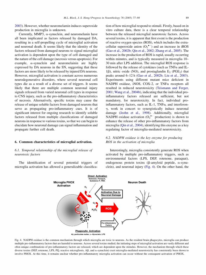
6.2. NADPH oxidase is the key enzyme for producing ROS in the activation of microglia . . . . . . . . . . . . . . . . . . . . . . . 89
6.2.1. Extracellular superoxide is the key factor mediating inflammation-related neurotoxicity . . . . . . . . . . . . . . . . 90
6.2.2. Intracellular ROS regulate the expression of pro-inflammatory factors . . . . . . . . . . . . . . . . . . . . . . . . . . . . 90
7. Microglial activation as a common mechanism in diverse neuropathology . . . . . . . . . . . . . . . . . . . . . . . . . . . . . . . . . . . . 91
8. Conclusions. . . . . . . . . . . . . . . . . . . . . . . . . . . . . . . . . . . . . . . . . . . . . . . . . . . . . . . . . . . . . . . . . . . . . . . . . . . . . . . . 92
References . . . . . . . . . . . . . . . . . . . . . . . . . . . . . . . . . . . . . . . . . . . . . . . . . . . . . . . . . . . . . . . . . . . . . . . . . . . . . . . . 92
1. Introduction
Inflammation occurs in multiple neurodegenerative
diseases, where each disease has unique pathology and
symptoms. There is an extensive list of specific triggers of
neuronal damage, where each environmental toxin or
genetic mutation is specific for a selected disease.
However, the gradual accumulation of neuronal death
and the increase in disease severity across time is a
unifying theme across the diverse classifications of
neurodegenerative disease. Previously, inflammation was
viewed as only a passive response to neuronal damage.
However, increasing reports demonstrate that inflamma-
tion is capable of actively causing neuronal death and
damage, which then fuels a self-propelling cycle of
neuronal death. Thus, while the triggers of various
neurodegenerative diseases are diverse, inflammation
may be a basic mechanism driving the progressive nature
of multiple neurodegenerative diseases. Several cell types
have been listed as contributors to inflammation-mediated
neurodegeneration, but microglia are implicated as critical
components of the immunological insult to neurons. In the
following review, we discuss the role of microglia in
neuronal death and describe the evidence implicating
microglia as a critical mechanism driving the self-
propelling nature of neurodegenerative disease.
2. Glial cells are inflammatory mediators ofneurodegenerative disease
Early reports described the brain as an immune privileged
organ, due to its compartmentalization and separation from
the peripheral blood system, as provided by the blood–brain-
barrier. However, most neurodegenerative diseases are
characterized by both local inflammation from resident cell
types in the brain and by the infiltration of leucocytes from
the periphery (Kurkowska-Jastrzebska et al., 1999; McGeer
et al., 1989). While infiltrating peripheral immune cells can
be significantly toxic to neurons (Freude et al., 2002; Wu and
Proia, 2004), leukocyte infiltration is not always associated
with neurotoxicity (Boztug et al., 2002; Trifilo and Lane,
2003), indicating a critical role for the local glial cells
(astroglia and microglia) in the inflammatory response
associated with neurodegeneration.
2.1. Astroglia
In the normal brain, astroglia play essential roles in
providing glia-neuron contact, maintaining ionic home-
ostasis, buffering excess neurotransmitters, secreting neuro-
trophic factors, and serving as a critical component of the
blood–brain barrier (Aloisi, 1999; Hansson and Ronnback,
1995; Vernadakis, 1988). Although the pro-inflammatory
function of astroglia is not as prominent as that of microglia
(Barde, 1989; Lindsay, 1994; Streit et al., 1999), astroglia
become activated in response to immunologic challenges or
brain injuries (Aloisi, 1999; Tacconi, 1998). Astroglia also
produce a host of trophic factors (Friedman et al., 1990;
Lindsay, 1994), which are crucial for the survival of neurons.
However, activated astroglia become hypertrophic, exhibit
increased production of glial fibrillary acidic protein, and
form glial scars, which hinder axonal regeneration. While
there is a clear relationship between astroglia and microglia
in both resting and activated conditions (Kahn MA et al.,
1995; Rezaie et al., 2002), efforts to understand the detailed
mechanisms of this complex association are ongoing.
2.2. Microglia
Microglia were originally described by del Rio-Hortega
(1932) as a unique cell type differing in morphology from
other glia and neurons, comprising approximately 12% of
the brain. While the precise origin of microglia in the brain is
M.L. Block, J.-S. Hong / Progress in Neurobiology 76 (2005) 77–98 79
a source of debate, it is generally accepted that microglia are
derived from myeloid origin (del Rio-Hortega, 1932) and are
responsible for the innate immune response in the brain. The
majority of microglial function goes unnoticed, as they
perform general maintenance and clean cellular debris (Beyer
et al., 2000). Additionally, microglia have active roles in late
embryonic brain development and early postnatal brain
maturation, where microglia enforce the programmed
elimination of neural cells (Barron, 1995; Milligan et al.,
1991). In mature brains, resting microglia exhibit a
characteristic ramified morphology and are responsible for
immune surveillance. Microglia become readily activated in
response to brain injuries or to immunological stimuli
(Kreutzberg, 1996; Liu and Hong, 2003; Streit et al., 1988,
1999) and undergo dramatic morphologic alterations upon
activation, changing from resting ramified microglia into
activated amoeboid microglia (Kreutzberg, 1996). Further,
surface molecules, such as complement receptors and major
histocompatibility complex molecules, are also upregulated
when microglia are activated (Graeber et al., 1988;
Oehmichen and Gencic, 1975). In addition, activated
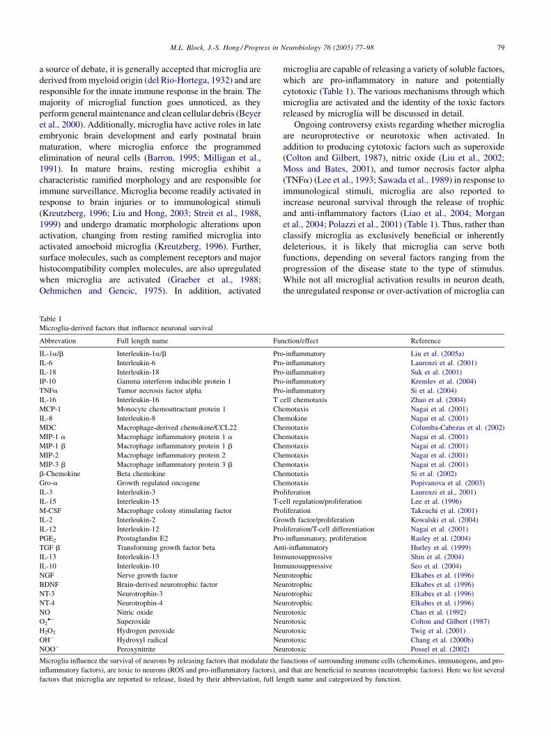
Table 1
Microglia-derived factors that influence neuronal survival
Abbrevation Full length name Fu
IL-1a/b Interleukin-1a/b Pro
IL-6 Interleukin-6 Pro
IL-18 Interleukin-18 Pro
IP-10 Gamma interferon inducible protein 1 Pro
TNFa Tumor necrosis factor alpha Pro
IL-16 Interleukin-16 T c
MCP-1 Monocyte chemoattractant protein 1 Ch
IL-8 Interleukin-8 Ch
MDC Macrophage-derived chemokine/CCL22 Ch
MIP-1 a Macrophage inflammatory protein 1 a Ch
MIP-1 b Macrophage inflammatory protein 1 b Ch
MIP-2 Macrophage inflammatory protein 2 Ch
MIP-3 b Macrophage inflammatory protein 3 b Ch
b-Chemokine Beta chemokine Ch
Gro-a Growth regulated oncogene Ch
IL-3 Interleukin-3 Pro
IL-15 Interleukin-15 T-c
M-CSF Macrophage colony stimulating factor Pro
IL-2 Interleukin-2 Gr
IL-12 Interleukin-12 Pro
PGE2 Prostaglandin E2 Pro
TGF b Transforming growth factor beta An
IL-13 Interleukin-13 Im
IL-10 Interleukin-10 Im
NGF Nerve growth factor Ne
BDNF Brain-derived neurotrophic factor Ne
NT-3 Neurotrophin-3 Ne
NT-4 Neurotrophin-4 Ne
NO Nitric oxide Ne
O2�� Superoxide Ne
H2O2 Hydrogen peroxide Ne
OH� Hydroxyl radical Ne
NOO� Peroxynitrite Ne
Microglia influence the survival of neurons by releasing factors that modulate the
inflammatory factors), are toxic to neurons (ROS and pro-inflammatory factors), a
factors that microglia are reported to release, listed by their abbreviation, full le
microglia are capable of releasing a variety of soluble factors,
which are pro-inflammatory in nature and potentially
cytotoxic (Table 1). The various mechanisms through which
microglia are activated and the identity of the toxic factors
released by microglia will be discussed in detail.
Ongoing controversy exists regarding whether microglia
are neuroprotective or neurotoxic when activated. In
addition to producing cytotoxic factors such as superoxide
(Colton and Gilbert, 1987), nitric oxide (Liu et al., 2002;
Moss and Bates, 2001), and tumor necrosis factor alpha
(TNFa) (Lee et al., 1993; Sawada et al., 1989) in response to
immunological stimuli, microglia are also reported to
increase neuronal survival through the release of trophic
and anti-inflammatory factors (Liao et al., 2004; Morgan
et al., 2004; Polazzi et al., 2001) (Table 1). Thus, rather than
classify microglia as exclusively beneficial or inherently
deleterious, it is likely that microglia can serve both
functions, depending on several factors ranging from the
progression of the disease state to the type of stimulus.
While not all microglial activation results in neuron death,
the unregulated response or over-activation of microglia can
nction/effect Reference
-inflammatory Liu et al. (2005a)
-inflammatory Laurenzi et al. (2001)
-inflammatory Suk et al. (2001)
-inflammatory Kremlev et al. (2004)
-inflammatory Si et al. (2004)
ell chemotaxis Zhao et al. (2004)
emotaxis Nagai et al. (2001)
emokine Nagai et al. (2001)
emotaxis Columba-Cabezas et al. (2002)
emotaxis Nagai et al. (2001)
emotaxis Nagai et al. (2001)
emotaxis Nagai et al. (2001)
emotaxis Nagai et al. (2001)
emotaxis Si et al. (2002)
emotaxis Popivanova et al. (2003)
liferation Laurenzi et al., 2001)
ell regulation/proliferation Lee et al. (1996)
liferation Takeuchi et al. (2001)
owth factor/proliferation Kowalski et al. (2004)
liferation/T-cell differentiation Nagai et al. (2001)
-inflammatory, proliferation Rasley et al. (2004)
ti-inflammatory Hurley et al. (1999)
munosuppressive Shin et al. (2004)
munosuppressive Seo et al. (2004)
urotrophic Elkabes et al. (1996)
urotrophic Elkabes et al. (1996)
urotrophic Elkabes et al. (1996)
urotrophic Elkabes et al. (1996)
urotoxic Chao et al. (1992)
urotoxic Colton and Gilbert (1987)
urotoxic Twig et al. (2001)
urotoxic Chang et al. (2000b)
urotoxic Possel et al. (2002)
functions of surrounding immune cells (chemokines, immunogens, and pro-
nd that are beneficial to neurons (neurotrophic factors). Here we list several
ngth name and categorized by function.
M.L. Block, J.-S. Hong / Progress in Neurobiology 76 (2005) 77–9880
have disastrous neurotoxic consequences. Here we summar-
ize the evidence detailing the role of microglial activation in
several neurodegenerative diseases.
3. The role of microglia in neurodegenerative disease
3.1. Alzheimer’s disease
Alzheimer’s disease (AD) is the leading cause of
dementia, where neural damage begins in the temporal
and parietal lobes of the cerebral cortex and progresses with
time to the hippocampus and the amygdala (Braak and
Braak, 1994). The result is the loss of language skills,
followed by memory decline, and finally delusion in the
latter stages. Pathological diagnosis of AD requires
identification of insoluble extracellular plaques containing
b-amyloid (Ab) and intraneuronal neurofibrilary tangles in
the cortical region of the brain. Early work demonstrated
that 100 amino acids of the carboxy terminal of the Ab
peptide can be directly toxic to neurons in vitro and initiated
the hypothesis that Ab was a driving force behind the
neurodegeneration of AD (Yankner, 1989; Yankner et al.,
1990). Furthermore, pioneering work by McGeer et al.
(1987) proposed an additional explanation for neuronal
death in AD when they identified the increased presence of
activated microglia (those staining more intensively for
HLD-DR, a marker for monocytes) around the Ab contain-
ing plaques in postmortem AD tissue, when compared to
similar tissue from control brains without neurodegenera-
tive pathology. While the microglial reaction to Ab was
initially perceived as a passive response to neuronal death,
ongoing research has shown that the microglial response
to Ab (Combs et al., 2000; Qin et al., 2002) and the senile
plaques (Van Everbroeck et al., 2004; Veerhuis et al., 1999)
can be detrimental to neuronal survival.
AD was one of the first neurodegenerative diseases
associated with neurotoxic microglial activation. As a
consequence, the traditionally perceived passive role of
microglia as bystander maintenance cells has been ques-
tioned and microglia are now accepted as active mediators of
neurodegeneration. Ab will both recruit and activate
microglia (Davis et al., 1992; Meda et al., 1995; Sasaki
et al., 1997), where Ab has been reported to result in the
release of neurotoxic factors from microglia, such as NO (Ii
et al., 1996), TNFa (Dheen et al., 2004) and superoxide (Qin
et al., 2002). In fact, use of synthetic Ab has determined that
the amino acids 10–16 are critical for microglial activation
(Giulian et al., 1996). Several receptors have been
implicated as necessary for the interaction of microglia
and Ab, such as the CD14 receptor (Bate et al., 2004) and the
b1-integrin receptor (Koenigsknecht and Landreth, 2004).
In fact, Koenigsknecht and Landreth (2004) suggest that
microglia interact with Ab through a cell surface receptor
complex consisting of B-class scavenger receptor CD36, a6-
b1 integrin, and CD47 (integrin-associated protein).
Interestingly, Koenigsknecht and Landreth (2004) also
report that this receptor complex is responsible for the
internalization of Ab through a non-traditional pathway.
Additionally, the receptor complex reported to be respon-
sible for the internalization of Ab (CD36, a6-b1 integrin,
and CD47) has also been identified as critical for Ab-
induced superoixde production in microglia (Bamberger
et al., 2003). While many studies indicate that the phago-
cytosis of Ab is neuroprotective (Das et al., 2003), as
microglia may take up and degrade Ab (Paresce et al.,
1997), it has been recently suggested that the process of
phagocytosis can be associated with neurotoxic consequences
(Block et al., 2004; Zhang et al., 2005). This mechanism
of phagocytic microglial activation is generalizable to a
diverse set of microglia stimuli and is discussed in detail later.
Human clinical trials using Ab peptide immunization
demonstrate the delicate balance between the helpful aspects
of microglial activation and potential deleterious conse-
quences of unregulated inflammatory responses (Dodel
et al., 2003; Schenk, 2002). The human clinical trials began
as a result of earlier promising animal studies demonstrating
that immunization with the Ab peptide in mice over
expressing Ab resulted in protection against AD-like
pathology (Schenk et al., 1999), where the mechanism of
protection was believed to be through the clearance of Ab by
microglia. However, while Ab plaques were cleared in
human immunized patients, phase IIa clinical trials were
halted when 18 of 298 patients immunized with the Ab
peptide demonstrated meningoencephalitis symptoms
(Senior, 2002). It has recently been shown that immuniza-
tion of C57 mice with the Ab peptide will result in an animal
model of autoimmune encephalitis, where these immunized
animals display features similar to those reported in the
human clinical trials (Furlan et al., 2003). While the cell type
responsible for the harmful inflammatory response as a
result of Ab immunization remains a debate (T cells or
microglia), these studies support that deleterious potential of
microglial activation in the process of Ab clearance warrants
caution and deserves further inquiry.
There has been some success with clinical studies
investigating the effects of anti-inflammatory therapy
against cognitive decline in AD patients (McGeer and
McGeer, 1996; Perry et al., 2003), but the promise of anti-
inflammatory treatment for AD is more evident in
experimental studies. For example, inhibition of glial
activation in animal AD models attenuates neurotoxicity.
Murine models employing intraventricular infusion of
human Ab1–42 peptide replicate many of the hallmarks
of AD pathology (neuro-inflammation, neuronal and
synaptic degeneration, and amyloid deposition). Adminis-
tration of aminopyridazines is shown to both attenuate glial
inflammation and result in reduction of neuronal neuro-
toxicity in the murine intraventricular human Ab1–42
infusion model (Craft et al., 2004). This supports the concept
that therapeutic inhibition of Ab-induced inflammation
could be neuroprotective. In a separate study, non-steroidal
M.L. Block, J.-S. Hong / Progress in Neurobiology 76 (2005) 77–98 81
anti-inflammatory drug (NSAID) treatment in mice over-
expressing Ab was able to lower Ab deposition, inhibit
microglial activation, and provide neuroprotection (Yan
et al., 2003), also indicating that inflammation induced by
Ab contributes to neurotoxicity. In vitro studies also support
that Ab is pro-inflammatory, where inhibition of synthetic
Ab-induced microglial activation with dextromethorphan
(Liu et al., 2003) results in a reduction of Ab-induced
neurotoxicity. Together, these studies indicate that inflam-
mation and microglia are critical for the ongoing process of
neurodegeneration in AD.
3.2. HIV-associated dementia
Dementia associated with human immunodeficiency
virus (HIV) infection is a debilitating condition of cognitive,
behavioral, and motor dysfunction seen in the later stages of
the acquired immunodeficiency syndrome (AIDS). The
hallmarks of HIV-associated dementia (HAD) is neuronal
loss, reactive astrogliosis, activated microglia, multinu-
cleated giant cells, and leukocyte infiltration (Budka, 1991).
Microglia are essential to the progression of the dementia, as
HIV will enter the brain on infected circulating monocytes
and is stored in microglia (Jordan et al., 1991; Kure et al.,
1990; Michaels et al., 1988; Ryzhova et al., 2002). The
microglia then serve as a reservoir for viral replication,
resulting in an increase in expression of pro-inflammatory
factors (Cosenza et al., 2002) and progression of the disease.
While the hallmark symptomology of HIV is immunode-
ficiency, ironically, the later stage of HAD is known to be an
inflammation-mediated neurodegenerative complication of
HIV infection. Activated microglia are found in the early
stages of HIV infection (Chakrabarti et al., 1991), which
then increases in intensity with the progression of the
disease. The result of microglial HIV infection and viral
replication is an increased release of neurotoxic pro-
inflammatory cytokines and enhanced microglial activation
(Sopper et al., 1996). Microglia are activated by interaction
with viral proteins, such as Tat (D’Aversa et al., 2004) and
gp120 (Garden et al., 2004; Kong et al., 1996), the HIV
infection itself (Sopper et al., 1996), and soluble factors
released from infected cells (Lipton and Gendelman, 1995).
While the majority of the literature investigating HIV-
induced neurotoxicity focuses on the direct neuronal toxicity
that occurs and the inflammatory consequence of infiltrating
macrophages, it is becoming increasingly evident that
microglia play a substantial role in neurodegeneration
associated with HAD.
3.3. Multiple sclerosis
Multiple sclerosis (MS) is the most prevalent inflamma-
tion-mediated demyelinating disease. MS is typically
characterized as a disease of the young adult, where
approximately 10% of those diagnosed will display
symptoms that worsen over time, despite periods of
remission. Initial clinical symptoms begin with vertigo,
fatigue, optic neuritis, and weakness in the limb extremities.
The disease progresses to further debilitating symptoms
including ataxia, paraparesis, limb spasms, and cognitive
impairment. The neurodegeneration associated with MS
occurs as lesions in the white matter of the CNS.
Specifically, the myelin sheath is damaged in MS in what
is believed to be an autoimmune response. While the cause
of MS is currently unknown, there is a clear inflammatory
component. For example, lymphocytes and activated
myeloid cells are localized in the area of demyelization
(Hill et al., 2004; Rose et al., 2004; Schonrock et al., 1998).
Using nuclear magnetic resonance imaging, positron
emission tomography, and [11C](R)-PK11195 (a microglia
marker) Banati et al. (2000) analyzed the brain of MS
patients and showed increased microglia activity around the
site of the MS lesion (Banati et al., 2000). The role of
microglia in MS is also supported in the MS animal model of
experimental autoimmune encephalomyelitis, where micro-
glia are shown to proliferate and increase lysosome activity
around active sites of demyelization (Matsumoto et al.,
1992). Once at the site of lesion in MS, microglia increase
cyclooxygenase 2 (Rose et al., 2004) and inducible nitric
oxide synthase (iNOS) (Hill et al., 2004) expression, which
is required for the production of neurotoxic PGE2 and NO,
respectively. In addition to being a source of neurotoxic
factors upon activation, microglia have been implicated in
the initiation and progression of MS as one of the antigen
presenting cells that sparks the autoimmune response
targeting myelin (Mack et al., 2003). Thus, while infiltrating
T-cells and macrophages have a clear role in MS associated
demyelination, lesions, and neuronal damage, microglia are
also critically involved in this process.
3.4. Frontotemporal lobe dementia
Frontotemporal dementias (FTD) are considered to be a
classification of five unique neuropathologies that are
categorized by: (1) the presence or absence of Pick’s
bodies; (2) neuronal tau-positive inclusions; (3) ubiquitin-
positive neuronal inclusions; (4) the number of tau repeats in
a neuronal inclusion. However, despite variable pathology,
FTDs express a similar clinical symptomology and
phenotype. In general, FTDs are dementia syndromes that
result in both progressive changes in behavior and language
dysfunction typically associated with frontal and/or anterior
temporal atrophy and loss of neurons (Munoz et al., 2003).
Earlier work identified that microglia were indeed activated
in frontotemporal dementias (Cooper et al., 1996; Mann,
1998), but not all pathologies were considered. Recent work
by Schofield et al. (2003) characterized the inflammatory
profiles of these five pathologies to discern a common thread
of similarity uniting them. While different protein deposi-
tion and degrees of astrogliosis were present in the
postmortem patient brain, activated microglia were elevated
and present in the white matter of the frontal lobe to similar
M.L. Block, J.-S. Hong / Progress in Neurobiology 76 (2005) 77–9882
degrees across all classifications of neurodegenerative
disease (Schofield et al., 2003). Of further interest, at
earlier stages of the disease, the microglia activation was
also present, providing rare evidence of an early and perhaps
initiating role of microglial inflammation in frontotemporal
dementias (Schofield et al., 2003). Currently, the specific
mechanisms through which microglia are activated in FTDs
are unknown.
3.5. Parkinson’s disease
Parkinson’s disease (PD) is characterized by progressive
degeneration of the nigro-striatal dopaminergic (DA)
pathway, which regulates body movement (Olanow and
Tatton, 1999). Degeneration of the neuronal cell bodies of
DA neurons in the substantia nigra (SN) and the nerve
terminals in the striatum results in resting tremor, rigidity,
bradykinesia, and gait disturbance in the PD patient
(Jellinger, 2001). Postmortem analysis of brains from PD
patients frequently shows cytoplasmic inclusions (Lewy
bodies) in the DA neurons localized in the SN (Holdorff,
2002; Schiller, 2000), which is the pathological hallmark of
PD (Takahashi and Wakabayashi, 2001).
Epidemiological studies, pathological analysis, and
biochemical characterization indicate that approximately
95% of PD cases are sporadic with late onset (Tanner, 2003)
and only 5% of PD cases occur in familial clusters with early
onset (Mizuno et al., 2001). Familial PD is associated with
mutations in several genes, including parkin, ubiquitin C-
terminal hydrolase L1, and a-synuclein (Gwinn-Hardy,
2002). However, idiopathic PD may represent a long and
cumulative process, where the final outcome is the result of a
complex set of interactions between genetic predisposition,
the innate vulnerabilities of the nigro-striatal DA system,
and exposure to environmental toxins. Among the environ-
mental toxins, infectious agents (Duvoisin et al., 1963, 1972;
Pradhan et al., 1999), pesticides (Barbeau et al., 1985; Elbaz
et al., 2004), and heavy metals (Hudnell, 1999; Iregren,
1999; Sadek et al., 2003) have been implicated in the
development and progression of PD. Similar to the case with
AD, the role of inflammation in the pathogenesis of PD was
not extensively questioned until the work of McGeer et al.
(1988) where increased HLD-DR staining in the SN of PD
patients suggested that microglia may play a role in yet
another neurodegenerative disease.
Microglia and inflammation-mediated neurodegenera-
tion have been implicated in numerous other diseases, such
as hypoxia (Olson and McKeon, 2004), stroke (Morioka
et al., 1993), amyotrophic lateral sclerosis (Banati et al.,
1995; Hall et al., 1998), and neuropathic pain (Tsuda et al.,
2004). However, the five diverse examples of neurodegen-
erative diseases presented here detail how the similar feature
of microglial activation can result in diverse localization,
pathology, and clinical symptoms for each unique disease.
To begin to elucidate the common mechanisms of microglia-
mediated neurotoxicity, PD will be used as a representative
neurodegenerative disease as we address the progressive
nature of microglia-mediated neurotoxicity and the common
characteristics/mechanism of microglial activation in
response to several diverse toxins.
4. Microglia-mediated dopaminergic neurotoxicity
Dopaminergic neurons are inherently susceptible to the
deleterious effects of microglial activation. While the
detailed mechanism remains debated, one hypothesis is
that the selective mechanism of microglia-mediated
dopaminergic neurotoxicity is due to the generation of
oxidative insult from microglia. In particular, DA neurons
possess reduced antioxidant capacity, as evidenced by low
intracellular glutathione, which renders DA neurons more
vulnerable to oxidative stress and microglial activation
relative to other cell types (Loeffler et al., 1994). While
oxidative stress is clearly toxic to multiple cell types,
according to this hypothesis, DA neurons will succumb first
at lower levels of oxidative stress, followed by other
neuronal and cell populations. Additionally, the SN contains
4.5 times as many microglia when compared to the cortex
and other regions of the brain (Kim et al., 2000), suggesting
that the localization of microglia in the SN predisposes them
to vulnerability to immunological insult.
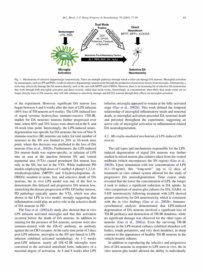
Interestingly, there are multiple factors/toxins that are
shown to selectively damage DA neurons through microglial
activation such as rotenone (Gao et al., 2003a), diesel
exhaust particles (DEP) (Block et al., 2004), paraquat (PQ)
(Wu et al., 2005), and Ab (Qin et al., 2002). However, the
majority of these toxins are dual mode toxins in that they
damage DA neurons primarily through microglia activation
at lower concentrations, but directly kill DA neurons at
higher concentrations (Fig. 1). The identification of multiple
dual mode DA toxins is critical to the understanding the
mechanisms of both selective DA neurotoxicity and the
general pathways through which microglia become acti-
vated. The initial work investigating the role of microglia in
selective DA neurodegeneration began with testing the
immunogen lipopolysacharide (LPS), where LPS was
shown to be toxic to DA neurons only in the presence of
microglia (Gao et al., 2002b) and was one of the first
microglia-mediated selective DA toxins identified.
4.1. Chronic LPS infusion produces selective and
progressive DA toxicity
In an effort to determine whether microglia activation
plays an active role in DA neurotoxicity, Gao et al. (2002a,b)
infused LPS, a commonly used immunogen derived from
the bacterial wall of gram-negative bacteria (for 2 weeks at
5 ng/h) into an area directly above the SN pars compacta of
the rat brain. Infusion of LPS induced a delayed,
progressive, and selective loss of nigral DA neurons (Gao
et al., 2002b). Neuronal loss was absent early in the first days
M.L. Block, J.-S. Hong / Progress in Neurobiology 76 (2005) 77–98 83
Fig. 1. Mechanisms of selective dopaminergic neurotoxicity. There are multiple pathways through which a toxin can damage DA neurons. Microglial activation
by immunogens, such as LPS and PMA, results in selective dopaminergic neurotoxicity through the production of neurotoxic factors from microglia. Alternatively, a
toxin may selectively damage the DA neuron directly, such as the case with MPTP and 6-OHDA. However, there is an increasing list of selective DA neurotoxins
that work through both microglial activation and direct toxicity, called dual mode toxins. Interestingly, at concentrations when these dual mode toxins are no
longer directly toxic to DA neurons, they will still continue to selectively damage and kill DA neurons through their effects on microglial activation.
of the experiment. However, significant DA neuron loss
began between 4 and 6 weeks after the start of LPS infusion
(40% loss of TH neurons at 6 weeks). The LPS-induced loss
of nigral tyrosine hydroxylase immuno-reactive (TH-IR,
marker for DA neurons) neurons further progressed over
time, where 60% and 70% losses were observed at the 8- and
10-week time point. Interestingly, the LPS-induced neuro-
degeneration was specific for DA neurons; the loss of Neu-N
immuno-reactive (IR) neurons (an index for total number of
neurons) in the SN was limited to 20% at 10-week time
point, where this decrease was attributed to the loss of DA
neurons (Gao et al., 2002b). Furthermore, the LPS-induced
DA neuron death was region-specific, as infusion of LPS
into an area at the junction between SN and ventral
tegmental area (VTA) caused prominent DA neuron loss
only in the SN, but not in the VTA. While traditional PD
models employing high doses of 1-methyl-4-phenyl-1,2,3,6-
tetrahydropyridine (MPTP) and 6-hydroxydopamine (6-
OHDA) resulted in acute, fast, and selective death of DA
neurons, the in vivo LPS model was one of the first to
demonstrate this delayed and progressive DA neuron loss,
mimicking the disease progression of PD. Of further interest,
PD pathology typically spares the VTA, which was also
shown in the LPS in vivo model, strongly suggesting that
inflammation could play an active role in the selective death
of DA neurons in PD.
The Gao et al. (2002a,b) study also showed that in vivo
LPS infusion activated microglia and that this activation
occurred before the death of DA neurons. In addition to
staining for the presence of DA neurons, sections were also
immuno-stained with the OX-42 antibody, an antibody
against the rat CR3 receptor. At the early time point of 3 days
post-LPS infusion, microglia in the SN ipsi-lateral to LPS-
infusion exhibited activated morphology. At 1–2 weeks
post-LPS infusion, nearly all OX-42-IR microglia were
converted to the activated amoeboid form, indicative of a
maximal degree of activation. At 4 and 8 weeks after LPS
infusion, microglia appeared to remain at the fully activated
stage (Gao et al., 2002b). This work defined the temporal
relationship of microglial inflammatory insult and neuronal
death, as microglial activation preceded DA neuronal death
and persisted throughout the experiment, suggesting an
active role of microglial activation in inflammation-related
DA neurodegeneration.
4.2. Microglia-mediated mechanism of LPS-induced DA
toxicity
The cell types and mechanisms responsible for the LPS-
induced degeneration of nigral DA neurons was further
studied in mixed neuron-glia cultures taken from the ventral
midbrain (which encompasses the SN region) (Gao et al.,
2002b). Upon stimulation with low concentrations of LPS
(0.1–10 ng/ml), this ‘‘long-term’’ (up to 12 days of
treatment) in vitro culture system allowed for the study of
progressive DA neurodegeneration. Time course study
revealed that the lower the concentration of LPS, the longer
it took to induce a significant reduction in DA uptake. In
vitro comparison of neuron-glia cultures for DA, GABA, or
5-HT neurotoxicity following treatment with LPS showed
greater selectivity for DA neurotoxicity, which is consistent
with the in vivo findings (Gao et al., 2002b). Immuno-
cytochemical analysis demonstrated that LPS-induced
degeneration of DA neurons involved a significant loss of
TH-IR perikarya and destruction of TH-IR dendrites, while
no significant damage was observed for the other types of
neurons (Gao et al., 2002a). Even the surviving TH-IR
neurons in the LPS-treated cultures exhibited shrunken cell
bodies, rough perimeters, and very short dendrites, in sharp
contrast to the appearance of healthy TH-IR neurons in the
vehicle-treated cultures.
In addition to reproducing the selective and progressive
loss of DA neurons in response to LPS seen in vivo, the in
vitro neuron-glia model allowed the ability to individually
M.L. Block, J.-S. Hong / Progress in Neurobiology 76 (2005) 77–9884
determine the cell types responsible for the DA neurotoxi-
city using ‘‘reconstituted’’ cell cultures. In the work by Gao
et al. (2002a,b), low concentrations of LPS (1–10 ng/ml)
produced damage to DA neurons in the neuron-glia cultures,
which are comprised of astroglia, microglia, and neurons.
However, across several studies the same concentrations of
LPS failed to produce any toxicity in neuron-enriched
cultures, which are depleted of microglia and astroglia,
indicating that the presence of glia is essential for LPS-
induced neurotoxicity (Block et al., 2004; Gao et al., 2002b;
Qin et al., 2004). Further, experiments depleting only the
microglia indicated that LPS is not toxic in these culture
systems (Qin et al., 2004). However, there have been several
experiments indicating that by adding enriched microglia to
neuron-enriched cultures, or microglia-depleted cultures,
the LPS-induced neurotoxicity is reinstated (Gao et al.,
2002b; Qin et al., 2004). Together, these results emphasize
that microglia, but not astroglia, are necessary for LPS-
induced neurotoxicity, which is a critical concept in defining
the relative contribution of glial cells to inflammation-
mediated neuron death.
Thus, the salient features of these LPS in vivo and in vitro
models are: (a) prominent inflammation preceding neuronal
death; (b) a delayed and progressive nature of DA neuronal
death; (c) a critical role for microglia in neurotoxicity. These
models were some of the first to mimic the delayed and
progressive nature of the disease symptoms in PD patients
and support that microglia activation can actively contribute
to neuronal injury and degeneration.
4.3. Early developmental exposure to LPS: critical
period of microglia development
Recently, there have been several indications that early
life exposure to LPS can activate microglia to cause the loss
of dopaminergic neurons (Gayle et al., 2002; Ling et al.,
2002, 2004b), where these changes persist from the neonate
through to adulthood (Carvey et al., 2003). Ling et al. (2002)
defined the critical period of maximal DA neuron cell loss in
response to LPS as E10.5, the time period during embryonic
development when DA neurons are being born. Addition-
ally, in a separate study, Ling et al. (2004a,b) demonstrated
that prenatal LPS can work in concert with other toxins to
amplify neurotoxicity, where prenatal (E10.5) LPS exposure
produced both a long-lasting DA cell loss and perpetual
inflammation, which results in synergistic DA neurotoxicity
following subsequent rotenone exposure (Ling et al., 2004a).
Interestingly, in another study, prenatal LPS exposure
combined with postnatal 6-hydroxydopamine exposure
failed to show synergy, suggesting that the mechanisms
through which prenatal LPS induces susceptibility to further
environmental insult are toxin specific (Ling et al., 2004b).
However, neonatal microglial activation has also been linked
with the amplification of neurotoxicity, where systemic
neonatal exposure to LPS has been shown to significantly
amplify neuronal death associated with ischemic insult
(Lehnardt et al., 2003). Together, this work suggests that
while a critical period of microglial activation exists for
maximal impact on DA neuron survival, microglia can
respond to LPS throughout development to harm DA
neurons and act synergistically with other neurotoxic
stimuli, depending on the toxin involved.
5. Triggers of microglia activation and
neurodegeneration
It has become increasingly evident that there are diverse
triggers through which microglia are activated to exert their
neurotoxicity. Interestingly, while these diverse toxins
elucidate several mechanisms of microglial activation,
NADPH oxidase activation is also a common pathway
through which microglia exert neurotoxicity that is shared
across these toxins. These diverse triggers of microglial
activation include immunological insult, such as LPS;
environmental toxins; endogenous disease proteins; neuro-
nal injury.
5.1. Environmental toxins
5.1.1. Rotenone
Rotenone, a common pesticide, is implicated as an
environmental risk factor for the development of PD.
Betarbet et al. (2000) and Greenamyre et al. (1999) reported
that chronic administration of rotenone resulted in a
selective destruction of the nigro-striatal DA system,
formation of cytoplasmic inclusions in nigral neurons,
and induction of hypokinesia and rigidity in rats, reprodu-
cing the key features of human PD (Betarbet et al., 2000;
Greenamyre et al., 1999). Rotenone’s selective DA neuron
toxicity has been attributed to the unique vulnerability of DA
neurons to oxidative damage, as rotenone is reported to
inhibit the activity of complex I of the mitochondrial
respiratory chain (Greenamyre et al., 1999; Jenner, 2001). It
is generally believed that rotenone directly impacts the
neurons to induce toxicity. However, recent work from our
laboratory and others has indicated that rotenone can also
activate microglia (Gao et al., 2002a; Sherer et al., 2003),
which is deleterious to neurons.
Recent work by Gao et al. (2002a,b) indicates that while
higher concentrations of rotenone results in direct neuro-
toxicity, treatment of neuron-enriched cultures (with no
microglia present) with up to 20 nM rotenone for 8 days
results in little direct DA toxicity. In contrast, neuron-glia
cultures (containing both neurons and glia) treated with
concentrations of rotenone as low as 1 nM showed selective
DA neurotoxicity (Gao et al., 2002a). The enhanced
neurodegenerative capacity of rotenone was attributed to
the presence of microglia, as the addition of microglia to
neuron-enriched cultures markedly increased rotenone-
induced DA neurotoxicity. Rotenone was also shown to
stimulate superoxide release from microglia. Additionally,
M.L. Block, J.-S. Hong / Progress in Neurobiology 76 (2005) 77–98 85
NADPH oxidase inhibition significantly reduced rotenone-
induced neurotoxicity (Gao et al., 2002a). Thus, rotenone
was shown to exert neurotoxicity by two mechanisms: first,
in high concentrations (greater than 25 nM) rotenone will
directly damage neurons; second, in much lower concentra-
tions (less than 10 nM), this pesticide will enhance toxicity
by activating microglia. At this time, the detailed mechan-
ism through which rotenone activates microglia is unclear.
5.1.2. Paraquat
The herbicide paraquat (PQ, 1,10-dimethyl-4,40-bypyr-
idinium) has been implicated as a risk factor for PD, and
while there is controversy in the literature as to whether PQ
is selectively toxic in vivo, there are increasing reports
defining PQ as a trigger for DA neuron cell death. For
example, exposure to PQ in early development has been
shown to induce long lasting DA neurodegeneration
persisting into the adult animal’s life (Thiruchelvam
et al., 2003). In a separate study, IC injections of PQ
directly to the striatum resulted in a dose dependent decrease
in DA neurons 2 weeks after the treatment (Liou et al.,
1996), where the DA loss was both long lasting and
irreversible. Further, Liou et al. (1996) reported glial
activation and changes in motor behavior in response to IC
injected PQ, as evidenced by rotational behavior. In human
cases of fatal PQ poisoning, postmortem analysis revealed
microglia and astrocyte activation (Grant et al., 1980).
Initially, the herbicide paraquat was assumed to be toxic to
DA neurons because of its structural similarity to the
selective and direct neurotoxin, 1-methyl-4-phenylpyridi-
nium (MPP+). Thus, while there is evidence of microglial
activation in PQ-associated neurotoxicity, the mechanism of
neuronal death was initially believed to be through direct
interaction with the neuron.
Recent work from our laboratory revealed that PQ (0.5–
1 mM) is selectively toxic to DA neurons through the
activation of microglial nicotinamide adenine dinucleotide
phosphate (NADPH) oxidase and the generation of super-
oxide (Wu et al., 2005). Microglia-depleted cultures exposed
to 1 mM PQ failed to demonstrate a reduction in DA uptake,
indicating that microglia are the critical cell type mediating
PQ neurotoxicity. Further, neuron-glia cultures treated with
PQ failed to generate TNFa and NO. However, microglia-
enriched cultures exposed to PQ produced extracellular
superoxide, supporting that microglia are an essential source
of PQ-derived oxidative stress. Finally, Wu et al. (2005)
showed that low concentrations of PQ failed to show toxicity
in NADPH oxidase deficient (PHOX�/�, phagocytic
oxidase, another name for NADPH oxidase) mice, indicat-
ing the critical role of NADPH oxidase in PQ neurotoxicity
at lower concentrations. NADPH oxidase an enzymatic
complex responsible for the production of extracellular
superoxide in phagocytes (Babior, 2000). Thus, while higher
concentrations of PQ were directly toxic to DA neurons, at
lower doses, the indirect superoxide insult generated from
microglial NADPH oxidase is the essential factor mediating
PQ-induced DA neurotoxicity. However, at this time, how
rotenone and PQ activate microglia remains unknown.
5.1.3. Particulate matter and the phagocytic activation
of microglia
Air pollution is epidemiologically associated with
increased morbidity and mortality in respiratory and
cardiovascular disease (Ma and Ma, 2002). Particulate
matter (PM) is a ubiquitous particle component of urban air
pollution responsible for the deleterious respiratory and
cardiovascular effects of air pollution. Diesel exhaust
particles (DEP) are a category of PM derived from diesel
fossil fuels and combustible engines (Ma and Ma, 2002).
DEP is a complex toxin consisting of a carbon core with over
300 potential adsorbed compounds, including polyaromatic
hydrocarbons, quinones, and transition metals (Ma and Ma,
2002). However, there are reports that many of the biological
effects of PM relate to the physiochemical features of the
particles, such as surface charge (Veronesi et al., 2002,
2003). There have been increasing reports that PM can enter
the brain and that PM may be associated with neurodegen-
erative pathology in vivo (Calderon-Garciduenas et al.,
2002, 2003; Finch et al., 2002; Jensen et al., 1989). In
particular, PM administration has been associated with
selective DA neuron loss in the SN in APOE�/� mice
(Veronesi et al., 2005). In humans, exposure to high amounts
of air pollution in Mexico City is associated with increased
markers of brain inflammation (Calderon-Garciduenas et al.,
2004). Additionally, mice exposed to concentrated particu-
late matter showed an increase in TNFa, interleukin-1 beta
(IL-1b), and NFkB expression (Campbell et al., 2005).
However, until recently, the mechanisms of how particulate
matter induces the pro-inflammatory response in the brain
and the cell types responsible for the neurotoxicity were
unclear.
Recent work from our laboratory reported that mesence-
phalic neuron-glia cultures treated with diesel exhaust
particles (DEP) (<0.22 mM) (5–50 mg/ml) resulted in a
selective dose dependent decrease in DA neurons (Block
et al., 2004). Microglia were also shown to be a critical
component of the neurotoxicity, as was demonstrated by the
failure of neuron-enriched cultures (containing only
neurons) to exhibit DEP-induced DA neurotoxicity at lower
concentrations, where DEP-induced DA neuron death was
reinstated with the addition of microglia to neuron-enriched
cultures (Block et al., 2004). Further, DEP treatment resulted
in activated microglia morphology and the production of
intracellular reactive oxygen species and superoxide. Addi-
tionally, similar to previously reported toxins, neuron-glia
cultures from NADPH oxidase deficient (PHOX�/�) mice
were insensitive to DEP neurotoxicity when compared to
control mice (PHOX+/+) (Block et al., 2004). However,
unlike other environmental toxins, cytochalasin D inhibited
DEP-induced superoxide production in enriched-microglia
cultures, implying that DEP induces microglia to produce
superoxide through the process of phagocytosis (Block et al.,
M.L. Block, J.-S. Hong / Progress in Neurobiology 76 (2005) 77–9886
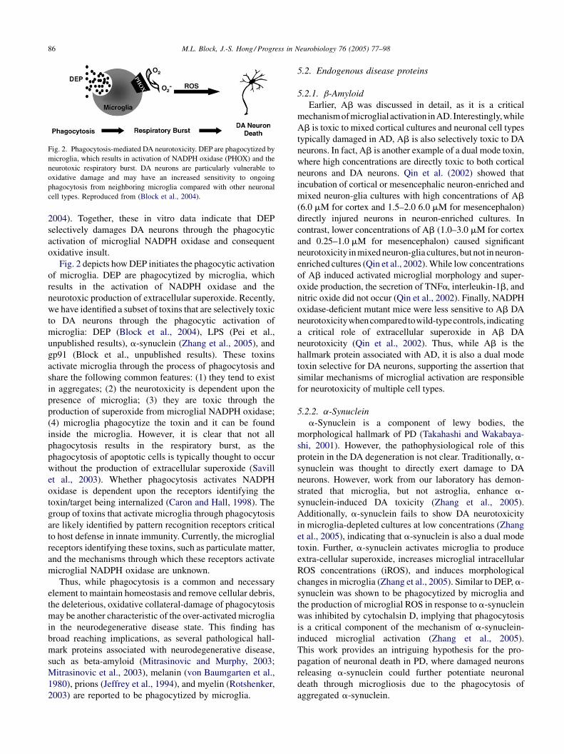
Fig. 2. Phagocytosis-mediated DA neurotoxicity. DEP are phagocytized by
microglia, which results in activation of NADPH oxidase (PHOX) and the
neurotoxic respiratory burst. DA neurons are particularly vulnerable to
oxidative damage and may have an increased sensitivity to ongoing
phagocytosis from neighboring microglia compared with other neuronal
cell types. Reproduced from (Block et al., 2004).
2004). Together, these in vitro data indicate that DEP
selectively damages DA neurons through the phagocytic
activation of microglial NADPH oxidase and consequent
oxidative insult.
Fig. 2 depicts how DEP initiates the phagocytic activation
of microglia. DEP are phagocytized by microglia, which
results in the activation of NADPH oxidase and the
neurotoxic production of extracellular superoxide. Recently,
we have identified a subset of toxins that are selectively toxic
to DA neurons through the phagocytic activation of
microglia: DEP (Block et al., 2004), LPS (Pei et al.,
unpublished results), a-synuclein (Zhang et al., 2005), and
gp91 (Block et al., unpublished results). These toxins
activate microglia through the process of phagocytosis and
share the following common features: (1) they tend to exist
in aggregates; (2) the neurotoxicity is dependent upon the
presence of microglia; (3) they are toxic through the
production of superoxide from microglial NADPH oxidase;
(4) microglia phagocytize the toxin and it can be found
inside the microglia. However, it is clear that not all
phagocytosis results in the respiratory burst, as the
phagocytosis of apoptotic cells is typically thought to occur
without the production of extracellular superoxide (Savill
et al., 2003). Whether phagocytosis activates NADPH
oxidase is dependent upon the receptors identifying the
toxin/target being internalized (Caron and Hall, 1998). The
group of toxins that activate microglia through phagocytosis
are likely identified by pattern recognition receptors critical
to host defense in innate immunity. Currently, the microglial
receptors identifying these toxins, such as particulate matter,
and the mechanisms through which these receptors activate
microglial NADPH oxidase are unknown.
Thus, while phagocytosis is a common and necessary
element to maintain homeostasis and remove cellular debris,
the deleterious, oxidative collateral-damage of phagocytosis
may be another characteristic of the over-activated microglia
in the neurodegenerative disease state. This finding has
broad reaching implications, as several pathological hall-
mark proteins associated with neurodegenerative disease,
such as beta-amyloid (Mitrasinovic and Murphy, 2003;
Mitrasinovic et al., 2003), melanin (von Baumgarten et al.,
1980), prions (Jeffrey et al., 1994), and myelin (Rotshenker,
2003) are reported to be phagocytized by microglia.
5.2. Endogenous disease proteins
5.2.1. b-Amyloid
Earlier, Ab was discussed in detail, as it is a critical
mechanismofmicroglialactivationinAD.Interestingly,while
Ab is toxic to mixed cortical cultures and neuronal cell types
typically damaged in AD, Ab is also selectively toxic to DA
neurons. In fact, Ab is another example of a dual mode toxin,
where high concentrations are directly toxic to both cortical
neurons and DA neurons. Qin et al. (2002) showed that
incubation of cortical or mesencephalic neuron-enriched and
mixed neuron-glia cultures with high concentrations of Ab
(6.0 mM for cortex and 1.5–2.0 6.0 mM for mesencephalon)
directly injured neurons in neuron-enriched cultures. In
contrast, lower concentrations of Ab (1.0–3.0 mM for cortex
and 0.25–1.0 mM for mesencephalon) caused significant
neurotoxicity in mixed neuron-glia cultures, but not in neuron-
enriched cultures (Qin et al., 2002). While low concentrations
of Ab induced activated microglial morphology and super-
oxide production, the secretion of TNFa, interleukin-1b, and
nitric oxide did not occur (Qin et al., 2002). Finally, NADPH
oxidase-deficient mutant mice were less sensitive to Ab DA
neurotoxicitywhencomparedtowild-typecontrols, indicating
a critical role of extracellular superoxide in Ab DA
neurotoxicity (Qin et al., 2002). Thus, while Ab is the
hallmark protein associated with AD, it is also a dual mode
toxin selective for DA neurons, supporting the assertion that
similar mechanisms of microglial activation are responsible
for neurotoxicity of multiple cell types.
5.2.2. a-Synuclein
a-Synuclein is a component of lewy bodies, the
morphological hallmark of PD (Takahashi and Wakabaya-
shi, 2001). However, the pathophysiological role of this
protein in the DA degeneration is not clear. Traditionally, a-
synuclein was thought to directly exert damage to DA
neurons. However, work from our laboratory has demon-
strated that microglia, but not astroglia, enhance a-
synuclein-induced DA toxicity (Zhang et al., 2005).
Additionally, a-synuclein fails to show DA neurotoxicity
in microglia-depleted cultures at low concentrations (Zhang
et al., 2005), indicating that a-synuclein is also a dual mode
toxin. Further, a-synuclein activates microglia to produce
extra-cellular superoxide, increases microglial intracellular
ROS concentrations (iROS), and induces morphological
changes in microglia (Zhang et al., 2005). Similar to DEP, a-
synuclein was shown to be phagocytized by microglia and
the production of microglial ROS in response to a-synuclein
was inhibited by cytochalsin D, implying that phagocytosis
is a critical component of the mechanism of a-synuclein-
induced microglial activation (Zhang et al., 2005).
This work provides an intriguing hypothesis for the pro-
pagation of neuronal death in PD, where damaged neurons
releasing a-synuclein could further potentiate neuronal
death through microgliosis due to the phagocytosis of
aggregated a-synuclein.
M.L. Block, J.-S. Hong / Progress in Neurobiology 76 (2005) 77–98 87
5.3. Reactive microgliosis
Microglial activation after CNS injury or in response to
neurodegeneration was initially perceived as a transient and
self-limited event (Streit et al., 1999). However, it has
become increasingly evident that the microglial response to
neuronal damage is both long-lived and self propelling (Gao
et al., 2003b; Huh et al., 2003; McGeer et al., 2003). The
neurotoxic response of microglia to central nervous system
(CNS) injury is a critical component of microglia-mediated
neurotoxicity across multiple diseases (Eikelenboom et al.,
2002; Sanchez-Moreno et al., 2004; Wenk, 2003). In
general, dying or damaged neurons have the potential to
activate microglia, regardless of how the neurons were
damaged (environmental toxin, endogenous disease protein,
or reactive microgliosis) or the neurodegenerative disease in
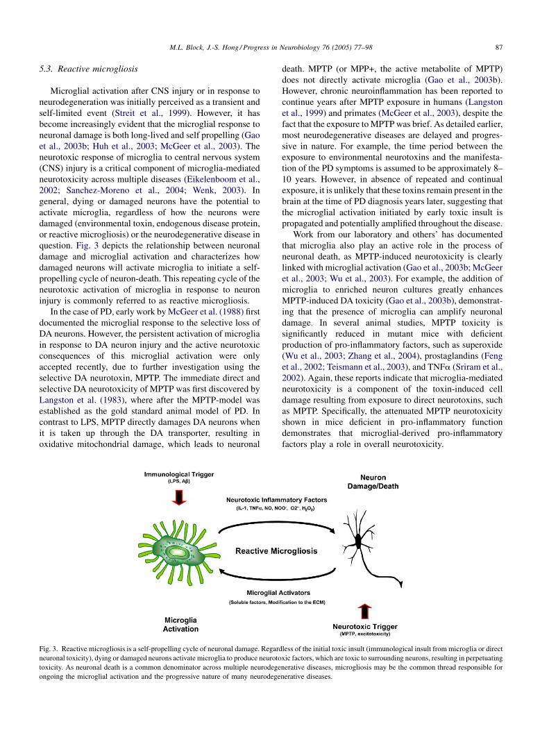
question. Fig. 3 depicts the relationship between neuronal
damage and microglial activation and characterizes how
damaged neurons will activate microglia to initiate a self-
propelling cycle of neuron-death. This repeating cycle of the
neurotoxic activation of microglia in response to neuron
injury is commonly referred to as reactive microgliosis.
In the case of PD, early work by McGeer et al. (1988) first
documented the microglial response to the selective loss of
DA neurons. However, the persistent activation of microglia
in response to DA neuron injury and the active neurotoxic
consequences of this microglial activation were only
accepted recently, due to further investigation using the
selective DA neurotoxin, MPTP. The immediate direct and
selective DA neurotoxicity of MPTP was first discovered by
Langston et al. (1983), where after the MPTP-model was
established as the gold standard animal model of PD. In
contrast to LPS, MPTP directly damages DA neurons when
it is taken up through the DA transporter, resulting in
oxidative mitochondrial damage, which leads to neuronal
Fig. 3. Reactive microgliosis is a self-propelling cycle of neuronal damage. Regard
neuronal toxicity), dying or damaged neurons activate microglia to produce neuroto
toxicity. As neuronal death is a common denominator across multiple neurodege
ongoing the microglial activation and the progressive nature of many neurodege
death. MPTP (or MPP+, the active metabolite of MPTP)
does not directly activate microglia (Gao et al., 2003b).
However, chronic neuroinflammation has been reported to
continue years after MPTP exposure in humans (Langston
et al., 1999) and primates (McGeer et al., 2003), despite the
fact that the exposure to MPTP was brief. As detailed earlier,
most neurodegenerative diseases are delayed and progres-
sive in nature. For example, the time period between the
exposure to environmental neurotoxins and the manifesta-
tion of the PD symptoms is assumed to be approximately 8–
10 years. However, in absence of repeated and continual
exposure, it is unlikely that these toxins remain present in the
brain at the time of PD diagnosis years later, suggesting that
the microglial activation initiated by early toxic insult is
propagated and potentially amplified throughout the disease.
Work from our laboratory and others’ has documented
that microglia also play an active role in the process of
neuronal death, as MPTP-induced neurotoxicity is clearly
linked with microglial activation (Gao et al., 2003b; McGeer
et al., 2003; Wu et al., 2003). For example, the addition of
microglia to enriched neuron cultures greatly enhances
MPTP-induced DA toxicity (Gao et al., 2003b), demonstrat-
ing that the presence of microglia can amplify neuronal
damage. In several animal studies, MPTP toxicity is
significantly reduced in mutant mice with deficient
production of pro-inflammatory factors, such as superoxide
(Wu et al., 2003; Zhang et al., 2004), prostaglandins (Feng
et al., 2002; Teismann et al., 2003), and TNFa (Sriram et al.,
2002). Again, these reports indicate that microglia-mediated
neurotoxicity is a component of the toxin-induced cell
damage resulting from exposure to direct neurotoxins, such
as MPTP. Specifically, the attenuated MPTP neurotoxicity
shown in mice deficient in pro-inflammatory function
demonstrates that microglial-derived pro-inflammatory
factors play a role in overall neurotoxicity.
less of the initial toxic insult (immunological insult from microglia or direct
xic factors, which are toxic to surrounding neurons, resulting in perpetuating
nerative diseases, microgliosis may be the common thread responsible for
nerative diseases.
M.L. Block, J.-S. Hong / Progress in Neurobiology 76 (2005) 77–9888
While the microglia pro-inflammatory response likely
involves multiple toxic factors, work from our laboratory
and others has identified that microglia-derived ROS are a
prominent component of reactive microgliosis (Gao et al.,
2003b; Wu et al., 2003). For example, while MPP+ and
MPTP do not directly affect microglial activation, Gao et al.
(2003b) demonstrated that the addition of both MPP+ and
MPTP to neuron-glia cultures induced the production of
superoxide at 4 days posttreatment. Gao et al. (2003b)
propose that this time delay in superoxide production in
response to MPP+ and MPTP occurred due to significant
accumulation of neuronal damage at 4 days posttreatment.
Further, the extracellular superoxide in response to MPTP
was produced only in animals with functioning NADPH
oxidase and was attenuated by NADPH oxidase inhibitors
(Gao et al., 2003b). However, no detectable amounts of
TNFa, NO, or PGE2 were produced in neuron-glia cultures
exposed to MPP+ or MPTP at any time point measured,
indicating the essential role of superoxide in microgliosis
(Gao et al., 2003b). Gao et al. (2003a,b,c) also showed that in
neuron-glia cultures from mice lacking functional NADPH
oxidase, MPTP and MPP+ both showed reduced DA toxicity
(Gao et al., 2003b), confirming that the production of
extracellular superoxide contributes to MPP+ and MPTP-
induced neurotoxicity. Further work by Gao et al. (2003c)
reports the amplifying nature of microgliosis, where LPS
and MPTP administered simultaneously or in tandem
resulted in synergystic neurotoxicity. Interestingly, the
synergyistic neurotoxicity of LPS and MPTP was also
demonstrated to be mediated through NADPH oxidase,
again emphasizing the critical role of this enzyme in the
microglia activation and DA neurotoxicity associated with
reactive microgliosis (Gao et al., 2003c). While there is
strong support that microglia become activated by neuronal
death to produce neurotoxic superoxide (Gao et al., 2003b),
the mechanisms through which neuronal damage induce
microglial activation are not completely understood.
5.3.1. Matrix metalloproteinase-3
While it is clear that microglia become activated upon
neuronal damage, there is a dearth of information on the
neuronal injury signals responsible for the chronic
microglial inflammatory response. Recent work suggests
that proteases known to modify the extracellular matrix
(ECM) may be a critical mechanism through which
damaged neurons activate microglia to produce extra-
cellular superoxide. Previous work from our laboratory
has emphasized the critical role of ECM proteins in the
interactions between microglia and neurons (Chang et al.,
2000a). In current work by Kim et al. (2005b) matrix
metalloproteinase-3 (MMP-3), a proteinase known to
degrade ECM components, was shown to be released upon
DA cell damage with MPP+ and to be toxic to DA neurons.
Mesencephalic neuron/glia cultures treated with MPP+
resulted in a dose dependent increase in the MMP-3
protein both in cell lysates and in conditioned media,
implying that DA neuron death upregulates the expression
of MMP-3. Mesencephalic neuron-glia cultures treated
with catalytically active MMP-3 showed DA neurotoxicity
and activated microglia morphology that preceded neuron
death (Kim et al., in review). Moreover, enriched
microglia produced extracellular superoxide in response
to MMP-3. This finding is critical to understanding the
mechanism of microgliosis, as previous studies from our
laboratory have identified that the activation of PHOX is a
mandatory component of the microglia contribution to
MPP+ and MPTP induced death. Further supporting this
premise, midbrain neuron-glia cultures from PHOX�/�
mice, lacking the catalytic subunit of PHOX and unable to
produce the phagocytic respiratory burst, were protected
from MMP-3-induced DA neurotoxicity in vitro when
compared to control (Kim et al., in review). In vivo
experiments showed that MMP-3 deficient mice were less
susceptible to SN DA neuronal degeneration and showed a
less pronounced microglial response induced in vivo by
MPTP. Together, these data suggest that MMP3 is released
upon DA neuron damage and activates microglia to further
propagate neuronal death.
5.3.2. Neuromelanin
Neuromelanin is also reported to be released by damaged
or dying DA neurons to activate microglia (Zecca et al.,
2003). In the normal, healthy human SN, neuromelanin is
located within dopaminergic neurons, accumulates in the SN
with age, and is responsible for the pigmented color of the
SN. Functionally, it has been suggested that neuromelanin
plays a protective role intracellularly, where neuromelanin
will bind toxins (D’Amato et al., 1986; Lindquist et al.,
1988; Zecca et al., 1994) and serves as an antioxidant
(Fornstedt et al., 1989; Wilczok et al., 1999). However, it has
also been suggested that neuromelanin has the potential to be
toxic, as excess neruomelanin inhibits the function of the DA
neuron proteasome (Shamoto-Nagai et al., 2004). Analysis
of the postmortem PD patient SN indicates that neurome-
lanin levels are significantly reduced, which is consistent
with the loss of DA neurons (Hirsch et al., 1988; Zecca et al.,
2002). Zecca et al. propose that neuromelanin is released by
damaged or dying DA neurons to initiate microglial
activation and that neuromelanin may be one of the factors
released by DA neurons responsible for the self-propelling
cycle of microgliosis (Zecca et al., 2003). Indeed,
neuromelanin is insoluble, is localized in high concentra-
tions in the SN (Lindquist, 1972), and it has been found in
the extracellular spaces in the SN of PD patients (Calabrese
and Hadfield, 1991), presenting an ideal opportunity for
neuromelanin to interact with microglia. Using exogenous
neuromelanin purified from the human brain, Wilms et al.
(2003) have shown that neuromelanin is chemotactic for
microglia. Further, Wilms et al. (2003) also showed that
neuromelanin added to rat enriched microglia cultures
activates microglial NFkB and induces the production of
toxic factors, such as TNFa, IL-6, and NO (Wilms et al.,
M.L. Block, J.-S. Hong / Progress in Neurobiology 76 (2005) 77–98 89
2003). However, whether neuromelamin induces superoxide
production in microglia is unknown.
Currently, MMP3, a-synuclein, and neuromelanin have
all been implicated as factors released by damaged DA,
resulting in a self-propelling cycle of microglial activation
and neuronal death. It seems likely that the identity of the
factors released from damaged neurons to signal microglial
activation is dependent upon the type of cell damaged and
the nature of the cell damage (necrosis versus apoptosis). For
example, a-synuclein and neuromelainin are highly
expressed by DA neurons in the SN, suggesting that these
factors are more likely to be released by injured DA neurons.
However, microglial activation is constant across numerous
neurodegenerative disorders, where several neuronal cell
types die as a result of a diverse set of triggers. It seems
likely that there are multiple common neuronal injury
signals released from varied neuronal cell types in response
to CNS injury, such as the pro-inflammatory characteristics
of necrosis. Alternatively, specific toxins may cause the
release of unique soluble factors from damaged neurons that
serve as propagating pro-inflammatory cues. It is of
significant interest for ongoing research to identify soluble
factors released from multiple classifications of damaged
neurons in response to various toxins, so that we can begin to
elucidate how neuronal damage can signal inflammation and
propagate further cell death.
6. Common characteristics of microglial activation.
6.1. Temporal relationship of the microglial release of
neurotoxic factors
The identification of several potential triggers of
microglia activation has allowed a generalizable classifica-
Fig. 4. NADPH oxidase is the common mechanism through which microglia are
multiple pro-inflammatory factors that are harmful to neurons. Across several toxin
often unique combinations of pro-inflammatory factors are released, which are de
diverse toxins (DEP, rotenone, LPS, PQ, reactive microgliosis, Ab, and a-synucle
involve PHOX. At this time, it remains unclear whether pro-inflammatory micro
tion of how microglial respond to stimuli. Firstly, based on in
vitro culture data, there is a clear temporal relationship
between the released microglial neurotoxic factors. Across
several toxins, it is apparent that first event is the production
of reactive oxygen species (ROS), which includes the extra-
cellular superoxide anion (O2��) and an increase in iROS
(Gao et al., 2002b; Qin et al., 2002; Zhang et al., 2005). The
increase in the production of ROS is rapid, usually occurring
within minutes, and is typically measured in microglia 10–
30 min after LPS addition. The microglial ROS response is
followed by the release of cytokines (such as TNFa and IL-
1b), nitric oxide (NO), and prostaglandin E2 (PGE2) that
peaks around 6–12 h (Gao et al., 2002b; Liu et al., 2003).
Experiments using different mutant mice deficient in
NADPH oxidase, iNOS, COX-2, or TNFa receptors all
resulted in reduced neurotoxicity (Teismann and Ferger,
2001; Wang et al., 2004b), indicating that the individual pro-
inflammatory factors released are sufficient, but not
mandatory, for neurotoxicity. In fact, individual pro-
inflammatory factors, such as IL-1, TNFa, and interferon-
g, work in concert to synergistically induce neuronal
damage (Jeohn et al., 1998). Additionally, microglial
NADPH oxidase activation (O2�� production) is shown to
enhance the release of other pro-inflammatory factors from
microglia (Qin et al., 2004), identifying this enzyme as a key
regulating factor of microglia-mediated neurotoxicity.
6.2. NADPH oxidase is the key enzyme for producing
ROS in the activation of microglia
Interestingly, microglia consistently generate ROS when
activated by multiple pro-inflammatory triggers, such as
environmental factors (LPS, DEP, rotenone, paraquat),
endogenous protein toxins (b-amyloid peptide, a-synu-
clein), and neuronal injury (Fig. 4). On the other hand, the
toxic to neurons. As the resident brain phagocytes, microglia can produce
s studied, the initiating steps of microglial activation are vastly different and
pendent upon the stimulus. However, the mechanism through which these
in) exert microglia-mediated neurotoxicity has consistently been shown to
glia activation can occur without the consequent activation of PHOX.
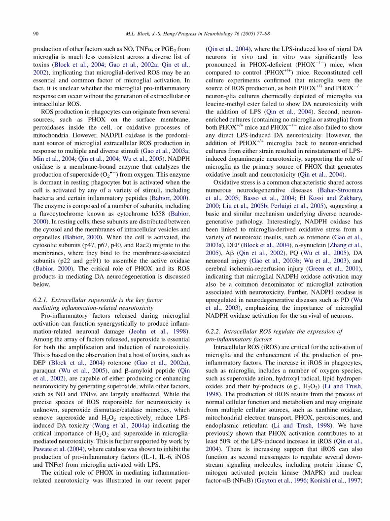
M.L. Block, J.-S. Hong / Progress in Neurobiology 76 (2005) 77–9890
production of other factors such as NO, TNFa, or PGE2 from
microglia is much less consistent across a diverse list of
toxins (Block et al., 2004; Gao et al., 2002a; Qin et al.,
2002), implicating that microglial-derived ROS may be an
essential and common factor of microglial activation. In
fact, it is unclear whether the microglial pro-inflammatory
response can occur without the generation of extracellular or
intracellular ROS.
ROS production in phagocytes can originate from several
sources, such as PHOX on the surface membrane,
peroxidases inside the cell, or oxidative processes of
mitochondria. However, NADPH oxidase is the predomi-
nant source of microglial extracellular ROS production in
response to multiple and diverse stimuli (Gao et al., 2003a;
Min et al., 2004; Qin et al., 2004; Wu et al., 2005). NADPH
oxidase is a membrane-bound enzyme that catalyzes the
production of superoxide (O2��) from oxygen. This enzyme
is dormant in resting phagocytes but is activated when the
cell is activated by any of a variety of stimuli, including
bacteria and certain inflammatory peptides (Babior, 2000).
The enzyme is composed of a number of subunits, including
a flavocytochrome known as cytochrome b558 (Babior,
2000). In resting cells, these subunits are distributed between
the cytosol and the membranes of intracellular vesicles and
organelles (Babior, 2000). When the cell is activated, the
cytosolic subunits (p47, p67, p40, and Rac2) migrate to the
membranes, where they bind to the membrane-associated
subunits (p22 and gp91) to assemble the active oxidase
(Babior, 2000). The critical role of PHOX and its ROS
products in mediating DA neurodegeneration is discussed
below.
6.2.1. Extracellular superoxide is the key factor
mediating inflammation-related neurotoxicity
Pro-inflammatory factors released during microglial
activation can function synergystically to produce inflam-
mation-related neuronal damage (Jeohn et al., 1998).
Among the array of factors released, superoxide is essential
for both the amplification and induction of neurotoxicity.
This is based on the observation that a host of toxins, such as
DEP (Block et al., 2004) rotenone (Gao et al., 2002a),
paraquat (Wu et al., 2005), and b-amyloid peptide (Qin
et al., 2002), are capable of either producing or enhancing
neurotoxicity by generating superoxide, while other factors,
such as NO and TNFa, are largely unaffected. While the
precise species of ROS responsible for neurotoxicity is
unknown, superoxide dismutase/catalase mimetics, which
remove superoxide and H2O2 respectively, reduce LPS-
induced DA toxicity (Wang et al., 2004a) indicating the
critical importance of H2O2 and superoxide in microglia-
mediated neurotoxicity. This is further supported by work by
Pawate et al. (2004), where catalase was shown to inhibit the
production of pro-inflammatory factors (IL-1, IL-6, iNOS
and TNFa) from microglia activated with LPS.
The critical role of PHOX in mediating inflammation-
related neurotoxicity was illustrated in our recent paper
(Qin et al., 2004), where the LPS-induced loss of nigral DA
neurons in vivo and in vitro was significantly less
pronounced in PHOX-deficient (PHOX�/�) mice, when
compared to control (PHOX+/+) mice. Reconstituted cell
culture experiments confirmed that microglia were the
source of ROS production, as both PHOX+/+ and PHOX�/�
neuron-glia cultures chemically depleted of microglia via
leucine-methyl ester failed to show DA neurotoxicity with
the addition of LPS (Qin et al., 2004). Second, neuron-
enriched cultures (containing no microglia or astroglia) from
both PHOX+/+ mice and PHOX�/� mice also failed to show
any direct LPS-induced DA neurotoxicity. However, the
addition of PHOX+/+ microglia back to neuron-enriched
cultures from either strain resulted in reinstatement of LPS-
induced dopaminergic neurotoxicity, supporting the role of
microglia as the primary source of PHOX that generates
oxidative insult and neurotoxicity (Qin et al., 2004).
Oxidative stress is a common characteristic shared across
numerous neurodegenerative diseases (Bahat-Stroomza
et al., 2005; Basso et al., 2004; El Kossi and Zakhary,
2000; Liu et al., 2005b; Perluigi et al., 2005), suggesting a
basic and similar mechanism underlying diverse neurode-
generative pathology. Interestingly, NADPH oxidase has
been linked to microglia-derived oxidative stress from a
variety of neurotoxic insults, such as rotenone (Gao et al.,
2003a), DEP (Block et al., 2004), a-synuclein (Zhang et al.,
2005), Ab (Qin et al., 2002), PQ (Wu et al., 2005), DA
neuronal injury (Gao et al., 2003b; Wu et al., 2003), and
cerebral ischemia-reperfusion injury (Green et al., 2001),
indicating that microglial NADPH oxidase activation may
also be a common denominator of microglial activation
associated with neurotoxicity. Further, NADPH oxidase is
upregulated in neurodegenerative diseases such as PD (Wu
et al., 2003), emphasizing the importance of microglial
NADPH oxidase activation for the survival of neurons.
6.2.2. Intracellular ROS regulate the expression of
pro-inflammatory factors
Intracellular ROS (iROS) are critical for the activation of
microglia and the enhancement of the production of pro-
inflammatory factors. The increase in iROS in phagocytes,
such as microglia, includes a number of oxygen species,
such as superoxide anion, hydroxyl radical, lipid hydroper-
oxides and their by-products (e.g., H2O2) (Li and Trush,
1998). The production of iROS results from the process of
normal cellular function and metabolism and may originate
from multiple cellular sources, such as xanthine oxidase,
mitochondrial electron transport, PHOX, peroxisomes, and
endoplasmic reticulum (Li and Trush, 1998). We have
previously shown that PHOX activation contributes to at
least 50% of the LPS-induced increase in iROS (Qin et al.,
2004). There is increasing support that iROS can also
function as second messengers to regulate several down-
stream signaling molecules, including protein kinase C,
mitogen activated protein kinase (MAPK) and nuclear
factor-kB (NFkB) (Guyton et al., 1996; Konishi et al., 1997;
M.L. Block, J.-S. Hong / Progress in Neurobiology 76 (2005) 77–98 91
Schreck et al., 1991). Using PHOX�/� neuron-glia cultures,
we have shown that activation of the PHOX initiates an
intracellular ROS signaling pathway (Forman and Torres,
2002) that can activate microglia and amplify the production
of pro-inflammatory cytokines, such as TNFa (Qin et al.,
2004) or PGE2 (Wang et al., 2004b). Additionally, Min et al.
(2004) demonstrated that ganglioside induces the activation
of microglia, where the production of IL-1b, TNFa, and
iNOS are attenuated by the addition of the NADPH oxidase
inhibitor, diphenyleneiodonium. Furthermore, NADPH
oxidase inhibitors and catalase are shown to suppress
LPS-induced expression of cytokines (IL-1, IL-6, and
TNFa), iNOS expression, MAP kinases, and NFkB
phosphorylation (Pawate et al., 2004). NADPH oxidase
has also been implicated as critical for the morphological
changes associated with the early phase of microglial
activation, where immuno-staining for F4/80 (marker for
mouse microglia) in midbrain neuron-glia cultures treated
with LPS revealed that PHOX�/� microglia show much less
activated morphology in response to LPS treatment than the
PHOX+/+ (Qin et al., 2004). While the majority of research
has been completed in models of LPS-induced microglial
activation, NADPH oxidase is essential across diverse
triggers of microglial activation (environmental toxins,
endogenous disease proteins, and neuronal injury) indicat-
ing that ROS are likely an essential signaling mechanism
regulating general microglial activation.
Accumulating evidence supports that NADPH oxidase
contributes to microglia-mediated neurotoxicity through
two mechanisms (Fig. 5). First, activation of NADPH
oxidase results in the production of extracellular ROS that is
toxic to neurons. Second, activation of NADPH oxidase
causes an increase in the microglial intracellular ROS, which
enhances the production of pro-inflammatory factors that are
toxic to neurons. This intracellular ROS increase is a critical
component of pro-inflammatory signaling in phagocytes
Fig. 5. Dual mechanisms of NADPH oxidase-mediated neurotoxicity. Here we
(PHOX) mediates neurotoxicity. Extracellular ROS produced as a consequence of P
ROS that occurs in microglia as a response to PHOX activation enhances the pr
(kupher cells, monocytes, microglia, macrophages) and has
been called the PHOX-ROS pathway. The dual impact of
NADPH oxidase activation on neurotoxicity and the
prevalence of NADPH oxidase activation upon microglial
activation, regardless of the stimulus, suggest that microglial
NADPH oxidase is a critical mechanism of neuronal death
across multiple neurodegenerative diseases.
7. Microglial activation as a common mechanism in
diverse neuropathology
While we have highlighted the common mechanisms of
how microglia respond to various toxins and signals to result
in selective DA neuron toxicity, we have also presented
evidence indicating that microglial activation contributes to
unique pathology associated with multiple neurodegenera-
tive diseases. The intriguing question remains as to how the
generalized phenomena of microglial activation can result in
diverse and localized neurodegenerative pathology. While
this is a complex issue that is likely specific to each
neurodegenerative disease, there are several hypotheses as to
why microglial activation would be localized in unique brain
regions that vary both across and within neurodegenerative
disease types.
It is important to note that microglial activation can be
localized, where activation of microglia in one brain region
does not instigate activation across the entire brain. A classic
example is the case of LPS injection into the nigral area,
where microglial activation and consequent neuronal
damage are limited to this region (Gao et al., 2002b). Thus,
the localization of the initiating stimulus (neuronal death, an
environmental factor, or an endogenous disease protein) will
be a critical factor in determining where microglia will
become activated to propagate neuronal toxicity and likely
contributes to the regional compartmentalization of many
depict the critical mechanisms through which microglial NADPH oxidase
HOX activation is toxic to neurons. Additionally, the increase in intracellular
oduction of neurotoxic pro-inflammatory factors from microglia.
M.L. Block, J.-S. Hong / Progress in Neurobiology 76 (2005) 77–9892
neurodegenerative disorders. In addition to the location of
the original neurotoxic event/trigger, the unique character-
istics of the neurotoxic triggers associated with neurode-
generative diseases may determine which cell types will be
damaged. For example, while a lower grade inflammatory
response selectively kills DA neurons, higher doses of the
same toxin (such as LPS) begins to kill multiple cell types
(Gao et al., 2002b). Thus, the degree of the microglial
inflammatory response may also determine which cell types
are damaged, as certain cell types localized in select brain
regions may be preferentially vulnerable to microglia-
derived insult. Again, while this is particularly evident in the
case of DA neurons and PD, the hierarchy of susceptible cell
types in the localized region of other neurodegenerative
diseases may also play a role in the specificity of disease
phenotype.
Finally, regional differences in microglial populations
and their pro-inflammatory responses also offer further
insight into the potential mechanisms mediating the
compartmentalization of both neurodegenerative pathology
and microglial activation seen across the numerous
neurodegenerative diseases. Again, DA neurons localized
in the SN are the extreme example, as there are 4.5 times as
many more localized in this region (Kim et al., 2000),
making cells there more susceptible to inflammatory insult.
In the case of other neurodegenerative disorders, differences
of regional microglia density could also explain localized
neurotoxicity. In addition, regional differences in sub-
classes of microglia (Ladeby et al., 2005; Mittelbronn et al.,
2001) have been reported, where microglial population
differences in reactivity could also offer insight into
neurodegenerative disease localization. Furthermore, brain
region differences in microglial regulatory factors, such as
astrocyte populations, could also explain how the general-
ized phenomenon of microglial activation could be localized
by brain region.
It is not surprising that multiple interacting factors will
likely determine the regional activation of microglia and the
specific pathology of neurodegenerative diseases. Unfortu-
nately, the identity of the initial neurotoxic triggers remains
unknown for most neurodegenerative diseases. As we begin
to learn more about the initiating factors for neurodegen-
erative diseases and further characterize both preserved and
affected brain areas, future research will have the ability to
address the potential mechanistic role of microglia in
specific neurodegenerative pathology in more detail.
8. Conclusions
Microglia are the critical actors of self-propelling
mechanisms of neurotoxicity (reactive microgliosis), which
is an underlying contributing mechanism to multiple
neurodegenerative disorders. Microglia can be activated
by two mechanisms: (1) direct stimulation of microglia from
environmental or endogenous toxins; (2) activation through
a reactive microgliosis process. Oxidative stress is pre-
dominant in neurodegenerative disease and microglia are
critical sources of oxidative insult. In particular, the
activation of microglial NADPH oxidase is a common
source of microglial-derived oxidative stress, regardless of
the stimulus activating the microglia. Additionally, ROS
generated from NADPH oxidase are an essential pathway
regulating the microglial expression of neurotoxic pro-
inflammatory factors. In summary, while the diversity of
neurodegenerative disorders can be explained by unique
localized triggers of neuronal death, the concentration of
microglia in the brain area of neurodegeneration, the
inherent vulnerability of the degenerating neuronal cell type,
the severity of the toxic insult, and many other character-
istics that will localize the self-propelling neuronal death, it
remains constant that once a neuron dies, this death can
activate microglia. Thus, multiple triggers of microglial
activation may share a common mechanism of microglial-
derived oxidative stress fueling the progressive nature of
several independent neurodegenerative diseases.
References
Aloisi, F., 1999. The role of microglia and astrocytes in CNS immune
surveillance and immunopathology. Adv. Exp. Med. Biol. 468, 123–
133.
Babior, B.M., 2000. Phagocytes and oxidative stress. Am. J. Med. 109, 33–
44.
Bahat-Stroomza, M., Gilgun-Sherki, Y., Offen, D., Panet, H., Saada, A.,
Krool-Galron, N., Barzilai, A., Atlas, D., Melamed, E., 2005. A novel
thiol antioxidant that crosses the blood brain barrier protects dopami-
nergic neurons in experimental models of Parkinson’s disease. Eur. J.
Neurosci. 21, 637–646.
Bamberger, M.E., Harris, M.E., McDonald, D.R., Husemann, J., Land-
reth, G.E., 2003. A cell surface receptor complex for fibrillar
beta-amyloid mediates microglial activation. J. Neurosci. 23,
2665–2674.
Banati, R.B., Gehrmann, J., Kellner, M., Holsboer, F., 1995. Antibodies
against microglia/brain macrophages in the cerebrospinal fluid of a
patient with acute amyotrophic lateral sclerosis and presenile dementia.
Clin. Neuropathol. 14, 197–200.
Banati, R.B., Newcombe, J., Gunn, R.N., Cagnin, A., Turkheimer, F.,
Heppner, F., Price, G., Wegner, F., Giovannoni, G., Miller, D.H., Perkin,
G.D., Smith, T., Hewson, A.K., Bydder, G., Kreutzberg, G.W., Jones, T.,
Cuzner, M.L., Myers, R., 2000. The peripheral benzodiazepine binding
site in the brain in multiple sclerosis: quantitative in vivo imaging of
microglia as a measure of disease activity. Brain 123 (Pt 11), 2321–
2337.
Barbeau, A., Dallaire, L., Buu, N.T., Poirier, J., Rucinska, E., 1985.
Comparative behavioral, biochemical and pigmentary effects of MPTP,
MPP+ and paraquat in Rana pipiens. Life Sci. 37, 1529–1538.
Barde, Y.A., 1989. Trophic factors and neuronal survival. Neuron 2, 1525–
1534.
Barron, K.D., 1995. The microglial cell. A historical review. J. Neurol. Sci.
134 (Suppl.), 57–68.
Basso, M., Giraudo, S., Corpillo, D., Bergamasco, B., Lopiano, L., Fasano,
M., 2004. Proteome analysis of human substantia nigra in Parkinson’s
disease. Proteomics 4, 3943–3952.
Bate, C., Veerhuis, R., Eikelenboom, P., Williams, A., 2004. Microglia kill
amyloid-beta1-42 damaged neurons by a CD14-dependent process.
Neuroreport 15, 1427–1430.
M.L. Block, J.-S. Hong / Progress in Neurobiology 76 (2005) 77–98 93
Betarbet, R., Sherer, T.B., MacKenzie, G., Garcia-Osuna, M., Panov, A.V.,
Greenamyre, J.T., 2000. Chronic systemic pesticide exposure repro-
duces features of Parkinson’s disease. Nat. Neurosci. 3, 1301–1306.
Beyer, M., Gimsa, U., Eyupoglu, I.Y., Hailer, N.P., Nitsch, R., 2000.
Phagocytosis of neuronal or glial debris by microglial cells: upregula-
tion of MHC class II expression and multinuclear giant cell formation in
vitro. Glia 31, 262–266.
Block, M.L., Wu, X., Pei, Z., Li, G., Wang, T., Qin, L., Wilson, B., Yang, J.,
Hong, J.S., Veronesi, B., 2004. Nanometer size diesel exhaust particles
are selectively toxic to dopaminergic neurons: the role of microglia,
phagocytosis, and NADPH oxidase. FASEB J. 18, 1618–1620.
Boztug, K., Carson, M.J., Pham-Mitchell, N., Asensio, V.C., DeMartino, J.,
Campbell, I.L., 2002. Leukocyte infiltration, but not neurodegeneration,
in the CNS of transgenic mice with astrocyte production of the CXC
chemokine ligand 10. J. Immunol. 169, 1505–1515.
Braak, H., Braak, E., 1994. Morphological criteria for the recognition of
Alzheimer’s disease and the distribution pattern of cortical changes
related to this disorder. Neurobiol. Aging 15, 355–356 (Discussion 379–
380).
Budka, H., 1991. The definition of HIV-specific neuropathology. Acta
Pathol. Jpn. 41, 182–191.
Calabrese, V.P., Hadfield, M.G., 1991. Parkinsonism and extraocular motor
abnormalities with unusual neuropathological findings. Mov. Disord. 6,
257–260.
Calderon-Garciduenas, L., Azzarelli, B., Acuna, H., Garcia, R., Gambling,
T.M., Osnaya, N., Monroy, S., MR FD.E.L.T., Carson, J.L., Villarreal-
Calderon, A., Rewcastle, B., 2002. Air pollution and brain damage.
Toxicol. Pathol. 30, 373–389.
Calderon-Garciduenas, L., Maronpot, R.R., Torres-Jardon, R., Henriquez-
Roldan, C., Schoonhoven, R., Acuna-Ayala, H., Villarreal-Calderon, A.,
Nakamura, J., Fernando, R., Reed, W., Azzarelli, B., Swenberg, J.A.,
2003. DNA damage in nasal and brain tissues of canines exposed to air
pollutants is associated with evidence of chronic brain inflammation and
neurodegeneration. Toxicol. Pathol. 31, 524–538.
Calderon-Garciduenas, L., Reed, W., Maronpot, R.R., Henriquez-Roldan,
C., Delgado-Chavez, R., Calderon-Garciduenas, A., Dragustinovis, I.,
Franco-Lira, M., Aragon-Flores, M., Solt, A.C., Altenburg, M., Torres-
Jardon, R., Swenberg, J.A., 2004. Brain inflammation and Alzheimer’s-
like pathology in individuals exposed to severe air pollution. Toxicol.
Pathol. 32, 650–658.
Campbell, A., Oldham, M., Becaria, A., Bondy, S.C., Meacher, D., Sioutas,
C., Misra, C., Mendez, L.B., Kleinman, M., 2005. Particulate matter in
polluted air may increase biomarkers of inflammation in mouse brain.
Neurotoxicology 26, 133–140.
Caron, E., Hall, A., 1998. Identification of two distinct mechanisms of
phagocytosis controlled by different Rho GTPases. Science 282, 1717–
1721.
Carvey, P.M., Chang, Q., Lipton, J.W., Ling, Z., 2003. Prenatal exposure to
the bacteriotoxin lipopolysaccharide leads to long-term losses of dopa-
mine neurons in offspring: a potential, new model of Parkinson’s
disease. Front Biosci. 8, s826–s837.
Chakrabarti, L., Hurtrel, M., Maire, M.A., Vazeux, R., Dormont, D.,
Montagnier, L., Hurtrel, B., 1991. Early viral replication in the brain
of SIV-infected rhesus monkeys. Am. J. Pathol. 139, 1273–1280.
Chang, R.C., Hudson, P., Wilson, B., Haddon, L., Hong, J.S., 2000a.
Influence of neurons on lipopolysaccharide-stimulated production of
nitric oxide and tumor necrosis factor-alpha by cultured glia. Brain Res.
853, 236–244.
Chang, R.C., Rota, C., Glover, R.E., Mason, R.P., Hong, J.S., 2000b. A
novel effect of an opioid receptor antagonist, naloxone, on the produc-
tion of reactive oxygen species by microglia: a study by electron
paramagnetic resonance spectroscopy. Brain Res. 854, 224–229.
Chao, C.C., Hu, S., Molitor, T.W., Shaskan, E.G., Peterson, P.K., 1992.
Activated microglia mediate neuronal cell injury via a nitric oxide
mechanism. J. Immunol. 149, 2736–2741.
Colton, C.A., Gilbert, D.L., 1987. Production of superoxide anions by a
CNS macrophage, the microglia. FEBS Lett. 223, 284–288.
Columba-Cabezas, S., Serafini, B., Ambrosini, E., Sanchez, M., Penna, G.,
Adorini, L., Aloisi, F., 2002. Induction of macrophage-derived chemo-
kine/CCL22 expression in experimental autoimmune encephalomyelitis
and cultured microglia: implications for disease regulation. J. Neuroim-
munol. 130, 10–21.
Combs, C.K., Johnson, D.E., Karlo, J.C., Cannady, S.B., Landreth, G.E.,
2000. Inflammatory mechanisms in Alzheimer’s disease: inhibition of
beta-amyloid-stimulated proinflammatory responses and neurotoxicity
by PPARgamma agonists. J. Neurosci. 20, 558–567.
Cooper, P.N., Siddons, C.A., Mann, D.M., 1996. Patterns of glial cell
activity in fronto-temporal dementia (lobar atrophy). Neuropathol.
Appl. Neurobiol. 22, 17–22.
Cosenza, M.A., Zhao, M.L., Si, Q., Lee, S.C., 2002. Human brain par-
enchymal microglia express CD14 and CD45 and are productively
infected by HIV-1 in HIV-1 encephalitis. Brain Pathol. 12, 442–455.
Craft, J.M., Watterson, D.M., Frautschy, S.A., Van Eldik, L.J., 2004.
Aminopyridazines inhibit beta-amyloid-induced glial activation and
neuronal damage in vivo. Neurobiol. Aging 25, 1283–1292.
D’Amato, R.J., Lipman, Z.P., Snyder, S.H., 1986. Selectivity of the par-
kinsonian neurotoxin MPTP: toxic metabolite MPP+ binds to neuro-
melanin. Science 231, 987–989.
Das, P., Howard, V., Loosbrock, N., Dickson, D., Murphy, M.P., Golde, T.E.,
2003. Amyloid-beta immunization effectively reduces amyloid deposi-
tion in FcRgamma�/� knock-out mice. J. Neurosci. 23, 8532–8538.
D’Aversa, T.G., Yu, K.O., Berman, J.W., 2004. Expression of chemokines
by human fetal microglia after treatment with the human immunode-
ficiency virus type 1 protein Tat. J. Neurovirol. 10, 86–97.
Davis, J.B., McMurray, H.F., Schubert, D., 1992. The amyloid beta-protein
of Alzheimer’s disease is chemotactic for mononuclear phagocytes.
Biochem. Biophys. Res. Commun. 189, 1096–1100.
del Rio-Hortega, P., 1932. Cytology and cellular pathology of the nervous
system. Penfeild Wed, New York.
Dheen, S.T., Jun, Y., Yan, Z., Tay, S.S., Ang Ling, E., 2004. Retinoic acid
inhibits expression of TNF-alpha and iNOS in activated rat microglia.
Glia.
Dodel, R.C., Hampel, H., Du, Y., 2003. Immunotherapy for Alzheimer’s
disease. Lancet Neurol. 2, 215–220.
Duvoisin, R.C., Lobo-Antunes, J., Yahr, M.D., 1972. Response of patients
with postencephalitic Parkinsonism to levodopa. J. Neurol. Neurosurg.
Psychiatry 35, 487–495.
Duvoisin, R.C., Yahr, M.D., Schweitzer, M.D., Merritt, H.H., 1963. Par-
kinsonism before and since the epidemic of encephalitis lethargica.
Arch. Neurol. 30, 232–236.
Eikelenboom, P., Bate, C., Van Gool, W.A., Hoozemans, J.J., Rozemuller,
J.M., Veerhuis, R., Williams, A., 2002. Neuroinflammation in Alzhei-
mer’s disease and prion disease. Glia 40, 232–239.
El Kossi, M.M., Zakhary, M.M., 2000. Oxidative stress in the context of
acute cerebrovascular stroke. Stroke 31, 1889–1892.
Elbaz, A., Levecque, C., Clavel, J., Vidal, J.S., Richard, F., Amouyel, P.,
Alperovitch, A., Chartier-Harlin, M.C., Tzourio, C., 2004. CYP2D6
polymorphism, pesticide exposure, and Parkinson’s disease. Ann. Neu-
rol. 55, 430–434.
Elkabes, S., DiCicco-Bloom, E.M., Black, I.B., 1996. Brain microglia/
macrophages express neurotrophins that selectively regulate microglial
proliferation and function. J. Neurosci. 16, 2508–2521.
Feng, Z.H., Wang, T.G., Li, D.D., Fung, P., Wilson, B.C., Liu, B., Ali, S.F.,
Langenbach, R., Hong, J.S., 2002. Cyclooxygenase-2-deficient mice are
resistant to 1-methyl-4-phenyl-1,2,3,6-tetrahydropyridine-induced
damage of dopaminergic neurons in the substantia nigra. Neurosci.
Lett. 329, 354–358.
Finch, G.L., Hobbs, C.H., Blair, L.F., Barr, E.B., Hahn, F.F., Jaramillo,
R.J., Kubatko, J.E., March, T.H., White, R.K., Krone, J.R., Menache,
M.G., Nikula, K.J., Mauderly, J.L., Van Gerpen, J., Merceica, M.D.,
Zielinska, B., Stankowski, L., Burling, K., Howell, S., 2002. Effects
of subchronic inhalation exposure of rats to emissions from a diesel
engine burning soybean oil-derived biodiesel fuel. Inhalib. Toxicol.
14, 1017–1048.
M.L. Block, J.-S. Hong / Progress in Neurobiology 76 (2005) 77–9894
Forman, H.J., Torres, M., 2002. Reactive oxygen species and cell signaling:
respiratory burst in macrophage signaling. Am. J. Respir. Crit. Care
Med. 166, S4–S8.
Fornstedt, B., Brun, A., Rosengren, E., Carlsson, A., 1989. The apparent
autoxidation rate of catechols in dopamine-rich regions of human brains
increases with the degree of depigmentation of substantia nigra. J.
Neural. Transm. Park Dis. Dement Sect. 1, 279–295.
Freude, S., Hausmann, J., Hofer, M., Pham-Mitchell, N., Campbell, I.L.,
Staeheli, P., Pagenstecher, A., 2002. Borna disease virus accelerates
inflammation and disease associated with transgenic expression of
interleukin-12 in the central nervous system. J. Virol. 76, 12223–12232.
Friedman, W.J., Larkfors, L., Ayer-LeLievre, C., Ebendal, T., Olson, L.,
Persson, H., 1990. Regulation of beta-nerve growth factor expression by
inflammatory mediators in hippocampal cultures. J. Neurosci. Res. 27,
374–382.
Furlan, R., Brambilla, E., Sanvito, F., Roccatagliata, L., Olivieri, S.,
Bergami, A., Pluchino, S., Uccelli, A., Comi, G., Martino, G., 2003.
Vaccination with amyloid-beta peptide induces autoimmune encepha-
lomyelitis in C57/BL6 mice. Brain 126, 285–291.
Gao, H.M., Hong, J.S., Zhang, W., Liu, B., 2002a. Distinct role for microglia
in rotenone-induced degeneration of dopaminergic neurons. J. Neurosci.
22, 782–790.
Gao, H.M., Jiang, J., Wilson, B., Zhang, W., Hong, J.S., Liu, B., 2002b.
Microglial activation-mediated delayed and progressive degeneration of
rat nigral dopaminergic neurons: relevance to Parkinson’s disease. J.
Neurochem. 81, 1285–1297.
Gao, H.M., Liu, B., Hong, J.S., 2003a. Critical role for microglial NADPH
oxidase in rotenone-induced degeneration of dopaminergic neurons. J.
Neurosci. 23, 6181–6187.
Gao, H.M., Liu, B., Zhang, W., Hong, J.S., 2003b. Critical role of microglial
NADPH oxidase-derived free radicals in the in vitro MPTP model of
Parkinson’s disease. FASEB J..
Gao, H.M., Liu, B., Zhang, W., Hong, J.S., 2003c. Synergistic dopaminergic
neurotoxicity of MPTP and inflammogen lipopolysaccharide: relevance
to the etiology of Parkinson’s disease. FASEB J. 17, 1957–1959.
Garden, G.A., Guo, W., Jayadev, S., Tun, C., Balcaitis, S., Choi, J., Montine,
T.J., Moller, T., Morrison, R.S., 2004. HIVassociated neurodegeneration
requires p53 in neurons and microglia. FASEB J. 18, 1141–1143.
Gayle, D.A., Ling, Z., Tong, C., Landers, T., Lipton, J.W., Carvey, P.M.,
2002. Lipopolysaccharide (LPS)-induced dopamine cell loss in culture:
roles of tumor necrosis factor-alpha, interleukin-1beta, and nitric oxide.
Brain Res. Dev. Brain Res. 133, 27–35.
Giulian, D., Haverkamp, L.J., Yu, J.H., Karshin, W., Tom, D., Li, J.,
Kirkpatrick, J., Kuo, L.M., Roher, A.E., 1996. Specific domains of
beta-amyloid from Alzheimer plaque elicit neuron killing in human
microglia. J. Neurosci. 16, 6021–6037.
Graeber, M.B., Streit, W.J., Kreutzberg, G.W., 1988. The microglial cytos-
keleton: vimentin is localized within activated cells in situ. J. Neuro-
cytol. 17, 573–580.
Grant, H., Lantos, P.L., Parkinson, C., 1980. Cerebral damage in paraquat
poisoning. Histopathology 4, 185–195.
Green, S.P., Cairns, B., Rae, J., Errett-Baroncini, C., Hongo, J.A., Erickson,
R.W., Curnutte, J.T., 2001. Induction of gp91-phox, a component of the
phagocyte NADPH oxidase, in microglial cells during central nervous
system inflammation. J. Cereb. Blood Flow Metab. 21, 374–384.
Greenamyre, J.T., MacKenzie, G., Peng, T.I., Stephans, S.E., 1999. Mito-
chondrial dysfunction in Parkinson’s disease. Biochem. Soc. Symp. 66,
85–97.
Guyton, K.Z., Gorospe, M., Kensler, T.W., Holbrook, N.J., 1996. Mitogen-
activated protein kinase (MAPK) activation by butylated hydroxyto-
luene hydroperoxide: implications for cellular survival and tumor
promotion. Cancer Res. 56, 3480–3485.
Gwinn-Hardy, K., 2002. Genetics of parkinsonism. Mov. Disord. 17, 645–
656.
Hall, E.D., Oostveen, J.A., Gurney, M.E., 1998. Relationship of microglial
and astrocytic activation to disease onset and progression in a transgenic
model of familial ALS. Glia 23, 249–256.
Hansson, E., Ronnback, L., 1995. Astrocytes in glutamate neurotransmis-
sion. FASEB J. 9, 343–350.
Hill, K.E., Zollinger, L.V., Watt, H.E., Carlson, N.G., Rose, J.W., 2004.
Inducible nitric oxide synthase in chronic active multiple sclerosis
plaques: distribution, cellular expression and association with myelin
damage. J. Neuroimmunol. 151, 171–179.
Hirsch, E., Graybiel, A.M., Agid, Y.A., 1988. Melanized dopaminergic
neurons are differentially susceptible to degeneration in Parkinson’s
disease. Nature 334, 345–348.
Holdorff, B., 2002. Friedrich Heinrich Lewy (1885–1950) and his work. J.
Hist. Neurosci. 11, 19–28.
Hudnell, H.K., 1999. Effects from environmental Mn exposures: a review of
the evidence from non-occupational exposure studies. Neurotoxicology
20, 379–397.
Huh, Y., Jung, J.W., Park, C., Ryu, J.R., Shin, C.Y., Kim, W.K., Ryu, J.H.,
2003. Microglial activation and tyrosine hydroxylase immunoreactivity
in the substantia nigral region following transient focal ischemia in rats.
Neurosci. Lett. 349, 63–67.
Hurley, S.D., Walter, S.A., Semple-Rowland, S.L., Streit, W.J., 1999.
Cytokine transcripts expressed by microglia in vitro are not expressed
by ameboid microglia of the developing rat central nervous system. Glia
25, 304–309.
Ii, M., Sunamoto, M., Ohnishi, K., Ichimori, Y., 1996. beta-Amyloid
protein-dependent nitric oxide production from microglial cells and
neurotoxicity. Brain Res. 720, 93–100.
Iregren, A., 1999. Manganese neurotoxicity in industrial exposures: proof of
effects, critical exposure level, and sensitive tests. Neurotoxicology 20,
315–323.
Jeffrey, M., Goodsir, C.M., Bruce, M.E., McBride, P.A., Farquhar, C., 1994.
Morphogenesis of amyloid plaques in 87V murine scrapie. Neuropathol.
Appl. Neurobiol. 20, 535–542.
Jellinger, K.A., 2001. The pathology of Parkinson’s disease. Adv. Neurol.
86, 55–72.
Jenner, P., 2001. Parkinson’s disease, pesticides and mitochondrial dysfunc-
tion. Trends Neurosci. 24, 245–247.
Jensen, L.K., Klausen, H., Elsnab, C., 1989. Organic brain damage in garage
workers after long-term exposure to diesel exhaust fumes. Ugeskr
Laeger 151, 2255–2258.
Jeohn, G.H., Kong, L.Y., Wilson, B., Hudson, P., Hong, J.S., 1998.
Synergistic neurotoxic effects of combined treatments with cytokines
in murine primary mixed neuron/glia cultures. J. Neuroimmunol. 85,
1–10.
Jordan, C.A., Watkins, B.A., Kufta, C., Dubois-Dalcq, M., 1991. Infection
of brain microglial cells by human immunodeficiency virus type 1 is
CD4 dependent. J. Virol. 65, 736–742.
Kahn MA, E.J., Speight, G.J., de Vellis, J., 1995. CNTF regulaation of
astrogliosis and the activation of microglia in the developing rat central
nervous system. Brain Res. 685, 55–67.
Kim, W.G., Mohney, R.P., Wilson, B., Jeohn, G.H., Liu, B., Hong, J.S.,
2000. Regional difference in susceptibility to lipopolysaccharide-
induced neurotoxicity in the rat brain: role of microglia. J. Neurosci.
20, 6309–6316.
Kim, Y.S., Choi, D.H., Block, M.L., Yang, L., Lorenz, S., Shin, D.H.,
Sugama, S., Browne, S.E., Hong, J.-S., Beal, M.F., Joh, T.H., in review.
Matrix metalloproteinase-3 inhibition prevents dopamine neuronal
degeneration. Neuroscience.
Kim, Y.S., Kim, S.S., Cho, J.J., Choi, D.H., Hwang, O., Shin, D.H., Chun,
H.S., Beal, M.F., Joh, T.H., 2005b. Matrix metalloproteinase-3: a novel
signaling proteinase from apoptotic neuronal cells that activates micro-
glia. J. Neurosci. 25, 3701–3711.
Koenigsknecht, J., Landreth, G., 2004. Microglial phagocytosis of fibrillar
beta-amyloid through a beta1 integrin-dependent mechanism. J. Neu-
rosci. 24, 9838–9846.
Kong, L.Y., Wilson, B.C., McMillian, M.K., Bing, G., Hudson, P.M., Hong,
J.S., 1996. The effects of the HIV-1 envelope protein gp120 on the
production of nitric oxide and proinflammatory cytokines in mixed glial
cell cultures. Cell Immunol. 172, 77–83.
M.L. Block, J.-S. Hong / Progress in Neurobiology 76 (2005) 77–98 95
Konishi, H., Tanaka, M., Takemura, Y., Matsuzaki, H., Ono, Y., Kikkawa,
U., Nishizuka, Y., 1997. Activation of protein kinase C by tyrosine
phosphorylation in response to H2O2. Proc. Natl. Acad. Sci. U.S.A. 94,
11233–11237.
Kowalski, J., Labuzek, K., Herman, Z.S., 2004. Flupentixol and trifluperidol
reduce interleukin-1beta and interleukin-2 release by rat mixed glial and
microglial cell cultures. Pol. J. Pharmacol. 56, 563–570.
Kremlev, S.G., Roberts, R.L., Palmer, C., 2004. Differential expression of
chemokines and chemokine receptors during microglial activation and
inhibition. J. Neuroimmunol. 149, 1–9.
Kreutzberg, G.W., 1996. Microglia: a sensor for pathological events in the
CNS. Trends Neurosci. 19, 312–318.
Kure, K., Weidenheim, K.M., Lyman, W.D., Dickson, D.W., 1990. Mor-
phology and distribution of HIV-1 gp41-positive microglia in subacute
AIDS encephalitis. Pattern of involvement resembling a multisystem
degeneration. Acta Neuropathol. (Berl.) 80, 393–400.
Kurkowska-Jastrzebska, I., Wronska, A., Kohutnicka, M., Czlonkowski, A.,
Czlonkowska, A., 1999. MHC class II positive microglia and lympho-
cytic infiltration are present in the substantia nigra and striatum in mouse
model of Parkinson’s disease. Acta Neurobiol. Exp. (Wars) 59, 1–8.
Ladeby, R., Wirenfeldt, M., Garcia-Ovejero, D., Fenger, C., Dissing-Olesen,
L., Dalmau, I., Finsen, B., 2005. Microglial cell population dynamics in
the injured adult central nervous system. Brain Res. Brain Res. Rev. 48,
196–206.
Langston, J.W., Ballard, P., Tetrud, J.W., Irwin, I., 1983. Chronic Parkin-
sonism in humans due to a product of meperidine-analog synthesis.
Science 219, 979–980.
Langston, J.W., Forno, L.S., Tetrud, J., Reeves, A.G., Kaplan, J.A., Karluk,
D., 1999. Evidence of active nerve cell degeneration in the substantia
nigra of humans years after 1-methyl-4-phenyl-1,2,3,6-tetrahydropyr-
idine exposure. Ann. Neurol. 46, 598–605.
Laurenzi, M.A., Arcuri, C., Rossi, R., Marconi, P., Bocchini, V., 2001.
Effects of microenvironment on morphology and function of the
microglial cell line BV-2. Neurochem. Res. 26, 1209–1216.
Lee, S.C., Liu, W., Dickson, D.W., Brosnan, C.F., Berman, J.W., 1993.
Cytokine production by human fetal microglia and astrocytes. Differ-
ential induction by lipopolysaccharide and IL-1 beta. J. Immunol. 150,
2659–2667.
Lee, Y.B., Satoh, J., Walker, D.G., Kim, S.U., 1996. Interleukin-15 gene
expression in human astrocytes and microglia in culture. Neuroreport 7,
1062–1066.
Lehnardt, S., Massillon, L., Follett, P., Jensen, F.E., Ratan, R., Rosenberg,
P.A., Volpe, J.J., Vartanian, T., 2003. Activation of innate immunity in
the CNS triggers neurodegeneration through a Toll-like receptor 4-
dependent pathway. Proc. Natl. Acad. Sci. U.S.A. 100, 8514–8519.
Li, Y., Trush, M.A., 1998. Diphenyleneiodonium, an NAD(P)H oxidase
inhibitor, also potently inhibits mitochondrial reactive oxygen species
production. Biochem. Biophys. Res. Commun. 253, 295–299.
Liao, H., Bu, W.Y., Wang, T.H., Ahmed, S., Xiao, Z.C., 2004. Tenascin-R
plays a role in neuroprotection via its distinct domains coordinately
modulating the microglia function. J. Biol. Chem..
Lindquist, N.G., 1972. Accumulation in vitro of 35 S-chlorpromazine in the
neuromelanin of human substantia nigra and locus coeruleus. Arch Int.
Pharmacodyn. Ther. 200, 190–195.
Lindquist, N.G., Larsson, B.S., Lyden-Sokolowski, A., 1988. Autoradio-
graphy of [14C]paraquat or [14C]diquat in frogs and mice: accumula-
tion in neuromelanin. Neurosci. Lett. 93, 1–6.
Lindsay, R.M., 1994. Neurotrophic growth factors and neurodegenerative
diseases: therapeutic potential of the neurotrophins and ciliary neuro-
trophic factor. Neurobiol. Aging 15, 249–251.
Ling, Z., Chang, Q.A., Tong, C.W., Leurgans, S.E., Lipton, J.W., Carvey,
P.M., 2004a. Rotenone potentiates dopamine neuron loss in animals
exposed to lipopolysaccharide prenatally. Exp. Neurol. 190, 373–383.
Ling, Z., Gayle, D.A., Ma, S.Y., Lipton, J.W., Tong, C.W., Hong, J.S.,
Carvey, P.M., 2002. In utero bacterial endotoxin exposure causes loss of
tyrosine hydroxylase neurons in the postnatal rat midbrain. Mov. Disord.
17, 116–124.
Ling, Z.D., Chang, Q., Lipton, J.W., Tong, C.W., Landers, T.M., Carvey,
P.M., 2004b. Combined toxicity of prenatal bacterial endotoxin expo-
sure and postnatal 6-hydroxydopamine in the adult rat midbrain.
Neuroscience 124, 619–628.
Liou, H.H., Chen, R.C., Tsai, Y.F., Chen, W.P., Chang, Y.C., Tsai, M.C.,
1996. Effects of paraquat on the substantia nigra of the wistar rats:
neurochemical, histological, and behavioral studies. Toxicol. Appl.
Pharmacol. 137, 34–41.
Lipton, S.A., Gendelman, H.E., 1995. Seminars in medicine of the Beth
Israel Hospital, Boston. Dementia associated with the acquired immu-
nodeficiency syndrome. N. Engl. J. Med. 332, 934–940.
Liu, B., Gao, H.M., Hong, J.S., 2003. Parkinson’s disease and exposure to
infectious agents and pesticides and the occurrence of brain injuries:
role of neuroinflammation. Environ. Health Perspect 111, 1065–
1073.
Liu, B., Gao, H.M., Wang, J.Y., Jeohn, G.H., Cooper, C.L., Hong, J.S., 2002.
Role of nitric oxide in inflammation-mediated neurodegeneration. Ann.
N.Y. Acad. Sci. 962, 318–331.
Liu, B., Hong, J.S., 2003. Role of microglia in inflammation-mediated
neurodegenerative diseases: mechanisms and strategies for therapeutic
intervention. J. Pharmacol. Exp. Ther. 304, 1–7.
Liu, L., Li, Y., Van Eldik, L.J., Griffin, W.S., Barger, S.W., 2005a. S100B-
induced microglial and neuronal IL-1 expression is mediated by cell
type-specific transcription factors. J. Neurochem. 92, 546–553.
Liu, Q., Smith, M.A., Avila, J., Debernardis, J., Kansal, M., Takeda, A., Zhu,
X., Nunomura, A., Honda, K., Moreira, P.I., Oliveira, C.R., Santos,
M.S., Shimohama, S., Aliev, G., de la Torre, J., Ghanbari, H.A., Siedlak,
S.L., Harris, P.L., Sayre, L.M., Perry, G., 2005b. Alzheimer-specific
epitopes of tau represent lipid peroxidation-induced conformations.
Free Radic. Biol. Med. 38, 746–754.
Loeffler, D.A., DeMaggio, A.J., Juneau, P.L., Havaich, M.K., LeWitt, P.A.,
1994. Effects of enhanced striatal dopamine turnover in vivo on
glutathione oxidation. Clin. Neuropharmacol. 17, 370–379.
Ma, J.Y., Ma, J.K., 2002. The dual effect of the particulate and organic
components of diesel exhaust particles on the alteration of pulmonary
immune/inflammatory responses and metabolic enzymes. J. Environ.
Sci. Health Part C: Environ. Carcinog. Ecotoxicol. Rev. 20, 117–
147.
Mack, C.L., Vanderlugt-Castaneda, C.L., Neville, K.L., Miller, S.D., 2003.
Microglia are activated to become competent antigen presenting and
effector cells in the inflammatory environment of the Theiler’s virus
model of multiple sclerosis. J. Neuroimmunol. 144, 68–79.
Mann, D.M., 1998. Dementia of frontal type and dementias with subcortical
gliosis. Brain Pathol. 8, 325–338.
Matsumoto, Y., Ohmori, K., Fujiwara, M., 1992. Microglial and astroglial
reactions to inflammatory lesions of experimental autoimmune ence-
phalomyelitis in the rat central nervous system. J. Neuroimmunol. 37,
23–33.
McGeer, P.L., Akiyama, H., Itagaki, S., McGeer, E.G., 1989. Immune
system response in Alzheimer’s disease. Can. J. Neurol. Sci. 16, 516–
527.
McGeer, P.L., Itagaki, S., Boyes, B.E., McGeer, E.G., 1988. Reactive
microglia are positive for HLA-DR in the substantia nigra of Parkin-
son’s and Alzheimer’s disease brains. Neurology 38, 1285–1291.
McGeer, P.L., Itagaki, S., Tago, H., McGeer, E.G., 1987. Reactive microglia
in patients with senile dementia of the Alzheimer type are positive for
the histocompatibility glycoprotein HLA-DR. Neurosci. Lett. 79, 195–
200.
McGeer, P.L., McGeer, E.G., 1996. Anti-inflammatory drugs in the fight
against Alzheimer’s disease. Ann. N.Y. Acad. Sci. 777, 213–220.
McGeer, P.L., Schwab, C., Parent, A., Doudet, D., 2003. Presence of
reactive microglia in monkey substantia nigra years after 1-methyl-4-
phenyl-1,2,3,6-tetrahydropyridine administration. Ann. Neurol. 54,
599–604.
Meda, L., Cassatella, M.A., Szendrei, G.I., Otvos Jr., L., Baron, P., Villalba,
M., Ferrari, D., Rossi, F., 1995. Activation of microglial cells by beta-
amyloid protein and interferon-gamma. Nature 374, 647–650.
M.L. Block, J.-S. Hong / Progress in Neurobiology 76 (2005) 77–9896
Michaels, J., Price, R.W., Rosenblum, M.K., 1988. Microglia in the giant
cell encephalitis of acquired immune deficiency syndrome: prolifera-
tion, infection and fusion. Acta Neuropathol. (Berl.) 76, 373–379.
Milligan, C.E., Cunningham, T.J., Levitt, P., 1991. Differential immuno-
chemical markers reveal the normal distribution of brain macrophages
and microglia in the developing rat brain. J. Comp. Neurol. 314, 125–
135.
Min, K.J., Pyo, H.K., Yang, M.S., Ji, K.A., Jou, I., Joe, E.H., 2004.
Gangliosides activate microglia via protein kinase C and NADPH
oxidase. Glia 48, 197–206.
Mitrasinovic, O.M., Murphy, G.M., 2003. Microglial overexpression of the
M-CSF receptor augments phagocytosis of opsonized Abeta. Neurobiol.
Aging 24, 807–815.
Mitrasinovic, O.M., Vincent, V.A., Simsek, D., Murphy Jr., G.M., 2003.
Macrophage colony stimulating factor promotes phagocytosis by mur-
ine microglia. Neurosci. Lett. 344, 185–188.
Mittelbronn, M., Dietz, K., Schluesener, H.J., Meyermann, R., 2001. Local
distribution of microglia in the normal adult human central nervous
system differs by up to one order of magnitude. Acta Neuropathol.
(Berl.) 101, 249–255.
Mizuno, Y., Hattori, N., Kitada, T., Matsumine, H., Mori, H., Shimura, H.,
Kubo, S., Kobayashi, H., Asakawa, S., Minoshima, S., Shimizu, N.,
2001. Familial Parkinson’s disease. Alpha-synuclein and parkin. Adv.
Neurol. 86, 13–21.
Morgan, S.C., Taylor, D.L., Pocock, J.M., 2004. Microglia release activators
of neuronal proliferation mediated by activation of mitogen-activated
protein kinase, phosphatidylinositol-3-kinase/Akt and delta-Notch sig-
nalling cascades. J. Neurochem. 90, 89–101.
Morioka, T., Kalehua, A.N., Streit, W.J., 1993. Characterization of micro-
glial reaction after middle cerebral artery occlusion in rat brain. J.
Comp. Neurol. 327, 123–132.
Moss, D.W., Bates, T.E., 2001. Activation of murine microglial cell lines by
lipopolysaccharide and interferon-gamma causes NO-mediated
decreases in mitochondrial and cellular function. Eur. J. Neurosci.
13, 529–538.
Munoz, D.G., Dickson, D.W., Bergeron, C., Mackenzie, I.R., Delacourte,
A., Zhukareva, V., 2003. The neuropathology and biochemistry of
frontotemporal dementia. Ann. Neurol. 54 (Suppl. 5), 24–S28.
Nagai, A., Nakagawa, E., Hatori, K., Choi, H.B., McLarnon, J.G., Lee,
M.A., Kim, S.U., 2001. Generation and characterization of immorta-
lized human microglial cell lines: expression of cytokines and chemo-
kines. Neurobiol. Dis. 8, 1057–1068.
Oehmichen, W., Gencic, M., 1975. Experimental studies on kinetics and
functions of monuclear phagozytes of the central nervous system. Acta
Neuropathol. Suppl. (Berl.) (Suppl. 6), 285–290.
Olanow, C.W., Tatton, W.G., 1999. Etiology and pathogenesis of Parkin-
son’s disease. Annu. Rev. Neurosci. 22, 123–144.
Olson, E.E., McKeon, R.J., 2004. Characterization of cellular and neuro-
logical damage following unilateral hypoxia/ischemia. J. Neurol. Sci.
227, 7–19.
Paresce, D.M., Chung, H., Maxfield, F.R., 1997. Slow degradation of
aggregates of the Alzheimer’s disease amyloid beta-protein by micro-
glial cells. J. Biol. Chem. 272, 29390–29397.
Pawate, S., Shen, Q., Fan, F., Bhat, N.R., 2004. Redox regulation of glial
inflammatory response to lipopolysaccharide and interferongamma. J.
Neurosci. Res. 77, 540–551.
Perluigi, M., Fai Poon, H., Hensley, K., Pierce, W.M., Klein, J.B., Calabrese,
V., De Marco, C., Butterfield, D.A., 2005. Proteomic analysis of 4-
hydroxy-2-nonenal-modified proteins in G93A-SOD1 transgenic mice-
A model of familial amyotrophic lateral sclerosis. Free Radic. Biol.
Med. 38, 960–968.
Perry, N.S., Bollen, C., Perry, E.K., Ballard, C., 2003. Salvia for dementia
therapy: review of pharmacological activity and pilot tolerability clin-
ical trial. Pharmacol. Biochem. Behav. 75, 651–659.
Polazzi, E., Gianni, T., Contestabile, A., 2001. Microglial cells protect
cerebellar granule neurons from apoptosis: evidence for reciprocal
signaling. Glia 36, 271–280.
Popivanova, B.K., Koike, K., Tonchev, A.B., Ishida, Y., Kondo, T., Ogawa,
S., Mukaida, N., Inoue, M., Yamashima, T., 2003. Accumulation of
microglial cells expressing ELR motif-positive CXC chemokines and
their receptor CXCR2 in monkey hippocampus after ischemia-reperfu-
sion. Brain Res. 970, 195–204.
Possel, H., Noack, H., Keilhoff, G., Wolf, G., 2002. Life imaging of
peroxynitrite in rat microglial and astroglial cells: role of superoxide
and antioxidants. Glia 38, 339–350.
Pradhan, S., Pandey, N., Shashank, S., Gupta, R.K., Mathur, A., 1999.
Parkinsonism due to predominant involvement of substantia nigra in
Japanese encephalitis. Neurology 53, 1781–1786.
Qin, L., Liu, Y., Cooper, C., Liu, B., Wilson, B., Hong, J.S., 2002. Microglia
enhance beta-amyloid peptide-induced toxicity in cortical and mesen-
cephalic neurons by producing reactive oxygen species. J. Neurochem.
83, 973–983.
Qin, L., Liu, Y., Wang, T., Wei, S.J., Block, M.L., Wilson, B., Liu, B., Hong,
J.S., 2004. NADPH oxidase mediates lipopolysaccharide-induced neu-
rotoxicity and proinflammatory gene expression in activated microglia.
J. Biol. Chem. 279, 1415–1421.
Rasley, A., Marriott, I., Halberstadt, C.R., Bost, K.L., Anguita, J., 2004.
Substance P augments Borrelia burgdorferi-induced prostaglandin E2
production by murine microglia. J. Immunol. 172, 5707–5713.
Rezaie, P., Trillo-Pazos, G., Greenwood, J., Everall, I.P., Male, D.K., 2002.
Motility and ramification of human fetal microglia in culture: an
investigation using time-lapse video microscopy and image analysis.
Exp. Cell Res. 274, 68–82.
Rose, J.W., Hill, K.E., Watt, H.E., Carlson, N.G., 2004. Inflammatory cell
expression of cyclooxygenase-2 in the multiple sclerosis lesion. J.
Neuroimmunol. 149, 40–49.
Rotshenker, S., 2003. Microglia and macrophage activation and the regula-
tion of complement-receptor-3 (CR3/MAC-1)-mediated myelin phago-
cytosis in injury and disease. J. Mol. Neurosci. 21, 65–72.
Ryzhova, E.V., Crino, P., Shawver, L., Westmoreland, S.V., Lackner, A.A.,
Gonzalez-Scarano, F., 2002. Simian immunodeficiency virus encepha-
litis: analysis of envelope sequences from individual brain multinu-
cleated giant cells and tissue samples. Virology 297, 57–67.
Sadek, A.H., Rauch, R., Schulz, P.E., 2003. Parkinsonism due to manganism
in a welder. Int. J. Toxicol. 22, 393–401.
Sanchez-Moreno, C., Dashe, J.F., Scott, T., Thaler, D., Folstein, M.F.,
Martin, A., 2004. Decreased levels of plasma vitamin C and increased
concentrations of inflammatory and oxidative stress markers after
stroke. Stroke 35, 163–168.
Sasaki, A., Yamaguchi, H., Ogawa, A., Sugihara, S., Nakazato, Y., 1997.
Microglial activation in early stages of amyloid beta protein deposition.
Acta Neuropathol. (Berl.) 94, 316–322.
Savill, J., Gregory, C., Haslett, C., 2003. Cell biology. Eat me or die. Science
302, 1516–1517.
Sawada, M., Kondo, N., Suzumura, A., Marunouchi, T., 1989. Production of
tumor necrosis factor-alpha by microglia and astrocytes in culture. Brain
Res. 491, 394–397.
Schenk, D., 2002. Amyloid-beta immunotherapy for Alzheimer’s disease:
the end of the beginning. Nat. Rev. Neurosci. 3, 824–828.
Schenk, D., Barbour, R., Dunn, W., Gordon, G., Grajeda, H., Guido, T., Hu,
K., Huang, J., Johnson-Wood, K., Khan, K., Kholodenko, D., Lee, M.,
Liao, Z., Lieberburg, I., Motter, R., Mutter, L., Soriano, F., Shopp, G.,
Vasquez, N., Vandevert, C., Walker, S., Wogulis, M., Yednock, T.,
Games, D., Seubert, P., 1999. Immunization with amyloid-beta attenu-
ates Alzheimer-disease-like pathology in the PDAPP mouse. Nature
400, 173–177.
Schiller, F., 2000. Fritz Lewy and his bodies. J. Hist. Neurosci. 9, 148–151.
Schofield, E., Kersaitis, C., Shepherd, C.E., Kril, J.J., Halliday, G.M., 2003.
Severity of gliosis in Pick’s disease and frontotemporal lobar degenera-
tion: tau-positive glia differentiate these disorders. Brain 126, 827–
840.
Schonrock, L.M., Kuhlmann, T., Adler, S., Bitsch, A., Bruck, W., 1998.
Identification of glial cell proliferation in early multiple sclerosis
lesions. Neuropathol. Appl. Neurobiol. 24, 320–330.
M.L. Block, J.-S. Hong / Progress in Neurobiology 76 (2005) 77–98 97
Schreck, R., Rieber, P., Baeuerle, P.A., 1991. Reactive oxygen intermediates
as apparently widely used messengers in the activation of the NF-kappa
B transcription factor and HIV-1. EMBO J. 10, 2247–2258.
Senior, K., 2002. Dosing in phase II trial of Alzheimer’s vaccine suspended.
Lancet Neurol. 1, 3.
Seo, D.R., Kim, K.Y., Lee, Y.B., 2004. Interleukin-10 expression in
lipopolysaccharide-activated microglia is mediated by extracellular
ATP in an autocrine fashion. Neuroreport 15, 1157–11561.
Shamoto-Nagai, M., Maruyama, W., Akao, Y., Osawa, T., Tribl, F., Gerlach,
M., Zucca, F.A., Zecca, L., Riederer, P., Naoi, M., 2004. Neuromelanin
inhibits enzymatic activity of 26S proteasome in human dopaminergic
SH-SY5Y cells. J. Neural Transm. 111, 1253–1265.
Sherer, T.B., Betarbet, R., Kim, J.H., Greenamyre, J.T., 2003. Selective
microglial activation in the rat rotenone model of Parkinson’s disease.
Neurosci. Lett. 341, 87–90.
Shin, W.H., Lee, D.Y., Park, K.W., Kim, S.U., Yang, M.S., Joe, E.H., Jin,
B.K., 2004. Microglia expressing interleukin-13 undergo cell death and
contribute to neuronal survival in vivo. Glia 46, 142–152.
Si, Q., Cosenza, M., Zhao, M.L., Goldstein, H., Lee, S.C., 2002. GM-CSF
and M-CSF modulate beta-chemokine and HIV-1 expression in micro-
glia. Glia 39, 174–183.
Si, Q., Zhao, M.L., Morgan, A.C., Brosnan, C.F., Lee, S.C., 2004. 15-deoxy-
Delta12, 14-prostaglandin J2 inhibits IFN-inducible protein 10/CXC
chemokine ligand 10 expression in human microglia: mechanisms and
implications. J. Immunol. 173, 3504–3513.
Sopper, S., Demuth, M., Stahl-Hennig, C., Hunsmann, G., Plesker, R.,
Coulibaly, C., Czub, S., Ceska, M., Koutsilieri, E., Riederer, P., Brink-
mann, R., Katz, M., ter Meulen, V., 1996. The effect of simian
immunodeficiency virus infection in vitro and in vivo on the cytokine
production of isolated microglia and peripheral macrophages from
rhesus monkey. Virology 220, 320–329.
Sriram, K., Matheson, J.M., Benkovic, S.A., Miller, D.B., Luster, M.I.,
O’Callaghan, J.P., 2002. Mice deficient in TNF receptors are protected
against dopaminergic neurotoxicity: implications for Parkinson’s dis-
ease. FASEB J. 16, 1474–1476.
Streit, W.J., Graeber, M.B., Kreutzberg, G.W., 1988. Functional plasticity of
microglia: a review. Glia 1, 301–307.
Streit, W.J., Walter, S.A., Pennell, N.A., 1999. Reactive microgliosis. Prog.
Neurobiol. 57, 563–581.
Suk, K., Yeou Kim, S., Kim, H., 2001. Regulation of IL-18 production by
IFN gamma and PGE2 in mouse microglial cells: involvement of NF-kB
pathway in the regulatory processes. Immunol. Lett. 77, 79–85.
Tacconi, M.T., 1998. Neuronal death: is there a role for astrocytes?
Neurochem. Res. 23, 759–765.
Takahashi, H., Wakabayashi, K., 2001. The cellular pathology of Parkin-
son’s disease. Neuropathology 21, 315–322.
Takeuchi, A., Miyaishi, O., Kiuchi, K., Isobe, K., 2001. Macrophage
colony-stimulating factor is expressed in neuron and microglia after
focal brain injury. J. Neurosci. Res. 65, 38–44.
Tanner, C.M., 2003. Is the cause of Parkinson’s disease environmental or
hereditary? Evidence from twin studies. Adv. Neurol. 91, 133–142.
Teismann, P., Ferger, B., 2001. Inhibition of the cyclooxygenase isoenzymes
COX-1 and COX-2 provide neuroprotection in the MPTP-mouse model
of Parkinson’s disease. Synapse 39, 167–174.
Teismann, P., Vila, M., Choi, D.K., Tieu, K., Wu, D.C., Jackson-Lewis, V.,
Przedborski, S., 2003. COX-2 and neurodegeneration in Parkinson’s
disease. Ann. N.Y. Acad. Sci. 991, 272–277.
Thiruchelvam, M., McCormack, A., Richfield, E.K., Baggs, R.B., Tank,
A.W., Di Monte, D.A., Cory-Slechta, D.A., 2003. Age-related irrever-
sible progressive nigrostriatal dopaminergic neurotoxicity in the para-
quat and maneb model of the Parkinson’s disease phenotype. Eur. J.
Neurosci. 18, 589–600.
Trifilo, M.J., Lane, T.E., 2003. Adenovirus-mediated expression of
CXCL10 in the central nervous system results in T-cell recruitment
and limited neuropathology. J. Neurovirol. 9, 315–324.
Tsuda, M., Mizokoshi, A., Shigemoto-Mogami, Y., Koizumi, S., Inoue, K.,
2004. Activation of p38 mitogen-activated protein kinase in spinal
hyperactive microglia contributes to pain hypersensitivity following
peripheral nerve injury. Glia 45, 89–95.
Twig, G., Jung, S.K., Messerli, M.A., Smith, P.J., Shirihai, O.S., 2001. Real-
time detection of reactive oxygen intermediates from single microglial
cells. Biol. Bull. 201, 261–262.
Van Everbroeck, B., Dobbeleir, I., De Waele, M., De Leenheir, E., Lubke,
U., Martin, J.J., Cras, P., 2004. Extracellular protein deposition corre-
lates with glial activation and oxidative stress in Creutzfeldt-Jakob and
Alzheimer’s disease. Acta Neuropathol. (Berl.) 108, 194–200.
Veerhuis, R., Janssen, I., De Groot, C.J., Van Muiswinkel, F.L., Hack, C.E.,
Eikelenboom, P., 1999. Cytokines associated with amyloid plaques in
Alzheimer’s disease brain stimulate human glial and neuronal cell
cultures to secrete early complement proteins, but not C1-inhibitor.
Exp. Neurol. 160, 289–299.
Vernadakis, A., 1988. Neuron-glia interrelations. Int. Rev. Neurobiol. 30,
149–224.
Veronesi, B., Haar, C., Lee, L., Oortgiesen, M., 2002. The surface charge of
visible particulate matter predicts biological activation in human bron-
chial epithelial cells. Toxicol. Appl. Pharmacol. 178, 144–154.
Veronesi, B., Makwana, O., Pooler, M., Chen, L.C., 2005. Effects of
subchronic exposures to concentrated ambient particles. VII. Degen-
eration of dopaminergic neurons in Apo E�/� mice. Inhalib. Toxicol. 17,
235–241.
Veronesi, B., Wei, G., Zeng, J.Q., Oortgiesen, M., 2003. Electrostatic charge
activates inflammatory vanilloid (VR1) receptors. Neurotoxicology 24,
463–473.
von Baumgarten, F., Baumgarten, H.G., Schlossberger, H.G., 1980. The
disposition of intraventricularly injected 14C-5,6-DHT-melanin in, and
possible routes of elimination from the rat CNS. VII. Degeneration of
dopaminergic neurons in Apo E�/� mice. Cell Tissue Res. 212, 279–
294.
Wang, T., Liu, B., Qin, L., Wilson, B., Hong, J.S., 2004a. Protective effect of
the SOD/catalase mimetic MnTMPyP on inflammation-mediated dopa-
minergic neurodegeneration in mesencephalic neuronal-glial cultures. J.
Neuroimmunol. 147, 68–72.
Wang, T., Qin, L., Liu, B., Liu, Y., Wilson, B., Eling, T.E., Langenbach, R.,
Taniura, S., Hong, J.S., 2004b. Role of reactive oxygen species in LPS-
induced production of prostaglandin E2 in microglia. J. Neurochem. 88,
939–947.
Wenk, G.L., 2003. Neuropathologic changes in Alzheimer’s disease. J. Clin.
Psychiatry 64 (Suppl. 9), 7–10.
Wilczok, T., Stepien, K., Dzierzega-Lecznar, A., Zajdel, A., Wilczok, A.,
1999. Model neuromelanins as antioxidative agents during lipid per-
oxidation. Neurotox. Res. 1, 141–147.
Wilms, H., Rosenstiel, P., Sievers, J., Deuschl, G., Zecca, L., Lucius, R.,
2003. Activation of microglia by human neuromelanin is NF-kappaB
dependent and involves p38 mitogen-activated protein kinase: implica-
tions for Parkinson’s disease. FASEB J. 17, 500–502.
Wu, D.C., Teismann, P., Tieu, K., Vila, M., Jackson-Lewis, V., Ischiropou-
los, H., Przedborski, S., 2003. NADPH oxidase mediates oxidative
stress in the 1-methyl-4-phenyl-1,2,3,6-tetrahydropyridine model of
Parkinson’s disease. Proc. Natl. Acad. Sci. U.S.A. 100, 6145–6150.
Wu, X., Block, M.L., Zhang, W., Qin, L., Wilson, B., Zhang, W., Veronesi,
B., Hong, J.S., 2005. The role of microglia in paraquat-induced dopa-
minergic neurotoxicity. Antioxid. Redox. Signal. 7, 654–661.
Wu, Y.P., Proia, R.L., 2004. Deletion of macrophage-inflammatory protein 1
alpha retards neurodegeneration in Sandhoff disease mice. Proc. Natl.
Acad. Sci. U.S.A. 101, 8425–8430.
Yan, Q., Zhang, J., Liu, H., Babu-Khan, S., Vassar, R., Biere, A.L., Citron,
M., Landreth, G., 2003. Anti-inflammatory drug therapy alters beta-
amyloid processing and deposition in an animal model of Alzheimer’s
disease. J. Neurosci. 23, 7504–7509.
Yankner, B.A., 1989. Amyloid and Alzheimer’s disease—cause or effect?
Neurobiol. Aging 10, 470–471 (Discussion 477–478).
Yankner, B.A., Duffy, L.K., Kirschner, D.A., 1990. Neurotrophic and
neurotoxic effects of amyloid beta protein: reversal by tachykinin
neuropeptides. Science 250, 279–282.
M.L. Block, J.-S. Hong / Progress in Neurobiology 76 (2005) 77–9898
Zecca, L., Fariello, R., Riederer, P., Sulzer, D., Gatti, A., Tampellini, D.,
2002. The absolute concentration of nigral neuromelanin, assayed by
a new sensitive method, increases throughout the life and is drama-
tically decreased in Parkinson’s disease. FEBS Lett. 510, 216–
220.
Zecca, L., Pietra, R., Goj, C., Mecacci, C., Radice, D., Sabbioni, E., 1994.
Iron and other metals in neuromelanin, substantia nigra, and putamen of
human brain. J. Neurochem. 62, 1097–1101.
Zecca, L., Zucca, F.A., Wilms, H., Sulzer, D., 2003. Neuromelanin of the
substantia nigra: a neuronal black hole with protective and toxic
characteristics. Trends Neurosci. 26, 578–580.
Zhang, W., Wang, T., Pei, Z., Miller, D.S., Wu, X., Block, M.L., Wilson, B.,
Zhou, Y., Hong, J.S., Zhang, J., 2005. Aggregated alpha-synuclein
activates microglia: a process leading to disease progression in Parkin-
son’s disease. FASEB J. 19, 533–542.
Zhang, W., Wang, T., Qin, L., Gao, H.M., Wilson, B., Ali, S.F., Hong, J.S.,
Liu, B., 2004. Neuroprotective effect of dextromethorphan in the MPTP
Parkinson’s disease model: role of NADPH oxidase. FASEB J. 18, 589–
591.
Zhao, M.L., Si, Q., Lee, S.C., 2004. IL-16 expression in lymphocytes and
microglia in HIV-1 encephalitis. Neuropathol. Appl. Neurobiol. 30,
233–242.