Original Article Mesenchymal stem cells preconditioned ... stem cells preconditioned with...
16
Int J Clin Exp Med 2015;8(9):16991-17005 www.ijcem.com /ISSN:1940-5901/IJCEM0012542 Original Article Mesenchymal stem cells preconditioned with trimetazidine promote neovascularization of hearts under hypoxia/reoxygenation injury Xiaowu Hu 1,4* , Junjie Yang 2* , Ying Wang 2 , You Zhang 1 , Masaaki Ii 3 , Zhenya Shen 2 , Jie Hui 1 1 Department of Cardiology of The First Affiliated Hospital, Soochow University, Suzhou 215006, China; 2 Institute for Cardiovascular Science & Department of Cardiovascular Surgery of The First Affiliated Hospital, Soochow University, Suzhou 215006, China; 3 Department of Pharmacology, Group of Translational Stem Cell Research, Osaka Medical College, Osaka, Japan; 4 Present address: Department of Cardiology of Xinyu People’s Hospital, Xinyu, Jiangxi Province, 338000, China. * Equal contributors. Received July 7, 2015; Accepted September 6, 2015; Epub September 15, 2015; Published September 30, 2015 Abstract: Background: Cell-based angiogenesis is a promising treatment for ischemic diseases; however, survival of implanted cells is impaired by the ischemic microenvironment. In this study, mesenchymal stem cells (MSCs) for cell transplantation were preconditioned with trimetazidine (TMZ). We hypothesized that TMZ enhances the survival rate of MSCs under hypoxic stimuli through up-regulation of HIF1-α. Methods and results: Bone marrow-derived rat mesenchymal stem cells were preconditioned with 10 μM TMZ for 6 h. TMZ preconditioning of MSCs remarkably increased cell viability and the expression of HIF1-α and Bcl-2, when cells were under hypoxia/reoxygenation (H/R) stimuli. But the protective effects of TMZ were abolished after knocking down of HIF-1α. Three days after implanta- tion of the cells into the peri-ischemic zone of rat myocardial ischemia-reperfusion (I/R) injury model, survival of the TMZ-preconditioned MSCs was high. Furthermore, capillary density and cardiac function were significantly better in the rats implanted with TMZ-preconditioned MSCs 28 days after cell injection. Conclusions: TMZ preconditioning increased the survival rate of MSCs, through up-regulation of HIF1-α, thus contributing to neovascularization and improved cardiac function of rats subjected to myocardial I/R injury. Keywords: Angiogenesis, stem cells, neovascularization, ischemia, hypoxia/reoxygenation injury, trimetazidine Introduction The phenomenon of exacerbated tissue and organ damage produced by the restoration of blood flow after ischemia is known as isch- emia/reperfusion (I/R) injury. Studies have demonstrated that this phenomenon takes place in a variety of tissues and organs such as the brain, heart, liver, lungs and skin. Myocardial I/R injury is a pathophysiological phenomenon commonly seen after ischemic heart disease and heart surgery [1, 2]. Reducing and eliminat- ing this damage has become a hot topic in the field. Mesenchymal stem cells (MSCs), capable of self-renewal and differentiating into various mesenchymal tissues, have emerged as a promising tool for I/R injury treatment. In par- ticular, the tissue repair functions of MSCs could act to dampen the inflammation process and promote vascular supply during ischemia and reperfusion. However, the harsh ischemic and cytokine-rich microenvironment in the isch- emic myocardium, infiltrated by the inflamma- tory and immune cells, offers a significant chal- lenge to the transplanted donor stem cells. Massive cell death occurs during transplanta- tion as well as following engraftment which sig- nificantly lowers the effectiveness of the heart cell therapy. Therefore, increasing the survival ratio of cells after transplantation into the isch- emic microenvironment could be a feasible strategy for enhancing the therapeutic efficacy [3]. Various approaches have been adopted to overcome this problem nevertheless with mul- tiple limitations in each of these current approaches. Cellular preconditioning by physi- cal, chemical [4], genetic [5-7], and pharmaco- logical [8] manipulation of the cells has shown
Transcript of Original Article Mesenchymal stem cells preconditioned ... stem cells preconditioned with...

Int J Clin Exp Med 2015;8(9):16991-17005www.ijcem.com /ISSN:1940-5901/IJCEM0012542
Original ArticleMesenchymal stem cells preconditioned with trimetazidine promote neovascularization of hearts under hypoxia/reoxygenation injury
Xiaowu Hu1,4*, Junjie Yang2*, Ying Wang2, You Zhang1, Masaaki Ii3, Zhenya Shen2, Jie Hui1
1Department of Cardiology of The First Affiliated Hospital, Soochow University, Suzhou 215006, China; 2Institute for Cardiovascular Science & Department of Cardiovascular Surgery of The First Affiliated Hospital, Soochow University, Suzhou 215006, China; 3Department of Pharmacology, Group of Translational Stem Cell Research, Osaka Medical College, Osaka, Japan; 4Present address: Department of Cardiology of Xinyu People’s Hospital, Xinyu, Jiangxi Province, 338000, China. *Equal contributors.
Received July 7, 2015; Accepted September 6, 2015; Epub September 15, 2015; Published September 30, 2015
Abstract: Background: Cell-based angiogenesis is a promising treatment for ischemic diseases; however, survival of implanted cells is impaired by the ischemic microenvironment. In this study, mesenchymal stem cells (MSCs) for cell transplantation were preconditioned with trimetazidine (TMZ). We hypothesized that TMZ enhances the survival rate of MSCs under hypoxic stimuli through up-regulation of HIF1-α. Methods and results: Bone marrow-derived rat mesenchymal stem cells were preconditioned with 10 μM TMZ for 6 h. TMZ preconditioning of MSCs remarkably increased cell viability and the expression of HIF1-α and Bcl-2, when cells were under hypoxia/reoxygenation (H/R) stimuli. But the protective effects of TMZ were abolished after knocking down of HIF-1α. Three days after implanta-tion of the cells into the peri-ischemic zone of rat myocardial ischemia-reperfusion (I/R) injury model, survival of the TMZ-preconditioned MSCs was high. Furthermore, capillary density and cardiac function were significantly better in the rats implanted with TMZ-preconditioned MSCs 28 days after cell injection. Conclusions: TMZ preconditioning increased the survival rate of MSCs, through up-regulation of HIF1-α, thus contributing to neovascularization and improved cardiac function of rats subjected to myocardial I/R injury.
Keywords: Angiogenesis, stem cells, neovascularization, ischemia, hypoxia/reoxygenation injury, trimetazidine
Introduction
The phenomenon of exacerbated tissue and organ damage produced by the restoration of blood flow after ischemia is known as isch-emia/reperfusion (I/R) injury. Studies have demonstrated that this phenomenon takes place in a variety of tissues and organs such as the brain, heart, liver, lungs and skin. Myocardial I/R injury is a pathophysiological phenomenon commonly seen after ischemic heart disease and heart surgery [1, 2]. Reducing and eliminat-ing this damage has become a hot topic in the field.
Mesenchymal stem cells (MSCs), capable of self-renewal and differentiating into various mesenchymal tissues, have emerged as a promising tool for I/R injury treatment. In par-ticular, the tissue repair functions of MSCs
could act to dampen the inflammation process and promote vascular supply during ischemia and reperfusion. However, the harsh ischemic and cytokine-rich microenvironment in the isch-emic myocardium, infiltrated by the inflamma-tory and immune cells, offers a significant chal-lenge to the transplanted donor stem cells. Massive cell death occurs during transplanta-tion as well as following engraftment which sig-nificantly lowers the effectiveness of the heart cell therapy. Therefore, increasing the survival ratio of cells after transplantation into the isch-emic microenvironment could be a feasible strategy for enhancing the therapeutic efficacy [3]. Various approaches have been adopted to overcome this problem nevertheless with mul-tiple limitations in each of these current approaches. Cellular preconditioning by physi-cal, chemical [4], genetic [5-7], and pharmaco-logical [8] manipulation of the cells has shown

Neovascularization by stem cells transplantation
16992 Int J Clin Exp Med 2015;8(9):16991-17005
promise and “prime” the cells to the “state of readiness” to withstand the rigors of lethal ischemia post-transplantation.
Trimetazidine [1-(2,3,4-trimethoxybenzyl) piper-azine; TMZ] is an anti-ischemic drug that modi-fies metabolic function without affecting the hemodynamic determinants of myocardial oxy-gen consumption (e.g., heart rate, systolic blood pressure, and rate-pressure product) [9]. TMZ optimizes the cardiac metabolism by reducing fatty acid oxidation through the selec-tive inhibition of mitochondrial 3-ketoacyl CoA thiolase. As a result, TMZ attenuates the adverse effects of free fatty acid-associated oxidative stress [10], lessens oxygen demand by decreasing oxygen consumption [11], and improves mitochondrial metabolism and cardi-ac performance during ischemia [12]. TMZ has also shown cytoprotective efficacy in several models of myocardial infarction [13]. The phar-macological efficacy of TMZ in augmenting myocardial stem cell therapy has been report-ed in two research groups [14, 15]. They dem-onstrated that preconditioning of MSCs by TMZ before implantation offered a significant enhancement in the functional recovery of infarcted myocardium. However, the mecha-nism by which TMZ rescued MSCs in I/R injury in vitro is not clarified, also the protective effect of TMZ on MSCs and the neovascularization of ischemic hearts is not deeply investigated. In our study, we found that TMZ preconditioning increased the survival ratio of MSCs, through activated HIF1-α, thus contributing to neovas-cularization and improved cardiac function of rats subjected to myocardial I/R injury.
Materials and methods
Isolation and identification of mesenchymal stem cells
Sprague Dawley (SD) rats were purchased from the Lab Animal Center of Soochow University (SuZhou, China). The procedures followed were in accordance with the ethical standards of the Ethic Committee of Soochow University. Bone marrow derived MSCs were isolated as previ-ously described [9]. Briefly, the SD rats were euthanatized and bone marrow from tibias and femurs was flushed with PBS. Mononuclear cells were separated by density-gradient cen-trifugation with Ficoll-Paque™ (Amersham Biosciences, Uppsala Sweden). Cells were resuspended in DMEM (Gibco, USA) supple-
mented with 10% fetal bovine serum (FBS) and antibiotics. Non-adherent cells were removed after 48 hours, replacing the media every two to three days. The passaged cells were cultured by using standard protocols. An MSC passage was chosen from among the 3rd to 5th pas-sage for all experimental uses. The morphologi-cal features and characteristic surface makers detected by flow cytometry were used to iden-tify the MSCs as reported before.
Isolation and culture of cardiomyocytes
Neonatal cardiomyocytes are generally isolated from rats that are 1-3 days old. A number of hearts can be digested simultaneously to increase the myocyte yield. Animals are decapi-tated, hearts removed, atria excised, and the ventricles then minced in Hanks buffer. The solution of ventricular tissue is then transferred to a spinner bottle, and a collagenase type II enzyme solution (Worthington) added. The bot-tle is then spun at low speed for 20 minutes, at which point the enzyme solution containing car-diomyocytes and other cell types is removed from the tissue chunks and set aside. A new enzyme solution is then added to the tissue, and the procedure repeated 8 times. The col-lected enzyme-solution is next centrifuged, the supernatant discarded, and the cardiomyocyte fraction re-suspended in 10% FBS/L-DMEM. Further purification of cardiomyocytes is attained using differential adhesion method. Cells are then cultured in 10% FBS/L-DMEM and media changed every two to three days.
In vitro hypoxia/reoxygenation treatment of cardiomyocytes and MSCs
For hypoxic culture, cells were cultured in serum-free DMEM in a gas mixture composed of 94% N2, 5% CO2, and 1% O2 for three hours and then transferred to normoxic culture for two hours. The H/R conditioned media was col-lected and the enzymatic activities of LDH, MDA and SOD in the H/R conditioned media were detected as instructed by the protocols of the manufactures. MSCs were preconditioned with 10 μM TMZ for 6 h and then cultured in the H/R conditioned media for 12 h. Non-treated MSCs and normal conditioned media were used as the controls.
Cell viability (mitochondrial activity) by CCK-8 assay
The effect of TMZ on the mitochondrial activity of MSCs was determined by CCK-8 assay using

Neovascularization by stem cells transplantation
16993 Int J Clin Exp Med 2015;8(9):16991-17005
a cell counting kit (CCK)-8 (Dojindo Laboratories, Kumamoto, Japan). Ten thousand cells were cultured in each well of a 96-well plate for the assay. After TMZ treatment and co-culture with H/R conditioned media, the culture media of MSCs was removed and 100 μL of fresh medi-um containing 10 μL CCK-8 was added to each well with triplicate experiments for each set of conditions. The cells were then incubated at 37°C for 2 h. The absorbance at 450 nm was measured with a plate reader (Multiskan GO Microplate Spectrophotometer; Thermo Fisher Scientific, Inc., Waltham, MA, USA).
HIF-1α siRNA transfection and TMZ treatment of MSCs
HIF-1α siRNA and scrambled HIF-1α siRNA sequences were synthesized from GenePharma (Suzhou, China). Cells were plated in 60-mm dishes 12 hours before transfection. The trans-fection of MSCs with HIF-1α siRNA (200 pmol for each dish) was performed with Lipofectamine 2000 (Invitrogen) with a standard protocol. Medium was changed 6 hours after transfec-tion. QRT-PCR was utilized to detect knockdown efficiency 24 h an 48 h after transfection. MSCs were further preconditioned with 10 μM TMZ for 6 h and then cultured in the H/R conditioned media for 12 h. Non-treated MSCs and normal conditioned media were used as the controls.
Quantitative RT-PCR
Total RNA was isolated from cell samples using RNeasy Mini kit (QIAGEN) according to the man-ufacturer’s instructions. cDNA was synthesized using PrimeScript RT reagent Kit (TAKARA BIO Inc., Japan). For quantitative RT-PCR, the con-verted cDNA samples (2 μl) were amplified in triplicate in a final volume of 10 μl using SYBR Green Master Mix reagent (Applied Biosystems) and gene-specific primers with StepOne plus (Applied Biosystems, CA, USA). Melting curve analysis was performed with Dissociation Curves software (Applied Biosystems) and the mean cycle threshold (Ct) values were used to calculate gene expression levels with normal-ization to GAPDH. Forward (F) and reverse (R) primer sequences were as follows: Bcl-2 (F) 5’-GAACTGGGGGAGGATTGTGG-3’ and (R) 5’- GGGGTGACATCTCCCTGTTG-3’; Bax (F) 5’-CTC- AAGGCCCTGTGCACTAA-3’ and (R) 5’-TAGGAA- AGGAGGCCATCCCA-3’; HIF-1α (F) 5’-CCGGCTCA- AAAGAAAACAGTCC-3’ and (R) 5’-ATACCAAC- AGGATAGGCAGAACATT-3’; GAPDH (F) 5’-CAAGG-
TCATCCATGACAACTTTG-3 and (R) 5’-GTCCACC- ACCCTGTTGCTGTAG-3’.
Western blot
RIPA buffer which contains protease inhibitors (cOmplete, ULTRA, Mini, EDTA-free, EASYpack Roche, Germany) was applied to extract protein from cells samples and heart tissues and BCA method was used to detected protein concen-tration. Equal amount of proteins (20 μg) were separated with 12% sodium dodecyl sulphate polyacrylamide gel electrophoresis (SDS-PAGE) and transferred to PVDF membrane (Roche Germany). Membrane was put into 5% skim- med milk for blocking. The primary antibodyies (HIF-1α, 1:1000; Bcl-2, 1:1000; Bax, 1:1000; Abcam, Cambridge, MA, USA) was then added at 4°C overnight. The secondary antibodies were anti-rabbit and anti-mouse HRP-linked which was purchased from Beyotime (Nantong, Jiangsu, China). The blots were developed using ECL reagent (Biological Industries). The β-actin antibody was used to confirmed equal amount of protein loading in each lane. The integrated density of the band was quantified by ImageJ software.
Ischemia-reperfusion in rats and echocardio-graphic measurements
This study was carried out in a strict accor-dance with the recommendations in the Guide for the Care and Use of Laboratory Animals (NIH 1996). The eight-week-old Sprague Dawley (SD) rats were anesthetized abdominally by 2% pentobarbital natrium (40 mg/kg). A left thora-cotomy was performed, and the left anterior descending coronary artery (LAD) was ligated with 6-0 silk suture ≈4 mm from its origin with a slipknot. A successful performance of coro-nary occlusion was confirmed by regional cya-nosis of the myocardial surface distal to the suture, accompanied by S-T segment elevation of more than 0.2 mv on the electrocardiogram (ECG). After 60 minutes, the LAD ligature was released and reperfusion was visually con-firmed. For short-term tracking of transplanted cells, MSCs were labeled by CM-Dil (Introvigen, C7000, USA) before transplantation. To track the injected cells in vivo for 28 days, MSCs were transduced with GFP lentivirus at an MOI of 100 (LV3:H1/GFP&Puro, Suzhou, Gene- Pharma). Rats were randomly divided into four groups for cell therapy, including 1) Sham with no I/R (Sham), 2) PBS-injected control group

Neovascularization by stem cells transplantation
16994 Int J Clin Exp Med 2015;8(9):16991-17005
who had I/R (PBS), 3) MSCs-injected group after exposure to I/R (MSCs), and 4) TMZ-preconditioned-MSCs-injected group after ex- posure to I/R (TMZ-MSCs). Three hours after surgery, 2 ml of blood was collected from femo-ral vein of each rat and the enzymatic activities of LDH, MDA and SOD in the blood serum were detected. Transthoracic M-mode echocardiog-raphy measurements were conducted at base-line and at 4 weeks after MSC transplantation using a GE Vivid 7 ultrasound imaging system equipped with a 15-MHz linear array transduc-er. Rats were anesthetized with 2% isoflurane in air for the duration of the procedure. Heart size and shape were calculated using the M-mode and two dimensional short-axis image plane of the LV. Measurements were averaged from three cardiac cycles. The data were used to estimate percentage LV ejection fraction (EF) and fractional shortening (FS).
Tissue harvesting
For cell survival studies, rat I/R hearts were harvested 24 hours after cell injection and pre-pared for frozen tissue sectioning after fixation with 4% PFA/PBS. The survival of implanted cells was identified by the number of CM-Dil-positive cells in the frozen sections (6 μm in thickness) under fluorescence microscope, and fluorescence intensity was used to reflect the transplanted cell numbers. For cell differentia-tion studies, rats were anesthetized 28 days after surgery. Griffonia (Bandeiraea) Sim- plicifolia lectin 1 (Vector, 0.2 mg per rat) was then injected systemically by direct cardiac puncture. Ten minutes later, the animals were euthanized, and hearts were harvested and prepared for paraffin tissue sectioning after fixation with 4% PFA/PBS.
Immunofluorescence staining
Sections were blocked in antibody dilution buf-fer 2% BSA/PBS for 1 hour at RT. After removal of the blocking solution, antibody to Griffonia (Bandeiraea) Simplicifolia lectin 1 (Vector, 1:100) was added and sections were kept at 4°C overnight. After washing three times with PBS for 5 min each, sections were incubated with Alexa Fluor 546 rabbit anti-goat IgG (MP/Invitrogen, 1:1000) for 30 min at RT. To detect arterioles, the sections were stained with anti-α smooth muscle actin (SMA) antibody (Dako, 1:250), then washed with PBS and stained with
Alexa Fluor 594 donkey anti-mouse IgG2a (MP/Invitrogen, 1:1000) at RT for one hour. After secondary antibodies were removed and sec-tions were washed with PBS for three times, DAPI solution (Sigma, 1:5000) was added and nuclei were stained for 10 min at RT. Five non-overlapping 400× magnification fields from four sections of each heart were randomly selected. The capillary density was averaged and expressed as the number of capillaries per unit area, and the data were finally statistically analyzed with SPSS.
TTC staining
The hearts were perfused with 5% solution of Phthalo blue dye in normal saline over 3 min-utes. The right ventricle of each heart was excised. The hearts were then frozen at -20°C for 20 minutes, followed by transverse section-ing into 2-mm slices. Sections were then incu-bated in 15 ml of 1.5% TTC for 20 minutes at 37°C. The sections were fixed in 10% formalde-hyde. Twenty-four hours later, the slices were weighed and photographed. Color digital imag-es of each transverse slice were obtained using a digital camera (Canon 640A). The blue regions represent non-ischemic normal tissue, red regions represent risk area (ischemic but non-infarcted), and unstained pale white regions represent infarct tissue. On each slice, the frac-tion of the LV area representing infarct-related tissue (average of 2 images) was multiplied by the weight of the section to determine the absolute weight of infarct-related tissue.
Statistical analyses
All the values are presented as mean ± stan-dard deviation. Results were compared by one-way analysis of variance (ANOVA) and least sig-nificant difference t test. Statistical analysis was performed using SPSS 19 software. A value of P<0.05 was considered to be statisti-cally significant.
Results
The characteristics of MSCs
Density gradient centrifugation culture method was used in the isolation and cultivation of MSCs. The morphological features were observed and the characteristic surface mak-ers were detected by flow cytometry. MSCs

Neovascularization by stem cells transplantation
16995 Int J Clin Exp Med 2015;8(9):16991-17005
used for in vivo study were transduced with GFP lentivirus and about 70 to 80% of MSCs were GFP positive (Figure 1A). The MSCs were uniformly positive for CD90 (99.9%), CD29 (99.7%), CD44 (99.6%) and CD105 (99.0%), and negative for CD34 (0.84%) and CD45 (2.54%) (Figure 1B).
TMZ preconditioning increases cell viability and up-regulates expression of survival genes in MSCs
After hypoxic culture of neonatal cardiomyo-cytes for three hours and normoxic culture for
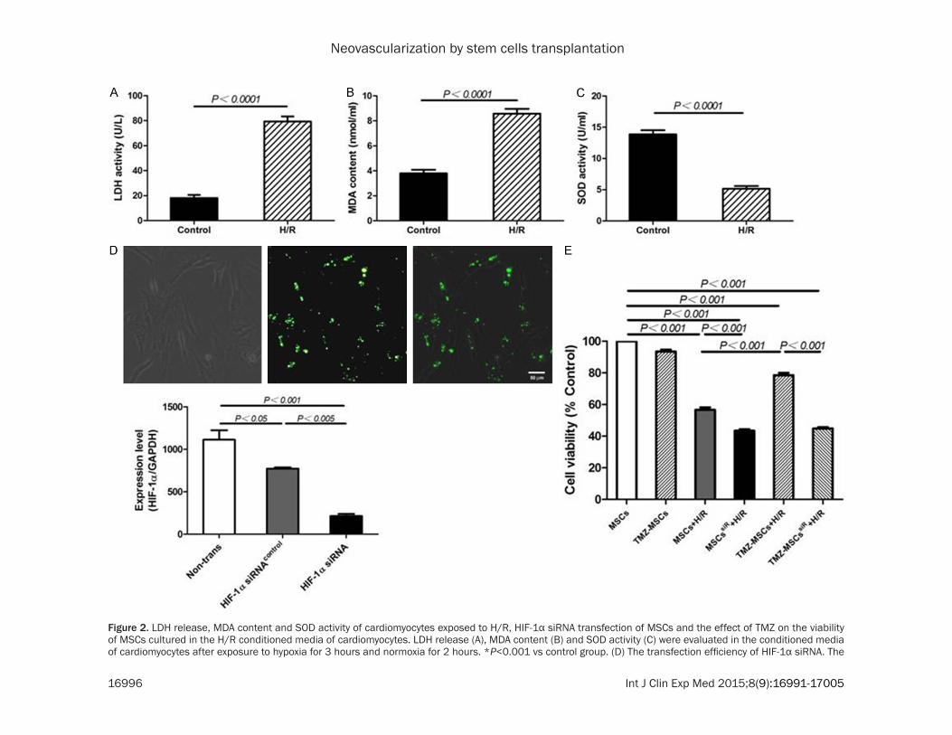
two hours, the H/R conditioned media were col-lected and the enzymatic activities of LDH and SOD and the MDA content in the media were detected. In the H/R conditioned media, the enzymatic activities of LDH were significantly increased compared with those in the control media (H/R: 79.3±4.1 vs control: 18.2±2.4, P<0.0001) (Figure 2A), and similar tendency was observed for the MDA content (H/R: 8.6±1.1 vs control: 3.8±0.8, P<0.0001) (Figure 2B), while the activity of SOD was decreased compared with that in the control media (H/R: 5.1±1.3 vs control: 13.9±1.8, P<0.0001) (Figure 2C). MSCs were transfected
Figure 1. GFP lentivirus transduction of MSCs and expression of MSCs surface markers measured by FACS analy-sis. A. Growth morphology of GFP lentivirus-transducted MSCs. B. Representative plots of CD90, CD29, CD34 and CD45, respectively. Data are expressed as relative mean fluorescence intensity ± SEM.
Neovascularization by stem cells transplantation
16996 Int J Clin Exp Med 2015;8(9):16991-17005
Figure 2. LDH release, MDA content and SOD activity of cardiomyocytes exposed to H/R, HIF-1α siRNA transfection of MSCs and the effect of TMZ on the viability of MSCs cultured in the H/R conditioned media of cardiomyocytes. LDH release (A), MDA content (B) and SOD activity (C) were evaluated in the conditioned media of cardiomyocytes after exposure to hypoxia for 3 hours and normoxia for 2 hours. *P<0.001 vs control group. (D) The transfection efficiency of HIF-1α siRNA. The

Neovascularization by stem cells transplantation
16997 Int J Clin Exp Med 2015;8(9):16991-17005
expression level of HIF-1α was inhibited to 20% by siRNA knocking down (0.19%±0.04, P<0.001). (E) The cell viability in the control group was normalized to 100%. The values represent the mean (% of control) ± SEM of each group in this experiment. MSCs vs MSCs+H/R, MSCssiR+H/R, TMZ-MSCs+H/R and TMZ-MSCssiR+H/R, P<0.001; MSCs+H/R vs MSCssiR+H/R and TMZ-MSCs+ H/R, P<0.001; TMZ-MSCs+H/R vs TMZ-MSCssiR+H/R, P<0.001.
Neovascularization by stem cells transplantation
16998 Int J Clin Exp Med 2015;8(9):16991-17005
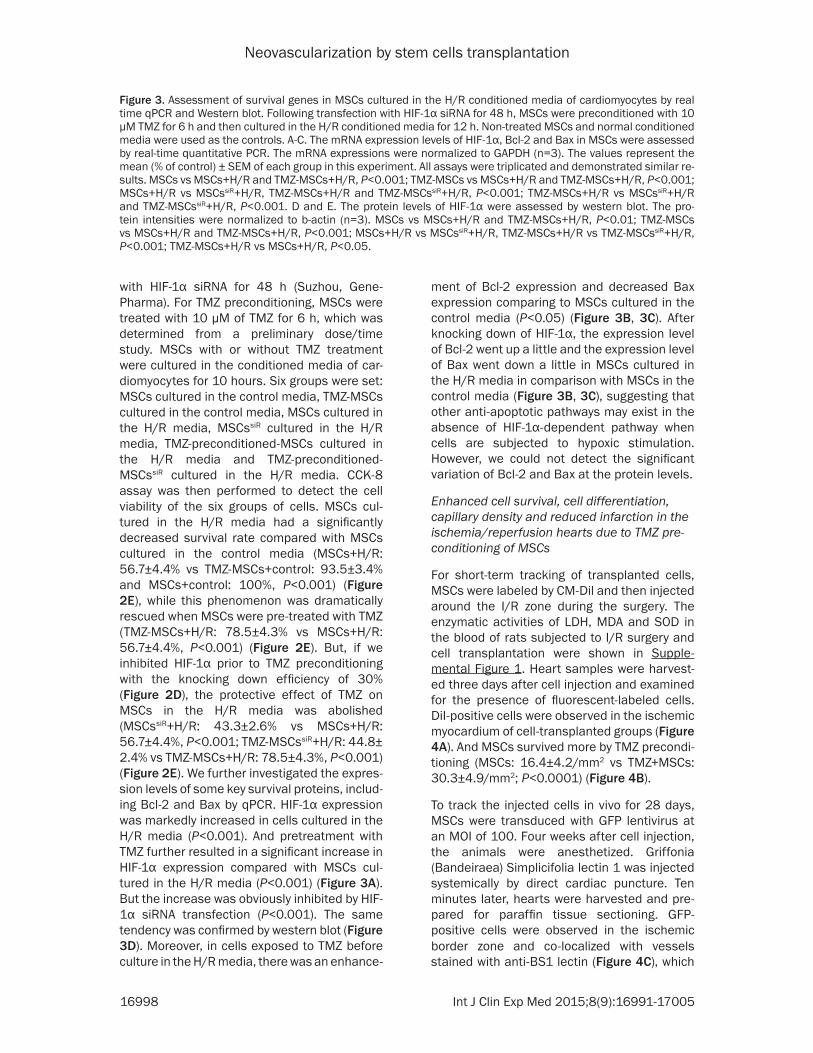
with HIF-1α siRNA for 48 h (Suzhou, Gene- Pharma). For TMZ preconditioning, MSCs were treated with 10 μM of TMZ for 6 h, which was determined from a preliminary dose/time study. MSCs with or without TMZ treatment were cultured in the conditioned media of car-diomyocytes for 10 hours. Six groups were set: MSCs cultured in the control media, TMZ-MSCs cultured in the control media, MSCs cultured in the H/R media, MSCssiR cultured in the H/R media, TMZ-preconditioned-MSCs cultured in the H/R media and TMZ-preconditioned-MSCssiR cultured in the H/R media. CCK-8 assay was then performed to detect the cell viability of the six groups of cells. MSCs cul-tured in the H/R media had a significantly decreased survival rate compared with MSCs cultured in the control media (MSCs+H/R: 56.7±4.4% vs TMZ-MSCs+control: 93.5±3.4% and MSCs+control: 100%, P<0.001) (Figure 2E), while this phenomenon was dramatically rescued when MSCs were pre-treated with TMZ (TMZ-MSCs+H/R: 78.5±4.3% vs MSCs+H/R: 56.7±4.4%, P<0.001) (Figure 2E). But, if we inhibited HIF-1α prior to TMZ preconditioning with the knocking down efficiency of 30% (Figure 2D), the protective effect of TMZ on MSCs in the H/R media was abolished (MSCssiR+H/R: 43.3±2.6% vs MSCs+H/R: 56.7±4.4%, P<0.001; TMZ-MSCssiR+H/R: 44.8± 2.4% vs TMZ-MSCs+H/R: 78.5±4.3%, P<0.001) (Figure 2E). We further investigated the expres-sion levels of some key survival proteins, includ-ing Bcl-2 and Bax by qPCR. HIF-1α expression was markedly increased in cells cultured in the H/R media (P<0.001). And pretreatment with TMZ further resulted in a significant increase in HIF-1α expression compared with MSCs cul-tured in the H/R media (P<0.001) (Figure 3A). But the increase was obviously inhibited by HIF-1α siRNA transfection (P<0.001). The same tendency was confirmed by western blot (Figure 3D). Moreover, in cells exposed to TMZ before culture in the H/R media, there was an enhance-
ment of Bcl-2 expression and decreased Bax expression comparing to MSCs cultured in the control media (P<0.05) (Figure 3B, 3C). After knocking down of HIF-1α, the expression level of Bcl-2 went up a little and the expression level of Bax went down a little in MSCs cultured in the H/R media in comparison with MSCs in the control media (Figure 3B, 3C), suggesting that other anti-apoptotic pathways may exist in the absence of HIF-1α-dependent pathway when cells are subjected to hypoxic stimulation. However, we could not detect the significant variation of Bcl-2 and Bax at the protein levels.
Enhanced cell survival, cell differentiation, capillary density and reduced infarction in the ischemia/reperfusion hearts due to TMZ pre-conditioning of MSCs
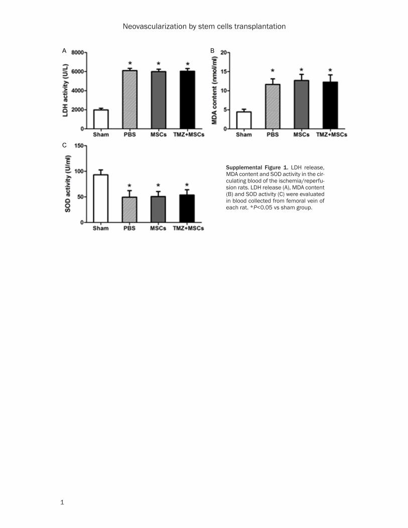
For short-term tracking of transplanted cells, MSCs were labeled by CM-Dil and then injected around the I/R zone during the surgery. The enzymatic activities of LDH, MDA and SOD in the blood of rats subjected to I/R surgery and cell transplantation were shown in Supple- mental Figure 1. Heart samples were harvest-ed three days after cell injection and examined for the presence of fluorescent-labeled cells. DiI-positive cells were observed in the ischemic myocardium of cell-transplanted groups (Figure 4A). And MSCs survived more by TMZ precondi-tioning (MSCs: 16.4±4.2/mm2 vs TMZ+MSCs: 30.3±4.9/mm2; P<0.0001) (Figure 4B).
To track the injected cells in vivo for 28 days, MSCs were transduced with GFP lentivirus at an MOI of 100. Four weeks after cell injection, the animals were anesthetized. Griffonia (Bandeiraea) Simplicifolia lectin 1 was injected systemically by direct cardiac puncture. Ten minutes later, hearts were harvested and pre-pared for paraffin tissue sectioning. GFP-positive cells were observed in the ischemic border zone and co-localized with vessels stained with anti-BS1 lectin (Figure 4C), which
Figure 3. Assessment of survival genes in MSCs cultured in the H/R conditioned media of cardiomyocytes by real time qPCR and Western blot. Following transfection with HIF-1α siRNA for 48 h, MSCs were preconditioned with 10 μM TMZ for 6 h and then cultured in the H/R conditioned media for 12 h. Non-treated MSCs and normal conditioned media were used as the controls. A-C. The mRNA expression levels of HIF-1α, Bcl-2 and Bax in MSCs were assessed by real-time quantitative PCR. The mRNA expressions were normalized to GAPDH (n=3). The values represent the mean (% of control) ± SEM of each group in this experiment. All assays were triplicated and demonstrated similar re-sults. MSCs vs MSCs+H/R and TMZ-MSCs+H/R, P<0.001; TMZ-MSCs vs MSCs+H/R and TMZ-MSCs+H/R, P<0.001; MSCs+H/R vs MSCssiR+H/R, TMZ-MSCs+H/R and TMZ-MSCssiR+H/R, P<0.001; TMZ-MSCs+H/R vs MSCssiR+H/R and TMZ-MSCssiR+H/R, P<0.001. D and E. The protein levels of HIF-1α were assessed by western blot. The pro-tein intensities were normalized to b-actin (n=3). MSCs vs MSCs+H/R and TMZ-MSCs+H/R, P<0.01; TMZ-MSCs vs MSCs+H/R and TMZ-MSCs+H/R, P<0.001; MSCs+H/R vs MSCssiR+H/R, TMZ-MSCs+H/R vs TMZ-MSCssiR+H/R, P<0.001; TMZ-MSCs+H/R vs MSCs+H/R, P<0.05.

Neovascularization by stem cells transplantation
16999 Int J Clin Exp Med 2015;8(9):16991-17005
Figure 4. Assessment of cell recruitment and cell differentiation in the ischemia/reperfusion hearts. MSCs were preconditioned with 10 μM TMZ for 6 h and stained with DiI. DiI-labeled cells were injected into the peri-infarct zone during the I/R surgery, and heart samples were examined histologically three days after cell in-jection. A. DiI positive cells (red) detected in ischemic myocardium under a fluorescence microscope. B. Number of DiI positive cells was counted in the ischemic

Neovascularization by stem cells transplantation
17000 Int J Clin Exp Med 2015;8(9):16991-17005
indicated the endothelial differentiation of MSCs. Hearts injected with MSCs by TMZ pre-conditioning had higher number of GFP+/BS1 lectin+ cells than those injected with non-treat-ed MSCs (TMZ+MSCs: 5±1.8 vs MSCs: 1.5±1.3, P<0.05) (Figure 4D). The staining of in vivo-per-fused BS1 lectin and α-smooth muscle actin reflects angiogenesis in functional vessels in the peri-infarct myocardium four weeks after I/R in all groups (Figure 5). The averaged capil-lary density in the ischemic border zones of LV, an index of neovascularization, was significant-ly greater in the TMZ-preconditioned-MSCs-injected group compared to the MSCs-injected
group (TMZ+MSCs: 78±3 vs MSCs: 61±4 and PBS: 49±4, P<0.001) (Figure 5B). Although the averaged number of arterioles was higher in the group with TMZ-preconditioned-MSCs injec-tion, no significant difference could be found between the groups with TMZ-preconditioned-MSCs injection and MSCs injection (TMZ+MSCs: 7±1 vs MSCs: 5±1 and PBS: 2.7±0.5, P<0.005) (Figure 5D).
Reduced fibrosis in the ischemia/reperfusion hearts due to TMZ preconditioning of MSCs
Heart sections stained with TTC showed exten-sive infarction in the PBS-injected hearts
border zone (bilateral sides of peri-infarct area) and averaged (n=3). MSCs vs TMZ+MSCs, P<0.0001. MSCs were transduced with GFP lentivirus, and then preconditioned with 10 μM TMZ for 6 h. The cells were injected into the peri-infarct zone during the I/R surgery, and heart samples were examined histologically 28 days after cell injection. C. Capillaries were visualized by immunofluorescent staining for BS1 lectin (red). Cells co-localizing with capillaries were marked by arrows and shown in yellow. D. The numbers of cells co-localized with capillaries were counted under a fluorescence microscope separately and averaged. Arrows indicate the double positive cells. MSCs vs TMZ+MSCs, P=0.006.
Figure 5. Histological analysis for capillary density and arterioles in ischemic myocardium. Heart samples were har-vested following BS1 lectin systemic perfusion 28 days after cell injection. A. Capillaries were visualized as tubular structure perfused by BS1 lectin. B. The numbers of capillaries were counted in bilateral sides of the peri-infarct zone on LV cross sections and averaged in each group (n=3), respectively. PBS vs MSCs and TMZ+MSCs, P<0.005; MSCs vs TMZ+MSCs, P<0.001. C. Arterioles were visualized following a-smooth muscle actin staining. D. The num-bers of arterioles were counted in bilateral sides of the peri-infarct zone on LV cross sections and averaged in each group (n=3), respectively. PBS vs TMZ+MSCs, P<0.005.

Neovascularization by stem cells transplantation
17001 Int J Clin Exp Med 2015;8(9):16991-17005
(Figure 6A). The extent of infarction was signifi-cantly reduced in the MSCs group compared with the PBS group (MSCs: 22.9±2.2% vs PBS: 31.2±3.2%, P<0.05) (Figure 6B). The hearts that received MSCs pretreated with TMZ showed significantly further reduction of infarc-tion compared with the PBS group (TMZ+MSCs: 17.0±1.5% vs PBS: 31.2±3.2%, P<0.001) (Figure 6B).
Over-expression of survival proteins in the in-farct hearts transplanted with preconditioned MSCs
To clarify whether MSCs-transplantation pro-moted survival of the infarcted myocardium, we dissected the LV area of the infarct hearts and performed western blot analysis. In western blot analysis, HIF-1α and Bcl-2 dramatically over-expressed in hearts implanted with TMZ-treated MSCs compared with those implanted with MSCs and PBS (P<0.001), whereas the expression level of Bax was decreased due to the injection of TMZ-MSCs (P<0.05) (Figure 7A-D).
Preconditioned MSCs effectively preserve LV function after ischemia/reperfusion
Echocardiography performed four weeks after ischemia/reperfusion revealed significantly
higher values of LVEF and FS in the MSCs group as compared to the PBS group (Figure 8) and the preconditioned MSCs further attenuated LV remodeling. LVEF was 54.7±2.3% in the PBS group, 63.1±2.0% in the MSCs group and 70.7±2.8% in the TMZ+MSCs group (n=9, P<0.001 vs PBS and MSCs groups) (Figure 8B, 8C). Similarly, FS was 26.2±1.5% in the PBS group, 31.0±2.7% in the MSCs group and 35.2±1.7% in the TMZ+MSCs group (n=9, P<0.05 vs PBS and MSCs groups) (Figure 8B, 8C).
Discussion
The low survival ratio of transplanted stem cells becomes a hurdle for their clinical use. Besides the time point and route of transplantation, cell death in the acute phase is assumed to be the critical factor that restricts the protective effects of MSCs. Genetic modification and pre-conditioning of MSCs are the most popular and effective ways to promote the survival and keep the function of MSCs. Whether permanent genetic modification could cause tumor or not remains an unsolved question. On the contrary, preconditioning of stem cells could enhance the transplantation efficiency in a fast, simple and effective way. And pharmacological pre-conditioning of stem cells becomes a new
Figure 6. Histological analysis for fibrosis size in isch-emic myocardium. Heart samples were harvested 28 days after cell injection. A. Heart sections were stained by TTC staining. Red indicates intact myo-cardium and white indicates scared fibrosis area. B. The percent of infarct area in entire LV cross sec-tional area was calculated and averaged (n=3). PBS vs MSCs, P<0.05; PBS vs TMZ+MSCs, P<0.001.

Neovascularization by stem cells transplantation
17002 Int J Clin Exp Med 2015;8(9):16991-17005
promising method for functional maintenance. Trimetazidine (TMZ) is a partial inhibitor of lipid oxidation. It has been proposed as a metabolic regulator for several cardiovascular patholo-gies. TMZ has also shown cytoprotective effi-cacy in several models of myocardial infarction [13]. The pharmacological efficacy of TMZ in augmenting myocardial stem cell therapy has been reported in two research groups [14, 15]. They demonstrated that preconditioning of MSCs by TMZ before implantation offered a sig-nificant enhancement in the functional recov-ery of infarcted myocardium. However, the mechanism by which TMZ rescued MSCs in I/R injury in vitro is not clarified, also the protective effect of TMZ on MSCs and the neovasculariza-tion of ischemic hearts is not deeply investigat-ed. Therefore, we evaluated the protective role of TMZ precondition on stem cells for transplantation.
Hypoxia-inducible factor-1 (HIF-1), the most important factor involved in the cellular
response to hypoxia, has been extensively studied. HIF-1 is specifically activated in hypox-ia and induces target genes involved in cell pro-liferation, vascular development [16], vascular tone, and energy metabolism [17]. The broad impact of HIF-1 on cell biology is that, among all hypoxia target genes, over 70 have so far been identified as being regulated by HIF-1 [18]. HIF-1 directly activates the transcription of the VEGF gene by binding to a hypoxia response element located 50 to the gene [19]. Reports by Kim et al. and Papandreou et al. demon-strate that HIF-1 performs an active suppres-sion of mitochondrial pyruvate catabolism and O2 consumption in hypoxic cells [20, 21], while TMZ could optimize the cell metabolism by reducing fatty acid oxidation through the selec-tive inhibition of mitochondrial 3-ketoacyl CoA thiolase. Therefore, it is very likely that TMZ plays the role in a HIF-1α-dependent way. We proposed that TMZ could recover the function of MSCs exposed to ischemia/reperfusion inju-ry through up-regulation of HIF-1α.
Figure 7. Analysis of hypoxia and survival proteins expressed by the ischemia/reperfusion hearts. The LV area of the infarct hearts was dissected 28 days after cell transplantation and western blot analysis was performed. A. Representative blots of HIF-1α, Bcl-2 and Bax are shown. B-D. Quantitative analysis of HIF-1α, Bcl-2 and Bax using data obtained from three different blots and expressed as mean ± S.D. HIF-1α, MSCs vs Sham and PBS, P<0.05; TMZ+MSCs vs Sham, PBS and MSCs, P<0.001. Bcl-2, TMZ+MSCs vs Sham, PBS and MSCs, P<0.001. Bax, TMZ+MSCs vs Sham and PBS, P<0.05.

Neovascularization by stem cells transplantation
17003 Int J Clin Exp Med 2015;8(9):16991-17005
In our study, we used 10 μM of TMZ to treat MSCs for 6 hours. We found that short term exposure of MSCs to TMZ can significantly enhance cell viability in the H/R conditioned media. But, if we knocked down HIF-1α prior to TMZ preconditioning, surprisingly, this effect could be abolished (Figure 2D). The Bcl-2 family can be divided into three classes: BH3-only proteins that are activated by various forms of
cellular stress, Bax and Bak proteins that medi-ate mitochondrial membrane permeabilization, and inhibitory proteins such as Bcl-2 and Bcl-XL [22]. Certain members of the BCL-2 pro-tein family, such as Bcl-2, Bcl-xl and Mcl-1 are anti-apoptotic, whilst others like Bax are pro-apoptotic. Bcl-2 is specifically considered as an important anti-apoptotic protein and is thus classified as an oncogene. Apoptosis regulator
Figure 8. Recovery of cardiac function at 4 weeks after MSCs transplantation in the ischemia/reperfusion hearts. Transthoracic echocardiography was performed in the Sham group, the PBS-injected group, I/R hearts treated with MSCs (MSCs), and I/R hearts treated with MSCs preconditioned with TMZ (TMZ+MSCs). Representative recordings of M-mode echocardiogram (A), LV ejection fraction (EF) (B), and fraction shortening (FS) (C) are shown. I/R hearts treated with TMZ-preconditioned cells had greater functional recovery than those treated with non-preconditioned cells. EF, PBS vs Sham, P<0.001; MSCs vs Sham and PBS, P<0.001; TMZ+MSCs vs Sham, PBS and MSCs, P<0.001. FS, PBS vs Sham, P<0.001; MSCs vs Sham and PBS, P<0.005; TMZ+MSCs vs PBS and MSCs, P<0.05.

Neovascularization by stem cells transplantation
17004 Int J Clin Exp Med 2015;8(9):16991-17005
BAX promotes apoptosis by binding to and antagonizing the Bcl-2 protein [23]. Therefore, we further checked Bcl-2 and Bax expression in MSCs exposed to the H/R media by qPCR. There was a concomitant increase of Bcl-2 expression and a decrease of Bax expression when cells were cultured in the H/R media fol-lowing TMZ preconditioning. But after knocking down of HIF-1α, the expression level of Bcl-2 also went up a little and the expression level of Bax went down a little in MSCs cultured in the H/R media in comparison with MSCs in the control media, suggesting that other anti-apop-totic pathways may exist in the absence of HIF-1α-dependent pathway when cells are subject-ed to hypoxic stimulation. The exact interaction among HIF-1α, Bcl-2 and Bax needs further study. As for the in vivo study, TMZ precondi-tioning of MSCs could enhance cell survival and capillary density following transplantation into the ischemic myocardium, thus contributing to the reduced fibrosis and the preserved LV func-tion after ischemia/reperfusion.
In conclusion, TMZ-treated MSCs transplanta-tion exhibited a significant improvement of car-diac function with reduced infarct size following ischemia/reperfusion compared with non-treated MSCs transplantation. The favorable effect of TMZ-treatment to MSCs could be attributed to increased cell survival in ischemic myocardium. This simple and effective treat-ment to MSCs might be a promising strategy for autologous cell therapy in patients with isch-emic heart diseases.
Acknowledgements
This work was supported by the National Natural Science Foundation of China (No. 30972696 and No. 81400199) and Suzhou Municipal Science and Technology Project (No. SYS201414).
Disclosure of conflict of interest
None.
Address correspondence to: Dr. Jie Hui, Department of Cardiology of The First Affiliated Hospital, Soochow University, 188, Shizi Street, Suzhou 215006, China. Tel: +86-0512-6778-1112; E-mail: [email protected]; Dr. Zhenya Shen, Institute for Cardiovascular Science & Department of Ca- rdiovascular Surgery of The First Affiliated Hospital, Soochow University, 188, Shizi Street, Suzhou
215006, China. Tel: +86-0512-6778-0100; E-mail: [email protected]
References
[1] Mozaffari MS, Liu JY, Abebe W, Baban B. Mechanisms of load dependency of myocardi-al ischemia reperfusion injury. Am J Cardiovasc Dis 2013; 3: 180-196.
[2] Souidi N, Stolk M, Seifert M. Ischemia-reperfusion injury: beneficial effects of mesen-chymal stromal cells. Curr Opin Organ Transplant 2013; 18: 34-43.
[3] Haider H, Ashraf M. Preconditioning and stem cell survival. J Cardiovasc Transl Res 2010; 3: 89-102.
[4] Pasha Z, Wang Y, Sheikh R, Zhang D, Zhao T, Ashraf M. Preconditioning enhances cell sur-vival and differentiation of stem cells during transplantation in infarcted myocardium. Cardiovasc Res 2008; 77: 134-142.
[5] Kutschka I, Kofidis T, Chen IY, von Degenfeld G, Zwierzchoniewska M, Hoyt G, Arai T, Lebl DR, Hendry SL, Sheikh AY, Cooke DT, Connolly A, Blau HM, Gambhir SS, Robbins RC. Adenoviral human BCL-2 transgene expression attenu-ates early donor cell death after cardiomyo-blast transplantation into ischemic rat hearts. Circulation 2006; 114 Suppl: I174-80.
[6] Li W, Ma N, Ong LL, Nesselmann C, Klopsch C, Ladilov Y, Furlani D, Piechaczek C, Moebius JM, Lützow K, Lendlein A, Stamm C, Li RK, Steinhoff G. Bcl-2 engineered MSCs inhibited apoptosis and improved heart function. Stem Cells 2007; 25: 2118-2127.
[7] Matsumoto R, Omura T, Yoshiyama M, Hayashi T, Inamoto S, Koh k, Izumi Y, Nakamura Y, Akioka K, Kitaura Y, Takeuchi K, Yoshikawa J. Vascular endothelial growth factor-expressing mesenchymal stem cell transplantation for the treatment of acute myocardial infarction. Arterioscler Thromb Vasc Biol 2005; 25: 1168-1173.
[8] Niagara MI, Haider H, Jiang S, Ashraf M. Pharmacologically preconditioned skeletal myoblasts are resistant to oxidative stress and promote angiomyogenesis via release of para-crine factors in the infarcted heart. Circ Res 2007; 100: 545-555.
[9] McClellan KJ, Plosker GL. Trimetazidine. A re-view of its use in stable angina pectoris and other coronary conditions. Drugs 1999; 58: 143-157.
[10] Gambert S, Vergely C, Filomenko R, Moreau D, Bettaieb A, Opie LH, Rochette L. Adverse ef-fects of free fatty acid associated with in-creased oxidative stress in postischemic iso-lated rat hearts. Mol Cell Biochem 2006; 283: 147-152.
Neovascularization by stem cells transplantation
17005 Int J Clin Exp Med 2015;8(9):16991-17005
[11] Monteiro P, Duarte AI, Goncalves LM, Moreno A, Providencia LA. Protective effect of trimeta-zidine on myocardial mitochondrial function in an ex-vivo model of global myocardial isch-emia. Eur J Pharmacol 2004; 503: 123-128.
[12] Kantor PF, Lucien A, Kozak R, Lopaschuk GD. The antianginal drug trimetazidine shifts car-diac energy metabolism from fatty acid oxida-tion to glucose oxidation by inhibiting mito-chondrial long-chain 3-ketoacyl coenzyme A thiolase. Circ Res 2000; 86: 580-588.
[13] Pantos C, Bescond-Jacquet A, Tzeis S, Paizis I, Mourouzis I, Moraitis P, Malliopoulou V, Politi ED, Karageorgiou H, Varonos D, Cokkinos DV. Trimetazidine protects isolated rat hearts against ischemia-reperfusion injury in an ex-perimental timing-dependent manner. Basic Res Cardiol 2005; 100: 154-160.
[14] Wisel S, Khan M, Kuppusamy ML, Mohan IK, Chacko SM, Rivera BK, Sun BC, Hideg K, Kuppusamy P. Pharmacological precondition-ing of mesenchymal stem cells with trimeta- zidine (1-[2,3,4-trimethoxybenzyl]piperazine) protects hypoxic cells against oxidative stress and enhances recovery of myocardial function in infarcted heart through Bcl-2 expression. J Pharmacol Exp Ther 2009; 329: 543-550.
[15] Xu H, Zhu G, Tian Y. Protective effects of trimetazidine on bone marrow mesenchymal stem cells viability in an ex vivo model of hy-poxia and in vivo model of locally myocardial ischemia. J Huazhong Univ Sci Technolog Med Sci 2012; 32: 36-41.
[16] Lando D, Peet DJ, Whelan DA, Gorman JJ, Whitelaw ML. Asparagine hydroxylation of the HIF transactivation domain a hypoxic switch. Science 2002; 295: 858-861.
[17] Jain S, Maltepe E, Lu MM, Simon C, Bradfield CA. Expression of ARNT, ARNT2, HIF1 alpha, HIF2 alpha and Ah receptor mRNAs in the de-veloping mouse. Mech Dev 1998; 73: 117-123.
[18] Manalo DJ, Rowan A, Lavoie T, Natarajan L, Kelly BD, Ye SQ, Garcia JG, Semenza GL. Transcriptional regulation of vascular endothe-lial cell responses to hypoxia by HIF-1. Blood 2005; 105: 659-669.
[19] Chilov D, Camenisch G, Kvietikova I, Ziegler U, Gassmann M, Wenger RH. Induction and nu-clear translocation of hypoxia-inducible fac-tor-1 (HIF-1): heterodimerization with ARNT is not necessary for nuclear accumulation of HIF-1alpha. J Cell Sci 1999; 112 8: 1203-1212.
[20] Kim JW, Tchernyshyov I, Semenza GL, Dang CV. HIF-1-mediated expression of pyruvate dehy-drogenase kinase: a metabolic switch required for cellular adaptation to hypoxia. Cell Metab 2006; 3: 177-185.
[21] Papandreou I, Cairns RA, Fontana L, Lim AL, Denko NC. HIF-1 mediates adaptation to hy-poxia by actively downregulating mitochondrial oxygen consumption. Cell Metab 2006; 3: 187-197.
[22] Cory S, Huang DC, Adams JM. The Bcl-2 family: roles in cell survival and oncogenesis. Oncogene 2003; 22: 8590-8607.
[23] Leber B, Lin J, Andrews DW. Still embedded to-gether binding to membranes regulates Bcl-2 protein interactions. Oncogene 2010; 29: 5221-5230.
Neovascularization by stem cells transplantation
1
Supplemental Figure 1. LDH release, MDA content and SOD activity in the cir-culating blood of the ischemia/reperfu-sion rats. LDH release (A), MDA content (B) and SOD activity (C) were evaluated in blood collected from femoral vein of each rat. *P<0.05 vs sham group.