Origin of intrinsic irregular firing in cortical interneurons
6
Origin of intrinsic irregular firing in cortical interneurons Klaus M. Stiefel a,b,c,1 , Bernhard Englitz a,b,c,2 , and Terrence J. Sejnowski a,b,c,d,3 a Computational Neurobiology Laboratory and b Howard Hughes Medical Institute, c Salk Institute for Biological Studies, La Jolla, CA 92037; and d Division of Biological Sciences, University of California at San Diego, La Jolla, CA 92093 Contributed by Terrence J. Sejnowski, March 22, 2013 (sent for review July 29, 2012) Cortical spike trains are highly irregular both during ongoing, spontaneous activity and when driven at high firing rates. There is uncertainty about the source of this irregularity, ranging from intrinsic noise sources in neurons to collective effects in large-scale cortical networks. Cortical interneurons display highly irregular spike times (coefficient of variation of the interspike intervals >1) in response to dc-current injection in vitro. This is in marked con- trast to cortical pyramidal cells, which spike highly irregularly in vivo, but regularly in vitro. We show with in vitro recordings and computational models that this is due to the fast activation kinetics of interneuronal K + currents. This explanation holds over a wide parameter range and with Gaussian white, power-law, and Ornstein– Uhlenbeck noise. The intrinsically irregular spiking of interneurons could contribute to the irregularity of the cortical network. inhibitory interneuron | bistability | neural noise | fluctuations | cortex W hen spike trains of cortical pyramidal neurons are recorded in vivo from the cortices of awake or sleeping cats (1–3) or awake monkeys (4), the interspike intervals (ISIs) are highly vari- able, with coefficients of variation (CV ISI = ISI SD/ISI mean) >1. However, when the main cortical excitatory (pyramidal) neu- rons are depolarized in vitro by injection of constant current to above firing threshold, their spike trains are substantially more regular (CV ISI <0.5). The irregularity of pyramidal neuron firing in vivo arises from the intense, ongoing, temporally correlated synaptic activity that bombards cortical neurons (1, 2, 5–7). The lower in vitro irregularity can be raised to in vivo levels by using fluctuating current injection (8), hyperosmolar solution (9), and a neuromodulatory mixture (10, 11). Computational models of irregular neuronal firing typically include network synaptic activity represented by stochastic ag- gregate processes. Fluctuating inputs have been represented by Brownian motion (12) or an Ornstein–Uhlenbeck process (8) in both single- and multicompartmental neuronal models (5). These are externally induced fluctuations and do not mechanistically explain the variety of irregular discharge patterns observed in cortical interneurons. In this study we combined in vitro recordings and computational models of cortical interneurons to show how neuronal conductances interact with the stochastic input to produce the variety of observed neuronal phenotypes. Results Recordings from Cortical Inhibitory Interneurons. Cortical GABAergic interneurons have a range of functions including the gating (13) and entrainment (14) of neuronal firing, dendritic integration (15), synaptic plasticity (16), and the generation of network oscillations (10, 11). There are several morphologically and physiologically distinct classes of interneurons (17). In contrast to pyramidal neurons, a constant current injected into mouse visual cortex layer 2/3 interneurons in vitro fre- quently produced a spike train with a CV ISI that substantially exceeds 1, often producing highly irregular sequences of burst. This observation is in accord with earlier descriptions of spike trains of these interneurons and used as a criterion in inter- neuron classifications (17, 18). The irregular spike trains were observed even under blockade of glutamatergic and GABAergic synaptic transmission and thus must be a consequence of an intrinsic process. Their intrinsically irregular spike trains hence make interneurons plausible candidates for the origin of cor- tical spiking irregularity. We made multiple, long recordings from 14 interneurons and classified them according to their firing patterns as described in Markram et al. (17). In short, when depolarized by a step pulse just above rheobase, the firing patterns potentially display ac- commodation (successive lengthening of ISIs) and irregular clustering of spikes to different degrees (irregular spiking to stuttering). The accommodating and nonaccommodating neu- rons are further subdivided into classic, bursting, and delayed firing neurons (c, b, and d). Of the neurons we recorded from, four were stuttering neu- rons (ST; Fig. 1A), four were irregular spiking (IS; Fig. 1B), three were nonaccommodating (NA; one each of the c-, b- and d-NA type, Fig. 1C), and three were accommodating (AC; all c-AC, Fig. 1D). For comparison purposes, we also recorded from two pyramidal neurons (Fig. 1E). As expected, the pyramidal cells emitted regular spike trains in response to current-step injection, as did the AC and NA interneurons. In contrast, the ST and IS interneurons displayed highly irregular spike trains in response to current-step injection, some even at high discharge rates (CV ISI = 1.21 at 28 Hz). The spike trains of ST and IS interneurons firing at low fre- quencies consisted of individual spikes spaced at irregular intervals. Bursts occurred at higher frequencies, with the spikes within each burst separated by afterhyperpolarization (AHP) (Fig. 1F). There was no voltage envelope below the bursts as in thalamic relay neurons (19). At even higher frequencies these bursts fused, leading to continuous spiking and a drop in CV ISI below 0.5 in five of eight cells tested above a cell-specific fre- quency. Thus, we observed an inverse dependence of the CV ISI on the firing frequency in these cells. In a related study, we demonstrated, using nonlinear time- series analysis, that the interspike intervals of ST and IS in- terneuron spike trains were purely stochastic and showed no detectable signs of higher-order determinism or chaos (20). We therefore searched for alternative, stochastic mechanisms that could generate the irregular spike trains. Subthreshold voltage fluctuations occur in pyramidal cells and interneurons and are voltage-dependent (Fig. 2). The most likely source of membrane potential noise in pharmacologically iso- lated neurons in slices is fluctuating ion channel gating, which is voltage-dependent (21). Although the amplitude of the noise in the membrane potential is predicted to be small, the extrapolated Author contributions: K.M.S., B.E., and T.J.S. designed research; K.M.S. and B.E. performed research; K.M.S., B.E., and T.J.S. analyzed data; and K.M.S., B.E., and T.J.S. wrote the paper. The authors declare no conflict of interest. Freely available online through the PNAS open access option. 1 Present address: Bioelectronics and Neuroscience, MARCS Institute, University of Western Sydney, Penrith, NSW 2751, Australia. 2 Present address: Institute for Systems Research, University of Maryland, College Park, MD 20742. 3 To whom correspondence should be addressed. E-mail: [email protected]. This article contains supporting information online at www.pnas.org/lookup/suppl/doi:10. 1073/pnas.1305219110/-/DCSupplemental. 7886–7891 | PNAS | May 7, 2013 | vol. 110 | no. 19 www.pnas.org/cgi/doi/10.1073/pnas.1305219110
Transcript of Origin of intrinsic irregular firing in cortical interneurons

Origin of intrinsic irregular firing incortical interneuronsKlaus M. Stiefela,b,c,1, Bernhard Englitza,b,c,2, and Terrence J. Sejnowskia,b,c,d,3
aComputational Neurobiology Laboratory and bHoward Hughes Medical Institute, cSalk Institute for Biological Studies, La Jolla, CA 92037;and dDivision of Biological Sciences, University of California at San Diego, La Jolla, CA 92093
Contributed by Terrence J. Sejnowski, March 22, 2013 (sent for review July 29, 2012)
Cortical spike trains are highly irregular both during ongoing,spontaneous activity and when driven at high firing rates. There isuncertainty about the source of this irregularity, ranging fromintrinsic noise sources in neurons to collective effects in large-scalecortical networks. Cortical interneurons display highly irregularspike times (coefficient of variation of the interspike intervals >1)in response to dc-current injection in vitro. This is in marked con-trast to cortical pyramidal cells, which spike highly irregularly invivo, but regularly in vitro. We show with in vitro recordings andcomputational models that this is due to the fast activation kineticsof interneuronal K+ currents. This explanation holds over a wideparameter range andwith Gaussian white, power-law, and Ornstein–Uhlenbeck noise. The intrinsically irregular spiking of interneuronscould contribute to the irregularity of the cortical network.
inhibitory interneuron | bistability | neural noise | fluctuations | cortex
When spike trains of cortical pyramidal neurons are recordedin vivo from the cortices of awake or sleeping cats (1–3) or
awake monkeys (4), the interspike intervals (ISIs) are highly vari-able, with coefficients of variation (CVISI = ISI SD/ISI mean) >1.However, when the main cortical excitatory (pyramidal) neu-rons are depolarized in vitro by injection of constant current toabove firing threshold, their spike trains are substantially moreregular (CVISI <0.5). The irregularity of pyramidal neuron firingin vivo arises from the intense, ongoing, temporally correlatedsynaptic activity that bombards cortical neurons (1, 2, 5–7). Thelower in vitro irregularity can be raised to in vivo levels by usingfluctuating current injection (8), hyperosmolar solution (9), anda neuromodulatory mixture (10, 11).Computational models of irregular neuronal firing typically
include network synaptic activity represented by stochastic ag-gregate processes. Fluctuating inputs have been represented byBrownian motion (12) or an Ornstein–Uhlenbeck process (8) inboth single- and multicompartmental neuronal models (5). Theseare externally induced fluctuations and do not mechanisticallyexplain the variety of irregular discharge patterns observed incortical interneurons. In this study we combined in vitro recordingsand computational models of cortical interneurons to show howneuronal conductances interact with the stochastic input toproduce the variety of observed neuronal phenotypes.
ResultsRecordings from Cortical Inhibitory Interneurons.Cortical GABAergicinterneurons have a range of functions including the gating (13)and entrainment (14) of neuronal firing, dendritic integration(15), synaptic plasticity (16), and the generation of networkoscillations (10, 11). There are several morphologically andphysiologically distinct classes of interneurons (17).In contrast to pyramidal neurons, a constant current injected
into mouse visual cortex layer 2/3 interneurons in vitro fre-quently produced a spike train with a CVISI that substantiallyexceeds 1, often producing highly irregular sequences of burst.This observation is in accord with earlier descriptions of spiketrains of these interneurons and used as a criterion in inter-neuron classifications (17, 18). The irregular spike trains wereobserved even under blockade of glutamatergic and GABAergicsynaptic transmission and thus must be a consequence of an
intrinsic process. Their intrinsically irregular spike trains hencemake interneurons plausible candidates for the origin of cor-tical spiking irregularity.We made multiple, long recordings from 14 interneurons and
classified them according to their firing patterns as described inMarkram et al. (17). In short, when depolarized by a step pulsejust above rheobase, the firing patterns potentially display ac-commodation (successive lengthening of ISIs) and irregularclustering of spikes to different degrees (irregular spiking tostuttering). The accommodating and nonaccommodating neu-rons are further subdivided into classic, bursting, and delayedfiring neurons (c, b, and d).Of the neurons we recorded from, four were stuttering neu-
rons (ST; Fig. 1A), four were irregular spiking (IS; Fig. 1B), threewere nonaccommodating (NA; one each of the c-, b- and d-NAtype, Fig. 1C), and three were accommodating (AC; all c-AC,Fig. 1D). For comparison purposes, we also recorded from twopyramidal neurons (Fig. 1E). As expected, the pyramidal cellsemitted regular spike trains in response to current-step injection,as did the AC and NA interneurons. In contrast, the ST and ISinterneurons displayed highly irregular spike trains in responseto current-step injection, some even at high discharge rates(CVISI = 1.21 at 28 Hz).The spike trains of ST and IS interneurons firing at low fre-
quencies consisted of individual spikes spaced at irregularintervals. Bursts occurred at higher frequencies, with the spikeswithin each burst separated by afterhyperpolarization (AHP)(Fig. 1F). There was no voltage envelope below the bursts as inthalamic relay neurons (19). At even higher frequencies thesebursts fused, leading to continuous spiking and a drop in CVISIbelow 0.5 in five of eight cells tested above a cell-specific fre-quency. Thus, we observed an inverse dependence of the CVISIon the firing frequency in these cells.In a related study, we demonstrated, using nonlinear time-
series analysis, that the interspike intervals of ST and IS in-terneuron spike trains were purely stochastic and showed nodetectable signs of higher-order determinism or chaos (20).We therefore searched for alternative, stochastic mechanismsthat could generate the irregular spike trains.Subthreshold voltage fluctuations occur in pyramidal cells and
interneurons and are voltage-dependent (Fig. 2). The most likelysource of membrane potential noise in pharmacologically iso-lated neurons in slices is fluctuating ion channel gating, which isvoltage-dependent (21). Although the amplitude of the noise inthe membrane potential is predicted to be small, the extrapolated
Author contributions: K.M.S., B.E., and T.J.S. designed research; K.M.S. and B.E. performedresearch; K.M.S., B.E., and T.J.S. analyzed data; and K.M.S., B.E., and T.J.S. wrotethe paper.
The authors declare no conflict of interest.
Freely available online through the PNAS open access option.1Present address: Bioelectronics and Neuroscience, MARCS Institute, University of WesternSydney, Penrith, NSW 2751, Australia.
2Present address: Institute for Systems Research, University of Maryland, College Park,MD 20742.
3To whom correspondence should be addressed. E-mail: [email protected].
This article contains supporting information online at www.pnas.org/lookup/suppl/doi:10.1073/pnas.1305219110/-/DCSupplemental.
7886–7891 | PNAS | May 7, 2013 | vol. 110 | no. 19 www.pnas.org/cgi/doi/10.1073/pnas.1305219110

noise at firing threshold is actually larger in pyramidal neurons(Table 1), which have the most regular interspike intervals.Different amplitudes of voltage noise could therefore not explainthe differences in the CVISI. The difference between pyramidalneurons and interneurons must thus be found in their suscepti-bility to transform these subthreshold fluctuations into spike-time variability.
Biophysical Simulations of Pyramidal Neurons and Interneurons. Oneobvious difference in the spike waveforms of interneurons andpyramidal cells is the duration of the AHP (Fig.1), which is muchshorter in interneurons than in pyramidal cells. This is caused bythe faster activation time constants (τs) of the K+ currents me-diating the AHP. In pyramidal cells, the currents mediating theAHP are IAHP, IAHPs, and IM. These currents have τ time con-stants ranging from 70 ms to 200 ms (21, 22). In interneurons, theAHP is mediated by IKDR-type K+ currents and ID, with timeconstants ranging from 5 ms to 20 ms. We explored the roleof this difference in a single-compartmental neuronal model,which is an expansion of the classic Hodgkin–Huxley equations
(with parameters reproducing cortical spiking) by one con-ductance, a K+ current involved in the AHP (Methods). In thiscase, gKS does not represent a specific current, but serves asan abstraction for the sum of all K+ currents involved inthe AHP.We varied the time constant of this K+ current from the in-
terneuronal range (τs ∼6 ms) to the pyramidal range (τs ∼200 ms)with all of the other conductances remaining the same. The ki-netics of the K+ conductance used in the simulations is given inMethods. A single K+ conductance gating time constant τm wasvaried over the range found in the AHPs of interneurons (fast τs)and pyramidal cells (slow τs). We found that faster τs producedhigher output spike variability (Fig. 3). This effect was also notdependent on the different firing rates between models witha fast and a slow τs (Fig. 3E).To further investigate the connection between τs and spike-
time irregularity, we observed the dynamics of spiking neuronson a phase plane (Fig. 3, Center). The axes of the phase planewere the activation variable (s) of the K+ conductance mediatingthe AHP and the voltage (Vm). The state of the neuron at anypoint in time corresponds to a point on a phase plane. Thecomplete phase plane for this five-dimensional model wouldbe a five-dimensional hyperplane. However, we restricted theanalysis to Vm and s, because these are the most relevant statevariables for the question of spiking irregularity. During a peri-odic behavior such as spiking, the state of the neuron moves onthe phase plane around a closed orbit called a “limit cycle.”In the absence of noise, when τs was slow (200 ms, pyramidal
neuron), the range over which s changed was small (±0.006,0.036–0.042), leading to limit cycle compressed in the s-axis. Incontrast, when τs was fast (6 ms, interneuron), s ranged over awider extent (±0.159, 0.024–0.183) during the limit cycle. In bothcases, the trajectory on the phase plane followed a single limitcycle, corresponding to the spike.In simulations with slow τs (200 ms, pyramidal neuron) the
introduction of noise led to jitter in the path around the limitcycle, corresponding to spike-time jitter. However, the phaseplane trajectory still followed a single limit cycle. In contrast,with fast τs (6 ms, interneuron) the trajectory on the phase planeFig. 1. Regular and irregular action potential firing in cortical pyramidal
cells and interneurons. Firing pattern in response to a depolarizing currentpulse (Left), ISIs during a 30-second spike train in response to dc depolarization(Center) and the CV of the ISIs as a function of the firing frequency (Right).Data from (A) ST, (B) IS, (C) NAC, and (D) AC interneurons and (E) regularfiring pyramidal cells. Resting potential (mV) and input resistance (MΩ) inthese cell types were as follow: ST, −67 ± 6 mV/218 ± 154 MΩ; IS, −63 ± 11 mV/376 ± 171 MΩ; AC, −72 ± 13 mV/189 ± 36 MΩ; NAC, −79 ± 7 mV/165 ± 43 MΩ;and Py, −72 ± 13 mV/189 ± 35 MΩ. (F) Percentage of spikes in clusters asa function of the firing frequency in a ST interneuron. (Inset) Averagedspike waveforms of spikes within a cluster (solid black line) and in isolation(gray line).
Fig. 2. Subthreshold noise in cortical pyramids and interneurons. Subthresholdnoise amplitude as a function of voltage (Right) and example voltage traces(at the indicated dc voltage, Right) of ST (A) and IS (B) interneurons andpyramids (C). Different symbols correspond to individual neurons.
Stiefel et al. PNAS | May 7, 2013 | vol. 110 | no. 19 | 7887
NEU
ROSC
IENCE
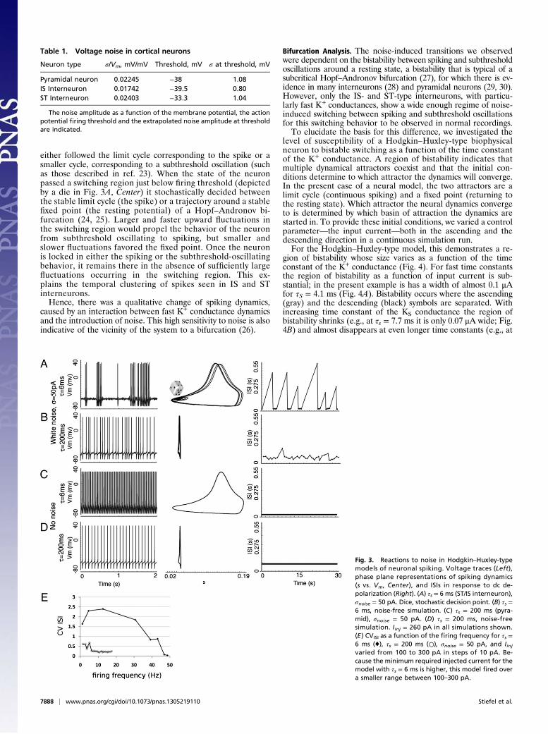
either followed the limit cycle corresponding to the spike or asmaller cycle, corresponding to a subthreshold oscillation (suchas those described in ref. 23). When the state of the neuronpassed a switching region just below firing threshold (depictedby a die in Fig. 3A, Center) it stochastically decided betweenthe stable limit cycle (the spike) or a trajectory around a stablefixed point (the resting potential) of a Hopf–Andronov bi-furcation (24, 25). Larger and faster upward fluctuations inthe switching region would propel the behavior of the neuronfrom subthreshold oscillating to spiking, but smaller andslower fluctuations favored the fixed point. Once the neuronis locked in either the spiking or the subthreshold-oscillatingbehavior, it remains there in the absence of sufficiently largefluctuations occurring in the switching region. This ex-plains the temporal clustering of spikes seen in IS and STinterneurons.Hence, there was a qualitative change of spiking dynamics,
caused by an interaction between fast K+ conductance dynamicsand the introduction of noise. This high sensitivity to noise is alsoindicative of the vicinity of the system to a bifurcation (26).
Bifurcation Analysis. The noise-induced transitions we observedwere dependent on the bistability between spiking and subthresholdoscillations around a resting state, a bistability that is typical of asubcritical Hopf–Andronov bifurcation (27), for which there is ev-idence in many interneurons (28) and pyramidal neurons (29, 30).However, only the IS- and ST-type interneurons, with particu-larly fast K+ conductances, show a wide enough regime of noise-induced switching between spiking and subthreshold oscillationsfor this switching behavior to be observed in normal recordings.To elucidate the basis for this difference, we investigated the
level of susceptibility of a Hodgkin–Huxley-type biophysicalneuron to bistable switching as a function of the time constantof the K+ conductance. A region of bistability indicates thatmultiple dynamical attractors coexist and that the initial con-ditions determine to which attractor the dynamics will converge.In the present case of a neural model, the two attractors are alimit cycle (continuous spiking) and a fixed point (returning tothe resting state). Which attractor the neural dynamics convergeto is determined by which basin of attraction the dynamics arestarted in. To provide these initial conditions, we varied a controlparameter—the input current—both in the ascending and thedescending direction in a continuous simulation run.For the Hodgkin–Huxley-type model, this demonstrates a re-
gion of bistability whose size varies as a function of the timeconstant of the K+ conductance (Fig. 4). For fast time constantsthe region of bistability as a function of input current is sub-stantial; in the present example is has a width of almost 0.1 μAfor τS = 4.1 ms (Fig. 4A). Bistability occurs where the ascending(gray) and the descending (black) symbols are separated. Withincreasing time constant of the KS conductance the region ofbistability shrinks (e.g., at τs = 7.7 ms it is only 0.07 μA wide; Fig.4B) and almost disappears at even longer time constants (e.g., at
Table 1. Voltage noise in cortical neurons
Neuron type σ/Vm, mV/mV Threshold, mV σ at threshold, mV
Pyramidal neuron 0.02245 −38 1.08IS Interneuron 0.01742 −39.5 0.80ST Interneuron 0.02403 −33.3 1.04
The noise amplitude as a function of the membrane potential, the actionpotential firing threshold and the extrapolated noise amplitude at thresholdare indicated.
Fig. 3. Reactions to noise in Hodgkin–Huxley-typemodels of neuronal spiking. Voltage traces (Left),phase plane representations of spiking dynamics(s vs. Vm, Center), and ISIs in response to dc de-polarization (Right). (A) τs = 6 ms (ST/IS interneuron),σnoise = 50 pA. Dice, stochastic decision point. (B) τs =6 ms, noise-free simulation. (C) τs = 200 ms (pyra-mid), σnoise = 50 pA. (D) τs = 200 ms, noise-freesimulation. Iinj = 260 pA in all simulations shown.(E) CVISI as a function of the firing frequency for τs =6 ms (♦), τs = 200 ms (○), σnoise = 50 pA, and Iinjvaried from 100 to 300 pA in steps of 10 pA. Be-cause the minimum required injected current for themodel with τs = 6 ms is higher, this model fired overa smaller range between 100–300 pA.
7888 | www.pnas.org/cgi/doi/10.1073/pnas.1305219110 Stiefel et al.

τs = 7.7 ms it is only 0.02 μA wide). The present analysis wasapplied to the Morris–Lecar model as well (SI Text and Fig. S1)with qualitatively similar results.Based on this bifurcation analysis we confirmed that a sig-
nificantly wider, bistable region in a spiking system with aHopf–Andronov bifurcation exists in both a biophysical Hodgkin–Huxley-type and the Morris–Lecar model only for fast, hyper-polarizing K+ conductances. Because noise-induced switching ispossible only in this bistable region, this phenomenon is depen-dent on fast, interneuronal K+ conductances. This is also con-sistent with the observation that intrinsically irregular spiketrains in cortical neurons are observed only in interneurons withfast K+ conductances.
Parameter Exploration. Finally, how robust are these results tovarying other parameters in the model? To determine the param-eter dependence of intrinsically irregular firing in the original,biophysical, neuron model, we ran two-dimensional param-eter sweeps over τs/gs, τs/cm, τs/Iinj, and τs/σnoise. When τs wassmaller, the CVISI was higher at all values of gs. When gs waslargest, the range of τs where irregular firing occurred was alsolargest (Fig. 5A). Smaller τs caused higher CVs at all values of cm.The same held true for all values of Iinj. The higher the Iinj, thesmaller τs had to be to allow for irregular firing and the higherthe maximum possible CVISI.To investigate the dependence of intrinsically irregular fir-
ing on the properties of the injected noise, we conductedsimulations using power-law and Ornstein–Uhlenbeck (OU,autocorrelated) noise in addition to the standard Gaussianwhite noise (Fig. 5B). The exponent of the power-law noisewas k = −0.7, as deduced from the recorded subthresholdvoltage fluctuations by deconvolution with the impulse re-sponse of the membrane. The parameters for the OU noisemimicked the synaptic bombardment experienced by cortical
neurons in vivo (31). The noise amplitude necessary forobtaining an identical CVISI was reduced an order of magni-tude with power-law noise and reduced another order ofmagnitude with OU noise. This is consistent with previousstudies showing that correlated fluctuations are highly effec-tive in evoking spikes (7, 9, 31). All qualitative observationsmade with Gaussian white noise were confirmed with power-law and OU noise as well.In parameter regions where CVISI ≈ 1, the interspike intervals
were approximately Poisson-distributed, similar to the IS behavior,whereas for CVISI >1.5, spikes were dominantly clustered, re-sembling the ST pattern. The discrete changes in spiking be-havior between regular firing pyramidal neurons, irregular firingIS interneurons, and highly irregular firing ST neurons can beexplained as a continuous change in a single parameter, the K+
conductance time constant τs.
DiscussionThe study of cortical spike-train statistics has previously focusedalmost entirely on pyramidal neurons. Pyramidal neurons and
0 0.2 0.4
−50
0
50A
AscendingDescending
Vol
tage
[V]
τS = 4.1ms
0 0.2 0.4
−50
0
50B
Vol
tage
[V]
τS = 7.7ms
0 0.2 0.4
−50
0
50C
Vol
tage
[V]
Input Current [μA]
τS = 31ms
Fig. 4. The time constant of the KS conductance modifies the bistableregion in the Hodgkin–Huxley-type neuron. We estimated a bifurcationdiagram by ramping the input current (as the control parameter) in anascending (gray) and a descending (black) direction for three differentvalues of the time constant of the KS channel. (A) For τs = 4.1 ms the modelexhibits a wide region of bistability (switching region), indicated by the re-gion where the descending parameter sweep still spikes (two dots indicatethe maxima/minima of the spike, whereas one dot indicates the stable fixedpoint below threshold), whereas the ascending sweep does not yet spike. (Band C) For larger time constants, the switching region becomes narrowerand almost disappears at even higher values (>20 ms).
Fig. 5. Dependence of the behavior of the Hodgkin–Huxley-type modelon parameter choice and noise type. (A) CVISI as a function of σnoise/τs, cm/τs,Iinj/τs and σnoise/τs. (B) Spiking behavior (Left) and ISI histogram (Right) forGaussian white (Top), power-law (λ = 0.7, Middle), and Ornstein–Uhlenbecknoise (Bottom).
Stiefel et al. PNAS | May 7, 2013 | vol. 110 | no. 19 | 7889
NEU
ROSC
IENCE

interneurons are markedly different with regard to the location ofthe axon (32) and the Na+ currents (32) and K+ currents (33, 34)involved in spike and AHP generation as well as their input re-sistance. We have presented recordings and biophysical simulationssuggesting that the faster time constant of the K+ currents could bethe decisive difference with respect to spike time variability.Faster τs lead to a more extended bistable region of the Hopf–
Andronov bifurcation. In this region, noise-induced switchingbetween spiking and subthreshold oscillating occurs, leading tointerneuronal spike trains consisting of bursts, interrupted bynonspiking periods of variable lengths. The mechanism wasdescribed in a Hodgkin–Huxley neuron model transitioning tospiking via a Hopf–Andronov bifurcation (24, 25). Here we havedemonstrated the importance of a fast K+ conductance for en-abling this mechanism. We further argue that this fast conduc-tance is what causes IS and ST interneurons to spike irregularly,but not other types of interneurons and pyramidal neurons, evenif they also spike via a Hopf–Andronov bifurcation.Interestingly, Badoual et al. (35) achieved high discharge
variability in pyramidal neurons by injecting low-pass filterednoise waveform. This represents an approach complementaryto ours. Whereas we observed increased discharge irregularityby recording from neurons (IS and ST interneurons) with afast τ of the K+ conductances, closer to the time scale of thenaturally occurring noise, Badoual et al. changed the time scaleof the noise to fit the τ of the slower pyramidal K+ conductances.Neural dynamics driven by noise have been reported pre-
viously (36).In a study of the firing of interneurons using a biophysical
model of interneurons including gNa, the persistent Na+ con-ductance gNAp, gKDR, and gKd, both the window current and thetotal gKd conductance shaped the spiking behavior of the modelneurons (37). In this model, a large gKd and a small window cur-rent were necessary for irregular firing. Depending on the exactparameter choice, they reproduced a noise-sensitive delay to firing,as seen in in vitro recordings from some interneurons. They alsoobserve subthreshold oscillations. The switching mechanismdescribed here could also occur in this more complex model (37).A more intuitive explanation of these dynamics is that with a
faster time constant of the K+ currents, these currents can interactwith the noise in a dynamic manner. Only a K+ current witha fast time constant can react quickly enough to a fast upward/downward fluctuation to move a neuron from/to the stableresting state to/from spiking. When the time constant of the K+
current is slower, it essentially becomes a constant hyperpolar-ization at the relevant time scales and does not influence theneuron’s fast dynamics anymore.A neuron “stuck” in the bistable resting state is in a condition
where its K+ current is just too strongly activated and its Na+current just too inactivated to allow it to spike. Fast K+ currentdynamics allow switching between this resting state and a spikingregime. Only if its K+ current is fast enough can this bistablestate persist over a range of applied current strengths. If the K+
currents are too slow, they act more or less like a constant hy-perpolarization, and the bistable resting state ceases to exist.These results raise the question of how a subpopulation of
intrinsically irregular interneurons could affect the overall spik-ing statistics in the cortex. Interneurons interact strongly amongthemselves and with pyramidal neurons (13, 18). The high sen-sitivity of interneurons to fluctuations will almost certainly affectthe temporal structure of firing in all cortical neurons. Thus,the intrinsically irregular output of ST and IS interneuronsmay partially account for the irregularity of cortical firing.
Additionally, if, as is likely, the fast fluctuations in cortical ac-tivity carry information, then ST and IS interneurons will beespecially sensitive to this information while their states are inthe switching region. This phase-sensitive read-out mechanismwould make them ideal candidates for processing informationrestricted to specific oscillatory phases, as proposed in severalcoding schemes (38, 39).
MethodsElectrophysiology. Pyramidal neurons and fast-spiking interneurons inlayer 2/3 were recorded intracellularly in the slices of mouse visual cortex.All procedures on animals were done in accordance with Salk InstituteInstitutional Animal Care and Use Committee ethical guidelines. Mice(B6D21/Hsd B6, “black 6,” age 28–35 d; Harlan) were anesthetized withhalothane and decapitated. The occipital forebrain was removed andglued to a plastic block. Coronal slices of the visual cortex (300 μm) were cutwith a Series 1000 vibratome (Pelco) in ice-cold artificial cerebrospinalfluid (ACSF; 125 mM NaCl, 2.5 mM KCl, 1.25 mM NaH2PO4, 25 mMNaHCO3, 2 mM CaCl2, 1.3 mM MgCl2, and 10 mM dextrose). Slices wereallowed to recover in ACSF at 35 °C for at least 30 min before the start ofthe recordings.
Recordings were performed under IR-differential interference contrastvideomicroscopy in oxygenated ACSF (flow 3 mL/min) at 32 °C. Whole-cellpatch-clamp recordings were performed with electrodes ranging from 6 to8 MΩ. The pipette solution contained 140 mM KMeSO4, 10 mM Hepes,1.5 mM NaCl, and 0.1 mM EGTA.
The voltage signal was recorded with an Axoclamp-2A amplifier (AxonInstruments), low-pass-filtered at 30 kHz, and digitized at 32 kHz with a PCI-MIO-16E-4 DAQ board (National Instruments). Data acquisition software wascustom-written in Lab View 6.1 (National Instruments).
Glutamatergic ionotropic synaptic transmission was blocked with DNQX(6,7-dinitroquinoxaline-2,3-dione) (20 μM) and AP5 [(2R)-amino-5-phos-phonopentanoate] (50 μM) and GABAergic ionotropic transmission withbicuculline (10 μM). Action potential firing was evoked by injecting dccurrent via the patch-electrode and series of 0.5-s pulses and continuousstretches spanning 32–64 s were recorded. Drugs were purchased fromSigma and Fisher.
Simulations. The Hodgkin–Huxley model of the interneuron had one com-partment with ion channels, described in SI Text.
The Morris–Lecar oscillator model (40) had two state variables, as de-scribed in SI Text.
We used the ordinary differential equation solvers NEURON (41) and XPP(42). All simulation code is available upon request from K.M.S. or the YaleModel Database (https://senselab.med.yale.edu/modeldb/).
Bifurcation Analysis.We studied the structure of the phase space as a functionof a control parameter using bifurcation analysis. For theMorris–Lecarmodel,we used XPP’s AUTO function to plot bifurcation diagrams for differentvalues of the injected current. For the biophysical neuron model, we directlyvaried the injected current in a continuous simulation run in NEURON. Inboth cases we then compared the width of the bistable region as a functionof the time constant of the KS conductance. The bistable region is defined asthe region where the neural behavior can alternatively be spiking or resting,depending on the initial conditions. For the biophysical model we rampedthe current both in an ascending and a descending direction, thus providinga resting or a spiking initial condition, respectively.
ACKNOWLEDGMENTS. We thank Dr. Lee Campbell for providing dataacquisition software; Drs. Jean-Marc Fellous, Peter J. Thomas, Paul Tiesinga,Jürgen Jost, Priscilla E. Greenwood, Peter Rowat, and Charles F. Stevens forhelpful discussions; and Dr. G. Bard Ermentrout for assistance with XPP. Finan-cial support was provided by the Deutsche Forschungsgemeinschaft (K.M.S.),the Studienstiftung des Deutschen Volkes (B.E.), and the Howard HughesMedical Institute and the Office of Naval Research N00014-97-1-0422 (T.J.S.).
1. Noda H, Adey WR (1970) Firing variability in cat association cortex during sleep and
wakefulness. Brain Res 18(3):513–526.2. Burns BD, Webb AC (1976) The spontaneous activity of neurones in the cat’s cerebral
cortex. Proc R Soc Lond B Biol Sci 194(1115):211–223.3. Destexhe A, Contreras D, Steriade M (1999) Spatiotemporal analysis of local field
potentials and unit discharges in cat cerebral cortex during natural wake and sleep
states. J Neurosci 19(11):4595–4608.
4. Knierim JJ, van Essen DC (1992) Neuronal responses to static texture patterns in area
V1 of the alert macaque monkey. J Neurophysiol 67(4):961–980.5. Softky WR, Koch C (1993) The highly irregular firing of cortical cells is inconsistent
with temporal integration of random EPSPs. J Neurosci 13(1):334–350.6. Singer W (1999) Time as coding space? Curr Opin Neurobiol 9(2):189–194.7. Salinas E, Sejnowski TJ (2000) Impact of correlated synaptic input on output firing
rate and variability in simple neuronal models. J Neurosci 20(16):6193–6209.
7890 | www.pnas.org/cgi/doi/10.1073/pnas.1305219110 Stiefel et al.

8. Fellous J-M, Rudolph M, Destexhe A, Sejnowski TJ (2003) Synaptic background noise
controls the input/output characteristics of single cells in an in vitro model of in vivo
activity. Neuroscience 122(3):811–829.9. Stevens CF, Zador AM (1998) Input synchrony and the irregular firing of cortical
neurons. Nat Neurosci 1(3):210–217.10. Buhl EH, Tamás G, Fisahn A (1998) Cholinergic activation and tonic excitation induce
persistent gamma oscillations in mouse somatosensory cortex in vitro. J Physiol
513(Pt 1):117–126.11. Fisahn A, Pike FG, Buhl EH, Paulsen O (1998) Cholinergic induction of network oscil-
lations at 40 Hz in the hippocampus in vitro. Nature 394(6689):186–189.12. Gerstein GL, Mandelbrot B (1964) Random walk models for the spike activity of
a single neuron. Biophys J 4:41–68.13. Thomson AM, West DC, Wang Y, Bannister AP (2002) Synaptic connections and small
circuits involving excitatory and inhibitory neurons in layers 2-5 of adult rat and cat
neocortex: Triple intracellular recordings and biocytin labelling in vitro. Cereb Cortex
12(9):936–953.14. Tamás G, Szabadics J, Lörincz A, Somogyi P (2004) Input and frequency-specific
entrainment of postsynaptic firing by IPSPs of perisomatic or dendritic origin.
Eur J Neurosci 20(10):2681–2690.15. Larkum ME, Zhu JJ, Sakmann B (1999) A new cellular mechanism for coupling inputs
arriving at different cortical layers. Nature 398(6725):338–341.16. Douglas RM, Goddard GV, Riives M (1982) Inhibitory modulation of long-term po-
tentiation: Evidence for a postsynaptic locus of control. Brain Res 240(2):259–272.17. Markram H, et al. (2004) Interneurons of the neocortical inhibitory system. Nat Rev
Neurosci 5(10):793–807.18. Gupta A, Wang Y, Markram H (2000) Organizing principles for a diversity of
GABAergic interneurons and synapses in the neocortex. Science 287(5451):273–278.19. Kim U, McCormick DA (1998) The functional influence of burst and tonic firing mode
on synaptic interactions in the thalamus. J Neurosci 18(22):9500–9516.20. Englitz B, Stiefel KM, Sejnowski TJ (2008) Irregular firing of isolated cortical interneurons
in vitro driven by intrinsic stochastic mechanisms. Neural Comput 20(1):44–64.21. Hille B (2001) Ion Channels of Excitable Membranes (Sinauer Associates, Sunderland,
MA), 3rd Ed.22. Coetzee WA, et al. (1999) Molecular diversity of K+ channels. Ann N Y Acad Sci 868:
233–285.23. Stiefel KM, Fellous JM, Thomas PJ, Sejnowski TJ (2010) Intrinsic subthreshold oscil-
lations extend the influence of inhibitory synaptic inputs on cortical pyramidal neu-
rons. Eur J Neurosci 31(6):1019–1026.24. Rowat PF, Greenwood PE (2011) Identification and continuity of the distributions of
burst-length and interspike intervals in the stochastic Morris-Lecar neuron. Neural
Comput 23(12):3094–3124.
25. Rowat P (2007) Interspike interval statistics in the stochastic Hodgkin-Huxley model:Coexistence of gamma frequency bursts and highly irregular firing. Neural Comput19(5):1215–1250.
26. Ermentrout GB, Terman TH (2010) Mathematical Foundations of Neuroscience(Springer, Berlin).
27. Izhikevich EM (2010) Dynamical Systems in Neuroscience: The Geometry of Excitabilityand Bursting (MIT Press, Cambridge, MA).
28. Tateno T, Robinson HPC (2007) Phase resetting curves and oscillatory stability in in-terneurons of rat somatosensory cortex. Biophys J 92(2):683–695.
29. Tsubo Y, Takada M, Reyes AD, Fukai T (2007) Layer and frequency dependencies ofphase response properties of pyramidal neurons in rat motor cortex. Eur J Neurosci25(11):3429–3441.
30. Stiefel KM, Gutkin BS, Sejnowski TJ (2008) Cholinergic neuromodulation changesphase response curve shape and type in cortical pyramidal neurons. PLoS ONE 3(12):e3947.
31. Destexhe A, Rudolph M, Fellous J-M, Sejnowski TJ (2001) Fluctuating synaptic con-ductances recreate in vivo-like activity in neocortical neurons. Neuroscience 107(1):13–24.
32. Martina M, Vida I, Jonas P (2000) Distal initiation and active propagation of actionpotentials in interneuron dendrites. Science 287(5451):295–300.
33. Martina M, Schultz JH, Ehmke H, Monyer H, Jonas P (1998) Functional and moleculardifferences between voltage-gated K+ channels of fast-spiking interneurons andpyramidal neurons of rat hippocampus. J Neurosci 18(20):8111–8125.
34. Lien CC, Martina M, Schultz JH, Ehmke H, Jonas P (2002) Gating, modulation andsubunit composition of voltage-gated K(+) channels in dendritic inhibitory inter-neurones of rat hippocampus. J Physiol 538(Pt 2):405–419.
35. Badoual M, Rudolph M, Piwkowska Z, Destexhe A, Bal T (2005) High discharge vari-ability in neurons driven by current noise. Neurocomputing 65–66:493–498.
36. Longtin A, Hinzer K (1996) Encoding with bursting, subthreshold oscillations, andnoise in mammalian cold receptors. Neural Comput 8(2):215–255.
37. Golomb D, et al. (2007) Mechanisms of firing patterns in fast-spiking cortical inter-neurons. PLOS Comput Biol 3(8):e156.
38. O’Keefe J, Recce ML (1993) Phase relationship between hippocampal place units andthe EEG theta rhythm. Hippocampus 3(3):317–330.
39. Vinck M, et al. (2010) Gamma-phase shifting in awake monkey visual cortex. J Neu-rosci 30(4):1250–1257.
40. Rinzel J, Ermentrout GB (1989) Methods in Neuronal Modeling, eds Koch C, Segev I(MIT Press, Cambridge, MA), pp 135–169.
41. Carnevale NT, Hines ML (2009) The NEURON Book (Cambridge Univ Press, Cambridge,UK), 1st Ed.
42. Ermentrout B (2002) Simulating, Analyzing, and Animating Dynamical Systems: A Guideto Xppaut for Researchers and Students (Software, Environments, Tools) (Society forIndustrial and Applied Mathematics, Philadelphia, PA), 1st Ed.
Stiefel et al. PNAS | May 7, 2013 | vol. 110 | no. 19 | 7891
NEU
ROSC
IENCE


![arXiv:1801.00062v1 [q-bio.NC] 30 Dec 2017...match via lateral (e.g. somatostatin-expressing, SST) interneurons the top-down feedback from downstream cortical areas. Synaptic learning](https://static.fdocuments.net/doc/165x107/607b5721eca8ef594f5a28da/arxiv180100062v1-q-bionc-30-dec-2017-match-via-lateral-eg-somatostatin-expressing.jpg)