ON THE ROLE OF PARVALBUMIN INTERNEURONS IN NEURONAL ...
92
From the Department of Neuroscience Karolinska Institutet, Stockholm, Sweden ON THE ROLE OF PARVALBUMIN INTERNEURONS IN NEURONAL NETWORK ACTIVITY IN THE PREFRONTAL CORTEX Nicolas Gustavo Guyon Stockholm 2021
Transcript of ON THE ROLE OF PARVALBUMIN INTERNEURONS IN NEURONAL ...
From the Department of Neuroscience
Karolinska Institutet, Stockholm, Sweden
ON THE ROLE OF PARVALBUMIN INTERNEURONS IN NEURONAL
NETWORK ACTIVITY IN THE PREFRONTAL CORTEX
Nicolas Gustavo Guyon
Stockholm 2021

All previously published papers were reproduced with permission from the publisher.
Published by Karolinska Institutet.
Printed by Universitetsservice US-AB, 2021
© Nicolas Gustavo Guyon, 2021
ISBN 978-91-8016-156-5
Cover illustration: “Secret Garden” by Nirupa Rao, 2021

On the role of parvalbumin interneurons in neuronal network activity in the prefrontal cortex
THESIS FOR DOCTORAL DEGREE (Ph.D.)
By
Nicolas Gustavo Guyon
The thesis will be defended in public at Eva & Georg Klein, Biomedicum, Solnavägen 9, Solna,
Friday the 21st of May 2021 at 14h00
Principal Supervisor:
Associate Professor Marie Carlén
Karolinska Institutet
Department of Neuroscience
Department of Biosciences and Nutrition
Co-supervisors: Professor Konstantinos Meletis
Karolinska Institutet
Department of Neuroscience
Professor Karl Deisseroth
Stanford University
Dept. of Psychiatry and Behavioral Sciences
Department of Bioengineering
Howard Hughes Medical Institute
Opponent:
Assistant Researcher Kathleen K.A. Cho
University of California, San Francisco
Department of Psychiatry and Behavioral Sciences
Examination Board:
Associate Professor Paolo Medini
Umeå Universitet
Department of Integrative Medical Biology
Principal Investigator Mª Victoria Puig
Institut Hospital del Mar d'Investigacions
Mèdiques
Dept. of Integrative Pharmacology and Systems
Neuroscience
Associate Professor Rochellys Diaz Heijtz
Karolinska Institutet
Department of Neuroscience
INSERM – Université de Rouen


Minha alma é uma orquestra oculta; não sei que instrumentos tange e range,
cordas e harpas, tímbales e tambores, dentro de mim. Só me conheço como sinfonia.
– “Livro do Desassossego, por Bernardo Soares,” Fernando Pessoa


POPULAR SCIENCE SUMMARY OF THE THESIS
The brain is a complex, enigmatic organ composed of a multitude of neurons. These neurons
are interconnected, composing a networked web of connections that in the human brain goes
up to the trillions. The overall activity of these intermingled networks of neurons leads to the
body being able to react to its environment. One of the ways that neurons use their network of
connections to communicate with each other is by releasing chemicals called neurotransmitters.
For example, when a recipient neuron receives the excitatory neurotransmitter glutamate via
receptors placed on its surface, the neuron responds by eliciting an electrical signal called action
potential. We say that the neuron “fired”. If, on the other hand, the recipient neuron receives
the inhibitory neurotransmitter GABA, it will be less likely to fire. These cycles in excitation
and inhibition create voltage fluctuations outside the neurons that we call “brain oscillations”.
These oscillations are heavily studied because they tell us information about what the brain is
doing. For instance, they can help us infer how brain activity is organized temporally, how the
brain responds to sensory input, or how it elicits a movement. Brain oscillations do not look to
be very informative at first sight. However, after being decomposed into smaller components,
based on the frequency of their fluctuation, one can find that different frequencies will have a
bigger or smaller amplitude depending on the behavior of the individual being recorded.
Moreover, these various classes of oscillations look different if a person is awake or asleep,
giving us an insight into how the brain is operating during these specific states. The difference
in amplitude for specific oscillations usually depends on how the neurons are activated or
inhibited and the timing of that cycle — the faster they are activated/inhibited, the higher the
frequency — but also whether they fire in synchrony or more randomly.
Oscillations are rhythmic and reflect the synchronization of the neurons’ activity. This could
be compared to the noise made by people clapping at a concert at the end of a song. It starts by
being uncoordinated, but when the clapping becomes synchronized — because people are
clapping at the same time/frequency — the sound gets louder at that specific beat or frequency.
When recording the brain oscillations with electrodes, we can therefore infer the activity of
neurons by the impact of specific frequencies. For example, some sensory stimuli are known
to specifically increase signals that oscillate at 30-80 cycles per second (30-80 hertz, or Hz).
These oscillations are called gamma oscillations and are strongly correlated to cognitive
processes like attention, working memory and visual processing. Indeed, their amplitude
specifically and narrowly increases when a human or an animal performs a cognitive task.
Significantly, these evoked gamma oscillations are weaker during the performance of similar
tasks in patients with schizophrenia. However, when we observe the brain activity of these
same patients between two tasks or when at rest, a constant, wide-ranging and increased noise
in the gamma range is detected. These aberrant gamma oscillations have been replicated in
animal models of schizophrenia, in which neurons that release the inhibitory neurotransmitter
GABA are dysfunctional. But very little is currently known about how the inhibitory neurons
generate proper narrow gamma oscillation during a task and, at the same time, are paradoxically
associated, when dysfunctional, with increased broad noise in the gamma range during rest.
In Paper I, we tried to solve this issue by testing the suggestion that the broadband increase in
amplitude spanning the entire gamma-band might not always be a rhythm but could result from

asynchronous and noisy communication between neurons. For this, we recorded the brain
activity of transgenic mice that lacked a receptor essential for the proper activity of inhibitory
neurons. Since this receptor had been removed only in the inhibitory neurons, it allowed us to
precisely observe the effect of impairing the activity of these neurons. We found that the
activity recorded during rest was increased and associated with the neurons' asynchronous
activity.
It is as if these specific neurons were not able anymore to hear that other cells around them
were clapping on cue after the song was finished, making it difficult for them to follow the
rhythm of collective synchronization. This led to the inhibitory neurons in our concert room
clapping at random and making noise during the song as well. Importantly, we replicated this
noisy activity by applying ketamine locally to the brain of normal mice, which might explain
how ketamine mimics the symptoms of schizophrenia in humans. Surprisingly, similar
ketamine application in transgenic mice did not cause any such changes. We explain this by
suggesting that ketamine needs the inhibitory neuron receptor removed in the transgenic mice
to have an effect on the brain.
In Paper II, we expressed a modified receptor in the same inhibitory neurons as in Paper I,
this time with the help of a virus injected in a specific area of the brain called the prefrontal
cortex. The virus we generated induces the expression of a modified receptor that is usually
critical for how brain connections are maintained. The modified receptor was injected in adult
transgenic mice and competed with the normal receptor, making it less effective. Thus, this
approach allowed us to target the neurons we wanted to study in the specific brain area
important for social behavior, and avoid interference with other brain areas as well as with
developmental processes. This precision was necessary, as alterations specific to this receptor
in inhibitory neurons of the prefrontal cortex have been found in postmortem examination of
patients with neuropsychiatric disorders such as schizophrenia.
We found that the modified receptor altered the inhibitory neurons in which it was expressed
both morphologically and functionally. When recording the brain activity of transgenic mice
while socially interacting with other mice, we found an increased number of excitatory
neurons and abnormal gamma oscillations within the prefrontal cortex, which was correlated
with unusually aggressive behavior. These results suggest that the modified receptor in
inhibitory neurons reduced their inhibitory connection with excitatory neurons, allowing
them to be activated imprecisely.
In conclusion, abnormal gamma oscillations were observed in both studies. Changes in gamma
are widely reported in both animal models and human studies, but at the same time, the
heterogeneity of gamma-band abnormalities so far recorded has limited the translation of these
findings into clinical settings. A better understanding of how to interpret gamma oscillation
results may thus be a helpful guide in developing approaches where we can use gamma
oscillations to track changes due to disorders but also changes elicited by drugs. Moreover, the
results in this thesis contribute to our understanding of the biological mechanisms behind
neuronal and circuit modifications due to dysfunctional receptors implicated in
neuropsychiatry disorders, especially schizophrenia. This information can be used to develop
targeted diagnoses, as well as interventions aimed at more specifically treating cognitive
impairments seen in neuropsychiatry disorders.
Résumé grand public de la thèse
Le cerveau est un organe complexe composé d'une multitude de neurones. Ces neurones sont
amplement interconnectés, constituant un réseau de connexions qui, dans le cerveau humain,
peut atteindre les billions. L'activité de ces réseaux entremêlés de neurones guide le corps dans
son interaction avec son milieu. L'un des procédés que les neurones utilisent pour communiquer
entre eux consiste à libérer des composés chimiques appelés neurotransmetteurs. Par exemple,
lorsqu'un neurone reçoit du glutamate, un neurotransmetteur excitateur, via des récepteurs
placés à sa surface, cela provoque un signal électrique appelé potentiel d'action. Nous disons
que le neurone a « déchargé ». Si, en revanche, le neurone reçoit le neurotransmetteur inhibiteur
GABA, il sera moins susceptible de produire un potentiel d’action. Ces cycles d'excitation et
d'inhibition génèrent des fluctuations du signal électrique en dehors des neurones appelées «
oscillations cérébrales ».
Ces oscillations sont très étudiées car elles fournissent des informations sur ce que fait le
cerveau. Par exemple, elles peuvent nous aider à déduire comment l'activité cérébrale est
organisée temporellement, mais aussi comment le cerveau répond à des influx sensoriels ou
déclenche un mouvement. Les oscillations cérébrales ne paraissent pas très informatives à
première vue. Cependant, après avoir été découpées en composantes plus petites, en fonction
de la fréquence de leur fluctuation, on peut constater que différentes fréquences auront une
amplitude plus ou moins grande en fonction du comportement du sujet. De plus, ces distinctes
classes d'oscillations semblent différentes si un individu est éveillé ou endormi, ce qui nous
donne un aperçu du fonctionnement du cerveau pendant ces états spécifiques. La différence
d'amplitude pour des oscillations spécifiques dépend généralement de la façon dont les
neurones sont activés ou inhibés et de la durée de ce cycle - plus le cycle activation / inhibition
est rapide, plus la fréquence est élevée - mais aussi s'ils se déchargent de manière synchronisée
ou de manière plus aléatoire.
Les oscillations sont généralement rythmiques et reflètent la synchronisation de l’activité des
neurones. Cela pourrait être comparé au bruit fait par des gens applaudissant lors d'un concert
à la fin d’un morceau de musique. Cela commence par être désorganisé, mais lorsque les
applaudissements s’harmonisent - parce que les gens applaudissent en même temps / à la même
fréquence - le son devient plus intense à cette fréquence spécifique. Lors de l'enregistrement
des oscillations avec des électrodes, on peut donc déduire l'activité des neurones par l'intensité
des différentes fréquences. Par exemple, certains stimuli sensoriels sont connus pour augmenter
spécifiquement les signaux qui oscillent à 30-80 cycles par seconde (30-80 hertz ou Hz).
Ces oscillations sont appelées oscillations gamma et sont fortement corrélées à des processus
cognitifs tels que l'attention, la mémoire de travail et le traitement visuel. En effet, leur
amplitude augmente spécifiquement et étroitement lorsqu'un humain ou un animal effectue une
tâche cognitive. De manière significative, ces oscillations gamma évoquées sont plus faibles
lors de l'exécution de tâches similaires chez les patients atteints de schizophrénie. Cependant,
lorsque nous observons l'activité cérébrale de ces mêmes patients entre deux tâches ou au repos,
on peut détecter une activité bruyante constante et élevée correspondant plus ou moins aux
ondes gamma. Ces oscillations gamma aberrantes ont été répliquées dans des modèles animaux
de schizophrénie, dans lesquels les neurones qui libèrent le neurotransmetteur inhibiteur
GABA sont dysfonctionnels. Malgré cela on sait actuellement très peu de choses sur la façon

dont les neurones inhibiteurs génèrent les ondes cérébrales gamma pendant une tâche et en
même temps sont paradoxalement associés, lorsqu'ils sont dysfonctionnels, à une augmentation
d’un rythme gamma associé à du bruit de fond au repos.
Dans l'étude I, nous avons essayé de résoudre ce problème en testant l’hypothèse selon laquelle
l'augmentation de la fréquence élevée couvrant toute la bande gamma pourrait ne pas toujours
être un rythme mais pourrait être le résultat d'une communication asynchrone et bruyante entre
les neurones. Pour cela, nous avons enregistré l'activité cérébrale de souris transgéniques
dépourvues d'un récepteur important pour l’activité des neurones inhibiteurs. Ce récepteur,
n'ayant été éliminé que dans les neurones inhibiteurs, nous a permis d'observer spécifiquement
l'effet de modifier l'activité de ces neurones. Nous avons constaté que l'activité enregistrée au
repos était augmentée et associée à une activité asynchrone des neurones.
En somme, ce serait comme si ces neurones spécifiques n’étaient plus capables d’entendre que
d’autres cellules autour d’eux applaudissaient après la fin de la musique, ce qui les empêche de
suivre la cadence collective. Cela conduit les neurones inhibiteurs de notre salle de concert à
applaudir au hasard et à faire du bruit pendant que les musiciens jouent. Nous avons notamment
reproduit cette activité bruyante en appliquant de la kétamine localement sur le cerveau de
souris normales, ce qui pourrait expliquer comment la kétamine imite les symptômes de la
schizophrénie chez l'homme. De manière surprenante, une application similaire de kétamine
chez des souris transgéniques n'a pas provoqué de tels changements. Nous expliquons cela en
suggérant que la kétamine a besoin du récepteur neuronal inhibiteur éliminé chez les souris
transgéniques pour avoir un effet sur le cerveau.
Dans l'étude II, nous avons exprimé un récepteur modifié dans les mêmes types de neurones
inhibiteurs que dans l'article I, mais cette fois à l'aide d'un virus qui a été injecté dans une zone
spécifique du cerveau appelée cortex préfrontal. Le virus que nous avons généré induit
l'expression d'un récepteur modifié qui est généralement crucial pour la façon dont les
connexions cérébrales sont maintenues. Le récepteur modifié a été injecté à des souris
transgéniques adultes et est entré en compétition avec le récepteur normal, le rendant moins
efficace. Ainsi, cette approche nous a permis de cibler les neurones que nous voulions étudier
dans une zone spécifique du cerveau connue pour être importante pour le comportement social,
et d'éviter les interférences avec d'autres zones cérébrales ainsi qu'avec les processus de
développement. Il était important d’être précis, car des altérations spécifiques de ce récepteur
dans les neurones inhibiteurs du cortex préfrontal ont été retrouvées lors de l'examen post-
mortem de patients souffrant de troubles neuropsychiatriques tels que la schizophrénie.
Nous avons donc constaté que le récepteur modifié changeait les neurones inhibiteurs dans
lesquels il était exprimé à la fois morphologiquement et fonctionnellement. Lors de
l'enregistrement de l'activité cérébrale des souris transgéniques alors qu'elles interagissent
socialement avec d'autres souris, nous avons trouvé un nombre accru de neurones excitateurs
et des oscillations gamma anormales dans le cortex préfrontal, le tout corrélé à un
comportement inhabituellement agressif. Ces résultats suggèrent que le récepteur modifié dans
les neurones inhibiteurs a réduit leur connexion inhibitrice avec les neurones excitateurs, leur
permettant d'être activés de manière imprécise.

En conclusion, des oscillations gamma anormales ont été observées dans les deux études. Les
altérations des ondes cérébrales gamma sont largement rapportées dans les modèles animaux
et dans les études humaines, mais cependant l'hétérogénéité des anomalies correspondant aux
oscillations gamma observées jusqu'à présent a limité le potentiel translationnel de ces résultats
dans des contextes cliniques. Une meilleure conception des différentes manières d'interpréter
les résultats des oscillations gamma peut donc être utile dans le développement d'approches où
nous pouvons utiliser ces oscillations afin de suivre les changements dus aux troubles mais
aussi les changements induits par la médication. De plus, les résultats de cette thèse ont pour
but de contribuer à une plus grande compréhension des mécanismes biologiques à l'origine des
modifications neuronales et des circuits, dues à des récepteurs dysfonctionnels impliqués dans
les troubles neuropsychiatriques, dont la schizophrénie. Nous espérons que ces résultats
pourront être utilisés dans le développement de diagnostics plus ciblés, ainsi que
d’interventions visant à traiter plus spécifiquement les déficiences cognitives observées dans
les troubles neuropsychiatriques.


ABSTRACT
The prefrontal cortex (PFC) is an area important for executive functions, the initiation and
temporal organization of goal-directed behavior, as well as social behaviors. Inhibitory
interneurons expressing parvalbumin (PV) have a vital role in modulating PFC circuit plasticity
and output, as inhibition by PV interneurons on excitatory pyramidal neurons regulates the
excitability of the network. Thus, dysfunctions of prefrontal PV interneurons are implicated in
the pathophysiology of a range of PFC-dependent neuropsychiatric disorders characterized by
excitation and inhibition (E/I) imbalance and impaired gamma oscillations.
In particular, the hypofunction of receptors important for neurotransmission and regulating
cellular functions, such as the N-methyl-D-aspartate receptors (NMDARs) and the tyrosine
receptor kinase B (trkB), has been implicated in PV dysfunction. Notably, this hypofunction is
known to impair the normal development of PV interneurons. However, it can also affect adult
brain activity. The effects of altered receptors on PV interneurons are multiple, from impaired
morphological connectivity to disruption of intrinsic activity, but have not yet been fully
characterized. Moreover, the effects of deficits of PV neuron-mediated inhibition on neuronal
network activity are complex, involved with compensatory mechanisms, and not fully
understood either. For instance, the E/I imbalance due to PV inhibition has been suggested to
functionally disrupt the cortex, which can be observed through an abnormal increase in
broadband gamma activity. But as the synchronous activity of cortical PV interneurons is
necessary for the generation of cortical gamma oscillations, it is paradoxical that deficient PV
inhibition is associated with increased broadband gamma power.
This thesis aims to examine the role of PV interneurons in shaping neuronal network activity
in the mouse PFC by investigating the microscopic to macroscopic functional effects of
disrupting receptors necessary for the proper activity of PV interneurons.
In paper I, we observed that the increase of broadband gamma power due to NMDAR
hypofunction in PV neurons is associated with asynchronies of network activity, confirming
that dysfunction of neuronal inhibition can cause desynchronization at multiple time scales
(affecting entrainment of spikes by the LFP, as well as cross-frequency coupling and brain
states fragmentation). In Paper II, we prompted and analyzed the rippling effect of PV
dysfunction in the adult PFC by expressing a dominant-negative trkB receptor specifically in
PV interneurons. Despite avoiding interfering with the development of the brain, we found
pronounced morphological and functional alterations in the targeted PV interneurons. These
changes were associated with unusual aggressive behavior coupled with gamma-band
alterations and a decreased modulation of prefrontal excitatory neuronal populations by PV
interneurons.
Thus, the work presented in this thesis furthers our understanding of the role of PV function in
PFC circuitry, particularly of two receptors that are central to the role of PV interneurons in
coordinating local circuit activity. A better understanding of the potential mechanisms that
could explain the neuronal changes seen in individuals with neuropsychiatric dysfunctions
could lead to using gamma oscillations or BDNF-trkB levels as biomarkers in psychiatric
disorders. It also presents possibilities for potential treatments designed around reestablishing
E/I balance by modifying receptor levels in particular cell types.

LIST OF SCIENTIFIC PAPERS
I. Network asynchrony underlying increased broadband gamma power.
The Journal of Neuroscience. 2021 Mar 31;41(13):2944-2963.
Nicolas Guyon, Leonardo Rakauskas Zacharias, Eliezyer Fermino de Oliveira,
Hoseok Kim, João Pereira Leite, Cleiton Lopes-Aguiar, Marie Carlén
II. Adult trkB signaling in parvalbumin interneurons is essential to prefrontal
network dynamics.
The Journal of Neuroscience. 2021 Apr 7;41(14):3120-3141.
Nicolas Guyon, Leonardo Rakauskas Zacharias, Josina Anna van Lunteren,
Jana Immenschuh, Janos Fuzik, Antje Märtin, Yang Xuan, Misha Zilberter,
Hoseok Kim, Konstantinos Meletis, Cleiton Lopes-Aguiar, Marie Carlén,

CONTENTS
1 INTRODUCTION ...................................................................................................... 1
1.1 THE PREFRONTAL CORTEX ........................................................................ 1
1.1.1 General organization of the mPFC ......................................................... 2
1.1.2 Prefrontal cell-types ............................................................................... 3
1.2 PARVALBUMIN INTERNEURONS .............................................................. 4
1.3 PREFRONTAL CIRCUIT ACTIVITY ............................................................. 6
1.3.1 Excitatory/inhibitory balance ................................................................. 6
1.3.2 Neuronal ensembles ............................................................................... 7
1.4 SYNCHRONY IN THE BRAIN ....................................................................... 8
1.4.1 Oscillatory activity ................................................................................. 8
1.4.2 Cortical deactivated and activated states .............................................. 10
1.4.3 UP and DOWN states .......................................................................... 12
1.4.4 Cross frequency coupling..................................................................... 13
1.5 N-METHYL-D-ASPARTATE RECEPTOR ................................................... 14
1.6 BDNF-TRKB SIGNALING ............................................................................ 17
1.6.1 trkB receptors....................................................................................... 18
1.6.2 BDNF-trkB signaling in prefrontal PV interneuron activity................. 19
1.7 REGULATION OF SOCIAL PROCESSING BY THE PREFRONTAL
PV INTERNEURONS .................................................................................... 21
2 RESEARCH AIMS ................................................................................................... 23
3 MATERIALS AND METHODS .............................................................................. 25
3.1 SPECIFIC TARGETING AND MANIPULATION OF PV NEURONS ........ 25
3.1.1 Cre-lox system and transgenic animals ................................................ 25
3.1.2 NR1 floxed transgenic line................................................................... 25
3.1.3 Viral delivery ....................................................................................... 27
3.1.4 Viral expression of trkB.DN-mCherry ................................................. 27
3.2 MOLECULAR AND CELLULAR READOUTS ........................................... 28
3.3 SOCIAL INTERACTION ............................................................................... 30
3.4 RECORDING THE ACTIVITY OF THE PFC ............................................... 31
3.4.1 Ex vivo electrophysiology .................................................................... 31
3.4.2 In vivo electrophysiology ..................................................................... 32
3.4.3 Electrophysiology data analysis ........................................................... 33
3.5 ETHICAL CONSIDERATIONS ..................................................................... 36
3.5.1 On the need to open sources................................................................. 37
3.5.2 On the need to open access .................................................................. 37
4 RESULTS AND DISCUSSION ............................................................................... 39
4.1 NMDAR ACTIVITY IN PV NEURONS AND ASYNCHRONOUS
mPFC NEURONAL ACTIVITY .................................................................... 39
4.1.1 Altered cortical states ........................................................................... 39
4.1.2 Asynchronous neuronal activity ........................................................... 40
4.1.3 Disorganization of single-unit activity ................................................. 40

4.1.4 Diverse asynchronies caused by ketamine application or by the
removal of NMDAR from PV neurons ................................................ 41
4.2 BDNF-TRKB SIGNALING IN PV INTERNEURONS IN THE ADULT
mPFC .............................................................................................................. 43
4.2.1 Molecular and morphological alterations ............................................. 43
4.2.2 Reduced sensitivity and firing activity of PV interneurons .................. 46
4.2.3 Social behavior dysfunctions ............................................................... 46
4.2.4 Altered prefrontal excitatory dynamics ................................................ 47
4.2.5 On potential sexual dimorphic differences ........................................... 50
5 CONCLUSION AND PERSPECTIVES .................................................................. 51
5.1 PV DYSFUNCTION AND ASYNCHRONOUS ACTIVITY ........................ 51
5.2 PV DYSFUNCTION AND CORTICAL STATES IMPAIRMENTS............. 53
5.3 PV DYSFUNCTION AND CIRCUIT ALTERATIONS ................................ 54
5.4 MANIPULATION OF RECEPTORS TO STUDY PV FUNCTION ............. 56
5.5 POTENCIAL CLINICAL RELEVANCE....................................................... 56
6 ACKNOWLEDGEMENTS ...................................................................................... 59
7 REFERENCES ......................................................................................................... 61
LIST OF ABBREVIATIONS
AAV Adeno-associated virus
ACA Anterior cingulate area
AMPA α-amino-3-hydroxy-5-methyl-4-
isoxazolepropionic acid
BDNF Brain-derived neurotrophic factor
CFC Cross-frequency coupling
DIO Double-floxed inverted open-
reading-frame
EEG Electroencephalograms
E/I Excitatory/inhibitory
EPSC Excitatory postsynaptic current
eYFP Enhanced yellow fluorescent
protein
FACS Fluorescence-activated cell
sorting
GABA Gamma-Aminobutyric acid
GAD Glutamic acid decarboxylase
GAT-1 GABA transporter-1
HFB High-frequency band
HFOs High-frequency oscillations
ILA Infralimbic area
IPSC Inhibitory postsynaptic current
LFP Local field potential
MK-801 Dizocilpine
mPFC Medial prefrontal cortex
NMDAR N-methyl-D-aspartate receptor
NR1 NMDA receptor subunit 1
NREM Non-rapid eye movement
ORB Orbital area
p75NTR p75 neurotrophin receptor
PCA Principal component analysis
PCP Phenylcyclohexyl piperidine or
Phencyclidine
PFC Prefrontal cortex
PL Prelimbic cortex
PSD Power spectral density
PV Parvalbumin
REM Rapid eye movement
snRNAseq Single nuclei RNA sequencing
trkB Tyrosine receptor kinase B
trkB.DN Dominant-negative trkB
trkB.FL Full-length trkB
trkB.T Truncated trkB
VIP Vaso-intestinal peptide
VTA Ventral tegmental area


1
1 INTRODUCTION
1.1 THE PREFRONTAL CORTEX
Located in the forefront of the brain, the prefrontal cortex (PFC) is a distinctive region involved
in various brain functions and processes linked to cognition and goal-oriented actions. The PFC
has been labeled a major evolutionary specialization, as its relative size peaks in primates – up
to 30% of the cortical domain is occupied by the PFC in humans (Carlén, 2017; Smaers et al.,
2017).
The work in this thesis has been performed in mice (Mus musculus) to, among other things,
make use of techniques allowing the spatially and timely restricted manipulation and recording
of cell-type-specific activity. I will therefore focus on the role of the PFC in this model
organism. However, a debate continues about the use of mice for studying the PFC, notably
whether one can translate the concept of prefrontal cortex between species, even though a
growing body of research has revealed functional homologies in rodents and primates (Carlén,
2017; Laubach et al., 2018). For example, hallmark functions of the PFC, like working
memory, attention, and behavioral flexibility, have been conceived based on findings in
primates and successfully replicated in rodents (Kamigaki and Dan, 2017; Kim et al., 2016b;
Liu et al., 2014). It is thus conceptually feasible to use the rodent PFC to shed light on the
functional properties of the primate brain, including the human brain (Carlén, 2017; Le Merre
et al., 2021). Besides, the mouse PFC is involved in sensory processing, the preparation of
motor functions, attention (Kim et al., 2016b), working memory (Kim et al., 2016a), and social
behavior (Felix-Ortiz et al., 2016; Levy et al., 2019; Yizhar and Levy, 2021; Yizhar et al.,
2011), among other cognitive behaviors (Le Merre et al., 2021).
Functionally, the PFC integrates internal and external information regarding the present state
in order to represent future goals and predict future actions. This capacity allows the temporal
organization of behavior in mammals as well as the initiation of goal-directed behaviors
(Fuster, 2015). Specifically, it is thought that sensory information flows from the periphery via
the thalamus and sensory cortical regions right up to the PFC, in a “bottom-up” fashion. In the
PFC, sensory information is then assimilated with information about the state and the goal, as
well as previous experience (Fuster, 2015). From the PFC, the information is then sent back to
other cortical regions, like the motor cortex, and to subcortical regions that are implicated in
the selection and execution of movement. This “top-down” or “executive” signal is thus
thought to be essential for guiding, biasing and modulating activity in downstream regions for
the appropriate action in response to a situation. For instance, pharmacological perturbation of
PFC activity causes disruptions of cortex-wide activity necessary for correctly performing a
task (Allen et al., 2017; Makino et al., 2017).
The pattern of connections to and from the medial prefrontal cortex reflects this functional
capacity to work as a highly integrative network. The primary inputs to the mouse PFC are
originated locally. However, the PFC is also densely interconnected with the rest of the cortex
and with numerous subcortical brain regions, receiving and projecting to a vast number of
regions in a reciprocal manner, making it the area with the highest proportion of feedback
projections (Ährlund-Richter et al., 2019; Harris et al., 2019; Le Merre et al., 2021). Common
connections to and from the PFC arise from the motor and sensory cortical regions but also

2
regions involved in arousal, memory, emotional and social responses, like the basal forebrain,
thalamus, amygdala, hippocampus dorsal raphe nucleus and locus coeruleus (Ährlund-Richter
et al., 2019; Collins et al., 2018; Hoover and Vertes, 2007).
Being an essential part of the integrative network underlying cognition, dysfunctions of the
prefrontal cortex have been causally implicated in a multitude of neuropsychiatric disorders.
Patients with prefrontal damage usually show signs of deficits in decision making, disrupted
selective attention for relevant inputs, and increased distractibility by irrelevant stimuli, as well
as impaired working memory (Lewis et al., 2005). For instance, epilepsy, autism spectrum
disorder, and schizophrenia have been related to malfunctions in the PFC neuronal circuitry,
particularly involving the disorganized firing of subsets of neurons, affecting its local and long-
range connectivity (Cho et al., 2015; Homayoun and Moghaddam, 2007; Lewis et al., 2005;
Schmitt et al., 2017; Yizhar et al., 2011). Research on the several mechanisms that could
produce pathological changes in the PFC circuitry is essential to link these PFC dysfunctions
to cognitive impairments in neuropsychiatric disorders (Gordon, 2016; Marín, 2012; Tang et
al., 2021).
1.1.1 General organization of the mPFC The PFC can be said to be an “umbrella term” for cortical regions located in the forefront of
the brain (Le Merre et al., 2021). The PFC has thus been historically divided into several sub-
regions - divisions based mainly on anatomical and histological examinations of the brain.
Mice possess fewer prefrontal regions than primates, and all regions in the prefrontal cortex of
mice lack the layer IV (e.g. the regions are agranular). In rodents, the cortical regions thus
identified as shaping the prefrontal cortex are the prelimbic area (PL), the infralimbic area
(ILA), the anterior cingulate areas (ACA) and the orbital areas (ORB). Both the papers
presented in this thesis use the term medial prefrontal cortex (mPFC) to depict the more medial
regions of the mice PFC (ventral ACA, PL, ILA and medial ORB) (Figs. 1a, b). However,
several ways of classifying the PFC still prevail today, primarily based on cytoarchitecture or
connectivity (Ährlund-Richter et al., 2019), but no clear function has yet been given to each
specific area – in humans, as in their homologous regions in rodents (Euston et al., 2012). More
research is thus needed in order to define the functions of each sub-region of the PFC (Carlén,
2017).
The cellular organization of the PFC is considered to be canonically organized by layers and
by columns. Organization conserved not only between species, but also similar to other cortical
areas (except for the lack of layer 4). However, although a general organization pattern is
observed (Figs. 1b, c), a definite circuit has not yet been defined for the PFC (Douglas and
Martin, 2007; Harris and Shepherd, 2015). Importantly, the syntax allowing the translation of
this structural organization into function is still not entirely known. It has nevertheless been
shown that cortical neurons within prefrontal columns are inter-connected, and receive
thalamic inputs, across all layers (Constantinople and Bruno, 2013), while larger excitatory
pyramidal neurons of the lower layers generate most of the output from the PFC to the thalamus
and other subcortical parts of the brain. Placed among the pyramidal neurons, gamma-
Aminobutyric acid (GABA)-ergic inhibitory interneurons are mostly found in layers 2 to 6,
locally restricting where they spread both their axonal and dendritic arbors (Tremblay et al.,
2016). Although highly interconnected locally, they still receive inputs from other cortical and
subcortical regions (Ährlund-Richter et al., 2019). Furthermore, there are distinct recruitment
3
patterns of GABAergic interneurons by local excitatory networks, forming feedforward and
feedback inhibitory loops, as well as disinhibitory paths due to the significant interconnections
between inhibitory interneurons. The mechanistic underpinnings of how these various
inhibitory circuits are formed and how the neuronal circuits process different inputs and shape
reliable output patterns are not fully understood. Studies considering the morphology and
connection patterns of specific cell-types while investigating their function are necessary to
pinpoint their possible implications for proper mPFC function.
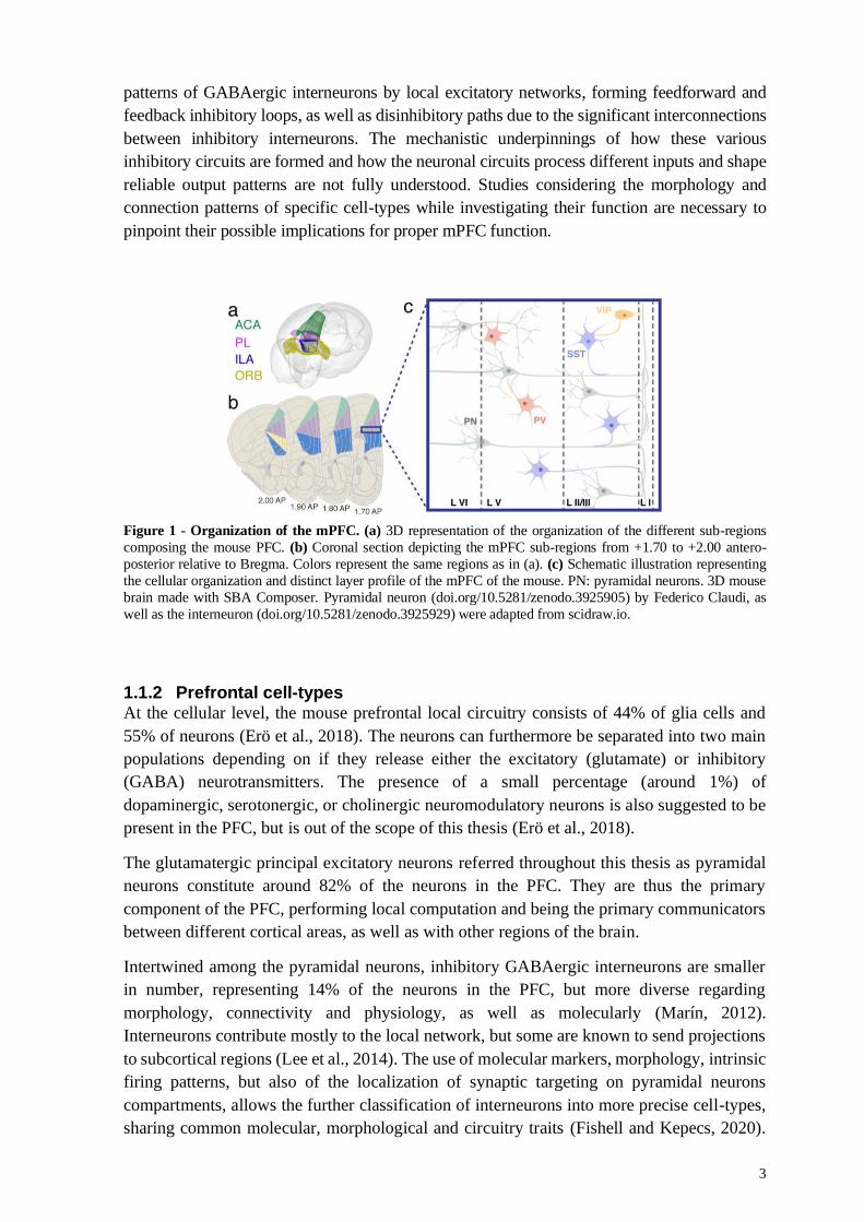
Figure 1 - Organization of the mPFC. (a) 3D representation of the organization of the different sub-regions
composing the mouse PFC. (b) Coronal section depicting the mPFC sub-regions from +1.70 to +2.00 antero-
posterior relative to Bregma. Colors represent the same regions as in (a). (c) Schematic illustration representing
the cellular organization and distinct layer profile of the mPFC of the mouse. PN: pyramidal neurons. 3D mouse
brain made with SBA Composer. Pyramidal neuron (doi.org/10.5281/zenodo.3925905) by Federico Claudi, as
well as the interneuron (doi.org/10.5281/zenodo.3925929) were adapted from scidraw.io.
1.1.2 Prefrontal cell-types At the cellular level, the mouse prefrontal local circuitry consists of 44% of glia cells and
55% of neurons (Erö et al., 2018). The neurons can furthermore be separated into two main
populations depending on if they release either the excitatory (glutamate) or inhibitory
(GABA) neurotransmitters. The presence of a small percentage (around 1%) of
dopaminergic, serotonergic, or cholinergic neuromodulatory neurons is also suggested to be
present in the PFC, but is out of the scope of this thesis (Erö et al., 2018).
The glutamatergic principal excitatory neurons referred throughout this thesis as pyramidal
neurons constitute around 82% of the neurons in the PFC. They are thus the primary
component of the PFC, performing local computation and being the primary communicators
between different cortical areas, as well as with other regions of the brain.
Intertwined among the pyramidal neurons, inhibitory GABAergic interneurons are smaller
in number, representing 14% of the neurons in the PFC, but more diverse regarding
morphology, connectivity and physiology, as well as molecularly (Marín, 2012).
Interneurons contribute mostly to the local network, but some are known to send projections
to subcortical regions (Lee et al., 2014). The use of molecular markers, morphology, intrinsic
firing patterns, but also of the localization of synaptic targeting on pyramidal neurons
compartments, allows the further classification of interneurons into more precise cell-types,
sharing common molecular, morphological and circuitry traits (Fishell and Kepecs, 2020).
4
As such, recent single-cell transcriptomics data showed that GABAergic interneurons could
be divided into six main sub-classes, further separated into 61 types (Tasic et al., 2018).
The most common class of GABAergic interneurons in the PFC are the parvalbumin (PV),
somatostatin, and vaso-intestinal peptide (VIP) neurons (Fig. 1c) (Tremblay et al., 2016). PV
interneurons make synaptic contact onto the soma or the initial segment of the axon of the
pyramidal neurons, while somatostatin neurons target the dendrites of pyramidal neurons
(Fishell and Kepecs, 2020). VIP expressing neurons send their inhibitory synapses onto other
GABAergic interneurons, having a disinhibitory effect on the circuit. Of note, the PFC has a
higher density of somatostatin neurons and a lower density of PV interneurons, unlike other
cortical regions (Kim et al., 2017). However, the functional ramifications of such a difference
are not known.
All in all, interneurons provide inhibitory input important for feedback inhibition, information
gating and other regulatory aspects of the microcircuit, making them vital to the control of
excitability and oscillatory rhythms in the PFC. It is, therefore, necessary to characterize in vivo
the activity of these different elementary neuronal components to improve our understanding
of the local computations performed by cortical circuits.
1.2 PARVALBUMIN INTERNEURONS
Among the several cell-types pertaining to the group of GABAergic inhibitory interneurons,
the ones expressing the calcium-binding protein parvalbumin have been considerably studied
due to their central role in several sets of PFC-dependent behaviors (Hu et al., 2014; Tremblay
et al., 2016). This magnified interest was made possible by their relatively easy identification
via their fast-spiking phenotype or the labeling with antibodies of the specific PV marker.
Furthermore, the specific targeting of the promoter for the PV gene via the use of genetic and
viral methods allows them to be labeled with fluorescent proteins or manipulated with
optogenetics methods (e.g. light-manipulation of neurons that have been genetically modified
to express light-sensitive receptors or channels) (Hu et al., 2014). The term “PV neuron” is
used here to refer to all PV-positive neurons found in the brain, including subcortical areas, as
these are not defined as interneurons. Whereas the term “PV interneurons” is used to refer
specifically to PV-positive neurons found in the cortex, including the PFC.
Therefore, quite a lot has been learned about the function of PV interneurons, notably in the
cortex (Bartos et al., 2007; Cardin et al., 2009; Hu et al., 2014; Lewis et al., 2005). PV activity
is crucial during development but also during the maintenance of cortical activity in the adult
brain. Some attention has been focused on the proper function of PV interneurons in the PFC
circuitry, as this has extensive implication in understanding normal cortical computation but
also in understanding impaired circuit dynamics underlying neuropsychiatric disorders (Kim
et al., 2016b; Lewis et al., 2005; Pafundo et al., 2018; Sohal et al., 2009).
PV interneurons are categorized by their fast firing rates and their narrow-spiking shape. Their
complex dendritic and axonal arborization allows them to integrate multiple layers inputs and,
at the same time, modulate the activity of several pyramidal neurons. PV interneurons are
densely interconnected through gap junctions and exert potent inhibition onto pyramidal
neurons. These two features are assumed to help the PV interneurons to generate an innate
firing range in the gamma range (30–80 Hz; Buzsáki and Draguhn 2004). They are therefore

5
known as potent regulators of local network activities (Hu et al., 2014), and synchronous
activation of PV interneurons is sufficient for the generation of gamma oscillations (Cardin et
al., 2009; Sohal et al., 2009). Furthermore, PV interneurons have been shown to mediate the
excitation–inhibition balance and regulate the timing of pyramidal neurons (Ferguson and Gao,
2018a; Hu et al., 2014; Moore et al., 2010; Yizhar et al., 2011).
More specifically, PV interneurons are characterized at the morphological level by their notable
axonal targeting near the soma of adjacent pyramidal neurons. This particularity allows them
to tightly control the output of pyramidal neurons, as they innervate them at the location where
action potentials are initiated (Hu et al., 2014). Furthermore, two sub-classes of PV
interneurons can be separated based on the axonal innervation area. The basket neurons have
their main direct inhibitory output onto the cell-body of the pyramidal neurons, their axons
forming a basket-like structure around the soma and proximal dendrites. In comparison,
chandelier neurons target the initial segment of the pyramidal neuron’s axon (Karube et al.,
2004).
Furthermore, PV interneuron axonal connections have been shown to be dense and nonspecific
(Karube et al., 2004; Packer and Yuste, 2011), allowing the PV interneuron to control local
circuits activity. For instance, PV’s axons show widespread arborization and have a high
number of boutons placed all along their extension, allowing them to make connections with a
large number of neurons. Thus, by balancing excitation within the area covered by their axons,
they form what is called a “blanket of inhibition” stretched over local pyramidal neurons
(Karnani et al., 2014). However, it has been demonstrated that PV interneurons adapt their
morphology and their synapses, depending on the local circuit activity (Dehorter et al., 2015;
Ferguson and Gao, 2018a), suggesting that despite making broad connections with a multitude
of neurons, they might be more specifically controlling the function of certain neuron types or
ensembles (Agetsuma et al., 2018; Fishell and Kepecs, 2020; Kim et al., 2016b; Kvitsiani et
al., 2013).
On the other end, cortical PV interneurons complex and long dendritic arborization allow them
to sample their inputs from local neurons. They receive numerous excitatory inputs from a large
population of local pyramidal neurons, as well as inhibitory inputs from other interneurons.
Moreover, helped by the fact that their dendritic arbor span across several cortical layers, PV
interneurons also receive inputs from diverse feedback and feedforward pathways originating
from numerous cortical and subcortical regions (Ährlund-Richter et al., 2019; Tremblay et al.,
2016).
Prefrontal PV activity is correlated with several cognitive behaviors, as PV firing activity can
be positively or negatively modulated during specific phases of a behavioral task (Lagler et al.,
2016), including during attention (Kim et al., 2016b), foraging (Kvitsiani et al., 2013) or social
behavior (Selimbeyoglu et al., 2017; Yizhar et al., 2011). The modulation of the activity of PV
interneurons is associated with the inhibition of certain pyramidal neurons and the increased
activity of other local pyramidal neurons (Kim et al., 2016b), suggesting that this specific
modulation of groups of neurons is essential for the proper delineation of neuronal
ensembles relevant for optimal performance during behavior. PV interneurons are thus
proposed to participate in neuronal ensembles formation by controlling the ensemble size
through inhibition of less efficiently recruited neurons (Holtmaat and Caroni, 2016).

6
1.3 PREFRONTAL CIRCUIT ACTIVITY
1.3.1 Excitatory/inhibitory balance
Homeostasis is a process that allows a system or living organism to adjust its internal
environment to resist and adapt to external forces of change through feedback control, acting
comparably in the same way as thermostats or autopilots.
There is a growing body of literature that recognizes the importance of homeostasis in local
circuits of the brain via the balanced interaction between excitatory and inhibitory neurons to
generate proper local operations and long-range neuronal communication (Hoftman et al.,
2017; Pozo and Goda, 2010; Rich and Wenner, 2007; Turrigiano, 2011). The role of this close
pairing between excitation and inhibition is not entirely clear, but it is thought to be necessary
for how fast and accurate neurons can respond, as it could work as a fine-tuning mechanism at
the network level (Okun and Lampl, 2008). Specifically, the excitatory output of neurons can
be modulated by excitatory and inhibitory feedback from adjacent neurons, offering a more
controlled local network response (Hennequin et al., 2017; Turrigiano and Nelson, 2004).
Therefore, the E/I balance seems essential for how brain networks respond to stimuli by
regulating the excitatory inputs, and how it communicates by controlling the neuronal output
(Froemke, 2015; Turrigiano, 2011). Indeed, balanced inhibition is known to be important in
shaping the tuning of neurons to specific sensory cues (Atallah et al., 2012; Tao et al., 2014).
It is also central to information transmission by allowing activity to propagate through the
network without losing or enhancing too much of the activity in the system. In the same fashion,
E/I balance is known to be essential for brain plasticity and the capacity of the brain to change
at the cellular and network level. For example, there is an over-excitation of the brain during
its early development. But subsequently, the maturation of neurons that release inhibitory
neurotransmitters leads to an increased inhibition that balances the E/I level. This stabilization
of the neuronal networks continues and is shaped by environmental experiences during the
critical period (Reh et al., 2020; Takesian and Hensch, 2013). Concomitantly, brain states, like
sleep or wake, are known to gate the homeostatic processes that help in stabilizing neuronal
circuits. This stability is maintained by controlling firing rates within a normal set-point range,
but only during some brain states, for example, during sleep (Hengen et al., 2016; Tononi and
Cirelli, 2014).
The mechanisms underlying the maintenance of an accurate E/I balance are diverse and
intricate. Previous research suggests that homeostatic regulation of neuronal firing could be
achieved by two different mechanisms – either by synaptic changes that adjust the balance
between excitatory and inhibitory inputs, or by intrinsic modification of the balance of inward
and outward voltage-dependent currents (Turrigiano, 2011). The change in synaptic strength,
also called synaptic scaling, is thus believed to be accompanied by changes in the accumulation
of receptors like NMDA, AMPA, or trkB, at synaptic sites (Rich and Wenner, 2007; Turrigiano
and Nelson, 2004), or by adjustment in neurotransmitters or neurotrophins content of synaptic
vesicles (Pozo and Goda, 2010).
Consequently, abnormalities in the neuron structure, dendritic arborization, deficits at the
synapses including changes in the formation and placement of receptors, altered
neurotransmitters synthesis and transport, or alterations in long-range communication between

7
brain structures, are known to be involved in the E/I imbalances that can result in various
malfunctions at the cellular and network level. This imbalance has been observed in multiple
psychiatric and neurological conditions such as autism, schizophrenia, and epilepsy, among
others (Hoftman et al., 2017; Lee et al., 2017a). For example, when inhibition is blocked
pharmacologically, improper inhibition leads to generalized neuronal firing, aberrant neuronal
oscillatory activity, cognitive deficits and several psychiatric comorbidities found in epilepsy
(Marín, 2012; Valero et al., 2017).
1.3.2 Neuronal ensembles While there is no commonly accepted proper definition of neuronal ensemble, it can be
defined as a stable group of co-active neurons dynamically involved in particular neuronal
computations (Carrillo-Reid and Yuste, 2020a). Neurons belonging to a neuronal ensemble
fire together in a time window that allows the consolidation of the connections between them.
A sensory stimulus would be therefore denoted by the overall change of activity of a
population of neurons, instead of individual neurons, and different sensory stimuli could be
represented in the activation of different neuronal ensembles. Manipulation of neuronal
ensembles with stimulation of selected patterns of neurons with holographic optogenetics has
been shown to be sufficient to control behavior in a Go/No-Go task (Carrillo-Reid and Yuste,
2020b).
As neuronal ensembles are constituted of recurrent connections between excitatory neurons
and inhibitory interneurons, the activity of inhibitory interneurons can strengthen the
connectivity of most engaged neurons, while weakening less engaged neurons, leading to a
spatial definition of the ensemble. They can also help define the temporal aspect of the
activation of neuronal ensemble, or orchestrate the transitions between neuronal ensembles,
by firing at a different phase of oscillations or brain states (Buzsáki, 2010). In other words,
interneurons could orchestrate which and when neuronal ensembles play (Agetsuma et al.,
2018), and thus modifying interneurons function can alter neuronal ensembles (Agetsuma et
al., 2018; Hamm et al., 2017). Therefore, proper functioning of interneurons and balanced
excitatory/inhibitory activity might be critical for the temporal and spatial manutention of
neuronal ensemble dynamics (Agetsuma et al., 2018).
As referred previously, prefrontal cortical activity seems to follow the cellular organization of
the mPFC, going through layers and within columns. However, how prefrontal neurons are
recruited and maintained among different ensembles during behavior remains unclear. Several
lines of evidence suggest that active maintenance of a specific neuronal representation, or
ensemble, in the mPFC is necessary for the performance of behaviors. Previous works have
thus recorded sustained increase or decrease of the firing rate of a population of neurons during
a variety of behavioral tasks, notably during attentional processing (Fujisawa et al., 2008; Kim
et al., 2016b) or working memory (Kamigaki and Dan, 2017; Liu et al., 2014). Furthermore,
manipulating specifically the behaviorally responsive neuronal population, notably by
stimulating neighboring PV interneurons, is enough to disrupt the behavioral outcome (Kim et
al., 2016b). Of note, although the activity of distinct neuronal ensembles can be collectively
reflected in neuronal oscillations, these are temporally defined and lack spatial resolution, thus
representing a powerful but indirect way to measure neuronal ensemble activity (Buzsáki,
2010).

8
Lastly, aberrant neuronal ensemble size, ensemble hyperactivity, as well as non-physiological
synchronization or temporal variability of ensembles, have been suggested to be illustrations
of neuronal ensemble dysfunction underlying various disorders such as epilepsy (Wenzel et al.,
2019a) or neuropsychiatric disorders (Hamm et al., 2017, 2020). Moreover, isoflurane
anesthesia disrupts population activity patterns by causing the fragmentation of the neuronal
ensembles into separate individual neuronal activity. Interestingly, this reversible process is
suggested to be a required effect for the loss of consciousness induced by drugs (Wenzel et al.,
2019b). Together, these studies indicate that local neuronal ensembles could provide a circuit
substrate for the brain’s formation and selection of cognitive processes, which are key for
behaviors and deficient in many neuropsychiatric disorders.
1.4 SYNCHRONY IN THE BRAIN
The synchronous activity of local neuronal populations or neuronal ensembles, but also
between multiple regionally distributed neuronal ensembles, can be observed through
synchronized oscillations in LFP recordings. This synchronization of activity is largely thought
to depend on the alternation of excitation and inhibition that paces ensembles of neurons and
thus on the activity of PV interneurons. Synchronization of activity is thus supported by the
important networks of local feedforward and feedback connections between neurons and long-
range connections between regions (Mathalon and Sohal, 2015).
The coordinated organization of neuronal activities, and coordinated interaction between
neuronal oscillations, are important to understand the temporal and spatial functional
organization of neurons and are suggested to be a fundamental organizing principle of the brain
(Buzsáki and Watson, 2012; Wilson et al., 2018).
1.4.1 Oscillatory activity
The rhythmic activity of neurons creates what is called neuronal oscillations. More specifically,
they are the result of membrane currents, movements of ions like sodium and potassium across
the cell membrane of neurons (Buzsáki et al., 2012). Oscillations are measured as
electroencephalograms (EEGs), on the scalp of a subject, or local field potentials (LFPs), when
recorded intracranially, and represent highly coordinated neuronal activity that occurs
rhythmically over a variety of frequency bands (<1Hz to up to 500 Hz) in the intact mammalian
brain. It is thus suggested that neuronal oscillations reflect the recurrent variations in neuronal
excitability of brain circuits. The different oscillatory frequency bands are proposed to echo the
various hierarchies of brain processing (Cannon et al., 2014).
Therefore, oscillations would reflect the fluctuating synchronous activity of a local circuit of
neurons at different time scales, alternations between brain states, or faster fluctuation between
depolarized and hyperpolarized states. These variations are suggested to create a temporal
organization of activity, with inputs having a higher or smaller chance to activate certain
neuronal populations depending on the state of the circuit. Neuronal oscillations are also
proposed to help separate information from noise by entraining neuronal spiking and thus
causally maintaining neuronal ensembles, e.g. arranging populations of neurons into groups
whose synchrony exceeds the overall recurring noise (Wilson et al., 2018). Here noise denotes
brain activity not directly related to the measured event but that may be critical in other
situations. Therefore, when the coordinated output from a neuronal ensemble reaches the same

9
target neuron at the same time, the impact on that postsynaptic neuron will be more significant
(Buzsáki, 2006). Thus, the synchronous firing of neurons with reciprocal interaction improves
the output of the neuronal population to a downstream target. Concurringly, LFP synchronicity
between two separate brain areas are interpreted as improving their connectivity and making
their communication more selective and effective, a concept called “communication through
coherence” (Fries, 2015; Harris and Gordon, 2015).
Theoretical framework and empirical evidence have implicated neuronal dysfunction in the
generation and coordination of brain oscillations in the pathophysiology of neuropsychiatric
disorders (Buzsáki and Wang, 2012; Buzsáki and Watson, 2012; Gonzalez-Burgos et al.,
2015; Mathalon and Sohal, 2015). These dysfunctions have been detected in different levels
of neuronal organizations, from LFP and brain states readouts to single-neuron activity. It is,
therefore, essential to understand the biological causes underlying these oscillatory changes
and to pinpoint the neuronal circuitry responsible for the concomitant symptoms.
1.4.1.1 Oscillatory activities of the PFC
The mouse PFC can be considered a rich oscillatory hub, as prefrontal oscillations are found to
be strongly coupled to distinct behavioral and cognitive states (Sohal, 2016). The work in this
thesis focuses on the main oscillations found in the mouse PFC, particularly the delta (0.5-4
Hz), the gamma (30-80 Hz) and the high-frequency oscillations (HFOs; 100-150 Hz). Of note,
LFP frequency bands have been conventionally and artificially defined by frequency range
criteria rather than mechanisms (Belluscio et al., 2012), which can conceal the links between
physiological and circuit mechanisms and behavioral effect.
The delta oscillation (0.5-4 Hz) is a slow oscillation prominent in slow-wave sleep or when
mice are quiet (restful wake). This slow oscillation is thought to be critical in grouping other
brain rhythms, notably modulating the amplitude of gamma and HFOs, and organizing
neuronal synchrony by allowing periods of depolarization and periods of hyperpolarization
(Steriade, 2006). The cortically generated delta oscillation is thought to depend on
thalamocortical inputs, but a part of delta oscillation still exists in the cortex after removing the
thalamus (Steriade, 2006). This persistent delta could be due to an often-overlooked oscillation
around 4 Hz caused by breathing activity that has been demonstrated to overlap with delta
oscillations in the mouse PFC. This breathing-entrained oscillation can modulate the amplitude
of HFOs (Tort et al., 2018). Thus, delta oscillations should be carefully analyzed to detect the
presence of breathing signals to avoid potential confounds (Jung and Carlén, 2021).
The gamma oscillation is defined as a synchronized rhythmic high-frequency activity (Buzsáki
and Wang, 2012) and is thus usually easily identified as a narrow peak in the gamma band (30-
80 Hz). Gamma oscillation is believed to play a causal role in many cognitive behaviors,
including those dependent on the mPFC (Kim et al., 2016b; Sohal, 2016; Steriade, 2006;
Uhlhaas and Singer, 2010). The timescale of the gamma cycle, approximately 10-30
milliseconds, is hypothesized to be ideal for synchronizing the activity of PFC neuronal
ensembles, enabling them to efficiently transmit and receive information (de Almeida et al.,
2013; Buzsáki and Watson, 2012; Fujisawa et al., 2008). This rapid cycle is thought to be
primarily generated locally by the synchronized inhibition of pyramidal neurons by PV

10
interneurons, resulting in synchronous entrainment of excitatory firing within local cortical
circuits (Cardin et al., 2009; Sohal et al., 2009).
Reduced task-evoked narrow-gamma power has been observed in both clinical studies as well
as in mouse models of neuropsychiatric disorders (Gonzalez-Burgos et al., 2015; Senkowski
and Gallinat, 2015), and optogenetic restoration of local narrow gamma activity has been found
to rescue impairments in cognitive flexibility in a model of autism spectrum disorder (Cho et
al., 2015). However, impairments in prefrontal circuits also increase the power of LFP in a
broadband manner, including the gamma-band (Billingslea et al., 2014; Carlén et al., 2012;
Cho et al., 2015; del Pino et al., 2013), making it difficult to pinpoint the mechanisms behind
typical and pathological gamma activity (Cardin, 2016; Sohal, 2016). We refer to “broadband”
as a term describing the LFP activities spanning across several frequency bands, in contrast to
“narrow” which is defined by a peak centered in a specific frequency.
HFOs are LFPs that are found around 100-150 Hz, and although there are often called “epsilon”
or as “high” or “fast” gamma oscillations (Jung and Carlén, 2021), it is assumed that the
generation of gamma (30-80 Hz) and HFOs oscillations are mechanistically distinct (Buzsáki
and Wang, 2012; Ray and Maunsell, 2011). They are thought to play several key roles in brain
functions such as sleeping and waking cycles, cognitive processing, and memory consolidation.
However, studies have suggested that HFOs may reflect action potentials from synchronous
neuronal firing (Buzsáki et al., 2012). It is further thought that increased spike frequency and
synchrony increases spectral power over a broad range, particularly in frequencies higher than
100 Hz, thus making HFOs an index of spiking synchrony (Belluscio et al., 2012; Manning et
al., 2009). However, Scheffer-Teixeira and colleagues argue against the idea that all high-
frequency LFP activity stems from spike contamination and that HFOs can also contain
genuine oscillatory activity (Scheffer-Teixeira et al., 2013). Despite the importance of HFOs,
there remains a paucity of evidence on its mechanisms.
1.4.2 Cortical deactivated and activated states
The brain is constantly active and shows spontaneous patterns of activity even when not
receiving any sensory input. Thus, brain activity is influenced by the interaction of external
stimuli together with spontaneous patterns, called brain states, that are produced endogenously
(Harris and Thiele, 2011). This spontaneous electrical activity shift between states, largely
between several stages of sleep, restful and aroused waking. Because neuronal spiking
dynamics can be contingent as much on the brain state as on sensory inputs, it is necessary to
always consider the brain state to understand how information is processed by a population of
neurons. Because the work presented in this thesis is based on cortical activity, we will use the
term cortical states to define these dynamics of network activity. Furthermore, it is important
to consider that cortical states are not binomial but are defined along a continuum of network
dynamics.
While a variety of terminologies are used to define cortical states, this thesis will use the terms
“activated” state instead of “asynchronized” or “desynchronized” state to name wakefulness
and rapid eye movement (REM) sleep, and “deactivated” states in place of “synchronized” or
“inactivated” state to designate slow-wave sleep (also known as nonrapid eye movement
(NREM) sleep) or restful wake. Historically, the activated state had been defined as a state

11
associated with a global cortical desynchronization concomitant with the occurrence of low-
voltage, high-frequency (> 20 Hz) oscillations. In contrast, the deactivated state was compared
to a state of global cortical synchronization marked by high-voltage, low-frequency (< 10Hz)
activities (Sanchez-Vives and McCormick, 2000). However, wakefulness was found not to be
necessarily desynchronized, as several high-frequency oscillations are found to be
synchronized between cortical regions, and slow-wave sleep, not completely synchronized
either, activated and deactivated states were preferred as to avoid unwarranted confusion
(Harris and Thiele, 2011; Steriade, 2000).
Several oscillatory activity patterns can be thus identified within cortical states, usually well
separated between deactivated states, which are primarily marked by slow oscillation (0.5–1
Hz), delta (1–4 Hz), and spindles (7–15 Hz), and activated states that are associated with beta
(20–30 Hz) and gamma (30–80 Hz) oscillations, as well as HFOs (> 100 Hz) (Harris and
Thiele, 2011; McKenna et al., 2017; Steriade, 2006). They can thus be used to separate the
different spontaneous activity into states. However, fast oscillations (> 30 Hz) also appear, with
lower incidence, during deactivated states (Compte et al., 2008), while low-frequency
oscillations have been recorded during activated states (Poulet and Crochet, 2019). These low-
frequency oscillations recorded during activated states in the mPFC could be, as previously
stated, due to breathing-associated signal conductance.
Figure 2 - Cortical states recorded under anesthesia (a) Example of single-unit activity (spikes; up) and
oscillatory activity (LFP, bottom) during a typical transition between activated and deactivated states produced by
urethane. (b) Power spectral density analyses unveil that deactivated states are mainly marked by slow oscillations
(<2 Hz), while activated states are associated with the increased activity in higher frequencies (> 20 Hz).
Cortical states can also be recorded under anesthesia. For example, urethane-anesthetized mice
display spontaneous cyclic state alternation between deactivated states that resemble slow-
wave sleep oscillations and activated states resembling REM sleep oscillations (Clement et al.,
2008). This opens up the possibility of using urethane to better study how neuronal activity is
associated with states and possibly network synchronization while avoiding the noise due to
motor activity seen in freely moving recordings (Fig. 2).
Overall, the mechanisms behind the apparition of local cortical states are not clear, as states are
shown to influence neuronal activity but may also emerge from the interactions between
populations of neurons (Goldman et al., 2019; Harris and Thiele, 2011). Pharmacological
perturbation of neuronal activity has been found to lead to a switch of state. For example, acute
administration of the NMDAR antagonist ketamine alters the cyclic alternations between

12
deactivated and activated states in urethane anesthetized rats (Lopes-Aguiar et al., 2020).
Furthermore, ascending projection from reticular formation or the basal forebrain to the cortex
regulates the transition between states recorded in the mPFC (Kim et al., 2015; McKenna et
al., 2017). Still, several studies looking at cortical states have demonstrated that the different
patterns of activity underlying cortical states influence the synchrony of local neuron activity
as well as the processing of sensory input during different behaviors and levels of arousal
(Poulet and Crochet, 2019). For example, in the barrel cortex of behaving mice, it has been
shown that fast-spiking GABAergic interneurons (putative PV interneurons) fire
synchronously at a high frequency during restful wake and are largely driven by the
synchronous slow oscillations. Therefore, they inhibit non-fast-spiking interneurons as well as
pyramidal neurons, who fire sparsely and uncorrelated during brief but large and cell-specific
depolarizations. Whereas during active wakefulness, the increased ascending glutamatergic or
neuromodulatory inputs lead to a more active phase with the slow large-amplitude oscillations
being suppressed and the membrane potential synchrony being reduced as well, causing the
fast-spiking GABAergic interneurons to decrease their firing rates, possibly due to enhanced
inhibition by non-fast-spiking interneurons, while pyramidal neurons are less restricted to fire
(Gentet et al., 2010). An implication of this is the possibility that the cortical states are
controlling local neuronal activity by principally modulating fast-spiking GABAergic
interneurons firing patterns. However, if and how PV interneurons and other interneurons
influence cortical states alternations is not clear yet. Therefore, it would be interesting to
investigate if E/I imbalance due to dysfunctional PV interneurons might affect back cortical
states by desynchronizing these patterns of hyperpolarization and depolarization.
1.4.3 UP and DOWN states
As indicated above, the deactivated state is also known as the synchronized state, as it fluctuates
strongly and synchronously in a timescale of 100 milliseconds or slower. This slow fluctuation
is constituted by cyclic alternations between DOWN and UP states (Fig. 3). During DOWN
states, a marked period of populational inhibition can be observed, while during UP states,
cortical neurons are more actively firing (Haider et al., 2006; Harris and Thiele, 2011; Steriade
et al., 1993). Additionally, gamma oscillations are found to be nested to the delta oscillations
(they are principally found at the peak of delta wave, and thus are modulated by the delta
oscillation phase), possibly linked to the higher activity of PV interneurons during the UP states
(McKenna et al., 2017; Puig et al., 2008; Steriade, 2006).
Figure 3 - Cyclic alternations between DOWN and UP states. (a) The deactivated states can be further divided
between DOWN states, defined by hyperpolarization and low single-unit activity, and UP states, defined by
depolarization, units firing and (b) nested high-frequency activities. Adapted with permission from Guyon et al.
2021 (Paper I).

13
Thus, the slow oscillation has been tightly correlated with the activity of both cortical pyramidal
neurons and inhibitory interneurons and is made up of a prolonged depolarizing UP-state, when
pyramidal cells fire, followed by a long-lasting hyperpolarizing DOWN-state, with no
pyramidal spiking activity due to the inhibitory action of the interneurons (Steriade, 2006). The
mechanisms responsible for generating slow oscillations are not entirely understood, but
ascending thalamocortical excitatory and neuromodulatory inputs and a balanced excitation
and inhibition seems to be a major prerequisite for the persistent activity during the UP-state
(Jercog et al., 2017).
Interestingly, previous results indicate that synaptic inhibition by PV interneurons has a
significant role in the termination of the UP-state, as well as in synchronizing the onsets of
DOWN-states (Zucca et al., 2017). Thus, it should be possible that the dysfunction of the PV
interneuron diminishes the synchronous alternation between UP and DOWN, for instance,
through E/I imbalance and/or hypoactivation by neuromodulators, and underlie some of the
clinical sleep disturbance found in animal models of schizophrenia (Phillips et al., 2012).
Furthermore, the transition between UP and DOWN states allows to precisely observe the
firing patterns of the different cell types (Massi et al., 2012), and therefore how well the E/I
balance is maintained by the interneuron’s control over pyramidal cell activity. This close link
between neuronal activity and UP and DOWN states suggests that the synchronous activity
during this state transition is an identifiable marker of E/I imbalance in the dysfunctional brain
activity.
1.4.4 Cross frequency coupling
It has also been shown that neuronal oscillations of different frequency bands can interact in a
synchronous manner (Hyafil et al., 2015; Jensen and Colgin, 2007; Lisman and Buzsáki, 2008).
This interaction, called cross-frequency coupling (CFC), has been described not only at the
local circuitry but also between regions across the brain (Canolty and Knight, 2010; Harris and
Gordon, 2015). Locally, it is suggested that the interactions between oscillatory frequency
bands could be controlling baseline excitability and stimulus-related responses in a neuronal
circuit by structuring the temporal activity pattern of the neuronal population to better process
the periodic inputs it receives from external sensory input or other brain areas (Lakatos et al.,
2005; Lisman, 2012; Wilson et al., 2018). Globally, synchrony between active regions would
allow for effective exchange of information across these regions (Buzsáki, 2006).
Among the described interactions, “phase-amplitude coupling” looks precisely at how the
amplitude of high-frequency oscillations is modulated by the phase of low-frequency
oscillations (Tort et al., 2010). The gamma power modulation by theta or alpha oscillations has
been extensively studied, since it is thought that nested gamma cycles may serve as
mechanisms for sustaining working memory activity or perceptual functions (Fujisawa et al.,
2008; Steriade, 2006).
It is suggested that nonspecific increased oscillatory power over a large range of frequencies,
as well as increased synchrony or cross-frequency coupling, could all be correlated with an
aberrant state of hyperexcitability and altered information flow, causing cognitive dysfunctions
and psychotic symptoms seen in schizophrenia-like disorders (Caixeta et al., 2013). For
instance, studies blocking the NMDA receptors with low doses of ketamine show not only an

14
increase in the power of gamma and delta oscillations, but also that ketamine produced an
increase in phase-amplitude coupling between these two bands. However, a higher dose of
ketamine disrupts the coupling between these two oscillations (Caixeta et al., 2013; Lopes-
Aguiar et al., 2020). Caixeta and colleagues thus suggested that NMDAR hypofunction could
lead to functionally hyper-connected, and synchronized, structures concomitant with an over-
processing of information, as seen in epilepsy. However, the neuronal mechanisms behind
these cross-frequency coupling alterations and their functional consequences are still not
entirely clear (Hyafil et al., 2015).
1.5 N-METHYL-D-ASPARTATE RECEPTOR
The NMDA receptors are glutamate-gated ion channels that form, with the AMPA receptors,
the predominant types of receptors found in neurons. NMDARs are encountered throughout
cortical and subcortical areas. Importantly, they are present in the mPFC, including in PV
interneurons (Paoletti et al., 2013).
NMDARs are composed of tetrameric complexes, consisting of fourteen different subunit
types, incorporated into three subfamilies, NR1, NR2 and NR3 (Also known as GluN1, GluN2
and GluN3). These subunits combine in a semi-flexible way, resulting in a myriad of receptor
subtypes, typically associating two obligatory NR1 with a mix of NR2 or NR3 subunits
(Paoletti et al., 2013). NR1 and NR3 subunits bind to glycine, while the NR2 subunits bind to
glutamate and control the electrophysiological properties of the NMDAR.
This diversity in subunit composition and expression leads to varied permeation, gating and
trafficking properties. NMDARs are involved in several important circuits and cognitive
processes that involve plasticity, learning, and memory (Paoletti et al., 2013). Previous research
has established that the alterations of NMDAR signaling properties are linked to neurological
and neuropsychiatric disorders (Cohen et al., 2015; Lau and Zukin, 2007; Moghaddam and
Javitt, 2012; Paoletti et al., 2013). For instance, evidence from the expression of NMDARs in
postmortem tissue of individuals who had schizophrenia show a reduction in the NR1 subunits
in the PFC (Catts et al., 2016). Furthermore, NMDAR hyperactivity is known to contribute to
neuronal death by allowing excessive Ca2+ influx into the cell. However, NMDAR
hypofunction is also detrimental (Mohn et al., 1999), as reduced NMDAR presence or
decreased NMDAR activity in GABAergic neurons leads to an imbalance between excitation
and inhibition in the neuronal network (Billingslea et al., 2014; Cohen et al., 2015; Homayoun
and Moghaddam, 2007; Pafundo et al., 2018).
Besides, the disruption of the E/I balance by acute treatment with non-competitive NMDAR
antagonists, e.g., dizocilpine (MK-801), ketamine, and phencyclidine (PCP), is considered a
useful pharmacological model of schizophrenia (Krystal et al., 1994; Lisman et al., 2008).
Administration of this class of drugs reproduces both positive and negative, as well as
cognitive, symptoms displayed in the disorder, e.g., stereotyped behaviors, working memory
deficits, sensory-motor gating disruption, as well as emotional and social impairments
(Moghaddam and Javitt, 2012). It has been suggested that NMDAR antagonists more
effectively target GABAergic inhibitory neurons than the pyramidal excitatory neurons. In
accordance with this idea, it has been demonstrated that systemic injection of MK-801 impacts
the activity in the mPFC by enhancing the firing rate of pyramidal neurons due to decreasing

15
activity of putative PV interneurons (Homayoun and Moghaddam, 2007). These results support
the assumption that PV interneurons control the activity of neuronal networks through
NMDAR-dependent disinhibition of local pyramidal neurons. In a like manner, NMDAR
antagonists are also known to trigger aberrant gamma oscillations associated with stereotypy
and hyper-locomotion, depending on the dose. Furthermore, long-term dysfunctions induced
by NMDAR antagonists (hours to days after treatment) on sensory-motor gating disruption,
social interaction, emotional memory, and working memory have been correlated with the
disruption of gamma oscillations (Moghaddam and Javitt, 2012).
1.5.1.1 NMDARs in PV interneuron and gamma oscillations
NMDA receptors in PV interneurons have been implicated in the emergence of gamma
oscillations (Gonzalez-Burgos and Lewis, 2012; Lisman et al., 2008), as NMDAR antagonists
were early associated with increased power in fast-frequency oscillations, including the gamma
range (30-80 Hz).
As such, ketamine is associated with an increase in gamma oscillations power in cortical and
subcortical regions in human EEG studies (Rivolta et al., 2015), as well as in pre-clinical
recordings (Caixeta et al., 2013; Hakami et al., 2009; Lopes-Aguiar et al., 2020; Picard et al.,
2019). The gamma oscillation abnormalities caused by NMDAR antagonists included not only
deficits in evoked oscillations but also an abnormal increase in baseline power (Gonzalez-
Burgos and Lewis, 2012; Lazarewicz et al., 2010). This effect was seen in both gamma (30−80
Hz) and high-frequency- band (> 80 Hz). However, questions have been raised whether the
effects observed on gamma oscillations are specifically caused by NMDAR hypofunction
(Gonzalez-Burgos and Lewis, 2012). NMDAR antagonists, such as Ketamine, PCP, and MK-
801, have been shown to bind to other receptors types apart from NMDARs and impact the
effectiveness of other neurotransmitters or signaling pathways.
The use of mutant mice lacking NMDAR globally or in specific cell-types allowed to isolate
potential mechanisms of altered gamma oscillations, and directly test the hypothesis that
NMDAR dysfunction in PV interneurons is critical for the manifestation of pathological
gamma abnormalities. Thus, previous works addressed this question by studying mice lacking
NMDAR selectively in PV neurons (Carlén et al., 2012; Korotkova et al., 2010). In short, these
animals displayed enhanced baseline cortical (Fig. 4a) and hippocampal gamma oscillations.
They also showed decreased induction of gamma by the optogenetic stimulation of PV
interneurons (Fig. 4b). NMDAR hypofunction affected not only gamma oscillatory activities
but also cross-frequency coupling between the phase of theta oscillation and amplitude of
gamma oscillation (Korotkova et al., 2010). At the behavioral level, the mice exhibited
compromised social, spatial and working memory (Billingslea et al., 2014; Carlén et al., 2012;
Korotkova et al., 2010). Importantly, the use of mutant mice showed that lack of NMDARs in
PV neurons blunts the gamma surge in response to NMDAR antagonist (Fig. 4c), proposing
that cortical PV interneurons are a central target of NMDAR antagonists (Carlén et al., 2012;
Hudson et al., 2020; Picard et al., 2019). The gamma increase after NMDAR antagonist could
therefore be due to desynchrony generated by disinhibition of pyramidal activity. However, it
is unclear if the increase in baseline gamma oscillations (>30 Hz) by NMDAR antagonist are
distinct from the increased spontaneous broadband gamma oscillations (>30 Hz also) observed
in genetic models of deficient NMDAR activity in PV interneurons.
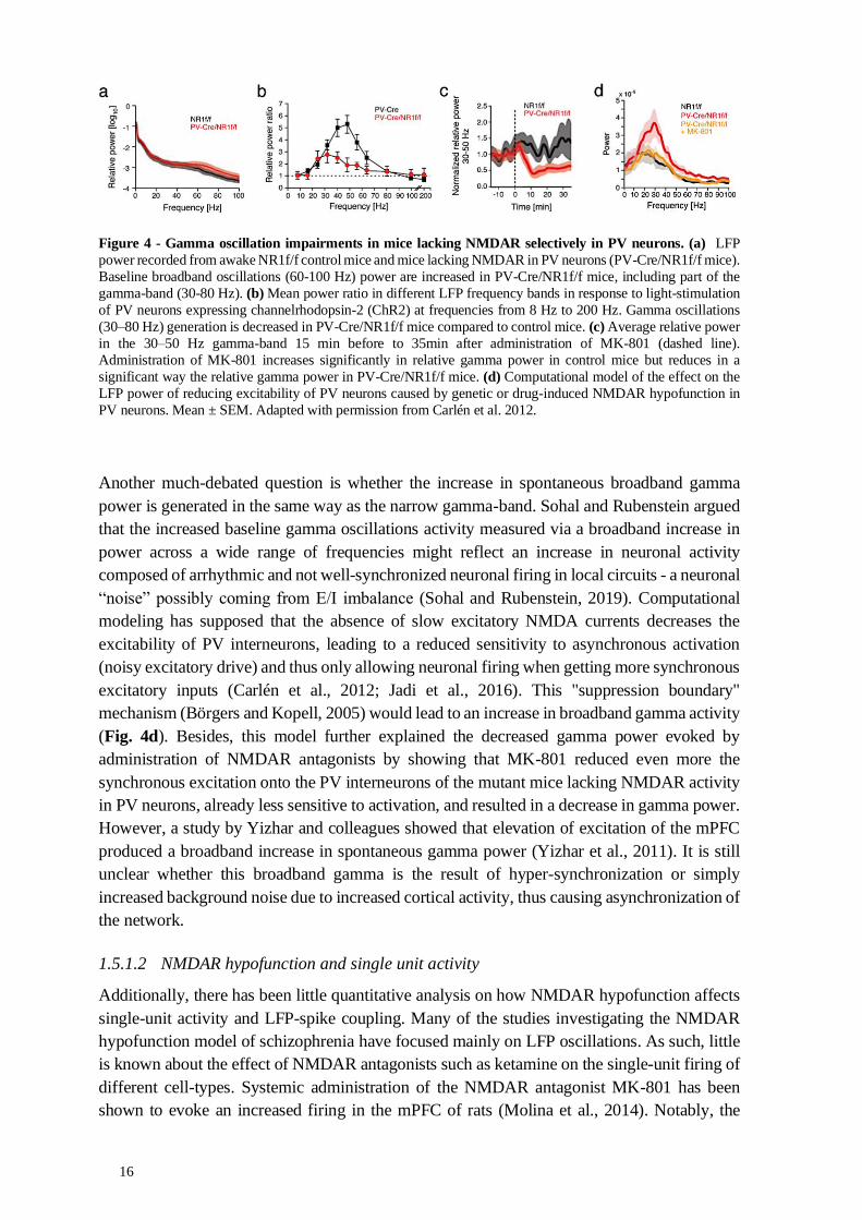
16
Figure 4 - Gamma oscillation impairments in mice lacking NMDAR selectively in PV neurons. (a) LFP
power recorded from awake NR1f/f control mice and mice lacking NMDAR in PV neurons (PV-Cre/NR1f/f mice).
Baseline broadband oscillations (60-100 Hz) power are increased in PV-Cre/NR1f/f mice, including part of the
gamma-band (30-80 Hz). (b) Mean power ratio in different LFP frequency bands in response to light-stimulation
of PV neurons expressing channelrhodopsin-2 (ChR2) at frequencies from 8 Hz to 200 Hz. Gamma oscillations
(30–80 Hz) generation is decreased in PV-Cre/NR1f/f mice compared to control mice. (c) Average relative power
in the 30–50 Hz gamma-band 15 min before to 35min after administration of MK-801 (dashed line).
Administration of MK-801 increases significantly in relative gamma power in control mice but reduces in a
significant way the relative gamma power in PV-Cre/NR1f/f mice. (d) Computational model of the effect on the
LFP power of reducing excitability of PV neurons caused by genetic or drug-induced NMDAR hypofunction in
PV neurons. Mean ± SEM. Adapted with permission from Carlén et al. 2012.
Another much-debated question is whether the increase in spontaneous broadband gamma
power is generated in the same way as the narrow gamma-band. Sohal and Rubenstein argued
that the increased baseline gamma oscillations activity measured via a broadband increase in
power across a wide range of frequencies might reflect an increase in neuronal activity
composed of arrhythmic and not well-synchronized neuronal firing in local circuits - a neuronal
“noise” possibly coming from E/I imbalance (Sohal and Rubenstein, 2019). Computational
modeling has supposed that the absence of slow excitatory NMDA currents decreases the
excitability of PV interneurons, leading to a reduced sensitivity to asynchronous activation
(noisy excitatory drive) and thus only allowing neuronal firing when getting more synchronous
excitatory inputs (Carlén et al., 2012; Jadi et al., 2016). This "suppression boundary"
mechanism (Börgers and Kopell, 2005) would lead to an increase in broadband gamma activity
(Fig. 4d). Besides, this model further explained the decreased gamma power evoked by
administration of NMDAR antagonists by showing that MK-801 reduced even more the
synchronous excitation onto the PV interneurons of the mutant mice lacking NMDAR activity
in PV neurons, already less sensitive to activation, and resulted in a decrease in gamma power.
However, a study by Yizhar and colleagues showed that elevation of excitation of the mPFC
produced a broadband increase in spontaneous gamma power (Yizhar et al., 2011). It is still
unclear whether this broadband gamma is the result of hyper-synchronization or simply
increased background noise due to increased cortical activity, thus causing asynchronization of
the network.
1.5.1.2 NMDAR hypofunction and single unit activity
Additionally, there has been little quantitative analysis on how NMDAR hypofunction affects
single-unit activity and LFP-spike coupling. Many of the studies investigating the NMDAR
hypofunction model of schizophrenia have focused mainly on LFP oscillations. As such, little
is known about the effect of NMDAR antagonists such as ketamine on the single-unit firing of
different cell-types. Systemic administration of the NMDAR antagonist MK-801 has been
shown to evoke an increased firing in the mPFC of rats (Molina et al., 2014). Notably, the

17
synchronization of single-unit firing was found to be decreased, and the spike trains
disorganized. This adds to both modeling and experimental findings showing that genetic
knockout of NMDARs in PV neurons results in increased variability of cortical excitatory
activity (Fig. 5) (Carlén et al., 2012; Jadi et al., 2016). Together, these studies indicate that
NMDAR hypofunction could lead to desynchronization of neuronal activity concomitant with
increased baseline broadband oscillation power.
Figure 5 - Impaired inhibition of single-unit PN activity after light activation of PV units in PV-Cre/NR1f/f
mice. (a) Overlaid pyramidal neuron (PN; n = 17 neurons) and PV (n = 9 neurons) population averages from the
PV-Cre control mice, with 0 set as the time of the light flash on each cycle. PV population average shows a light
evoked peak in firing at 1-2 ms. The PN population average shows a decrease in spike probability around 4-10 ms
after the light flash and an increase in spike probability around 20 ms. (b) Overlaid PN (n = 4) and PV (n = 6)
population averages from the PV-Cre/NR1f/f mice. PV population average shows a larger light evoked peak in
firing at 1-5 ms, while PN population average shows a slightly elevated spontaneous firing rate, a smaller decrease
in spike probability around 4-10 ms after the light flash, and a modest increase in spike probability around 20 ms.
(c) Mean light-evoked spike latency for PV interneurons in control mice was 2.62 ± 1.2 ms at low power (31
mW/mm2) and 1.18 ± 1.2 ms at high power (68 mW/mm2). Mean light-evoked spike latency for PV interneurons
in PV-Cre/NR1f/f mice was significantly longer (4.52 ± 2.2 ms at low power and 2.74 ± 2.2 ms at high power;
Mann-Whitney test; P < 0.05 in both cases). In addition, the variance of the spike latencies was significantly higher
across cells in PV-Cre/NR1f/f mice than in control mice (F test; P < 0.05). Adapted with permission from Carlén
et al. 2012.
1.6 BDNF-TRKB SIGNALING
The brain-derived neurotrophic factor (BDNF) is a member of the family of growth factors
called neurotrophins that influence the proliferation, differentiation, survival, and death of
neurons and glia. BDNF is secreted in most parts of the central nervous system, with the highest
levels detected in the cortex and the hippocampus (Armanini et al., 1995). Although BDNF
was first characterized as instrumental in the development of the brain (Huang et al., 1999), it
is now clear that BDNF has multiple roles in the adult nervous system, such as regulating
synaptic structure and connections, the release of neurotransmitter, and synaptic plasticity
(Kowiański et al., 2018; Nagappan and Lu, 2005; Park and Poo, 2013), Thus, BDNF signaling
is thought to be critical for the maintenance and refinement of neuronal circuits as one of its
main roles is to regulate neuronal morphology.
Much research on the role of BDNF has focused on plasticity processes. BDNF-trkB signaling
(Fig. 6), elicited through the binding of BDNF to trkB, causes intracellular signaling cascades
that contribute to the transcription of genes central for synaptic plasticity and processes like
long-term potentiation (Minichiello, 2009). But these diverse intracellular signaling cascades
are also known to regulate widely diverse functions, such as cell migration, survival and death,

18
as well as the outgrowth and pruning of neurites, formation of synapses, neuronal
communication and synaptic plasticity (Chao, 2003; Kowiański et al., 2018; Lu et al., 2005,
2014), making it difficult to categorize the full function of BDNF-trkB signaling. Moreover,
impaired BDNF-trkB signaling has been extensively associated with the pathophysiology of
many neurodegenerative disorders, depression, anxiety and other psychiatric disorders (Autry
and Monteggia, 2012; Castrén, 2014; Mitre et al., 2016; Woo and Lu, 2006). However, little is
known about the role of BDNF-trkB signaling at the network level, particularly in the precise
mechanisms underlying trkB dysfunction in the mature brain.
Figure 6 – Overview of the BDNF-trkB signaling pathways. Binding of BDNF to the full-length trkB (a)
triggers the homodimerization and phosphorylation of trkBs, which elicits three main intracellular signaling
pathways (b), through the activation of: Ras–mitogen-activated protein kinase (MAPK) which promotes
extracellular signal-regulated kinase (ERK) signaling; phospholipase Cγ (PLC-γ), which promotes the generation
of diacylglycerol (DAG) and Ca2+/calmodulin-dependent protein kinase II (CaMKII); phosphatidylinositol 3-
kinase (PI3K)-Akt cascade. The activation of these pathways leads to numerous physiological processes, of which
some are shown in (c). Image of neuron was adapted from scidraw.io (doi.org/10.5281/zenodo.3925929).
1.6.1 trkB receptors
The trkB gene NTRK2 is able to transcribe several splice variants, including the full-length trkB
(trkB.FL), but also truncated variants (trkB.T), i.e., receptors lacking the catalytic kinase
domain (Klein et al., 1990; Stoilov et al., 2002). TrkB.FL is composed of an extracellular
ligand-binding domain, a transmembrane domain, and an intracellular domain that includes the
highly conserved catalytic kinase domain (Benito-Gutiérrez et al., 2006; Klein et al., 1990).
The C‐terminal truncated trkB receptors (of which the trkB.T1 is the most studied) hold the
same extracellular and transmembrane domains, but their intracellular part consists of a unique
short sequence of amino acid residues (Fenner, 2012; Ohira and Hayashi, 2009).
For this reason, overexpressing truncated versions of trkB might offer the possibility to impair
BDNF-trkB signalization. Indeed, the truncated receptors were found to function as inhibitory
modulators of neurotrophins responsiveness (Eide et al., 1996; Ohira and Hayashi, 2009;
Stoilov et al., 2002). Truncated trkBs behave as dominant-negative isoforms by competing for
BDNF and forming nonfunctional heterodimers with the full-length version of the receptor
(Haapasalo et al., 2002). For example, overexpressing trkB.T1 has been shown to reduce trkB
phosphorylation (Saarelainen et al., 2003), adding to the findings indicating that truncated
19
trkBs negatively regulate the full-length trkB signaling (Carim-Todd et al., 2009). However,
trkB.T1 also holds BDNF independent functions and its involvement in regulating neuronal
and glial mechanisms is not yet completely understood (Fenner, 2012).
1.6.2 BDNF-trkB signaling in prefrontal PV interneuron activity
It has previously been suggested that a potential role for BDNF is to control the neuronal
activity in the PFC (Galloway et al., 2008). Galloway and colleagues further hypothesize that
this control of prefrontal neural activity is dependent on BDNF-trkB signaling in PV
interneurons. In the same vein, it has been suggested that BDNF-trkB signaling in the cortex
could serve as a way to stabilize the inhibitory drive of PV interneurons (Marty et al., 1997)
and thus mediate the excitatory/inhibitory balance of the local network (Woo and Lu, 2006).
However, while trkB is expressed in pyramidal neurons and in inhibitory GABAergic neurons
- PV neurons express trkB abundantly - BDNF is essentially secreted by pyramidal neurons
(Gorba and Wahle, 1999; Hashimoto et al., 2005; Marty et al., 1997). As in situ expression of
BDNF confirmed that cortical GABAergic interneurons, including PV interneurons, do not
synthesize BDNF themselves, it has been proposed that BDNF act in a paracrine manner,
modulating the presynaptic inhibitory activity, the GABA expression levels, as well as the
morphology, of cortical PV interneurons. This has been shown to occur via the release of
BDNF from postsynaptic pyramidal neurons (Huang et al., 1999; Jiao et al., 2011).
Functional evidence of this interaction between BDNF-trkB signaling and PV interneurons
have been observed in neuropsychiatric patients. For instance, changes in the expression of
trkB in PV interneurons have been associated with the pathophysiology of schizophrenia. Most
postmortem studies indicate that both BDNF and trkB proteins are downregulated in the brain
tissue of individuals who had schizophrenia (Fig. 7a) (Hashimoto et al., 2005; Ray et al., 2014;
Takahashi et al., 2000; Weickert et al., 2003, 2005), although a recent study report that trkB
mRNA levels in the PFC did not differ between postmortem tissue of people with
schizophrenia and healthy comparison subjects (Reinhart et al., 2015). Furthermore, the
expression of truncated trkB isoforms was found to be increased in the prefrontal cortex (Wong
et al., 2013). Both the decrease in trkB.FL and the increase in truncated trkBs are correlated
with decreased GABA markers in PFC tissue of schizophrenic patients (Wong et al., 2013).
Taken overall, these changes have been suggested to lead to the hypofunction of the prefrontal
cortex (Hashimoto et al., 2005; Lewis et al., 2005)
The results from postmortem studies of neuropsychiatric patients are corroborated by animal
studies showing a reduction in GABA markers when trkB function is inhibited for 20 days in
the adult brain, suggesting that the maintenance of GABA levels involves BDNF-trkB
signaling (Chen et al., 2011; Porcher et al., 2018). Furthermore, findings obtained by knockout
of trkB (knockout affecting all isoforms) specifically in PV neurons (Xenos et al., 2018; Zheng
et al., 2011) and cortical GABAergic interneurons (Tan et al., 2018) reported several
morphological as well as functional alterations, including neuronal desynchronizations
affecting the LFP gamma-band and E/I imbalance. The absence of trkB in PV interneurons
during the development also led to a significant loss of inhibitory synapses onto prefrontal
pyramidal neurons, indicating an essential role of BDNF-trkB signaling in the clustering of
GABAergic synapses (Xenos et al., 2018). Additionally, Zheng and colleagues found in the

20
hippocampus of mice lacking trkB in PV neurons a reduced inhibitory drive onto pyramidal
neurons, as well as disrupted gamma oscillations (Fig. 7b). They further observed impaired
spiking entrainment to the gamma LFP in PV interneurons (Fig. 7c) (Zheng et al., 2011).
Reduced induced gamma oscillations were also found in in vivo recordings of the sensory
cortex of transgenic mice, along with the increased firing of pyramidal neurons (Xenos et al.,
2018). Collectively, these studies outline a critical role for BDNF-trkB signaling in PV
interneurons for proper GABAergic control of the local circuit, reducing the reliability of
pyramidal firing and leading to E/I imbalance. However, these physiological changes might be
caused by global and developmental adaptations caused by the genetic deletion of trkB in PV
neurons.
Figure 7 - BDNF-trkB signaling and PV interneurons. (a) Reduced BDNF, trkB and GAD67 and PV mRNA
expression in the dorsolateral PFC of an individual with schizophrenia compared to a healthy control. Solid lines:
pial surface. Broken lines: border between the grey and white matter. Right: Expression of each transcript in
individuals with schizophrenia (Scz) and matched control subjects (Ctr). Mean ± SD. F: Fisher’s F distribution.
nCi/g: nanoCuries/gram. (b) Representative traces (up) of oscillations induced by carbachol. PSD (bottom)
indicating a significant reduction in the peak amplitude at a frequency corresponding to the gamma-band (~40 Hz)
in trkB knockout mice compared to control mice. (c) (left) Impaired firing phase distribution in PV interneurons
in trkB knockout mice. (right) The average spike incidence per gamma cycle in trkB knockout mice was
significantly reduced compared with control mice. *p < 0.05. The spike recorded in fast-spiking interneurons was
wider in trkB knockout mice than in control mice. (a) was adapted with permission from Lewis et al. 2005 and (b,
c) from Zheng et al. 2011, PNAS.
In terms of behavior, despite the extensive links with several brain disorders (Andero et al.,
2014; Ninan, 2014; Qi et al., 2015), the role of trkB signaling in mPFC behavioral correlates
are still poorly understood. For instance, unspecific deletion of trkB in corticolimbic
GABAergic interneurons (affecting the cortex and hippocampus, but not the cerebellum) has
been correlated with PV dysfunctions in the mPFC and social dominance abnormalities and
aggressive behavior (Tan et al., 2018), but as these alterations are not cell and region-specific,
they cannot pinpoint precisely the mechanisms behind the behavioral alterations.

21
Therefore, despite what we know of the role of BDNF-trkB signaling in neuronal development
and synaptic function, the question of how trkB in PV interneurons might modulate and control
pyramidal neurons excitatory and oscillatory activity has so far not been answered. Further
studies are thus needed to determine how BDNF-trkB signaling impairments lead to functional
changes in PV interneurons and disrupt excitation/inhibition balance that could lie at the core
of both network and behavioral disorders.
1.7 REGULATION OF SOCIAL PROCESSING BY THE PREFRONTAL PV INTERNEURONS
Social behavior is a fundamental process across species that requires the integration of a wide
variety of behaviors, including attention, reward-seeking, motivation, awareness of self and
others, social hierarchy, agonistic behavior, and flexibility in adapting one’s behavior to others
in a group (Bicks et al., 2015; Yizhar and Levy, 2021). Changes in social behaviors, along with
defects in decision making and the processing of emotion, were observed after the railroad
worker Phineas Gage’s skull was penetrated by a metal rod in 1849 (Damasio et al., 1994).
Four decades later, Ferrier and Yeo found by surgically removing the prefrontal part of the
brain of a baboon that he lost interest in other baboons and that his interactions with other
baboons were changed (Ferrier and Yeo, 1884).
Since then, several brain regions like the hypothalamus or the ventral tegmental area (VTA),
or neuropeptides like oxytocin, have been found to have a fundamental role in social
interactions (Dölen et al., 2013), but the PFC remains one of the critical regions for social
processing (Bicks et al., 2015; Wang et al., 2011; Yizhar and Levy, 2021). Indeed, it has been
shown that the optogenetic manipulation of projection pathways linking regions like the
amygdala or nucleus accumbens to the mPFC can modulate social behaviors (Felix-Ortiz et al.,
2016; Murugan et al., 2017), highlighting the integrative and functional importance of the
mPFC in these behaviors. However, manipulation of the projection from VTA to the nucleus
accumbens was shown to sufficient to promote prosocial interactions, while optogenetic
activation of the projection from VTA to mPFC had no effect on social interaction but was
associated with anxiety-like effects (Gunaydin et al., 2014). Therefore, although the mPFC has
been implicated in social behavior, understanding which neurons or pathways are relevant or
how they contribute has been challenging, in part due to the complexity of the underlying
circuitry.
At the local circuit level, synchronous oscillatory activity and correct E/I balance are thought
to be a crucial mechanism for the role of mPFC in social behaviors (Cao et al., 2018; Liu et al.,
2020; Selimbeyoglu et al., 2017; Wang et al., 2011; Yizhar et al., 2011). For example,
potentiating or depressing AMPA receptor activity, leading to higher excitability, primarily in
pyramidal neurons in the mPFC, was sufficient to modulate dominance behavior and hierarchy
in a group of mice (Wang et al., 2011). The increase of E/I ratio in the rat mPFC was also linked
with reduced social preference after the impairment of inputs from the mediodorsal thalamus
to mPFC GABAergic neurons. However, the preference for social cues was recovered after
pharmacogenetically increasing the activity of PV neurons, suggesting that proper PV
interneurons activity in the rat PFC is required for social behavior (Ferguson and Gao, 2018b).
Similarly, Bicks and colleagues found that juvenile social isolation caused alteration of mice
prefrontal PV interneurons. They also observed that prefrontal PV interneurons were active
22
during social interaction and that manipulating their activity modulated social approach (Bicks
et al., 2020). However, it is still not known whether the role of prefrontal PV interneurons in
social behaviors is specific to the social domain or is due to their involvement in a range of
cognitive processes dependent on the PFC, like attention, that are used during social behaviors.
Besides, it is still unclear which functional or compensatory factors could induce PV
dysfunction, circuit impairments and deficiencies in social processing. While some research
has been carried out on how manipulating PV interneurons might impact mPFC activity, the
mechanism by which it affects neuronal ensembles, coordinating communications with other
cortical and subcortical regions involved in regulating different social functions, has not been
established.
This section has attempted to provide a summary of the literature relating to the role of
prefrontal PV interneurons in the formation and maintenance of local circuit activity, with an
emphasis on the role played by NMDAR and trkB receptors. However, several questions
remain, which both works in this thesis have tried to address. For example, research to date has
not yet determined if and how increases in broadband gamma are fundamentally different from
narrow-band increases in gamma power caused by rhythmic feedback inhibition by PV
interneurons (e.g. in response to sensory stimulus). Also, the mechanism by which
asynchronous neuronal activity caused by deficient PV inhibition could manifest as an
increased power over a broad range of LFP frequencies (that includes the gamma-band), or
affects cortical states, has not been established. Additionally, much regarding the role of
BDNF-trkB signaling in the adult brain is unclear. Moreover, it has not been possible to
conclusively pinpoint functional or behavioral alterations to cell and region-specific changes
in BDNF-trkB signaling, notably in the PV interneurons of the mPFC.

23
2 RESEARCH AIMS
The general aim of this thesis is to understand the role of parvalbumin interneurons in shaping
the neuronal network activity in the mouse prefrontal cortex. We are particularly interested in
how receptors important for proper activity of parvalbumin interneurons lead to deficiency
affecting various aspects, from molecular, circuit to behavior. More specifically, the aims are:
I - To understand if and how increased prefrontal broadband gamma power could be
associated with neuronal activity with decreased synchronicity. For this reason, we aim to
evaluate how the NMDAR hypofunction in PV neurons in the PFC leads to altered neuronal
and oscillatory activity, notably affecting faster oscillations that include the gamma-band.
Additionally, we aim to evaluate if the electrophysiological changes observed following the
administration of NMDAR antagonists (such as ketamine) are consistent with oscillatory
alterations observed in genetic models of NMDAR deficiency (PAPER I).
II - To understand how alterations in mature PV interneurons specifically in the PFC lead to
circuit and behavior alterations. For this, we aim to investigate how the dysfunction of
BDNF-trkB signaling affects PV interneuron activity and its role in the PFC circuitry in the
adult brain (PAPER II).


25
3 MATERIALS AND METHODS
This section will briefly present and discuss the main techniques applied in both papers, as well
as possible alternatives and novel approaches that are becoming more viable with further
development. The research presented in Paper I used mainly electrophysiology recordings,
while Paper II used cellular, physiological, and behavioral readouts for the functional dissection
of the role of PV in the PFC. Please refer to the already well-described methods sections in
both papers for further details. Lastly, I will succinctly discuss ethical considerations emanating
from the use of animals, neuroscience tools, and publication practices.
3.1 SPECIFIC TARGETING AND MANIPULATION OF PV NEURONS
As it became possible to characterize populations of neurons that express specific molecular
markers, such as PV protein, it became feasible to target specific classes of neurons genetically.
This ability to experimentally be able to target PV neurons in a precise manner allowed a better
understanding of the morphology and function of PV neurons (Cardin et al., 2009; Hu et al.,
2014; Sohal et al., 2009).
3.1.1 Cre-lox system and transgenic animals
One of the main technologies used to perform selective genetic targeting of a defined neuronal
population is the Cre-lox system. Cre recombinase is an enzyme that executes the
recombination between two DNA recognition sites (called loxP sites). DNA sequences found
between two loxP sites are said to be "floxed". Thus, placing loxP sites correctly around a
specific gene allows its activation, deletion, or replacement with other genes. The Cre-lox
system allows switching the orientation of DNA sequences as two loxP sites in opposite
orientation to each other invert the intervening piece of DNA. It can also remove the DNA
sequence as two loxP sites in direct orientation dictate excision of the intervening DNA
between the sites leaving one loxP site behind. The fact that the activity of the Cre enzyme can
be controlled so that it is expressed in a particular cell type allowed us to delete the NR1 subunit
of the NMDAR or express the truncated version of trkB specifically in PV neurons. Targeted
DNA changes are extremely useful to perform modifications that affect only specific cell types,
as some knockout, such as NR1 or trkB, are lethal if expressed globally (Forrest et al., 1994;
Luikart et al., 2003).
3.1.2 NR1 floxed transgenic line
In Paper I, we studied the role of NMDAR in PV neurons using a transgenic mouse line initially
created and characterized in our research group (Carlén et al., 2012). PV-Cre/NR1f/f mice were
obtained by crossing mice with Cre recombinase expression in PV neurons (PV-Cre mice) with
mice carrying floxed alleles of the NR1 subunit (NR1f/f mice). This mouse line lacked the
NMDAR subunit NR1 specifically in PV-expressing neurons. Since NR1 is the essential
subunit for NMDAR, NR1 deletion results in loss of functional NMDARs (Forrest et al., 1994).
Unlike mouse models of NMDAR hypofunction in all neurons, no severe developmental,
anatomical (Figs. 8a, b) or behavioral abnormalities, except for selective cognitive
impairments, were found in PV-Cre/NR1f/f mice. Nevertheless, loss of NMDAR currents in
cortical PV interneurons was previously confirmed by ex-vivo recordings in PV-Cre/NR1f/f
mice (Fig. 8c), leading to functional impairments at the circuit level (Carlén et al., 2012).

26
The PV-Cre-driven recombination was previously quantified and validated in PV-Cre mice
crossed to the R26R-EYFP Cre reporter mouse line by counting PV neurons co-labeled with
eYFP (Carlén et al., 2012). This showed that Cre-recombination happened postnatally and
increased during development, attaining almost complete recombination at week 8 postnatal in
the somatosensory cortex (Figs. 8d, e). From this, we can assume that PV-Cre-driven
recombination of NR1 followed the same time course and that the circuit impairments seen in
Paper I could have been due to increased NMDAR hypofunction during development.
Furthermore, as we briefly discuss in Paper I, NMDAR hypofunction in PV-Cre/NR1f/f affects
all PV neurons throughout the brain. The global knockout of NMDAR in PV neurons,
therefore, affects PV neurons in several brain areas with significant anatomical and functional
connections to the PFC, such as the basal forebrain, hippocampus or thalamus (Cohen et al.,
2015; Kim et al., 2015) and thus could be behind the impairment seen in the PFC. Therefore,
the chronic and global effect of the NMDAR hypofunction might make it challenging to
pinpoint precisely when or in which area of the brain PV neuron dysfunction causes the
impairments we observed in the PFC.
Figure 8 - Genetic ablation of NMDAR specifically in PV neurons. (a) Layer distribution of PV neurons
in NR1f/f control and PV-Cre/NR1f/f mice at 11 weeks. (b) Immunohistochemistry for PV neurons in the
somatosensory cortex shows similar distribution of PV neurons in an adult NR1f/f and PV-Cre/NR1f/f mouse.
(c) Functional loss of NMDAR currents in PV neurons in PV-Cre/NR1f/f mice while the AMPA currents are
preserved. (left) Sample EPSC traces mediated by the AMPAR (downward) and NMDAR (upward) from a
control PV-Cre mouse and a PV-Cre/NR1f/f mouse. (right) NMDAR EPSC/AMPAR EPSC ratio in control
PV-Cre and PV-Cre/NR1f/f mice. (d) Recombination in PV-Cre mice. At 13d, only scattered PV interneurons
have recombined in somatosensory cortex (up; example pointed by white arrowhead), while near-complete
recombination was observed at 8w (bottom). (e) Quantification of recombination from the PV locus in
somatosensory cortex at different time points. * p < 0.05; error bars, mean ± S.E.M. Scale bars: (b,d) 200 μm.
Adapted with permission from Carlén et al. 2012.
27
It is thus desirable to control both spatial and temporal aspects of transgene expression. This
precision can be reached by using region-specific molecular markers for spatial specificity (Tan
et al., 2018) or using techniques that regulate temporal expression of Cre recombinase, such as
the activation of Cre recombinase fused to the estrogen receptor by the addition of tamoxifen.
One of the approaches we decided to use to control the spatial and temporal aspects of our
manipulation was to utilize a viral delivery method.
3.1.3 Viral delivery
The viral delivery of genetic material to the brain has been made possible by replacing part of
viruses with an expression cassette containing the genes of interest. Placing the gene of interest
between loxP sites and injecting the virus in transgenic animals with Cre recombinase
expression in specific cell-types allows the precise expression of the gene of interest in a local
and temporal manner (Heldt and Ressler, 2009). This has allowed the significant development
of Cre-dependent viruses carrying modified receptors, opsins, and fluorescent markers. We
used the Cre/loxP dependent viral vector strategy, via adeno-associated viruses (AAV), in both
papers presented in this thesis. It is worth noticing that AAVs are known to have a low level of
pathogenicity, having no association with diseases, but are still able to reach an effective level
of transgene expression (Urban and Rossier, 2012). More specifically, our vectors had the
sequence of interest flanked by two sets of incompatible lox sites, called double-floxed inverted
open-reading-frame (DIO). In the presence of Cre recombinase, the open-reading-frame is
inverted and can be expressed under the promoter present in the AAV, for instance, the
elongation factor 1-alpha (EF1α) promoter.
In Paper I, we used an AAV vector with Cre-dependent expression of channelrhodopsin-2
(ChR2) fused to mCherry (AAV-DIO-ChR2-mCherry) to identify PV interneurons via light-
activation (opto-tagging). The use of an AAV vector in Paper II allowed us to bypass the
developmental and more long-term changes due to trkB signaling and offered us a chance to
induce local manipulations in an adult brain. In both papers, we used the AAV-DIO-eYFP virus
as control by simply expressing the fluorophore eYFP in PV interneurons.
3.1.4 Viral expression of trkB.DN-mCherry
We decided to express a truncated version of the trkB specifically in PV interneurons. This
truncated receptor can compete with the normal receptor for BDNF, thus reducing BDNF-trkB
signaling in these neurons. For this, we used a trkB.T1 plasmid in which the small intracellular
part of the truncated receptor was replaced by the monomeric red fluorescent protein mCherry.
This allowed the easy detection of neurons expressing this trkB construct. As such, our
engineered receptor retains the capacity of sequestering BDNF as the standard trkB.T1, but its
complete lack of intracellular part does not elicit the still relatively unknown trkB.T1
intracellular signaling cascades (Fenner, 2012). We chose to call our engineered receptor
trkB.DN-mCherry, “DN” standing for dominant-negative, as a dominant-negative receptor is
a receptor that is able to disrupt the activity of the wild-type receptor when overexpressed.
To deliver trkB.DN-mCherry in a temporally, spatially, and cell-type-specific controlled
manner, we generated an AAV vector with Cre-dependent expression of trkB.DN-mCherry,
AAV-DIO-trkB.DN-mCherry (full name AAV5-Ef1a-DIO-TrkB.DN-TM570-mCherry) (Fig.
9). By bilateral injection of 0.5 µl of AAV-DIO-trkB.DN-mCherry into the PFC of 2-month-

28
old mice, the conditional transduction of PV interneurons was spatially restricted to the PFC
and allowed us to manipulate the BDNF-trkB signaling after normal development, and thus
provide a more accurate assessment for its role in the mature brain.
Figure 9 – Information about AAV-DIO-trkB.DN-mCherry and AAV-DIO-eYFP viruses (a) Full sequence
map of the vector pAAV-DIO-trkB.DN-TM570-mCherry for Cre-inducible expression of trkB.DN-mCherry
(Image created with SnapGene). (b) Highlight of the sequence part around the link of mCherry and the
transmembrane domain (TM domain) of trkB.T1, forming the vector AAV-DIO-trkB.DN-mCherry. (c) The viral
vectors used in Paper II. AAV-DIO-trkB.DN-TM570-mCherry: AAV vector with Cre-dependent expression of a
truncated version of the trkB receptor fused to mCherry, AAV-DIO-eYFP (full name AAV5-Ef1a-DIO-eYFP):
AAV vector with Cre-dependent expression of eYFP. L-ITR and R-ITR: Left and Right Inverted Terminal Repeat
(ITR) sequences; EF1a: elongation factor 1a promoter; WPRE: Woodchuck hepatitis virus Posttranscriptional
Regulatory Element.
Altogether, we successfully managed to manipulate two kinds of receptors expressed in PV
neurons and study their aftermath on the function of those neurons. In the meantime, new
methods have emerged and will make this kind of manipulation cheaper, less time-consuming,
and able to deal with longer sequences of genes than AAVs. The development of viral (but also
non-viral) delivery methods for technologies like the CRISPR-Cas9 system that can be
intracranially injected and safely edit genes in any cell type in the brains of adult animals offers
new possibilities to manipulate NMDAR or trkB in a spatial and time selective manner. For
example, the focal knockout of mGluR5, a metabotropic NMDA receptor, in adult mice has
been successful, reversing the behavioral phenotype in a fragile X mental retardation 1 KO
mice (Sandoval et al., 2020).
3.2 MOLECULAR AND CELLULAR READOUTS
Genetic manipulation studies are widely used to grasp the mechanisms underlying complex
behaviors and model molecular, cellular, and circuit alterations resulting from aberrant gene
expression. Previous work suggests that increased expression of truncated trkB results in

29
decreased levels of trkB.FL (Haapasalo et al., 2002). Most previous works used Western blot
to compare neurotrophic protein levels in transgenic mice versus control mice brain tissue.
However, since transduced PV interneurons represented only 56% of PFC PV interneurons,
we decided to examine if the expression of trkB.DN-mCherry altered the levels of endogenous
trkB by using two immunohistochemistry steps instead. As trkB.DN-mCherry has an
extracellular domain that is 100% homologous to the extracellular domain of trkB.FL (Fenner,
2012), but lacks the intracellular trkB domain, we used antibodies targeting the extracellular
domain and intracellular domain of trkB receptors to compare their amounts in the soma of
trkB.DN-mCherry or eYFP virus-transduced PV interneurons. This allowed us to obtain the
corrected total cell fluorescence (Mean fluorescence of the area of interest minus mean
fluorescence of the background), measured with the widely used open-source FIJI software
(Schindelin et al., 2012), and calculate an intracellular/ extracellular ratio.
We further used immunostaining to examine if the expression of trkB.DN-mCherry led to
changes in GABAergic proteins, as trkB is known to control the gene transcription of GABA-
related proteins, as well as parvalbumin protein level, in cortical interneurons (Lewis et al.,
2005; Sánchez-Huertas and Rico, 2011; Woo and Lu, 2006). Although we managed to get an
overview of the alterations at the level of proteins, further quantification is needed to
understand molecular changes better. The use of fluorescence-activated cell sorting (FACS) to
sort PV interneurons based on the mCherry/eYFP fluorescence could be a viable alternative to
compare protein levels with an enzyme-linked immunosorbent assay (known as ELISA) or
quantify alterations at the mRNA level with snRNA-seq.
Additionally, we checked for potential morphological alteration in transduced PV interneurons
in the mPFC, including dendritic and axonal arborization and the number of axonal inhibitory
terminals. After filling patched PV interneurons with biocytin, we cleared 200 µm tick mPFC
slices with CUBIC (Figs. 10a-d), an easy 2-steps histology tissue clearing method (Ariel, 2017;
Susaki et al., 2014), and stained them for PV, mCherry or eYFP, and gephyrin, a marker
specific to postsynaptic inhibitory synapses, before imaging them with a confocal microscope.
This allowed us to perform Sholl analysis (counting of the number of neurite intersections that
occur at fixed distances from the soma in concentric circles, see Fig. 10e) and quantify the
number of axonal boutons (see method section of the Paper II, for a full description).
Figure 10 - Cubic and Sholl analysis.
(a-d) Clearing of the brain slices. (a)
coronal brain slice (40 μm thick) before
clearing; (b) after incubation with
CUBIC reagent 1 overnight; (c) after
washing of brain slice before blocking;
(d) after incubation with CUBIC reagent
2 overnight. Imaging is done with
CUBIC reagent 2. (e) Sholl analysis.
Digitally applied concentric rings spaced
10 μm apart are centered on the soma
center, and the number of intersections
between the neuronal process and ring at
a given radius is counted. Estimates for
morphological attributes of the neuron
are then analyzed.
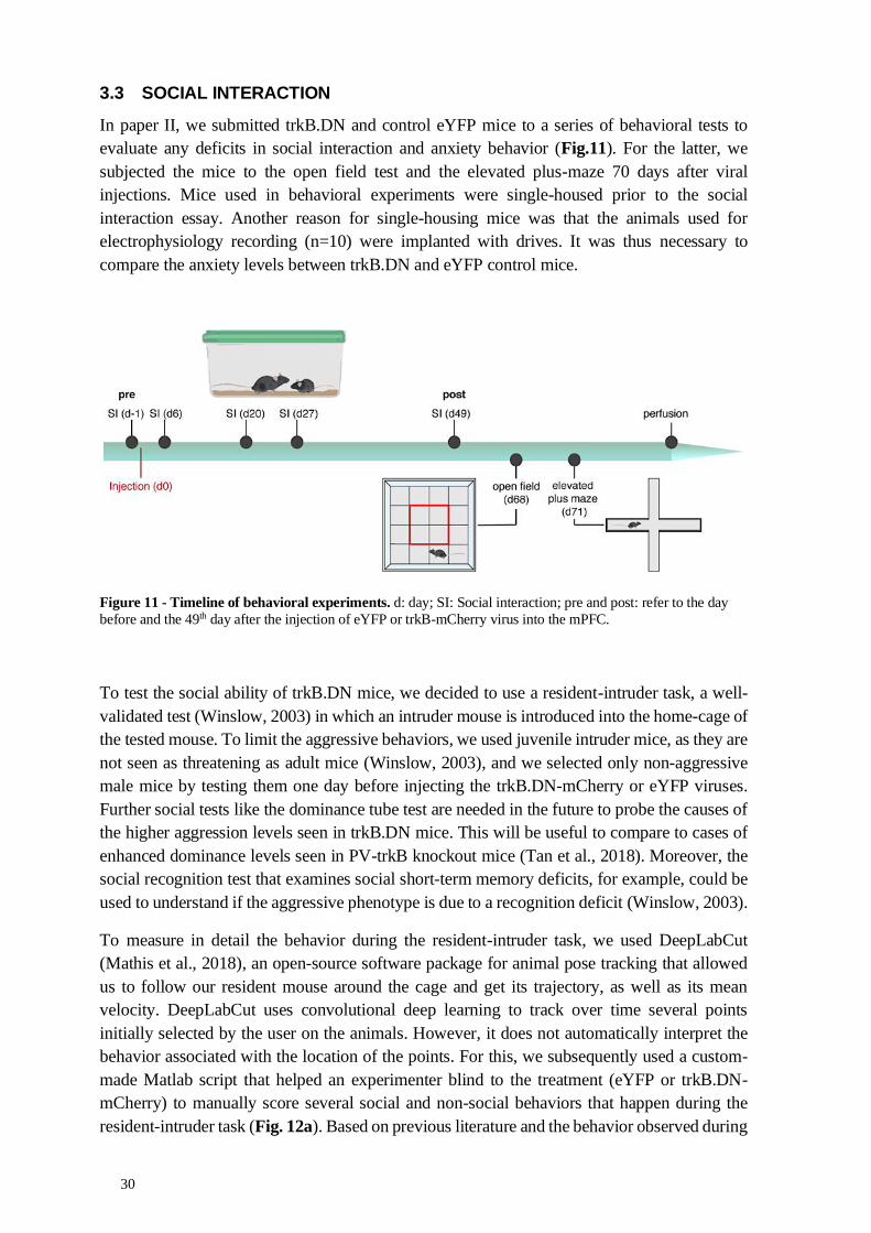
30
3.3 SOCIAL INTERACTION
In paper II, we submitted trkB.DN and control eYFP mice to a series of behavioral tests to
evaluate any deficits in social interaction and anxiety behavior (Fig.11). For the latter, we
subjected the mice to the open field test and the elevated plus-maze 70 days after viral
injections. Mice used in behavioral experiments were single-housed prior to the social
interaction essay. Another reason for single-housing mice was that the animals used for
electrophysiology recording (n=10) were implanted with drives. It was thus necessary to
compare the anxiety levels between trkB.DN and eYFP control mice.
Figure 11 - Timeline of behavioral experiments. d: day; SI: Social interaction; pre and post: refer to the day
before and the 49th day after the injection of eYFP or trkB-mCherry virus into the mPFC.
To test the social ability of trkB.DN mice, we decided to use a resident-intruder task, a well-
validated test (Winslow, 2003) in which an intruder mouse is introduced into the home-cage of
the tested mouse. To limit the aggressive behaviors, we used juvenile intruder mice, as they are
not seen as threatening as adult mice (Winslow, 2003), and we selected only non-aggressive
male mice by testing them one day before injecting the trkB.DN-mCherry or eYFP viruses.
Further social tests like the dominance tube test are needed in the future to probe the causes of
the higher aggression levels seen in trkB.DN mice. This will be useful to compare to cases of
enhanced dominance levels seen in PV-trkB knockout mice (Tan et al., 2018). Moreover, the
social recognition test that examines social short-term memory deficits, for example, could be
used to understand if the aggressive phenotype is due to a recognition deficit (Winslow, 2003).
To measure in detail the behavior during the resident-intruder task, we used DeepLabCut
(Mathis et al., 2018), an open-source software package for animal pose tracking that allowed
us to follow our resident mouse around the cage and get its trajectory, as well as its mean
velocity. DeepLabCut uses convolutional deep learning to track over time several points
initially selected by the user on the animals. However, it does not automatically interpret the
behavior associated with the location of the points. For this, we subsequently used a custom-
made Matlab script that helped an experimenter blind to the treatment (eYFP or trkB.DN-
mCherry) to manually score several social and non-social behaviors that happen during the
resident-intruder task (Fig. 12a). Based on previous literature and the behavior observed during

31
the experiment, we chose to focus on exploration, digging, grooming, tail rattling, sniffing, and
aggressive (attack, fighting as kicking, biting, and wrestling) behaviors (Fig. 12b). We used
these categories to score the amount and percentage of time spent in each social and non-social
behavior, as reported in Figure 5 of Paper II). This manual scoring was sufficient to allow
behavioral comparisons between groups. However, the temporal resolution of manual
annotation of behavior does not allow for optimum synchronization to recorded neural data
from in vivo electrophysiological recording.
Several new open-source tools are attempting to close the gap between tracking and behavior
classification by inferring what actions animals perform by analyzing the output of an
automated tracker. Besides, different new strategies are allowing the simultaneous tracking of
multiple animals, using radio frequency identification (RFID) tags or multi-camera 3D systems
(de Chaumont et al., 2019). The development and increased usage of these open-source
software will hopefully allow growing a library of pose estimation and behavioral predictive
classifiers for the easy categorizations of social behaviors in groups of several animals (Nilsson
et al., 2020). These new tools will also allow us to monitor and describe the complex repertoire
parts of behaviors in a more detailed way and hopefully be able to match them with neuronal
activity (Datta et al., 2019).
3.4 RECORDING THE ACTIVITY OF THE PFC
3.4.1 Ex vivo electrophysiology
In paper II, we used whole-cell patch-clamp electrophysiology to investigate the
electrophysiological effect of trkB-mCherry expression in PV interneurons. These experiments
were done (by colleagues) on ex vivo brain tissue. Current-clamp electrophysiology allowed
the recording of the activity of PV interneurons present in the mPFC at a highly temporal,
electrical, and spatial resolution, giving us knowledge about the intrinsic properties of the PV
interneurons and their firing patterns. We used voltage-clamp to record pyramidal neurons to
Figure 12 - Example
of movement tracking
and behavioral
classification.
(a) The trkB.DN mouse
movement was tracked
(yellow dots in videos)
during the resident-
intruder protocol while
interacting with a C57
juvenile mouse. (b)
Manual classification
of behaviors over time.
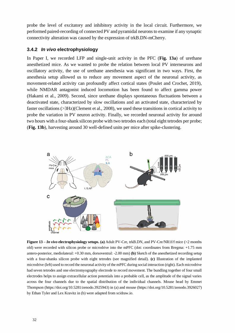
32
probe the level of excitatory and inhibitory activity in the local circuit. Furthermore, we
performed paired-recording of connected PV and pyramidal neurons to examine if any synaptic
connectivity alteration was caused by the expression of trkB.DN-mCherry.
3.4.2 In vivo electrophysiology
In Paper I, we recorded LFP and single-unit activity in the PFC (Fig. 13a) of urethane
anesthetized mice. As we wanted to probe the relation between local PV interneurons and
oscillatory activity, the use of urethane anesthesia was significant in two ways. First, the
anesthesia setup allowed us to reduce any movement aspect of the neuronal activity, as
movement-related activity can profoundly affect cortical states (Poulet and Crochet, 2019),
while NMDAR antagonist induced locomotion has been found to affect gamma power
(Hakami et al., 2009). Second, since urethane displays spontaneous fluctuations between a
deactivated state, characterized by slow oscillations and an activated state, characterized by
faster oscillations (>3Hz)(Clement et al., 2008), we used these transitions in cortical activity to
probe the variation in PV neuron activity. Finally, we recorded neuronal activity for around
two hours with a four-shank silicon probe with two tetrodes each (total eight tetrodes per probe;
(Fig. 13b), harvesting around 30 well-defined units per mice after spike-clustering.
Figure 13 – In vivo electrophysiology setups. (a) Adult PV-Cre, trkB.DN, and PV-Cre/NR1f/f mice (>2 months
old) were recorded with silicon probe or microdrive into the mPFC (dot: coordinates from Bregma: +1.75 mm
antero-posterior, mediolateral: +0.30 mm, dorsoventral: -2.00 mm) (b) Sketch of the anesthetized recording setup
with a four-shanks silicon probe with eight tetrodes (see magnified detail). (c) Illustration of the implanted
microdrive (left) used to record the neuronal activity of the mPFC during social interaction (right). Each microdrive
had seven tetrodes and one electromyography electrode to record movement. The bundling together of four small
electrodes helps to assign extracellular action potentials into a probable cell, as the amplitude of the signal varies
across the four channels due to the spatial distribution of the individual channels. Mouse head by Emmet
Thompson (https://doi.org/10.5281/zenodo.3925943) in (a) and mouse (https://doi.org/10.5281/zenodo.3926027)
by Ethan Tyler and Lex Kravitz in (b) were adapted from scidraw.io.
33
In Paper II, we recorded LFP and single-unit activity during behavior and under urethane
anesthesia. For the freely moving recordings, we used a handmade drive with seven tetrodes
and one electromyography recording electrode (Fig. 13c) based on the open-source FlexDrive
(Voigts et al., 2013). For the urethane recordings, we used the same four-shank silicon probes
as for Paper I (Fig. 13b). Both methods provided us with an adequate number of units recorded
per animal (~10 units per mice during the social interaction and ~35 units per mice during the
urethane recording), with a lower yield during the social interaction caused by the restricted
time of the recording. PV interneurons usually represent a small fraction of recorded units (<
10%), and the number of putative PV units we recorded was not sufficient for proper
comparison between animals or groups. We, therefore, focused analysis on pyramidal neurons.
I will discuss alternative methods for recording neuronal activity in the perspectives chapter of
this thesis.
Moreover, to elicit a change in neuronal activity within the mPFC circuitry and follow the
response of the pyramidal neurons, we utilized the tail pinch protocol, which is a classic method
using a short 7s pinch to the tail of the mouse to produce an arousal signal traveling from the
reticular system to the cortex, including the mPFC (Carceller et al., 2020; Mantz et al., 1988;
Massi et al., 2012).
3.4.3 Electrophysiology data analysis
In Paper I, we focused heavily on electrophysiology data analysis, and as such, we tried to
describe the reasons for each analysis as much as possible. I will, therefore, only briefly present
here some of the analyses we performed.
3.4.3.1 Power spectral density of LFP
As the LFP is an electric signal, a majority of tools applied for its analysis come from the field
of signal-processing and electronic communication. One of the main tools is the use of Fourier
transform to convert a signal from its original domain, like time, to a representation in the
frequency domain. In our case, the LFP can be thus decomposed into several discrete temporal
frequency components in hertz (gamma, delta, theta, etc.). The LFP is commonly visualized
with power spectral density (PSD) plots. In short, we can use a power spectrum to describe the
distribution of power of the LFP into its frequency components. This helps identify which
frequency of the signal is the most dominant. For example, we can observe with a PSD which
frequency of the LFP is increased by a task, like the typical narrow peak around 40 Hz when
the power in the gamma oscillation is increased during cognitive tasks. There are several ways
to estimate the PSD of a signal. Here we mostly use Welch's method. This approach, averaging
the Fourier transform with some overlapping, reduces the noise in the estimated power spectra
in exchange for reducing the frequency resolution. As we are primarily interested in frequencies
from 0.5 to 150 Hz, Welch's method is thus an adequate method for comparing the LFP.
There has been a discussion over the interpretation of PSD, as diverse events can be displayed
in a similar fashion on a PSD. For example, the increase of power in a particular frequency
band of the PSD could result from the increased number of oscillations in that frequency but
could also be due to a higher firing rate of local neurons (Manning et al., 2009). To understand
the origin of the increased power in the PV-Cre/NR1f/f mice, we decided to use, along with
the PSD, other analyses of the synchronous nature of the oscillation.

34
3.4.3.2 Cross-frequency coupling analyses
One of such analyses is the CFC analysis. In a nutshell, analysis of CFC focuses on the
synchronous relationships between the amplitude, phase, and frequency of two rhythms from
different frequency bands. There are several ways to measure different types of cross-frequency
coupling. In the LFP, the amplitude of fast oscillations, typically 30–100 Hz, are routinely
coupled to the phase of oscillatory activity at slower frequencies, most commonly at the theta
or delta frequency. To see if the lack of NMDAR in PV neurons altered this coupling, we first
used phase-amplitude comodulation maps (comodulograms), which allowed us to identify at
which phase of the slower frequencies we could find the strongest high-frequency amplitude
modulation by scanning multiple frequency pairs searching for cross-frequency coupling (Tort
et al., 2010). For this, we calculated a modulation index, which is a measure of the intensity of
the coupling. Therefore, if the mean amplitude of fast oscillation is uniformly distributed over
the phases of slower oscillation (meaning lack of phase-amplitude coupling), we have MI = 0.
On the other hand, an MI value of 1 would denote an oscillation that just exists in a single-
phase bin and disappears at the other phases of the slower oscillation (Tort et al., 2010).
Therefore, the low-frequency band (0.5-2 Hz) used for the MI analyses was identified by
building phase-amplitude comodulation maps aimed to identify the strongest phase modulation
of the broadband gamma and high-frequency band (HFB) amplitudes. The comodulation maps
were constructed based on the calculation of the MI at phase frequencies from 0.5 Hz to 6 Hz
(in steps of 1 Hz) and amplitude frequencies from 30 Hz to 150 Hz (in steps of 5 Hz). There
are several discussions on the potential limitations of the different methodological toolbox that
exist to analyze cross-frequency coupling (Aru et al., 2015). For example, as most only focus
on one type of cross-frequency coupling (Phase-amplitude here, but also phase-phase or
amplitude-amplitude), it has been argued that the choice of method restricts the type of cross-
frequency coupling detectable in data (Nadalin et al., 2019; Tort et al., 2010). In our case, we
found the MI to be suitable to show that synchrony was modified in PV-Cre/NR1f/f or after
ketamine application in PV-Cre controls.
3.4.3.3 Spectral entropy and high-frequency index
We used two measures to quantify and qualify the level of frequency dispersion (and
consequently the level of synchronization in the neuronal activity) in the PSD. First, we
performed an analysis of the spectral entropy (based on Shannon entropy) of the LFP (30-
150Hz), where a narrower power distribution results in lower entropy values, while a large
power distribution, as in the case of broadband activity, results in higher entropy values.
Secondly, we calculated a high-frequency index (the power ratio between the 200-300 Hz and
the 30-150 Hz band - the latter including the aberrant bands in PV-Cre/NR1f/f mice) to quantify
the contribution of spectral leakage caused by increased spiking activity to the LFP power in
the 30-150 Hz frequency band. We focused on 200-300 Hz to avoid the typical multi-unit
activity frequency band (>300 Hz) and to as far as possible exclude frequencies possibly
reflecting true oscillations (<200 Hz) (Menendez de la Prida et al., 2015). The 200-300 Hz band
is considered to reflect noisy activity typical of spectral leakage (Ray and Maunsell, 2011;
Scheffer-Teixeira et al., 2013), and we, therefore, focused on this band. These measures were
adapted from methods previously used to define events with epileptic signals from control LFP
(Valero et al., 2017) and allowed us to separate events with asynchronous signals from more
synchronous events.
35
3.4.3.4 Single-unit activity analyses
For single-unit activity, after spike sorting and cell classification (well described in both
papers), we predominantly focused our analysis on the mean firing rate (in spike/s or z-scored
against its baseline activity) of individual neurons and of populations of neurons. This allowed
us to straightforwardly quantify changes in activity during UP and DOWN states in Paper I or
after social interaction or tail pinch stimuli in Paper II. However, the relatively low single-unit
yield did not allow us to analyze single-unit data for the PV neuron population in Paper II,
which might in the future warrant the use of recently developed probes that can record up to
400 units (see perspectives).
Moreover, the mean firing rate is only one of the many factors defining the activity pattern of
a neuronal population. With this in mind, in Paper II, we used neural trajectories to analyze the
activity dynamics of wide spiking neurons recorded during social interaction as well as during
the tail pinch experiment. In general, to understand the dynamics of a neuronal population, it
is important to measure how the pattern of many different neuronal activity variables evolves
over time. However, this neuronal activity data is high-dimensional, can be variable with
patterns evolving along time, and can be organized at multiple temporal and spatial scales,
making it difficult to be visualized. To better picture the neuronal population dynamics, we
thus reduced its complexity by using a principal component analysis (PCA). This
dimensionality reduction allowed us to take the single-unit activity dataset, which is assumed
to be made of many variables and collapses its highly complex number of variables into a
lower-dimensional space where these are more easily visualized. Given a set of data about
single-unit activity features, the PCA can distinguish an ordered set of principal components
(PCs), each of which forms a different “axis” representing progressively less variance present
in the data (Datta et al., 2019; Pang et al., 2016). Therefore, this trimmed version of the dataset
can serve as a useful lower-dimensional approximation of the original data and allows the
analysis of the dynamic of complex datasets by following fewer variables. In our case, we
chose to restrict to the two principal components, PC1 and PC2, which explained the variability
of the dataset the most. Furthermore, to consider the evolution of the neuronal dynamic
alongside the social interaction or the tail pinch, we plotted the evolution of the PC1 and PC2
along time. These neuronal trajectories allowed us to visualize the change in the dynamics of
the neuronal activity along the experimental timeline (Pessoa, 2019).
3.4.3.5 Considerations on neurophysiology data analyses
Finally, I will end this section on electrophysiology data by shortly considering some
opportunities to improve data sharing. In our two projects, we mainly relied on homemade
scripts to analyze our data. This is beneficial, as we tailored our analysis to the format of our
data and did not have to rely on expensive and somewhat limited software. However, it is
difficult and time-consuming, and added efforts are needed to make the scripts available for
anyone to use. Furthermore, the lack of a standardized system to collect and share the vast
amount of produced data among researchers, as well as the lack of a standard data format,
makes comparison across laboratories and replication of experiments difficult. As the
electrophysiology data collected becomes larger and more complex, its analysis relies more
and more on heavy computational methods. There is, therefore, a necessity for an improvement
in data analysis standards to not only allow collaborations around easily shareable datasets, but
36
also to allow the development of open-source hardware and analysis software for
neurophysiology data. Several open-source and collaborative projects have been set up, and we
used several in our analysis, but further efforts seem to be needed. One example is the
development of a common data standard for neurophysiology called Neurodata Without
Borders. It is designed to store neurophysiology data from diverse sources, including data from
ex vivo and in vivo electrophysiology experiments, data from optical physiology experiments,
as well as movement or eye tracking and stimulus data. As more labs join this movement to
share data, the easier it will be to find ways to do so, but till then, it is still time-consuming and
requires somewhat advanced coding skills, which adds quite a lot of strain on top of routine
data analysis. Efforts should be made to incentivize such procedures by contributing special
funding specifically for such initiatives, for example. Research institutes should also emphasize
this, making sure they develop structures for data storage that allow such data to be uploaded,
curated and shared, as well as employ software developers to help with such measures.
3.5 ETHICAL CONSIDERATIONS
For the purpose of the research carried out for this thesis, we used many techniques based on
invasive approaches, recording both in vitro and in vivo neuronal activity in transgenic animals
after modifying their brain activity with genetic engineering, optogenetics, and viral injection
techniques. Our main aim is to understand the primary function of the prefrontal cortex in the
integration and coordination of information being internally generated with that received from
the external world. Consequently, we aim to identify potential cellular and molecular targets
for novel therapies for neuropsychiatric disorders by studying how cellular changes lead to
altered neuronal communication and distorted brain activity. We hope this will lead future
research to identify better cellular and network targets for medical interventions in
neuropsychiatric disorders. While there are serious ethical questions to consider regarding the
use of animals to help answer these questions, we believe the benefits of such research
outweigh the ethical issues that emerge when we use invasive techniques in animals. However,
for this to hold good, we should ensure that the scientific research question and methods are as
fitting as possible. This is not only important for the ethical management of animals but also
for the proper scientific quality of results.
One of the ways we aimed to do so, in this case, was to apply the 3 R Principle (Reduction,
Replacement, Refinement) by aiming to use the minimal number of animals possible to reach
a conclusion; with adequate care before, during, and after any operation; and by analyzing the
results obtained during the study as effectively as possible. Animal research at Karolinska
Institutet is strictly regulated and comes under both Swedish and EU legislation on animal
welfare. Additionally, taking courses on laboratory animals is necessary to be able to carry out
animal experiments at Karolinska Institutet. Therefore, any experiment included in this thesis
has been weighed against their impact on the animals and performed as thoughtfully as
possible.
Another way to reduce the number of animal experiments is to avoid the unnecessary repetition
of studies (while still keeping in mind that replication is fundamental) by sharing negative data,
and by making material, methods, and data as transparent as possible via open-source and open-
access initiatives. This open science aspect has been key to the elaboration of our two articles,
as it was important to be able to share tools, data, and software between collaborators.

37
3.5.1 On the need to open sources
Money should not be a determining factor of the prospects of a project being published in a
high-impact factor journal. But the development and use of new technologies do cost money,
and therefore funding agencies and research institutes should make sure to finance the open-
source sharing of such resources. Ensuring that technologies are cheap and easily usable
anywhere in the world is one of the main steps for higher equity and diversity in
science. Further, this allows one to be more transparent about the work and analysis, helping
in the replication of the data harvested and thus contributing to cumulative science (Gleeson et
al., 2017).
Examples of open-source tools not yet cited that we used in our projects include open-access
repositories like Zenodo, where we uploaded supplementary data as well as code for data
analysis. We used the Allen Mouse Brain Atlas to check their gene expression maps and
register the location of our electrophysiology probes. We used several open-source tools for
neuroscience experiments available at Open Ephys, like the FlexDrive, and now have started
using the Neuropixel recording probes developed by the International Brain Laboratory. These
initiatives promote open-source data acquisition, analysis, management, and sharing, allowing
the implementation of cheaper experiments, easier replication of experiments and analysis
across labs. Please refer to https://open-neuroscience.com/ for more open source projects
related to neuroscience.
3.5.2 On the need to open access
The research in this thesis has been primarily funded by Swedish, Brazilian and European
grants, of which most are originally financed by taxpayers. Moreover, Ph.D. students in
Sweden have the status of state employee. This gave me enhanced responsibility to share and
make available the results of our work to the public. To make results available to the greatest
number of people, publishing in open-access is fundamental. Open-access allows any
researcher or layman to read the results without having to pay a fee, thus not having to pay
twice for the research they helped to fund. Our choice to publish with The Journal of
Neuroscience meets these values, as it is a journal owned and published by the Society of
Neuroscience and not a traditional for-profit publisher. Also, as a delayed open-access journal,
The Journal of Neuroscience allows publication under “gold open access” for a fee, thereby
allowing the choice to make an article freely available from its publication date. More
importantly, all papers are made freely available after six months. Furthermore, the Journal of
Neuroscience has adopted the Creative Commons Attribution 4.0 International (CC BY)
license, responding positively to an initiative by a group of neuroscientists (of which I was part)
(McKiernan et al., 2014), meaning that the copyrights are kept with the authors and that
anybody can use the data for scientific purposes, provided that the original article is credited.
This allows researchers to upload several versions (including an accepted, peer-reviewed
version) of their manuscripts to a preprint repository like bioRxiv.
In addition, research funders like European Research Council (ERC) and Karolinska Institutet
demand open access publishing. Researchers have to make their peer-reviewed publications
openly and freely available by either publishing their article open access or by self-archiving
their manuscript in an open archive within six months of publication. We thus followed

38
Karolinska Institutet’s open-access mandate by self-archiving both articles presented in this
thesis on the KI Open Archive (http://openarchive.ki.se/).
Finally, as science comes from shared resources and should be returned to society at large, we
made efforts to disseminate our methods and results to the lay public by contributing to popular
science articles with the help of science journalists. This should also lead to the sharing of ideas
and hypotheses with scientists in other fields and help foster cross-disciplinary collaborations.

39
4 RESULTS AND DISCUSSION
This thesis set out to investigate the role of PV interneurons in the neuronal activity of the
mPFC. Here I summarize the main results obtained in both papers and offer additional possible
explanations of findings not included in the articles.
4.1 NMDAR ACTIVITY IN PV NEURONS AND ASYNCHRONOUS MPFC NEURONAL ACTIVITY
The goal of this study was to investigate if and how increased prefrontal broadband gamma
power caused by PV dysfunction could be associated with asynchronous neuronal activity. For
this, we used not only male and female mice lacking NMDAR activity specifically in PV
neurons, but also local application of the NMDAR antagonist ketamine to model deficient PV
inhibition. These approaches allowed us to understand better the impact of PV dysfunction due
to NMDAR hypofunction on the PFC’s neuronal circuit function and its link with brain
oscillations, notably in the gamma-band and HFB.
Our postulate was based on previous works in which it was demonstrated that light stimulating
PV interneurons specifically could generate narrow gamma oscillations at 40Hz (Cardin et al.,
2009) and that lack of NMDAR activity in PV neurons led to altered gamma oscillations and
cognitive deficits (Carlén et al., 2012). More specifically, these changes in the LFP included
decreased narrow gamma power induced by optogenetic stimulation but also a baseline power
increase in a broad range of frequencies that included the gamma-band.
We performed in vivo acute recordings of the mPFC of mice lacking the NMDAR subunit NR1
in PV neurons (PV-Cre/NR1f/f) and in control animals with intact NMDARs (PV-Cre) to
examine how the lack of NMDARs in PV neurons affects the synchrony of the neuronal
activity, from the cortical states to the relationship between LFPs and spikes. The mice were
anesthetized with urethane and fixed in a stereotaxic frame to allow positioning in the mPFC
of an optical fiber and a silicon probe. We recorded basal LFP and single-unit activity in the
mPFC. We used the fact that, like during natural sleep, urethane-anesthetized oscillatory
activity fluctuates between two main cortical states to investigate how PV dysfunction affects
the different states of activity in the cortex and possible changes in the natural transition
between low and high neuronal activity.
4.1.1 Altered cortical states
We found that the lack of NMDAR activity in PV neurons affected the dynamics of the cortical
states. The differences between PV-Cre/NR1f/f mice and PV-Cre mice were specifically found
in the deactivated states, with increased state frequency and shorter state duration in mice
lacking NMDAR in PV neurons. As deactivated states can be further divided into DOWN and
UP states, we found that the duration of the states was also affected, with more variable DOWN
states and shorter UP state duration. As noted previously, the deactivated state is also known
as the synchronized state (Harris and Thiele, 2011), as its UP and DOWN activity follows
precise slow fluctuation patterns of around 100 milliseconds or more. Thus, it is interesting that
the alterations we observed were primarily seen in this synchronous state, while we did not
observe any changes in the asynchronous/activated state. We thus decided to look further into
the deactivated state to investigate the causes of this loss of synchronous activity.

40
4.1.2 Asynchronous neuronal activity
We first confirmed that long-term deficiency of PV inhibition is a contributor to the abnormal
broadband increase, as mice lacking NMDAR activity in PV neurons displayed increased
power across a broad frequency band, including the broadband gamma-band (30-60Hz) and
the HFB (100-150Hz), in the deactivated state compared to the PV-Cre mice. In PV-
Cre/NR1f/f mice, this abnormal increased high-frequency power was apparent during DOWN
states, usually defined by low high-frequency power. This result hinted that the LFP activity
was not fluctuating in a standard periodic manner in these animals.
We next found that this increased broadband gamma was associated with reduced coupling
between the phase of slow oscillation and the amplitude of fast oscillations (>30Hz). It was
important to avoid the main confounds when analyzing phase-amplitude CFC (Aru et al.,
2015). For this, we first checked the presence of oscillations by making sure that the PSD
showed a clear peak in the frequency band defining the phase. We also verified that the power
in the low-frequency band used for the phase (0.5-2 Hz) was not different between PV-Cre and
PV-Cre/NR1f/f, as changes in power in the bandwidth used for the phase can affect measures
of phase-amplitude CFC (Nadalin et al., 2019). Finally, it was also essential to select the
bandwidth of both the phase and the amplitude by analyzing the comodulogram based on our
data (here, we based it on the prominent bands in the PV-Cre mice comodulogram) and not by
canonically fixing them based on the literature. This lower coordination between faster and
slower oscillations of the LFP of PV-Cre/NR1f/f confirms that the neuronal activity is less
synchronized, as per the continuous presence of increased gamma and HFB power during both
UP and DOWN states.
We then decided to quantify and qualify the level of synchronization in the neuronal activity
in the UP and DOWN events by analyzing the spectral entropy and the level of spectral leakage
by spiking activity. We observed that the lack of NMDAR in PV neurons increased the
presence of events with high spectral entropy/high HF index with increased 30-150 Hz power.
These events were also positively correlated with higher firing rate and increased power in the
gamma-band (30-60Hz) and the HFB. This finding suggests that events of lower synchrony are
caused by a shift in spectral leakage due to a higher firing rate, as seen in Manning 2009, Ray
& Maunsell 2011, and Scheffer-Teixeira et al., 2013. However, as discussed in Paper I, work
in other species indicates that broadband high-frequency activity can also reflect increased
voltage fluctuations subthreshold to neural firing (Leszczyński et al., 2020), so still consistent
with an increased E/I ratio.
4.1.3 Disorganization of single-unit activity
As we observed asynchronous activity correlated with a higher firing rate, we next decided to
investigate the single-unit activity during the transition between the UP and DOWN states. We
found that the lack of NMDAR activity in PV neurons is accompanied by increased firing rates
of pyramidal neurons during DOWN states, as well as significantly increased variability in the
spike-timing of pyramidal neurons and significantly increased spike latency of PV
interneurons. This disorganization of single-unit activity in PV-Cre/NR1f/f mice was further
observed in the LFP-spike entrainment, as spiking activity was more broadly entrained, in
particular by frequencies < 40 Hz, while control and previous results show that neuronal
spiking is usually coupled with higher LFP frequencies (50-180Hz).

41
The observed increased PV spike latency and the increase in pyramidal neurons’ firing rate
during DOWN states are consistent with previous findings (Fig. 5; Carlén et al., 2012). It also
supports the idea that the increased E/I ratio is related to the increased spontaneous broadband
gamma power (Sohal and Rubenstein, 2019; Yizhar et al., 2011). Different mechanisms of
NMDAR hypofunction in PV neurons have been proposed to be implicated in this increase in
excitatory activity (Jadi et al., 2016). Both the decrease of postsynaptic NMDAR activity in
PV neurons, resulting in a reduced PV excitability (Carlén et al., 2012), and the decrease in
presynaptic NMDAR activity of PV neurons, reducing the inhibitory postsynaptic currents
(IPSCs) and the strength of synaptic inhibition of PV to pyramidal neuron (Pafundo et al.,
2018), could explain the increased spiking asynchrony associated with a decrease of power in
low-frequency LFP oscillations and abnormal increase in high-frequency LFP oscillations.
4.1.4 Diverse asynchronies caused by ketamine application or by the removal of NMDAR from PV neurons
The second set of results showed that disparate mechanisms underlie the increased power of
broadband gamma caused by genetic alteration of PV neurons and ketamine application. In
mice with intact NMDAR activity in PV neurons, local ketamine application resulted in a
desynchronized state, characterized by lower amplitude and higher frequencies of LFP
oscillations, and reduced modulation of gamma amplitude by the 0.5- 2Hz phase. These results
were similar to those obtained when recording PV-Cre/NR1f/f mice (without ketamine). But
surprisingly, local application of ketamine failed to induce a state of increased broadband
gamma power and to induce the increase of neuronal activity in PV-Cre/NR1f/f mice.
Furthermore, ketamine application completely disorganized the CFC between the amplitude of
the gamma-band and the phase of slow-oscillations in mice with intact NMDAR in PV neurons
while not changing the CFC in PV-Cre/NR1f/f mice.
As previously mentioned in the literature review, many electrophysiological studies with
NMDAR antagonists report alterations in LFP oscillations, but the effects on single-unit
activity are still discussed (Fitzgerald and Watson, 2019). Several studies looking at NMDAR
antagonists in the mPFC reported disparate effects on single-unit activity, with some studies
reporting increased and more irregular firing (Homayoun and Moghaddam, 2007; Molina et
al., 2014; Wood et al., 2012), while some did not report any increase in firing activity (Kiss et
al., 2011). Here, we show that the single-unit firing activity of both putative pyramidal and PV
interneurons in mice with intact NMDAR activity in PV neurons is modified by local
application of ketamine. Ketamine prompted a desynchronized state, diminishing the presence
of UP and DOWN activity and thus eliciting the single-unit firing to be more persistent (Fig.
14). This was reflected in the observed increased firing rate of both putative pyramidal and PV
neurons of the PV-Cre mice.
Additionally, while analysis of the entrainment of spikes to the LFP showed that ketamine
application was able to shift the spiking entrainment to higher frequencies (>200Hz) in mice
with intact NMDAR activity in PV neurons, no changes in the spike entrainment were observed
after ketamine in mice lacking NMDAR activity in PV neurons. These results confirmed that
NMDAR in PV interneurons are primordial for ketamine potentiation of single-unit activity,
increasing their firing rate and potentially disconnecting the spike-discharge from ongoing
oscillations. It also supports the view that the increased asynchronous activity elicited by

42
ketamine application might seem similar to the baseline asynchrony found in mice lacking
NMDAR activity in PV neurons but relies on separate circuit mechanisms depending on PV
neurons.
Furthermore, these LFP and single-unit changes were observed after we applied ketamine
locally on the PFC. Kiss and colleagues reported that no changes in LFP were seen after local
microinjection in the mPFC of the NMDAR antagonist MK-801, suggesting that the cortex is
not the primary site of action of systemically administered NMDAR antagonists (Kiss et al.,
2011). We cannot exonerate that the ketamine application affected other brain regions, mainly
through their afferents in the mPFC, for example, inputs from the mediodorsal thalamus,
hippocampus or nucleus accumbens (Jodo, 2013; Kulikova et al., 2012; Lee et al., 2017b).
However, our results clearly show that local disruption of NMDAR by ketamine affects the
PFC circuits.
The increased broadband gamma power seen in both cases (chronic NMDAR knockout and
acute ketamine) are dependent on the proper activity of NMDAR in PV neurons, but in the
case of the PV-Cre/NR1f/f, NMDAR activity has been chronically deficient, including during
the development phase of the brain. This chronic effect must induce proper structural changes,
not only on PV interneurons morphology and activity, but also on other local cells that need
proper PV activity to develop. NMDAR levels have been shown to be influenced by activity
and/or experience (Lee and Zhou, 2019). Thus, it will be interesting to manipulate NMDAR at
different time points to separate the long-term and short-term NMDAR deficits effects on the
mPFC circuit.
Together, the result from Paper I provide important insights into how PV dysfunction by
modulating NMDAR affected the mPFC dynamics at different timescales, notably adding to
the literature on the role of NMDAR hypofunction in PV neurons in neuronal activity
(Nakazawa et al., 2017) the presence of impaired spike-LFP coupling and LFP phase-amplitude
coupling.
Figure 14 - Prefrontal single-unit
activity after local application of
ketamine. Representative (6 seconds)
single-unit activity, filtered LFP in the
30-80 Hz band, and raw LFP traces
for a PV-Cre (Up) and a PV-
Cre/NR1f/f mouse (Bottom) after
ketamine application. Right panel
shows 1 second close-up of the
activity highlighted on the left panel.
Adapted with permission from Paper I
– Guyon et al. 2021a
43
4.2 BDNF-TRKB SIGNALING IN PV INTERNEURONS IN THE ADULT MPFC
Our second study aimed to further our understanding of the role of BDNF-trkB signaling in PV
interneurons on the PFC circuitry dynamics, in the adult brain precisely. We focused on the
role of trkB receptors in PV interneurons as trkB knockout, specifically in PV interneurons,
leads to morphology, oscillatory and behavioral alterations. Specifically, we examined the
effect of expressing a dominant-negative version of trkB, a truncated form of the trkB receptor
that binds BDNF but does not trigger any intracellular signaling pathways, specifically in
prefrontal PV interneurons. The hypothesis was that the expression of the DN receptor in the
mPFC PV interneurons of mice would compete with full-length trkB receptors and impact the
normal cellular functions of the PV interneurons. We postulated that the sustained expression
of the DN receptor would change the inhibitory output of transduced PV interneurons, possibly
leading to a change in the architecture and E/I balance of the local neuronal network, affecting
how the PFC would process a different kind of inputs.
We first used single nuclei RNA sequencing (snRNAseq) to confirm that PV interneurons
expressed trkB and did not express any BDNF, as reported by previous immunohistochemistry
experiments (Gorba and Wahle, 1999; Huang et al., 1999). This result was important to choose
how to manipulate BDNF-trkB signaling in PV interneurons precisely. Based on the snRNAseq
and the several approaches previously reported to impair BDNF-trkB signaling in PV
interneurons, we decided to target trkB activity by expressing a truncated version of trkB that
would compete for the endogenous BDNF released by neighbor cells, including pyramidal
neurons (Jiao et al., 2011). A reduction of the trkB.FL/trkB.T ratio has been shown to lead to
circuits and functional alterations (Carim-Todd et al., 2009; Eide et al., 1996; Fenner, 2012;
Heimel et al., 2010).
To alter BDNF-trkB signaling, we thus generated a virus that triggers the expression of
trkB.DN. By injecting the AAV-DIO-trkB.DN-mCherry in adult mice (~2 months old), we
avoided interfering with the BDNF-trkB signaling necessary for normal development of the
brain, and could therefore study the effects of altered BDNF-trkB signaling specifically in PV
interneurons in the adult PFC. The viral injections were specifically aimed at the PFC, targeting
the PL and IL sub-areas, transducing 57% of PV interneurons in these areas. Furthermore, 96%
of trkB.DN-mCherry neurons were positive for PV immunohistochemistry staining, showing
that the expression of our virus is restricted to PV interneurons.
4.2.1 Molecular and morphological alterations
We first confirmed with immunohistochemistry that trkB.DN competed with the full-length
trkB receptors in the transduced PV interneurons, as we found reduced expression of trkB.FL
on their surface. We also checked how it affected proteins necessary for regular inhibitory
activity of PV interneurons and found lower fluorescence intensity of GABA while levels of
PV protein in the transduced neurons remained similar to the control neurons. The fact that PV
levels were not lower contrasted with previous PV-trkB knockout studies (Xenos et al., 2018;
Zheng et al., 2011), possibly because in our case, PV interneuron development was not
impaired.
A possible explanation for the reduced levels of GABA in PV interneurons expressing
trkB.DN-mCherry is the reduced transcription of the glutamic acid decarboxylase (GAD)
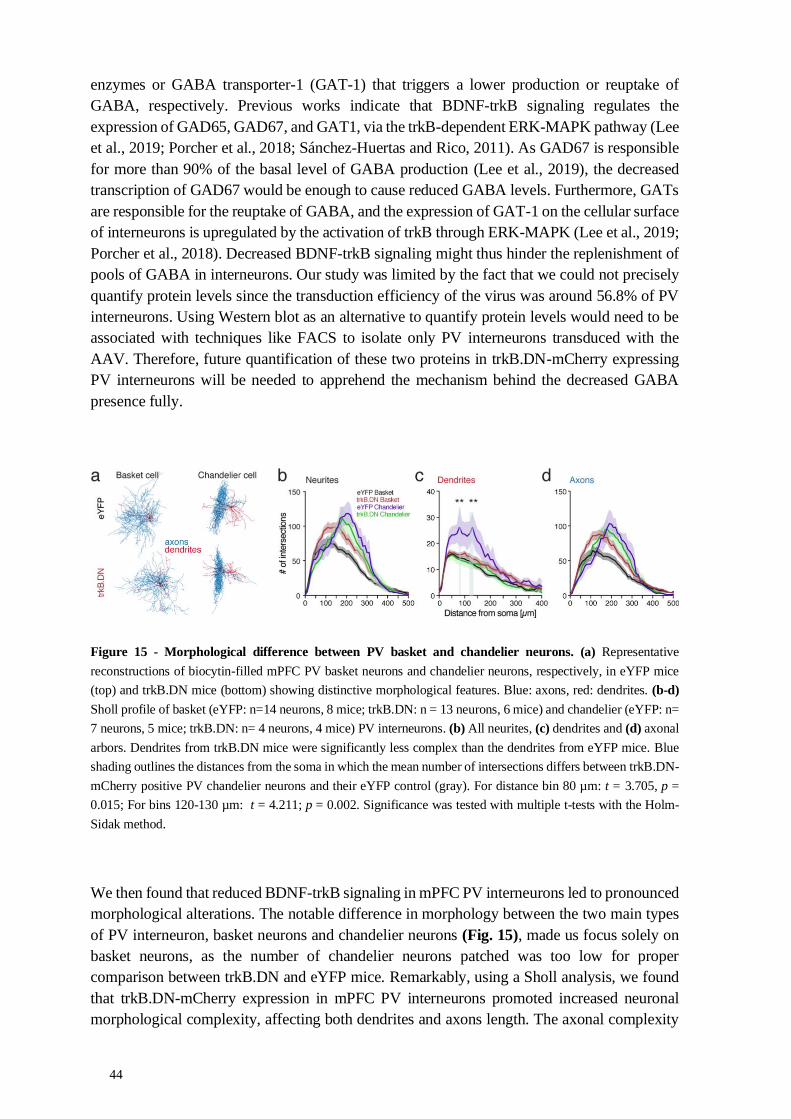
44
enzymes or GABA transporter-1 (GAT-1) that triggers a lower production or reuptake of
GABA, respectively. Previous works indicate that BDNF-trkB signaling regulates the
expression of GAD65, GAD67, and GAT1, via the trkB-dependent ERK-MAPK pathway (Lee
et al., 2019; Porcher et al., 2018; Sánchez-Huertas and Rico, 2011). As GAD67 is responsible
for more than 90% of the basal level of GABA production (Lee et al., 2019), the decreased
transcription of GAD67 would be enough to cause reduced GABA levels. Furthermore, GATs
are responsible for the reuptake of GABA, and the expression of GAT-1 on the cellular surface
of interneurons is upregulated by the activation of trkB through ERK-MAPK (Lee et al., 2019;
Porcher et al., 2018). Decreased BDNF-trkB signaling might thus hinder the replenishment of
pools of GABA in interneurons. Our study was limited by the fact that we could not precisely
quantify protein levels since the transduction efficiency of the virus was around 56.8% of PV
interneurons. Using Western blot as an alternative to quantify protein levels would need to be
associated with techniques like FACS to isolate only PV interneurons transduced with the
AAV. Therefore, future quantification of these two proteins in trkB.DN-mCherry expressing
PV interneurons will be needed to apprehend the mechanism behind the decreased GABA
presence fully.
Figure 15 - Morphological difference between PV basket and chandelier neurons. (a) Representative
reconstructions of biocytin-filled mPFC PV basket neurons and chandelier neurons, respectively, in eYFP mice
(top) and trkB.DN mice (bottom) showing distinctive morphological features. Blue: axons, red: dendrites. (b-d)
Sholl profile of basket (eYFP: n=14 neurons, 8 mice; trkB.DN: n = 13 neurons, 6 mice) and chandelier (eYFP: n=
7 neurons, 5 mice; trkB.DN: n= 4 neurons, 4 mice) PV interneurons. (b) All neurites, (c) dendrites and (d) axonal
arbors. Dendrites from trkB.DN mice were significantly less complex than the dendrites from eYFP mice. Blue
shading outlines the distances from the soma in which the mean number of intersections differs between trkB.DN-
mCherry positive PV chandelier neurons and their eYFP control (gray). For distance bin 80 µm: t = 3.705, p =
0.015; For bins 120-130 µm: t = 4.211; p = 0.002. Significance was tested with multiple t-tests with the Holm-
Sidak method.
We then found that reduced BDNF-trkB signaling in mPFC PV interneurons led to pronounced
morphological alterations. The notable difference in morphology between the two main types
of PV interneuron, basket neurons and chandelier neurons (Fig. 15), made us focus solely on
basket neurons, as the number of chandelier neurons patched was too low for proper
comparison between trkB.DN and eYFP mice. Remarkably, using a Sholl analysis, we found
that trkB.DN-mCherry expression in mPFC PV interneurons promoted increased neuronal
morphological complexity, affecting both dendrites and axons length. The axonal complexity

45
was more pronounced in distances from the soma (100–200 mm). A distance where basket
neurons are known to densely innervate local pyramidal neurons (Tremblay et al., 2016). The
longer length of axons observed in trkB.DN mice affected the density of synaptic inhibitory
boutons. The boutons were less dense in trkB.DN mice, but the total number of synaptic
boutons per entire axon did not finally differ between the trkB.DN and eYFP mice. PV boutons
were colocalized with the postsynaptic marker gephyrin, thus suggesting that they kept their
ability to make proper connections with postsynaptic structures.
The longer axons and dendrites could be due to adaptations aiming to compensate for the
decreased responsiveness in the PV interneurons. The total dendritic length of a PV interneuron
can range from 4 to 9 mm, while the axons can measure up to 50 mm (usually around 24 mm
in the PFC; see review by Hu, Gan, and Jonas 2014). Here, we observed that the mean length
of dendrites (5.3 mm) and axons (34.3 mm) measured in trkB.DN were significantly higher
than the controls (3.9 mm and 24.9 mm, respectively). However, the observed lengths in
trkB.DN are situated within the upper limit of the standard length range, suggesting that these
changes are not morphologically abnormal and still being homeostatically controlled
(Samsonovich and Ascoli, 2006).
The morphology changes could also be due to the alteration of the trkB.FL/trkB.T ratio. BDNF-
trkB signaling is engaged in regulating the neuron morphology, and the truncated receptor
trkB.T1 concurs in the refinement of the growth or decrease of dendrites, axons, and synaptic
structures (Ohira and Hayashi, 2009). The overlapping expression of multiple neurotrophic
receptors and their ligands has been implicated in the growth and disintegration of axons
(Deinhardt and Chao, 2014; Park and Poo, 2013) and in the participation of the formation and
remodeling of cellular connectivity, a process that continues to be present in the adult brain
(Bellon et al., 2011). For example, the mechanisms that BDNF uses to alternatively promote
growth or pruning of axons depend on the various levels of trkB or p75 neurotrophin receptor
(p75NTR) activation. BDNF engages p75NTR in the absence of trkB to facilitate axon pruning
(Singh et al., 2008). The truncated receptor trkB.T1 is also involved, acting in this process by
not letting BDNF activate neither trkB nor p75NTR (Baho et al., 2019; Michaelsen et al., 2010).
Future studies looking at how the expression of trkB.DN affects the function of other proteins,
like p75NTR, in PV interneurons would be necessary to understand further the mechanisms by
which neurotrophins modulate axonal arborization in the adult brain.
Furthermore, modifying the trkB.FL-trkB.T ratio could be a way for the neurons to regulate
from where they get their information. The increase in dendritic arbor length and complexity
we observed fits with the role of the truncated trkBs regarding the extension of dendrites, as
trkB signaling is known to enhance the growth and branching of dendrites (Deinhardt and
Chao, 2014). In particular, previous research proposed that trkB.FL promotes the addition of
short branches in dendrites proximal to the soma, whereas trkB.T1 induces the extension of
dendrites in regions more distal to the soma (Yacoubian and Lo, 2000). As the trkB isoforms
appear to act in a mutually inhibitory manner, this data suggests that expression of the correct
set of trkB isoforms is needed for the normal development of dendrites (Bellon et al., 2011).
Expressing trkB.DN-mCherry in PV interneurons might have blocked the normal signaling
pathways necessary for the dendrites to adapt to their inputs. As a proper connectivity pattern
is acquired by adjusting already existing connections based on inputs (Bellon et al., 2011), the
incapacity to modify the neuronal structure in response to activity-dependent mechanisms by

46
fine-tuning the neurotrophic response could lead to aberrant network oscillatory activity and
abnormal generalized response of the mPFC circuit to external cues.
4.2.2 Reduced sensitivity and firing activity of PV interneurons
As our long-term expression of trkB.DN-mCherry in mPFC PV interneurons led to molecular
and morphological changes, we continued to investigate how these changes could affect the
intrinsic properties of transduced PV interneurons and the local circuitry activity. Principally,
we found reduced sensitivity of PV interneurons to excitatory inputs due to the significantly
increased membrane time constant and increased firing rate adaptation. The reduced
responsiveness of PV interneurons to fast frequency inputs, along with dendritic arbor
morphology alterations, can conceivably affect local network oscillatory activities (Jadi et al.,
2016; Otte et al., 2010; Wang, 2010).
The altered membrane properties of PV interneurons were concomitant with increased firing
adaptation and reduced spontaneous firing of PV interneurons, indicating a deficient inhibitory
activity of PV interneurons in the local network, as observed in the trending reduction in IPSC
frequency in pyramidal neurons. However, altered basal synaptic transmission was not evident,
as no effects of reduced BDNF-trkB signaling on the short-term synaptic plasticity between
PV interneurons and connected pyramidal neurons were observed in the paired whole-cell
recordings. Conversely, the removal of presynaptic trkB in neurons of the hippocampus has
been shown to result in an increased paired-pulse ratio, indicating a reduced presynaptic release
probability (Lin et al., 2018). Presynaptic and postsynaptic BDNF-trkB signaling in PV
interneurons might thus be affected differently by the expression of trkB.DN-mCherry.
Moreover, expression of trkB.DN-mCherry might trigger adaptations that preserve synaptic
strength (Lin et al., 2018).
4.2.3 Social behavior dysfunctions
We found these functional and circuit changes to be associated with increased aggression and
anxiety in behaving mice. TrkB.DN mice exhibited heightened aggression when tested in a
resident-intruder test with juvenile mice compared to control mice. It has been proposed that
impaired BDNF-trkB signaling in the prefrontal cortex plays an important role in aggression.
Particularly, several works report that aggressive behavior is correlated with reduced
trkB.FL/trkB.T ratio. For example, in the frontal cortex of aggressive rats, the level of the
trkB.FL receptor protein was decreased, while trkB.T protein levels were increased, suggesting
that a reduced trkB.FL/trkB.T ratio is prevalent in highly aggressive rats (Ilchibaeva et al.,
2018). Furthermore, restoring proper BDNF-trkB signaling in BDNF knockout mice by
decreasing the levels of trkB.T1 reduced aggressive behavior in the resident-intruder test
(Carim-Todd et al., 2009). Another study showed that abnormal aggression in a Post-Weaning
Social Isolation-Induced Aggression model is linked with lower BDNF, higher levels of trkB.T,
and a decreased trkB.FL/trkB.T ratio. Treatment with the drug fluoxetine, an antidepressant of
the selective serotonin reuptake inhibitor (SSRI) class, reduced trkB.T1 levels, restoring the
trkB.FL/trkB.T ratio and diminishing aggression (Mikics et al., 2018). Moreover, a study using
a conditional knockout mouse in which trkB (all isoforms) was ablated from a majority of
corticolimbic GABAergic interneurons postnatally shows that mutant mice did not display
enhanced aggression in the resident-intruder test. However, mutant mice were more dominant

47
and showed more aggression in another agonistic behavior test in which they analyzed the
behavior of group-housed mice after switching to a novel context, concurrent with the idea that
BDNF-trkB signaling impairment in inhibitory interneurons is linked with aggressive and
dominant behaviors (Tan et al., 2018).
TrkB.DN mice displayed normal locomotion (no hyperactivity). However, we observed higher
anxiety-related behaviors in trkB.DN mice, when compared to eYFP controls, relative to the
number of entry and head-dips in the open arms of the elevated plus-maze test, and to time
spent in the center in the first 3min of the open field test. As mice which were used in behavioral
experiments were single-housed prior to the social interaction essay, this could have affected
their anxiety levels and thus their BDNF-trkB activity, as prefrontal BDNF-trkB activity is
stress-modulated (Barfield and Gourley, 2018; Gray et al., 2013), and the lower level of BDNF
has been implicated in anxiety phenotype (Notaras and van den Buuse, 2020). Moreover, the
mPFC is part of circuits that are known to be involved in social interaction and anxiety-related
behaviors, as in the case of the reciprocal projections between the amygdala and the mPFC
(Allsop et al., 2014; Felix-Ortiz et al., 2016). Further research should be undertaken to
disentangle more specifically which components of these circuits influence both (or either)
social and anxiety-related aspects of behavior, and therefore develop a complete picture of the
links between affective and social cognition.
4.2.4 Altered prefrontal excitatory dynamics
To assess how the circuit impairments seen in ex vivo and in vivo acute recordings are linked
to behavioral changes, we recorded LFP and single-unit activity in the mPFC in freely moving
animals. We performed recordings during a nine-minute baseline period followed by four-
minute of social interaction with a juvenile intruder. After the removal of the intruder mouse,
we kept recording for ten more minutes. We focused on analyzing mPFC responses and circuit
dynamics in relation to the start of social interaction. We found that reduced trkB signaling in
mPFC PV interneurons affected the local oscillatory network activity. More specifically, mice
with the expression of trkB.DN in mPFC PV interneurons showed a broadband increase of
baseline LFP power and a reduced modulation of mPFC oscillations triggered by social
interaction, compared to eYFP mice. This deficient induction of synchronous activities over a
broad frequency band we observed in response to social stimuli is similar to the reduction in
local network gamma oscillatory induced by a drug or visual stimuli reported in mouse models
with genetic deletion of trkB in PV neurons (Xenos et al., 2018; Zheng et al., 2011). However,
Xenos and colleagues also reported that the ablation of trkB in PV neurons during adolescence
decreased spontaneous broadband power in a frequency range between 20 to 100 Hz,
contrasting with our observation during baseline. This disparity might be due to distinct
morphologic changes between the postnatal knock-out models and the sustained expression of
trkB.DN in the adult.
Furthermore, the reduction of trkB signaling directly affected prefrontal population dynamics,
with a significantly larger proportion of pyramidal neurons being positively modulated by
social interaction in trkB.DN mice than in eYFP mice. Specifically, we found that the mean
population firing rate of the mPFC pyramidal neurons was higher in trkB.DN than in eYFP
mice during social interaction. We divided the pyramidal population into three subpopulations
based on their firing during social interaction (yielding positively, negatively, and not
48
modulated neurons, respectively). The mean firing rates of the three subpopulations were not
significantly different between trkB.DN and eYFP mice. However, the proportion of positively
modulated pyramidal neurons was higher in trkB.DN mice than in eYFP mice. Thus, the
increased mean firing rate of the (whole) pyramidal population in trkB.DN mice was not a
result of increased spiking in individual pyramidal neurons but a result of more pyramidal
neurons being recruited by social interaction in trkB.DN mice. The positively modulated
pyramidal neurons were responsible for the higher variability in the population response to
social interaction in trkB.DN mice, compared to the eYFP mice, as analyzed with neuronal
trajectory plots.
Generalized neuronal ensemble activity, detected in the abnormal proportion of positively
modulated pyramidal neurons, might be underlying social impairments. Indeed, the oscillatory
and single-unit results are consistent with those of Liu and colleagues, who recently observed
that social interaction increased the firing of prefrontal PV interneurons as well as increased
LFP gamma (20 - 50 Hz) power. They also found that optogenetic synchronization of PV
interneurons, specifically at gamma frequency, improved sociability. Besides, specific
inhibition of mPFC PV interneurons with chemogenetics reduced gamma activity and impaired
sociability. Equally important, they found that most pyramidal neurons did not respond (60%),
while a small population showed increased (26%) or decreased (14%) firing rates during social
interaction (Liu et al., 2020).
Additionally, to ensure that the observed differences in the Euclidean distance between
trkB.DN and eYFP mice were not a mere result of their differences in the proportion of
positively modulated neurons, we performed a bootstrapped version of the population
trajectory analysis, where neurons from each of the three pyramidal subpopulations were
randomly selected and analyzed in every 10000 repetitions. This analysis identified that the
increased variability of the pyramidal population in trkB.DN mice was driven by altered
dynamics in the positively modulated subpopulation specifically. As the increased firing of
individual pyramidal neurons is not likely to underlie the increased Euclidean distance in
trkB.DN mice, other patterns of activity detected by principal components are responsible for
the variability in population dynamics.
Finally, we checked if the altered population-level dynamics observed in trkB.DN mice during
social interaction were limited to this behavior or could be generalized to other functional states
known to engage separate mPFC circuitry. For this, while recording in anesthetized mice, we
used a tail pinch stimulation and found that the change in PFC activity patterns caused by a tail
pinch induced similar abnormal modulation of pyramidal neurons and altered dynamics as seen
during social interaction.
Surprisingly, similar pyramidal population dynamics were observed in both social interaction
and tail pinch. A possible explanation for these results is that the sustained effect of trkB.DN-
mCherry expression in PV interneurons may lead to changes in the organization of pre-
established neuronal ensembles, usually differently activated by diverse stimuli. Thus, it can
be suggested that this disorganization of neuronal activity reflects the recruitment of typically
unresponsive neurons in the mPFC. For instance, morphological or functional adaptations
leading to E/I imbalance might explain this disorganization (Sohal and Rubenstein, 2019;
Turrigiano, 2011) by possibly reducing the inhibitory inputs of a portion of pyramidal neurons.

49
Moreover, the reduced firing rate of PV interneurons could increase the excitatory activity of
pyramidal neurons to which they are still connected (Dehorter et al., 2017; Fujisawa et al.,
2008; Hamm et al., 2017; Tremblay et al., 2016). Therefore, the association of these two
occurrences could provide the pyramidal neurons with the possibility to increase the range of
their excitation, and gather new neurons to previously assembled circuits through different
long-term potentiation processes. The disorganization of neuronal activity in trkB.DN mice
during the social interaction could thus lead to a behavioral response based on the
misinterpretation of stimuli or the unspecific activation of aggression-related networks
(Aleyasin et al., 2018).
Our PCA analyses revealed abnormalities in the population-level dynamics of the mPFC
pyramidal neurons in trkB.DN mice. Curiously, the first two principal components explained
only 23.4% and 14.0% of the variance seen in the eYFP mice during the social interaction
experiment. This is a low percentage compared, for example, to the 75% for the first two PCs
in Levy et al. 2019. This result could be explained by the heterogeneity of behaviors within the
social interaction or inter-mouse variability within the same group. If we could have
investigated the firing patterns separately during each behavior (Levy and colleagues used six
discrete odors), we might have found a better percentage of variance explained by the principal
components for the different behaviors. In contrast, in trkB.DN mice, the first two principal
components explain more of the variance (42.8% and 11.3% of variance, respectively). That
is, regardless of behavioral variability, the dynamics of these units seem to be more “uniform”
(generalized to the different behaviors). If we had analyzed the behaviors individually, we
might have observed that the recorded units in the eYFP mice describe one of the behaviors
very well while not explaining others as well. However, in trkB.DN mice, the activity recorded
during each behavior could be expected to have a similar percentage of explanation by the
principal components. Further recordings during discrete social and non-social behaviors, as
discussed briefly in the method section of this thesis, would be necessary to check this
hypothesis.
However, in contrast with the complexity inherent to the social behavior, the tail pinch is (a
priori) a unique variable, but the first two principal components in the eYFP mice still only
explained a small percentage of the variance seen in the neuronal activity (25.2% and 19.2%,
respectively), while PC1 and PC2 in the trkB.DN mice explained 68.4% and 8.6% of variance.
One hypothesis is that, as PV interneurons activity is altered in trkB.DN mice, allowing the
pyramidal neurons to become disorderly responsive to the tail pinch, the pyramidal activity
goes on to explain more of the increased variability found in the neuronal patterns. On the other
hand, the tail pinch has been shown to increase the firing rate of inhibitory interneurons but not
the firing of pyramidal neurons in the mPFC (Massi et al., 2012). It can thus be suggested that
if we had been able to observe the dynamics of PV interneurons in PV-Cre mice during the tail
pinch, they would have been more tuned to the tail pinch than local pyramidal neurons. It will
be required to analyze the dynamics of PV interneurons (see perspective) to evaluate their level
of impairment and understand how they can organize local circuit activity in response to the
tail pinch.

50
4.2.5 On potential sexual dimorphic differences
Additionally, BDNF-trkB signaling in PV interneurons might be influenced by sex-steroid
hormones, exposing the existence of differences caused by sexual dimorphism (Lucas et al.,
2014; Notaras and van den Buuse, 2020). Our study does not directly address this factor while
using both females and males in most experiments. We nevertheless verified that sex was not
a variable that could explain the differences between PV-Cre controls and trkB.DN mice in our
morphological (Fig. 16), molecular and ex vivo electrophysiology recordings. As only males
were used in behavioral and in vivo electrophysiological recordings experiments, further work
would be necessary to precisely check the role of sex differences in the altered BDNF-trkB
signaling effects in PV interneurons.
Figure 16 - Morphological comparison between female and male eYFP and trkB.DN mice. (a-d) Data based
on Sholl analysis of the biocytin-filled mPFC PV basket neurons (Female mice: n = 19; male mice: n = 7). (a) No
differences were detected in the total number of intersections of neurites of PV basket neurons from female mice
and male mice. Female: 2155 ± 130.2; Male: 2268 ± 198.5; U = 60, p = 0.7347. (b) No differences were detected
in the total neurite’s length of PV basket neurons from female mice and male mice. Female: 35024 ± 3085; Male:
35381 ± 3582 µm; U = 61, p = 0.7777. (c) No differences were detected in the number of neurites of PV basket
neurons from female mice and male mice. Female: 350.4 ± 29.51; Male: 336.3 ± 33.85; U = 66, p = 0.9999. (d)
No differences were detected in the ending radius of the neurites of PV basket neurons from female mice and male
mice. Female: 602.1 ± 34.10 µm; Male: 571.4 ± 32.91 µm; U = 56.50, p = 0.5812. (e) No differences were detected
in the number of axonal boutons/100 µm in biocytin-filled mPFC PV basket neurons (Female mice: n = 15; Male
mice: n = 5 neurons). Female: 12.21 ± 0.55; Male: 10.94 ± 0.73; U = 17, p = 0.0806. The Wilcoxon rank-sum test
was used to assess significance.
Altogether, the mechanistic relationship between all the observed changes in PV interneuron
physiology and alterations of network activity following PV-specific expression of a trkB
dominant-negative receptor remains hard to explain. It will be essential to be able to reverse
the effects of the trkB.DN on PV interneurons. The use of new tools like light-activated trkB
receptors or pharmacogenetics targeting specifically trkB in PV interneurons will allow such
spatially and cell-type restricted approach in reversing the abnormal phenotype (Kramer et al.,
2013; Mondoloni et al., 2019; Winkel et al., 2020).
51
5 CONCLUSION AND PERSPECTIVES
This thesis examined the role of PV interneurons in the mPFC, with a focus on two receptors
that are central for PV interneurons’ connectivity, activity, and role in coordinating local circuit
activity. The main goal was to investigate the causal links between molecular processes, circuit
function, and behavior, and understand how these different levels are coordinated towards the
proper functioning of the PFC. Thus, we used genetic and viral methods allowing for cell-type
selective knockout of NMDARs, or cell- and time-specific expression of a truncated version of
trkB, in order to disrupt PV function. We monitored the changes at the molecular, cellular,
circuit and behavioral levels, and our experiments confirmed that deleting NMDAR, or
manipulating the BDNF-trkB signaling pathway, specifically in PV neurons, is sufficient to
trigger significant abnormalities in mPFC circuit function, as well as behavioral alterations. We
also used this opportunity to understand better the physiological mechanisms by which
mesoscopic signals like gamma-band (30-80 Hz) and HFB (100-150 Hz) are generated at the
cellular level. Overall, the two studies presented in this thesis strengthen the idea that PV
interneurons are critical for regulating and delimiting prefrontal responses to different classes
of inputs.
5.1 PV DYSFUNCTION AND ASYNCHRONOUS ACTIVITY
One of the more significant findings to emerge from this thesis is that PV dysfunction
contributes to asynchronous activity underlying enhanced broadband gamma power. The term
"increased basal gamma oscillations'' used in literature to define an increased broadband power
in the gamma range (30-80Hz) is thus not the most appropriate way to describe this aberrant
activity. Additionally, our results suggest that the aberrant increase in broadband power can
originate from different sources, as distinct mechanisms are behind the altered inhibitory
activity. The increase in baseline broadband power was observed in both models of PV
dysfunction in a wide range of frequencies (30-150Hz in Paper I and > 6 Hz in Paper II),
including the gamma-band (30-80Hz).
Thus, it will be important to dissect the origin of gamma alterations and asynchronous activities
when observed in animal models or patients of neuropsychiatric disorders, as the circuit
modifications leading to these changes might differ depending on their source. A limitation of
the study presented in Paper II is that the NMDAR knockout might lead to developmental
alterations that are not fully assessed. Therefore, we do not fully understand which specific
alterations in PV interneurons lead to asynchronous activities. Particularly, it would be essential
to figure out the link between modified intrinsic properties and genetic factors, receptors
expression or morphological alterations, as well as dissect the potential compensatory
mechanisms due to developmental alterations initiated by the genetic knockout of NMDAR.
For instance, further research should be undertaken to explore how NMDAR hypofunction in
PV neurons affects both the inputs and outputs of PV interneurons by quantifying the strength
of their connections with target neurons, with paired patch-clamp recordings. Conversely, a
future study could assess the effects of the sustained expression of trkB.DN in PV interneurons
on the mPFC dynamics at different timescales, replicating the analysis used in Paper I to
examine if these changes lead to similar asynchronous activity.

52
Additionally, our results show that NMDAR in PV interneurons are critical for the
desynchronizing effect of ketamine in the mPFC, a region that is highly involved in psychiatric
disorders such as schizophrenia and autism. However, the mechanisms between the two kinds
of NMDAR hypofunction, caused by NMDAR antagonists or caused by genetic ablation of
NMDAR, seemed to differ. This was highlighted by the lack of response to ketamine in PV-
Cre/NR1f/f mice, as well as the difference in the entrainment of spikes to the LFP between the
two approaches. These findings have significant implications for the understanding of the
NMDAR hypofunction hypothesis of schizophrenia, as this hypothesis is based on observations
that NMDAR antagonists induce, in normal human subjects, symptoms similar to those with
schizophrenia (Krystal et al., 1994). Our results add more complexity to the heated debate
around NMDAR dysregulation and the phenomenology of schizophrenia (Bianciardi and
Uhlhaas, 2021; Gonzalez-Burgos and Lewis, 2012; Lee and Zhou, 2019; Lisman, 2012;
Pafundo et al., 2018). It will thus be important to investigate the mechanisms that differ
between the two kinds of NMDAR hypofunction so that we can pinpoint how the ensuing
neuronal circuit dysfunction relates to the phenomenology of the disease.
Importantly, we demonstrated that multiple synchronous and asynchronous activities could co-
exist within the neuronal activity, and thus, LFP or EEG activity in the gamma-band should
not be read uniformly. This asynchronous neuronal activity can appear and mask true rhythms
in the analysis of the power of specific oscillatory bands, for example, when the gamma-band
is defined canonically. Thus, it is important to verify from the start the presence of synchronous
oscillations and asynchronous activity (Donoghue et al., 2020). This can be done by detecting
if the signal in a PSD plot presents meaningful peaks, as genuine oscillations are usually
noticeable as narrowband power peaks above the aperiodic component of the signal.
It is also sensible to not idiosyncratically define specific bands prior to the analysis in order to
calculate and compare the power of these specific bands. It is recommended to detect potential
differences on the full spectrum of frequencies spanning from 1–200 Hz and then specify at
which range of frequencies (in hertz) these differences correspond. Consequently, reporting
differences this way would also allow better comparison between studies, as many use different
frequency ranges to define the gamma-band, for example.
Moreover, recorded neural oscillations can show variations in their peak frequencies,
depending on the brain region or brain state. It is important to consider such variations as many
studies also limit their analysis to predefined bands and miss reporting potential changes that
can happen just outside or across these bands. Description of the brain state in which one is
analyzing LFP signal is thus essential, as brain states might modulate not only the peak
frequency but also the overall signal-to-noise ratio. For example, inactive and possibly tired
subjects may show very different modulation in LFP compared to subjects engaged in active
tasks (Donoghue et al., 2020; Fitzgerald and Watson, 2018).
Our research has also shown that both NMDAR and trkB are critical for PV interneurons role
in proper synchronous activity, confirming previous results linking PV interneurons firing and
synchronous activity in the gamma range (30-80 Hz; Cardin et al. 2009; Sohal et al. 2009;
Zheng et al. 2011; Carlén et al. 2012). Abnormal PV activity due to dysfunction of trkB led to
impaired gamma rhythm induction after a social challenge, while specific dysfunction of
NMDAR in PV neurons blocked the effects of ketamine on faster oscillations, including the
53
gamma-band. This supports the notion that dysfunction of PV interneurons is enough to
generate neuronal activity changes, such as the reduced sensory-evoked gamma power
observed in individuals with schizophrenia (White and Siegel, 2016). Further work is needed
to fully understand the interactions of PV neurons with other cell types in the production of
synchrony in the local circuit, as well as between areas.
Synchronous activities, principally at higher frequencies, are important for local PFC circuitry,
but they are also involved in the coupling of cross-regional or cross-hemispheric activity
(Adhikari et al., 2011; Cho et al., 2020; Fernandez et al., 2017). As such, the role of mPFC
cannot be separated from the several cortical and subcortical regions from which it receives
and to which it sends projections. It will be interesting to look at cross-frequency coupling
between regions, including the PFC, thalamus, hippocampus and others, to see if synchronous
activities between highly connected regions are modified in both PV-Cre/NR1f/f mice or
trkB.DN mice.
Also, as locally recorded brain activity can reflect activity not only from the local circuit but
also from more distant sources that overlap both spatially as well as temporally (Donoghue et
al., 2021), it will be important to understand if the observed asynchrony, due to oscillatory
activity alterations, is restricted to the mPFC. This could give us an insight into how local
synchrony is. As discussed in Paper I, the ablation of NMDAR in PV neurons is global in PV-
Cre/NR1f/f mice, and as such, the abnormal oscillatory activity we recorded in the mPFC might
also be caused by PV neuron dysfunction in other regions like the thalamic reticular nucleus or
the basal forebrain. Further research should be carried out to record activity in these regions
simultaneously to pinpoint if asynchronous activities are purely instigated by local circuit
alterations or more global ones.
5.2 PV DYSFUNCTION AND CORTICAL STATES IMPAIRMENTS
NMDAR hypofunction in PV neurons led to impaired coordination of cortical states, with
notably more fragmented deactivated states. These results were obtained in urethane
anesthesia, a useful model of sleep-like brain oscillations (Clement et al., 2008; Hauer et al.,
2019). As such, the fragmentation of the deactivated state in Paper I parallels findings of
fragmentation specific to NREM sleep, in mouse models of schizophrenia involving PV
interneuron dysfunction (Phillips et al., 2012). The mPFC is one of the principal cortical
regions that receives projections from the subcortical ascending system (including the reticular
formation and basal forebrain) that promotes wakefulness (McKenna et al., 2017). Therefore,
cortical state impairments in the PFC are proposed to indicate impairment of sleep regulation
by the brain. Recording the activity of PV neurons in different regions involved in sleep/wake
transitions, including the mPFC, during sleep will be of importance. Non-only to understand
more closely the mechanisms linking PV interneurons with cortical state coordination, but also
to understand the pathological mechanisms affecting sleep architecture, as sleep and circadian
rhythm dysfunctions are comorbidities of neuropsychiatric disorders like schizophrenia
(Chouinard et al., 2004). Additionally, these recordings during sleep should be done not only
in PV-Cre/NR1f/f mice but also in trkB.DN mice as BDNF-trkB signaling has also been
implicated in sleep dysfunctions (Garner et al., 2018; Watson et al., 2015).

54
5.3 PV DYSFUNCTION AND CIRCUIT ALTERATIONS
Many more questions that we could answer experimentally appeared during the elaboration of
the work presented in Paper II. For instance, we observed significant morphological alterations
in PV interneurons after the expression of trkB.DN-mCherry in these neurons. Notably, the
dendritic branches were not only more complex but their radius was increased, thus covering a
bigger receptive field. This is functionally important since the dendrite arbor determines what
signals a neuron receives and how these signals are integrated. The local and distal connectivity
disruption caused by either hypoconnected or hyperconnected neurons has been suggested to
underlie neuropsychiatric disorders (Forrest et al., 2018). It is possible that the longer and more
complex dendritic trees we observed increased the number of unspecific synaptic contacts and
thus sampled excessive circuit inputs, leading to higher sensitivity to neuronal “noise”.
However, we did not evaluate further if dendritic alterations led to changes in dendritic spines
in the PV interneurons. A further study with more focus on the dendritic arborization of PV
interneurons is therefore suggested to gather the extent of dendritic modifications and
investigate the possible link with impaired circuit activity in mPFC recorded via LFP after
trkB.DN-mCherry expression.
Additionally, the extent of the possible alterations on other neurons due to PV dysfunction is
still unknown. Further investigation is needed to pinpoint how alterations found in models of
PV dysfunction lead to functional, and maybe morphological, changes in different cell types in
the circuit. Remarkably, a recent work utilizing mice with NR1 subunit knockout in cortical
and hippocampal GABAergic neurons showed reduced dendritic lengths in adult, but not
juvenile, local pyramidal neurons in the mPFC (Pafundo et al., 2021). Pafundo and colleagues
further propose that this impact on adult pyramidal neurons is observable only when the mPFC
receives increased activity, e.g., when recruited for more cognitively demanding tasks. They
further claim that this might be causing the altered computations underlying the behavioral
deficits observed in animals with NMDAR hypofunction. Interestingly, these findings parallel
our single-unit observation in Paper II, where the alterations in pyramidal neurons’ single-unit
and population dynamics in trkB.DN mice were surprisingly restricted to when inputs (from
the social interaction or the tail pinch) supposedly arrived at the mPFC. No differences in firing
rate were observed during the baseline between the trkB.DN and control mice. Therefore,
future work should not only consider how specific changes in PV interneurons alter their
function but also consider the effect on other pyramidal and GABAergic interneurons in the
vicinity.
Another question raised by the results in Paper II is how local the changes are due to spatially
restricted trkB.DN-mCherry expression in adult PV interneurons. Recording neuronal activity
within the area transduced by the AAV virus and compare it with the activity outside this area
might permit us to see if trkB.DN-mCherry expression in PV interneurons affects just the local
circuit or also disrupts adjacent cortical circuits and regions receiving afferent input from
mPFC. One suggestion would be to simultaneously record the several mPFC subdivisions
using the Neuropixel 2.0 probe with four shanks (Jun et al., 2017; Steinmetz et al., 2021). The
possibility to record from 384 sites distributed over four shanks could enable the recording of
around 500 or more well-isolated units and LFP activity simultaneously over several areas and
layers of the mPFC. This would allow us to compare alterations from recording sites inside the

55
virally transduced area versus areas outside by analyzing LFP traces patterns and spectral
power distribution along probe track, or by possibly constructing current source density maps,
as already done to explore differences between cortical layers (Senzai et al., 2019).
Furthermore, it will be essential to understand how the impairment of BDNF-trkB signaling
specific to PV interneurons alters PV single-unit activity. This could be done by checking
whether the PV interneurons actually have a decreased activity during a stimulus, or are
temporally or spatially disorganized. The use of probes with a higher yield in units recorded
would allow having enough neurons recorded to focus single-unit analysis on PV interneurons.
The study published in Paper II was limited by the absence of single-unit activity analysis on
PV interneurons due to the low number of recorded neurons of this cell-type. Consequently,
we focused on the much more numerous pyramidal neurons. We thus observed the results of
our manipulation in PV interneurons indirectly, following only the pyramidal neurons activity
in response to social interaction or tail pinch. Therefore, to pinpoint how dysfunction of PV
interneuron affect the architecture of neuronal ensembles, recording of enough PV interneurons
would be necessary not only to perform firing rate and population dynamics analysis as we did
with pyramidal neurons, but also analysis of cross-correlation between PV interneurons and
neighboring neurons (Agetsuma et al., 2018; Senzai et al., 2019).
Moreover, the precise mechanism of how selective impairment of PV function in the mPFC
alters behavior remains to be elucidated. We observed that the increase in pyramidal neurons
firing activity in trkB.DN mice is generalized to several stimuli. It will be important to
investigate how this generalization occurs. The use of fluorescence imaging to record in vivo
calcium activity from neurons in the mPFC will permit us to observe spatially if the pyramidal
neurons are activated nonspecifically over several stimuli (Agetsuma et al., 2018; Hamm et al.,
2017). Fluorescence imaging allows precise spatial mapping of activity and the rapid
improvement of genetically encoded calcium indicators like GCamp, voltage indicators
(Knöpfel and Song, 2019), or neurotransmitter and neuromodulator sensors (Sabatini and Tian,
2020), offers a plethora of tools to reveal spatiotemporal dynamics that were till now difficult
to record. For example, fluorescence microscope (miniscope) recordings while mice freely
explored social targets showed that mPFC pyramidal neurons formed two non-overlapping
populations with opposing neuronal activities (Liang et al., 2018). These distinct neuronal
populations were tuned to different social targets, and the systemic administration of the
NMDAR antagonist PCP disordered these mPFC neural ensembles. Recording with miniscope
during the resident-intruder test and further post-hoc confirmation of which PV interneurons
were trkB.DN-mCherry-positive would thus allow us to observe how neighboring cells are
affected by altered PV activity and follow the activity of these neuronal ensembles during the
diverse behaviors.
Additionally, there has been more attention on studying social behavior as synchronous
cognitive processes between interacting individual subjects (Dumas et al., 2010; Kingsbury
and Hong, 2020). Social interactions markedly influence individual neuronal activity and
behaviors (Liang et al., 2018). Recently, correlated neuronal activity has been observed
between the brains of socially interacting humans, but also mice or bats (Kingsbury et al., 2019;
Zhang and Yartsev, 2019), with particular emphasis on synchronous oscillatory activities,
including gamma oscillations (Kingsbury and Hong, 2020). Moreover, most results have been

56
focused on social dynamics between two mice, but there has been recent effort to study how
the interaction between individuals affects large groups. Research questions that could be asked
include if altered neuronal activity in one mouse can change the interactions between
individuals, including within a group (Torquet et al., 2018). For instance, it would be interesting
to understand how the observed altered oscillatory activities in the mPFC of trkB.DN mice
might be reducing synchronization of neural processes across subjects, affecting both the
resident as well as the intruder. New tracking and wireless recording technologies are allowing
researchers to record the neuronal activity of two or more mice simultaneously and possibly
manipulate the activity of one of them, as their interactions are tracked (de Chaumont et al.,
2019; Kingsbury and Hong, 2020; Torquet et al., 2018), opening the door to exciting future
research. Finally, these approaches could be applied to studies of NMDAR hypofunction — as
NMDAR are also found to be implicated in social behavior (Gao and Mack, 2021) — or other
mouse models of schizophrenia and autism spectrum disorders, paving the way for future
investigations into the neuronal mechanisms underlying social behavior deficits.
5.4 MANIPULATION OF RECEPTORS TO STUDY PV FUNCTION
An issue that was not addressed in this study was whether it is possible to restore proper BDNF-
trkB signaling in these neurons and see if we can rescue the morphological, functional and
behavioral impairments. Future work should thus use pharmacological or genetic approaches,
like the re-expression of NMDAR in NR1 knockout mice or light-control of photo-switchable
trkB receptors (Lee and Zhou, 2019; Leopold et al., 2019; Mielnik et al., 2020; Winkel et al.,
2020), to identify the mechanisms behind the abnormal phenotype observed after expression
of trkB.DN-mCherry or hypofunction of NMDAR in PV interneurons.
Finally, another key point for future research will be to check how several receptors are linked
and act together. We investigated in this thesis the function of only one receptor expressed by
PV interneurons at a time. However, PV interneuron activity is modulated by several co-active
receptors at the same time, including AMPA, p75, serotonergic, dopaminergic, or acetylcholine
receptors, among others. Notably, crosstalk between BDNF-trkB signaling and NMDAR
activity has been investigated (Björkholm and Monteggia, 2016; Minichiello, 2009; Ninan,
2014; Otis et al., 2014), and both receptors have been found to be the binding targets of drugs
like ketamine. In recent work, activation of trkB was shown to be necessary for the
antidepressant-related slow effect of ketamine (Casarotto et al., 2021), while NMDARs have
been implicated in the rapid antidepressant effect of ketamine (Abdallah et al., 2015). Further
research needs to examine the interactions between several co-receptors more closely by
following and manipulating the function of these receptors simultaneously.
5.5 POTENCIAL CLINICAL RELEVANCE
The findings included in this thesis pertain to the basic neuroscience field but have several
important implications for future practice. For instance, it is interesting to note that functional
aspects of circuitry such as neuronal types and brain oscillations are evolutionary conserved.
Despite changes in brain volume, the oscillatory patterns in the cortex, or synchronous activity
recorded between different brain regions, are tightly preserved among mammals (Buzsáki and
Watson, 2012; Buzsáki et al., 2013). Additionally, parvalbumin-like interneuron-types are
conserved in reptiles and mammals (Hodge et al., 2019; Tosches et al., 2018), while trkB is

57
highly conserved among vertebrates (Benito-Gutiérrez et al., 2006). So, understanding the
function of neuronal types and the circuitry in mice has the potential to be translated into other
species and further help understand human brain function. As such, the knowledge of the
mechanisms behind altered brain dynamics might provide promising ways to characterize
neuropsychiatric disorders from the perspective of brain activity.
Furthermore, a better understanding of how to interpret the power of particular oscillations, as
well as the synchronization of neuronal activity and the cross-frequency relationships between
the various frequency ranges, may be a useful guide in developing approaches where we can
use oscillations as biomarkers. It has also suggested that gamma power measurements can be
used to track changes due to disorders but also changes elicited by drugs.
For instance, increased baseline broadband gamma has been observed in animal models and
individuals with schizophrenia and autism spectrum disorders, and as such, may underlie some
of the common features among these two neuropsychiatric disorders. The increased baseline
LFP power might conceal different functional abnormalities seen in schizophrenia and autism
spectrum disorders depending on which stage of development it emerges. As such, increased
baseline broadband power emerging specifically in adults (post-adolescent) may lead to
hallucinations or delusions as observed in subjects with schizophrenia by interfering with
previously developed neuronal ensembles found in sensory and higher-order cognitive
processing areas (White and Siegel, 2016). However, it has been suggested that changes in
baseline broadband power happening postnatally would not interfere with already formed
ensembles of neurons. Instead, it would lead to neuronal circuit compensations that might
possibly evolve into functional impairments present in autism spectrum disorders. Therefore,
manipulating the function of PV interneurons at different development stages will help
understand the impaired mechanisms behind the increased baseline broadband activity.
This information can be used to develop targeted therapeutic interventions aimed at reversing
the increase in baseline broadband power or elevate the reduced evoked/induced gamma
power. Using neuropharmacological or noninvasive brain stimulation interventions like
transcranial magnetic stimulation to manipulate specific circuits remotely might improve
perceptual and cognitive processing (Nimpf and Keays, 2017).
Research on cell-specific, circuit-specific mechanisms will be crucial to advance the
neuropsychiatry field and enhance our understanding of how disorders develop (Ford and
Young, 2021). Studies using tools to directly or indirectly downregulate receptors functions or
signaling pathways in a temporal and spatial controlled manner may help reveal how genetic
and environmental factors lead to the dysfunction of specific neurons. Furthermore, spatially
manipulating neurons will give important insight on where and when alterations in circuit
activity occur during development, and how those changes lead to behavioral deficits. These
findings will hopefully move psychiatry beyond nonspecific diagnoses, as well as adopt more
targeted treatments (Fig. 17), as unspecified brain-wide pharmacological drugs produce
unwanted side effects on patients.

58
Figure 17 - Translational potential to apply circuit-level approaches into the diagnosis and treatment of
neuropsychiatric disorders. Non-specific pharmacological drugs affect indiscriminately all brain regions,
including pathways involved in social cognition: including the mPFC, rest of the cortex, amygdala (AMG),
paraventricular nucleus of the hypothalamus (PVN), thalamus (Tha), nucleus accumbens (NAc), hippocampus
(HPC), ventral tegmental area (VTA) and dorsal raphe nuclei (DRN). Applying knowledge from basic
neuroscience has the potential to improve diagnosis, use biomarkers to uncover and monitor pathological
alterations, and reduce side-effects by using cell and/or circuit-specific pharmacological drugs. Adapted with
permission from Ford and Young, 2021. Human brains (doi.org/10.5281/zenodo.3925925) by Macauley Smith
Breault, sagittal view of the mouse brain (doi.org/10.5281/zenodo.3925911) and pyramidal neuron
(doi.org/10.5281/zenodo.3925905) by Federico Claudi, pill (doi.org/10.5281/zenodo.3926307) by Daniel Clough,
as well as the interneuron (doi.org/10.5281/zenodo.3925929) were adapted from scidraw.io.
Finally, I am hopeful that our understanding of the biological processes that underlie brain
functions will reduce stigma about brain disorders. This increased knowledge about why
certain behaviors occur will lead not only to more evolved treatments, but also better societal
adaptations and acceptance.

59
6 ACKNOWLEDGEMENTS
My doctoral journey has been slow but steady, thanks to a huge collaborative effort. It’s
difficult to express how thankful I am to everyone who’s been there for me over these years.
But here’s giving it a shot...
Thank you Marie Carlén for all the guidance. For giving me space to grow as a scientist, but
always showing up to keep me on track. You and Dinos Meletis have done such a great job
building this lab and making it an environment that nurtures good people. To my co-
supervisors, Dinos and Karl Deisseroth, thanks for supporting me throughout this journey —
in particular Dinos, for all the retreats in Greece!
I am grateful to Cleiton Lopes Aguiar for his friendship, and for the great collaboration that
brought these two papers to life. To many more in the future! To Leonardo Zacharias for
being a top-class scientist and friend, for the long days in the animal facility and the long nights
on Zoom. And, of course, for all the memes.
Many thanks to all who contributed their valuable efforts to these projects. To Josina van
Lunteren, Janos Fuzik and Misha Zilberter — the “patch-clamp recordings clique” — for
helping me understand ex vivo ephys a bit better. Hoseok Kim for his support since the
beginning with “real” (in vivo) ephys, and for all the “xulé”. Yang Xuan, for all the viruses,
and for tagging along on football matches. Eliezyer de Oliveira, for the hardcore analyses, and
for being the Seu Jorge do ABC. João Leite, for our transatlantic collaboration, and for
receiving me so warmly in Ribeirão Preto along with Rafael Ruggiero and Matheus
Rossignoli. To the students I had the privilege of supervising — Joseph Clerke and Jana
Immenschuh, who taught me a lot more than I taught them and are now doing great PhDs out
there in the world.
Thank you to the DMC lab crew — Calvin Young, Åsa Konradsson, Laura Pozzi, Daniel
Fürth and Siew Kian Tai, for welcoming me in. Micke Corell for his generous presence
around the lab, the unlimited energy and beers, and sharing rooms in Greek islands. Niten
Olofsson, for expanding a short-term collaboration about thalamocortical interactions into a
long-lasting human interaction. Sofie Ährlund Richter, for all the great feedback, including
on this thesis, and being the best PhD-twin one could think of. Pierre le Merre, for being a
great teacher of ephys and more, but moreover for being a great human. Xinming Wang, for
appreciating my bad jokes. Moritz Weglage, for all the Günters hangouts, fencing classes, and
weird movies. Cantin Ortiz, for all the political rants, the bike rides, the “glouglous” in the
middle of the night. Iskra Pollak Dorocic, for forging the path forward, and for letting me
publish opinionated popular science articles. Iakovos Lazaridis, for the MacGyverism, the
good food and better wine. Ourania Tzortzi, for making me feel like a true Cretan. Antje
Märten, for being Antje Märten. Hans Brünner, for all that good stuff. Daniela Calvigioni,
for all the help and advice. Felix Jung, for always ending us up at the karaoke bar. Marc
Parent, Martin Hägglund, Johanna Stergiadou, Emil Wärnberg, Ana Paula Crestani —
it wouldn’t have been the same without you. Katharina Heining, Marina Slashcheva, Angelo
Guadagno, welcome to the DMC family. To all the great students who passed through the lab
bringing work and friendship — Ram Yahya, for helping me translate my research into
Swedish, Muaad Husein, for all the snacks, Solmaz Yazdani, Laura Heezen, Agnieszka

60
Limiszewska, Victor Salander, Andriana Mantzafou, Chase Clark, Naz Karadag,
Fredrik Wernstål, Hyunsoo Park, and last but not least Michal Miazga.
To the many friends I met through KI that made the institute and Stockholm such a great place
to work and live. Maya Ketzef, Ipsit Srivastava, Carolina Gonzalez, Anil Sharma, Maria
Papathanou, Gustaf Wigerblad, Giulia Gaudenzi, Hermany Munguba, Jil Protzmann,
Yvonne Johansson, Renan Mendes, Paschalis Efstathopoulos, Stefanos Stagkourakis.
Ayla De Paepe and Nigel Kee, for banter between wickets at the Roo & Elk. Thanos Eftaxias,
for being my fellow grumbler. Débora Masini, for all the foosball. Martin Becker, for tagging
along to all those Wu Tang Clan concerts. To the BCM crew, especially João Rosa and Miguel
Larguinho, for following me from Lisbon to Karolinska and making it feel like home. Thank
you, Fredrik Kristoffers, for agreeing to be my PhD mentor, for inviting me into your home
like one of the family, for all the sound advice, and the opportunity to learn invaluable
leadership skills. Malin Jonsson, for the great conversations and lessons on how to reach my
goals.
To my family: Merci Martine, Guy et Thomas, pour tout le soutient. Obrigado Zete, Carlo,
Elsa, Alexandre, Isabel, Anna e Rogério pelo apoio. Venu, Carol, Suniti, Alok for all the
love, jokes, road trips, cricket commentary and support. Nirupa, for this amazing cover — I
guess I owe you some stickers. Une pensée pour Mémé qui m’a depuis tout petit poussé à être
curieux dans toutes les situations, ainsi que dans nos longues balades avec les Dumond. Pour
Pépé, qui serait surement content de me voir défendre mon doctorat. Obrigado aos meus avós,
Lucinda e Júlio, que me deram o gosto pelo estudo do cérebro, com as histórias incríveis da
psiquiatria no Hospital Central de Maputo onde trabalhavam.
To my parents: Papa, pour m’avoir donné le goût de m’intéresser autant aux gens qu’aux
systèmes. Obrigado maman pelo apoio constante, teres dado muito para eu poder estar onde
estou hoje, pela abertura de espírito que me inculcaste, assim como por não me teres perguntado
assim tantas vezes quando é que eu ia acabar o doutoramento.
Thank you Nandini. For pushing me to start my PhD, and pushing me to finish it. And for
inspiring me to be best.

61
7 REFERENCES Abdallah, C.G., Sanacora, G., Duman, R.S., and
Krystal, J.H. (2015). Ketamine and rapid-acting
antidepressants: A window into a new neurobiology
for mood disorder therapeutics. Annu. Rev. Med. 66,
509–523.
Adhikari, A., Topiwala, M.A., and Gordon, J.A.
(2011). Single units in the medial prefrontal cortex
with anxiety-related firing patterns are preferentially
influenced by ventral hippocampal activity. Neuron
71, 898–910.
Agetsuma, M., Hamm, J.P., Tao, K., Fujisawa, S., and
Yuste, R. (2018). Parvalbumin-positive interneurons
regulate neuronal ensembles in visual cortex. Cereb.
Cortex 28, 1831–1845.
Ährlund-Richter, S., Xuan, Y., van Lunteren, J.A.,
Kim, H., Ortiz, C., Pollak Dorocic, I., Meletis, K., and
Carlén, M. (2019). A whole-brain atlas of
monosynaptic input targeting four different cell types
in the medial prefrontal cortex of the mouse. Nat.
Neurosci. 22, 657–668.
Aleyasin, H., Flanigan, M.E., and Russo, S.J. (2018).
Neurocircuitry of aggression and aggression seeking
behavior: nose poking into brain circuitry controlling
aggression. Curr. Opin. Neurobiol. 49, 184–191.
Allen, W.E., Kauvar, I.V., Chen, M.Z., Richman,
E.B., Yang, S.J., Chan, K., Gradinaru, V., Deverman,
B.E., Luo, L., and Deisseroth, K. (2017). Global
Representations of Goal-Directed Behavior in
Distinct Cell Types of Mouse Neocortex. Neuron 94,
891-907.e6.
Allsop, S.A., Vander Weele, C.M., Wichmann, R.,
and Tye, K.M. (2014). Optogenetic insights on the
relationship between anxiety-related behaviors and
social deficits. Front. Behav. Neurosci. 8, 241.
de Almeida, J., Jourdan, I., Murer, M.G., and Belforte,
J.E. (2013). Refinement of Neuronal Synchronization
with Gamma Oscillations in the Medial Prefrontal
Cortex after Adolescence. PLoS ONE 8, e62978.
Andero, R., Choi, D.C., and Ressler, K.J. (2014).
BDNF-TrkB receptor regulation of distributed adult
neural plasticity, memory formation, and psychiatric
disorders. Prog. Mol. Biol. Transl. Sci. 122, 169–192.
Ariel, P. (2017). A beginner’s guide to tissue clearing.
Int. J. Biochem. Cell Biol. 84, 35–39.
Armanini, M.P., McMahon, S.B., Sutherland, J.,
Shelton, D.L., and Phillips, H.S. (1995). Truncated
and Catalytic Isoforms of trkB are Co‐expressed in
Neurons of Rat and Mouse CNS. Eur. J. Neurosci. 7,
1403–1409.
Aru, J., Aru, J., Priesemann, V., Wibral, M., Lana, L.,
Pipa, G., Singer, W., and Vicente, R. (2015).
Untangling cross-frequency coupling in neuroscience.
Curr. Opin. Neurobiol. 31, 51–61.
Atallah, B.V., Bruns, W., Carandini, M., and
Scanziani, M. (2012). Parvalbumin-Expressing
Interneurons Linearly Transform Cortical Responses
to Visual Stimuli. Neuron 73, 159–170.
Autry, A.E., and Monteggia, L.M. (2012). Brain-
derived neurotrophic factor and neuropsychiatric
disorders. Pharmacol. Rev. 64, 238–258.
Baho, E., Chattopadhyaya, B., Lavertu-Jolin, M.,
Mazziotti, R., Awad, P.N., Chehrazi, P., Groleau, M.,
Jahannault-Talignani, C., Vaucher, E., Ango, F., et al.
(2019). p75 neurotrophin receptor activation regulates
the timing of the maturation of cortical parvalbumin
interneuron connectivity and promotes Juvenile-like
plasticity in adult visual cortex. J. Neurosci. 39, 4489–
4510.
Barfield, E.T., and Gourley, S.L. (2018). Prefrontal
cortical trkB, glucocorticoids, and their interactions in
stress and developmental contexts. Neurosci.
Biobehav. Rev. 95, 535–558.
Bartos, M., Vida, I., and Jonas, P. (2007). Synaptic
mechanisms of synchronized gamma oscillations in
inhibitory interneuron networks. Nat. Rev. Neurosci.
8, 45–56.
Bellon, A., Krebs, M.-O., and Jay, T.M. (2011).
Factoring neurotrophins into a neurite-based
pathophysiological model of schizophrenia. Prog.
Neurobiol. 94, 77–90.
Belluscio, M.A., Mizuseki, K., Schmidt, R., Kempter,
R., and Buzsáki, G. (2012). Cross-frequency phase-
phase coupling between theta and gamma oscillations
in the hippocampus. J. Neurosci. 32, 423–435.
Benito-Gutiérrez, È., Garcia-Fernàndez, J., and
Comella, J.X. (2006). Origin and evolution of the Trk
family of neurotrophic receptors. Mol. Cell. Neurosci.
31, 179–192.
Bianciardi, B., and Uhlhaas, P.J. (2021). Do NMDA-
R antagonists re-create patterns of spontaneous
gamma-band activity in schizophrenia? A systematic
review and perspective. Neurosci. Biobehav. Rev.
124, 308–323.
Bicks, L.K., Koike, H., Akbarian, S., and Morishita,
H. (2015). Prefrontal Cortex and Social Cognition in
Mouse and Man. Front. Psychol. 6, 1805.
Bicks, L.K., Yamamuro, K., Flanigan, M.E., Kim,
J.M., Kato, D., Lucas, E.K., Koike, H., Peng, M.S.,
Brady, D.M., Chandrasekaran, S., et al. (2020).
Prefrontal parvalbumin interneurons require juvenile
social experience to establish adult social behavior.
Nat. Commun. 11, 1003.

62
Billingslea, E.N., Tatard-Leitman, V.M., Anguiano,
J., Jutzeler, C.R., Suh, J., Saunders, J.A., Morita, S.,
Featherstone, R.E., Ortinski, P.I., Gandal, M.J., et al.
(2014). Parvalbumin Cell Ablation of NMDA-R1
Causes Increased Resting Network Excitability with
Associated Social and Self-Care Deficits.
Neuropsychopharmacology 39, 1603–1613.
Björkholm, C., and Monteggia, L.M. (2016). BDNF -
A key transducer of antidepressant effects.
Neuropharmacology 102, 72–79.
Börgers, C., and Kopell, N. (2005). Effects of Noisy
Drive on Rhythms in Networks of Excitatory and
Inhibitory Neurons. Neural Comput. 17, 557–608.
Buzsáki, G. (2006). Rhythms of the Brain (Oxford
University Press).
Buzsáki, G. (2010). Neural Syntax: Cell Assemblies,
Synapsembles, and Readers. Neuron 68, 362–385.
Buzsáki, G., and Draguhn, A. (2004). Neuronal
Oscillations in Cortical Networks. Science 304, 1926.
Buzsáki, G., and Wang, X.-J. (2012). Mechanisms of
Gamma Oscillations. Annu. Rev. Neurosci. 35, 203–
225.
Buzsáki, G., and Watson, B.O. (2012). Brain rhytms
and neural syntax: implications for efficient coding of
cognitive content and neuropsychiatric disease.
Dialogues Clin. Neurosci. 345–367.
Buzsáki, G., Anastassiou, C.A., and Koch, C. (2012).
The origin of extracellular fields and currents — EEG,
ECoG, LFP and spikes. Nat. Rev. Neurosci. 13, 407–
420.
Buzsáki, G., Logothetis, N., and Singer, W. (2013).
Scaling Brain Size, Keeping Timing: Evolutionary
Preservation of Brain Rhythms. Neuron 80, 751–764.
Caixeta, F.V., Cornélio, A.M., Scheffer-Teixeira, R.,
Ribeiro, S., and Tort, A.B.L. (2013). Ketamine alters
oscillatory coupling in the hippocampus. Sci. Rep. 3,
2348.
Cannon, J., Mccarthy, M.M., Lee, S., Lee, J., Börgers,
C., Whittington, M. a, and Kopell, N. (2014).
Neurosystems: Brain rhythms and cognitive
processing. Eur. J. Neurosci. 39, 705–719.
Canolty, R.T., and Knight, R.T. (2010). The
functional role of cross-frequency coupling. Trends
Cogn. Sci. 14, 506–515.
Cao, W., Lin, S., Xia, Q. qiang, Du, Y. lan, Yang, Q.,
Zhang, M. ying, Lu, Y. qing, Xu, J., Duan, S. min,
Xia, J., et al. (2018). Gamma Oscillation Dysfunction
in mPFC Leads to Social Deficits in Neuroligin 3
R451C Knockin Mice. Neuron 97, 1253-1260.e7.
Carceller, H., Guirado, R., Ripolles-Campos, E.,
Teruel-Marti, V., and Nacher, J. (2020). Perineuronal
Nets Regulate the Inhibitory Perisomatic Input onto
Parvalbumin Interneurons and γ Activity in the
Prefrontal Cortex. J. Neurosci. 40, 5008–5018.
Cardin, J.A. (2016). Snapshots of the brain in action:
Local circuit operations through the lens of γ
oscillations. J. Neurosci. 36, 10496–10504.
Cardin, J.A., Carlén, M., Meletis, K., Knoblich, U.,
Zhang, F., Deisseroth, K., Tsai, L.-H., and Moore, C.I.
(2009). Driving fast-spiking cells induces gamma
rhythm and controls sensory responses. Nature 459,
663–667.
Carim-Todd, L., Bath, K.G., Fulgenzi, G.,
Yanpallewar, S., Jing, D., Barrick, C. a, Becker, J.,
Buckley, H., Dorsey, S.G., Lee, F.S., et al. (2009).
Endogenous Truncated TrkB.T1 Receptor Regulates
Neuronal Complexity and TrkB Kinase Receptor
Function In Vivo. J. Neurosci. 29, 678–685.
Carlén, M. (2017). What constitutes the prefrontal
cortex? Science 358, 478–482.
Carlén, M., Meletis, K., Siegle, J.H., Cardin, J. a,
Futai, K., Vierling-Claassen, D., Rühlmann, C., Jones,
S.R., Deisseroth, K., Sheng, M., et al. (2012). A
critical role for NMDA receptors in parvalbumin
interneurons for gamma rhythm induction and
behavior. Mol. Psychiatry 17, 537–548.
Carrillo-Reid, L., and Yuste, R. (2020a). What Is a
Neuronal Ensemble? In Oxford Research
Encyclopedia of Neuroscience, (Oxford University
Press), pp. 1–23.
Carrillo-Reid, L., and Yuste, R. (2020b). Playing the
piano with the cortex: role of neuronal ensembles and
pattern completion in perception and behavior. Curr.
Opin. Neurobiol. 64, 89–95.
Casarotto, P.C., Girych, M., Fred, S.M., Kovaleva, V.,
Moliner, R., Enkavi, G., Biojone, C., Cannarozzo, C.,
Sahu, M.P., Kaurinkoski, K., et al. (2021).
Antidepressant drugs act by directly binding to TRKB
neurotrophin receptors. Cell 184, 1299-1313.e19.
Castrén, E. (2014). Neurotrophins and Psychiatric
Disorders. In Neurotrophic Factors, G.R. Lewin, and
B.D. Carter, eds. (Berlin, Heidelberg: Springer Berlin
Heidelberg), pp. 461–479.
Catts, V.S., Lai, Y.L., Weickert, C.S., Weickert, T.W.,
and Catts, S.V. (2016). A quantitative review of the
postmortem evidence for decreased cortical N-
methyl-d-aspartate receptor expression levels in
schizophrenia: How can we link molecular
abnormalities to mismatch negativity deficits? Biol.
Psychol. 116, 57–67.
Chao, M.V. (2003). Neurotrophins and their
receptors: A convergence point for many signalling
pathways. Nat. Rev. Neurosci. 4, 299–309.
de Chaumont, F., Ey, E., Torquet, N., Lagache, T.,
Dallongeville, S., Imbert, A., Legou, T., Le Sourd,
A.M., Faure, P., Bourgeron, T., et al. (2019). Real-
time analysis of the behaviour of groups of mice via a
depth-sensing camera and machine learning. Nat.
Biomed. Eng. 3, 930–942.

63
Chen, A.I., Nguyen, C.N., Copenhagen, D.R.,
Badurek, S., Minichiello, L., Ranscht, B., and
Reichardt, L.F. (2011). TrkB (Tropomyosin-Related
Kinase B) Controls the Assembly and Maintenance of
GABAergic Synapses in the Cerebellar Cortex. J.
Neurosci. 31, 2769–2780.
Cho, K.K.A., Hoch, R., Lee, A.T., Patel, T.,
Rubenstein, J.L.R., and Sohal, V.S. (2015). Gamma
Rhythms Link Prefrontal Interneuron Dysfunction
with Cognitive Inflexibility in Dlx5/6+/− Mice.
Neuron 85, 1332–1343.
Cho, K.K.A., Davidson, T.J., Bouvier, G., Marshall,
J.D., Schnitzer, M.J., and Sohal, V.S. (2020). Cross-
hemispheric gamma synchrony between prefrontal
parvalbumin interneurons supports behavioral
adaptation during rule shift learning. Nat. Neurosci.
23, 892–902.
Chouinard, S., Poulin, J., Stip, E., and Godbout, R.
(2004). Sleep in untreated patients with
schizophrenia: A meta-analysis. Schizophr. Bull. 30,
957–967.
Clement, E.A., Richard, A., Thwaites, M., Ailon, J.,
Peters, S., and Dickson, C.T. (2008). Cyclic and sleep-
like spontaneous alternations of brain state under
urethane anaesthesia. PLoS ONE 3, e2004.
Cohen, S.M., Tsien, R.W., Goff, D.C., and Halassa,
M.M. (2015). The impact of NMDA receptor
hypofunction on GABAergic neurons in the
pathophysiology of schizophrenia. Schizophr. Res.
167, 98–107.
Collins, D.P., Anastasiades, P.G., Marlin, J.J., and
Carter, A.G. (2018). Reciprocal Circuits Linking the
Prefrontal Cortex with Dorsal and Ventral Thalamic
Nuclei. Neuron 98, 366-379.e4.
Compte, A., Reig, R., Descalzo, V.F., Harvey, M.A.,
Puccini, G.D., and Sanchez-Vives, M.V. (2008).
Spontaneous high-frequency (10-80 Hz) oscillations
during up states in the cerebral cortex in vitro. J.
Neurosci. 28, 13828–13844.
Constantinople, C.M., and Bruno, R.M. (2013). Deep
Cortical Layers Are Activated Directly by Thalamus.
Science 340, 1591.
Damasio, H., Grabowski, T., Frank, R., Galaburda,
A., and Damasio, A. (1994). The return of Phineas
Gage: clues about the brain from the skull of a famous
patient. Science 264, 1102–1105.
Datta, S.R., Anderson, D.J., Branson, K., Perona, P.,
and Leifer, A. (2019). Computational Neuroethology:
A Call to Action. Neuron 104, 11–24.
Dehorter, N., Ciceri, G., Bartolini, G., Lim, L., del
Pino, I., and Marín, O. (2015). Tuning of fast-spiking
interneuron properties by an activity-dependent
transcriptional switch. Science 349, 1216.
Dehorter, N., Marichal, N., Marín, O., and Berninger,
B. (2017). Tuning neural circuits by turning the
interneuron knob. Curr. Opin. Neurobiol. 42, 144–
151.
Deinhardt, K., and Chao, M.V. (2014). Shaping
neurons: Long and short range effects of mature and
proBDNF signalling upon neuronal structure.
Neuropharmacology 76, 603–609.
Dölen, G., Darvishzadeh, A., Huang, K.W., and
Malenka, R.C. (2013). Social reward requires
coordinated activity of nucleus accumbens oxytocin
and serotonin. Nature 501, 179–184.
Donoghue, T., Haller, M., Peterson, E.J., Varma, P.,
Sebastian, P., Gao, R., Noto, T., Lara, A.H., Wallis,
J.D., Knight, R.T., et al. (2020). Parameterizing neural
power spectra into periodic and aperiodic
components. Nat. Neurosci. 23, 1655–1665.
Donoghue, T., Schaworonkow, N., and Voytek, B.
(2021). Methodological Considerations for Studying
Neural Oscillations. PsyArXiv 1–43.
Douglas, R.J., and Martin, K.A.C. (2007). Mapping
the matrix: the ways of neocortex. Neuron 56, 226–
238.
Dumas, G., Nadel, J., Soussignan, R., Martinerie, J.,
and Garnero, L. (2010). Inter-brain synchronization
during social interaction. PLoS ONE 5, e12166.
Eide, F.F., Vining, E.R., Eide, B.L., Zang, K., Wang,
X.Y., and Reichardt, L.F. (1996). Naturally occurring
truncated trkB receptors have dominant inhibitory
effects on brain-derived neurotrophic factor signaling.
J. Neurosci. 16, 3123–3129.
Erö, C., Gewaltig, M.-O., Keller, D., and Markram, H.
(2018). A Cell Atlas for the Mouse Brain. Front.
Neuroinformatics 12, 84.
Euston, D.R., Gruber, A.J., and McNaughton, B.L.
(2012). The role of medial prefrontal cortex in
memory and decision making. Neuron 76, 1057–
1070.
Felix-Ortiz, A.C., Burgos-Robles, A., Bhagat, N.D.,
Leppla, C.A., and Tye, K.M. (2016). Bidirectional
modulation of anxiety-related and social behaviors by
amygdala projections to the medial prefrontal cortex.
Neuroscience 321, 197–209.
Fenner, B.M. (2012). Truncated TrkB: Beyond a
dominant negative receptor. Cytokine Growth Factor
Rev. 23, 15–24.
Ferguson, B.R., and Gao, W.-J. (2018a). PV
Interneurons: Critical Regulators of E/I Balance for
Prefrontal Cortex-Dependent Behavior and
Psychiatric Disorders. Front. Neural Circuits 12, 37.
Ferguson, B.R., and Gao, W.-J. (2018b). Thalamic
Control of Cognition and Social Behavior Via
Regulation of Gamma-Aminobutyric Acidergic
Signaling and Excitation/Inhibition Balance in the
Medial Prefrontal Cortex. Biol. Psychiatry 83, 657–
669.

64
Fernandez, L.M.J., Comte, J.-C., Le Merre, P., Lin, J.-
S., Salin, P.-A., and Crochet, S. (2017). Highly
Dynamic Spatiotemporal Organization of Low-
Frequency Activities During Behavioral States in the
Mouse Cerebral Cortex. Cereb. Cortex N. Y. N 1991
27, 5444–5462.
Ferrier, D., and Yeo, G.F. (1884). XIX. A record of
experiments on the effects of lesion of different
regions of the cerebral hemispheres. Philos. Trans. R.
Soc. Lond. 175, 479–564.
Fishell, G., and Kepecs, A. (2020). Interneuron Types
as Attractors and Controllers. Annu. Rev. Neurosci.
43, 1–30.
Fitzgerald, P.J., and Watson, B.O. (2018). Gamma
oscillations as a biomarker for major depression: an
emerging topic. Transl. Psychiatry 8, 177.
Fitzgerald, P.J., and Watson, B.O. (2019). In vivo
electrophysiological recordings of the effects of
antidepressant drugs. Exp. Brain Res. 237, 1593–
1614.
Ford, C.L., and Young, L.J. (2021). Translational
opportunities for circuit-based social neuroscience:
advancing 21st century psychiatry. Curr. Opin.
Neurobiol. 68, 1–8.
Forrest, D., Yuzaki, M., Soares, H.D., Ng, L., Luk,
D.C., Sheng, M., Stewart, C.L., Morgan, J.I., Connor,
J.A., and Curran, T. (1994). Targeted disruption of
NMDA receptor 1 gene abolishes NMDA response
and results in neonatal death. Neuron 13, 325–338.
Forrest, M.P., Parnell, E., and Penzes, P. (2018).
Dendritic structural plasticity and neuropsychiatric
disease. Nat. Rev. Neurosci. 19, 215–234.
Fries, P. (2015). Rhythms for Cognition:
Communication through Coherence. Neuron 88, 220–
235.
Froemke, R.C. (2015). Plasticity of Cortical
Excitatory-Inhibitory Balance. Annu. Rev. Neurosci.
38, 195–219.
Fujisawa, S., Amarasingham, A., Harrison, M.T., and
Buzsáki, G. (2008). Behavior-dependent short-term
assembly dynamics in the medial prefrontal cortex.
Nat. Neurosci. 11, 823–833.
Fuster, J.M. (2015). The Prefrontal Cortex, 5th edition
(Elsevier).
Galloway, E.M., Woo, N.H., and Lu, B. (2008).
Chapter 15 Persistent neural activity in the prefrontal
cortex: A mechanism by which BDNF regulates
working memory? In Progress in Brain Research,
W.S. Sossin, J.-C. Lacaille, V.F. Castellucci, and S.
Belleville, eds. (Elsevier), pp. 251–266.
Gao, W.J., and Mack, N.R. (2021). From
Hyposociability to Hypersociability—The Effects of
PSD-95 Deficiency on the Dysfunctional
Development of Social Behavior. Front. Behav.
Neurosci. 15, 618397.
Garner, J.M., Chambers, J., Barnes, A.K., and Datta,
S. (2018). Changes in brain-derived neurotrophic
factor expression influence sleep-wake activity and
homeostatic regulation of rapid eye movement sleep.
Sleep 41.
Gentet, L.J., Avermann, M., Matyas, F., Staiger, J.F.,
and Petersen, C.C.H. (2010). Membrane Potential
Dynamics of GABAergic Neurons in the Barrel
Cortex of Behaving Mice. Neuron 65, 422–435.
Gleeson, P., Davison, A.P., Silver, R.A., and Ascoli,
G.A. (2017). A Commitment to Open Source in
Neuroscience. Neuron 96, 964–965.
Goldman, J.S., Tort-Colet, N., di Volo, M., Susin, E.,
Bouté, J., Dali, M., Carlu, M., Nghiem, T.A., Górski,
T., and Destexhe, A. (2019). Bridging Single Neuron
Dynamics to Global Brain States. Front. Syst.
Neurosci. 13, 75.
Gonzalez-Burgos, G., and Lewis, D.A. (2012).
NMDA receptor hypofunction, parvalbumin-positive
neurons, and cortical gamma oscillations in
schizophrenia. Schizophr. Bull. 38, 950–957.
Gonzalez-Burgos, G., Cho, R.Y., and Lewis, D.A.
(2015). Alterations in Cortical Network Oscillations
and Parvalbumin Neurons in Schizophrenia. Biol.
Psychiatry 77, 1031–1040.
Gorba, T., and Wahle, P. (1999). Expression of TrkB
and TrkC but not BDNF mRNA in neurochemically
identified interneurons in rat visual cortex in vivo and
in organotypic cultures. Eur. J. Neurosci. 11, 1179–
1190.
Gordon, J.A. (2016). On being a circuit psychiatrist.
Nat. Neurosci. 19, 1385–1386.
Gray, J.D., Milner, T.A., and McEwen, B.S. (2013).
Dynamic plasticity: The role of glucocorticoids,
brain-derived neurotrophic factor and other trophic
factors. Neuroscience 239, 214–227.
Gunaydin, L. a, Grosenick, L., Finkelstein, J.C.,
Kauvar, I.V., Fenno, L.E., Adhikari, A., Lammel, S.,
Mirzabekov, J.J., Airan, R.D., Zalocusky, K. a, et al.
(2014). Natural neural projection dynamics
underlying social behavior. Cell 157, 1535–1551.
Haapasalo, A., Sipola, I., Larsson, K., Akerman,
K.E.O., Stoilov, P., Stamm, S., Wong, G., Castren, E.,
Åkerman, K.E.O., Stoilov, P., et al. (2002).
Regulation of TRKB Surface Expression by Brain-
derived Neurotrophic Factor and Truncated TRKB
Isoforms. J. Biol. Chem. 277, 43160–43167.
Haider, B., Duque, A., Hasenstaub, A.R., and
McCormick, D.A. (2006). Neocortical network
activity in vivo is generated through a dynamic
balance of excitation and inhibition. J. Neurosci. 26,
4535–4545.

65
Hakami, T., Jones, N.C., Tolmacheva, E.A., Gaudias,
J., Chaumont, J., Salzberg, M., O’Brien, T.J., and
Pinault, D. (2009). NMDA receptor hypofunction
leads to generalized and persistent aberrant γ
oscillations independent of hyperlocomotion and the
state of consciousness. PLoS ONE 4, e6755.
Hamm, J.P., Peterka, D.S., Gogos, J.A., and Yuste, R.
(2017). Altered Cortical Ensembles in Mouse Models
of Schizophrenia. Neuron 94, 153-167.e8.
Hamm, J.P., Shymkiv, Y., Mukai, J., Gogos, J.A., and
Yuste, R. (2020). Aberrant Cortical Ensembles and
Schizophrenia-like Sensory Phenotypes in Setd1a+/−
Mice. Biol. Psychiatry 88, 215–223.
Harris, A.Z., and Gordon, J.A. (2015). Long-Range
Neural Synchrony in Behavior. Annu. Rev. Neurosci.
38, 171–194.
Harris, K.D., and Shepherd, G.M.G. (2015). The
neocortical circuit: themes and variations. Nat.
Neurosci. 18, 170–181.
Harris, K.D., and Thiele, A. (2011). Cortical state and
attention. Nat. Rev. Neurosci. 12, 509–523.
Harris, J.A., Mihalas, S., Hirokawa, K.E., Whitesell,
J.D., Choi, H., Bernard, A., Bohn, P., Caldejon, S.,
Casal, L., Cho, A., et al. (2019). Hierarchical
organization of cortical and thalamic connectivity.
Nature 575, 195–202.
Hashimoto, T., Bergen, S.E., Nguyen, Q.L., Xu, B.,
Monteggia, L.M., Pierri, J.N., Sun, Z., Sampson,
A.R., and Lewis, D.A. (2005). Relationship of brain-
derived neurotrophic factor and its receptor TrkB to
altered inhibitory prefrontal circuitry in
schizophrenia. J. Neurosci. 25, 372–383.
Hauer, B.E., Pagliardini, S., and Dickson, C.T.
(2019). The reuniens nucleus of the thalamus has an
essential role in coordinating slow-wave activity
between neocortex and hippocampus. ENeuro 6.
Heimel, J.A., Saiepour, M.H., Chakravarthy, S.,
Hermans, J.M., and Levelt, C.N. (2010). Contrast gain
control and cortical TrkB signaling shape visual
acuity. Nat. Neurosci. 13, 642–648.
Heldt, S.A., and Ressler, K.J. (2009). The use of
lentiviral vectors and Cre/loxP to investigate the
function of genes in complex behaviors. Front. Mol.
Neurosci. 2, 22.
Hengen, K.B., Torrado Pacheco, A., McGregor, J.N.,
Van Hooser, S.D., and Turrigiano, G.G. (2016).
Neuronal Firing Rate Homeostasis Is Inhibited by
Sleep and Promoted by Wake. Cell 165, 180–191.
Hennequin, G., Agnes, E.J., and Vogels, T.P. (2017).
Inhibitory Plasticity: Balance, Control, and
Codependence. Annu. Rev. Neurosci. 40, 557–579.
Hodge, R.D., Bakken, T.E., Miller, J.A., Smith, K.A.,
Barkan, E.R., Graybuck, L.T., Close, J.L., Long, B.,
Johansen, N., Penn, O., et al. (2019). Conserved cell
types with divergent features in human versus mouse
cortex. Nature 573, 61–68.
Hoftman, G.D., Datta, D., and Lewis, D.A. (2017).
Layer 3 Excitatory and Inhibitory Circuitry in the
Prefrontal Cortex: Developmental Trajectories and
Alterations in Schizophrenia. Cortical Excit.-Inhib.
Balance Dysfunct. Psychiatr. Disord. 81, 862–873.
Holtmaat, A., and Caroni, P. (2016). Functional and
structural underpinnings of neuronal assembly
formation in learning. Nat. Neurosci. 19, 1553–1562.
Homayoun, H., and Moghaddam, B. (2007). NMDA
Receptor Hypofunction Produces Opposite Effects on
Prefrontal Cortex Interneurons and Pyramidal
Neurons. J. Neurosci. 27, 11496–11500.
Hoover, W.B., and Vertes, R.P. (2007). Anatomical
analysis of afferent projections to the medial
prefrontal cortex in the rat. Brain Struct. Funct. 212,
149–179.
Hu, H., Gan, J., and Jonas, P. (2014). Fast-spiking,
parvalbumin+ GABAergic interneurons: From
cellular design to microcircuit function. Science 345,
1255263.
Huang, Z.J., Kirkwood, A., Pizzorusso, T., Porciatti,
V., Morales, B., Bear, M.F., Maffei, L., and
Tonegawa, S. (1999). BDNF regulates the maturation
of inhibition and the critical period of plasticity in
mouse visual cortex. Cell 98, 739–755.
Hudson, M.R., Sokolenko, E., O’Brien, T.J., and
Jones, N.C. (2020). NMDA receptors on
parvalbumin-positive interneurons and pyramidal
neurons both contribute to MK-801 induced gamma
oscillatory disturbances: Complex relationships with
behaviour. Neurobiol. Dis. 134, 104625.
Hyafil, A., Giraud, A.L., Fontolan, L., and Gutkin, B.
(2015). Neural Cross-Frequency Coupling:
Connecting Architectures, Mechanisms, and
Functions. Trends Neurosci. 38, 725–740.
Ilchibaeva, T.V., Tsybko, A.S., Kozhemyakina, R.V.,
Kondaurova, E.M., Popova, N.K., and Naumenko,
V.S. (2018). Genetically defined fear-induced
aggression: Focus on BDNF and its receptors. Behav.
Brain Res. 343, 102–110.
Jadi, M.P., Behrens, M.M., and Sejnowski, T.J.
(2016). Abnormal Gamma Oscillations in N-Methyl-
D-Aspartate Receptor Hypofunction Models of
Schizophrenia. Biol. Psychiatry 79, 716–726.
Jensen, O., and Colgin, L.L. (2007). Cross-frequency
coupling between neuronal oscillations. Trends Cogn.
Sci. 11, 267–269.
Jercog, D., Roxin, A., Barthó, P., Luczak, A., Compte,
A., and De La Rocha, J. (2017). UP-DOWN cortical
dynamics reflect state transitions in a bistable
network. ELife 6.
Jiao, Y., Zhang, Z., Zhang, C., Wang, X., Sakata, K.,
Lu, B., and Sun, Q.-Q. (2011). A key mechanism
underlying sensory experience-dependent maturation
of neocortical GABAergic circuits in vivo. Proc. Natl.
Acad. Sci. 108, 12131–12136.

66
Jodo, E. (2013). The role of the hippocampo-
prefrontal cortex system in phencyclidine-induced
psychosis: A model for schizophrenia. J. Physiol.
Paris 107, 434–440.
Jun, J.J., Steinmetz, N.A., Siegle, J.H., Denman, D.J.,
Bauza, M., Barbarits, B., Lee, A.K., Anastassiou,
C.A., Andrei, A., Aydin, Ç., et al. (2017). Fully
integrated silicon probes for high-density recording of
neural activity. Nature 551, 232.
Jung, F., and Carlén, M. (2021). Chapter Twelve -
Neuronal oscillations and the mouse prefrontal cortex.
In International Review of Neurobiology, A.T.
Brockett, L.M. Amarante, M. Laubach, and M.R.
Roesch, eds. (Academic Press), pp. 337–372.
Kamigaki, T., and Dan, Y. (2017). Delay activity of
specific prefrontal interneuron subtypes modulates
memory-guided behavior. Nat. Neurosci. 20, 854–
863.
Karnani, M.M., Agetsuma, M., and Yuste, R. (2014).
A blanket of inhibition: functional inferences from
dense inhibitory connectivity. Curr. Opin. Neurobiol.
26, 96–102.
Karube, F., Kubota, Y., and Kawaguchi, Y. (2004).
Axon Branching and Synaptic Bouton Phenotypes in
GABAergic Nonpyramidal Cell Subtypes. J.
Neurosci. 24, 2853–2865.
Kim, D., Jeong, H., Lee, J., Ghim, J.-W., Her, E.S.,
Lee, S.-H., and Jung, M.W. (2016a). Distinct Roles of
Parvalbumin- and Somatostatin-Expressing
Interneurons in Working Memory. Neuron 92, 902–
915.
Kim, H., Ährlund-Richter, S., Wang, X., Deisseroth,
K., and Carlén, M. (2016b). Prefrontal Parvalbumin
Neurons in Control of Attention. Cell 164, 208–218.
Kim, T., Thankachan, S., McKenna, J.T., McNally,
J.M., Yang, C., Choi, J.H., Chen, L., Kocsis, B.,
Deisseroth, K., Strecker, R.E., et al. (2015). Cortically
projecting basal forebrain parvalbumin neurons
regulate cortical gamma band oscillations. Proc. Natl.
Acad. Sci. 112, 3535–3540.
Kim, Y., Yang, G.R., Pradhan, K., Venkataraju, K.U.,
Bota, M., García del Molino, L.C., Fitzgerald, G.,
Ram, K., He, M., Levine, J.M., et al. (2017). Brain-
wide Maps Reveal Stereotyped Cell-Type-Based
Cortical Architecture and Subcortical Sexual
Dimorphism. Cell 171, 456-469.e22.
Kingsbury, L., and Hong, W. (2020). A Multi-Brain
Framework for Social Interaction. Trends Neurosci.
43, 651–666.
Kingsbury, L., Huang, S., Wang, J., Gu, K., Golshani,
P., Wu, Y.E., and Hong, W. (2019). Correlated Neural
Activity and Encoding of Behavior across Brains of
Socially Interacting Animals. Cell 178, 429-446.e16.
Kiss, T., Hoffmann, W.E., Scott, L., Kawabe, T.T.,
Milici, A.J., Nilsen, E.A., and Hajós, M. (2011). Role
of thalamic projection in NMDA receptor-induced
disruption of cortical slow oscillation and short-term
plasticity. Front. Psychiatry 2, 14.
Klein, R., Conway, D., Parada, L.F., and Barbacid, M.
(1990). The trkB tyrosine protein kinase gene codes
for a second neurogenic receptor that lacks the
catalytic kinase domain. Cell 61, 647–656.
Knöpfel, T., and Song, C. (2019). Optical voltage
imaging in neurons: moving from technology
development to practical tool. Nat. Rev. Neurosci. 20,
719–727.
Korotkova, T., Fuchs, E.C., Ponomarenko, A., von
Engelhardt, J., and Monyer, H. (2010). NMDA
Receptor Ablation on Parvalbumin-Positive
Interneurons Impairs Hippocampal Synchrony,
Spatial Representations, and Working Memory.
Neuron 68, 557–569.
Kowiański, P., Lietzau, G., Czuba, E., Waśkow, M.,
Steliga, A., and Moryś, J. (2018). BDNF: A Key
Factor with Multipotent Impact on Brain Signaling
and Synaptic Plasticity. Cell. Mol. Neurobiol. 38,
579–593.
Kramer, R.H., Mourot, A., and Adesnik, H. (2013).
Optogenetic pharmacology for control of native
neuronal signaling proteins. Nat. Neurosci. 16, 816–
823.
Krystal, J.H., Karper, L.P., Seibyl, J.P., Freeman,
G.K., Delaney, R., Bremner, J.D., Heninger, G.R.,
Bowers, M.B., and Charney, D.S. (1994).
Subanesthetic Effects of the Noncompetitive NMDA
Antagonist, Ketamine, in Humans: Psychotomimetic,
Perceptual, Cognitive, and Neuroendocrine
Responses. Arch. Gen. Psychiatry 51, 199–214.
Kulikova, S.P., Tolmacheva, E.A., Anderson, P.,
Gaudias, J., Adams, B.E., Zheng, T., and Pinault, D.
(2012). Opposite effects of ketamine and deep brain
stimulation on rat thalamocortical information
processing. Eur. J. Neurosci. 36, 3407–3419.
Kvitsiani, D., Ranade, S., Hangya, B., Taniguchi, H.,
Huang, J.Z., and Kepecs, A. (2013). Distinct
behavioural and network correlates of two interneuron
types in prefrontal cortex. Nature 498, 363–366.
Lagler, M., Ozdemir, A.T., Lagoun, S., Malagon-
Vina, H., Borhegyi, Z., Hauer, R., Jelem, A., and
Klausberger, T. (2016). Divisions of Identified
Parvalbumin-Expressing Basket Cells during
Working Memory-Guided Decision Making. Neuron
91, 1390–1401.
Lakatos, P., Shah, A.S., Knuth, K.H., Ulbert, I.,
Karmos, G., and Schroeder, C.E. (2005). An
Oscillatory Hierarchy Controlling Neuronal
Excitability and Stimulus Processing in the Auditory
Cortex. J. Neurophysiol. 94, 1904–1911.

67
Lau, C., and Zukin, R. (2007). NMDA receptor
trafficking in synaptic plasticity and neuropsychiatric
disorders. Nat. Rev. Neurosci. 8, 413–426.
Laubach, M., Amarante, L.M., Swanson, K., and
White, S.R. (2018). What, if anything, is rodent
prefrontal cortex? ENeuro 5.
Lazarewicz, M.T., Ehrlichman, R.S., Maxwell, C.R.,
Gandal, M.J., Finkel, L.H., and Siegel, S.J. (2010).
Ketamine modulates theta and gamma oscillations. J.
Cogn. Neurosci. 22, 1452–1464.
Le Merre, P., Ährlund-Richter, S., and Carlén, M.
(2021). The mouse prefrontal cortex: Unity in
diversity. Neuron 109.
Lee, G., and Zhou, Y. (2019). NMDAR Hypofunction
Animal Models of Schizophrenia. Front. Mol.
Neurosci. 12, 185.
Lee, A.T., Vogt, D., Rubenstein, J.L., and Sohal, V.S.
(2014). A class of GABAergic neurons in the
prefrontal cortex sends long-range projections to the
nucleus accumbens and elicits acute avoidance
behavior. J. Neurosci. 34, 11519–11525.
Lee, E., Lee, J., and Kim, E. (2017a).
Excitation/Inhibition Imbalance in Animal Models of
Autism Spectrum Disorders. Biological Psychiatry
81, 838–847.
Lee, J., Hudson, M.R., O’Brien, T.J.,
Nithianantharajah, J., and Jones, N.C. (2017b). Local
NMDA receptor hypofunction evokes generalized
effects on gamma and high-frequency oscillations and
behavior. Neuroscience 358, 124–136.
Lee, S.-E., Lee, Y., and Lee, G.H. (2019). The
regulation of glutamic acid decarboxylases in GABA
neurotransmission in the brain. Arch. Pharm. Res. 42,
1031–1039.
Leopold, A.V., Chernov, K.G., Shemetov, A.A., and
Verkhusha, V.V. (2019). Neurotrophin receptor
tyrosine kinases regulated with near-infrared light.
Nat. Commun. 10, 1129.
Leszczyński, M., Barczak, A., Kajikawa, Y., Ulbert,
I., Falchier, A.Y., Tal, I., Haegens, S., Melloni, L.,
Knight, R.T., and Schroeder, C.E. (2020).
Dissociation of broadband high-frequency activity
and neuronal firing in the neocortex. Sci. Adv. 6, 977–
989.
Levy, D.R., Tamir, T., Kaufman, M., Parabucki, A.,
Weissbrod, A., Schneidman, E., and Yizhar, O.
(2019). Dynamics of social representation in the
mouse prefrontal cortex. Nat. Neurosci. 22, 2013–
2022.
Lewis, D. a, Hashimoto, T., and Volk, D.W. (2005).
Cortical inhibitory neurons and schizophrenia. Nat.
Rev. Neurosci. 6, 312–324.
Liang, B., Zhang, L., Barbera, G., Fang, W., Zhang,
J., Chen, X., Chen, R., Li, Y., and Lin, D.T. (2018).
Distinct and Dynamic ON and OFF Neural Ensembles
in the Prefrontal Cortex Code Social Exploration.
Neuron 100, 700-714.e9.
Lin, P.Y., Kavalali, E.T., and Monteggia, L.M.
(2018). Genetic Dissection of Presynaptic and
Postsynaptic BDNF-TrkB Signaling in Synaptic
Efficacy of CA3-CA1 Synapses. Cell Rep. 24, 1550–
1561.
Lisman, J. (2012). Excitation, inhibition, local
oscillations, or large-scale loops: what causes the
symptoms of schizophrenia? Curr. Opin. Neurobiol.
22, 537–544.
Lisman, J., and Buzsáki, G. (2008). A neural coding
scheme formed by the combined function of gamma
and theta oscillations. Schizophr. Bull. 34, 974–980.
Lisman, J.E., Coyle, J.T., Green, R.W., Javitt, D.C.,
Benes, F.M., Heckers, S., and Grace, A.A. (2008).
Circuit-based framework for understanding
neurotransmitter and risk gene interactions in
schizophrenia. Trends Neurosci. 31, 234–242.
Liu, D., Gu, X., Zhu, J., Zhang, X., Han, Z., Yan, W.,
Cheng, Q., Hao, J., Fan, H., Hou, R., et al. (2014).
Medial prefrontal activity during delay period
contributes to learning of a working memory task.
Science 346, 458–463.
Liu, L., Xu, H., Wang, J., Li, J., Tian, Y., Zheng, J.,
He, M., Xu, T.L., Wu, Z.Y., Li, X.M., et al. (2020).
Cell type-differential modulation of prefrontal cortical
GABAergic interneurons on low gamma rhythm and
social interaction. Sci. Adv. 6, 4073–4095.
Lopes-Aguiar, C., Ruggiero, R.N., Rossignoli, M.T.,
Esteves, I. de M., Peixoto-Santos, J.E., Romcy-
Pereira, R.N., and Leite, J.P. (2020). Long-term
potentiation prevents ketamine-induced aberrant
neurophysiological dynamics in the hippocampus-
prefrontal cortex pathway in vivo. Sci. Rep. 10, 1–15.
Lu, B., Pang, P.T., and Woo, N.H. (2005). The yin and
yang of neurotrophin action. Nat. Rev. Neurosci. 6,
603–614.
Lu, B., Nagappan, G., and Lu, Y. (2014). BDNF and
Synaptic Plasticity, Cognitive Function, and
Dysfunction. In Handbook of Experimental
Pharmacology, (Springer, Berlin, Heidelberg), pp.
223–250.
Lucas, E.K., Jegarl, A., and Clem, R.L. (2014). Mice
lacking TrkB in parvalbumin-positive cells exhibit
sexually dimorphic behavioral phenotypes. Behav.
Brain Res. 1–7.
Luikart, B.W., Nef, S., Shipman, T., and Parada, L.F.
(2003). In vivo role of truncated trkB receptors during
sensory ganglion neurogenesis. Neuroscience 117,
847–858.
68
Makino, H., Ren, C., Liu, H., Kim, A.N.,
Kondapaneni, N., Liu, X., Kuzum, D., and
Komiyama, T. (2017). Transformation of Cortex-
wide Emergent Properties during Motor Learning.
Neuron 94, 880-890.e8.
Manning, J.R., Jacobs, J., Fried, I., and Kahana, M.J.
(2009). Broadband shifts in local field potential power
spectra are correlated with single-neuron spiking in
humans. J. Neurosci. 29, 13613–13620.
Mantz, J., Milla, C., Glowinski, J., and Thierry, A.M.
(1988). Differential effects of ascending neurons
containing dopamine and noradrenaline in the control
of spontaneous activity and of evoked responses in the
rat prefrontal cortex. Neuroscience 27, 517–526.
Marín, O. (2012). Interneuron dysfunction in
psychiatric disorders. Nat. Rev. Neurosci. 13, 107–
120.
Marty, S., da, M., and Berninger, B. (1997).
Neurotrophins and activity-dependent plasticity of
cortical interneurons. Trends Neurosci. 20, 198–202.
Massi, L., Lagler, M., Hartwich, K., Borhegyi, Z.,
Somogyi, P., and Klausberger, T. (2012). Temporal
dynamics of parvalbumin-expressing axo-axonic and
basket cells in the rat medial prefrontal cortex in vivo.
J. Neurosci. 32, 16496–16502.
Mathalon, D.H., and Sohal, V.S. (2015). Neural
oscillations and synchrony in brain dysfunction and
neuropsychiatric disorders it’s about time. JAMA
Psychiatry 72, 840–844.
Mathis, A., Mamidanna, P., Cury, K.M., Abe, T.,
Murthy, V.N., Mathis, M.W., and Bethge, M. (2018).
DeepLabCut: markerless pose estimation of user-
defined body parts with deep learning. Nat. Neurosci.
21, 1281–1289.
McKenna, J.T., Zielinski, M.R., and McCarley, R.W.
(2017). Neurobiology of REM Sleep, NREM Sleep
Homeostasis, and Gamma Band Oscillations. In Sleep
Disorders Medicine, S. Chokroverty, ed. (New York,
NY: Springer New York), pp. 55–77.
McKiernan, E.C., Herrera-Valdez, M.A., Madan,
C.R., Desjardins-Proulx, P., Eklund, A., Kubke, M.F.,
Holcombe, A.O., Steel, G., Marrone, D.F.,
Oppenheim, C., et al. (2014). Open letter to the
Society for Neuroscience. The Winnower 8:e140865.
Menendez de la Prida, L., Staba, R.J., and Dian, J.A.
(2015). Conundrums of High-Frequency Oscillations
(80–800 Hz) in the Epileptic Brain. J. Clin.
Neurophysiol. 32.
Michaelsen, K., Zagrebelsky, M., Berndt-Huch, J.,
Polack, M., Buschler, A., Sendtner, M., and Korte, M.
(2010). Neurotrophin receptors TrkB.T1 and p75NTR
cooperate in modulating both functional and structural
plasticity in mature hippocampal neurons. Eur. J.
Neurosci. 32, 1854–1865.
Mielnik, C.A., Binko, M.A., Chen, Y., Funk, A.J.,
Johansson, E.M., Intson, K., Sivananthan, N., Islam,
R., Milenkovic, M., Horsfall, W., et al. (2020).
Consequences of NMDA receptor deficiency can be
rescued in the adult brain. Mol. Psychiatry 1–14.
Mikics, É., Guirado, R., Umemori, J., Tóth, M., Biró,
L., Miskolczi, C., Balázsfi, D., Zelena, D., Castrén, E.,
Haller, J., et al. (2018). Social learning requires
plasticity enhanced by fluoxetine through prefrontal
Bdnf-TrkB signaling to limit aggression induced by
post-weaning social isolation.
Neuropsychopharmacology 43, 235–245.
Minichiello, L. (2009). TrkB signalling pathways in
LTP and learning. Nat. Rev. Neurosci. 10, 850–860.
Mitre, M., Mariga, A., and Chao, M.V. (2016).
Neurotrophin signalling: novel insights into
mechanisms and pathophysiology. Clin. Sci. 131, 13–
23.
Moghaddam, B., and Javitt, D. (2012). From
Revolution to Evolution: The Glutamate Hypothesis
of Schizophrenia and its Implication for Treatment.
Neuropsychopharmacology 37, 4–15.
Mohn, A.R., Gainetdinov, R.R., Caron, M.G., and
Koller, B.H. (1999). Mice with reduced NMDA
receptor expression display behaviors related to
schizophrenia. Cell 98, 427–436.
Molina, L.A., Skelin, I., and Gruber, A.J. (2014).
Acute NMDA Receptor Antagonism Disrupts
Synchronization of Action Potential Firing in Rat
Prefrontal Cortex. PLoS ONE 9, e85842.
Mondoloni, S., Durand-de Cuttoli, R., and Mourot, A.
(2019). Cell-Specific Neuropharmacology. Trends
Pharmacol. Sci. 40, 696–710.
Moore, C.I., Carlén, M., Knoblich, U., and Cardin, J.
a (2010). Neocortical Interneurons: From Diversity,
Strength. Cell 142, 189–193.
Murugan, M., Jang, H.J., Park, M., Miller, E.M., Cox,
J., Taliaferro, J.P., Parker, N.F., Bhave, V., Hur, H.,
Liang, Y., et al. (2017). Combined Social and Spatial
Coding in a Descending Projection from the
Prefrontal Cortex. Cell 171, 1663-1677.e16.
Nadalin, J.K., Martinet, L.-E., Blackwood, E.B., Lo,
M.-C., Widge, A.S., Cash, S.S., Eden, U.T., and
Kramer, M.A. (2019). A statistical framework to
assess cross-frequency coupling while accounting for
confounding analysis effects. ELife 8.
Nagappan, G., and Lu, B. (2005). Activity-dependent
modulation of the BDNF receptor TrkB: mechanisms
and implications. Trends Neurosci. 28, 464–471.
Nakazawa, K., Jeevakumar, V., and Nakao, K. (2017).
Spatial and temporal boundaries of NMDA receptor
hypofunction leading to schizophrenia. Npj
Schizophr. 3, 7.

69
Nilsson, S.R.O., Goodwin, N.L., Choong, J.J.,
Hwang, S., Wright, H.R., Norville, Z.C., Tong, X.,
Lin, D., Bentzley, B.S., Eshel, N., et al. (2020).
Simple Behavioral Analysis (SimBA) – an open
source toolkit for computer classification of complex
social behaviors in experimental animals. BioRxiv
2020.04.19.049452.
Nimpf, S., and Keays, D.A. (2017). Is
magnetogenetics the new optogenetics? EMBO J. 36,
1643–1646.
Ninan, I. (2014). Synaptic regulation of affective
behaviors; role of BDNF. Neuropharmacology 76 Pt
C, 684–695.
Notaras, M., and van den Buuse, M. (2020).
Neurobiology of BDNF in fear memory, sensitivity to
stress, and stress-related disorders. Mol. Psychiatry
25, 2251–2274..
Ohira, K., and Hayashi, M. (2009). A New Aspect of
the TrkB Signaling Pathway in Neural Plasticity.
Curr. Neuropharmacol. 7, 276–285.
Okun, M., and Lampl, I. (2008). Instantaneous
correlation of excitation and inhibition during
ongoing and sensory-evoked activities. Nat. Neurosci.
11, 535–537.
Otis, J.M., Fitzgerald, M.K., and Mueller, D. (2014).
Infralimbic BDNF/TrkB enhancement of GluN2B
currents facilitates extinction of a cocaine-conditioned
place preference. J. Neurosci. 34, 6057–6064.
Otte, S., Hasenstaub, A., and Callaway, E.M. (2010).
Cell type-specific control of neuronal responsiveness
by gamma-band oscillatory inhibition. J. Neurosci.
30, 2150–2159.
Packer, A.M., and Yuste, R. (2011). Dense,
Unspecific Connectivity of Neocortical Parvalbumin-
Positive Interneurons: A Canonical Microcircuit for
Inhibition? J. Neurosci. 31, 13260–13271.
Pafundo, D.E., Miyamae, T., Lewis, D.A., and
Gonzalez-Burgos, G. (2018). Presynaptic Effects of
N-Methyl-D-Aspartate Receptors Enhance
Parvalbumin Cell–Mediated Inhibition of Pyramidal
Cells in Mouse Prefrontal Cortex. Biol. Psychiatry 84,
460–470.
Pafundo, D.E., Pretell Annan, C.A., Fulginiti, N.M.,
and Belforte, J.E. (2021). Early NMDA Receptor
Ablation in Interneurons Causes an Activity-
Dependent E/I Imbalance in vivo in Prefrontal Cortex
Pyramidal Neurons of a Mouse Model Useful for the
Study of Schizophrenia. Schizophr. Bull.
Pang, R., Lansdell, B.J., and Fairhall, A.L. (2016).
Dimensionality reduction in neuroscience. Curr. Biol.
26, R656–R660.
Paoletti, P., Bellone, C., and Zhou, Q. (2013). NMDA
receptor subunit diversity: impact on receptor
properties, synaptic plasticity and disease. Nat. Rev.
Neurosci. 14, 383–400.
Park, H., and Poo, M. (2013). Neurotrophin regulation
of neural circuit development and function. Nat. Rev.
Neurosci. 14, 7–23.
Pessoa, L. (2019). Neural dynamics of emotion and
cognition: From trajectories to underlying neural
geometry. Neural Netw. 120, 158–166.
Phillips, K.G., Bartsch, U., McCarthy, A.P., Edgar,
D.M., Tricklebank, M.D., Wafford, K.A., and Jones,
M.W. (2012). Decoupling of Sleep-Dependent
Cortical and Hippocampal Interactions in a
Neurodevelopmental Model of Schizophrenia.
Neuron 76, 526–533.
Picard, N., Takesian, A.E., Fagiolini, M., and Hensch,
T.K. (2019). NMDA 2A receptors in parvalbumin
cells mediate sex-specific rapid ketamine response on
cortical activity. Mol. Psychiatry 24, 828–838.
del Pino, I., García-Frigola, C., Dehorter, N., Brotons-
Mas, J.R., Alvarez-Salvado, E., Martínez de Lagrán,
M., Ciceri, G., Gabaldón, M.V., Moratal, D.,
Dierssen, M., et al. (2013). Erbb4 Deletion from Fast-
Spiking Interneurons Causes Schizophrenia-like
Phenotypes. Neuron 79, 1152–1168.
Porcher, C., Medina, I., and Gaiarsa, J.-L. (2018).
Mechanism of BDNF Modulation in GABAergic
Synaptic Transmission in Healthy and Disease Brains.
Front. Cell. Neurosci. 12, 273.
Poulet, J.F.A., and Crochet, S. (2019). The Cortical
States of Wakefulness. Front. Syst. Neurosci. 12, 64.
Pozo, K., and Goda, Y. (2010). Unraveling
Mechanisms of Homeostatic Synaptic Plasticity.
Neuron 66, 337–351.
Puig, M.V., Ushimaru, M., and Kawaguchi, Y.
(2008). Two distinct activity patterns of fast-spiking
interneurons during neocortical UP states. Proc. Natl.
Acad. Sci. U. S. A. 105, 8428–8433.
Qi, X.-R., Zhao, J., Liu, J., Fang, H., Swaab, D.F., and
Zhou, J.-N. (2015). Abnormal Retinoid and TrkB
Signaling in the Prefrontal Cortex in Mood Disorders.
Cereb. Cortex 25, 1–9.
Ray, S., and Maunsell, J.H.R. (2011). Different
origins of gamma rhythm and high-gamma activity in
macaque visual cortex. PLoS Biol. 9, e1000610.
Ray, M.T., Shannon Weickert, C., and Webster, M.J.
(2014). Decreased BDNF and TrkB mRNA
expression in multiple cortical areas of patients with
schizophrenia and mood disorders. Transl. Psychiatry
4, e389.
Reh, R.K., Dias, B.G., Nelson, C.A., Kaufer, D.,
Werker, J.F., Kolb, B., Levine, J.D., and Hensch, T.K.
(2020). Critical period regulation across multiple
timescales. Proc. Natl. Acad. Sci. 117, 23242–23251.
70
Reinhart, V., Bove, S.E., Volfson, D., Lewis, D.A.,
Kleiman, R.J., and Lanz, T.A. (2015). Evaluation of
TrkB and BDNF transcripts in prefrontal cortex,
hippocampus, and striatum from subjects with
schizophrenia, bipolar disorder, and major depressive
disorder. Neurobiol. Dis. 77, 220–227.
Rich, M.M., and Wenner, P. (2007). Sensing and
expressing homeostatic synaptic plasticity. Trends
Neurosci. 30, 119–125.
Rivolta, D., Heidegger, T., Scheller, B., Sauer, A.,
Schaum, M., Birkner, K., Singer, W., Wibral, M., and
Uhlhaas, P.J. (2015). Ketamine dysregulates the
amplitude and connectivity of high-frequency
oscillations in cortical-subcortical networks in
humans: Evidence from resting-state
magnetoencephalography-recordings. Schizophr.
Bull. 41, 1105–1114.
Saarelainen, T., Hendolin, P., Lucas, G., Koponen, E.,
Sairanen, M., MacDonald, E., Agerman, K.,
Haapasalo, A., Nawa, H., Aloyz, R., et al. (2003).
Activation of the TrkB neurotrophin receptor is
induced by antidepressant drugs and is required for
antidepressant-induced behavioral effects. J.
Neurosci. 23, 349–357.
Sabatini, B.L., and Tian, L. (2020). Imaging
Neurotransmitter and Neuromodulator Dynamics
In Vivo with Genetically Encoded Indicators. Neuron
108, 17–32.
Samsonovich, A.V., and Ascoli, G.A. (2006).
Morphological homeostasis in cortical dendrites.
Proc. Natl. Acad. Sci. U. S. A. 103, 1569–1574.
Sánchez-Huertas, C., and Rico, B. (2011). CREB-
dependent regulation of gad65 transcription by
BDNF/TrκB in cortical interneurons. Cereb. Cortex
21, 777–788.
Sanchez-Vives, M.V., and McCormick, D.A. (2000).
Cellular and network mechanisms of rhytmic
recurrent activity in neocortex. Nat. Neurosci. 3,
1027–1034.
Sandoval, A., Elahi, H., and Ploski, J.E. (2020).
Genetically engineering the nervous system with
CRISPR-cas. ENeuro 7.
Scheffer-Teixeira, R., Belchior, H., Leao, R.N.,
Ribeiro, S., and Tort, A.B.L. (2013). On High-
Frequency Field Oscillations (>100 Hz) and the
Spectral Leakage of Spiking Activity. J. Neurosci. 33,
1535–1539.
Schindelin, J., Arganda-Carreras, I., Frise, E., Kaynig,
V., Longair, M., Pietzsch, T., Preibisch, S., Rueden,
C., Saalfeld, S., Schmid, B., et al. (2012). Fiji: an
open-source platform for biological-image analysis.
Nat. Methods 9, 676–682.
Schmitt, L.I., Wimmer, R.D., Nakajima, M., Happ,
M., Mofakham, S., and Halassa, M.M. (2017).
Thalamic amplification of cortical connectivity
sustains attentional control. Nature 545, 219–223.
Selimbeyoglu, A., Kim, C.K., Inoue, M., Lee, S.Y.,
Hong, A.S.O., Kauvar, I., Ramakrishnan, C., Fenno,
L.E., Davidson, T.J., Wright, M., et al. (2017).
Modulation of prefrontal cortex excitation/inhibition
balance rescues social behavior in CNTNAP2 -
deficient mice. Sci. Transl. Med. 9, eaah6733.
Senkowski, D., and Gallinat, J. (2015). Dysfunctional
Prefrontal Gamma-Band Oscillations Reflect
Working Memory and Other Cognitive Deficits in
Schizophrenia. Cortical Oscil. Cogn. Dysfunct.
Psychiatr. Disord. 77, 1010–1019.
Senzai, Y., Fernandez-Ruiz, A., and Buzsáki, G.
(2019). Layer-Specific Physiological Features and
Interlaminar Interactions in the Primary Visual Cortex
of the Mouse. Neuron 101, 500-513.e5.
Singh, K.K., Park, K.J., Hong, E.J., Kramer, B.M.,
Greenberg, M.E., Kaplan, D.R., and Miller, F.D.
(2008). Developmental axon pruning mediated by
BDNF-p75NTR-dependent axon degeneration. Nat.
Neurosci. 11, 649–658.
Smaers, J.B., Gómez-Robles, A., Parks, A.N., and
Sherwood, C.C. (2017). Exceptional Evolutionary
Expansion of Prefrontal Cortex in Great Apes and
Humans. Curr. Biol. 27, 714–720.
Sohal, V.S. (2016). How close are we to
understanding what (If anything) γ oscillations do in
cortical circuits? J. Neurosci. 36, 10489–10495.
Sohal, V.S., and Rubenstein, J.L.R. (2019).
Excitation-inhibition balance as a framework for
investigating mechanisms in neuropsychiatric
disorders. Mol. Psychiatry 24, 1248–1257.
Sohal, V.S., Zhang, F., Yizhar, O., and Deisseroth, K.
(2009). Parvalbumin neurons and gamma rhythms
enhance cortical circuit performance. Nature 459,
698–702.
Steinmetz, N.A., Aydin, C., Lebedeva, A., Okun, M.,
Pachitariu, M., Bauza, M., Beau, M., Bhagat, J.,
Böhm, C., Broux, M., et al. (2021). Neuropixels 2.0:
A miniaturized high-density probe for stable, long-
term brain recordings. Science 372, eabf4588.
Steriade, M. (2000). Corticothalamic resonance, states
of vigilance and mentation. Neuroscience 101, 243–
276.
Steriade, M. (2006). Grouping of brain rhythms in
corticothalamic systems. Neuroscience 137, 1087–
1106.
Steriade, M., Nunez, A., and Amzica, F. (1993). A
novel slow (< 1 Hz) oscillation of neocortical neurons
in vivo: Depolarizing and hyperpolarizing
components. J. Neurosci. 13, 3252–3265.
Stoilov, P., Castren, E., and Stamm, S. (2002).
Analysis of the human TrkB gene genomic
organization reveals novel TrkB isoforms, unusual
gene length, and splicing mechanism. Biochem.
Biophys. Res. Commun. 290, 1054–1065.

71
Susaki, E.A., Tainaka, K., Perrin, D., Kishino, F.,
Tawara, T., Watanabe, T.M., Yokoyama, C., Onoe,
H., Eguchi, M., Yamaguchi, S., et al. (2014). Whole-
brain imaging with single-cell resolution using
chemical cocktails and computational analysis. Cell
157, 726–739.
Takahashi, M., Shirakawa, O., Toyooka, K.,
Kitamura, N., Hashimoto, T., Maeda, K., Koizumi, S.,
Wakabayashi, K., Takahashi, H., Someya, T., et al.
(2000). Abnormal expression of brain-derived
neurotrophic factor and its receptor in the
corticolimbic system of schizophrenic patients. Mol.
Psychiatry 5, 293–300.
Takesian, A.E., and Hensch, T.K. (2013). Chapter 1 -
Balancing Plasticity/Stability Across Brain
Development. In Progress in Brain Research, M.M.
Merzenich, M. Nahum, and T.M. Van Vleet, eds.
(Elsevier), pp. 3–34.
Tan, S., Xiao, Y., Yin, H.H., Chen, A.I., Wah, T., Je,
H.S., Pozen, D.E., Soong, T.W., and Je, H.S. (2018).
Postnatal TrkB ablation in corticolimbic interneurons
induces social dominance in male mice. Proc. Natl.
Acad. Sci. 115, E9909–E9915.
Tang, X., Jaenisch, R., and Sur, M. (2021). The role
of GABAergic signalling in neurodevelopmental
disorders. Nat. Rev. Neurosci. 22, 1–18.
Tao, H.W., Li, Y., and Zhang, L.I. (2014). Formation
of excitation-inhibition balance: inhibition listens and
changes its tune. Trends Neurosci. 37, 528–530.
Tasic, B., Yao, Z., Graybuck, L.T., Smith, K.A.,
Nguyen, T.N., Bertagnolli, D., Goldy, J., Garren, E.,
Economo, M.N., Viswanathan, S., et al. (2018).
Shared and distinct transcriptomic cell types across
neocortical areas. Nature 563, 72–78.
Tononi, G., and Cirelli, C. (2014). Sleep and the Price
of Plasticity: From Synaptic and Cellular Homeostasis
to Memory Consolidation and Integration. Neuron 81,
12–34.
Torquet, N., Marti, F., Campart, C., Tolu, S., Nguyen,
C., Oberto, V., Benallaoua, M., Naudé, J., Didienne,
S., Debray, N., et al. (2018). Social interactions
impact on the dopaminergic system and drive
individuality. Nat. Commun. 9, 1–11.
Tort, A.B.L., Komorowski, R., Eichenbaum, H., and
Kopell, N. (2010). Measuring Phase-Amplitude
Coupling Between Neuronal Oscillations of Different
Frequencies. J. Neurophysiol. 104, 1195–1210.
Tort, A.B.L., Brankačk, J., and Draguhn, A. (2018).
Respiration-Entrained Brain Rhythms Are Global but
Often Overlooked. Trends Neurosci. 41, 186–197.
Tosches, M.A., Yamawaki, T.M., Naumann, R.K.,
Jacobi, A.A., Tushev, G., and Laurent, G. (2018).
Evolution of pallium, hippocampus, and cortical cell
types revealed by single-cell transcriptomics in
reptiles. Science 360, 881–888.
Tremblay, R., Lee, S., and Rudy, B. (2016).
GABAergic Interneurons in the Neocortex: From
Cellular Properties to Circuits. Neuron 91, 260–292.
Turrigiano, G. (2011). Too Many Cooks? Intrinsic
and Synaptic Homeostatic Mechanisms in Cortical
Circuit Refinement. Annu. Rev. Neurosci. 34, 89–
103.
Turrigiano, G.G., and Nelson, S.B. (2004).
Homeostatic plasticity in the developing nervous
system. Nat. Rev. Neurosci. 5, 97–107.
Uhlhaas, P.J., and Singer, W. (2010). Abnormal
neural oscillations and synchrony in schizophrenia.
Nat. Rev. Neurosci. 11, 100–113.
Urban, A., and Rossier, J. (2012). Chapter 9 - Genetic
targeting of specific neuronal cell types in the cerebral
cortex. In Progress in Brain Research, T. Knöpfel, and
E.S. Boyden, eds. (Elsevier), pp. 163–192.
Valero, M., Averkin, R.G., Fernandez-Lamo, I.,
Aguilar, J., Lopez-Pigozzi, D., Brotons-Mas, J.R.,
Cid, E., Tamas, G., and Menendez de la Prida, L.
(2017). Mechanisms for Selective Single-Cell
Reactivation during Offline Sharp-Wave Ripples and
Their Distortion by Fast Ripples. Neuron 94, 1234-
1247.e7.
Voigts, J., Siegle, J.H., Pritchett, D.L., and Moore,
C.I. (2013). The flexDrive: an ultra-light implant for
optical control and highly parallel chronic recording
of neuronal ensembles in freely moving mice. Front.
Syst. Neurosci. 7.
Wang, X.-J. (2010). Neurophysiological and
Computational Principles of Cortical Rhythms in
Cognition. Physiol. Rev. 90, 1195–1268.
Wang, F., Zhu, J., Zhu, H., Zhang, Q., Lin, Z., and Hu,
H. (2011). Bidirectional control of social hierarchy by
synaptic efficacy in medial prefrontal cortex. Science
334, 693–697.
Watson, A.J., Henson, K., Dorsey, S.G., and Frank,
M.G. (2015). The truncated TrkB receptor influences
mammalian sleep. Am. J. Physiol. - Regul. Integr.
Comp. Physiol. 308, R199–R207.
Weickert, C.S., Hyde, T.M., Lipska, B.K., Herman,
M.M., Weinberger, D.R., and Kleinman, J.E. (2003).
Reduced brain-derived neurotrophic factor in
prefrontal cortex of patients with schizophrenia. Mol.
Psychiatry 8, 592–610.
Weickert, C.S., Ligons, D.L., Romanczyk, T.,
Ungaro, G., Hyde, T.M., Herman, M.M., Weinberger,
D.R., and Kleinman, J.E. (2005). Reductions in
neurotrophin receptor mRNAs in the prefrontal cortex
of patients with schizophrenia. Mol. Psychiatry 10,
637–650.
Wenzel, M., Hamm, J.P., Peterka, D.S., and Yuste, R.
(2019a). Acute Focal Seizures Start As Local
Synchronizations of Neuronal Ensembles. J.
Neurosci. 39, 8562–8575.

72
Wenzel, M., Han, S., Smith, E.H., Hoel, E., Greger,
B., House, P.A., and Yuste, R. (2019b). Reduced
Repertoire of Cortical Microstates and Neuronal
Ensembles in Medically Induced Loss of
Consciousness. Cell Syst. 8, 467-474.e4.
White, R.S., and Siegel, S.J. (2016). Cellular and
Circuit Models of Increased Resting State Network
Gamma Activity in Schizophrenia. In The
Neurobiology of Schizophrenia, (Elsevier Inc.), pp.
237–259.
Wilson, C.J., Higgs, M.H., Simmons, D.V., and
Morales, J.C. (2018). Oscillations and Spike
Entrainment. F1000Research 7, 1960.
Winkel, F., Voigt, M.B., Didio, G., Matéo, S.,
Jetsonen, E., Pou, M.L., Steinzeig, A., Ryazantseva,
M., Harkki, J., Englund, J., et al. (2020). Optical TrkB
activation in Parvalbumin interneurons regulates
intrinsic states to orchestrate cortical plasticity.
BioRxiv 2020.04.27.063503.
Winslow, J.T. (2003). Mouse Social Recognition and
Preference. Curr. Protoc. Neurosci. 22, 1–16.
Wong, J., Rothmond, D.A., Webster, M.J., and
Weickert, C.S. (2013). Increases in two truncated
TrkB isoforms in the prefrontal cortex of people with
schizophrenia. Schizophr. Bull. 39, 130–140.
Woo, N.H., and Lu, B. (2006). Regulation of Cortical
Interneurons by Neurotrophins: From Development to
Cognitive Disorders. The Neuroscientist 12, 43–56.
Wood, J., Kim, Y., and Moghaddam, B. (2012).
Disruption of prefrontal cortex large scale neuronal
activity by different classes of psychotomimetic
drugs. J. Neurosci. 32, 3022–3031.
Xenos, D., Kamceva, M., Tomasi, S., Cardin, J.A.,
Schwartz, M.L., and Vaccarino, F.M. (2018). Loss of
TrkB Signaling in Parvalbumin-Expressing Basket
Cells Results in Network Activity Disruption and
Abnormal Behavior. Cereb. Cortex 28, 3399–3413.
Yacoubian, T.A., and Lo, D.C. (2000). Truncated and
full-length TrkB receptors regulate distinct modes of
dendritic growth. Nat. Neurosci. 3, 342–349.
Yizhar, O., and Levy, D.R. (2021). The social
dilemma: prefrontal control of mammalian
sociability. Curr. Opin. Neurobiol. 68, 67–75.
Yizhar, O., Fenno, L.E., Prigge, M., Schneider, F.,
Davidson, T.J., O’Shea, D.J., Sohal, V.S., Goshen, I.,
Finkelstein, J., Paz, J.T., et al. (2011). Neocortical
excitation/inhibition balance in information
processing and social dysfunction. Nature 477, 171–
178.
Zhang, W., and Yartsev, M.M. (2019). Correlated
Neural Activity across the Brains of Socially
Interacting Bats. Cell 178, 413-428.e22.
Zheng, K., An, J.J., Yang, F., Xu, W., Xu, Z.-Q.D.,
Wu, J., Hokfelt, T.G.M., Fisahn, A., Xu, B., and Lu,
B. (2011). TrkB signaling in parvalbumin-positive
interneurons is critical for gamma-band network
synchronization in hippocampus. Proc. Natl. Acad.
Sci. 108, 17201–17206.
Zucca, S., D’Urso, G., Pasquale, V., Vecchia, D.,
Pica, G., Bovetti, S., Moretti, C., Varani, S., Molano-
Mazón, M., Chiappalone, M., et al. (2017). An
inhibitory gate for state transition in cortex. ELife 6.