A Survey of Ocular Anatomy and Pathology of Vertebrate Species

Ocular Anatomy and Variations in Laboratory Animals
Rodent, Rabbit, Primate, Dog
Dick Dubielzig

Anatomy is Important• Is there an adequate body of experience in the
species of choice? • Is the ocular anatomy appropriate for the
procedures to be done?• What is the best species to answer the question?• Are there particular anatomic features that might
impact the experimental design?• What is unique about the ocular anatomy in the
different species?• What are the background lesions in each species?• Albino vs pigmented

MammalianEvolution

Schlemm’s Canal&
Atapetal Fundus

Holangiotic Retina
Merangiotic Retina
Rat
Rabbit

Is there an adequate body of experience in the species of choice?
• Species often used in toxicolgy studies where the eye is a target– Macaque, Rabbit, Rats of Mice, Dogs
• Species often used in basic vision science research but not toxicology studies– Cats, Ground Squirrels, Fruitfly, Chicken
• Species being put forward as having particular advantages in toxicology studies– Squirrel Monkey, Mini-pig
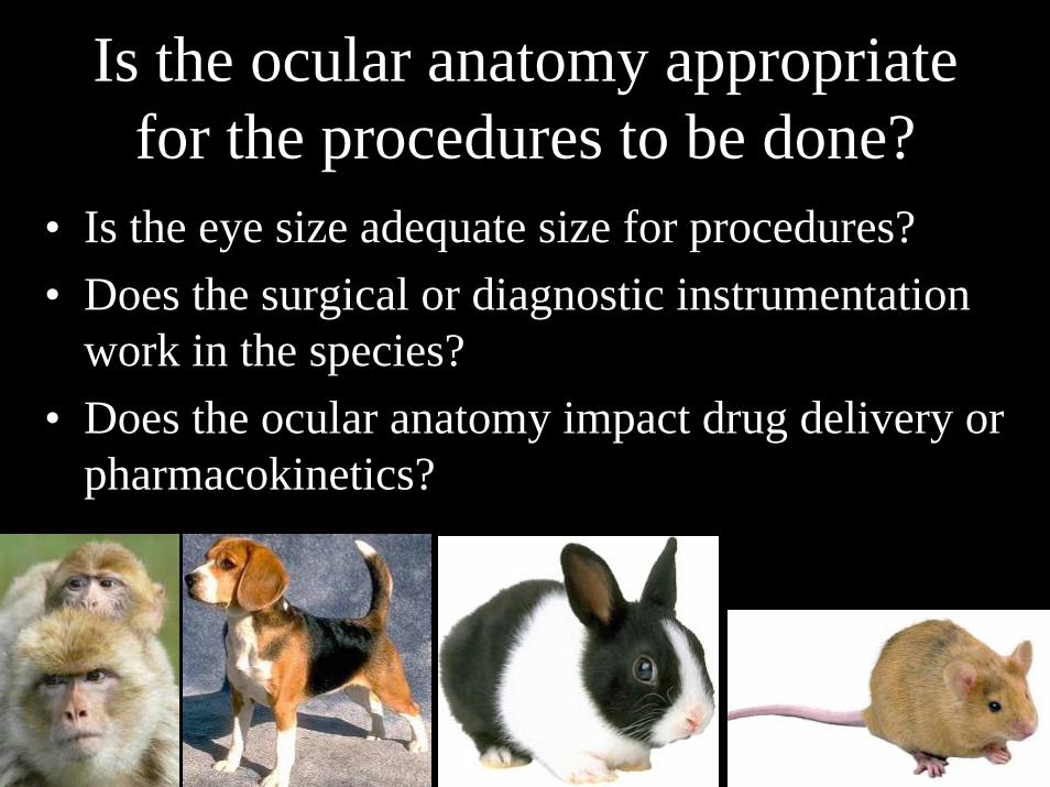
Is the ocular anatomy appropriate for the procedures to be done?
• Is the eye size adequate size for procedures?• Does the surgical or diagnostic instrumentation
work in the species?• Does the ocular anatomy impact drug delivery or
pharmacokinetics?

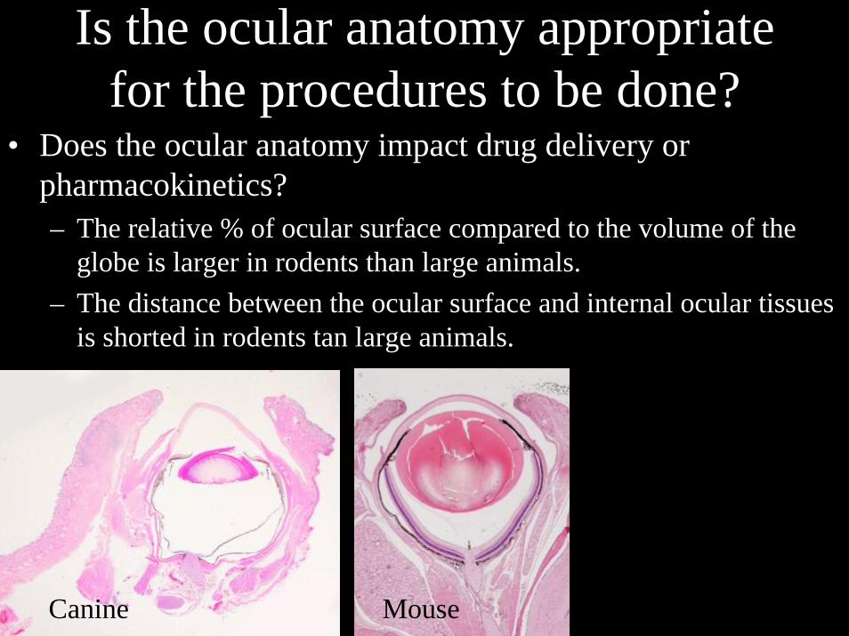
Is the ocular anatomy appropriate for the procedures to be done?
• Is the eye size adequate size for procedures?– Problems with using rodents because of the small eye size and
the inaccessibility of the vitreous• Intraocular pressure measurement is not easily done
– Rebound tonometry on trained mice or manometry• Intravitreous injection or sampling is not easily done
• Diagnostic examination and procedures require experience and training that may or may not be automatically available even with board certified specialists
– Ophthalmoscopy– Electrophysiology– Fluorescein angiography
Mouse Eye

Ocular Dimentions
Axial Length (mm)
Corneal Thickness
(mm)
Anterior Chamber
Depth (mm)
Lens Thickness
(mm)
Vitreous Chamber
Depth (mm) Reference
HUMAN 23.92 0.55 3.05 4.0 16.32A Photon Accurate Model of the Human Eye, Deering, ACM Transactions on Graphics, 2005
MONKEY 17.92 0.55 3.24 2.98 11.3
A Four-surface Schematic Eye of Macaque Money Obtained by An Optical Method, LAPUERTA, and SCHEIN, Vision Research, 1995
CAT 22.3 0.68 4.52 8.5 8.13 The Schematic Eye In The Cat Vakkur and Bishop, Vision Research, 1963
DOG 20.8 .64 4.29 7.85 10.02
Naturally Occurring Vitreous Chamber—Based Myopia in the Labrador Retriever, Mutti, Zadnik, and Murphy, Investigative Ophthalmology & Visual Science, 1999
RABBIT 18.1 0.4 2.9 7.9 6.2 A Schematic Eye for the Rabbit, HUGHES, Vision Research, 1972
RAT 5.98 0.25 0.87 3.87 1.51A Revision of the Rat Schematic Eye, MASSOF and CHANG, Vision Research, 1972

Is the ocular anatomy appropriate for the procedures to be done?
• Does the surgical or diagnostic instrumentation work in the species?– Tonometry in rodents– Devises designed for the human eye have to be
retooled to use the rodent model– Vitrectomy instrument: Macaques vs Human– Choosing the appropriate site for intravitrel
injection or aspiration– Glaucoma drainage devise in the rabbit eye
TonometryCanine
Rabbit
Is the ocular anatomy appropriate for the procedures to be done?
• Does the ocular anatomy impact drug delivery or pharmacokinetics?– The relative % of ocular surface compared to the volume of the
globe is larger in rodents than large animals.– The distance between the ocular surface and internal ocular tissues
is shorted in rodents tan large animals.
Canine Mouse

What is the best species to answer the question?
• Is a particular model of disease better defined or more authentic in a particular species?– Glaucoma models– Laser models for CNV– Transgenic mouse models of AMD
• Is a more human-like anatomy and physiology important?– There is a monkey bias in the ophthalmic drug delivery
world because of the marked similarity between monkey and human eyes
• Fovea• Accommodation• Outflow• Lids, tear film, orbital anatomy

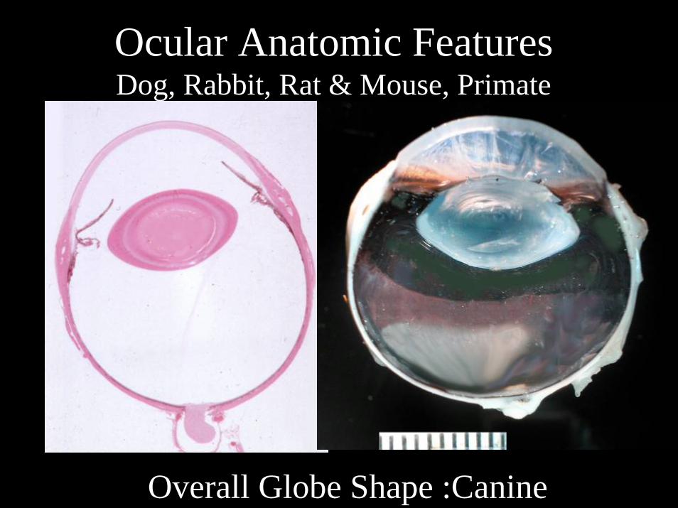
Ocular Anatomic FeaturesDog, Rabbit, Rat & Mouse, Primate
Rabbits are able to resist blinking forlong intervals because they have a verystable tear film. This is likely due to the contribution of a lipid contributionfrom the prominent Hardarian gland.Absent in the primate and dog.
Lacrimal & Hardarian Glands
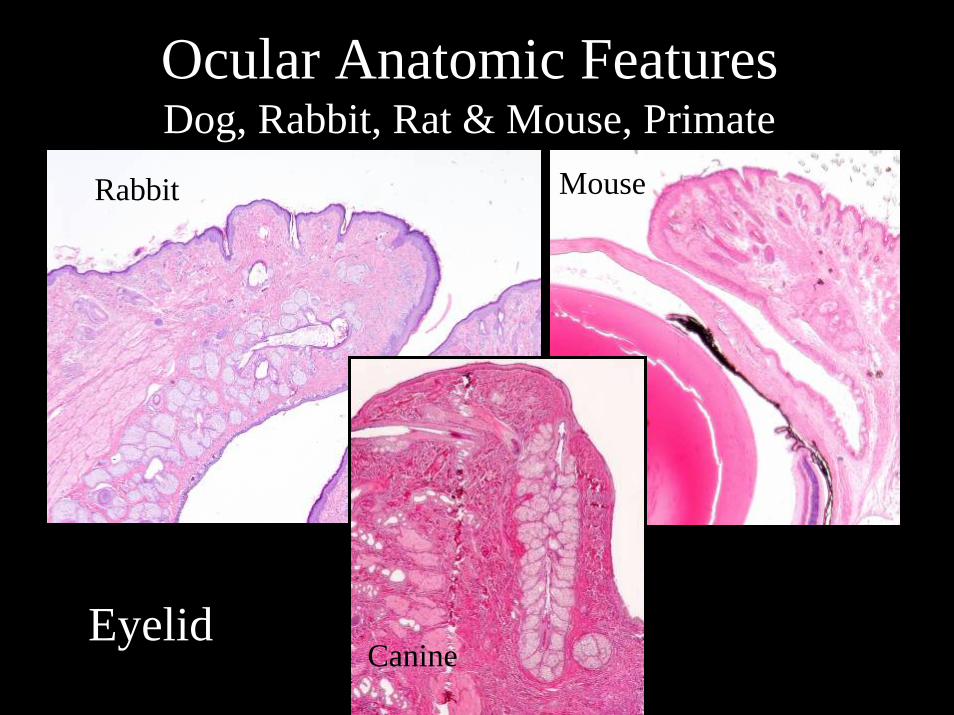
Ocular Anatomic FeaturesDog, Rabbit, Rat & Mouse, Primate
Rabbit Mouse
CanineEyelid

Ocular Anatomic FeaturesDog, Rabbit, Rat & Mouse, Primate
Human Macaque
Primate eyelid
Tarsal plate

Ocular Anatomic FeaturesDog, Rabbit, Rat & Mouse, Primate
Overall Globe Shape :PrimateFovea
Ocular Anatomic FeaturesDog, Rabbit, Rat & Mouse, Primate
Overall Globe Shape :Canine

Ocular Anatomic FeaturesDog, Rabbit, Rat & Mouse, Primate
Overall Globe Shape :Rodent

Ocular Anatomic FeaturesDog, Rabbit, Rat & Mouse, Primate
Primate Vestigial Nictitans
Nictitans (Third Eyelid)
Rabbit Nictitans
Rat Vestigial Nictitans
Hairs

Canine Nictitans
(Third Eyelid)

Ocular Anatomic FeaturesFiltration Apparatus Primate
Schlemm’s CannalScleral Spur

Ocular Anatomic FeaturesFiltration Apparatus Dog
Primary Pectinate
Ciliary Cleft
Angular Aqueous Plexus

Ocular Anatomic FeaturesFiltration Apparatus Rat & Rabbit
Rat Rabbit
Schlemm’s Cannal

Accommodation
Ciliary Muscle

Tapetum Lucidum
Nontapetal Tapetal Tapetal
Eye Shine Canine

RetinaFundus
Canine
Rabbit: Merangiotic
Primate
Rat

Retina: Primate Macula

Fovea
Primate Cone Arrestin
Canine Cone ArrestinmERG

The Primate Retina The Cone Mosaic
Mike Nork

Adaptive Optics
Roorda A and Williams DR, Nature 1999
Variability in Red/Green Cone Ratio among Individuals

Cone Opsoins
Green vs Red Green vs Blue

The Visual Streak and Superior Retina
Canine Area Centralis Rabbit Medullary Ray
Rat Superior Retina and Phototoxic Degeneration

Optic Nerve Lamina Cribrosa
Primate Dog
Rat: No Lamina Cribrosa


![ocular emergencies-1.ppt [Read-Only] - ocw.usu.ac.idocw.usu.ac.id/.../emd166_slide_ocular_emergencies.pdf · Objectives of presentation Review ocular anatomy Understand basic ophthalmic](https://static.fdocuments.net/doc/165x107/5b5334587f8b9a575f8b6a7a/ocular-emergencies-1ppt-read-only-ocwusuacidocwusuacidemd166slideocular.jpg)
