Clinical Anatomy of the Biliary Apparatus: Relations ... Biliary.pdf · Clinical Anatomy of the...
16
Clinical Anatomy of the Clinical Anatomy of the Biliary Apparatus: Biliary Apparatus: Relations & Variations Relations & Variations Lawrence M. Witmer, PhD Lawrence M. Witmer, PhD Professor of Anatomy Department of Biomedical Sciences College of Osteopathic Medicine Ohio University, Athens, Ohio 45701 [email protected] Handout download: http://www.oucom.ohiou.edu/dbms-witmer/gs-rpac.htm 27 March 2007
Transcript of Clinical Anatomy of the Biliary Apparatus: Relations ... Biliary.pdf · Clinical Anatomy of the...
Clinical Anatomy of theClinical Anatomy of theBiliary Apparatus:Biliary Apparatus:
Relations & VariationsRelations & Variations
Lawrence M. Witmer, PhDLawrence M. Witmer, PhDProfessor of AnatomyDepartment of Biomedical SciencesCollege of Osteopathic MedicineOhio University, Athens, Ohio [email protected]
Handout download:http://www.oucom.ohiou.edu/dbms-witmer/gs-rpac.htm
27 March 2007
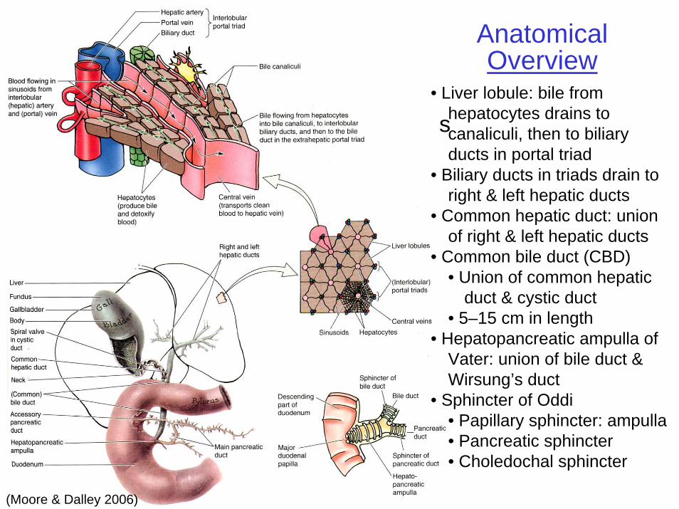
AnatomicalOverview
(Moore & Dalley 2006)
s• Liver lobule: bile from
hepatocytes drains to canaliculi, then to biliary ducts in portal triad
• Biliary ducts in triads drain to right & left hepatic ducts
• Common hepatic duct: union of right & left hepatic ducts
• Common bile duct (CBD)• Union of common hepatic
duct & cystic duct• 5–15 cm in length
• Hepatopancreatic ampulla of Vater: union of bile duct & Wirsung’s duct
• Sphincter of Oddi• Papillary sphincter: ampulla• Pancreatic sphincter• Choledochal sphincter
(Netter 2003)
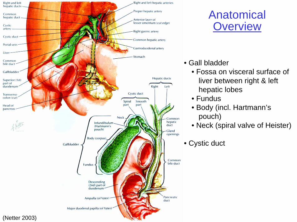
AnatomicalOverview
• Gall bladder• Fossa on visceral surface of
liver between right & left hepatic lobes
• Fundus• Body (incl. Hartmann’s
pouch)• Neck (spiral valve of Heister)
• Cystic duct

(Netter 2003)
AnatomicalOverview
• Peritoneal relations: visceral peritoneum passes over the gall bladder
• Blood supply• From celiac axis• Cystic artery to gall bladder
• Branch to peritoneal surface
• Branch to bare surface• Multiple branches to CBD
• Triangle of Calot• Cystic duct, common hepatic
duct, liver• Cystic artery usually within
Calot’s triangle
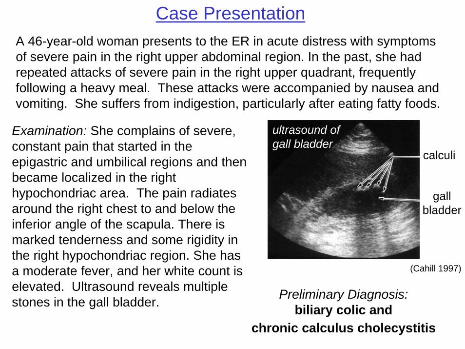
Preliminary Diagnosis:biliary colic and
chronic calculus cholecystitis
Case Presentation
(Cahill 1997)
A 46-year-old woman presents to the ER in acute distress with symptoms of severe pain in the right upper abdominal region. In the past, she had repeated attacks of severe pain in the right upper quadrant, frequently following a heavy meal. These attacks were accompanied by nausea and vomiting. She suffers from indigestion, particularly after eating fatty foods.
Examination: She complains of severe, constant pain that started in the epigastric and umbilical regions and then became localized in the right hypochondriac area. The pain radiates around the right chest to and below the inferior angle of the scapula. There is marked tenderness and some rigidity in the right hypochondriac region. She has a moderate fever, and her white count is elevated. Ultrasound reveals multiple stones in the gall bladder.
gallbladder
calculi
ultrasound ofgall bladder
1. How do you explain the location of the pain in the right hypochondriac region and its typical radiation to the ipsilateral back, particularly to the scapular and infrascapular regions? Why do some patients show ipsilateral pain in the neck and shoulder region?
2. What is the anatomical basis for the muscular rigidity overlying the affected area?
3. Given the anatomical relations of the gall bladder, what organs are most likely to form fistulas with the gall bladder and hence be the recipient of pus and/or stones?
4. Although surgery on the gall bladder is about as common as that for inguinal hernia and appendicitis, what anatomical fact accounts for the much higher frequency of surgical complications in gall bladder surgery?
Questions
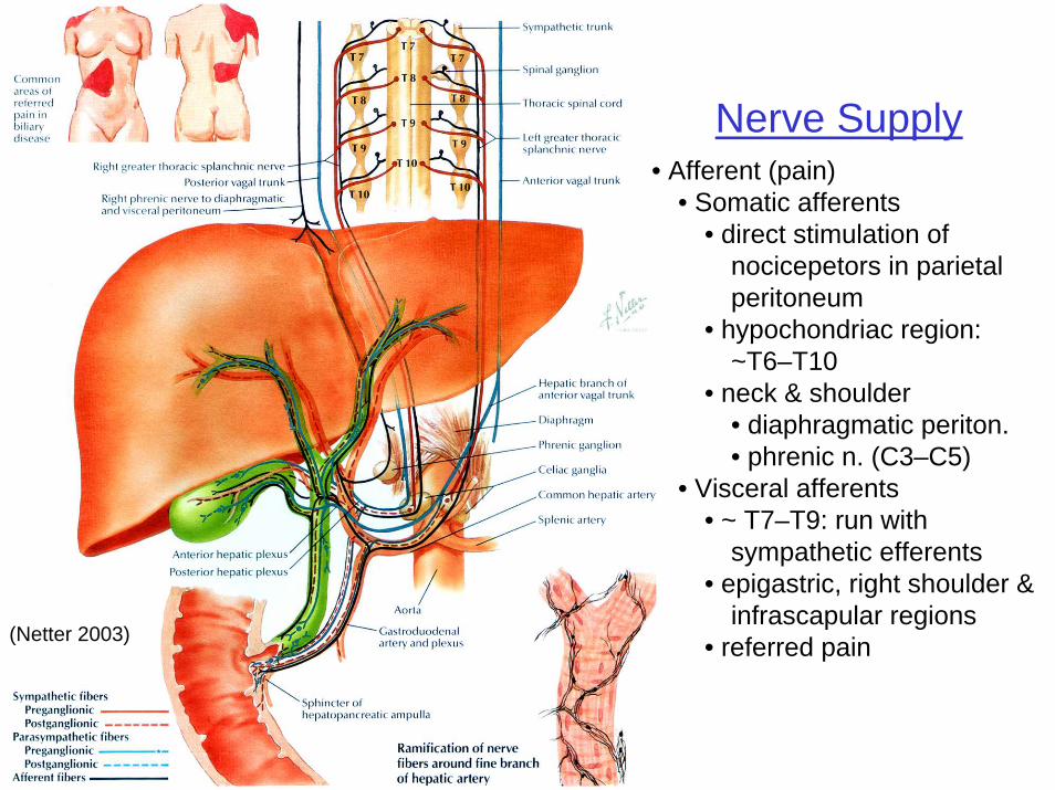
Nerve Supply• Afferent (pain)
• Somatic afferents• direct stimulation of
nocicepetors in parietal peritoneum
• hypochondriac region: ~T6–T10
• neck & shoulder• diaphragmatic periton.• phrenic n. (C3–C5)
• Visceral afferents• ~ T7–T9: run with
sympathetic efferents • epigastric, right shoulder &
infrascapular regions• referred pain(Netter 2003)
1. How do you explain the location of the pain in the right hypochondriac region and its typical radiation to the ipsilateral back, particularly to the scapular and infrascapular regions? Why do some patients show ipsilateral pain in the neck and shoulder region?
2. What is the anatomical basis for the muscular rigidity overlying the affected area?
3. Given the anatomical relations of the gall bladder, what organs are most likely to form fistulas with the gall bladder and hence be the recipient of pus and/or stones?
4. Although surgery on the gall bladder is about as common as that for inguinal hernia and appendicitis, what anatomical fact accounts for the much higher frequency of surgical complications in gall bladder surgery?
Questions
(Netter 2003)
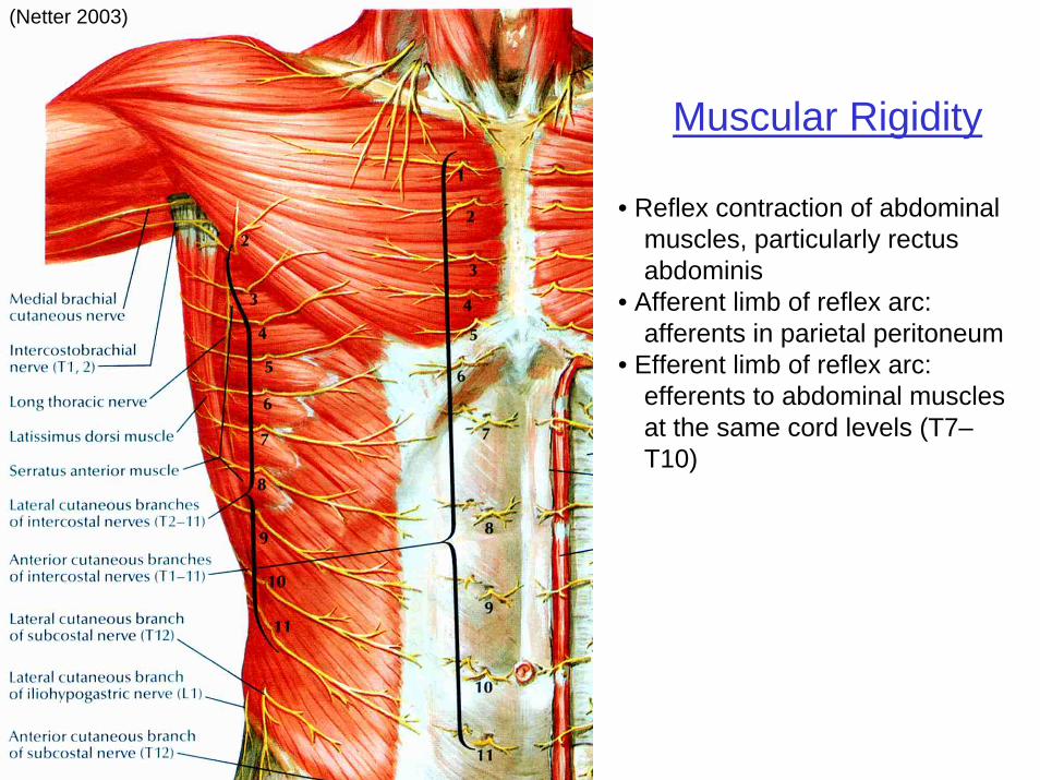
Muscular Rigidity
• Reflex contraction of abdominal muscles, particularly rectus abdominis
• Afferent limb of reflex arc: afferents in parietal peritoneum
• Efferent limb of reflex arc:efferents to abdominal muscles at the same cord levels (T7–T10)
1. How do you explain the location of the pain in the right hypochondriac region and its typical radiation to the ipsilateral back, particularly to the scapular and infrascapular regions? Why do some patients show ipsilateral pain in the neck and shoulder region?
2. What is the anatomical basis for the muscular rigidity overlying the affected area?
3. Given the anatomical relations of the gall bladder, what organs are most likely to form fistulas with the gall bladder and hence be the recipient of pus and/or stones?
4. Although surgery on the gall bladder is about as common as that for inguinal hernia and appendicitis, what anatomical fact accounts for the much higher frequency of surgical complications in gall bladder surgery?
Questions
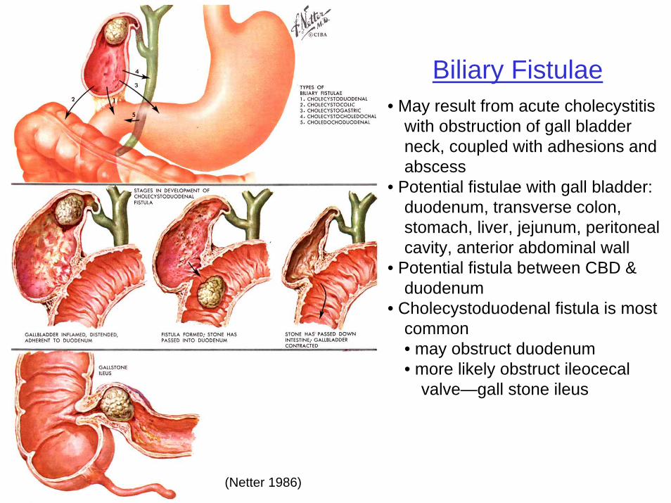
Biliary Fistulae• May result from acute cholecystitis
with obstruction of gall bladder neck, coupled with adhesions and abscess
• Potential fistulae with gall bladder:duodenum, transverse colon, stomach, liver, jejunum, peritoneal cavity, anterior abdominal wall
• Potential fistula between CBD & duodenum
• Cholecystoduodenal fistula is most common• may obstruct duodenum• more likely obstruct ileocecal
valve—gall stone ileus
(Netter 1986)
1. How do you explain the location of the pain in the right hypochondriac region and its typical radiation to the ipsilateral back, particularly to the scapular and infrascapular regions? Why do some patients show ipsilateral pain in the neck and shoulder region?
2. What is the anatomical basis for the muscular rigidity overlying the affected area?
3. Given the anatomical relations of the gall bladder, what organs are most likely to form fistulas with the gall bladder and hence be the recipient of pus and/or stones?
4. Although surgery on the gall bladder is about as common as that for inguinal hernia and appendicitis, what anatomical fact accounts for the much higher frequency of surgical complications in gall bladder surgery?
Questions
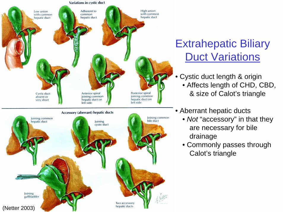
Extrahepatic BiliaryDuct Variations
• Cystic duct length & origin• Affects length of CHD, CBD,
& size of Calot’s triangle
• Aberrant hepatic ducts• Not “accessory” in that they
are necessary for bile drainage
• Commonly passes through Calot’s triangle
(Netter 2003)
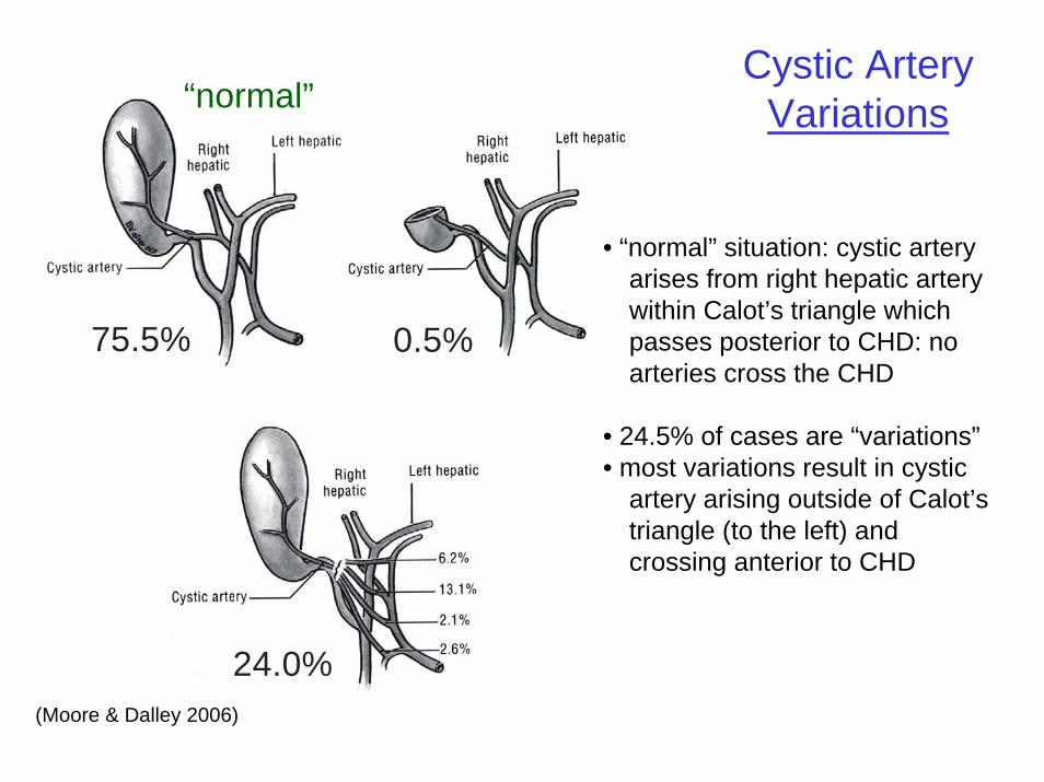
75.5% 0.5%
24.0%
“normal”
• “normal” situation: cystic artery arises from right hepatic artery within Calot’s triangle which passes posterior to CHD: no arteries cross the CHD
• 24.5% of cases are “variations”• most variations result in cystic
artery arising outside of Calot’s triangle (to the left) and crossing anterior to CHD
Cystic ArteryVariations
(Moore & Dalley 2006)
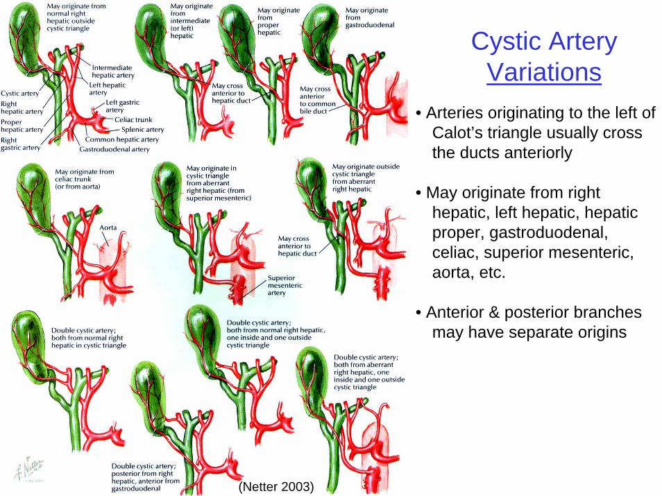
(Netter 2003)
Cystic ArteryVariations
• Arteries originating to the left of Calot’s triangle usually cross the ducts anteriorly
• May originate from right hepatic, left hepatic, hepatic proper, gastroduodenal, celiac, superior mesenteric, aorta, etc.
• Anterior & posterior branches may have separate origins

References
• Cahill, D. R. 1997. Lachman’s Case Studies in Anatomy. Oxford Univ. Press, New York.
• Moore, K. L. and A. F. Dalley. 2006. Clinically Oriented Anatomy, 5th Ed. Lippincott, Williams & Wilkins, Baltimore.
• Netter, F. H. 1986. The CIBA Collection of Medical Illustrations, Volume 3: Digestive System, Part III. CIBA-Geigy, Summit.
• Netter, F. H. 2003. Atlas of Human Anatomy, 3rd Ed. Icon Learning Systems, Teterboro.