Obstructive Lung Diseases
59
Obstructive Lung Diseases MARYAM JAMILAH BINTI ABDUL HAMID 082013100002 IMS BANGALORE 1
-
Upload
autumnpianist -
Category
Education
-
view
162 -
download
2
Transcript of Obstructive Lung Diseases

Obstructive Lung Diseases
MARYAM JAMILAH BINTI ABDUL HAMID
082013100002
IMS BANGALORE
1

Learning outcomes
Emphysema
Chronic bronchitis
Asthma
Bronchiectasis
Define
Etiology
Pathogenesis
Morphology
Clinical features
Conditions related
to the disease2

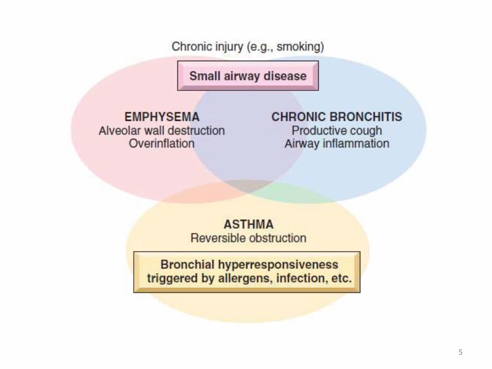
Definition of Obstructive Lung Disease
A lung disease characterized by chronic
obstruction of lung airflow that interferes with
normal breathing and is not fully reversible
3

-Air flow obstruction
(emphysema, bronchiectasis)
-Thickening of walls
-Deposition at the lumen
(chronic bronchitis)
-Fibrosis
4
5

6

Definition of emphysema
“Abnormal permanent enlargement of the air spaces distal to the terminal bronchioles, accompanied by destruction of their walls
without significant fibrosis”
7

Etiology of emphysema
• Tobacco smoke
• Marijuana smoke
• Air pollution
• Manufacturing fumes
• α1-antitrypsin deficiency
8
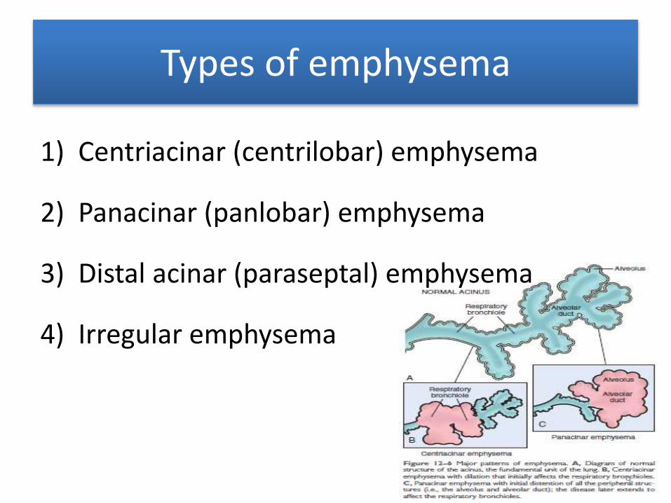
Types of emphysema
1) Centriacinar (centrilobar) emphysema
2) Panacinar (panlobar) emphysema
3) Distal acinar (paraseptal) emphysema
4) Irregular emphysema
9

Centriacinar (centrilobar) emphysema
• Most common; >20%
• Central or proximal parts of the acini, formed
by respiratory bronchioles, are affected,
while distal alveoli are spared
• Severe type affects the distal alveoli as well
• Seen in cigarette smokers
10

• Lower lung zone
• Acini are uniformly enlarged, from the level of
the respiratory bronchiole to the terminal blind
alveoli
• Usually seen in α1-antitrypsin deficiency
Panacinar (panlobar) emphysema
11

• The proximal portion of the acinus is normal but the distal part is primarily involved
• Unknown cause spontaneous pneumothorax in young adults
• Characteristic finding: multiple, contiguous, enlarged air spaces ranging in diameter from <0.5 mm to >2.0 cm
• Sometimes forming cystic structures that, with progressive enlargement, are referred to as bullae
Distal acinar (paraseptal) emphysema
12

• Acinus is irregularly involved, is almost
invariably associated with scarring
• Clinically asymptomatic
Irregular emphysema
13

14

Pathogenesis of emphysema
15
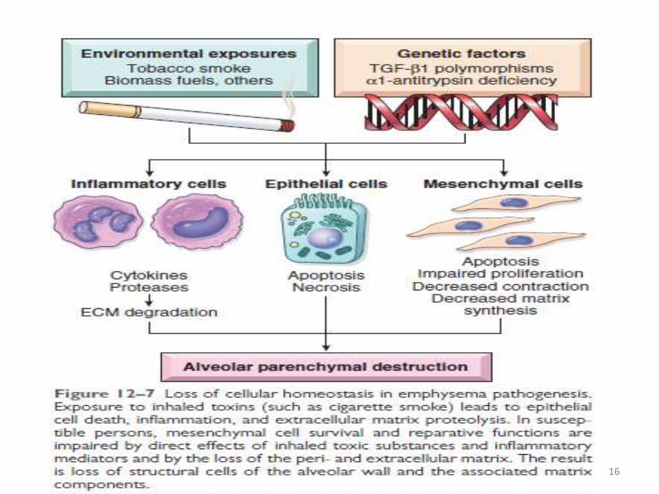
16
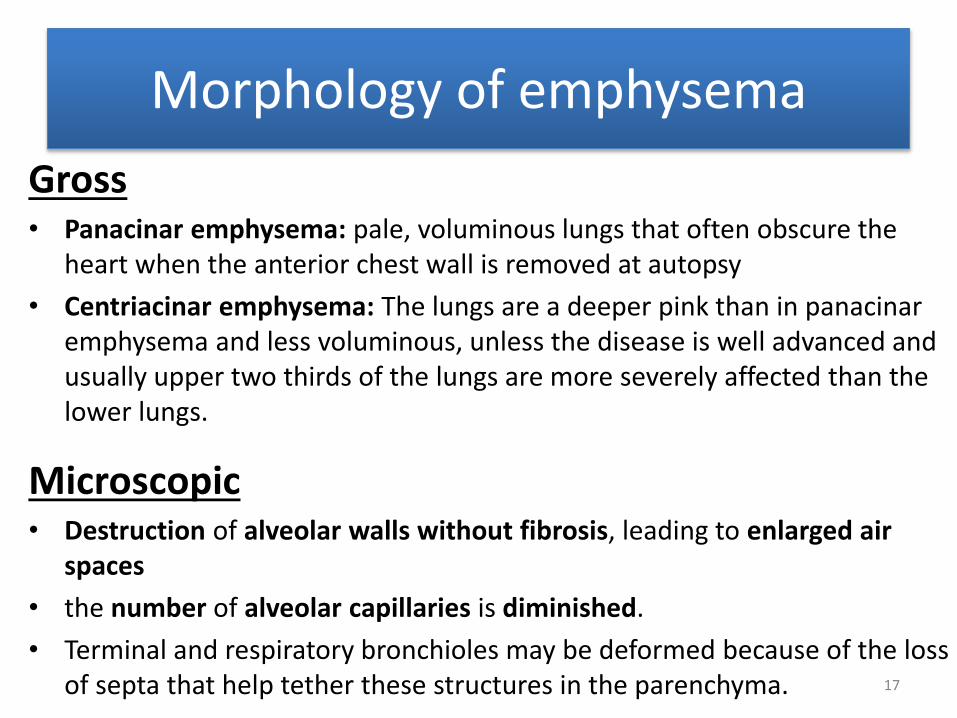
Morphology of emphysema
Gross• Panacinar emphysema: pale, voluminous lungs that often obscure the
heart when the anterior chest wall is removed at autopsy
• Centriacinar emphysema: The lungs are a deeper pink than in panacinaremphysema and less voluminous, unless the disease is well advanced and usually upper two thirds of the lungs are more severely affected than the lower lungs.
Microscopic• Destruction of alveolar walls without fibrosis, leading to enlarged air
spaces
• the number of alveolar capillaries is diminished.
• Terminal and respiratory bronchioles may be deformed because of the loss of septa that help tether these structures in the parenchyma. 17

18

19
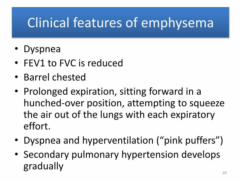
Clinical features of emphysema
• Dyspnea
• FEV1 to FVC is reduced
• Barrel chested
• Prolonged expiration, sitting forward in a hunched-over position, attempting to squeeze the air out of the lungs with each expiratory effort.
• Dyspnea and hyperventilation (“pink puffers”)
• Secondary pulmonary hypertension develops gradually
20

Complication of emphysema
• Pulmonary failure with respiratory acidosis,
• Hypoxia
• Coma
• Right-sided heart failure (cor pulmonale)
21

Conditions related to emphysema
• Compensatory emphysema
• Obstructive overinflation
• Bullous emphysema
• Mediastinal (interstitial) emphysema
22

23

24

Chronic Bronchitis
• 40- to 65-year-old (20-25% are suffering)
• Heavy smoker & pollutants
• Diagnose: presence of a persistent productive cough for at least 3 consecutive months in at least 2 consecutive years
• Early stage: mucoid sputum (w/o obstruction)
• Later stage: intermittent bronchospasm & wheezing 25

Pathogenesis of chronic bronchitis
1. Irritants
2. Hypertrophy of mucous glands in trachea & main
bronchi
3. Hypersecretion of mucus (begin in large airways)
4. Marked increase in mucin-secreting goblet cells
5. Small airway disease & coexist
6. Secondary microbial infection 26

Morphology of chronic bronchitis
Gross• Mucosal lining of the larger airways usually is
hyperemic and swollen by edema fluid
• Covered by a layer of mucinous or mucopurulent secretions
• Smaller bronchi and bronchioles also may
be filled with similar secretions
27

Microscopic
• Trachea and larger bronchi is enlargement of the mucus-secreting glands
• Increase in size is assessed by the ratio of the thickness of the submucosal
gland layer to that of the bronchial wall (the Reid index—normally 0.4)
• Inflammatory cells, largely mononuclear but sometimes admixed with
neutrophils
• Goblet cells metaplasia, mucous plugging, inflammation, and fibrosis
• Severe cases, there may be complete obliteration of the lumen as a
consequence of fibrosis (bronchiolitis obliterans). It is the submucosal
fibrosis that leads to luminal narrowing and airway obstruction.
28

29
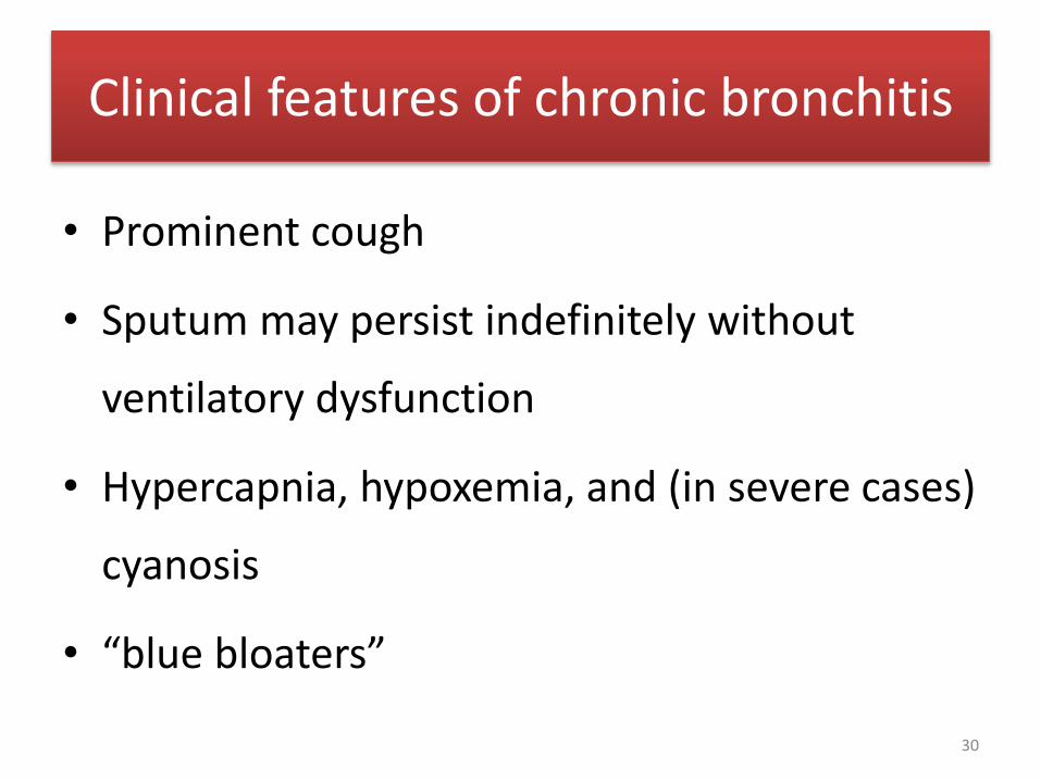
Clinical features of chronic bronchitis
• Prominent cough
• Sputum may persist indefinitely without
ventilatory dysfunction
• Hypercapnia, hypoxemia, and (in severe cases)
cyanosis
• “blue bloaters”
30

Complication of chronic bronchitis
• Pulmonary hypertension
• Cardiac failure
• Recurrent infections
• Respiratory failure
31

32

33

34

35

Asthma
• A chronic inflammatory disorder of the airways
• Recurrent episodes of wheezing, breathlessness, chest tightness, and cough, particularly at night and/or early in the morning
• The hallmarks of the disease are intermittent and reversible airway obstruction, chronic bronchial inflammation with eosinophils, bronchial smooth muscle cell hypertrophy and hyperreactivity, and increased mucus secretion
• Stimuli that trigger attacks in patients would have little or no effect in persons with normal airway
36

• Eosinophils, mast cells, macrophages, lymphocytes,
neutrophils, and epithelial cells. Of note, there has
significant increase in the incidence of asthma
• “hygiene hypothesis”
• Classification:-
– Atopic asthma -Drug induced asthma
– Nonatopic asthma -Occupational asthma
• Bronchospasm can be triggered by diverse mechanisms
• Environmental exposure to irritants (e.g., smoke,
fumes), cold air, stress, and exercise. 37

Pathogenesis of asthma
38

Atopic Asthma
• Most common• Usually beginning in childhood, classic example of
type I IgE–mediated HS• A positive family history; asthmatic attacks,
rhinitis, urticaria, or eczema• Triggered by environmental antigens; Dusts,
pollen, animal dander, and foods• Infections can also be a trigger• A skin test; immediate wheal-and flare reaction• Diagnosis based on serum radioallergosorbent
tests (RASTs) that identify the presence of IgEspecific for a panel of allergens.
39

Non-Atopic Asthma
• No evidence of allergen sensitization
• Skin test; usually negative
• A positive family history of asthma is less common
• Respiratory infections due to viruses and inhaled air pollutants are common triggers
• It is thought that virus-induced inflammation of the respiratory mucosa lowers the threshold of the subepithelial vagal receptors to irritants
• Humoral and cellular mediators of airway obstruction are common to both atopic and nonatopic variants of asthma
• So they are treated in a similar way 40

Drug-Induced Asthma
• Aspirin• Patients with aspirin sensitivity present with
recurrent rhinitis and nasal polyps, urticaria, and bronchospasm
• Precise mechanism remains unknown• But it is presumed that aspirin inhibits the
cyclooxygenase-1 pathway of arachidonic acid metabolism without affecting the lipoxygenaseroute
• Thereby shifting the balance of productiontoward leukotrienes that cause bronchial spasm
41

Occupational Asthma
• Stimulated by fumes (epoxy resins plastics),
organic and chemical dusts (wood, cotton,
platinum), gases (toluene), and other
chemicals
• Asthma attacks usually develop after repeated
exposure to the inciting antigen
42

Morphology of asthma
Gross
• Lungs are overdistended because of
overinflation
• Small areas of atelectasis
• Occlusion of bronchi and bronchioles by
thick, tenacious mucous plugs43

Microscopic• Mucous plugs contain whorls of shed epithelium
(Curschmann spirals)
• Numerous eosinophils and Charcot-Leyden crystals (collections of crystalloids made up of eosinophil proteins)
• “airway remodeling” include
• Thickening of airway wall
• Sub-basement membrane fibrosis (Fig. 12–12)
• Increased vascularity in submucosa
• An increase in size of the submucosal glands and goblet cell metaplasia of the airway epithelium
• Hypertrophy and/or hyperplasia of the bronchialmuscle
44

45

Clinical features of asthma
• Severe dyspnea with wheezing; the chief difficulty lies in expiration
• Progressive hyperinflation of the lungs• Attacks last from 1 to several hours and subside either• Spontaneously or with therapy, usually
bronchodilators and corticosteroids• Intervals between attacks are characteristically free
from overt respiratory difficulties, but persistent, subtle deficits can be detected by spirometry
• Occasionally a severe paroxysm occurs that does not respond to therapy and persists for days and even weeks (status asthmaticus)
• Hypercapnia, acidosis,severe hypoxia may be fatal46

47

Bronchiectasis
Bronchiectasis is the permanent dilation of bronchi and bronchioles caused by destruction of the
muscle and the supporting elastic tissue, resulting from or associated with chronic
necrotizing infections
• Secondary to persisting infection or obstruction caused by a variety of conditions
• Characteristic symptom: cough & expectoration of copious amounts of foul purulent sputum
• Diagnosis: patient history + radiographic bronchial dilation
48

Etiology of bronchiectasis
• Bronchial obstruction
• Congenital or hereditary conditions:-
In cystic fibrosis
In immunodeficiency state
Kartagener syndrome
• Necrotizing or suppurative pneumonia49

Pathogenesis
• Two processes are crucial
–Obstruction
– Chronic persistent infection
• Either of these may come first
• Usually affects the lower lobes bilaterally
50

Morphology
Gross• Most severe involvement is more distal
bronchi and bronchioles
• Airways are dilated to as much as 4x their usual
• Bronchioles can be seen on the pleural surfaces
51

52

Microscopic
• Vary with the activity and chronicity of the disease
• In the usual case, a mixed flora can be cultured from
the involved bronchi
• Hyperplasia of epithelium metaplasia of
epithelium into squamous cell
• Full-blown active case:-
– an intense acute and chronic inflammatory exudate within the walls
of the bronchi and bronchioles
– Desquamation of lining epithelium cause extensive areas of ulceration53

• Healing: lining epithelium may regenerate completely
• Healing in chronic case: Fibrosis of the bronchial and
bronchiolar walls and peribronchiolar fibrosis
• In some instances, necrosis destroys the bronchial or
bronchiolar walls formation of an abscess cavity
within which a fungus ball may develop
54

Clinical features of bronchiectasis
• Severe, persistent cough with expectoration of mucopurulent, sometimes fetid sputum
• Sputum; flecks of blood, frank hemoptysis
• Symptoms are episodic, precipitated by upper respiratory tract infections or new pathogenic agents
• Clubbing
• Hypoxemia, hypercapnia, pulmonary hypertension and cor pulmonale (rare)
55

Complication of bronchiectasis
• Metastatic brain abscesses
• Reactive amyloidosis
56

Conclusion
Points discussed:-
• Emphysema
• Chronic bronchitis
• Asthma
• Bronchiectasis
57

References
• Robbins Pathology 9th edition
• Internet
58

59


![PH Palliative Care April 2018 [Read-Only] · 3.1 Chronic obstructive pulmonary disease 3.2 Interstitial lung disease 3.3 Other pulmonary diseases with mixed restrictive and obstructive](https://static.fdocuments.net/doc/165x107/5f6082feb24ab0784a7d4434/ph-palliative-care-april-2018-read-only-31-chronic-obstructive-pulmonary-disease.jpg)