Obstructive jaundice with a biliary clot post-endoscopic ...
4
Obstructive jaundice with a biliary clot post-endoscopic sphincterotomy treated with clipping and endoscopic biliary stenting A 62-year-old man was admitted with epigastralgia. He had a history of laparo- scopic cholecystectomy for cholecystoli- thiasis. He had no coagulopathy and was not taking anticoagulants. Abdominal computed tomography (CT) showed a common bile duct (CBD) stone (▶ Fig. 1a). Endoscopic retrograde cholangiog- raphy (ERC) and intraductal ultrasono- graphy (IDUS) also showed a 2.8-mm CBD stone (▶ Fig. 1 b, ▶ Fig.2a,b). Endo- scopic sphincterotomy (EST) was per- formed (▶ Fig.2c) and the CBD stone was removed using a wire basket (▶ Fig. 2d). The patient complained of epigastralgia again after 4 days. Laboratory investiga- tions demonstrated elevated cholestatic parameters: total bilirubin 2.8 mg/dL (normal range 0.4 – 1.5 mg/dL), aspartate aminotransferase 176U/L (13 – 30 U/L), alanine aminotransferase 146 U/L (10 – 42 U/L), alkaline phosphatase 233U/L (38 – 113 U/L), and gamma-glutamyl transpeptidase 695U/L (9 – 32 U/L); he- moglobin was within the normal limit. CT showed a diffuse high-density structure in the CBD, with the bile duct mildly dila- ted (▶ Fig.3a). ERC revealed post-EST bleeding and a biliary clot in the CBD (▶ Fig. 3 b, ▶ Fig.4a). The clot was re- moved using a grasping forceps and wire basket (▶ Fig.3c, ▶ Fig.4b), and an en- doscopic biliary stent (EBS) was inserted into the CBD for biliary drainage. Clip- ping was applied to stop the bleeding (▶ Fig.3d, ▶ Fig.4c,d, ▶ Video 1). The patient progressed well after the proce- dures. The EBS was removed 8 days postoperatively and the patient was dis- charged 10 days postoperatively. E-Videos ▶ Fig. 1 Common bile duct stone (arrow). a Abdominal computed tomography. b Endoscopic retrograde cholangiography. Video 1 The biliary clot, caused by delayed bleeding after endoscopic sphincterotomy, was removed using a grasping forceps and wire basket, and clips were applied to stop the bleeding. Abe Takashi et al. Clipping and endoscopic biliary stenting for obstructive jaundice … Endoscopy 2021; 53: E297–E300 | © 2020. Thieme. All rights reserved. E297 This document was downloaded for personal use only. Unauthorized distribution is strictly prohibited. Published online: 2020-10-08
Transcript of Obstructive jaundice with a biliary clot post-endoscopic ...

Obstructive jaundice with a biliary clot post-endoscopic sphincterotomy treatedwith clipping and endoscopic biliary stenting
A 62-year-old man was admitted withepigastralgia. He had a history of laparo-scopic cholecystectomy for cholecystoli-thiasis. He had no coagulopathy and wasnot taking anticoagulants. Abdominalcomputed tomography (CT) showed acommon bile duct (CBD) stone (▶Fig.1 a). Endoscopic retrograde cholangiog-raphy (ERC) and intraductal ultrasono-graphy (IDUS) also showed a 2.8-mmCBD stone (▶Fig. 1b, ▶Fig. 2 a, b). Endo-scopic sphincterotomy (EST) was per-formed (▶Fig. 2 c) and the CBD stonewas removed using a wire basket (▶Fig.2d).The patient complained of epigastralgiaagain after 4 days. Laboratory investiga-tions demonstrated elevated cholestaticparameters: total bilirubin 2.8mg/dL(normal range 0.4–1.5mg/dL), aspartateaminotransferase 176U/L (13–30U/L),alanine aminotransferase 146U/L (10–42U/L), alkaline phosphatase 233U/L(38–113U/L), and gamma-glutamyltranspeptidase 695U/L (9–32U/L); he-moglobin was within the normal limit. CTshowed a diffuse high-density structurein the CBD, with the bile duct mildly dila-ted (▶Fig. 3 a). ERC revealed post-ESTbleeding and a biliary clot in the CBD(▶Fig. 3b, ▶Fig. 4 a). The clot was re-moved using a grasping forceps and wirebasket (▶Fig. 3 c, ▶Fig. 4b), and an en-doscopic biliary stent (EBS) was insertedinto the CBD for biliary drainage. Clip-ping was applied to stop the bleeding(▶Fig. 3d, ▶Fig. 4 c, d, ▶Video 1). Thepatient progressed well after the proce-dures. The EBS was removed 8 dayspostoperatively and the patient was dis-charged 10 days postoperatively.
E-Videos
▶ Fig. 1 Common bile duct stone (arrow). a Abdominal computed tomography. b Endoscopicretrograde cholangiography.
Video 1 The biliary clot, caused by delayed bleeding after endoscopic sphincterotomy,was removed using a grasping forceps and wire basket, and clips were applied to stop thebleeding.
Abe Takashi et al. Clipping and endoscopic biliary stenting for obstructive jaundice… Endoscopy 2021; 53: E297–E300 | © 2020. Thieme. All rights reserved. E297
Thi
s do
cum
ent w
as d
ownl
oade
d fo
r pe
rson
al u
se o
nly.
Una
utho
rized
dis
trib
utio
n is
str
ictly
pro
hibi
ted.
Published online: 2020-10-08

The incidence of post-EST delayed he-morrhage is 1.62% [1], and biliary ob-struction with a biliary clot caused bypost-EST bleeding is extremely rare [2–5]. Endoscopic hemostasis is currentlythe first treatment choice for post-ERCbleeding, with balloon dilation and bili-ary stent placement used for treatment[2–5]. To the best of our knowledge,
this is the first English case report of ob-structive jaundice with a biliary clotcaused by post-EST bleeding, treatedwith clipping and an EBS, which may bean effective endoscopic technique fortreating such cases.
Endoscopy_UCTN_Code_CPL_1AK_2AC
Competing interests
The authors declare that they have no con-flict of interest.
The authors
Takashi Abe1, Takehiko Nariyasu1, Takayuki
Nagai1, Marina Hamamoto1, Masato
Hanzawa1, Yasuhisa Hiroshima1, Kazunari
Murakami2
1 Department of Gastroenterology, Oita
Kouseiren Tsurumi Hospital, Beppu, Japan
2 Department of Gastroenterology, Faculty of
Medicine, Oita University, Yufu, Japan
Corresponding author
Takashi Abe, MD, PhD
Department of Gastroenterology, Oita
Kouseiren Tsurumi Hospital, Tsurumi 4333,
Beppu City, Oita 874-8585, Japan
Fax: +81-977-237884
▶ Fig. 2 Removal of the common bile duct (CBD) stone. Endoscopic views (a, c,d) and intra-ductal ultrasonography (IDUS) view (b). a The ampulla of Vater was intact (arrow). b IDUSshowed a CBD stone, approximately 2.8mm in size (arrow). c Immediately after endoscopicsphincterotomy (arrow). d The CBD stone was removed using a wire basket (arrow).
E298 Abe Takashi et al. Clipping and endoscopic biliary stenting for obstructive jaundice… Endoscopy 2021; 53: E297–E300 | © 2020. Thieme. All rights reserved.
E-Videos
Thi
s do
cum
ent w
as d
ownl
oade
d fo
r pe
rson
al u
se o
nly.
Una
utho
rized
dis
trib
utio
n is
str
ictly
pro
hibi
ted.

References
[1] Yan J, Zhou CX, Wang C et al. Risk factors fordelayed hemorrhage after endoscopicsphincterotomy. Hepatobiliary Pancreat DisInt 2020. doi:10.1016/j.hbpd.2019.12.010
[2] Mosenkis BN, Brandt LJ. Bleeding causingbiliary obstruction after endoscopic sphinc-terotomy. Am J Gastroenterol 1997; 92:708–709
[3] Ala A, Khin CC, van Someren N. Commonbile duct thrombus: a cause of persistingobstructive jaundice after endoscopicsphincterotomy. Gastrointest Endosc 1999;50: 285–286
[4] Ergül B, Koçak E, Köklü S. An unusual com-plication of ERCP: obstructive jaundice dueto a blood clot. Clin Res Hepatol Gastroen-terol 2012; 36: e40–e41
[5] Zhu Y, Wang S, Zhao S et al. Obstructivejaundice due to a blood clot after ERCP: acase report and review of the literature. BMCGastroenterol 2018; 18: 163
Bibliography
Endoscopy 2021; 53: E297–E300DOI 10.1055/a-1260-2903ISSN 0013-726Xpublished online 8.10.2020© 2020. Thieme. All rights reserved.Georg Thieme Verlag KG, Rüdigerstraße 14,70469 Stuttgart, Germany
▶ Fig. 3 Imaging after endoscopic sphincterotomy (EST) and common bile duct (CBD) stoneremoval. a Reconstructed coronal image of abdominal computed tomography on day 4 afterEST showed a diffuse high-density structure in the CBD with the bile duct being mildly dilated(arrows). b Endoscopic retrograde cholangiography revealed a diffuse filling defect in theCBD with the bile duct mildly dilated (arrows). c The CBD was cleaned up by removing thebiliary clot using a grasping forceps and wire basket. d An endoscopic biliary stent was in-serted into the CBD and clipping was applied for endoscopic hemostasis.
Abe Takashi et al. Clipping and endoscopic biliary stenting for obstructive jaundice… Endoscopy 2021; 53: E297–E300 | © 2020. Thieme. All rights reserved. E299
Thi
s do
cum
ent w
as d
ownl
oade
d fo
r pe
rson
al u
se o
nly.
Una
utho
rized
dis
trib
utio
n is
str
ictly
pro
hibi
ted.
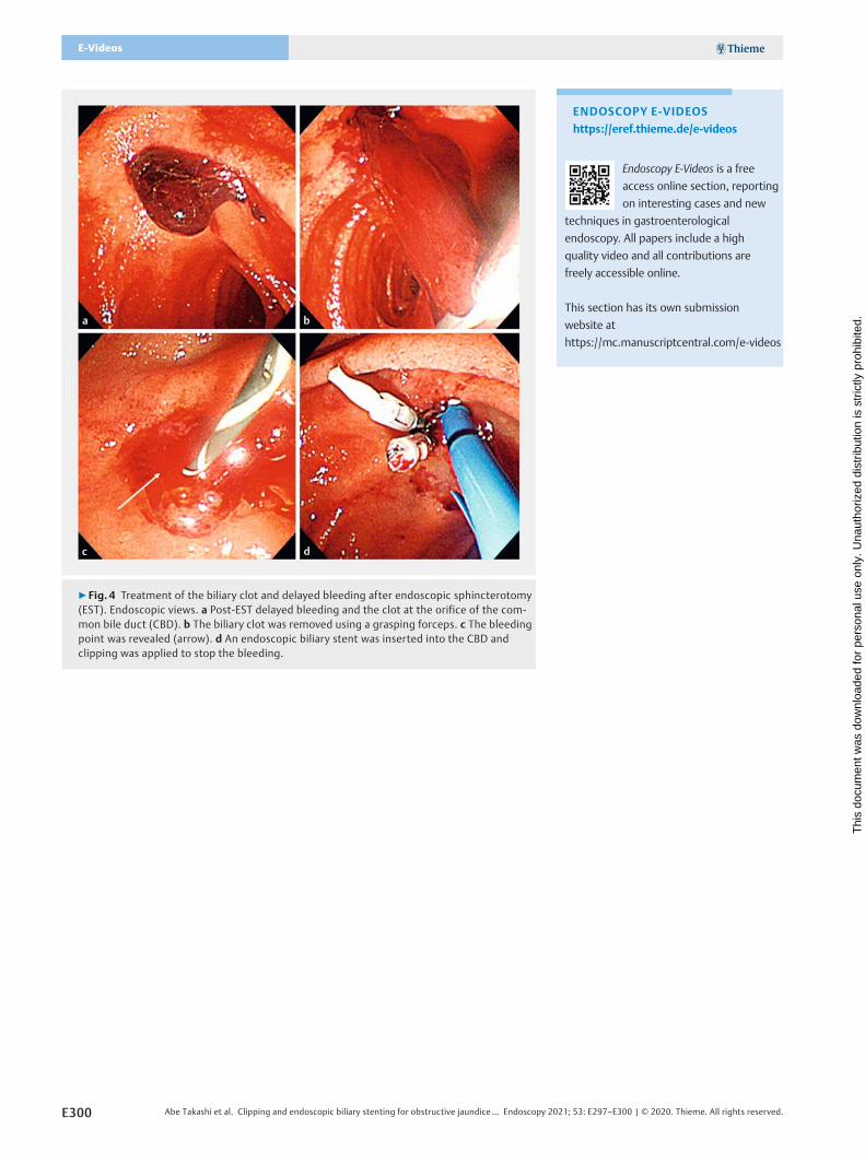
▶ Fig. 4 Treatment of the biliary clot and delayed bleeding after endoscopic sphincterotomy(EST). Endoscopic views. a Post-EST delayed bleeding and the clot at the orifice of the com-mon bile duct (CBD). b The biliary clot was removed using a grasping forceps. c The bleedingpoint was revealed (arrow). d An endoscopic biliary stent was inserted into the CBD andclipping was applied to stop the bleeding.
ENDOSCOPY E-VIDEOS
https://eref.thieme.de/e-videos
Endoscopy E-Videos is a free
access online section, reporting
on interesting cases and new
techniques in gastroenterological
endoscopy. All papers include a high
quality video and all contributions are
freely accessible online.
This section has its own submission
website at
https://mc.manuscriptcentral.com/e-videos
E300 Abe Takashi et al. Clipping and endoscopic biliary stenting for obstructive jaundice… Endoscopy 2021; 53: E297–E300 | © 2020. Thieme. All rights reserved.
E-Videos
Thi
s do
cum
ent w
as d
ownl
oade
d fo
r pe
rson
al u
se o
nly.
Una
utho
rized
dis
trib
utio
n is
str
ictly
pro
hibi
ted.