Animal Health Surveillance - Department of Primary · PDF fileCongenital biliary atresia and...
12
NEW SOUTH WALES April–June 2007 • Number 2007/2 ANIMAL HEALTH SURVEILLANCE In this issue! QUARTERLY HIGHLIGHTS 1 Fog fever in cattle 1 Congenital biliary atresia and jaundice in lambs 2 Nitrate toxicity 5 Bovine tuberculosis exclusion 6 Winter dysentery in a dairy herd 6 Botulism in horses 7 Campylobacter fetus subsp. fetus abortion in cattle 7 Exclusion of canine infuenza 7 Neurological disease in pigs 7 Vitamin A and E defciency in cattle 8 Hendra virus exclusions: Crofton weed poisoning and pneumonia in horses 8 NOTIFIABLE DISEASES 8 Anthrax 8 Low-level pathogenic avian infuenza (LPAI) detections 9 Bovine papular stomatitis: exotic disease exclusion 9 DISEASE SURVEILLANCE AND CONTROL PROGRAMS 9 NSW DPI QUARTERLY HIGHLIGHTS Fog fever in cattle Interstitial pneumonia was the cause of death in April 2007 of a 2-year-old Limousin bull running with a group of 26 cows and calves near Bathurst. The animals had been fed cabbages for a month and had been running in a paddock after cabbage harvest for 2 weeks before the incident. On clinical examination the bull was alert, reluctant to walk, and dyspnoeic; its heart rate and respiratory rate were increased and its mucous membranes were pale. No abnormal lung sounds were noticed at this stage. Blood was taken for haematology. Two days later the bull was re-examined; it had deteriorated, was severely dyspnoeic and showed subcutaneous emphysema. It died following examination, and autopsy revealed extensive pulmonary emphysema and pneumonia. Haematology results showed no evidence of anaemia. On histopathological examination of the lung severe, difuse, subacute interstitial pneumonia and bullous pulmonary emphysema were diagnosed. The lung pathology had features of more acute lesions—protein-rich oedema fuid and hyaline membranes in the alveolar lumina—as well as the subacute lesions, which included proliferation of type 2 alveolar epithelial cells, marked smooth muscle hypertrophy and fbroplasia. This case is consistent with Acute Respiratory Distress Syndrome, sometimes reported 7 to 10 days after cattle have grazed brassicas. Intoxication is caused by a sudden increase in tryptophan levels in the rumen. Tryptophan is metabolised in the rumen to 3-methylindole, which enters the lung via the bloodstream. In the lung it is further metabolised to the ultimate pulmonary toxin. In Europe and America fog fever occurs in autumn, 4 to 10 days after cattle have been moved from a dry to a lush green pasture. The species of grass or other plant can vary, but the disease does occur after cattle have had access to felds after harvest of Brassica species. It occurs almost exclusively in adult cattle. Some reports have suggested a breed incidence. Fog fever is rare in Australia. Treatment is of limited value once animals are sick, and handling of animals in respiratory distress for examination and treatment can lead to death. Attempts to prevent disease, if practical, include limited grazing on the new pasture, gradual introduction of, and supplementation with, other feeds, and supplementation with an ionophore to inhibit 3-methylindol production. The remainder of the herd were moved from the paddock of cabbages and no further cases were reported. Report written by Erika Bunker, Bruce Watt and Tom McClelland. For further information contact Bruce Watt, DV Central Tablelands RLPB, on (02) 6331 1377. Fog fever: 2 days before death the afected Limousin bull was clinically dyspnoeic, with marked exercise intolerance. Photo by Bruce Watt Information contributed by staf of the Rural Lands Protection Boards and the NSW Department of Primary Industries
Transcript of Animal Health Surveillance - Department of Primary · PDF fileCongenital biliary atresia and...
NEW
SOUT
H WA
LES
April–June 2007 • Number 2007/2 April–June 2007 • Number 2007/2 April–June 2007 • Number 2007/2
ANIMAL HEALTH SURVEILLANCE
In this issue!
QUARTERLY HIGHLIGHTS 1
Fog fever in cattle 1
Congenital biliary atresia and jaundice in lambs 2
Nitrate toxicity 5
Bovine tuberculosis exclusion 6
Winter dysentery in a dairy herd 6
Botulism in horses 7
Campylobacter fetus subsp. fetus abortion in cattle 7
Exclusion of canine influenza 7
Neurological disease in pigs 7
Vitamin A and E deficiency in cattle 8
Hendra virus exclusions: Crofton weed poisoning and pneumonia in horses 8
NoTIFIABLE DISEASES 8
Anthrax 8
Low-level pathogenic avian influenza (LPAI) detections 9
Bovine papular stomatitis: exotic disease exclusion 9
DISEASE SURVEILLANCE AND CoNTRoL PRoGRAmS 9
N S W D P I
QUARTERLY HIGHLIGHTS
Fog fever in cattle Interstitial pneumonia was the cause of death in April 2007 of a 2-year-old Limousin bull running with a group of 26 cows and calves near Bathurst.
The animals had been fed cabbages for a month and had been running in a paddock after cabbage harvest for 2 weeks before the incident.
on clinical examination the bull was alert, reluctant to walk, and dyspnoeic; its heart rate and respiratory rate were increased and its mucous membranes were pale. No abnormal lung sounds were noticed at this stage. Blood was taken for haematology.
Two days later the bull was re-examined; it had deteriorated, was severely dyspnoeic and showed subcutaneous emphysema. It died following examination, and autopsy revealed extensive pulmonary emphysema and pneumonia.
Haematology results showed no evidence of anaemia.
on histopathological examination of the lung severe, diffuse, subacute interstitial pneumonia and bullous pulmonary emphysema were diagnosed. The lung pathology had features of more acute lesions—protein-rich oedema fluid and hyaline membranes in the alveolar lumina—as well as the subacute lesions, which included proliferation of type 2 alveolar epithelial cells, marked smooth muscle hypertrophy and fibroplasia.
This case is consistent with Acute Respiratory Distress Syndrome, sometimes reported 7 to 10 days after cattle have grazed brassicas. Intoxication is caused
by a sudden increase in tryptophan levels in the rumen. Tryptophan is metabolised in the rumen to 3-methylindole, which enters the lung via the bloodstream. In the lung it is further metabolised to the ultimate pulmonary toxin.
In Europe and America fog fever occurs in autumn, 4 to 10 days after cattle have been moved from a dry to a lush green pasture. The species of grass or other plant can vary, but the disease does occur after cattle have had access to fields after harvest of Brassica species. It occurs almost exclusively in adult cattle. Some reports have suggested a breed incidence. Fog fever is rare in Australia.
Treatment is of limited value once animals are sick, and handling of animals in respiratory distress for examination and treatment can lead to death.
Attempts to prevent disease, if practical, include limited grazing on the new pasture, gradual introduction of, and supplementation with, other feeds, and supplementation with an ionophore to inhibit 3-methylindol production.
The remainder of the herd were moved from the paddock of cabbages and no further cases were reported.
Report written by Erika Bunker, Bruce Watt and Tom McClelland. For further information contact Bruce Watt, DV Central Tablelands RLPB, on (02) 6331 1377.
Fog fever: 2 days before death the affected Limousin bull was clinically dyspnoeic, with marked exercise intolerance. Photo by Bruce Watt
Information contributed by staff of the Rural Lands Protection Boards and the NSW Department of Primary Industries

PAGE2
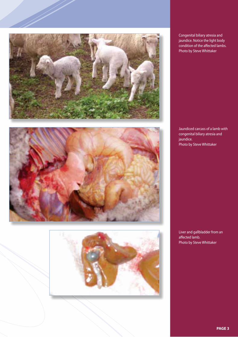
Congenital biliary atresia and jaundice in lambs DV Hume investigated an unusual case of lamb mortality on a property adjacent to the Hume Weir. First-cross ewes joined to Poll Dorset rams had extensively grazed previously water-covered river flats during gestation. Dysphania glomulifera subsp. glomulifera and a type of smartweed provided the predominant vegetation, and both these plants had been heavily grazed. Approximately 50 lambs from a mob of 550 ewes died, with a further 20 or 30 lambs affected. Affected lambs were 1 to 2 weeks old. They were very light in body condition, exhibited a white scour, and had jaundiced conjunctivae. Gross pathology confirmed severe jaundice; the liver had a yellow ochre appearance; the gallbladder was small and shrunken; the kidneys were khaki coloured (urine positive for bilirubin); and the spleen appeared to be enlarged. Largely undigested milk in the stomach and intestines was evidence that the lambs had been suckling.
Congenital aplasia of the bile ducts was suspected and duly confirmed by histopathology.
This condition has been recorded only twice previously. In 1964 perinatal lamb mortalities associated with congenital biliary atresia and jaundice occurred on a property on the foreshores of Burrinjuck Dam. The affected lambs were born to ewes that had been restricted to grazing the exposed foreshores of the northern banks of the Burrinjuck Dam when water levels fell. Sixty of 400 lambs died with congenital biliary atresia and jaundice. In 1988 the condition occurred on another two properties in the same area as the 1964 case. one of these properties adjoined the property on which the 1964 cases occurred. In 1988 flat country that was normally under water became exposed and some sheep were restricted to this area during pregnancy and lambing. A prolific growth of weeds, predominantly Centipeda cunninghamii and Dysphania glomulifera subsp. glomulifera, was noted. Approximately 300 lambs from 420 ewes died. The mortality was higher in lambs born later in the lambing period.
on the second property, located on the Goodrabidgee River, in the upper reaches of the Burrinjuck Dam, cows at varying stages of pregnancy were grazed on exposed flats where Dysphania glomulifera subsp. glomulifera predominated. Nine calves from 49 cows died. The lambs and calves affected in 1988 failed to thrive, developed jaundice and white scours, and died within 4 weeks of birth.
The common features in these outbreaks have been:
1. grazing plants on land previously under water
2. Dysphania glomulifera subsp. glomulifera identified in the 1988 cases and this case
3. clinical picture and histopathology.
The aetiology is thought to be a toxic insult to the fetus in early gestation, causing choledysgenesis and biliary atresia and leading to diffuse, subacute to chronic choliohepatopathy and cirrhosis. At this stage there is no definitive evidence that Dysphania glomulifera subsp. glomulifera is the source of the toxic insult, so other plants should still be considered as possible sources of toxin.
Report written by Sarah Robson. For further information contact Steve Whittaker on (02) 6040 4210.
PAGE3
Congenital biliary atresia and jaundice. Notice the light body condition of the affected lambs. Photo by Steve Whittaker
Jaundiced carcass of a lamb with congenital biliary atresia and jaundice. Photo by Steve Whittaker
Liver and gallbladder from an affected lamb. Photo by Steve Whittaker

PAGE4
The river flat where the ewes were. Photo by Steve Whittaker
Dysphania glomulifera subsp. glomulifera: close-up.
Photo by Steve Whittaker
Dysphania glomulifera subsp. glomulifera can be seen here as the predominant plant species.
Photo by Steve Whittaker
PAGE5
Nitrate toxicity There were 13 cases of nitrate toxicity in NSW confirmed at the Regional Veterinary Laboratories during the quarter. Details of some of these cases are given below.
A mob of 350 2-year-old ewes approximately 14 weeks pregnant was held in a shed ready for shearing. The ewes were very hungry when they were let out into a holding paddock containing predominantly capeweed (Arctotheca calendula) and small-flowered mallow (Malva parviflora). Later that afternoon the ewes became weak and collapsed, especially if they were moved. Some ewes displayed symptoms of respiratory distress. Twenty-five ewes were dead by 4 pm. The following morning another 132 had died. Four sick sheep recovered. DV Wagga examined the mob and noted that affected ewes had brown conjunctivae but their blood had a normal red appearance. Post mortem findings included muscle pallor, congested lungs with bronchi full of froth with flecks of blood; congestion of the renal cortex; sloughing of the rumen lining; autolysis of the liver; and a puffy spleen.
Nitrate-nitrite poisoning was confirmed by laboratory testing of the aqueous humor. Three out of three samples had high nitrate and nitrite, a finding consistant with toxicity.
Nitrate poisoning associated with capeweed (Arctotheca calendula) caused the death of cattle on two properties in the Wagga Wagga RLPB. on the first property a total of 16 out of 30 weaners were affected; five of the 16 died. on the second property 10 out of 40 weaners died. These cases occurred within a day of each other, and a press release was immediately issued to notify producers of the high risk of nitrate poisoning due to the abundance of capeweed in the district.
Thirty-eight out of 52 Dorper rams died overnight from nitrate toxicity in April 2007 on a property near Wanaaring. The
rams had arrived on the property in the afternoon. They were put in a yard with cathead (Tribulus terrestris), nightshade (Solanum spp.) and paddymelons (Cucumis myriocarpus subsp. leptodermis) and were found dead the next morning.
Nitrate, nitrite and calcium levels were measured in aqueous humour samples from two animals. Nitrate and nitrite levels were high: nitrate 209 and 84 mg/L (normal <10 mg/L); nitrite 18 and 19 mg/L (normal <1 mg/L). Calcium levels were within the normal range. Histopathology of the liver, lung and kidney showed no significant lesions.
Cathead is suspected as the source of nitrate, as it is a plant known to cause nitrate toxicity. Additionally, uptake of nitrate is facilitated in areas such as yards, where many animals have urinated and defaecated. Further contributing factors were that the animals were stressed and hungry after transport, and their rumen microbes had no time to adapt to an increased nitrate load.
Thirty-two out of 100 merino ewes died from nitrate intoxication in may 2007 on a property near manildra. The ewes were shorn over the previous 2 days, locked in a shed overnight, and let out the following morning into a paddock of predominantly native grasses with good green pick.
Aqueous humour taken from three sheep had nitrate levels of 299, 153 and 144 mg/L (normal <10 mg/L) and nitrite levels of 16, 11 and 23 mg/L (normal <1 mg/L); calcium and magnesium levels were within normal ranges.
Four out of 50 Hereford cattle of mixed ages were found dead in April 2007 on a property near Trunkey; additionally, five of a neighbour’s cattle were found dead at the same time. The cattle were running on creek country, and variegated thistle (Silybum marianum) was freely available near the creek and had been grazed by the cattle. There was some fresh hemlock within the thistle, and nightshade,
bracken fern (Pteridium esculentum), Paterson’s curse (Echium plantagineum) and castor oil plant (Ricinus communis) were also present but appeared untouched by the cattle.
A sample of the variegated thistle was tested; it had a nitrate concentration of greater than 2500 and less than 5000 mg/kg wet weight. A feed concentration of greater than 1500 mg/kg wet weight (assuming dry matter 30%) is considered to be potentially toxic.
Nitrate poisoning caused the death of 18 out of 250 two- to five-year-old merino ewes 6 weeks off lambing on a grazing property near Trundle. Nitrate in aqueous humour from a recently dead sheep was detected at 100 ppm. These sheep had been fed a high-grain diet for the previous few months. The sheep were then held overnight off feed and scanned the next day for pregnancy. After scanning the mob was placed onto roadside pasture for 5 hours. The grass species present at the roadside were barley grass, rye grass, couch and wild black oats.
When the sheep were mustered at the end of the day to be returned to their original paddock, the 18 dead animals were found, along with 100 sick animals. All the sick animals made a complete recovery after being removed from the roadside grazing. It appears that as grains are low in nitrates these animals lost their ability to digest nitrates properly. As the animals were hungry, they gorged on the higher-nitrate grass diet.
PAGE6
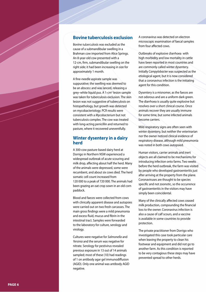
Bovine tuberculosis exclusion Bovine tuberculosis was excluded as the cause of a submandibular swelling in a Brahman cow imported from Alice Springs. An 8-year-old cow presented with a 12-cm, firm, submandibular swelling on the right side; it had been increasing in size for approximately 1 month.
A fine-needle aspirate sample was suppurative; the swelling was deemed to be an abscess and was lanced, releasing a grey–white liquid pus. A 1-cm2 lesion sample was taken for tuberculosis exclusion. The skin lesion was not suggestive of tuberculosis on histopathology, but growth was detected on mycobacteriology. PCR results were consistent with a Mycobacterium but not tuberculosis complex. The cow was treated with long-acting penicillin and returned to pasture, where it recovered uneventfully.
Winter dysentery in a dairy herd A 300-cow pasture-based dairy herd at Dorrigo in Northern NSW experienced a widespread outbreak of acute scouring and milk drop, affecting about half the herd. many of the animals were depressed, some were recumbent, and about six cows died. The herd somatic cell count increased from 120 000 to a peak of 720 000. The animals had been grazing an oat crop sown in an old corn paddock.
Blood and faeces were collected from cows with clinically apparent disease and autopsies were carried out on two fresh carcasses. The main gross findings were a mild pneumonia and excess fluid, mucus and fibrin in the intestinal tract. Samples were forwarded to the laboratory for culture, serology and virology.
Cultures were negative for Salmonella and Yersinia and the serum was negative for nitrate. Serology for pestivirus revealed previous exposure in 13 out of 14 animals sampled; most of these (10) had readings of 1 on antibody agar gel immunodiffusion (AGID). only one animal was antibody AGID negative.
A coronavirus was detected on electron microscopic examination of faecal samples from four affected cows.
outbreaks of explosive diarrhoea with high morbidity and low mortality in cattle have been reported in most countries and are commonly called winter dysentery. Initially Campylobacter was suspected as the etiological agent, but it is now considered that a coronavirus infection is the initiating agent for this condition.
Dysentery is a misnomer, as the faeces are not odorous and are a uniform dark green. The diarrhoea is usually quite explosive but resolves over a short clinical course. once animals recover they are usually immune for some time, but some infected animals become carriers.
mild respiratory signs are often seen with winter dysentery, but neither the veterinarian nor the owner noticed clinical evidence of respiratory disease, although mild pneumonia was noted in both cows autopsied.
Human visitors, carrier animals and inert objects are all claimed to be mechanisms for introducing infection onto farms. Two weeks before the herd outbreak, the farm was visited by people who developed gastroenteritis just after arriving at the property from the plane. Coronaviruses are thought to be species specific and not zoonotic, so the occurrence of gastroenteritis in the visitors may have simply been coincidental.
many of the clinically affected cows ceased milk production, compounding the financial loss to the owner. Coronavirus infection is also a cause of calf scours, and a vaccine is available in some countries to provide protection.
The private practitioner from Dorrigo who investigated this case took particular care when leaving the property to clean his footwear and equipment and did not go to another farm. As this condition is reported to be very contagious these steps may have prevented spread to other herds.
cause of a submandibular swelling in a Brahman cow imported from Alice Springs. An 8-year-old cow presented with a 12-cm, firm, submandibular swelling on the right side; it had been increasing in size for right side; it had been increasing in size for
suppurative; the swelling was deemed to be an abscess and was lanced, releasing a
lesion sample
The diarrhoea is usually quite explosive but resolves over a short clinical course. once animals recover they are usually immune for some time, but some infected animals become carriers.
mild respiratory signs are often seen with winter dysentery, but neither the veterinarian nor the owner noticed clinical evidence of respiratory disease, although mild pneumonia
Bovine tuberculosis exclusion Bovine tuberculosis exclusion Bovine tuberculosis was excluded as the
microscopic examination of faecal samples from four affected cows. from four affected cows.
outbreaks of explosive diarrhoea with outbreaks of explosive diarrhoea with outbreaks of explosive diarrhoea with outbreaks of explosive diarrhoea with high morbidity and low mortality in cattle have been reported in most countries and
Bovine tuberculosis was excluded as the cause of a submandibular swelling in a Brahman cow imported from Alice Springs. An 8-year-old cow presented with a 12-cm, firm, submandibular swelling on the right side; it had been increasing in size for approximately 1 month. approximately 1 month.
A fine-needle aspirate sample was A fine-needle aspirate sample was suppurative; the swelling was deemed to suppurative; the swelling was deemed to be an abscess and was lanced, releasing a
lesion sample was taken for tuberculosis exclusion. The skin lesion was not suggestive of tuberculosis on
animals recover they are usually immune for some time, but some infected animals become carriers.
mild respiratory signs are often seen with winter dysentery, but neither the veterinarian nor the owner noticed clinical evidence of respiratory disease, although mild pneumonia was noted in both cows autopsied.
are commonly called winter dysentery. Campylobacter was suspected as the Campylobacter was suspected as the Campylobacter
etiological agent, but it is now considered that a coronavirus infection is the initiating
Bovine tuberculosis was excluded as the Bovine tuberculosis was excluded as the cause of a submandibular swelling in a Brahman cow imported from Alice Springs.
high morbidity and low mortality in cattle have been reported in most countries and have been reported in most countries and have been reported in most countries and are commonly called winter dysentery.
Bovine tuberculosis was excluded as the Bovine tuberculosis was excluded as the
approximately 1 month.
A fine-needle aspirate sample was suppurative; the swelling was deemed to be an abscess and was lanced, releasing a be an abscess and was lanced, releasing a grey–white liquid pus. A 1-cmwas taken for tuberculosis exclusion. The skin lesion was not suggestive of tuberculosis on histopathology, but growth was detected on mycobacteriology. PCR results were
but not tuberculosis complex. The cow was treated
mild respiratory signs are often seen with winter dysentery, but neither the veterinarian nor the owner noticed clinical evidence of respiratory disease, although mild pneumonia was noted in both cows autopsied.
Bovine tuberculosis was excluded as the cause of a submandibular swelling in a Brahman cow imported from Alice Springs.
have been reported in most countries and have been reported in most countries and have been reported in most countries and are commonly called winter dysentery. are commonly called winter dysentery.
Campylobacter was suspected as the Campylobacter was suspected as the Campylobacteretiological agent, but it is now considered that a coronavirus infection is the initiating
Human visitors, carrier animals and inert objects are all claimed to be mechanisms for introducing infection onto farms. Two weeks before the herd outbreak, the farm was visited
Bovine tuberculosis was excluded as the
are commonly called winter dysentery. Campylobacter
PAGE7
Botulism in horses Two yarded stock horses in the Coonabarabran RLPB district died suddenly overnight. one horse had been lightly worked the previous day and the other had been spelled. Both horses were fed in the evening and found dead the following morning, with evidence of profuse diarrhoea during the night.
Post mortem findings were limited to increased fluid in the caecum and caecal mucosa sloughing. Initial investigations did not find any point source contact of poisons or toxins. Histology results were unremarkable.
on further questioning the owner of the horses remembered finding a dead rabbit squashed between two bales of wheaten hay several weeks previously. A portion of one of the hay bales involved had been fed out at the time of the horse deaths. Fresh horse spleen, liver and serum and the desiccated remains of the rabbit were sent to a Queensland laboratory for botulinum toxin ELISA. Both the spleen and liver returned positives for botulinum toxin type C or D. The rabbit submission was negative, probably because of poor specimen quality.
This case is unusual, as botulinum toxicity does not normally present as peracute sudden death with diarrhoea. most animals die over a few days, and constipation is a normal finding. It is supposed that ingestion of a large amount of botulinum toxin may have caused colic before death.
Campylobacter fetus subsp. fetus abortion in cattle A cattle reproductive disorder consisting of abortions and the birth of ‘dumb calves’ was investigated by DV Nyngan in may 2007. Several abortions per year had been occurring in various mobs of cows and heifers on the sheep/beef property west of Nyngan. ‘Dumb calves’ had been born over several years. The first known abortion for 2007 occurred in mid April, the second followed 3 weeks later, and a third fetus was aborted at approximately 6 months
of gestation at the end of may. The three abortions occurred in a mob of 15 heifers that had been drought-fed in a paddock with ewes.
The third aborted fetus was sent to RVL orange for post mortem examination, and a blood sample was collected from the dam. A blood sample was also collected from the dam of the second aborted fetus, as well as from five heifers that had not aborted.
Leptospirosis and Neospora infection were excluded by serology. Campylobacter fetus subspecies fetus was confirmed as the cause of this abortion by bacteriological culture of foetal stomach contents.
Sporadic bovine abortions caused by Campylobacter fetus subsp. fetus have been reported. It is likely that the affected heifers ingested an infective dose of Campylobacter fetus subsp. fetus in sheep faeces contaminating the feed.
For further information contact Monique Bloemers, DV Nyngan RLPB on (02) 6832 1008.
Exclusion of canine influenza Canine influenza was excluded as a cause of an acute outbreak of respiratory disease and pneumonia in a greyhound facility near Sydney.
Eighty out of 200 greyhounds showed mild to severe coughing. Three dogs developed severe respiratory signs over a 12-hour period and subsequently died of fibrino-haemorrhagic bacterial bronchopneumonia. Streptococcus equi subsp. zooepidemicus was cultured from the lung of one dog. Since this bacterium has been closely associated with canine influenza outbreaks in US greyhounds, a sample of lung tissue from one dog and nasal and blood samples from 19 affected cohorts were examined at EmAI and AAHL for influenza A virus. No virus was detected.
For further information contact Diane Ryan, NSW DPI, on (02) 4640 6378.
Neurological disease inpigs Severe nutritional deficiencies caused unusual nervous signs in porkers from southern NSW consigned to an abattoir.
most of the 160 pigs delivered to the abattoir were affected. The pigs were difficult to load and unload. many were ataxic and unable to walk. Some exhibited apparent blindness, reluctance to stand, dog-sitting, poor balance and convulsions. Post mortem findings were normal. No viruses or bacteria were isolated from laboratory samples. Histology showed moderate encephalomalacia. An absence of cerebral eosinophilia ruled out salt poisoning and blood chemistry ruled out arsenic and lead poisoning. Tests on the feed showed a complete absence of vitamin A and on-farm investigations revealed that nutritional pre-mixes had been left out of the feed. The feed balance was corrected, and multivitamin injections of the remaining pigs at the piggery led to clinical improvement.
PAGE8
Vitamin A and E deficiency incattle Vitamin A and E deficiency caused illness in 15 to 20 animals and death in two animals out of a group of 80 Poll Hereford steers and heifers of mixed young ages in a large feedlot near Forbes in may 2007.
Cattle were fed a mixed ration by self-feeders. Clinical signs started about 1 month before the first death occurred. many animals showed infectious bovine keratitis (pinkeye). most affected animals exhibited neurological signs: head tilt, circling, dilated pupils not responsive to light, apparent blindness, loss of awareness, and falling when pushed. Some animals not showing neurological signs were panting and drooling. Some animals had body temperatures over 40 °C.
A blood sample was taken from one diseased animal for Chlamydia complement fixation testing; the animal’s titre was 16. No gross lesions were reported on necropsy. Kidney and blood lead levels were within the normal range. Bacterial culture of liver and brain revealed no significant growth. on histopathological examination of the brain, lesions were confined to the optic tract and consisted of moderate Wallerian degeneration, scattered swollen axons, and gemistocytic astrocytes with areas of astrocytosis. The orbital part of the optic nerve showed lesions similar to those in the optic tract. There was a mild acute suppurative keratoconjunctivitis. The retina appeared normal. No significant histopathological lesions were present in the liver, lung, heart, kidney, spleen or intestines.
Vitamin A deficiency causing stenosis of the optic nerve foraminae was suspected. A low liver Vitamin A concentration of 0.5 mg/kg was measured (reference range 5.7 to 286 mg/kg). The concentration of Vitamin E in the liver was also measured and found to be low (1.59 mg/kg; reference range 9 to 44 mg/kg).
In feedlots, grain- and silage-based diets require vitamin supplementation to provide adequate amounts of beta carotene and vitamin E to maintain rapidly growing animals.
Hendra virus exclusions: Crofton weed poisoning and pneumonia in horses A private practitioner investigated the sudden death of a 12-year-old gelding on a property at Berry. Another horse on the property had died 2 weeks previously and reportedly exhibited nasal discharge before death. Three goats on the property had also died recently. Crofton weed (Eupatorium adenophorum) was growing in abundance on the property and the horses had had access to it. Although Crofton weed was the most likely cause of death, the practitioner considered Hendra virus and undertook a post mortem, taking adequate precautions. The histological findings of severe toxic interstitial pneumonopathy and fibrosis confirmed Crofton weed toxicity.
Hendra virus was excluded as the cause of respiratory distress in a 5-year-old pony at Kempsey. The pony was very depressed and had a mild nasal discharge. Flying-foxes roosted in trees in the paddock in which it was kept. It was treated for pneumonia for a number of days but died. In the meantime, samples forwarded to AAHL excluded Hendra virus as the cause.
NoTIfIAbLE DISEASES
Anthrax one case of anthrax was diagnosed during the quarter. Three out of 45 animals from a cow–calf herd in Condobolin died over a 1-week period at the beginning of April. The owner initially thought that the deaths were due to heat stress. A number of sheep from the property had been recently sold to slaughter; they were released for domestic consumption following a detailed risk assessment by AQIS and NSW Food Authority.
This property neighboured a property where there had been another recent anthrax case (in march) and advice to vaccinate had been ignored. The case was managed in accordance with NSW DPI anthrax policy. Carcasses were disposed of by burning, all animals on the property were vaccinated, and neighbours were notified and advised to vaccinate.
PAGE9
There were 13 investigations of mortalities during the quarter in which anthrax was excluded. Four of these involved sheep, and one case was suspected to be due to cyanide toxicity following recent rains. Fuschia bush (Eremophila maculata) samples from the affected paddock tested positive for cyanide.
Nine investigations involved cattle and, in two of these, aqueous humour samples from affected animals tested positive for nitrates.
For further information contact Barbara Moloney, NSW DPI, on (02) 6391 3687.
Low-level pathogenic avian influenza (LPAI) detections Samples from a commercial chicken flock in Sydney tested serologically positive for antibodies to an H6 avian influenza virus. The positive serology followed investigations into an episode of 10% to 15% egg drop and a slight transient increase in mortality (cumulative mortality less than 0.5%) within the flock. No gross pathological lesions indicative of avian influenza infection were found in the birds, and significant respiratory signs (normally seen in clinical episodes associated with LPAI) were not observed.
The flock has since been depopulated. Extensive investigations at the time of depopulation did not find any evidence of virus or active infection.
meanwhile, as part of a national wild bird surveillance program, H7 strain was identified in April 2007 in a healthy grey teal in Jerilderie, NSW. In may 2005 an H6 isolate had been identified in a wild Pacific black duck, also from Jerilderie.
Waterbirds such as ducks, geese and swans are regarded as important reservoir hosts and disseminators of various subtypes of avian influenza virus but are usually not clinically affected. Strains of LPAI (not H5/ H7) are also isolated at times from a variety of bird species but are associated with little or no clinical disease. Nevertheless, all avian influenza is notifiable in NSW.
The finding of H6 antibodies in a commercial flock and H7 in wild waterfowl highlights the need for awareness and attention to biosecurity details on poultry farms.
For further information contact George Arzey, NSW DPI, on (02) 4640 6402.
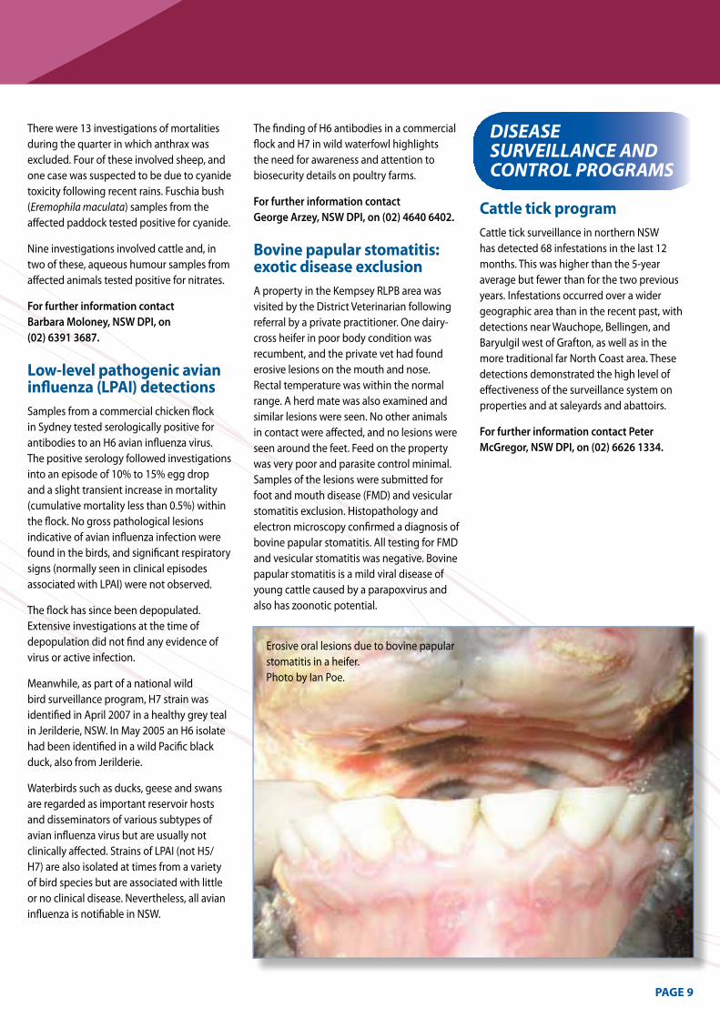
Bovine papular stomatitis: exotic disease exclusion A property in the Kempsey RLPB area was visited by the District Veterinarian following referral by a private practitioner. one dairy-cross heifer in poor body condition was recumbent, and the private vet had found erosive lesions on the mouth and nose. Rectal temperature was within the normal range. A herd mate was also examined and similar lesions were seen. No other animals in contact were affected, and no lesions were seen around the feet. Feed on the property was very poor and parasite control minimal. Samples of the lesions were submitted for foot and mouth disease (FmD) and vesicular stomatitis exclusion. Histopathology and electron microscopy confirmed a diagnosis of bovine papular stomatitis. All testing for FmD and vesicular stomatitis was negative. Bovine papular stomatitis is a mild viral disease of young cattle caused by a parapoxvirus and also has zoonotic potential.
DISEASE SURvEILLANcE AND coNTRoL PRoGRAmS
Cattle tick program Cattle tick surveillance in northern NSW has detected 68 infestations in the last 12 months. This was higher than the 5-year average but fewer than for the two previous years. Infestations occurred over a wider geographic area than in the recent past, with detections near Wauchope, Bellingen, and Baryulgil west of Grafton, as well as in the more traditional far North Coast area. These detections demonstrated the high level of effectiveness of the surveillance system on properties and at saleyards and abattoirs.
For further information contact Peter McGregor, NSW DPI, on (02) 6626 1334.
Erosive oral lesions due to bovine papular stomatitis in a heifer. Photo by Ian Poe.
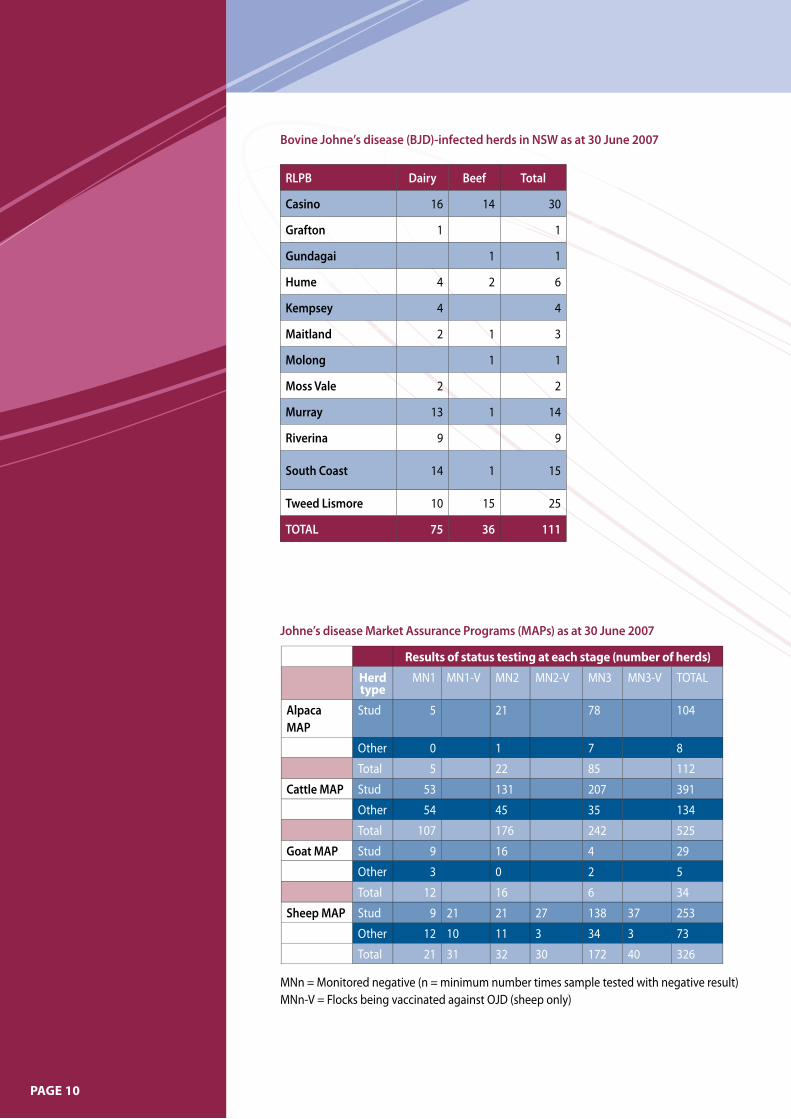
Results of status testing at each stage (number of herds)
Herd type
mN1 mN1-V mN2 mN2-V mN3 mN3-V ToTAL
Alpaca MAP
Stud 5 21 78 104
other 0 1 7 8
Total 5 22 85 112
Cattle MAP Stud 53 131 207 391
other 54 45 35 134
Total 107 176 242 525
Goat MAP Stud 9 16 4 29
other 3 0 2 5
Total 12 16 6 34
Sheep MAP Stud 9 21 21 27 138 37 253
other 12 10 11 3 34 3 73
Total 21 31 32 30 172 40 326
Johne’s disease Market Assurance Programs (MAPs) as at 30 June 2007
RLPB Dairy Beef Total
Casino 16 14 30
Grafton 1 1
Gundagai 1 1
Hume 4 2 6
Kempsey 4 4
Maitland 2 1 3
Molong 1 1
Moss Vale 2 2
Murray 13 1 14
Riverina 9 9
South Coast 14 1 15
Tweed Lismore 10 15 25
TOTAL 75 36 111
Bovine Johne’s disease (BJD)-infected herds in NSW as at 30 June 2007
mNn = monitored negative (n = minimum number times sample tested with negative result) mNn-V = Flocks being vaccinated against oJD (sheep only)
PAGE10
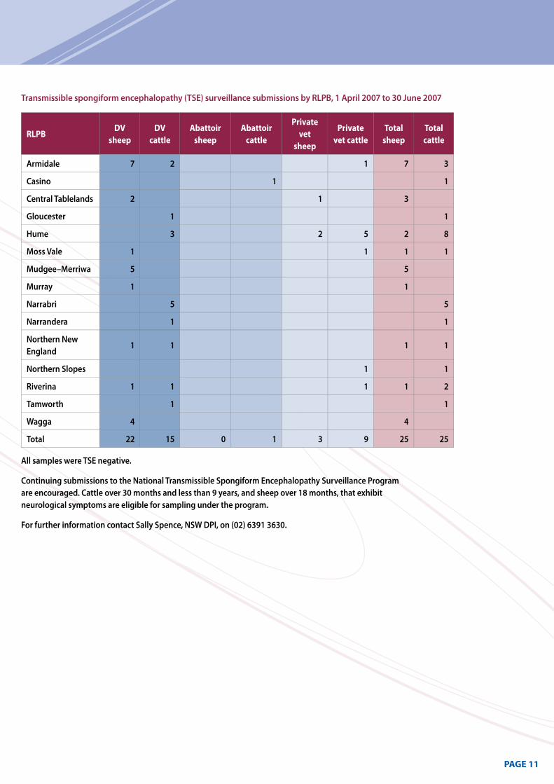
Transmissible spongiform encephalopathy (TSE) surveillance submissions by RLPB, 1 April 2007 to 30 June 2007
RLPB DV sheep
DV cattle
Abattoir sheep
Abattoir cattle
Private vet sheep
Private vet cattle
Total sheep
Total cattle
Armidale 7 2 1 7 3
Casino 1 1
Central Tablelands 2 1 3
Gloucester 1 1
Hume 3 2 5 2 8
Moss Vale 1 1 1 1
Mudgee–Merriwa 5 5
Murray 1 1
Narrabri 5 5
Narrandera 1 1
Northern New England
1 1 1 1
Northern Slopes 1 1
Riverina 1 1 1 1 2
Tamworth 1 1
Wagga 4 4
Total 22 15 0 1 3 9 25 25
All samples were TSE negative.
Continuing submissions to the National Transmissible Spongiform Encephalopathy Surveillance Program are encouraged. Cattle over 30 months and less than 9 years, and sheep over 18 months, that exhibit neurological symptoms are eligible for sampling under the program.
For further information contact Sally Spence, NSW DPI, on (02) 6391 3630.
PAGE11
Prepared by:
Sarah Robson Regional Animal Health Leader, Wagga Wagga Agricultural Institute, Wagga Wagga NSW 2650
Phone (02) 6938 1967 or fax (02) 6938 1995 e-mail: [email protected]
copies of NSW Animal Health Surveillance reports are available on the internet at: http://www.dpi.nsw.gov.au/reader/ah-surveillance
09/2007 - 7839
GettingInformationonAnimalDiseasesThis surveillance report can convey only
a very limited amount of information
about the occurrence and distribution
of livestock diseases in New South
Wales. If you would like more specific
information about diseases occurring
in your part of the State, contact your
local Rural Lands Protection Board
District Veterinarian, Departmental
Senior Regional Animal Health Manager,
Regional Health Leader, or Regional
Veterinary Laboratory.
for Statewide information, contact
NSW DPI’s Animal and Plant
biosecurity branch in orange on
(02) 6391 3237 or
fax (02) 6361 9976.
For more information on national disease status, check the National Animal Health Information System (NAHIS) via the internet at: http://www.animalhealthaustralia. com.au/status/nahis.cfm
Disclaimer
The information contained in this publication is based on
knowledge and understanding at the time of writing
(September 2007). However, because of advances in
knowledge, users are reminded of the need to ensure that
information upon which they rely is up-to date and to check
the currency of the information with the appropriate officer
of New South Wales Department of Primary Industries or the
user s independent adviser.